dissertation submitted to the -...
TRANSCRIPT
Dissertation
submitted to the
Combined Faculties for the Natural Sciences and for Mathematics
of the Ruperto-Carola University of Heidelberg, Germany
for the degree of
Doctor of Natural Sciences
Presented by
M.Sc. Lei Zhang, born in Hancheng, China
Oral-examination:

Construction of infectious full-length cDNA clones
of apple viruses and plant viral vector development
Referees: Prof. Dr. Wilhelm Jelkmann
Prof. Dr. Thomas Rausch

Is viral infection passive or active?
Abstract
Apple chlorotic leaf spot virus (ACLSV) and Apple stem pitting virus (ASPV) are important
viral pathogens in apple. The aim of the present work was to construct full-length cDNA
clones of ACLSV and ASPV and to agroinoculate apple seedlings with the constructed
infectious clones using a newly developed vacuum infiltration method. A further goal was to
explore and create viral vectors based on the obtained infectious full-length cDNA clones of
ACLSV.
In the thesis, the full-length cDNA clones of ACLSV and ASPV were constructed using
different methods. The methods contained circular polymerase extension cloning (CPEC),
Gibson assembly and In-Fusion cloning. In total 17 full-length cDNA clones of ACLSV and
ASPV were obtained. Four of the 17 clones were infectious on Nicotiana occidentalis 37B, i.e.
pIF3-15, pIF3-19, pIF14-23 and pIF4-4. The viral genomic cDNAs in these infectious clones
were completely sequenced, and the sequence data were analyzed by alignment with
published sequences in NCBI. The results indicated that three isolates of ACLSV were
rescued: ACLSV isolate 38/85A (pIF3-15), 38/85B (pIF3-19) and (36)/88 (pIF14-23). One
ASPV isolate was rescued: ASPV 40/87.
A protocol of agroinoculation of apple seedlings by vacuum infiltration was developed to
inoculate the infectious clones (pIF3-15, pIF3-19, pIF14-23 and pIF4-4) to apple seedlings. In
the protocol, the treatment of seedlings, preparation of inocula and parameters of vacuum
infiltration were evaluated. The highest PCR-positive rate (infection) of 78% (11/14), 100%
(11/11), 25% (2/8) and 50% (9/18) were observed for the infectious clones of pIF3-15, pIF3-
19, pIF14-23 and pIF4-4, respectively. The infection of virus was determined by RT-PCR.
The existence of viral particles in PCR-positive plants was determined by immunosorbent
electron microscopy.
To explore and develop plant viral vectors based on ACLSV, marker genes of Emerald GFP,
mCherry or iLov were inserted into the genomic cDNA of ACLSV. Nine plasmids with

marker genes were constructed using three different strategies, including pIF13-9, pIF18-2,
pIF25-7, pG11-15, pIF24-6, pIF23-1, pIF16-1, pIF20-16 and pIF27-10. After agroinoculation
of N. occidentalis 37B with the constructed plasmids, it was found that deletion of marker
genes in pIF13-9 and pG11-15 occurred due to homologous recombination between
duplicated fragments of ACLSV genomic cDNA. Typical ACLSV symptoms were observed
on the test-plants inoculated with pIF13-9 and pG11-15. By western blot, viral proteins of CP
of identical size with wild type virus (pIF3-19) were detected for the two constructs in
symptomatic plants. In one trial in winter, the pIF25-7 caused systemic infection in one plant.
The other plasmids of pIF18-2, pIF24-6, pIF23-1, pIF16-1, pIF20-16 and pIF27-10 did not
cause local or systemic infection in any test-plant.

Zusammenfassung
Apple chlorotic leaf spot virus (ACLSV) und Apple stem pitting virus (ASPV) sind wichtige
virale Erreger in Apfelkulturen. Das Ziel dieser Arbeit war die Konstruktion von Volllängen
cDNA Klonen von ACLSV und ASPV und deren Infektion an Apfelsämlingen durch eine neu
entwickelte Vakuuminfiltrations-Methode zur Agroinokulation. Ein weiteres Ziel war die
Untersuchung und Konstruktion viraler Vektoren basierend auf den hergestellten infektiösen
Volllängen cDNA Klonen von ACLSV.
In dieser Arbeit wurden Volllängen cDNA Klone von ACLSV und ASPV über verschiedene
Methoden hergestellt. Diese Methoden waren das Circular Polymerase Extension Cloning
(CPEC), Gibson Assemblierung und In-Fusion Klonierung. Insgesamt konnten 17 Volllängen
cDNA Klone von ACLSV und ASPV hergestellt werden. Vier dieser 17 Klone waren
infektiös auf Nictoiana occidentalis 37B (pIF3-15, pIF3-19, pIF14-23 und pIF4-4). Die virale
genomische cDNA dieser Klone wurde vollständig sequenziert und mit bereits
veröffentlichen Sequenzen in NCBI verglichen. Die Ergebnisse deuteten darauf hin, dass drei
verschiedene Isolate von ACLSV vorlagen: ACLSV Isolate 38/85A (pIF3-15), 38/85B (pIF3-
19) und (36)/88 (pIF14-23). Von ASPV konnte ein Isolat identifiziert werden (ASPV 40/87).
Um Apfelsämlinge mit den infektiösen Klonen pIF3-15, pIF3-19, pIF14-23 und pIF4-4
inokulieren zu können, wurde ein Vakuuminfiltrations-Protokoll zur Agroinokulation
entwickelt. Für das Protokoll wurden die Behandlung der Sämlinge, die Präparation des
Inokulums und die Parameter der Vakuuminfiltration optimiert. Für die infektiösen Klone
pIF3-15, pIF3-19, pIF14-23 bzw. pIF4-4 wurden 78% (11/14), 100% (11/11), 25% (2/8) und
50% (9/18) positiv getesteter Sämlinge beobachtet. Die Virusinfektion wurde dabei mit Hilfe
von RT-PCR überprüft. Das Vorkommen viraler Partikel in PCR-positiven Pflanzen wurde
über Immun-Elektronenmikroskopie bestätigt.

Um pflanzliche virale Vektoren, basierend auf ACLSV zu entwickeln und zu untersuchen,
wurden Markergene von Emerald GFP, mCherry oder iLov in die genomische cDNA von
ACLSV inseriert. Insgesamt neun Plasmide mit Markergenen wurden über drei verschiedene
Klonierungsstrategien hergestellt, die mit pIF13-9, pIF18-2, pIF25-7, pG11-15, pIF24-6,
pIF23-1, pIF16-1, pIF20-16 und pIF27-10 bezeichnet wurden. Nach der Agroinokulation von
N. occidentalis 37B mit den Plasmidkonstrukten konnte festgestellt werden, dass in pIF13-9
und pG11-15 eine Deletion der Markergene über homologe Rekombination zwischen
duplizierten Fragmenten der genomischen cDNA von ACLSV stattgefunden hat. Auf den mit
pIF13-9 und pG11-15 inokulierten Testpflanzen konnten charakteristische ACLSV
Symptome beobachtet werden. Über Western Blot konnten in den symptomatischen Pflanzen
aus beiden Konstrukten virale CP Proteine mit der identischen Größe des Wildtyp Virus‘
(pIF3-19) detektiert werden. In einem Versuchsdurchlauf im Winter verursachte pIF25-7 eine
systemische Infektion in einer einzigen Pflanze. Die anderen Plasmide von pIF18-2, pIF24-6,
pIF23-1, pIF16-1, pIF20-16 und pIF27-10 verursachten weder lokale noch systemische
Infektionen in den Testpflanzen.

i
Contents
Contents ....................................................................................................................................... i
List of Figures ............................................................................................................................ v
List of Tables ............................................................................................................................. vi
Abbreviations ........................................................................................................................... vii
1. Introduction ............................................................................................................................ 1
1.1 Properties of ACLSV and ASPV ...................................................................................... 1
1.1.1 Virion properties ........................................................................................................ 1
1.1.2 Host range .................................................................................................................. 2
1.1.3 Symptoms and impacts .............................................................................................. 2
1.1.4 Transmission and geographical distribution .............................................................. 4
1.2 Construction of infectious full-length cDNA clones of plant viruses .............................. 4
1.2.1 Historical background ................................................................................................ 4
1.2.2 Methodology .............................................................................................................. 5
1.2.3 Difficulties in construction of full-length cDNA clones for viruses from woody
plants ................................................................................................................................... 6
1.3 Inoculation of woody plants with viruses ......................................................................... 7
1.3.1 Methods of Transmission of plant viruses ................................................................. 7
1.3.2 Difficulties of transmission of viruses to woody plants ............................................. 7
1.4 Utilization of the plant viral vectors ................................................................................. 8
1.4.1 The movement of ACLSV in plants .......................................................................... 8
1.4.2 Development of plant viral vectors ............................................................................ 9
1.5 Aims of this thesis .......................................................................................................... 10
2. Materials and methods ......................................................................................................... 11
2.1 Materials ......................................................................................................................... 11

ii
2.1.1 Virus source ............................................................................................................. 11
2.1.2 Plant materials .......................................................................................................... 11
2.1.3 Bacterial strains and plasmids .................................................................................. 11
2.1.4 Molecular biological reagents .................................................................................. 12
2.1.5 Apparatus, consumable and software ...................................................................... 17
2.2 Methods .......................................................................................................................... 19
2.2.1 Molecular biology .................................................................................................... 19
2.2.1.1 Total nucleic acids extraction ........................................................................ 19
2.2.1.2 Reverse transcription polymerase chain reaction (RT-PCR)......................... 20
2.2.1.3 Polymerase chain reaction (PCR) .................................................................. 21
2.2.1.4 Agarose gel electrophoresis ........................................................................... 22
2.2.1.5 DNA purification ........................................................................................... 23
2.2.1.6 Cloning of the target fragments ..................................................................... 23
2.2.2 Molecular cloning of viruses and viral vectors ........................................................ 24
2.2.2.1 Construction of full-length cDNA clones ...................................................... 24
2.2.2.2 Strategies of labeling viral proteins with fluorescent proteins ...................... 26
2.2.2.3 Construction of labeled plasmids................................................................... 27
2.2.2.3.1 Construction of plasmids using strategy A ................................................. 27
2.2.2.3.2 Construction of plasmids using strategy B ................................................. 28
2.2.2.3.3 Construction of plasmids using strategy C ................................................. 30
2.2.2.4 Sequencing of obtained plasmids .................................................................. 31
2.2.3 Microbiology ............................................................................................................ 31
2.2.3.1 Preparation of electrocompetent bacteria cells .............................................. 31
2.2.3.2 Electroporation .............................................................................................. 32
2.2.3.3 Heat shock ..................................................................................................... 32
2.2.4 Inoculation of plants ................................................................................................ 33
2.2.4.1 Mechanical agroinoculation........................................................................... 33
2.2.4.2 Sap inoculation .............................................................................................. 33
2.2.4.3 Agroinoculation by vacuum infiltration ........................................................ 33
2.2.4.4 Grafting .......................................................................................................... 34

iii
2.2.4.5 Detection of viral infection in plants ............................................................. 35
2.2.5 Protein immunoblot ................................................................................................. 35
2.2.5.1 Protein preparation......................................................................................... 35
2.2.5.2 Sodium dodecyl sulfate polyacrylamide gel electrophoresis (SDS-PAGE) .. 35
2.2.5.3 Transmembrane ............................................................................................. 36
2.2.5.4 Western blot ................................................................................................... 36
2.2.6 Immunosorbent electron microscopy (ISEM) ......................................................... 36
3. Results .................................................................................................................................. 38
3.1 Construction of full-length cDNA clones ....................................................................... 38
3.1.1 Construction of ACLSV clones isolate (27)/85 ....................................................... 39
3.1.2 Construction of ACLSV clones isolate 38/85 .......................................................... 39
3.1.3 Construction of ACLSV clones isolate (36)/88 ....................................................... 40
3.1.4 Construction of ASPV clones isolate 40/87 ............................................................. 40
3.2 Infectivity of the constructed clones on herbaceous plants ............................................ 41
3.2.1 Symptoms development on N. occidentalis 37B ..................................................... 41
3.2.2 RT-PCR detection of viruses in agroinoculated test-plants ..................................... 42
3.2.3 Detection of viral particles by ISEM ....................................................................... 43
3.2.4 Transmission of viruses by sap inoculation ............................................................. 43
3.3 Agroinoculation of woody plants with infectious clones ............................................... 44
3.3.1 Infectivity of the clones on apple seedlings ............................................................. 45
3.3.2 Transmission of viruses to woody plants by grafting .............................................. 46
3.4 Sequence analysis ........................................................................................................... 47
3.4.1 Sequences of ACLSV .............................................................................................. 47
3.4.2 Sequences of ASPV ................................................................................................. 49
3.5 Labeling of ACLSV isolate 38/85 with marker genes ................................................... 51
3.5.1 Construction of the plasmids containing marker genes ........................................... 51
3.5.2 Detection of marker genes in constructed plasmids and transformed A. tumefaciens
........................................................................................................................................... 53
3.5.3 Agroinoculation of N. occidentalis 37B with labeled plasmids .............................. 54
3.5.3.1 First agroinoculation of N. occidentalis 37B ................................................. 54

iv
3.5.3.2 Second agroinoculation of N. occidentalis 37B ............................................ 56
3.5.3.2.1 Infection in inoculated leaves ..................................................................... 56
3.5.3.2.2 Infection in noninoculated leaves ............................................................... 58
4. Discussion ............................................................................................................................ 59
4.1 Investigation of different cloning techniques to construct full-length cDNA clones of
ACLSV and ASPV ............................................................................................................... 59
4.2 Agroinoculation of herbaceous and woody plants ......................................................... 61
4.3 Phylogenetic analysis of ACLSV and ASPV ................................................................. 64
4.4 Development of ACLSV infectious cDNA clones as expression vector for foreign genes
.............................................................................................................................................. 65
References ................................................................................................................................ 68
Supplementary .......................................................................................................................... 81
Publication ................................................................................................................................ 83
Acknowledgements .................................................................................................................. 84

v
List of Figures
Figure 2.1 The structure of the binary vector pV297 ............................................................... 25
Figure 2.2 Apple seedlings and pump system used in vacuum infiltration .............................. 34
Figure 3.1 The structures of the constructed full-length cDNA clones.................................... 38
Figure 3.2 Symptoms on N. occidentalis 37B 18 days after agroinoculation with ACLSV
clones ............................................................................................................................ 42
Figure 3.3 Symptoms on N. occidentalis 37B 10 days after agroinoculation with the ASPV
clone ............................................................................................................................. 42
Figure 3.4 Virions of ACLSV detected in infected N. occidentalis 37B using ISEM ............. 43
Figure 3.5 The efficiency of agroinoculation of apple seedlings with obtained infectious
cDNA clones by vacuum infiltration ........................................................................... 46
Figure 3.6 The genome organizations of the three obtained ACLSV isolates ......................... 48
Figure 3.7 The phylogenetic tree based on complete genome sequences of ACLSV.............. 49
Figure 3.8 The genome organization of the obtained ASPV ................................................... 50
Figure 3.9 The phylogenic tree based on complete genome sequences of ASPV ................... 51
Figure 3.10 The labeling of ACLSV MP at C-terminus with EmGFP .................................... 51
Figure 3.11 The labeling of ACLSV CP at N-terminus with mCherry .................................... 52
Figure 3.12 The labeling of ACLSV CP at C-terminus with mCherry or iLov ....................... 53
Figure 3.13 PCR detection of marker genes before performing agroinoculation .................... 54
Figure 3.14 RT-PCR detection of marker genes in agroinoculated plants ............................... 55
Figure 3.15 Detection of ACLSV CP in agroinoculated plants by western blot ..................... 56
Figure 3.16 RT-PCR detection of marker genes in agroinoculated leaves .............................. 57
Figure 3.18 RT-PCR detection of marker genes in noninoculated leaves ............................... 58
Figure S1 Systemic symptoms on Chenopodium quinoa infected with ACLSV by sap
inoculation .................................................................................................................... 81
vi
List of Tables
Table 2.1 Bacterial strains of Escherichia coli and Agrobacterium tumefaciens .................... 11
Table 2.2 Plasmids used in labeling of ACLSV ....................................................................... 12
Table 2.3 Oligonucleotides used in the present work .............................................................. 12
Table 2.4 Standard markers ...................................................................................................... 14
Table 2.5 Enzymes and enzyme-based kits .............................................................................. 14
Table 2.6 Primary and secondary antibodies used in western blot .......................................... 15
Table 2.7 Silica-based kits ....................................................................................................... 15
Table 2.8 Buffers and chemicals .............................................................................................. 15
Table 2.9 Apparatus ................................................................................................................. 17
Table 2.10 Consumables .......................................................................................................... 18
Table 2.11 In silico tools .......................................................................................................... 19
Table 2.12 The reaction mixture and cycling conditions of RT-PCR ...................................... 20
Table 2.13 The reaction mixture and cycling conditions of PRT-PCR.................................... 20
Table 2.14 PCR reaction mixture using KAPA Taq DNA polymerase ................................... 21
Table 2.15 Cycling conditions using KAPA Taq DNA polymerase ........................................ 21
Table 2.16 PCR reaction mixture using PRECISOR high-fidelity DNA polymerase ............. 22
Table 2.17 Cycling conditions using PRECISOR high-fidelity DNA polymerase .................. 22
Table 2.18 The reaction mixture of the CPEC ......................................................................... 24
Table 2.19 The cycling conditions in the CPEC ...................................................................... 24
Table 3.1 Number of full-length cDNA clones constructed using different methods ............. 38
Table S1 Primers used for sequencing the obtained ACLSV and ASPV cDNA clones .......... 81

vii
Abbreviations
Items Abbreviations Full text
Viruses ACLSV Apple chlorotic leaf spot virus
ALSV Apple latent spherical virus
ALSV Apple latent spherical virus
AMV Alfalfa mosaic virus
ApMV Apple mosaic virus
ASGV Apple stem grooving virus
ASPV Apple stem pitting virus
BYDV Barley yellow dwarf virus
CaMV Cauliflower mosaic virus
CLBV Citrus leaf blotch virus
CMV Cucumber mosaic virus
CPMV Cowpea mosaic virus
CTV Citrus tristeza virus
GRSPaV Grapevine rupestris stem pitting-associated virus
GVA Grapevine virus A
GVB Grapevine virus B
MRFV Maize rayado fino virus
PLDMV Papaya leaf distortion mosaic virus
PPV Plum pox virus
PVX Potato virus X
PVY Potato virus Y
TMV Tobacco mosaic virus
ToBMV Tomato blistering mosaic virus
Units bp Base pair
cm Centermeter
ca. Circa
hPa Hectopascal
kb Killobase
kDa Killodalton
min Minute
ml Mililiter
mm Milimeter
mM Millimolar
ng Nanogram
nm Nanometer
nt Nucleotide

viii
Items Abbreviations Full text
nts Nucleotides
rpm Revolutions per minute
sec Second
V Volt
μF Microfarad
μg Microgram
μl Microliter
μM Micromolar
Others APS Ammonium persulfate
CP Coat protein
CPEC Circular polymerase extension cloning
DNA Deoxyribonucleic acid
DTT Dithiothreitol
EDTA Ethylenediaminetetraacetic acid
EmGFP Emerald-GFP
GFP Green fluorescent protein
HDVpA Hepatitis delta viral ribozyme followed by a CaMV 35S
polyadenylation signal
ISEM Immunosorbent electron microscopy
JKI Julius Kuehn Institute
LB Luria-Bertani medium
MP Movement protein
ORF Open reading frame
PAG Polyacrylamide gel
PAGE Polyacrylamide gel electrophoresis
PCR Polymerase chain reaction
polyA Polyadenylated
PRT Premium reverse transcription
PRTase Premium reverse transcriptase
RdRp RNA-dependent RNA ploymerase
RNA Ribonucleic acid
RT Reverse transcription
RTase Reverse transcriptase
SDS Sodium dodecyl sulfate
SOC Super optimal broth with catabolite repression (SOC)
spp. Species
ssDNA Single-stranded DNA
ssRNA Single-stranded RNA
subsp. Subspecies
TEMED Tetramethylethylenediamine
TGB Triple gene block
UTR Untranslated region
YEB Yeast extract broth

1
1. Introduction
Apple chlorotic leaf spot virus (ACLSV) and Apple stem pitting virus (ASPV) were first
described in the 1950s (Luckwill and Campbell 1959; Smith 1954). They are latent (eliciting
no symptom) in most commercial apple cultivars (Eastwell and Howell 2014a, b; Jelkmann
and Paunovic 2011; Myrta et al. 2011). Without use of certificaion systems (EPPO 1999)
latent viruses are propagated and get widely distributed in orchards over time. Therefore,
these viruses are found throughout pome fruit-growing regions in the world.
This work focuses on the construction of infectious full-length cDNA clones of ACLSV and
ASPV. Furthermore experiments were conducted to develop a plant viral expression vector
based on ACLSV clones.
1.1 Properties of ACLSV and ASPV
1.1.1 Virion properties
ACLSV is the type species of the genus Trichovirus within the family Betaflexiviridae. It has
virions of very flexuous filaments, usually 640-890 nm in length and 10-12 nm in diameter,
with distinct cross banding. ACLSV virions contain a single molecule of linear positive
single-stranded (ss) RNA about 7500 nucleotides (nts) in size excluding the polyadenylated
(polyA) tract at 3’ terminus, accounting for about 5% of the particle weight (Adams et al.
2012). The genome has three slightly overlapping open reading frames (ORFs). ORF1, 2 and
3 encode a RNA-dependent RNA polymerase (RdRp, ca. 217 kDa), a movement protein (MP,
ca. 50 kDa), and a coat protein (CP, ca. 22 kDa), respectively (German et al. 1990; Sato et al.
1993). ACLSV shows a very large molecular variability among isolates. The genomes show
an overall identity between 76 and 82% in their nucleotide sequences. The CP is more
conserved (87-93% identity) than the MP (77-85% identity) (Adams et al. 2012; Candresse et
al. 1996; German-Retana et al. 1997).

2
ASPV is the type species of the genus Foveavirus within the family Betaflexiviridae. Its
virions are flexuous filaments, 800-1000 nm in length and 12-15 nm in diameter with a
surface pattern of cross-banding (Yanase et al. 1988). The virions contain a single molecule of
positive-sense ssRNA of around 9500 nts excluding the polyA tail at 3’ terminus (Jelkmann
1994; Jelkmann et al. 1991). The genome has five ORFs. The ORF1 encodes an RdRp (247
kDa). The ORFs 2-4 as triple gene block (TGB) encode proteins of 25, 13 and 8 kDa,
respectively, constituting putative MP. The ORF5 encodes a CP (44 kDa).
1.1.2 Host range
The natural hosts of ACLSV mainly include Rosaceae. For example, ACLSV infects several
Prunus species, including peach, apricot, almond, cherry, and plum (Myrta et al. 2003),
blackthorn (P. spinosa) (Sweet 1980), Himalayan wild cherry (P. cerasoides) (Rana et al.
2008) and dwarf flowering almond (P. glandulosa ‘Sinensis’) (Spiegel et al. 2005). It also
occurs in Malus species, such as cultivated apples and ornamental species of M. platycarpa,
M. floribunda, M. robusta, M. coronaria (Desvignes et al. 1999). It is detected in hawthorn
(Crataegus spp.) (Sweet 1980), pear (Pyrus spp.) and quince (Cydonia oblonga) too (Lister
1970; Németh 1986).
The experimental host range of ACLSV is limited to a few herbaceous species, including
Chenopodium quinoa. C. amaranticolor, Leguminosae and Nicotiana occidentalis (Lister et al.
1965; Yoshikawa 2001).
The natural host range of ASPV is largely restricted to Maloideae, such as Malus spp.,
Crataegus spp., Sorbus spp., Pyrus spp. [P. communis (European pear), P. serotinia (Japanese
pear), P. ussuriensis (Chinese pear), and P. sinkiangensis)], and Cydonia oblonga (quince)
(Dhir et al. 2010; Ma et al. 2008; Mathioudakis et al. 2010; Mathioudakis et al. 2012). The
most commonly used experimental hosts are N. occidentalis 37B and N. occidentalis subsp.
obliqua (Koganezawa and Yanase 1990; Van der Meer 1985).
1.1.3 Symptoms and impacts
ACLSV causes cytopathological changes in infected cells to varying extents (Adams et al.
2012; Ohki et al. 1989). The virions are found in mesophyll, phloem and parenchyma cells of
leaves and roots. They accumulate in the cytoplasm or between tonoplast and cytoplasm in
bundles or paracrystalline aggregates. They were found in the nucleus as well. No inclusion
bodies are formed.

3
Common isolates of ACLSV do not cause observable symptoms in most commercial cultivars
of pome and stone fruit trees currently in production. In some susceptible species or cultivars,
the symptom severity depends on the plant species and virus isolates, and symptoms appear
mainly on leaves, fruits and more rarely on the trunk (Myrta et al. 2011; Németh 1986). For
example, severe isolates elicit a apricot disease of ‘pseudopox’ (Desvignes and Boyé 1988),
deforming the fruit. Some isolates induce ‘false plum pox’ on leaves and fruit of some plum
cultivars (Jelkmann and Kunze 1994; Lebas et al. 2003). Maruba kaido (Malus prunifolia var.
ringo) is susceptible to ACLSV, causing the particular occurrence of the apple topworking
disease in Japan. The species of M. sylvestris cv. R12740-7A, M. platycarpa, M. hupehensis,
M. prunifolia var. ringo can be used as diagnostic indexing varieties. In addition, ACLSV is
associated with apple russet ring disease and pear ring pattern mosaic (Cropley et al. 1963;
Desvignes and Boyé 1988; Yanase 1974).
ACLSV causes considerable yield losses in apple. In mixed infections with ASPV and Apple
stem grooving virus (ASGV), a yield loss of 12 to 30% was observed on Malus domesticus cv.
Golden Delicious in USA (Meijneke et al. 1973; Van Oosten et al. 1982). It has been reported
that in China mixed infection with ASGV led to a yield loss up to 40% (Wu et al. 1997).
ASPV elicits a severe derangement of the cytology of infected cells. The virions of ASPV are
found in mesophyll, epidermal and vascular parenchyma cells of infected plants. They
accumulate in bundles in the cytoplasm, but no specific cytopathic structures or inclusion are
formed (Adams et al. 2012; Koganezawa and Yanase 1990).
ASPV is latent in most common commercial apple cultivars, but susceptible cultivars do react
with a variety of symptoms. For example, ASPV causes xylem pitting in the stem of M.
pumila cv. Virginia crab, and elicits epinasty and decline on M. pumila cv. Spy 227. It also
causes symptoms on susceptible pear and quince cultivars. For example, on infected P.
communis, symptoms of narrow chlorotic bending of veins and red mottling are expressed
(Cameron 1989). It induced symptoms of black sooty lines and rings bordering veins, pale
yellow spots on leaves and fruit malformations on susceptible quince cultivars (Desvignes
1971; Paunovic 1994). These sensitive varieties are used as serological indicators (EPPO
1999).
Since often mixed infection occurred, it is difficult to assess the effect of ASPV alone (Hadidi
et al. 2011). When in combination with ACLSV and ASGV, a negative impact can be seen in
production with most commercial cultivars. There are reports of reduced budding success on

4
pear cultivars (5 to 50%) (Lemoine and Michelesi 1990; Lemoine and Michelesi 1995) and
reduced growth of nursery trees (10 to 55%) (Cropley and Posnette 1973; Thomsen 1973)
1.1.4 Transmission and geographical distribution
ACLSV is transmitted by grafting and vegetative propagation (Németh 1986). It is believed
that ACLSV is only transmitted by grafting in the field between woody plants (Yaegashi et al.
2011). There is no evidence of vector-, seed- or pollen-borne transmission in any of its hosts
(Yoshikawa 2001). In addition, ACLSV can be transmitted by sap inoculation with limited
efficiency from woody plants to herbaceous hosts (N. occidentialis and C. quinoa) (Yaegashi
et al. 2011).
ACLSV is one of the most widely distributed viruses of fruit trees (Sutton et al. 2014). It is
distributed worldwide and probably present wherever susceptible species grow. For example,
an infection rate up to 80-100% in mixed infections with ASGV is reported in China (Wu et al.
1997). A disease incidence ranging from 85% to 90% is reported in Himachal Pradesh, India
(Rana et al. 2010). Its infection rates range up to 80-100% in many commercial apple
cultivars in USA (Cembali et al. 2003; Németh 1986).
Spread of ASPV is very similar to that of ACLSV. Mixed infections with other apple viruses
occur frequently (Sutton et al. 2014). The incidence of ASPV and ACLSV (84.21%) has been
reported as the most common mixed infection in apple; the incidence of ASPV together with
Apple mosaic virus (ApMV) is rare and was only reported in 5.26% of identified mixed
infections; the incidence of the mixed infection of ASPV, ASGV and ACLSV is 26.32%
(Çağlayan et al. 2006). No vector is known for ASPV.
1.2 Construction of infectious full-length cDNA clones of plant
viruses
1.2.1 Historical background
An infectious clone (as cDNAs or as in vitro-transcribed RNA copies) of a RNA virus is a
plasmid containing the full-length genomic cDNA of the virus. Under control of suitable
promoters, the viral genomic cDNA can be transcribed in vitro or in vivo into the viral
genomic RNA, establishing infection in host plants.

5
Infectious cDNA clones are powerful tools for investigating plant viruses. They can be
applied to basic studies of viral life cycles and virus-host interactions, and serve as pools of
viral genes for the design of antiviral strategies and for the development of viral vectors
(Boyer and Haenni 1994; Nagyová and Subr 2007).
The first infectious clone of a plant RNA virus, Brome mosaic virus (BMV), was obtained in
1984 (Ahlquist et al. 1984). Since then, clones to a wide range of different virus species
belonging to several virus families such as Alphaflexiviridae, Betaflexiviridae, Bromoviridae,
Closteroviridae, Luteoviridae, Potyviridae and Tymoviridae have been produced, such as
Tobacco mosaic virus (TMV) (Dawson et al. 1986), Cucumber mosaic virus (CMV) (Rizzo
and Palukaitis 1990), Plum pox virus (PPV) (Riechmann et al. 1990), Potato virus X (PVX)
(Hemenway et al. 1990), Barley yellow dwarf virus (BYDV) (Young et al. 1991), Potato virus
Y (PVY) (Jakab et al. 1997), Citrus tristeza virus (CTV) (Satyanarayana et al. 2001), Papaya
leaf distortion mosaic virus (PLDMV) (Tuo et al. 2015), Tomato blistering mosaic virus
(ToBMV) (Blawid and Nagata 2015) and Maize rayado fino virus (MRFV) (Edwards et al.
2015). The first infectious clone of ACLSV (isolate P-205) was constructed in 1999 (Satoh et
al. 1999). An infectious cDNA clone of ASPV was constructed by Arntjen and Jelkmann
(2009).
1.2.2 Methodology
The nature of constructing full-length cDNA clones of viruses is a process of precisely
assembling single or multiple DNA fragments of viral genomic cDNAs and vectors. The
technique of precise assembly of specific DNA fragments is a critical step in molecular
biology. A number of methods for the assembly have been developed, serving various
purposes in research.
Given the properties of manipulated DNA fragments, these methods can be grouped roughly
into three categories. As one of the simplest and most efficient method, TA cloning has been
widely used for cloning (Yao et al. 2016). This method is based on the additional single
thymine (T) residue at each end of DNA fragments added by some Taq DNA polymerases
during polymerase chain reaction (PCR). This method is used rarely in the construction of
full-length cDNA clones of plant viruses. But it is reported that an infectious clone of
Muscovy duck parvovirus, having a ssDNA genome of ca. 5.1 kb, is obtained by TA cloning
(Yen et al. 2015). A widely used method to generate infectious clones is traditional cloning
method, using restriction enzymes and ligation of DNA in vitro. For example, the first

6
infectious clone of plant RNA viruses, BMV, is constructed using this method (Ahlquist et al.
1984). More recently introduced methods are ligation-independent cloning methods. These
methods rely on the generation of complementary overhangs by DNA polymerase, without
requiring specific restriction sites or ligation. Several such methods have been developed,
including sequence and ligation-independent cloning (Li and Elledge 2012), polymerase
incomplete primer extension cloning (Klock et al. 2008), overlap extension cloning (Bryksin
and Matsumura 2010), circular polymerase extension cloning (CPEC) (Stevenson et al. 2013)
and an in vivo yeast homologous recombination (Youssef et al. 2011a). Commercial kits are
also available, such as Gibson assembly (Gibson et al. 2010; Gibson et al. 2009) and In-
Fusion cloning (Takara Bio Europe, France).
1.2.3 Difficulties in construction of full-length cDNA clones for viruses from
woody plants
There are several challenges in construction of full-length cDNA clones of viruses of woody
plants (MacKenzie et al. 1997; Meng et al. 2013). First, the titer of viruses is low in woody
hosts, making it difficult to extract high concentration viral genomic RNA. Second, the length
of the viral genomic cDNA for some viruses (ca. 7.5 kb of ACLSV and 9.5 kb of ASPV)
makes one-step cloning poorly efficient. Third, the presence of inhibitory factors (such as
polyphenolic compounds or polysaccharides) in fruit trees results in poor quality of viral
products in PCR amplification. In order to overcome these difficulties the techniques of
CPEC, Gibson assembly and In-Fusion cloning were applied to construction of cDNA clones
of the two apple viruses.
The three methods (CPEC, Gibson assembly and In-Fusion cloning) work on fragments
having homologous ends, but the mechanism of CPEC is different from that of Gibson
assembly and In-Fusion cloning. The CEPC method relies entirely on the polymerase
extension mechanism, extending overlapping regions between the insert and vector fragments
to form a complete circular plasmid (Stevenson et al. 2013). Gibson assembly uses three
enzymes together in a single reaction: the T5 exonuclease removes nucleotides (15 nt) at the 5’
termini of the target DNA fragments, creating single-stranded overhangs at 3’ termini. The
DNA polymerase then fills in the gaps and the DNA ligase seals the nicks (Gibson et al. 2010;
Gibson et al. 2009). The In-Fusion cloning is similar to Gibson assembly.

7
1.3 Inoculation of woody plants with viruses
1.3.1 Methods of Transmission of plant viruses
Plant viruses can be transmitted from plants to plants in different ways. Modes of
transmission include mechanical inoculation with sap containing virus particles or viral
nucleic acids, vegetative propagation (by bulbs, corms, rhizomes, tubers and runners), through
seed, pollen, root, dodder and human behaviors (such as grafting and pruning) and by specific
insects, mites, nematodes, and fungi (Agrios 2005).
In addition, molecular biological technologies have been developed to induce virus infections,
couples of new methods of transmission of viruses have been invented. These include
agroinoculation (or agroinfection) methods (Grimsley et al. 1986; Turpen et al. 1993) and
physical methods of electroporation, microinjection, and biolistic inoculation (see for review
Rivera et al. 2012). For agroinoculation several techniques are used such as syringe
infiltration (Turpen et al. 1993), agrodrench (Ryu et al. 2004) and vacuum infiltration (Rossi
et al. 1993).
Among these methods, mechanical inoculation is commonly used, but for those viruses that
are not mechanically transmissible, other methods such as agroinoculation, the use of the
insect vectors, or grafting may be used. Agroinoculation has become a preferred delivery tool
for a variety of viral genomes of interest via expression in plants through Agrobacterium
binary vectors (Vaghchhipawala et al. 2011).
1.3.2 Difficulties of transmission of viruses to woody plants
It is difficult to transmit viruses to woody fruit trees and grapevine, by conventional
mechanical inoculation methods (Yamagishi et al. 2010). This is independent if the inocula
are viral particles, nucleic acids or infectious clones. For example, the Apple latent spherical
virus (ALSV) is transmitted to apple trees in poor infection efficiency (Ito et al. 1992; Li et al.
2004). Grapevine virus B (GVB) is transmitted by sap inoculation to hosts with great
difficulty (Boscia et al. 1993; Conti et al. 1980). PVX is mechanically transmitted to several
citrus species with difficulty (Holmes 1959). The infectious transcripts of ACLSV failed to be
transmitted to peach by stem slashing (Youssef et al. 2011a).
Other inoculation methods have been applied to woody hosts, such as biolistic inoculation,
syringe infiltration and agrodrench. For example, total RNAs, extracted from ALSV- or
ACLSV- infected C. quinoa leaves, established infection in apple seedlings with high

8
efficiency by particle bombardment (Yamagishi et al. 2010). A full-genome cDNA clone of
Citrus leaf blotch virus (CLBV) was inoculated to Etrog citron plants in high efficiency by
syringe infiltration (Vives et al. 2008). Full-length cDNA clones of Grapevine rupestris stem
pitting-associated virus (GRSPaV) and Grapevine virus A (GVA) were introduced to Vitis
vinifera plantlets by modified agrodrench (Meng et al. 2013; Muruganantham et al. 2009).
Transmission of viruses to woody plants by conventional mechanical inoculation and with
molecular methods has always been laborious and time-consuming. None of the current
methods will fit all types of viruses and their woody hosts. For example, an infectious cDNA
clone of GVA was inoculated by agroinfiltration to Vitis vinifera with great difficulty
(Muruganantham et al. 2009), while this method was effectively used for CLBV (Vives et al.
2008). CTV can be effectively transmitted to Etrog citron by cutting or slashing the plant stem
with a contaminated blade (Garnsey et al. 1977), whilst ALSV cannot be transmitted using
this method (Yamagishi et al. 2010).
Thus, it is necessary to establish effective inoculation methods available to different viruses
and hosts. In this study, a highly efficient method of agroinoculation of full-length cDNA
clones of apple viruses to apple seedlings by vacuum infiltration was established.
1.4 Utilization of the plant viral vectors
Plant viral vectors are constructed for various purposes: to mark viruses for visualization of
virus movement and distribution in plants; to express foreign genes in plants; and to examine
the relationship between specific genes and plant phenotypes by silencing (Dawson 2014).
1.4.1 The movement of ACLSV in plants
The incorporation of non destructive fluorescent proteins (e.g. the green fluorescent protein,
GFP) into viral vectors has allowed an exponential progress on the knowledge of movement
of plant viruses. About 75% of reports about transport of plant viruses have been published
after the first use of the jellyfish GFP in plant virology (Pallάs et al. 2011).
The protein of ca. 50 kDa encoded by ORF2 is suggested to be a MP of ACLSV (German et
al. 1990; Sato et al. 1993; Sato et al. 1995). The MP performs multiple functions in host cells,
such as increasing the plasmodesmatal size exclusion limit and inducing tubules in infected
protoplasts (Satoh et al. 2000). An excellent review on the multiple functions of the MP has
been published (Isogai et al. 2007).

9
To examine the localization, subcellular distribution and cell-to-cell trafficking, the MP of
ACLSV P-205 has been expressed. The C-terminus of the MP is fused to the N-terminus of
GFP and, depending on purposes, expressed in plants or mesophyll protoplasts of C. quinoa
and N. occidentalis. The results of fluorescence and confocal laser scanning microscopy
indicate that the MP of ACLSV targets to plasmodesmata, is distributed as small irregular
spots or a fibrous network structure on the periphery of epidermal cells and protoplasts,
accumulates in sieve elements in plants, and induces formation of tubular structures on the
surface of protoplasts (Satoh et al. 2000; Yoshikawa et al. 1999).
1.4.2 Development of plant viral vectors
Plant viral vectors have been developed as promising alternatives to other methods of gene
expression, such as the use of stably transformed transgenic or transplastomic plants, because
of some obvious anticipated advantages. For example, compared with the stable nuclear
genetic transformation, the transient expression utilizing viral vectors allows for a more
efficient, versatile, controlled and safe process (Gleba and Giritch 2011). In addition, viral
vectors have anticipated advantages such as speed of expression, high yield, reduced costs and
duration of research and development, and extremely high throughput (Gleba et al. 2004;
Gleba and Giritch 2011).
So far most viral vectors have been modified from RNA viruses, such as TMV, PVX, Alfalfa
mosaic virus (AMV), CTV and Cowpea mosaic virus (CPMV) (Dawson 2014; Gleba and
Giritch 2011).
Different strategies have been used in viral vector development. The vectors function as
viruses with extra genes. For example, in construction of the first generation of add-a-gene
vectors based on TMV, all of the viral genes needed for replication and movement were kept
(Dawson et al. 1989), and the foreign ORF of chloramphenicol acetyltransferase was
engineered into TMV either before or after the CP gene under the control by an additional CP
subgenomic RNA promoter. Advanced vectors are produced utilizing the properties of
deconstructed viruses. For example, expression vectors were designed with a duplication of
the three pseudoknots of the TMV 3’ nontranslated RNA internally between the foreign ORF
and the CP gene (Shivprasad et al. 1999). A series of research suggests that proximity of the
gene to the pseudoknots is a key to increase protein expression, see for review (Dawson 2014).

10
1.5 Aims of this thesis
In this thesis, we aimed to 1) Construction of full-length cDNA clones of ACLSV and ASPV;
2) Optimizing the protocol of constructing full-length cDNA clones of RNA viruses of woody
plants; 3) Develop an economic and high-efficiency protocol available for agroinoculation of
apple seedlings; 4) Explore and create viral vectors based on the obtained infectious full-
length cDNA clones of ACLSV to study ACLSV systemic movement in host plants and to
express foreign proteins in plants.

11
2. Materials and methods
2.1 Materials
2.1.1 Virus source
Four different virus isolates were used in the present work. All isolates are kept under
glasshouse or field conditions at the Julius Kuehn Institute (JKI), Dossenheim: Apple
chlorotic leaf spot virus (ACLSV) isolate (27)/85 in peach; ACLSV isolate 38/85 in apple;
ACLSV isolate (36)/88 in pear; Apple stem pitting virus (ASPV) isolate 40/87 in apple.
2.1.2 Plant materials
Herbaceous and woody plants were used as test or host plants in this work. All plants were
produced in JKI, Dossenheim. These plants were virus-free. The herbaceous plants were
Nicotiana occidentalis 37B and Chenopodium quinoa. They were germinated and grown in
the glasshouse of JKI. Growing conditions of the glasshouse were 20-26°C and 50-60%
humidity with daily luminous flux from 120 to 560 kilolux. Unless mentioned otherwise four
to six-leaf-stage N. occidentalis 37B and four to six-leaf stage C. quinoa were the test-plants.
Woody plants contained one to three-month seedlings of Malus domesticus cv. Golden
Delicious, and one-year seedlings of G. Delicious, Prunus persica and P. armeniaca. The one
to three-month G. Delicious were germinated from seeds in sterile sand in darkness at 4°C
(refrigerator). They were maintained and grown in the refrigerator until use. The one-year-old
G. Delicious, P. persica and P. armeniaca were kept in screen house in JKI.
2.1.3 Bacterial strains and plasmids
Table 2.1 Bacterial strains of Escherichia coli and Agrobacterium tumefaciens
Bacterial strain Application Source
E. coli NM 522 Electro competent cells Lab stored
E. coli NEB® 5-alpha (High
Efficiency)
Chemically competent cells New England Biolabs
GmbH, Germany
E. coli NEB® 10-beta (High Chemically competent cells New England Biolabs

12
Bacterial strain Application Source
Efficiency) GmbH, Germany
E. coli Steller™ Chemically competent cells Takara Bio Europe, France
A. tumefaciens strain ATHV Electro competent cells Lab stored
A. tumefaciens strain GV2260 Electro competent cells Lab stored
Table 2.2 Plasmids used in labeling of ACLSV
Name Application Obtained from References
pV297 Binary vector modified
from pCB301
E. Maiss (Xiang et al. 1999)
pDoc-G Donor of Ermald GFP A. Wensing (Lee et al. 2009)
pK2GW7 Donor of mCherry Borja Garnelo-Goméz (Karimi et al. 2002)
p2488 Donor of iLov E. Maiss -
pIF3-15 Infectious clone of ACLSV
38/85A
Cloned during this study section 2.2.2.3 and
section 3.1.2
pIF3-19 Infectious clone of ACLSV
38/85B
Cloned during this study section 2.2.2.3 and
section 3.1.2
pIF13-9 Label MP at C-terminus
with GFP
Cloned during this study section 2.2.2.3 and
section 3.5.1
pIF18-2 Label MP at C-terminus
with GFP
Cloned during this study section 2.2.2.3 and
section 3.5.1
pIF25-7 Label MP at C-terminus
with GFP
Cloned during this study section 2.2.2.3 and
section 3.5.1
pG11-15 Label CP at N-terminus
with mCherry
Cloned during this study section 2.2.2.3 and
section 3.5.1
pIF24-6 Label CP at N-terminus
with mCherry
Cloned during this study section 2.2.2.3 and
section 3.5.1
pIF23-1 Label CP at N-terminus
with mCherry
Cloned during this study section 2.2.2.3 and
section 3.5.1
pIF16-1 Label CP at C-terminus
with mCherry
Cloned during this study section 2.2.2.3 and
section 3.5.1
pIF20-16 A linker of (EAAAK)4
between CP and mCherry
Cloned during this study section 2.2.2.3 and
section 3.5.1
pIF27-10 Label CP at C-terminus
with iLov
Cloned during this study section 2.2.2.3 and
section 3.5.1
2.1.4 Molecular biological reagents
Table 2.3 Oligonucleotides used in the present work
Referen
ce no. in
text
Primer names Tm
(°C)
Sequence*
#001 ZL-ACLSV-F 51 TGATACTGATACAGTGTACACTCAC
#002 ZL-ACLSV-R 51 GTAGTAAAATATTTAAAAGTCTACAGG
#003 297exACLSV-IF-F 55 **taaatattttactacGGGTCGGCATGGCATCTC

13
Referen
ce no. in
text
Primer names Tm
(°C)
Sequence*
#004 297exACLSV-IF-R 55 actgtatcagtatcaCCTCTCCAAATGAAATGAAC
#005 ZL-ASPV-F 55 GGATACGCAAACAAACTCTGAA
#006 ZL-ASPV-R 55 GAAAATCTAGTTAAAACAAAAATAAG
#007 ZL-pV297 ex
ASPV2-F
55 tttaactagattttcGGGTCGGCATGGCATCTC
#008 ZL-pV297 ex
ASPV2-R
55 tttgtttgcgtatccCCTCTCCAAATGAAATGAACTTC
CTTATATAG
#009 ZL140813-f-01 52 AACGCTCTTTTCTCTTAGGT
#010 ZL140813-r-02 52 GAGGCGTTACGTCAATCTGT
#011 ZL140814-f-03 48 GGTATTTAATTGGAGTGTTT
#012 ZL140814-r-04 48 TACAAATACAAATACATACTAAGG
#013 ZL-ASPV-05-F 45 TAAAGGAAAGGCTATCGTT
#014 ZL-ASPV-06-R 45 CGGTAGGAGTGGGGGCTGAGGT
#015 ZL-ASPV-07-F 53 CTCATGCTGCAAACTCAAAGTC
#016 ZL-ASPV-08-R 53 TCCCTTAGCCATCCGAGTG
#027 ZL-ATHV10-G15-F 60 TGAGGCGTTCGCCCCTGA
#028 ZL-ATHV10-G16-R 60 CATACTTGGCGGAAAGTCATG
#029 ZL-ATHV10-G17-F 60 tttccgccaagtatgAGCAAGGGCGAGGAGCTG
#030 ZL-ATHV10-G18-R 60 cttcttgcccatcatTTACTTGTACAGCTCGTCCATG
#031 ZL-ATHV10-G19-F 52 gagctgtacaagtaaATGATGGGCAAGAAGAAAG
#032 ZL-ATHV10-G20-R 52 ggggcgaacgcctcaCATACTTGGCGGAAAGTC
#036 ZL-ATHV10-G24-R 60 ggggcgaacgcctcaTTACTTGTACAGCTCGTCCAT
G
#037 ZL-ATHV10-G25-R 60 TTACTTGTACAGCTCGTCCATG
#038 ZL-ATHV10-G26-F 51 AGCAAGGGCGAGGAGCTG
#046 ACLSV-FP-6860 55~60 TTCATGGAAAGACAGGGGCAA
#047 ACLSV-RP-7507 55~60 AAGTCTACAGGCTATTTATTATAAGTCTAA
#048 ASPV-F-8869 55 ATGTCTGGAACCTCATGCTGCAA
#049 ASPV-R-9211 55 TTGGGATCAACTTTACTAAAAAGCATAA
#050 IF3-19-nurMP-F 55 ACCTGATGATCCATTGGAATG
#054 ZL-ATHV10-m01-F 52 TAATCTGATGAAGAGGTTTGG
#055 ZL-ATHV10-m02-R 52 AACGCAAAGATCAGTCGTAAC
#056 ZL-ATHV10-m03-F 55 actgatctttgcgttATGGTGAGCAAGGGCGAG
#057 ZL-ATHV10-m04-R 55 ctcttcatcagattaCTACTTGTACAGCTCGTCC
#059 ZL-ATHV10-m08-R 50 ctcttcatcagattaAACGCAAAGATCAGTCGTAAC
#060 ZL-ATHV10-m09-R 51~56 CTACTTGTACAGCTCGTCCATGC
#061 ZL-ATHV10-m10-F 51~56 ATGGTGAGCAAGGGCG
#064 ZL-ATHV10-m11-F 58 ATGGCGGCAGTGTTGAACC
#065 ZL-ATHV10-m12-R 58 CCTTCGCCTTCTGATCTTGTC
#066 ZL-ATHV10-m13-F 56 atcagaaggcgaaggATGGTGAGCAAGGGCGAG
#067 ZL-ATHV10-m14-R 56 caacactgccgccatCTTGTACAGCTCGTCCATGC
#068 ZL-ATHV10-m15-F 50 ATGATGGGCAAGAAGAAAG
#071 ZL-ATHV10-m18-R 50 cttcttgcccatcatTCACATACTTGGCGGAAAG
#080 ZL-ATHV10-
mp636-F
52 CAAGGGAGCATGAGATACC
#083 ZL-ATHV10-i01-F 55 TAATCTGATGAAGAGGTTTGG
#084 ZL-ATHV10-i02-R 55 AACGCAAAGATCAGTCGTAAC

14
Referen
ce no. in
text
Primer names Tm
(°C)
Sequence*
#085 ZL-ATHV10-i03-F 52 actgatctttgcgttATAACAATGATAGAGAAGAATT
TCG
#086 ZL-ATHV10-i04-R 52 ctcttcatcagattaTACATGATCACTTCCATCG
#087 ZL-Re-01-F 53 TCTGATGAAGAGGTTTGGTTC
#088 ZL-Re-02-R 53 TTACTTGTACAGCTCGTCCA
#090 ZL-Re-03p-F 56 gagctgtacaagtaaCGAAATCCATTACTTCAGAG
#091 ZL-Re-04-R 56 aacctcttcatcagaCTAAATGCAAAGATCAGTTGTA
AC
#093 ZL-Re-06-R 55 TCACACACCTGGCGGAAAG
#094 ZL-Re-07-F 51 ccgccaggtgtgtgaATGATGGGCAAGAAGAAAGTC
#096 ZL-ATHV9-
MP1050-F
54 AAGGAGGATGGCAGCAGTTC
#097 ZL-ATHV10-CP-
mCh-R
54 CTGCCGCCATCTTGTACAGC
#098 ZL-ATHV9-CP-R 51 CTTACTTCCTACTTCCGGCATG
#099 ZL-ATHV10-
mp636-F
49 CAAGGGAGCATGAGATACC
#100 ZL-Re-09-F 58 cagaatctattctgaTATCAGCCCTTTCAAAAAGGC
#101 ZL-Re-10-R 58 cttcttgcccatcatTCACACACCTGGCGGAAAG
#102 ZL-Re-11-F 53 ATGATGGGCAAGAAGAAAGTC
#103 ZL-Re-12-R 53 TCAGAATAGATTCTGGAGCTTTTCACC
#104 ZL-Re-13-F 62 cagaatctattctgaCATAAGCCCCTTCAAGAGGG
#105 ZL-Re-14-R 62 gaaatctctctgactTTACTTGTACAGCTCGTCCATGC
#106 ZL-Re-15-F 52 AGTCAGAGAGATTTCCCTGG
#107 ZL-Re-16-R 52 TCAGAATAGATTCTGGAGC
#108 Oligo(dT)18 - TTTTTTTTTTTTTTTTTT
#109 M13 49 CACGACGTTGTAAAACGAC
#110 seq-r-04 49 GACCTTTTGTTGGGCCTTATTCAT
#111 Seq-f 55 GAGGAGCATCGTGGAAAAAGAAG
#112 Seq-r 55 GACTGGTGATTTTTGCGGACTCT
* Unless mentioned otherwise the applied concentration of each oligonucleotide was 10 µM.
** Small letters of 15 nts indicate the nucleotides additional to their templates
Table 2.4 Standard markers
Marker Company
GeneRuler™1 kb plus DNA ladder Thermo Scientific
PageRuler™ prestained protein ladder Thermo Scientific
Table 2.5 Enzymes and enzyme-based kits
Enzyme Concentration Company
FastDigest DpnI -* Thermo scientific
FastDigest XbaI - Thermo scientific
RevertAid Premium reverse transcriptase 200 U/μl Thermo Scientific

15
Enzyme Concentration Company
ReverseAid reverse transcriptase 200 U/μl Thermo Scientific
KAPA Taq DNA polymerase 5 U/μl Kapa Biosystems, US
PRECISOR high-fidelity DNA
polymerase
2 U/μl BioCat GmbH, Germany
Gibson Assembly®
master mix 2× New England Biolabs GmbH,
Germany.
In-Fusion® HD enzyme premix 5× Clontech, Takara Bio Europe,
France
T4 DNA ligase (pGEM®
-T easy vector
system)
3 U/μl Promega GmbH, Germany
*Concentration is not provided by manufacturer
Table 2.6 Primary and secondary antibodies used in western blot
Name Antigen / Conjugate Source Dilution Reference / Company
Anti-
mCherry
Red fluorescent
protein
Rabit,
polyclonal
(IgG)
1:1000 US Biological life sciences
Anti-CP ACLSV CP 1:2000 Kind gift from N. Yoshikawa
(Sato et al. 1995; Yoshikawa
and Takahashi 1989) Anti-50K ACLSV MP 1:2000
Anti-Rabit
IgG
Alkaline phosphotase Goat 1:1000 Sigma-Aldrich Chemie
GmbH, Germany
Table 2.7 Silica-based kits
Name Company
RNeasy® plant mini kit QIAGEN GmbH, Germany
QIAquick® PCR purification kit QIAGEN GmbH, Germany
QIAquick® gel extraction kit QIAGEN GmbH, Germany
QIAEX® II gel extraction kit QIAGEN GmbH, Germany
QIAprep® spin miniprep kit QIAGEN GmbH, Germany
Table 2.8 Buffers and chemicals
Buffer / Solution /
Reagents
Utilities Composition / Company
Grinding buffer Silica capture 6 M guanidine hydrochlorid, 0.2 M NaOAc, 25
mM EDTA, 1.0 M KOAc, 2.5% (w/v) PVP40 and
H2O
NLS buffer Silica capture 10 g N-Lauroylsarcosine sodium salt, 100 ml
distilled H2O, optional 5% β-mercaptoethanol
1× Silica capture
buffer
Silica capture 150 µl ethanol, 300 µl 6 M NaI and 25 µl
resuspended silica
NaI solution Silica capture 0.75 g Na2S03 in 40 ml Milli-Q H2O and 36 g NaI

16
Buffer / Solution /
Reagents
Utilities Composition / Company
Resuspended silica Silica capture 1 g silicon dioxide in 1 ml Milli-Q H2O, pH 2.0
Wash buffer Silica capture 10 mM Tris-HCl, pH 7.5, 0.5 mM EDTA, 50.0 mM
NaCl, 50% (v/v) ethanol
1× TAE buffer Electrophoresis 40 mM Tris-acetate, 1 mM EDTA
DNA loading dye
(6x orange)
Electrophoresis 50% (v/v) glycerol, 0.25% (w/v) Orange G and
Mili-Q H2O
DNA loading dye
(6x blue)
Electrophoresis Thermo Scientific
SOC medium Transformation 2% (w/v) tryptone, 0.5% (w/v) yeast extract, 0.05%
(w/v) NaCl, 2.5 mM KCl, 10 mM MgCl2 and 20
mM glucose
LB liquid Culture
medium
1% (w/v) tryptone, 0.5% (w/v) yeast extract, 1%
(w/v) NaCl and ddH2O; pH 7.0
LB solid Culture
medium
LB media liquid, 1.5% (w/v) agar
LB glycerol buffer Store medium LB media liquid, 50% (v/v) glycerol
YEB liquid Culture
medium
0.5% (w/v) nutrient broth, 0.5% (w/v) bacto
tryptone, 0.1% (w/v) yeast extract and 0.5% (w/v)
sucrose; pH 7.4
YEB solid Culture
medium
YEB liquid, 1.5% (w/v) agar
GSM buffer Store medium 50% (v/v) glycerol, 100 mM MgSO4 and 25 mM
Tris pH 7.5
Herb-herb
inoculation buffer
Mechanical
inoculation
0.07 M phosphate buffer, 0.01 M sodium
diethyldithiocarbamate trihydrate and 0.02 M
sodium thioglycolate, pH 7.0
Woody-herb
inoculation buffer
Mechanical
inoculation
0.05 M sodium phosphate buffer, 0.02 M sodium
diethyldithiocarbamate trihydrate, 0.04 M sodium
thioglycolate and 2.5% (v/v) nicotin, pH 7.0
1× Protein extraction
buffer
Western blot 100 mM Tris (pH 8.0), 100 mM NaCl, 5 mM
EDTA and 0.5% (v/v) Tween 20, 20 mM DTT and
Mili-Q H2O
1× Protein loading
buffer
Western blot 1% (v/v) SDS and 0.025 mg/ml bromophenol blue
SDS-PAGE running
buffer
Western blot 23 mM Tris (pH 8.0), 190 mM glycine, 0.1% (w/v)
SDS
1× Transfer buffer Western blot 23 mM Tris (pH 8.0), 190 mM glycine, 10% (v/v)
methanol
1× PBS buffer Western blot 137 mM NaCl, 2.7 mM KCl, 8.1 mM Na2HPO4,
1.5 mM KH2PO4 and Mili-Q H2O
1× PBS-T buffer Western blot 1 × PBS buffer, 0.1% (v/v) Tween 20

17
Buffer / Solution /
Reagents
Utilities Composition / Company
Blocking buffer Western blot 1 × PBS-T buffer, 5% (w/v) milk powder
AP buffer Western blot 5 mM MgCl2, 100 mM NaCl and 100 mM Tris (pH
9.6)
Silicon dioxide Silica capture 99%, 0.5-10 µm (approx. 80% between 1~5 µm),
Sigma-Aldrich Co. LLC.
Universial Agrose Electrophoresis Bio&SELL GmbH, Germany
H2O HPLC grade* - PanReac AppliChem, Illinois Tool Works Inc.
Mili-Q H2O - Lab produce
dNTPs - Fermentas
Midori Green
advanced DNA stain
Electrophoresis Nippon Genetics Europe GmbH, Germany
Skim milk powder Western blot Merck KGaA, Germany
BCIP/NBT Western blot SERVA Electrophoresis GmbH, Germany
Rotiphorese® Gel40
(37.5:1)
SDS-PAGE Carl Roth GmbH, Germany
Mikrozid®
AF liquid Disinfection Schülke & Mayr GmbH, Germany
antifect®
N liquid Disinfection Schülke & Mayr GmbH, Germany
Maywax Graft Hermann Meyer, Germany
Carborundum (600
mesh)
Mechanical
inoculation
-
* Unless mention otherwise H2O referred to H2O HPLC grade in the present work.
2.1.5 Apparatus, consumable and software
Table 2.9 Apparatus
Equipment Type Company
Benchtop fluorometer Invitrogen™ Qubit® 2.0 Thermo Scientific
PCR cycler Mastercycler® personal Eppendorf AG, Germany
PCR cycler GeneTouch thermal cycle Bioer, China
Benchtop centrifuge Heraeus Biofuge Fresco DJB Labcare, UK
Benchtop centrifuge Centrifuge 5804 Eppendorf AG, Germany
Water bath TW12 Thermo Haake® DC10-P14 Sigma-Aldrich Chemie GmbH,
Germany
UV transilluminator Reprostar 3 CAMAG, Germany,
Incubator shaker Innova 4430 GMI Inc., USA
Heating block Thermomixer comfort Eppendorf AG, Germany
Electrophoresis power
supply A
PowerPac 300 Bio-Rad Laboratories GmbH,
Germany
Electrophoresis cells Wide Mini-Sub® Cell GT Bio-Rad Laboratories GmbH,
Germany
Horizontal agarose gel Sub-Cell GT systems Bio-Rad Laboratories GmbH,

18
Equipment Type Company
caster Germany
Vacuum pump Büchi® V-500 Sigma-Aldrich Chemie GmbH,
Germany
Vacuum controller Büchi® B-721 Sigma-Aldrich Chemie GmbH,
Germany
Dessicator - Glaswerk Wertheim, Germany
High-Speed centrifuge Avanti® J-26S XP Beckman Coulter GmbH,
Germany
Centrifuge rotor JA-25.50 Beckman Coulter GmbH,
Germany
Centrifuge rotor JA-14 Beckman Coulter GmbH,
Germany
Centrifuge rotor JS-5.3 Beckman Coulter GmbH,
Germany
Electrophoresis Power
Supply
PS9009 Gibco BRL
Electrophoresis Power
Supply
Consort E844 Sigma-Aldrich GmbH,
Germany
Electrophoretic Transfer
system
Mini Trans-Blot®
Bio-Rad Laboratories GmbH,
Germany
Vertical Electrophoresis
System
SE250 Hofer Inc., USA
Dual gel caster SE245 Hofer Inc., USA
Biological safety
cabinets
SterilGARD® Hood, Class II,
Type A/B3
Baker Company Inc, USA
Rocking Shaker Duomax 1030 Heidolph Instruments GmbH,
Germany
Homogenizer Hand
Model
- BIOREBA AG, Switzerland
Sterilized needles - -
Mortars and pestles - -
Table 2.10 Consumables
Experimental consumable Company
Extraction bag Universal BIOREBA AG, Switzerland
Hybond® ECL
® Nitrocellulose membrane GE Healthcare UK Limited, UK
Immobilon®
-P transfer membrane Milipore Corporation, USA
Filter tips Neptune Scientific
Parafilm M Singma-Aldrich GmbH, Germany
Rubber (Fleicoband ‘A’ 240 × 60 mm) Hermann Meyer, Germany
Petri dishes Greiner Bio-One GmbH, Germany
Semi-micro cuvette Sarstedt AG & Co., Germany
Mix2Seq Kit Eurofins Genomics, Germany

19
Table 2.11 In silico tools
Software Company / Reference
MEGA5 (Tamura et al. 2011)
Lasergene 7.1.0 DNASTAR, Inc.
NCBI tools https://www.ncbi.nlm.nih.gov/
CLC Main Workbench 7.8.1 QIAGEN Aarhus A/S
2.2 Methods
2.2.1 Molecular biology
2.2.1.1 Total nucleic acids extraction
To extract the total nucleic acids from plant materials, two methods were used in the present
work, i.e. silica capture and RNeasy plant mini kit (Table 2.7). The two methods served
different purposes.
For the purpose of diagnosis of viral infection, the silica capture was used following the
protocol by Rott and Jelkmann (2001) with modification. Fresh leaf blades of ca. 300 mg
were ground in 3 ml grinding buffer (Table 2.8) in a universal extraction bag (Table 2.10).
The homogenized plant material of 500 µl was then transferred to 100 µl NLS buffer (Table
2.8) to a 1.5 ml reaction tube. The mixture was incubated in a heating block at 70°C with
intermittent shaking (300 rpm) for 10 min, followed by cooling on ice for 5 min. By
centrifugation at 13,000 rpm for 10 min, the residual plant materials were separated from the
mixture. The supernatant of 300 µl was pipetted to 1× silica capture buffer (Table 2.8) to a
new tube. This silica mixture was incubated at room temperature for 10 min with shaking.
The silica particles were collected by centrifugation at 6,000 rpm for 30 sec. They were
washed twice by resuspension in 500 µl wash buffer (Table 2.8). After that, the pellet of silica
particles was shortly centrifuged to pipette the residual wash buffer, followed by air-drying in
a biological safety cabinet for several minutes. The dry pellet was resuspended in 150 µl H2O
and incubated at 70 °C for 5 min with intermittent shaking. Through separating the silica
particles by centrifugation at 13,000 rpm for 3 min, the nucleic acids solution was transferred
to a new tube and stored at -20°C.
For the purpose of producing full-length genomic cDNAs of viruses, the RNeasy plant mini
kit was used for RNA extraction. To obtain optimal RNA yield and purity, the amount of 100
20
mg plant material was processed. Buffer RLT (see the RNeasy mini handbook for more
information) was the lysis buffer of choice in the present work.
The concentration of the RNAs was measured using a Qubit 2.0 fluorometer (Table 2.9)
according to the manufacturer’s instructions. The RNAs were stored at -20°C until use.
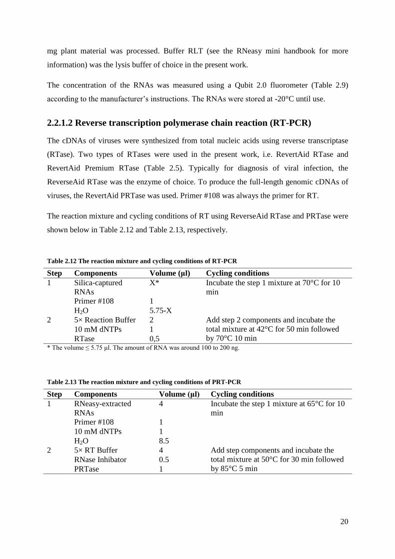
2.2.1.2 Reverse transcription polymerase chain reaction (RT-PCR)
The cDNAs of viruses were synthesized from total nucleic acids using reverse transcriptase
(RTase). Two types of RTases were used in the present work, i.e. RevertAid RTase and
RevertAid Premium RTase (Table 2.5). Typically for diagnosis of viral infection, the
ReverseAid RTase was the enzyme of choice. To produce the full-length genomic cDNAs of
viruses, the RevertAid PRTase was used. Primer #108 was always the primer for RT.
The reaction mixture and cycling conditions of RT using ReverseAid RTase and PRTase were
shown below in Table 2.12 and Table 2.13, respectively.
Table 2.12 The reaction mixture and cycling conditions of RT-PCR
Step Components Volume (μl) Cycling conditions
1 Silica-captured
RNAs
X* Incubate the step 1 mixture at 70°C for 10
min
Primer #108 1
H2O 5.75-X
2 5× Reaction Buffer 2 Add step 2 components and incubate the
total mixture at 42°C for 50 min followed
by 70°C 10 min 10 mM dNTPs 1
RTase 0,5 * The volume ≤ 5.75 μl. The amount of RNA was around 100 to 200 ng.
Table 2.13 The reaction mixture and cycling conditions of PRT-PCR
Step Components Volume (μl) Cycling conditions
1 RNeasy-extracted
RNAs
4 Incubate the step 1 mixture at 65°C for 10
min
Primer #108 1
10 mM dNTPs 1
H2O 8.5
2 5× RT Buffer 4 Add step components and incubate the
total mixture at 50°C for 30 min followed
by 85°C 5 min RNase Inhibator 0.5
PRTase 1
21
2.2.1.3 Polymerase chain reaction (PCR)
Two types of polymerases were used in the present work, i.e. KAPA Taq DNA polymerase
and PRECISOR high-fidelity DNA polymerase (Table 2.5). To detect virus fragments, to
perform colony PCR or for TA cloning purpose, KAPA Taq DNA polymerase was the
enzyme of choice. To amplify fragments with blunt ends, such as full-length fragments of
viral genomic cDNAs and linear vectors, PRECISOR high-fidelity DNA polymerase was
used in PCRs.
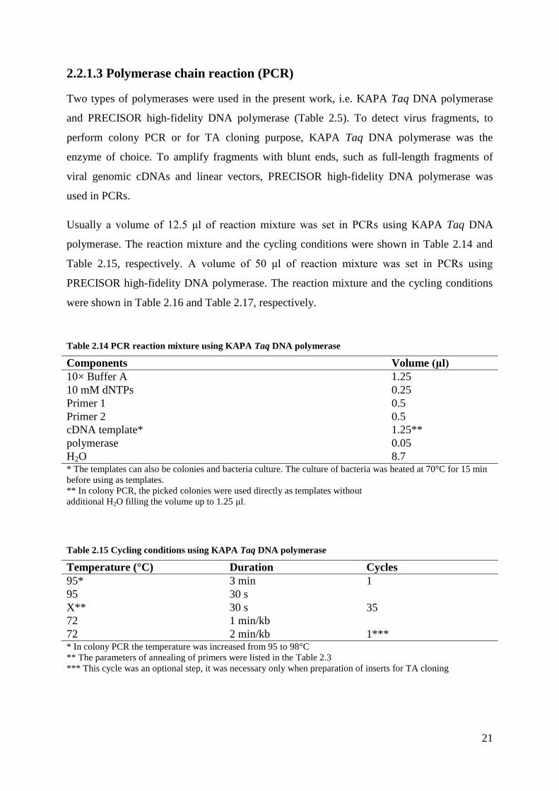
Usually a volume of 12.5 μl of reaction mixture was set in PCRs using KAPA Taq DNA
polymerase. The reaction mixture and the cycling conditions were shown in Table 2.14 and
Table 2.15, respectively. A volume of 50 μl of reaction mixture was set in PCRs using
PRECISOR high-fidelity DNA polymerase. The reaction mixture and the cycling conditions
were shown in Table 2.16 and Table 2.17, respectively.
Table 2.14 PCR reaction mixture using KAPA Taq DNA polymerase
Components Volume (μl)
10× Buffer A 1.25
10 mM dNTPs 0.25
Primer 1 0.5
Primer 2 0.5
cDNA template* 1.25**
polymerase 0.05
H2O 8.7 * The templates can also be colonies and bacteria culture. The culture of bacteria was heated at 70°C for 15 min
before using as templates. ** In colony PCR, the picked colonies were used directly as templates without
additional H2O filling the volume up to 1.25 μl.
Table 2.15 Cycling conditions using KAPA Taq DNA polymerase
Temperature (°C) Duration Cycles
95* 3 min 1
95 30 s
35 X** 30 s
72 1 min/kb
72 2 min/kb 1*** * In colony PCR the temperature was increased from 95 to 98°C ** The parameters of annealing of primers were listed in the Table 2.3 *** This cycle was an optional step, it was necessary only when preparation of inserts for TA cloning

22
Table 2.16 PCR reaction mixture using PRECISOR high-fidelity DNA polymerase
Components Volume (μl)
5× Buffer* 10
2 mM dNTPs 6.25
Primer 1 2
Primer 2 2
Template X**
Polymerase 1
H2O 28.75-X
* Two types of buffer were offered with the polymerase. To amplify genomic cDNAs of viruses from cDNA
templates, the GC buffer was the choice. To linearize plasmids of < 5 kb, the Hifi buffer was used; to linearize
plasmids of > 5 kb, the GC buffer was used. To produce partial fragments < 3 kb from plasmid template, the Hifi
buffer was used; otherwise GC buffer was used.
** Usually to amplify genomic cDNAs of viruses, cDNA template of 4 μl was used. To linearize plasmid of < 5
kb, template of plasmid of 0.1-0.5 ng was used; to linearize plasmid of > 5 kb or amplify partial fragments from
plasmids, template of plasmid of 20-30 ng was used.
Table 2.17 Cycling conditions using PRECISOR high-fidelity DNA polymerase
Temperature (°C) Duration Cycles
98 2 min 1
98°C 30 s
25-35*** X* 30 s
72°C 15-30 sec/kb**
72°C 10 min 1 * The annealing temperature of each primer was listed in Table 2.3. ** For cDNA templates, to linearize plasmids and to amplify partial fragments of > 3 kb from plasmid templates,
the time was 30 sec/kb. To amplify partial fragments < 3 kb from plasmid templates, the time was 15 sec/kb. *** The number of cycles was optimized for different assays.
To amplify the short fragment of 90 bp of (EAAAK)4 linker from synthesized
oligonucleotides. A 25 μl reaction mixture was prepared: 5 μl 10× Hifi Buffer, 3.25 μl 2 mM
dNTPs, 5 μl of primer #075, 5 μl of the Oligonucleotides #076 as template, 0.2 μl PRECISOR
high-fidelity DNA polymerase and filled up with H2O to the final volume. The cycling
conditions were 95°C for 2 min, 3 cycles of 50°C for 20 sec and 72°C for 10 sec.
2.2.1.4 Agarose gel electrophoresis
Electrophoresis was conducted in Sub-Cell GT system. The system consisted of power supply
PowerPac 300, Wide Mini-Sub Cell GT and horizontal agarose gel casters (Table 2.9). Gels
were prepared ahead of the run and supplemented with 20 μl/l Midori Green advanced DNA
stain (Table 2.8). Samples were mixed with orange or blue loading dye, and loaded on the gel
together with a standard marker of 1 kb plus DNA ladder (Table 2.4) for size determination.
23
Electrophoresis was performed through 1× TAE buffer (Table 2.8) at 90-110 V for 30-40 min.
DNA was visualized on a Reprostar 3 UV transilluminator system (Table 2.9).
The concentration of gels was decided according to the purposes and the size of fragments. In
general detection purposes, 1% (w/v) agarose gel was used. To purify fragments by gel
extraction, 2% (w/v) agarose gel was used for fragments of ca. 90 to 150 bp. Gels of 1% (w/v)
were used for fragments of ca. 200 bp to 8 kb. Gels of 0.7% (w/v) were prepared for
fragments of > 8 kb.
2.2.1.5 DNA purification
DNA purification was conducted using different gel extraction kits, i.e. QIAquick and QIAEX
II gel extraction kit (Table 2.7). The separation of DNAs in agarose gels were described above,
see section 2.2.1.4.
If the size of target DNA fragments was between 90 and 150 bp or > 8 kb, the DNAs in the
cut gel were extracted using the QIAEX II gel extraction kit. The extraction was performed
according to the manufacturer’s protocols.
If the size of the target DNA fragments was between 200 bp and 8 kb, the DNAs were
extracted from the cut gels using QIAquick gel extraction Kit. The extraction was conducted
according to the manufacturer’s protocols.
The concentration of the purified DNAs was measured using a Qubit 2.0 fluorometer (Table
2.9) according to the manufacturer’s instructions. The purified DNAs were stored at -20°C for
use.
2.2.1.6 Cloning of the target fragments
For fusion of inserts and vectors, three different methods were used in the present work:
circular polymerase extension cloning (CPEC) (Quan and Tian 2009), Gibson assembly and
In-Fusion cloning (Table 2.5). The three methods achieved cloning based on the homologous
ends of inserts and vectors (see section 1.2.3). Before fusion, the inserts and vectors were
purified by Gel extraction (sections 2.2.1.4 and 2.2.1.5).
In CPEC assays the PRECISOR high-fidelity DNA polymerase (Table 2.5) was the enzyme
of choice. The CPEC reaction mixture (25 μl) and cycling conditions were as below.

24
Table 2.18 The reaction mixture of the CPEC
Components Volume (μl)
5× Hifi Buffer 5
2 mM dNTPs 3.25
Polymerase 0.5
Inserts X*
Vectors Y*
H2O** fill up to 25 μl * The volumes of inserts and vectors were determined according to their concentration. X+Y ≤ 16.25. ** H2O was an optional component. The volume could be 0 μl.
Table 2.19 The cycling conditions in the CPEC
Temperature (°C) Duration Cycles
98 30 sec 1
98 10 sec 15
55 20 sec
72 30 sec/kb
The In-Fusion cloning and Gibson assembly reaction mixture were prepared according to the
manufacturer’s instructions, in the present work the preparation of each reaction mixture was
described for each cloning. The prepared reaction mixtures were incubated at an isothermal
condition of 50°C for 1 to 2 hours.
After the fusion step, the assembled product was directly used for transformation of
competent E. coli cells or stored at -20°C.
2.2.2 Molecular cloning of viruses and viral vectors
2.2.2.1 Construction of full-length cDNA clones
To rescue the viruses of ACLSV and ASPV from their host plants, full-length cDNA clones
were constructed. In the present work, the binary vector of pV297 was used as vectors (Table
2.2). It possesses a Cauliflower mosaic virus (CaMV) 35S promoter and the hepatitis delta
viral ribozyme (HDV) sequence followed by a CaMV 35S polyadenylation (pA) signal
(Figure 2.1).

25
Figure 2.1 The structure of the binary vector pV297. The insertion site of genomic cDNA of viruses was
between a CaMV 35S promoter and a HDVpA (indicated by the red arrow).
The plasmid pV297 was linearized by PCR. To produce linear vectors for ACLSV, the
primers #003 and #004 were used (Table 2.3). Using this primer pair, extensions of 15 bp
were attached to the 5’ and 3’ ends of the linear vectors. The extensions were homologous to
the 3’ and 5’ ends of genomic cDNAs of ACLSV, respectively. To produce linear vectors for
ASPV, the primers #007 and #008 were used (Table 2.3). Linear vectors with extensions
homologous to ASPV were generated too. In the PCR assays the enzyme of PRECISOR high-
fidelity DNA polymerase was the choice. The reaction mixture and cycling conditions were
set according to Table 2.16 and Table 2.17 (see section 2.2.1.3).
The full-length genomic cDNAs of the viruses were amplified from total nucleic acids of
infected plants by RT-PCR. The total nucleic acids of infected plant materials were extracted
using RNeasy plant mini kit (see section 2.2.1.1). With templates of the total nucleic acids
extraction, cDNAs were generated by reverse transcription assays (see section 2.2.1.2). With
the templates of cDNAs, genomic cDNAs of viruses were amplified by PCR. To amplify
genomic cDNAs of ACLSV, the primers #001 and #002 (Table 2.3) were used. The primers
#005 and #006 (Table 2.3) were used for amplification of genomic cDNAs of ASPV. In the
PCR assays, the PRECISOR high-fidelity DNA polymerase was used. The reaction mixture
and cycling conditions were as in Table 2.16 and Table 2.17 (see section 2.2.1.3).
The fragments of the vectors and the full-length genomic cDNAs of viruses were purified
using a gel extraction kit (see section 2.2.1.5). Before purification, the templates of plasmid

26
pV297 were digested with DpnI. The enzyme of DpnI of 1 μl was added directly to the PCR
reaction mixture. This mixture was incubated for 15 min at 37°C.
The gel purified fragments of the vectors and full-length genomic cDNAs were fused using
CPEC, Gibson assembly or In-Fusion cloning (see section 2.2.1.6).
The fusion reaction mixture was used for transformation of competent E. coli cells by heat
shock. Heat shock was as described in section 2.2.3.2.
Positive colonies were selected by colony PCRs (see section 2.2.1.3). For selecting full-length
cDNA clones of ACLSV, primer pairs of #009/#010 and #011/#012 were used. Each primer
pair encompassed the fusion parts of pV297 and genomic cDNAs of ACLSV. In addition, the
primer pair of #046/#047 (Menzel et al. 2002) was used for the selection too. For selecting
full-length cDNA clones of ASPV, primer pairs of #048/#049, #013/#014 and #015/#016
were used.
The plasmids of positive colonies were extracted from liquid cultures using QIAprep spin
miniprep kit according to the manufacturer’s protocol.
The plasmids were delivered into competent A. tumefaciens strain ATHV by electroporation
(see section 2.2.3.2). Then the transformed A. tumefaciens were used for agroinoculation of
test-plants (see section 2.2.4.1 and section 2.2.4.3).
2.2.2.2 Strategies of labeling viral proteins with fluorescent proteins
To fuse fluorescent proteins to the proteins of ACLSV, recombinant plasmids containing
marker genes, i.e. Emerald-GFP (EmGFP), mCherry and iLov, were constructed. Three
different strategies were used for labeling ACLSV proteins.
Strategy A: the overlapping open reading frames of movement protein (MP) and coat protein
(CP) genes were selected for insertion of the fluorescent genes. To keep the genes intact, the
overlapping part was duplicated. The potential promoter for CP was retained.
Strategy B: as in strategy A the overlapping open reading frames of MP and CP genes were
also the insertion site. To keep the gene intact and to avoid the deletion of the marker gene
insert caused by homologous recombination, the heterogeneous genes of MP and CP in pIF3-
15 and pIF3-19 (Table 2.2) were recombined.
Strategy C: the 3’ end of CP gene was selected as the insertion site.
27
2.2.2.3 Construction of labeled plasmids
Using different strategies (see section 2.2.2.2), recombinant plasmids were constructed by
inserting marker genes to the infectious full-length cDNA clones of ACLSV. The plasmid of
pDoc-G was the donor of EmGFP gene. The pK2GW7 was the donor of mCherry gene. The
plasmid of p2488 was the donor of iLov gene (Table 2.2). The infectious full-length cDNA
clones of ACLSV, i.e. pIF3-15 or pIF3-19, were used as backbones.
The plasmids were constructed using Gibson assembly or In-Fusion cloning (see section
2.2.1.6). The general working flow was similar to the construction of full-length cDNA clones
of viruses (see section 2.2.2.1). First the target fragments having homologous ends of 15 bp
were produced by PCR. In the PCRs, PRECISOR high-fidelity DNA polymerase was the
enzyme of choice. The PCR reaction mixture and cycling conditions were set according to
Table 2.16 and Table 2.17 (see section 2.2.1.3). Before cloning, the target fragments were gel
purified (see section 2.2.1.5). The reaction mixture was directly used for transformation of
competent E. coli cells by heat shock (see section 2.2.3.3). Positive colonies were selected by
colony PCRs (section 2.2.1.3). In the PCRs, designed primers encompassed fused parts were
used. The KAPA Taq DNA polymerase was used. The PCR reaction mixture and cycling
conditions were set according to Table 2.14 and Table 2.15. The plasmids were isolated using
QIAprep spin miniprep kit (Qiagen) according to the manufacturer’s protocol. The insert parts
were sequenced using Mix2Seq kit (see section 2.2.2.4). By comparison with the sequence of
marker genes, the plasmids having correct sequence (no mutation in the insert) were
agroinoculated to test-plants of N. occidentalis 37B (section 2.2.4.1).
2.2.2.3.1 Construction of plasmids using strategy A
To insert the marker gene of EmGFP to the 3’ end of MP gene, two steps were conducted. As
first step, EmGFP fragments with 15 bp extensions were amplified using primers #029 and
#036. The plasmid pIF3-19 (Table 2.2) was linearized using primers #027 and #028. The
linearized position was in front of the stop codon of MP gene, exactly between nucleotide (nt)
7090 and nt 7091 of genomic cDNA of ACLSV on pIF3-19 (see section 3.1.2). The two
fragments were cloned using In-Fusion. The fusion reaction mixture consisted of 6 μl (215 ng)
of linear pIF3-19, 2 μl (30 ng) of EmGFP with extensions and 2 μl of In-Fusion enzyme
premix. Positive colonies were selected by colony PCR using primers #029 and #030. By
sequencing the inserted parts, the plasmid having correct sequence of EmGFP was selected
for next step and named pIF12-28. As second step, the plasmid pIF12-28 was linearized using

28
primers #027 and #037. The position was just behind the stop codon of the GFP gene in
pIF12-28. The partial CP gene (plus the short fragment of 304 bp upstream) with extensions
of 15 bp was amplified from pIF3-19 using primers #031 and #032. The target fragments
were cloned using In-Fusion. The fuison mixture consisted of the linear pIF12-28 of 6.5 μl
(350 ng), partial CP gene of 0.5 μl (60ng) and In-Fusion enzyme premix of 2 μl, H2O filled up
to 10 μl. Positive colonies were selected by colony PCR using primers #029 and #030. The
fuison part was sequenced. Finally the plasmid having correct sequences was named pIF13-9.
To insert the marker gene of mCherry to the 5’ end of CP gene, two steps were conducted. As
first step, pIF3-19 was linearized using primers #064 and #065. The position was just in front
of the start codon of the CP gene. The mCherry gene with extensions of 15 bp was amplified
using primers #066 and #067. The fragments were cloned using Gibson assembly. The
reaction mixture consisted of the linear pIF3-19 of 4.5 μl (126 ng), the mCherry with
extensions of 1 μl (63 ng) and 5.5 μl Gibson master mix. The mixture was incubated for 2 h at
37°C, and used for transformation of competent E. coli NEB 5-alpha cells. The positive
colonies were selected by colony PCR using primers #060 and #061. The plasmids of positive
colonies were sequenced for the cloned part of the mCherry gene. The one with correct
sequence was named pG5-4. As second step, the fragment of mCherry:CP (plus a short
fragment of 304 bp upstream) containing extensions of 15 bp was amplified using primers
#059 and #068. The plasmid of pG5-4 was used as templates. The fragment containing
pV297:RdRp:MP: 3’ untranslated region (UTR) was amplified using primers #054 and #071.
The template was the linear pIF16-1 (section 2.2.2.3.3), which were produced by restriction
digestion of plasmids pIF16-1 with XbaI. The target fragments were cloned using Gibson
assembly. The reaction mixture consisted of the fragment of mCherry:CP of 1 μl (120 ng), the
fragment containing pV297:RdRp:MP: 3’ untranslated region of 9 μl (171 ng) and 10 μl
Gibson master mix. The mixture was incubated for 2 h at 37°C, and used directly for
transformation of E. coli Steller competent cells. Positive colonies were screened by colony
PCR using primers #059 and #068. The fusion parts of the plasmids from positive colonies
were sequenced. The expected plasmid was named pG11-15.
2.2.2.3.2 Construction of plasmids using strategy B
To prevent the deletion of inserts in ACLSV genomes, which could be caused by homologous
recombination of the duplicated parts of overlapping ORFs, heterogeneous MP or CP genes
were introduced into the genomic cDNAs of ACLSV.

29
Based on the plasmid of pIF13-9 (section 2.2.2.3.1), two other plasmids were constructed. In
one case, the heterogeneous CP gene (plus a short fragment of 87 bp upstream) was amplified
from pIF3-15 using primers #090 and #091. The plasmid of pIF3-15 was constructed during
this thesis (see section 3.1.2). With the primers #090 and #091, an extension of 15 bp was
added to the target CP genes. Using primers #087 and #088, the fragments of linear pIF13-9
excluding CP genes were generated. The target fragments were cloned using In-Fusion
cloning. The reaction mixture consisted of the heterogeneous CP fragments of 0.4 μl (316 ng),
the linear reduced pIF13-9 of 5.3 μl (250.2 ng) and In-Fusion enzyme premix of 1.4 μl.
Positive colonies were selected using primers #099 and #110. The inserted part of the CP of
pIF3-15 was sequenced for selecting clones without mutation(s). The expected plasmid was
named pIF18-2. In the second case, partial fragment of pIF13-9 was amplified using primers
#104 and #105. The fragments contained partial MP genes and the complete EmGFP genes.
The 5’ end of the partial MP genes started just behind the stop codon of RdRp genes. The
plasmid of pIF3-15 was linearized using primers #106 and #107. The linear fragments
contained all genes originally on pIF3-15 except for a partial MP gene. The target fragments
were cloned using In-Fusion cloning. The reaction mixture consisted of 1 μl (326 ng)
fragments of the partial MP gene plus EmGFP gene of pIF13-9, the linear fragments of partial
pIF3-15 of 3 μl (393 ng) and the In-Fusion enzyme premix of 1 μl. Positive colonies were
selected by colony PCR using primers #038 and #098. The cloning part of isolated plasmids
was sequenced and the plasmid with correct sequence was named pIF25-7.
Based on the plasmid of pG11-15, two other plasmids were constructed. In the first case, the
plasmid of pG11-15 was linearized using the primers #102 and #103. The linear pG11-15
contained all genes originally on it, except for the partial MP gene between the RdRp gene
and the possible promoter (German et al. 1992) of the CP gene (304 bp upstream). Using
primers #100 and #101, the partial MP gene on pIF3-15 was amplified with extensions of 15
bp at ends. The 5’ end of partial MP gene was just behind the stop codon of the RdRp gene on
pIF3-15. The 3’ end of the partial MP gene was intact. The fragments were cloned using In-
Fusion cloning. The reaction mixture consisted of the fragments of the partial pG11-15 of 5 μl
(250 ng), the fragments of partial pIF3-15 of 1 μl (442 ng) and the In-Fusion premix of 1.5 μl.
Positive colonies were selected using primers #096 and #097. The fused parts of the plasmids
were sequenced. The expected plasmid was named pIF24-6. In the second case, the plasmid
of pIF3-15 was linearized using primers #003 and #093. The linear fragments of pIF3-15
contained the original genes on it, except for the partial CP gene and 3’ UTR. The partial CP
gene was just behind the stop codon of MP gene. The fragments of mCherry:CP:3’ UTR

30
genes were amplified from pG11-15 using primers #002 and #094. The target fragments were
cloned using In-Fusion cloning. The reaction mixture consisted of 5 μl (370 ng) of the
fragments of partial pIF3-15, 1 μl (426 ng) of the fragments of mCherry:CP:3’ UTR and 1.5
μl of In-Fusion premix. Positive colonies were selected by colony PCR using primers #096
and #097. The cloning parts of isolated plasmids were sequenced. The expected plasmid was
named pIF23-1.
2.2.2.3.3 Construction of plasmids using strategy C
The plasmid of pIF3-19 was linearized using primers #054 and #055. The position was just in
front of the stop codon of CP gene of ACLSV. The mCherry with extensions of 15 bp was
amplified using primers #056 and #057. The target fragments were purified and cloned using
In-Fusion cloning. The reaction mixture consisted of the linear pIF3-19 of10 μl (50 ng), the
mCherry of 1 μl (50 ng) and 2.2 μl of In-Fusion enzyme premix. Positive colonies were
selected by colony PCR using primers #060 and #061. The fragment of the insert was
sequenced and the plasmid with correct sequence was named pIF16-1.
Based on the plasmid of pIF16-1, the second plasmid was constructed. The plasmid of pIF16-
1 was linearized using primers #061 and #055. The linearized position was the connection of
CP and mCherry genes. Fragments of a linker of (EAAAK)4 were produced by a PCR step
(see section 2.2.1.3). The linear pIF16-1 and the linker were cloned using In-Fusion cloning.
The reaction mixture consisted of 0.2 μl (53.6 ng) of the linker, 5.4 μl (310 ng) of the linear
pIF16-1 and 1.4 μl of the In-Fusion enzyme premix. Positive colonies were selected by
colony PCR using primers #080 and #110. The cloning part was sequenced, and plasmid with
correct sequence was named pIF20-16.
Based on the plasmid of pIF16-1, the third plasmid was constructed. The plasmid of pIF16-1
was linearized using the primers #083 and #084. The linear fragments contained all the
original genes on pIF16-1, except for the gene of mCherry. The fragments of iLov gene were
amplified using primers #085 and #086. Extensions of 15 bp were added to the ends of iLov
genes. The target fragments were cloned using In-Fusion cloning. The reaction mixture
consisted of 1 μl (254 ng) of the iLov fragments, 6 μl (258 ng) of the linear pIF16-1 and 1.8 μl
of the In-Fusion premix. The positive colonies were selected using primers #085 and #086
(annealing at 61°C). The cloning part of the plasmids was sequenced for selecting clones
without mutation(s). The expected one was named pIF27-10.
31
2.2.2.4 Sequencing of obtained plasmids
For the purpose of sequencing, the plasmids in a volume of 15 μl were mixed with sequencing
primers (see Supplementary Table S1) of 2 μl (10 µM) in a Mix2Seq tube (see information
about the Mix2Seq kit in Table 2.10). The samples were then sent to Eurofins Genomics for
sequencing. The first sequencing primers of #111 and #112 were bound to vector pV297. The
next primers were designed step by step based on the obtained sequence data. The obtained
partial fragments were assembled using SeqMan (DNASTAR Inc.) based on the overlapping
data. The sequences of the complete genomic cDNAs were then annotated using the NCBI
Basic Local Alignment Search Tool (BLAST) and NCBI Conserved Domain Search. The
phylogeny analyses were performed between the obtained sequences and the published
sequences in NCBI database using MEGA5 (Table 2.11).
2.2.3 Microbiology
2.2.3.1 Preparation of electrocompetent bacteria cells
Electrocompetent cells of A. tumefaciens GV2260, A. tumefaciens ATHV and E. coli NM522
were used in the present work. For preparation of electrocompetent A. tumefaciens GV2260,
the cells were plated on a yeast extract broth (YEB) agar plate containing 25 μg/ml rifampicin
and 50 μg/ml carbenicillin and grown at 28°C for around 45 hours. And then 50 ml YEB
medium containing the same antibiotics were inoculated with one single colony picked from
the plate and again grown at 28°C with 200 rpm for around 45 hours. Then 6 flasks with 250
ml YEB medium containing the same antibiotics were inoculated with 5 ml of the pre-culture
and incubated at 28°C until the OD600 value reached 0.5. The culture was incubated on ice for
15 min and then harvested by centrifugation for 15 min at 4,000 rpm and 4°C. The bacterial
pellets were resuspended in 250 ml ice-cold sterile Mili-Q H2O. This wash step was repeated
once. After that the pellet was resuspended in 200 ml ice-cold sterile 10% glycerol. The
resuspending was centrifuged again. By discarding the supernatant, the volume of the wet
pellet was reduced to 1 to 3 ml. The pellet was mixed with the liquid by gently hand-vortex.
Aliquots of 50 µl bacteria were frozen in liquid nitrogen and immediately transferred to -80°C
until electroporation experiments.
The electrocompetent A. tumefaciens ATHV and E. coli NM522 cells were equally prepared.
Cultivation of A. tumefaciencs ATHV was performed on YEB medium with 25 µg/ml
rifampicin at 28°C. E. coli NM522 was cultivated on Luria-Bertani (LB) medium at 37°C
without antibiotics.

32
2.2.3.2 Electroporation
For transformation of electrocompetent Agrobacterium cells, an aliquot of 50 µl
electrocompetent cells was thawed on ice. Then plasmids of 2 µl were slightly mixed with the
cells. Immediately the mixture was transferred to a pre-cooled electroporation cuvette (2 mm
gap). The cuvette was placed into a Gene pulser electroporation system and pulsed with 2500
V, 25 µF and 200 Ω. Once done 950 µl of prewarmed SOC (super optimal broth with
catabolite repression) medium was added to the bacteria. The culture was transferred to a 2 ml
reaction tube and was incubated for 2 hours at 28°C and 200 rpm. Afterwards, culture of 10 µl
and 50 µl was plated on antibiotic-containing YEB agar plates, respectively. The remaining
culture was centrifuged for 30 sec at 4000 rpm. The pellet was resuspended in 100 µl medium
and also plated. Plates were incubated at 28°C for above 40 hours. Single colonies were
picked for further experiments.
Transformation of electrocompetent E. coli NM522 cells was performed in the same way. The
optimal incubation temperature was 37°C and LB medium was used. The cells were agitated
for 1 hour at 37°C and 200 rpm after electroporation. The culture of 50 µl, 100 µl and rest
were plated on LB agar plates, respectively. The plates were incubated overnight at 37°C.
2.2.3.3 Heat shock
In the present work, commercial chemo-competent E. coli cells were used, including NEB 5-
alpha, NEB 10-beta and Steller (Table 2.1). Unless mentioned otherwise NEB 10-beta was the
cells of choice. The transformation was performed mainly according to the manufacturer’s
protocols. The competent cells were thawed on ice. Five µl of a Gibson or In-Fusion reaction
mixture was added into 50 µl of the thawed cells. The mixture was incubated for 30 min on
ice. Then a heat shock was performed for 30 to 45 sec in a water bath at 42°C, and the cells
were then cooled down on ice for 2 to 5 min. Afterwards the cells were incubated in 950 µl
prewarmed SOC medium for 1 hour at 37°C and 200 rpm. The culture of 100 µl, 200 µl and
rest was plated on LB agar plates containing 30 μg/ml kanamycin, respectively (see section
2.2.3.2). The plates were incubated overnight at 37°C. Single colonies were picked for further
experiments.

33
2.2.4 Inoculation of plants
2.2.4.1 Mechanical agroinoculation
To test the infectivity of the constructed full-length cDNA clones of viruses, they were
agroinoculated to test-plants. The herbaceous test-plants included Nicotiana occidentalis 37B
and Chenopodium quinoa (section 2.1.2).
To prepare the inocula of agrobacteria containing the tested cDNA clones, a single colony
(section 2.2.3.2) was inoculated to 50 ml antibiotic-containing YEB medium (Table 2.8). The
culture was incubated for around 45 hours at 28°C and 190 rpm. The agrobacteria were
harvested by centrifugation for 15 min at 5000 rpm, and then the pellet was resuspended in 10
mM MgSO4. This suspension was inoculated directly to test-plants.
To perform a mechanical inoculation, the upper surface of leaves was dusted with
carborundum (600 mesh). After that the inoculum was rubbed onto the leaf surface. On each
leaf, about 500 μl of inoculum was used. Symptom development was observed during three
weeks.
2.2.4.2 Sap inoculation
To consequently transmit the viruses generated from infectious full-length cDNA clones to
new host plants, sap inoculation was conducted. The juice of infected leaves was extracted as
inoculum. To prepare this, fresh symptomatic leaf blades were ground in herb-herb
inoculation buffer (Table 2.8) in a universal extraction bag. Approximately 1 g fresh leaf
blades was ground in 100 ml inoculation buffer, the amount of leaf materials and buffer can
be increased in proportion. The juice (about 200 μl) was then slightly rubbed onto leaf
surfaces of test-plants that were dusted with carborundum (600 mesh). Symptom development
was observed during three weeks.
2.2.4.3 Agroinoculation by vacuum infiltration
To inoculate woody plants, the protocol of agroinoculation of infectious cDNA clones by
vacuum infiltration was developed in the present work.
The seedlings of one to three-month old Malus domestica cv. Golden Delicious were used as
test-plants (section 2.1.2). Before suffering vacuum infiltration, the brown seed coats on
cotyledons were removed, and the seedlings were taken to room temperature (ca. 20°C) for
around 20 hours under natural light. Wounded or unwounded seedlings were used in the

34
experiments. Of wounded seedlings, each cotyledon was stuck by a sterilized needle to
introduce four to six pin holes (Figure 2.2A and B). The entire wounded or unwounded
seedlings were immersed in sterilized Milli-Q water containing 1% (v/v) Tween 80 for 10 min,
and rinsed two times with sterilized Milli-Q water. Prepared seedlings were then immediately
immersed in the inoculum for vacuum infiltration.
The inocula of transformed A. tumefaciens containing test cDNA clones were prepared as
described above (sections 2.2.3.2 and 2.2.4.1), additionally 0.2% (v/v) Tween 20 was added.
The vacuum was generated in a closed desiccator using a Büchi V-500 vacuum pump with
vacuum controller B-721 (Table 2.9) (Figure 2.2C and D). Different pressure and duration of
vacuum were used in independent treatments.
The infiltrated seedlings were immediately planted in virus-free soil after the vacuum
infiltration and were grown in an insect-proof glasshouse at 60-65% humidity and 20-25°C.
Figure 2.2 Apple seedlings and pump system used in vacuum infiltration. Apple seedlings with unwounded
(A) and wounded (B) cotyledons were used in the vacuum infiltration. Red arrow indicates a magnification of a
wounded cotyledon. The vacuum system consisted of a dessicator (C) and a pump system (D).
2.2.4.4 Grafting
To transmit virus particles from agroinfected apple to healthy apple, cleft grafting was
conducted. The rootstocks (ca. 25 cm) were prepared from one-year old apple seedlings (ca.
70 cm) (see section 2.1.2). The top branches of the seedlings were removed using sterilized
guarder knife and the rootstocks were split 3 to 5 cm. The scions were prepared from the
agroinfected apple seedlings (see section 2.2.4.3). The selected branches of ca. 8 cm were
pruned off the apple seedlings, and a V-shaped appearance that will fit tightly into the split on
the stocks were made by long tapering cuts on both sides of the bottom of the sticks. The
scions were then carefully placed into the split of rootstocks, and the graft unions were
wrapped with parafilm and rubber. The top of the scions was sealed with grafting wax
35
(Maywax). The grafted plants were kept in an insect-proof glasshouse with 60-65% humidity
at ca. 22°C.
2.2.4.5 Detection of viral infection in plants
The host-plants were tested by RT-PCR for virus infection. First total RNAs of noninoculated
leaves were extracted using silica capture method (section 2.2.1.1). And then the cDNAs
templates were produced from total RNAs using ReverseAid RTase (section 2.2.1.2). Virus
fragments were detected using selected primers: for ACLSV detection, the primers #046/#047
were used; for detection of ASPV, the primers #048/#006 were used. PCR reaction mixture
and cycling conditions were described in section 2.2.1.3.
2.2.5 Protein immunoblot
2.2.5.1 Protein preparation
Fresh leaf blades were ground in 1× protein extraction buffer (Table 2.8) using a mortar and a
pestle (250 mg leaf blades : 250 μl buffer). The ground plant materials were centrifuged for 3
min at 13000 rpm. The supernatant of 200 μl was transferred into a new reaction tube. The
supernatant was centrifuged for 3 min at 13000 rpm, and then 150 μl were transferred into
another reaction tube.
The raw protein extraction of 40 μl was transferred into a reaction tube containing 1× protein
loading buffer (Table 2.8). The mixture was incubated for 10 min at 99°C.
2.2.5.2 Sodium dodecyl sulfate polyacrylamide gel electrophoresis (SDS-
PAGE)
To separate the proteins (section 2.2.5.1) extracted from test-plants, SDS-polyacrylamide gels
(PAGs) were prepared. Firstly resolving gels of 12% SDS-PAGs were prepared in SE245 dual
gel casters. Four volumes of resolving gels consisted of 8.7 ml of H2O, 5 ml of 1.5 M Tris (pH
8.8, plus 1% SDS), 6 ml of Rotiphorese® Gel 40 (37.5:1), 20 μl of
tetramethylethylenediamine (TEMED) and 100 μl of 10% ammonium persulfate (APS). The
gel pre-mixture was poured between the two glass plates and overlaid with isopropanol. Once
the resolving gel was polymerized, the isopropanol was removed. The stacking gel was
poured and a comb with the required number of wells (10 or 15) was inserted. Four volumes
of stacking gels consisted of 6.43 ml of H2O, 2.5 ml of 0.5 M Tris (pH 6.8, plus 1% SDS), 1

36
ml of 40% Rotiphorese® Gel 40 (37.5:1), 10 μl of TEMED, 50 μl of 10% APS and 50 μl blue
loading dye.
Electrophoresis was performed using a SE250 vertical electrophoresis system with a Consort
E844 power supply (Table 2.9). The gels ran in SDS-PAGE running buffer (Table 2.8) for 1.5
hours at 150 V. After the run the gels were processed for western blotting (see section 2.2.5.4).
2.2.5.3 Transmembrane
After SDS-PAGE electrophoresis (see section 2.2.5.2), the separated proteins were transferred
to an Immobilon-P transfer membrane through tank blotting. The transfer was conducted
using a mini Trans-Blot transfer cell with a Consort E844 power supply. The resolving gels
were cut off from the glass plates. Each gel was assembled with the membrane, filter papers
and foam pads in a tank transfer cassette holder. Before the assembly the filter papers and
foam pads were equilibrated in transfer buffer. The blotting was performed in iceblock-
cooling 1× transfer buffer for 1.5 hours at 120 V. The efficiency of transfer was determined
through PageRuler prestained protein ladder (Table 2.4).
2.2.5.4 Western blot
The membranes were blocked in 1x PBS-T buffer containing 5 % (w/v) milk powder for
around 1 hour at room temperature with shaking. The membranes were then washed 3 x 15
min with 1x PBS-T buffer. Afterwards the washed membranes were incubated with diluted
primary antibodies overnight at 4°C. The primary antibodies were diluted in blocking buffer
according to Table 2.6. Next day, the membranes were washed 3x 15 min with AP buffer.
During the wash step, secondary antibodies were diluted in blocking buffer according to
Table 2.6. The membranes were incubated in secondary antibody dilution for at least 1 hour at
room temperature with shaking. After a subsequent washing step, the membranes were
visualized. The membranes were washed 3 x 10 min in AP buffer. To visualize, the
membrane was incubated for 2 min with BCIP/NBT reagent.
2.2.6 Immunosorbent electron microscopy (ISEM)
Viral particles of ACLSV were visualized from N. occidentalis 37B and G. Delicious test-
plants by ISEM as described previously (Jelkmann et al. 1990). The polyclonal antiserum
used was ACLSV-AS1236 (JKI, Institute for Epidemiology and Pathogen Diagnostics). For
ISEM the antiserum was diluted 1:1000 in 0.1 M K-Na phosphate buffer pH 7.0. Nickel grids

37
were incubated for 5 min at room temperature in the diluted antiserum, and then were washed
with 1.5 ml the same buffer. The grids were transferred to leaf homogenate and incubated
overnight at room temperature. Decoration of particles was performed by incubation in 1 : 50
diluted antiserum at room temperature for 15 min. Excess liquid was carefully removed with
filter paper, and the grids were washed with 7% uranyl acetate in ultrapure water. Dry grids
were examined with a Zeiss EM 10C electron microscope.

38
3. Results
3.1 Construction of full-length cDNA clones
In total 17 full-length cDNA clones were obtained in the present work. In each plasmid the
viral genomic cDNA was inserted between the CaMV 35S promoter and HDVpA (Figure 3.1).
For each virus isolate, the number of obtained clones is shown in Table 3.1.
Figure 3.1 The structures of the constructed full-length cDNA clones. The genomic cDNAs of ACLSV (A)
and ASPV (B) are inserted between a CaMV 35S promoter and an HDVpA.
Table 3.1 Number of full-length cDNA clones constructed using different methods
Virus species Isolates Number of obtained clones
CPEC Gibson In-Fusion
ACLSV (27)/85 1 1 3
ACLSV 38/85 4 - 4
ACLSV (36)/88 - - 2
ASPV 40/87 - - 2

39
3.1.1 Construction of ACLSV clones isolate (27)/85
In total five clones were obtained for ACLSV isolate (27)/85 (Table 3.1). One was obtained
using CPEC method, one was obtained using Gibson assembly and three were obtained using
In-Fusion cloning. The preparation of the genomic cDNAs and linear pV297 were as
described in section 2.2.2.1.
In CPEC, 5 µl (43 ng) of genomic cDNA fragments and 3 µl (42 ng) of linear pV297 were
used in the reaction mixture (see section 2.2.1.6). After transformation of NEB 5-alpha E. coli
cells, 50 colonies developed on LB plate inoculated with 100 µl transformants. Using primers
#009/#010, #011/#012, and #046/#047, respectively (section 2.2.1.3), PCR products of about
600, 340 and 680 bp were amplified from 1 out of 25 colonies. The plasmid was named
pCPEC2-1.
In Gibson assembly, the reaction mixture consisted of 3.5 µl (200 ng) of genomic cDNA
fragments, 0.8 µl (100 ng) of linear pV297 and 10 µl 2× Gibson MasterMix, H2O filled up to
20 µl. The reaction mixture was incubated for 1 hour at 50°C. The mixture of 5 µl was
directly used for transformation of NEB 10-beta E. coli cells. By colony PCRs with primer
pairs of #009/#010, #011/#012 and #046/#047 (section 2.2.1.3), 1 positive colony was
selected out of 27 colonies. The plasmid was named pG2-138.
In In-Fusion cloning, the reaction mixture consisted of 8 µl (100 ng) of genomic cDNA
fragments, 3 µl (51 ng) of linear pV297 and 2.75 µl of 5× In-Fusion enzyme premix. The
reaction mixture was incubated for 1 hour at 50°C. The mixture of 5 µl was directly used for
transformation of NEB 10-beta E. coli cells. Three positive colonies were selected out of 50
colonies by colony PCR using primers primer pairs of #009/#010, #011/#012 and #046/#047
(section 2.2.1.3). The plasmids were named pIF15-13, pIF15-15 and pIF15-26.
3.1.2 Construction of ACLSV clones isolate 38/85
Eight clones were obtained for ACLSV isolate 38/85 (Table 3.1). Four clones were obtained
using CPEC method, and the others were obtained using In-Fusion cloning. The preparation
of genomic cDNAs and linear pV297 was as described in section 2.2.2.1.
Two independent CPEC were performed. In the two CPEC assays, the reaction mixture was
prepared according to Table 2.18, except for the amount of inserts and vectors. In the first
CPEC, 10 µl (55 ng) of genomic cDNA fragments and 0.5 µl (135 ng/ µl) of linear pV297
were used in the reaction mixture. In the second CPEC, 10 µl (55 ng) of genomic cDNA
40
fragments and 3.6 µl (18.7 ng/µl) of linear pV297 were used in the reaction mixture. The
cycling conditions were shown in Table 2.19. The reaction mixture of 5 µl was used for
transformation of NEB 10-beta E. coli cells. The colony PCR assays were performed using
primer pairs of #009/#010, #011/#012 and #046/#047 according to section 2.2.1.3. Finally,
two positive colonies were selected out of more than 300 colonies in the first CPEC. Two
other positive colonies were selected out of around 100 colonies in the second CPEC. The
plasmids were named pCPEC6-11, pCPEC6-18, pCPEC7-39 and pCPEC7-91.
In In-Fusion cloning, the reaction mixture consisted of 10 µl (55 ng) of genomic cDNA
fragments, 3.6 µl (67 ng) of linear pV297 and 3.4 µl of 5× In-Fusion enzyme premix. After
incubation for 2 hours at 50°C, the reaction mixture of 3.5 µl was directly used for
transformation of NEB 10-beta E. coli cells. Finally, 4 positive colonies were selected out of
the 36 colonies by colony PCR using primer pairs #009/#010, #011/#012 and #046/#047 (see
section 2.2.1.3). The plasmids were named pIF3-12, pIF3-14, pIF3-15 and pIF3-19.
3.1.3 Construction of ACLSV clones isolate (36)/88
Two clones of ACLSV isolate (36)/88 were constructed using In-Fusion cloning (Table 3.1).
The preparation of genomic cDNAs and linear pV297 were described in section 2.2.2.1. The
reaction mixture consisted of 3 µl (51 ng) of genomic cDNA fragments, 7 µl (101 ng) of
linear pV297 and 2.5 µl of 5× In-Fusion enzyme premix. The reaction mixture was incubated
for 1 hour at 50°C. Afterwards the reaction mixture of 5 µl was directly used for
transformation of NEB 10-beta E. coli cells. Two positive colonies were selected out of 90
colonies by colony PCR using primers #009/#010, #011/#012 and #046/#047 (see section
2.2.1.3). The plasmids were named pIF14-14 and pIF14-23.
3.1.4 Construction of ASPV clones isolate 40/87
Two clones of ASPV isolate 40/87 were constructed using In-Fusion cloning (Table 3.1). The
preparation of genomic cDNAs and linear pV297 were as described in section 2.2.2.1. The
reaction mixture consisted of 15 µl (90 ng) of genomic cDNA fragments, 3 µl (54 ng) of
linear pV297 and 4.5 µl of 5× In-Fusion enzyme premix. The reaction mixture was then
incubated for 2 hours at 50°C. After that the mixture of 4 µl was used for transformation of
NEB 10-beta E. coli cells. By colony PCR using primers #013/#014, #015/#016 and
#048/#049 (see section 2.2.1.3), fragments of around 480, 500 and 360 bp were amplified
from two out of 23 colonies. The plasmids were named pIF4-4 and pIF4-16.

41
3.2 Infectivity of the constructed clones on herbaceous plants
To test the infectivity, the obtained full-length cDNA clones of ACLSV and ASPV were
agroinoculated to ten plants of Nicotiana occidentalis 37B and ten plants of Chenopodium
quinoa, respectively. The inoculum of pV297-transformed agrobacteria was used as a
negative control. The agroinoculation was performed according to section 2.2.4.1. The tests
were performed independently three times.
After agroinoculation the symptom development was observed every day during three weeks.
The infection of viruses was determined by RT-PCR (section 2.2.4.5). In PCR positive plants
the existence of virus particles was confirmed by ISEM (section 2.2.6.1).
The results of agroinoculation demonstrated that four full-length cDNA clones were
infectious on N. occidentalis 37B, i.e. pIF3-15, pIF3-19, pIF4-4 and pIF14-23. But no
infection was observed for any clone on the test-plants of C. quinoa after the agroinoculation.
Moreover, infected N. occidentalis 37B were further inoculated to healthy N. occidentalis 37B
plants by sap inoculation with 100% successful rate. No transmission was observed on C.
quinoa by sap inoculation.
3.2.1 Symptoms development on N. occidentalis 37B
On N. occidentalis 37B test-plants, symptoms caused by pIF3-15, pIF3-19 and pIF14-23 were
similar. For each clone, all test-plants were infected via agroinoculation. Seven to nine days
after inoculation, infected plants developed the first symptom of yellow spots on
noninoculated leaves. Over time symptoms of chlorosis, crinkle and slight necrotic spots were
observed on upper leaves (Figure 3.2). No symptom developed on the inoculated leaves.
The clone of pIF4-4 caused systemic symptoms on all the ten test-plants of N. occidentalis
37B after mechanical agroinoculation. Eight to nine days after inoculation, symptoms started
to appear on the noninoculated leaves. The symptoms included chlorosis and vein-banding on
upper leaves (Figure 3.3). No symptoms developed on the inoculated leaves.
The control plasmid pV297 did not cause symptoms on test-plants. The following
inoculations remained without symptoms on test-palnts: pCPEC2-1, pG2-138, pIF15-13,
pIF15-15, pIF15-26, pCPEC6-11, pCPEC6-18, pCPEC7-39, pCPEC7-91, pIF3-12, pIF3-14,
pIF14-14 and pIF4-16. They also tested negative in PCR.

42
Figure 3.2 Symptoms on N. occidentalis 37B 18 days after agroinoculation with ACLSV clones. The plants
were agroinoculated with the infectious clones of pIF3-15 (A), pIF3-19 (B), pIF14-23 (C) and pV297 (D)
respectively. Symptoms of mosaic and chlorosis were observed on plants inoculated with pIF3-15 (A), pIF3-19
(B), pIF14-23 (C). No infection was observed on the plants inoculated with pV297 control (D). i was the side
view of the whole infected plants. ii was one of the symptomless inoculated leaves of each plant. iii was a
separated leaf indicated by red arrow on symptomatic plants.
Figure 3.3 Symptoms on N. occidentalis 37B 10 days after agroinoculation with the ASPV clone. The plants
were agroinoculated with control pV297 (A) and pIF4-4 (B) respectively. No symptoms developed on the
pV297-inoculated plants (A and C). The symptoms of chlorosis and vein-banding were observed on pIF4-4
inoculated plants (B and D). C and D are magnifications of the red box- indicated in A and B respectively.
3.2.2 RT-PCR detection of viruses in agroinoculated test-plants
The agroinoculated plants were analyzed by RT-PCR for ACLSV and ASPV infections
(section 2.2.4.5). In symptomatic plants agroinoculated with pIF3-15, pIF3-19 and pIF14-23,
PCR fragments of around 650 bp were amplified using primers #046/#047. The results
suggested the infection of ACLSV in the test-plants. In the plants inoculated with control
pV297, no PCR product was generated using the same primers. All inoculated test-plants with
the other 12 plasmids of the full-length clones of pCPEC2-1, pG2-138, pIF15-13, pIF15-15,

43
pIF15-26, pCPEC6-11, pCPEC6-18, pCPEC7-39, pCPEC7-91, pIF3-12, pIF3-14 and pIF14-
14 remained negative in PCR. This result indicated that no ACLSV infection was established
from the agroinoculation of the 12 clones in the test-plants.
In the symptomatic plants agroinoculated with pIF4-4, the band of around 430 bp was
amplified using primers #048/#006, suggesting the existence of ASPV in the inoculated plants.
In the plants inoculated with pV297, no PCR product was generated using primer pair
#048/#006. In the test-plants inoculated with pIF4-16, no PCR product of around 430 bp was
generated. This result indicated no ASPV infection was caused by pIF4-16 in the test-plants.
No infection of ACLSV or ASPV was detected in the test-plants of C. quinoa by RT-PCR.
This result suggested that no infection of ACLSV or ASPV was established in the C. quinoa.
3.2.3 Detection of viral particles by ISEM
Virus particles of ACLSV in the symptomatic and PCR-positive N. occidentalis 37B plants
were detected using ISEM (section 2.2.6.1). The virions were very flexuous filaments, the
length of the virions ranged from 840 nm to 1210 nm. The width was approximately 12 nm
(Figure 3.4).
Figure 3.4 Virions of ACLSV detected in infected N. occidentalis 37B using ISEM. A: virions trapping with
antiserum was used as standard, scale bar = 100 nm. B: virions definitively decorated with antiserum; scale bar =
200 nm. Red arrows indicated virions.
3.2.4 Transmission of viruses by sap inoculation
ACLSV generated from infectious clones of pIF3-15, pIF3-19 and pIF14-23 in infected N.
occidentalis 37B plants can be further transmitted to healthy N. occidentalis 37B plants. The
efficiency of transmission was 100%. The symptoms on the sap-inoculated plants were more
severe than those on the agroinoculated plants.

44
ACLSV extracts from N. occidentalis 37B were rubbed onto C. quinoa plants as well.
However, the establishment of infection was inconclusive. Among three trials, one time the
infection was successful in test-plants. In the successful one, a positive PCR result was
obtained in two out of four plants for pIF3-15 and pIF3-19, respectively, as well as in one out
of four plants for pIF14-23. Systemic symptoms of chlorosis and necrotic spots were observed
on the infected plants (Figure S1).
Similar to the ACLSVs, the ASPV generated from pIF4-4 can be further mechanically
transmitted to N. occidentalis 37B by sap inoculation at 100% infection rate. No transmission
to C. quinoa was successful.
3.3 Agroinoculation of woody plants with infectious clones
To inoculate woody hosts, the infectious full-length cDNA clones of pIF3-15, pIF3-19, pIF4-
4 and pIF14-23 were first mechanically agroinoculated upon the one-year-old G. Delicious, P.
persica and P. armeniaca respectively. At least eight plants were agroinoculated in each
treatment. However, two months after the inoculation, no viral infection was detected in the
test-plants by RT-PCR (section 2.2.4.5).
The infectious full-length cDNA clones of pIF3-15, pIF3-19, pIF4-4 and pIF14-23 were then
agroinoculated to three-month-old apple seedlings of G. Delicious by vacuum infiltration.
Except for the parameters of air pressure and duration of vacuum time, the protocol of
vacuum infiltration was described in section 2.2.4.3.
Different parameters were used in three vacuum infiltration assays. In the vacuum infiltration
1, wounded and unwounded apple seedlings were used as the host plants. The infectious full-
length cDNA clone of pIF3-19 was used as inoculum, whilst pV297 was used as a negative
control. The vacuum infiltration was conducted for 10 min at 50 hPa. In the vacuum
infiltration 2, the wounded apple seedlings were used as host plants. The inocula contained
cDNA clones pIF3-15, pIF3-19 and pV297, respectively. The infiltration was performed at
500 hPa for 2, 5 and 10 min, respectively. In the vacuum infiltration 3, the wounded apple
seedlings were used as host plants. The inoculums contained pIF3-15, pIF3-19, pIF14-23,
pIF4-4 and pV297, respectively. The infiltration was performed for 5 min at 500 hPa.

45
The infection of viruses in infiltrated apple seedlings was detected two months after
inoculation by RT-PCR (section 2.2.4.5). In PCR-positive samples, the existence of viral
particles was confirmed by ISEM (section 2.2.6.1).
3.3.1 Infectivity of the clones on apple seedlings
In vacuum infiltration 1, 23 wounded and 35 unwounded apple seedlings were agroinoculated
with the infectious clone of pIF3-19. Two weeks after the infiltration, most of the 35 grew
well, whilst nearly half of the 23 wounded seedlings wilted and died. Around 50 days after the
infiltration, 5 out of the 23 and 18 out of the 35 seedlings survived (Figure 3.5A). ACLSV
infection in survived seedlings was then detected by RT-PCR. The PCR results indicated that
4 out of the 5 seedlings and 2 out of the 18 seedlings were positive for ACLSV infection
(Figure 3.5A). Three of the four PCR-positive seedlings were analyzed by ISEM for viral
particles, and ACLSV particles were observed in the samples (data not shown). One PCR
negative sample was tested as well and no viral particle was observed (data not shown).
In the control group, ten wounded and nine unwounded apple seedlings were agroinoculated
with pV297. Two weeks after the infiltration, all seedlings died.
In vacuum infiltration 2, 15 or 10 wounded seedlings were agroinoculated with the infectious
clones of pIF3-15, pIF3-19 and control pV297, respectively. The duration of vacuum time
was 2, 5 and 10 min, respectively. After the infiltration most of the seedlings grew well, only
a few wilted in two weeks. Around 50 days after the infiltration the survival rates, defined as
the ratio of the number of survived seedlings to the number of infiltrated seedlings, of above
66% (10/15, pIF3-19) were observed for all the tests (Figure 3.5B). The PCR-positive rates,
defined as the ratio of the number of the PCR-positive samples to the number of the survived
seedlings, of above 21% (3/14, pIF3-15) were observed (Figure 3.5B) for all the tests.
In vacuum infiltration 3, 10, 15 or 20 wounded seedlings were agroinoculated with the
infectious clones of pIF3-15, pIF3-19, pIF14-23, pIF4-4 and control pV297, respectively. The
results are shown in Figure 3.5C. After the infiltration most seedlings grew well, showing
survival rates of above 70% (7/10, pV297). For pIF3-15, the PCR-positive rate was 78%
(11/14). For pIF3-19, the PCR-positive rate was 87% (7/8). For pIF4-4, the PCR-positive rate
was 50% (9/18). For pIF14-23, the PCR-positive, however, was 25% (2/8). For pIF3-15 and
pIF3-19, three out of the PCR-positive seedlings were detected by ISEM: ACLSV particles
were observed in the samples (data not shown). ASPV particles could not be observed in the

46
PCR-positive samples of pIF4-4 inoculated. One PCR negative sample was analyzed as well
and no viral particle was observed.
Figure 3.5 The efficiency of agroinoculation of apple seedlings with obtained infectious cDNA clones by
vacuum infiltration. A: inoculation of wounded and unwounded seedlings with infectious clone pIF3-19, the
vacuum infiltration was performed at 50 hPa for 5 min. B: inoculation of wounded apple seedlings with
infectious clones pIF3-15 and pIF3-19, respectively; the infiltration was performed at 500 hPa for 2, 5 and 10
min respectively. C: vacuum infiltration of wounded apple seedlings at 500 hPa for 5 min with pIF3-15, pIF3-19,
pIF14-23 and pIF4-4, respectively. The vector pV297 was used as a negative control. The number of survived
seedlings and the number of PCR-positive seedlings were obtained around 50 days after the vacuum infiltration.
3.3.2 Transmission of viruses to woody plants by grafting
To test the transmission of the viruses generated from the infectious full-length cDNA clones
of pIF3-15, pIF3-19 and pIF4-4 in apple seedlings (section 3.3.1), grafting was performed
(section 2.2.4.4).
To test the transmission of ACLSVs, the branches of virus-infected apple seedlings were used
as scions for grafting. Healthy one-year-old apple seedlings were used as rootstocks. Six
grafted plants were produced for each ACLSV isolate (clone pIF3-15 and pIF3-19). Four
plants were propagated using the infected scions and two as healthy controls. Four months
after the grafting, the grafted plants were analyzed for ACLSV infection by RT-PCR (section
2.2.4.5). The PCR results demonstrated that all four trees grafted by virus-infected scions
47
were successfully infected with the viruses. In addition one of the PCR-positive samples was
analyzed by ISEM. The existence of ACLSV particles was confirmed in the test-sample.
Transmission of ASPV was tested in the same way as just described for ACLSV. Nine grafted
plants were produced, containing eight grafts using the infected scions and one using the
healthy scion. Three months after grafting, ASPV infection in the grafted plants was detected
by RT-PCR (section 2.2.4.5). The eight trees grafted with the infected scions were PCR-
positive. But in the ISEM, no viral particles were observed in the PCR-positive samples.
3.4 Sequence analysis
The genomic cDNAs of the infectious full-length cDNA clones of pIF3-15, pIF3-19, pIF14-
23 and pIF4-4 were sequenced (see section 2.2.2.4).
3.4.1 Sequences of ACLSV
The genomic cDNA of ACLSV in pIF3-15 was assembled and analyzed. The corresponding
virus was designed ACLSV isolate 38/85A. The genome of ACLSV 38/85A, excluding the
polyadenylated (polyA) tail, consists of 7561 nucleotides (nts). Besides the 5’ and 3’
untranslated regions (UTRs), there are three open reading frames (ORFs) in the genome, i.e.
ORF1, ORF2 and ORF3 (Figure 3.6). The 5’ UTR consists of 149 nts, the position in the
genome is from (nucleotide) nt 1 to 149. The ORF1 consists of 5658 nts, encoding the RNA-
dependent RNA polymerase (RdRp), is located at position from nt 150 to 5807. The ORF2
consists of 1383 nts, encoding the movement protein (MP), is located at position from nt 5719
to 7101. The ORF3 consists of 582 nts, encoding the (coat protein) CP unit, is located at
position from nt 6785 to 7366. The 3’ UTR consists of 195 nts, the position in the genome is
from nt 7367 to 7561. The ORF1 and ORF2 overlap from nt 5719 to 5807 (89 nts in length).
ORF2 and ORF3 overlap from nt 6785 to 7101 (317 nts in length).
The genomic cDNA in pIF3-19 corresponded to the genome of ACLSV isolate 38/85B. The
genome organization of ACLSV 38/85B is shown in Figure 3.6. The genome consists of 7544
nts excluding the polyA tail. The 5’ UTR consists of 150 nts, the position in the genome is
from nt 1 to 150. The ORF1 consists of 5652 nts, encoding the RdRp, and is located at
position from nt 151 to 5802. The ORF2 consists of 1380 nts, encoding the MP, is located
from nt 5714 to 7093. The ORF3 consists of 582 nts, encoding a unit of CP, is located at
position from nt 6777 to 7358. The 3’ UTR consists of 186 nts, the position in the genome is

48
from nt 7359 to 7544. The ORF1 and ORF2 overlap from nt 5714 to 5,802 (89 nt in length).
The ORF2 and ORF3 overlap from nt 6777 to 7093 (317 nts in length).
The genomic cDNA in pIF14-23 corresponded to the genome of ACLSV isolate (36)/88. The
genome organization of ACLSV (36)/88 is shown in Figure 3.6. The genome consists of 7549
nts excluding the polyA tail. The 5’ UTR consists of 148 nts, the position in the genome is
from nt 1 to 148. The ORF1 consists of 5661 nts, encoding the RdRp, is located at position
from nt 149 to 5809. The ORF2 consisted of 1383 nts, encoding the MP, is located at postion
from nt 5721 to 7103. The ORF3 consists of 582 nts, encoding a unit of CP, is located at
position from nt 6787 to 7368. The 3’ UTR consists of 181 nts, the position in the genome
started from nt 7369 to 7549. The ORF1 and ORF2 overlap from nt 5721 to 5809 (89 nts in
length). The ORF2 and ORF3 overlap from nt 6787 to 7103 (317 nts in length).
Figure 3.6 The genome organizations of the three obtained ACLSV isolates. The genome is made up of three
open reading frames (ORFs), encoding RdRp, MP and CP respectively. Heterogeneity of the genomic sequences
of the three isolates of ACLSV 38/85A (in pIF3-15), 38/85B (in pIF3-19) and (36)/88 (in pIF14-23) is described.
The three obtained ACLSV genomes from the infectious cDNA clones shared identities
between 77.0% and 77.5% among each other. ACLSV (36)/88 (pIF14-23) shared identity of
77.0% with either ACLSV 38/85A or 38/85B. ACLSV 38/85A and 38/85B shared an identity
of 77.5% with each other.
When aligned with the 18 sequences of complete ACLSV genomes published in NCBI, the
sequence identities ranged from 66.5% to 79.4%. The 18 published sequences were:
AB326223, AB326224, AB326225, D14996, EU223295, HE980332, JN634760, JN634761,
KC935954, KC935955, KC935956, KJ522693, KM207212, KU870524, KU870525, M58152,
NC_001409 and X99752. ACLSV 38/85A (pIF3-15) shared identities of 67.2%-78.5 with the
18 sequences, ACLSV 38/85B (pIF3-19) had identities of 68.1%-79.4%, and ACLSV (36)/88
(pIF14-23) shared identities of 66.5%-79.1%.
The sequences of ACLSV 38/85A and ACLSV 38/85B originally from the same sample were
compared using MegAlign (DNASTAR Inc.). The genomes shared an identity of 77.5% with

49
each other. Their 5’ UTR sequences were of 87.3% identical, their RdRp genes showed 76.1%
identity, their MP genes 80.9% identity, and the CP genes 85.2% identity. The 3’ UTR
sequences were of 75.9% identical.
A phylogenetic tree was constructed for the three obtained ACLSV isolates of 38/85A,
38/85B and (36)/88, and the 18 sequences of complete ACLSV genome published in NCBI
using MEGA5 (Figure 3.7). The tree contained two major clusters. ACLSV 38/85A, 38/85B
and (36)/88 were in the same cluster. ACLSV (36)/88 was closely related to the ACLSV Bal 1
(X99752). The relationship of the ACLSV sequences does not indicate a geographic
association.
Figure 3.7 The phylogenetic tree based on complete genome sequences of ACLSV. The complete genome
sequences used for analysis contained 18 published in NCBI and the sequences of ACLSV 38/85A (in pIF3-15),
38/85B (in pIF3-19) and (36)/88 (in pIF14-23). Red triangles indicate the ACLSV isolates obtained in the
present work. The tree was generated using neighbor-joining method with bootstrap value of 1000.
3.4.2 Sequences of ASPV
The genomic cDNA in pIF4-4 was assembled and analyzed. The corresponding virus was
ASPV isolate 40/87. The genome of ASPV 40/87, excluding the polyA tail, consists of 9270
nts. Besides the 5’ and 3’ UTRs, there are five ORFs in the genome, i.e. ORF1, ORF2, ORF3,
ORF4 and ORF5 (Figure 3.8). The 5’ UTR consists of 60 nts, the position in the genome is
from nt 1 to 60. The ORF1 consists of 6546 nts, encoding the RdRp, is located at position
from nt 61 to 6606. The ORF2 consists of 672 nts, encoding the triple gene block protein
(TGB) 1, is located at position from nt 6705 to 7376. The ORF3 consists of 363 nts, encoding
the TGB 2, is located at position from nt 7378 to 7740. The ORF4 consists of 228 nts,

50
encoding the TGB 3, is located at position from nt 7649 to 7876. The ORF5 consists of 1185
nts, encoding a unit of CP, is located at position from nt 7952 to 9136. The 3’ UTR consists of
134 nts, the position in the genome is from nt 9137 to 9270. The ORF3 and ORF4 overlap
from nt 7649 to 7740 (92 nts in length).
Figure 3.8 The genome organization of the obtained ASPV. ASPV 40/87 is in the clone pIF4-4. The genome
consists of five open reading frames (ORFs), encoding RdRp, TGB proteins 1-3 and the CP. The nt position of
each gene is shown above the diagram.
The complete sequence of ASPV 40/87 was aligned with the 12 complete sequences of ASPV
published in NCBI. The 12 sequences were AB045371, EU095327, FR694186, JF946775,
JF946772, KF319056, KF321966, KF321967, KF915809, KJ522472, KU308398, and
LM999967. The sequence of ASPV 40/87 shared identities of 71.2% to 79.4% with the 12
published sequences. The highest identity of 79.4% was with KF915809 (ASPV isolate YT
from apple in China), whilst the lowest identity of 71.2% was with EU095327 (ASPV isolate
PR1 from pear in China).
A phylogenetic tree was constructed based on the sequences of ASPV 40/87 and the other 12
sequences in NCBI (Figure 3.9). From the tree, it was seen that ASPV 40/87 was close to the
ASPV Hannover (KF915809) and ASPV YT (KF321967). ASPV Hannover was from M.
domestica at JKI, Dossenheim, Germany. The ASPV YT was found in apple in China. The
relationship of the ASPV viral sequences does not indicate a geographic association.

51
Figure 3.9 The phylogenic tree based on complete genome sequences of ASPV. The sequences used for
analysis contained 12 sequences published in NCBI and the sequence of ASPV 40/87 (in pIF4-4). Red triangle
indicates ASPV 40/87. The tree was constructed using a neighbor-joining method with bootstrap value of 1000.
3.5 Labeling of ACLSV isolate 38/85 with marker genes
3.5.1 Construction of the plasmids containing marker genes
To label the MP of ACLSV 38/85B at the C-terminus, the gene of Emerald-GFP (EmGFP)
was fused to the 3’ end of MP gene using strategy A and B (see section 2.2.2.3). The resulted
plasmids were pIF13-9, pIF18-2 and pIF25-7 (Figure 3.10). In the plasmid of pIF13-9, the
overlapping part (nt 8109-8422) of MP and CP genes and a possible promoter (nt 7805-8108)
of CP in pIF3-19 was duplicated, flanking the insert of EmGFP (nt 7091-7804). The plasmid
of pIF18-2 was modified from the pIF13-9 by substituting the CP and a possible promoter in
pIF3-15 (nt 7805-8473) for CP and the possible promoter in pIF13-9. The plasmid of pIF25-7
was constructed by substituting the MP:EmGFP in pIF13-9 (nt 5808-7809) for the MP of
ACLSV 38/38A in pIF3-15.
Figure 3.10 The labeling of ACLSV MP at C-terminus with EmGFP. The constructed plasmids were pIF13-
9, pIF18-2 and pIF25-7. The clones pIF3-15 and pIF3-19 were used as backbones for modification. Nt positions
of genes are shown on the diagrams. The 35S promoter and the HDVpA of pV297 vector are shown in black
arrows.

52
To label the CP at the N-terminus, the gene of mCherry was fused to the 5’ end of CP gene
using strategy A and B (see section 2.2.2.3). The resulting plasmids were pG11-15, pIF23-1
and pIF24-6 (Figure 3.11). On plasmid pG11-15, the overlapping part of MP (nt 6777 – 7093)
and CP genes (nt 8106 – 8422) and a possible promoter for CP (nt 7094 – 7397, identical to nt
6473 – 6776 of MP) was duplicated, flanking the insert of mCherry gene (without stop codon).
The plasmid of pIF23-1 was constructed by substituting the promoter:mCherry:CP of pG11-
15 (nt 7102 – 8695 in pIF23-1, nt 7094 – 8187 in pG11-15) for the CP in pIF3-15. The
plasmid of pIF24-6 was constructed by substituting partial MP in pIF3-15 for the partial MP
in pG11-15 (nt 5803 – 7096).
Figure 3.11 The labeling of ACLSV CP at N-terminus with mCherry. The obtained plasmids were pG11-15,
pIF23-1 and pIF24-6. The clones pIF3-15 and pIF3-19 were used as backbones for modification. Nt positions of
genes are shown on the diagrams. The genes of the 35S promoter and the HDVpA of vector pV297 are shown in
black arrows.
To label the CP at the C-terminus, the gene of mCherry or iLov was fused to the 3’ end of CP
gene using Strategy C (see section 2.2.2.3). The resulting plasmids were pIF16-1, pIF20-16
and pIF27-10. The plasmid pIF16-2 was constructed by inserting a mCherry gene between the
3’ end of CP gene and the 3’ UTR in pIF3-19. The insertion site was in front of the stop
codon of the CP. The pIF20-16 was modified from pIF16-2 by inserting a linker of
(EAAAK)4 between CP and mCherry. The plasmid pIF27-10 was modified from pIF16-2 by
substituting the iLov gene for the mCherry gene (Figure 3.12).

53
Figure 3.12 The labeling of ACLSV CP at C-terminus with mCherry or iLov. The obtained plasmids were
pIF16-1, pIF20-16 and pIF27-10. The clone pIF3-19 was used as backbone for modification. Nt positions of
genes are shown on the diagrams. The 35S promoter and the HDVpA of pV297 vector are shown in black
arrows.
3.5.2 Detection of marker genes in constructed plasmids and transformed
A. tumefaciens
The existence of the marker genes in the constructed plasmids was confirmed by PCR (see
section 2.2.1.3) using primers #050/#047 (Figure 3.13A). The primer #050 binding site was nt
5829 – 5849 in the genomic cDNA of ACLSV 38/85B (in pIF3-19), whilst the primer #047
binding site was nt 8831 – 8860. The expected bands contained partial viral fragments and the
complete marker genes of EmGFP, mCherry or iLov. The PCR products of expected sizes of
3032, 2815, 2942, 3029, 3032, 3032, 2411, 2468 and 2039 bp were amplified from pIF13-9,
pIF18-2, pIF25-7, pG11-15, pIF24-6, pIF23-1, pIF16-1, pIF20-16 and pIF27-10, respectively.
From pIF3-19 (the clone used as backbone for labeling), a product of 1700 bp was generated.
No PCR product was amplified using pV297 as template.
Before performing agroinoculation, the marker genes were also detected in the inocula
(agrobacteria) as described above. The same result was obtained. (Figure 3.13B).

54
Figure 3.13 PCR detection of marker genes before performing agroinoculation. The existence of marker
genes in the constructed plasmids (A) and the transformed A. tumefaciens (B) was confirmed by PCR. Lanes 1 to
9: pIF13-9, pIF18-2, pIF25-7, pG11-15, pIF24-6, pIF23-1, pIF16-1, pIF20-16 and pIF27-10, respectively. Lane
10: pIF3-19. Lane 11: pV297 vector. M: GeneRuler 1 kb plus DNA ladder. The sizes of these bands were 3032,
2815, 2942, 3029, 3032, 3032, 2411, 2468, 2039, and 1700 bp, respectively.
3.5.3 Agroinoculation of N. occidentalis 37B with labeled plasmids
The agroinoculation of N. occidentalis 37B with the constructed plasmids (pIF13-9, pIF18-2,
pIF25-7, pG11-15, pIF24-6, pIF23-1, pIF16-1, pIF20-16 and pIF27-10) was performed four
times. The results, however, were different; mainly two results were observed. One result (see
section 3.5.3.1) was that pIF13-9 and pG11-15 caused systemic infection in agroinoculated
plants, and this result was observed in three independent trials. Therefore one construct
(pIF13-9), using EmGFP fused to the C-terminus of the ACLSV MP and one construct
(pG11-15), using mCherry fused to the N-terminus of the CP resulted in systemic ACLSV
infection. The other result (see section 3.5.3.2) was that pG11-15 and pIF25-7 caused
systemic infection in test-plants, and this was observed in a trial at winter time. Furthermore,
the inoculated and noninoculated leaves were examined for viral infection.
3.5.3.1 First agroinoculation of N. occidentalis 37B
After agroinoculation with the constructed plasmids (pIF13-9, pIF18-2, pIF25-7, pG11-15,
pIF24-6, pIF23-1, pIF16-1, pIF20-16 and pIF27-10), the total nucleic acid of the test-plants
were extracted using silica capture method (see section 2.2.1.1). The expected marker genes
were detected in the extracted nucleic acids by RT-PCR using primers #55/#80 (see sections
2.2.1.2 and 2.2.1.3). The binding site of #55 was nt 7335 – 7355 of the ACLSV genomic
cDNA in pIF3-19 (CP gene), whilst the binding site of #80 was nt 6438 – 6456 (MP gene).
The vector pV297 and the infectious clone of pIF3-19 were the controls.
Four days after agroinoculation with pIF13-9 and pG11-15, the marker genes in the test-plants
were detected for the marker genes by RT-PCR using primers #55/#80. The PCR products of

55
expected sizes of 2250 and 2247 bp were generated from pIF13-9- and pG11-15-inoculated
samples respectively. In addition, products of unexpected size of around 1000 bp were
generated too (Figure 3.14A and C). No band was amplified from pV297-inoculated samples.
Seven days after agroinoculation, PCR products of 918 bp were amplified from pIF13-9-
inoculated plants (Figure 3.14B), which were identical to that amplified from the pIF3-19-
inoculated (the unmodified infectious clone of ACLSV) plants. The result of pG11-15-
inoculated plants was similar (Figure 3.14D). No band was amplified from pV297-inoculated
samples.
Figure 3.14 RT-PCR detection of marker genes in agroinoculated plants. Five or four plants of N.
occidentalis 37B were agroinoculated with the constructed clones. The detection was performed four day (A)
and seven days (B) after agroinoculation, respectively. Lanes 1: pIF13-9. Lanes 2: pG11-15. Lanes 3: pIF3-19
(wild type). Lanes 4: pV297 (negative control). Lanes M: GeneRuler 1 kb plus DNA ladder. The expected bands
were indicated by red lines.
Around nine days after agroinoculation with pIF13-9 and pG11-15, the test-plants started to
develop symptoms. The symptoms were similar to those caused by pIF3-15 or pIF3-19 (the
unmodified infectious clones of ACLSV). Using Anti-CP antibody, the proteins of ca. 22 kDa
were detected in the symptomatic plants (Figure 3.15). Viral particles of ACLSV were
detected in the symptomatic plants by ISEM (data not shown).
Agroinoculation of pIF18-2, pIF25-7, pIF24-6, pIF23-1, pIF16-1, pIF20-16 and pIF27-10 did
not cause infection on any test-plants. The fragments of marker genes cannot be detected by
RT-PCR. No viral fragments were detected by RT-PCR.

56
Figure 3.15 Detection of ACLSV CP in agroinoculated plants by western blot. Around nine days after
agroinoculation, the ACLSV CP was detected in the inoculated N. occidentalis 37B. The used antiserum was
Anti-CP antibody. The test-plants were inoculated with: pIF13-9 (lane 1), pG11-15 (lane 2), pIF3-19 (wild type)
(lanes 3) and pV297 (negative control) (lanes 4). Red arrows indicate the CPs of ca. 22 kDa.
3.5.3.2 Second agroinoculation of N. occidentalis 37B
3.5.3.2.1 Infection in inoculated leaves
In the inoculated leaves, the fused genes of EmGFP, mCherry and iLov can be detected by
RT-PCR. For detection, total nucleic acids of the inoculated leaves were extracted 16 days
after agroinoculation, followed by generation of cDNAs (see sections 2.2.1.1 and 2.2.1.2).
The cDNAs were used as templates in the detection of the marker genes using primers
#050/#047. The primer #050 binding site was nt 5829 – 5849 in the genomic cDNA of
ACLSV 38/85B (in pIF3-19), whilst the primer #047 binding site was nt 8831 – 8860. To
avoid potential contamination of plasmid DNA or T-DNA from agrobacteria, the total nucleic
acids were directly used as templates in PCR amplification too. From the cDNA templates,
PCR products of expected sizes of 3032, 2815, 2942, 3029, 3032, 3032, 2411, 2468, 2039 and
1700 bp were generated for pIF13-9, pIF18-2, pIF25-7, pG11-15, pIF24-6, pIF23-1, pIF16-1,
pIF20-16, pIF27-10 and pIF3-19, respectively (Figure 3.16A). The band pattern was similar
to that in Figure 3.13 (see section 3.5.2). When total nucleic acids were used as templates in
PCR, no PCR product was generated. Therefore, the PCR products of the expected size
resulted from the expression of the modified ACLSV genome, but not from DNA
contamination.
The PCR detection of the marker genes was repeated once 36 days after agroinoculation by
the method described. From the cDNA templates, products of 3032, 2815, 1700, 1700, 3032,
3032, 2411, 2468, 2039 and 1700 bp were amplified (Figure 3.16B). No product was
amplified from the total nucleic acids.

57
Figure 3.16 RT-PCR detection of marker genes in agroinoculated leaves. The detection was performed 16
days (A) and 36 days (B) after agroinoculation, respectively. The samples (N. occidentalis 37B) were
agroinoculated with pIF13-9 (Lane 1), pIF18-2 (Lane 2), pIF25-7 (Lane 3), pG11-15 (Lane 4), pIF24-6 (Lane 5),
pIF23-1 (Lane 6), pIF16-1 (Lane 7), pIF20-16 (Lane 8), pIF27-10 (Lane 9), pIF3-19 (wild type) (Lane 10) and
pV297 (negative control) (Lane 11) respectively. The sizes of the fragments in each lane were 3032, 2815 (1700
bp in B), 2942 (1700 bp in B), 3029, 3032, 3032, 2411, 2468, 2039 and 1700 bp, respectively.
The protein expression of the constructed plasmids was determined by western blot 36 days
after inoculation (see section 2.2.5). The antibodies of Anti-CP, Anti-MP and Anti-mCherry
were used for detection of ACLSV CP, MP and the fluorescent protein of mCherry,
respectively (Table 2.6).
The results (Figure 3.17) indicated that proteins of expected size of ca. 22 kDa (the CP of
ACLSV) were detected in the leaves agroinoculated with pIF23-1 and pIF3-19. In the plants
agroinoculated with pIF3-19 only proteins of ca. 50 kDa (the MP of ACLSV) were detected.
A band of ca. 26 kDa was seen from the leaves agroinoculated with pIF3-19 using the anti-
mCherry antibody.
Figure 3.17 Detection of viral proteins in agroinoculated leaves by western blot. The antibodies were Anti-
CP (A), Anti-MP (B) and Anti-mCherry (C), respectively. The proteins were extracted from N. occidentalis 37B
agroinoculated with pIF13-9 (Lane 1), pIF18-2 (Lane 2), pIF25-7 (Lane 3), pG11-15 (Lane 4), pIF24-6 (Lane 5),
pIF23-1 (Lane 6), pIF16-1 (Lane 7), pIF20-16 (Lane 8), pIF27-10 (Lane 9), pIF3-19 (Lane 10) and pV297 (Lane
11), respectively. The target bands are indicated by red boxes.

58
3.5.3.2.2 Infection in noninoculated leaves
Sixteen days after the agroinoculation, noninoculated leaves of test-plants were detected by
RT-PCR using primers #050/#047, viral fragments of ca. 1700 bp were amplified from the
plants agroinoculated with pG11-15 and pIF3-19 (Figure 3.18A). Thirty-six days after
inoculation, the detection was performed again. The same result was obtained as shown in
Figure 3.18B. No product was amplified from the total nucleic acids of plants agroinoculated
with pIF13-9, pIF18-2, pIF25-7, pIF24-6, pIF23-1, pIF16-1, pIF20-16 and pIF27-10.
Figure 3.18 RT-PCR detection of marker genes in noninoculated leaves. The detection was performed 16
days (A) and 36 days (B) after agroinoculation, respectively. The test-plants of N. occidentalis 37B were
agroinoculated with pIF13-9 (lane 1), pIF18-2 (lane 2), pIF25-7 (lane 3), pG11-15 (lane 4), pIF24-6 (lane 5),
pIF23-1 (lane 6), pIF16-1 (lane 7), pIF20-16 (lane 8), pIF27-10 (lane 9), pIF3-19 (wild type) (lane 10) and
pV297 (negative control) (lane 11), respectively. The target bands are indicated by red arrow.
The clones of pG11-15 and pIF3-19 caused systemic infection on N. occidentalis 37B plants
around 12 days after agroinoculation, whilst pIF25-7 caused symptoms 40 days after
agroinoculation. The symptoms caused by pG11-15 and pIF25-7 were similar to those caused
by pIF3-19 (the wild-type control).
In summary, the constructs of pIF13-9 and pG11-15 can cause systemic symptoms on
agroinoculated N. occidentalis 37B. The symptoms were similar to those caused by pIF3-19
(the wild type). However, marker genes cannot be detected in the infected plants. The other
constructs (pIF18-2, pIF25-7, pIF24-6, pIF23-1, pIF16-1, pIF20-16 and pIF27-10) did not
cause any symptoms on agroinoculated N. occidentalis 37B, but the marker genes can be
detected in inoculated leaves, not in noninoculated leaves, by RT-PCR in 36 days. However
the expression of viral proteins or fluorescent proteins needs to be further investigated.

59
4. Discussion
4.1 Investigation of different cloning techniques to construct full-
length cDNA clones of ACLSV and ASPV
Different methods were used in the cloning of ACLSV and ASPV in the present work. These
methods included circular polymerase extension cloning (CPEC), Gibson assembly and In-
Fusion cloning. The protocols of these methods were optimized during the work. Several
considerations were taken into account. First, in all techniques to amplify the full-length
genomic cDNA of the viruses, the primers should not contain any extension part which is
homologous to the ends of the linearized vector. Otherwise, failed amplification or low yield
of the viral genomic cDNAs can be expected during the PCR. Second, in the linearization of
pV297 by PCR, the amount of the plasmid template should be as low as possible, such as 0.1
ng in 50 μl. Furthermore the PCR product of linear pV297 should be digested with DpnI
which cleaves only E. coli Dam methylase-methylated plasmid DNA, but does not cleave the
PCR product since it is not methylated. With successful control of the template amount and
performance of the DpnI digestion, the number of false colonies can be greatly reduced to a
very low level or even be totally eliminated. This made the selection of positive colonies more
efficient. Finally gel extraction was recommended for the purification of target fragments of
the insert and the vector. Gel purification can increase the purity of target fragments, thus
reducing unexpected fusion during Gibson assembly and In-Fusion cloning or the unspecific
annealing of insert and vector during CPEC.
It was found that In-Fusion cloning worked more efficient than Gibson assembly and CPEC
in the present work. On one hand, the number of obtained full-length cDNA clones by In-
Fusion cloning was higher than that by CPEC or Gibson assembly. For example, in
construction of full-length clones of ACLSV (27)/85, three clones were obtained by In-Fusion,
one was obtained by CPEC and Gibson assembly respectively (Table 3.1). On the other hand,
the In-Fusion cloning was exacter than the CPEC and Gibson assembly. The cloning parts of
pV297 vector and viral genomic cDNAs of all the obtained clones were sequenced, and the

60
results indicated that all the clones constructed by In-Fusion have exact cloning parts of the 15
bp, whilst the clones constructed by CPEC or Gibson have extra non-viral nucleotides
introduction or reduction at the ends of viral genomic cDNAs (data not shown). Based on this
hypothesis, In-Fusion was decided the main method in the present work, and it worked well
throughout the whole project.
Compared with cloning systems that depend on several cloned or PCR amplified cDNA
fragments and their fusion, In-Fusion cloning has an advantage of simplicity and is ligation
independent. In previous work of construction of infectious full-length cDNA clones of
ACLSV P-205, restriction digestion and a ligation-based method (Satoh et al. 1999), or an in
vivo yeast homologous recombination system (Youssef et al. 2011a) were used. The
restriction digestion and ligation-based method requires unique and specific sites in both the
insert and the vector and the ligation steps are usually inefficient (Stevenson et al. 2013). The
in vivo yeast system has the advantage of avoiding ligation, however, the strategy requires a
ternary yeast-E. coli-A. tumefaciens vector. Multiple transformation steps in E. coli, yeast and
A. tumefaciens increase the risk of mutations of the cDNA clones. In comparison with the
restriction digestion and ligation-based method and the in vivo yeast system for ACLSV full-
length clone construction, the In-Fusion cloning is simpler as specific restriction sites in the
insert and the vector are not required, and inefficient fusion steps and a ternary vector are
avoided. The overlapping 15 nucleotides between vector and viral insert can be exactly cloned
by In-Fusion. This was confirmed by sequencing for the cloning sites of the four ACLSV full-
length clones pIF3-12, pIF3-14, pIF3-15 and pIF3-19, and the two ASPV full-length clones
pIF4-4 and pIF4-16 generated in this work. Extra non-viral nucleotides might significantly
reduce virus infectivity (Hans et al. 1992; Janda et al. 1987). The lack of infectivity for the
ACLSV clones pIF3-12 and pIF3-14 was not due to problems at the cloning sites. For clone
pIF3-14 a frameshift in the RNA-dependent RNA polymerase region was determined and
assumed to be the reason for lack of infectivity. For example, Ishikawa et al. (1986) found
that frame-shift mutants with an intact 130K but a defective 180K protein (putative TMV
replicase) gene of TMV were not infectious. Clone pIF3-12 showed considerable variation in
the 3’ and 5’ terminal nucleotides in comparison to the two infectious clones. Either this
variation or possible unexpected mutation(s) during PCR, or during E. coli or A. tumefaciens
transformation steps might explain lack of infectivity for this construct.
The infectious full-length cDNA clone of Papaya leaf distortion mosaic virus (PLDMV) was
recently constructed using In-Fusion cloning (Tuo et al. 2015). PLDMV belongs to the genus

61
Potyvirus and its genome comprises 10,153 nucleotides excluding the polyA tail. Therefore,
In-Fusion cloning has the potential of application to most filamentous RNA viruses.
Another major advantage of using full-length PCR to produce the ACLSV (ca. 7.5 kb) and
ASPV (9.5 kb) genome is its independence of the highly variable genome of ACLSV and
ASPV isolates. Their conserved terminal end sequences allow design and use of primers
which can be applied to a wide range of isolates if not all ACLSV or ASPV strains. In NCBI
there were other 18 complete sequences of ACLSV isolates, in comparison with ACLSV
38/85A, 38/85B and (36)/88, although identity of 66.5 to 79.4% was observed between theses
ACLSV isolates (see section 3.4.1), the 5’ and 3’ termini are highly conserved. It was seen
that 12 of 21 isolates (including the three in present work) have the identical sequences of first
32 nts at their 5’ termini, except for four isolates (accession no. KC935954, KC935955,
KM202212 and KU870525) were not reported with the complete first 32 nts. Otherwise this
number may increase. Moreover 18 of 21 isolates have the identical sequences of 34 nts at 3’
termini, except for the last 34 nts of one isolate (accession no. HE980332) were not reported
in GenBank. It is sure that 32 nts or 34 nts were long enough for primer design. Similarly
except for the sequences of 6 isolates (accession no. LM999967, JF946775, JF946772,
FR694186, KJ522472 and EU095327) were not reported, the other 7 of 13 ASPV isolates
(including ASPV 40/87) have the identical sequences of 27 nts at their 5’ termini, whilst 8
isolates have the identical sequences of 25 nts at 3’ termini. The conserved sequences are long
enough to be used for designing primers too. This approach can be applied to other viruses
that show high variability but with conserved terminal sequences (Yoon et al. 2014; Youssef
et al. 2011b).
4.2 Agroinoculation of herbaceous and woody plants
Chenopodium quinoa is a useful experimental host of ACLSV (Myrta et al. 2011). It has
played an important role in propagation and purification of ACLSV (German et al. 1990;
Yoshikawa and Takahashi 1988) or as assay host of ACLSV (Dhir et al. 2013). In the present
work, the infectious cDNA clones of pIF3-15, pIF3-19 and pIF14-23 were agroinoculated to
C. quinoa. However, no infection of any ACLSV isolate was established in C. quinoa. The
reason of the failed infection was unknown. We first examined the virus isolates based on
sequence data of the obtained genomic cDNAs (see section 3.4.1). It is said that combination
of two amino acids (Ala40
- Phe75
or Ser40
- Tyr75
) in CP is necessary for infection to C.
quinoa by mechanical inoculation (Yaegashi et al. 2007). The ACLSV isolates 38/85A (pIF3-

62
15), 38/85B (pIF3-19) and (36)/88 (pIF14-23) had the combination of amino acid sequences
of Ser40
-Tyr75
, Ala40
-Phe75
, and Ser40
-Tyr75
, respectively. Based on this knowledge, the CP
amino acids of the tested ACLSV isolates were not the reason for failed infection in C. quinoa.
Other possibilities were that the C. quinoa may preform structural barriers or toxic
compounds that confine infection establishment or the pathogen (transformed agrobacteria or
the nucleic acids of ACLSV) perishes before the establishment of the infection (Hammond-
Kosack and Jones 2015), but there was no available evidence in publications supporting the
hypotheses as so far.
The agroinoculation experiments of apple seedlings were successful and highly efficient. The
overall survival rate of the infiltrated seedlings was between 21% (5/23, pIF3-19, Figure 3.5A)
and 93% (14/15, pIF3-15, Figure 3.5B-C). The PCR positive rate was between 21% (3/14,
pIF3-15) and 100% (11/11, pIF3-19) (Figure 3.5B). Compared with the mechanical
agroinoculation of 6-month apple seedlings, in which the establishment of infection of
ACLSV infectious clones failed mostly (at least 5 failure in 5 independent experiments), but
once we observed a PCR-positive rate of 2.5% (2/80) (data now shown in the work), the result
of the vacuum infiltration above was satisfactory. Moreover, it is reported that biolistic
inoculation is an efficient method of inoculation of viral RNAs (total RNAs from infected
tissues or virus RNAs from purified virus) to apple seedlings (Yamagishi et al. 2010). For
example, in the case of ACLSV, it showed that 6 out of 7 plants (86%) were infected, the
efficiency of the biolistic inoculation was comparable to the vacuum infiltration described in
the present work. However, given the consumption of gold particles and other consumables,
the vacuum infiltration is scalable and low-cost. Although agroinoculated full-length viral
cDNA clones could cause false positives in PCR detection, due to systemic spread of the
bacterium in host species (Cubero et al. 2006), infections of ACLSV in woody plants were
confirmed by ISEM in addition to PCR detection.
The vacuum infiltration parameters may influence the survival rates of infiltrated seedlings.
With the vacuum parameters of treatment 1 the survival rates of 21% (5/23) and 51% (18/35)
were observed for wounded and unwounded seedlings, respectively (Figure 3.5A). While the
much lower vacuum used in treatment 2 and 3 resulted in survival rates above 66% (10/15,
pIF3-19, Figure 3.5B). These results indicate that the high air pressure of 500 hPa may be
more beneficial to the survival of apple seedlings. However, the effect of air pressure
(strength of vacuum) on survival rate of infiltrated apple seedlings remained unclear. During
vacuum infiltration, air is forced out of the intercellular spaces within the leaves through

63
stomata and possibly through wounding sites, and pressure difference forces the agrobacteria
suspension into the leaves (Simmons et al. 2009). There is assumption that water injection of
the air spaces in leaf tissue neither are injuring cell membranes nor are altering the cellular
metabolism (MacDonald 1975). But it is known that if drop in pressure is too rapid (because
of the power of the pump), or when the vacuum is suddenly broken, it can cause death of
Arabidopsis thaliana plants after vacuum infiltration (Bechtold and Pelletier 1998). Moderate
pressure reduction is used in food storage by vacuum cooling. It had positive effects on
storage quality of iceberg lettuce by retaining tissue firmness (He et al. 2004).
The duration of vacuum may not greatly influence the success of agroinoculation of apple
seedlings with the infectious clones as expected. In the vacuum infiltration 2, vacuum time of
2, 5 and 10 min was tested in the agroinoculation of wounded seedlings with pIF3-15 and
pIF3-19 respectively (Figure 3.5B). In the agroinoculation with pIF3-15, the survival rates
were respective 93% (14/15), 86% (13/15) and 80% (12/15) under the time conditions of 2, 5
and 10 min, whilst the corresponding PCR-positive rates were 21% (3/14), 23% (3/13) and 50%
(6/12). The results indicated that the survival rates decreased from 93% (14/15) to 80% (12/15)
with the increase of vacuum time from 2 to 10 min, and the PCR-positive rates rose up with
the increase of the vacuum time. However, in the agroinoculation with pIF3-19, the results
indicated that the survival rates increased from 66 (10/15) to 73% (11/15) with the
prolongation of the vacuum time, and the PCR-positive rate also increased from 80% (8/10) to
100% (11/11). From these results we cannot confirm the influence of vacuum time with the
survival rates of the infiltrated seedlings, and the longer duration of vacuum may be beneficial
to the PCR-positive rates, but not obvious.
The wounding of cotyledons resulted in higher infection rates with the virus but this was only
done in treatment 1. With wounding the PCR positive rate of 80% (4/5) for wounded
seedlings was significantly higher than the 11% (2/18) for unwounded seedlings (Figure
3.5A). This result indicates that wounding of cotyledons considerably increased the efficiency
of agroinoculation of the infectious full-length cDNA clones of ACLSV to woody plants. It
has been reported that physical wounding of plants could significantly influence
transformation efficiency (Norelli et al. 1996; Rashid et al. 1996). Crushing apple leaves by
using nontraumatic forceps before inoculation resulted in a 10-fold increase in the amount of
transformation when compared with the unwounded control (Norelli et al. 1996).
The cold pretreatment of apple seedlings might also influence the rates of infectivity. It is
known that exposure of plants to low temperatures before inoculation may increase their

64
susceptibility to disease. For example, cold pretreatment of seeds of bean, pea, maize, or
cucumber, in the early stages of germination prior to being exposed to infection by
Rhizoctonia solani Kühn, increased their susceptibility (Schulz and Bateman 1969). This cold
treatment apparently reduced the vigor of the plants, and thus, they were more sensitive to
infection (Colhoun 1973). In another report using ACLSV inoculation of apple seedlings
germinated in darkness at 4°C were inoculated by particle bombardment of total RNAs
extracted from ACLSV-infected C. quinoa. After the biolistic inoculation, the seedlings were
again placed in the dark at 4°C for 1-2 days. An efficiency of 86% (6/7) was observed
(Yamagishi et al. 2010).
The virus isolate may influence the success of agroinoculation of the apple seedlings with the
infectious clones. In the vacuum infiltration treatment 3, a PCR-positive rate of 25% (2/8) was
observed for ACLSV (36)/88 (pIF14-23), which was lower than the 78% for ACLSV 38/85A
and 87% for ACLSV 38/85B (Figure 3.5C) (see section 3.3.1). The low infection efficiency
might be because ACSLV (36)/88 was from pear, and the test-plant was apple. The isolate
(36)/88 might be better adapted to peach than to apple. It is reported that the lab strain of
Tobacco etch virus (TEV) is adapted to tobacco, but when TEV was adapted to pepper,
virulence increased, but was found to decrease in the tobacco host, suggesting a tradeoff in
becoming more specialized (Bedhomme et al. 2012; Elena et al. 2008). ACLSV (36)/88 might
be specialized in peach, resulting in the low infection rate in test-plants of apple seedlings.
The protocol of agroinoculation of infectious full-length ACLSV cDNA clones described here
can be applicable to other woody hosts. ACLSV has a wide range of woody hosts such as
apple, pear, quince, peach, plum, almond, apricot, cherry and hawthorns (Myrta et al. 2011;
Yaegashi et al. 2011). It is reported that GF305 peach seedlings were infected with ACLSV P-
205 by agroinfiltration (Youssef et al. 2011a), but at a low efficiency.
4.3 Phylogenetic analysis of ACLSV and ASPV
It is not unexpected that two variants of ACLSV were obtained from source 38/85. Single-
strand conformation polymorphism analysis of the 3'-terminal 1.8 kb genomic cDNAs of 15
ACLSV isolates revealed that at least two to four sequence variants were found in declining
apple trees, while in nondeclining trees, a major type sequence was dominant (Nakahara et al.
2011). Similarly coexistence of divergent viral variants of Grapevine virus A (GVA), type
member of the genus Vitivirus in the family Betaflexiviridae, in individual grapevines is

65
observed too: the number of distinct haplotypes specific to RdRp and CP present in individual
grapevines ranged from two to nine and two to eight, respectively (Alabi et al. 2014). In
addition, it is known that ACLSV isolates show a high variability in their nt sequence with an
overall identity between 76 and 82% (Adams et al. 2012; German-Retana et al. 1997). The
three obtained ACLSV isolates of 38/85A, 38/85B and (36)/88 shared identities between 77.0%
and 77.5% among each other. The identity numbers were in commonly observed variabilities
between different strains of 76 and 82% (Adams et al. 2012; German-Retana et al. 1997).
Previous studies showed the genetic diversity of ASPV CP genes ranged from 70.7 to 93.5%
(Komorowska et al. 2011). However, the criteria demarcating species in the genus Foveavirus
is less than about 72% nt identity (or 80% aa identity) between their CP or polymerase genes
(Adams et al. 2012). Comparison analysis of complete genome sequences of ASPV in the
present work indicated that the sequence of ASPV 40/87 shared overall identities of 71.2% to
79.4% (section 3.4.2) with the other 12 isolates published in NCBI. It is concluded that
ASPV40/87 is a diverged strain of ASPV infecting apple. Comparison analysis of the
complete genome indicated that ASPV 40/87 shared a sequence identity of 78.7% with ASPV
PB66, another strain kept in the virus collection of JKI, Dossenheim. An infectious cDNA
clone of ASPV PB66 was constructed using CPEC method (Arntjen and Jelkmann 2009).
4.4 Development of ACLSV infectious cDNA clones as expression
vector for foreign genes
Systemic infection of N. occidentalis 37B was successful with constructs pIF13-9 and pG11-
15. In the construct of pIF13-9, a maker gene of EmGFP was fused to the 3’ end of MP gene
and the overlapped ORFs between CP and MP genes were duplicated (see section 2.2.2.3.1
and section 3.5.1). In the construct of pG11-15, a marker gene of mCherry was fused to the 5’
end of CP gene, and the overlapped ORFs between CP and MP genes were also duplicated
(see section 2.2.2.3.1 and section 3.5.1). The result of RT-PCR detection of marker genes in
ACLSV in symptomatic N. occidentialis 37B suggested deletion of inserted EmGFP and
mCherry (see section 3.5.3). Although there was no direct evidence, the deletion of marker
genes can be a result from homologous recombination of the duplicated fragments of the
overlapping ORFs. A similar phenomenon has been observed for TMV. Dawson et al. (1989)
attempted to build a TMV-based vector using add-a-gene strategy. In one trial, the ORF of
chloramphenicol acetyltransferase (CAT) under control by a subgenomic promoter of TMV
was inserted before the coat protein gene (CAT-CP). The constructed vector was too unstable

66
for the CAT gene which was surrounded by the repeated sequences of the coat protein
promoter (around 250 bp), and homologous recombination between those sequences resulted
in deletion of the inserted sequences. In plasmids of pIF13-9 and pG11-15, the marker genes
were inserted into duplicated fragments of the possible promoter of CP of ACLSV (300 bp).
Therefore, it was assumed that the deletion of the marker genes (see section 3.5.3) could be
due to the homologous recombination of the duplicated fragments.
The deletion of marker genes via homologous recombination can be reduced by increasing the
heterogeneity of sequences. For example, recombination - deletion between homologous
cassettes in retrovirus can be efficiently suppressed via a strategy of degenerate codon
substitution in the two-chain single-vector (Im et al. 2014). In addition, it is reported that
recombination frequency to DNA divergence fitted a log-linear function until approximately
10% sequence divergence in Agrobacteria (Costechareyre et al. 2009). To avoid the deletion
of marker genes in pIF13-9 and pG11-15, which may cause by the homologous recombination
of duplicated fragments, the plasmids of pIF18-2, pIF25-7, pIF24-6 and pIF23-1 were
constructed. In these plasmids, the genes of ACLSV 38/85A (from the clone pIF3-15) and
ACLSV 38/85B (from the clone pIF3-19) were recombined (see section 2.2.2.3). The results
of RT-PCR detection of the marker genes (see section 3.5.3 and 3.5.4) indicated that the
deletion of marker genes in the plasmids of pIF18-2, pIF24-6 and pIF23-1 has been avoided.
However, no infection of N. occidentalis 37B was observed for any construct.
The expression of marker genes at 3’ terminus of CP was not successful. It is known that
enkephalin can be produced in tobacco protoplasts using TMV vectors, and the enkephalin is
fused to the C-terminus of CP of TMV (Takamatsu et al. 1990). Moreover, CAT can be
expressed when fused to C-terminus of CP of TMV, although CP-CAT moved poorly
(Dawson et al. 1989). In the plasmids of pIF16-1 the marker gene of mCherry was inserted
between CP gene (without stop codon) and 3’ UTR of ACLSV. However, the results of
western blot (see section 3.5.3) indicated that no viral protein or fluorescent protein was
expressed from the three constructed plasmids. Given the possibilities that the function of CP
was inhibited by the mCherry protein or the size of mCherry protein is too large to be a label
for CP, the two other plasmids of pIF20-16 and pIF27-10 were constructed. In pIF20-16, a
rigid linker of the alpha helix-forming linkers of (EAAAK)4 was inserted between CP gene
and mCherry gene of pIF16-1 to avoid interaction between the two proteins. Rigid linkers can
serve to keep a fixed distance between functional domains, and therefore maintain their
independent functions (Arai et al. 2001; Chen et al. 2013). In pIF27-10, a marker gene of

67
smaller size of 342 bp than mCherry (711 bp) was used instead of the mCherry in pIF16-1.
However still there was no infection or viral protein expression was observed in the
agroinoculated plants. Therefore the C-terminus of ACLSV CP may not be available for
expression of foreign genes.

68
References
Adams, M. J., Candresse, T., Hammond, J., Kreuze, J. F., Martelli, G. P., Namba, S., Pearson,
M. N., Ryu, K. H., Saldarelli, P., and Yoshikawa, N. 2012. Betaflexiviridae. Pages
920-941 in: Virus Taxonomy: Classification and Nomenclature of Viruses. A. M. Q.
King, M. J. Adams, E. B. Carstens and E. J. Lefkowitz, eds. Elsevier, USA.
Agrios, G. N. 2005. Plant diseases caused by viruses. Pages 723 - 824 in: Plant pathology fifth
edition. Elsevier Academic Press.
Ahlquist, P., French, R., Janda, M., and Loesch-Fries, L. S. 1984. Multicomponent RNA plant
virus infection derived from cloned viral cDNA. Proc. Natl. Acad. Sci. U S A.
81:7066-7070.
Alabi, O. J., Rwahnih, M. A., Mekuria, T. A., and Naidu, R. A. 2014. Genetic diversity of
Grapevine virus A in Washington and California vineyards. Phytopathology 104:548-
560.
Arai, R., Ueda, H., Kitayama, A., Kamiya, N., and Nagamune, T. 2001. Design of the linkers
which effectively separate domains of a bifunctional fusion protein. Protein
engineering 14:529-532.
Arntjen, A., and Jelkmann, W. 2009. Construction of an infectious full-length cDNA clone
and investigations on variability of Apple stem pitting virus. Mitteilungen aus dem
Julius Kühn-Institut.
Bechtold, N., and Pelletier, G. 1998. In Planta Agrobacterium Mediated Transformation of
Adult Arabidopsis thaliana Plants by Vacuum Infiltration. Pages 259 - 566 in:
Methods in Molecular Btology, Arabrdopsrs Protocols, vol. 82. J. Martmez-Zapater
and J. Salinas, eds. Humana Press Inc, Totowa, NJ.
Bedhomme, S., Lafforgue, G., and Elena, S. F. 2012. Multihost experimental evolution of a
plant RNA virus reveals local adaptation and host-specific mutations. Mol. Biol. Evol.
29:1481-1492.

69
Blawid, R., and Nagata, T. 2015. Construction of an infectious clone of a plant RNA virus in
a binary vector using one-step Gibson Assembly. J. Virol. Methods 222:11-15.
Boscia, D., Savino, V., Minafra, A., Namba, S., Elicio, V., Castellano, M., Gonsalves, D., and
Martelli, G. 1993. Properties of a filamentous virus isolated from grapevines affected
by corky bark. Arch. Virol. 130:109-120.
Boyer, J.-C., and Haenni, A.-L. 1994. Infectious transcripts and cDNA clones of RNA viruses.
Virology 198:415-426.
Bryksin, A. V., and Matsumura, I. 2010. Overlap extension PCR cloning: a simple and
reliable way to create recombinant plasmids. BioTechniques 48:463.
Çağlayan, K., SERÇE, Ç. U., Gazel, M., and Jelkmann, W. 2006. Detection of four apple
viruses by ELISA and RT-PCR assays in Turkey. Turkish journal of agriculture and
forestry 30:241-246.
Cameron, H. 1989. Pear vein yellows. Pages 175-181 in: Virus and virus-like diseases of
pome fruits and simulating non-infectious disorders. P. R. Fridlund, ed. Cooperative
Extension College of Agriculture and Home Econimics, Washington State University,
Pullman.
Candresse, T., German, S., Lanneau, M., and Dunez, J. 1996. In vitro translation of apple
chlorotic leaf spot virus (ACLSV) RNA. Arch. Virol. 141:2031-2043.
Cembali, T., Folwell, R. J., Wandschneider, P., Eastwell, K. C., and Howell, W. E. 2003.
Economic implications of a virus prevention program in deciduous tree fruits in the
US. Crop Protect. 22:1149-1156.
Chen, X., Zaro, J. L., and Shen, W.-C. 2013. Fusion protein linkers: property, design and
functionality. Adv. Drug Del. Rev. 65:1357-1369.
Colhoun, J. 1973. Effects of environmental factors on plant disease. Annu. Rev. Phytopathol.
11:343-364.
Conti, M., Milne, R., Luisoni, E., and Boccardo, G. 1980. A closterovirus from a stem-pitting-
diseased grapevine. Phytopathology 70:394-399.
Costechareyre, D., Bertolla, F., and Nesme, X. 2009. Homologous recombination in
Agrobacterium: potential implications for the genomic species concept in bacteria.
Mol. Biol. Evol. 26:167-176.

70
Cropley, R., and Posnette, A. 1973. The effect of viruses on growth and cropping of pear trees.
Ann. Appl. Biol. 73:39-43.
Cropley, R., Wolfswinkel, L. D., and Posnette, A. F. 1963. The identification of some viruses
infecting apple, pear and quince. Phytopathol. Mediterr. 2:132-136.
Cubero, J., Lastra, B., Salcedo, C., Piquer, J., and López, M. 2006. Systemic movement of
Agrobacterium tumefaciens in several plant species. J. Appl. Microbiol. 101:412-421.
Dawson, W., Lewandowski, D., Hilf, M., Bubrick, P., Raffo, A., Shaw, J., Grantham, G., and
Desjardins, P. 1989. A tobacco mosaic virus-hybrid expresses and loses an added gene.
Virology 172:285-292.
Dawson, W. O. 2014. A Personal History of Virus-Based Vector Construction. Pages 1-18 in:
Current Topics in Microbiology and Immunology: Plant Viral Vectors, vol. 375. K.
Palmer and Y. Gleba, eds. Springer.
Dawson, W. O., Beck, D. L., Knorr, D. A., and Grantham, G. L. 1986. cDNA cloning of the
complete genome of tobacco mosaic virus and production of infectious transcripts.
Proc. Natl. Acad. Sci. U S A. 83:1832-1836.
Desvignes, J.-C., Boyé, R., Cornaggia, D., and Grasseau, N. 1999. Maladies à virus des arbres
fruitiers. Editions Ctifl, Paris, France.
Desvignes, J. 1971. Observations on some pear virus diseases: vein yellows, quince sooty
ringspot, ring pattern mosaic. Ann. Phytopath. No. hors série:295-304.
Desvignes, J., and Boyé, R. 1988. Different diseases caused by the chlorotic leaf spot virus on
the fruit trees. Pages 31-38 in: XIV International Symposium on Fruit Tree Virus
Diseases 235, P. E. Kyriakopoulou, F. P. Bem and C. L. Varveri, eds.
Dhir, S., Zaidi, A. A., and Hallan, V. 2013. Molecular characterization and recombination
analysis of the complete genome of Apple chlorotic leaf spot virus. J. Phytopathol.
161:704-712.
Dhir, S., Tomar, M., Thakur, P., Ram, R., Hallan, V., and Zaidi, A. 2010. Molecular evidence
for Apple stem pitting virus infection in India. Plant Pathol. 59:393-393.
Eastwell, K. C., and Howell, W. E. 2014a. Chlorotic leaf spot (Apple topworking disease).
Pages 95-96 in: Compendium of apple and pear diseases and pests, second edition. T.
B. Sutton, H. S. Aldwinckle, A. M. Agnello and J. F. Walgenbach, eds. The American
Phytopathological Society, USA.

71
Eastwell, K. C., and Howell, W. E. 2014b. Stem pitting. Page 97 in: Compendium of apple
and pear diseases and pests, second edition. T. B. Sutton, H. S. Aldwinckle, A. M.
Agnello and J. F. Walgenbach, eds. The Amierican Phytopathological Society, USA.
Edwards, M. C., Weiland, J. J., Todd, J., and Stewart, L. R. 2015. Infectious Maize rayado
fino virus from Cloned cDNA. Phytopathology 105:833-839.
Elena, S., Agudelo-Romero, P., Carrasco, P., Codoner, F., Martin, S., Torres-Barcelo, C., and
Sanjuán, R. 2008. Experimental evolution of plant RNA viruses. Heredity 100:478-
483.
EPPO. 1999. Certification schemes-Pathogen-tested material of Malus, Pyrus and Cydonia
EPPO Bull. 29:239-252.
Garnsey, S., Gonsalves, D., and Purcifull, D. 1977. Mechanical transmission of citrus tristeza
virus. Phytopathology 67:965-968.
German-Retana, S., Bergey, B., Delbos, R., Candresse, T., and Dunez, J. 1997. Complete
nucleotide sequence of the genome of a severe cherry isolate of apple chlorotic leaf
spot trichovirus (ACLSV). Arch. Virol. 142:833-841.
German, S., Candresse, T., Le Gall, O., Lanneau, M., and Dunez, J. 1992. Analysis of the
dsRNAs of apple chlorotic leaf spot virus. J. Gen. Virol. 73:767-773.
German, S., Candresse, T., Lanneau, M., Huet, J. C., Pernollet, J. C., and Dunez, J. 1990.
Nucleotide sequence and genomic organization of apple chlorotic leaf spot
closterovirus. Virology 179:104-112.
Gibson, D. G., Young, L., Chuang, R.-Y., Venter, J. C., Hutchison, C. A., and Smith, H. O.
2009. Enzymatic assembly of DNA molecules up to several hundred kilobases. Nat.
Methods 6:343-345.
Gibson, D. G., Glass, J. I., Lartigue, C., Noskov, V. N., Chuang, R.-Y., Algire, M. A.,
Benders, G. A., Montague, M. G., Ma, L., and Moodie, M. M. 2010. Creation of a
bacterial cell controlled by a chemically synthesized genome. science 329:52-56.
Gleba, Y., Marillonnet, S., and Klimyuk, V. 2004. Engineering viral expression vectors for
plants: the ‘full virus’ and the ‘deconstructed virus’ strategies. Curr. Opin. Plant Biol.
7:182-188.

72
Gleba, Y. Y., and Giritch, A. 2011. Plant viral vectors for protein expression. Pages 387-412
in: Recent advances in plant virology. C. Caranta, M. A. Aranda, M. Tepfer and J. J.
López-Moya, eds. Caister Academic Press, Norfolk, UK.
Grimsley, N., Hohn, B., Hohn, T., and Walden, R. 1986. “Agroinfection,” an alternative route
for viral infection of plants by using the Ti plasmid. Proceedings of the National
Academy of Sciences 83:3282-3286.
Hadidi, A., Barba, M., Candresse, T., and Jelkmann, W. 2011. Virus and virus-like diseases of
pome and stone fruits. The American Phytopathological Society.
Hammond-Kosack, K., and Jones, J. D. G. 2015. Responses to plant pathogens. Pages 964-
1050 in: Biochemistry & molecular biology of plants, second edition. B. B. Buchanan,
W. Gruissem and R. L. Jones, eds. John Wiley & Sons, UK.
Hans, F., Fuchs, M., and Pinck, L. 1992. Replication of grapevine fanleaf virus satellite RNA
transcripts in Chenopodium quinoa protoplasts. J. Gen. Virol. 73:2517-2523.
He, S., Feng, G., Yang, H., Wu, Y., and Li, Y. 2004. Effects of pressure reduction rate on
quality and ultrastructure of iceberg lettuce after vacuum cooling and storage.
Postharvest Biol. Technol. 33:263-273.
Hemenway, C., Weiss, J., O'Connell, K., and Tumer, N. E. 1990. Characterization of
infectious transcripts from a potato virus X cDNA clone. Virology 175:365-371.
Holmes, F. 1959. Transmission of potato mottle virus to, and from, citrus plants by
mechanical inoculation. Phytopathology 49:729-731.
Im, E. J., Bais, A. J., Yang, W., Ma, Q., Guo, X., Sepe, S. M., and Junghans, R. P. 2014.
Recombination–deletion between homologous cassettes in retrovirus is suppressed via
a strategy of degenerate codon substitution. Molecular Therapy-Methods & Clinical
Development 1:14022.
Ishikawa, M., Meshi, T., Motoyoshi, F., Takamalsu, N., and Okada, Y. 1986. In vitro
mutagenesis of the putative replicase genes of tobacco mosaic virus. Nucleic Acids
Res. 14:8291-8305.
Isogai, M., Yaegashi, H., and Yoshikawa, N. 2007. The multifunctional roles of Apple
chlorotic leaf spot virus 50KP movement protein. Plant viruses:135-141.

73
Jakab, G., Droz, E., Brigneti, G., Baulcombe, D., and Malno, P. 1997. Infectious in vivo and
in vitro transcripts from a full-length cDNA clone of PVY-N605, a Swiss necrotic
isolate of potato virus Y. J. Gen. Virol. 78:3141-3145.
Janda, M., French, R., and Ahlquist, P. 1987. High efficiency T7 polymerase synthesis of
infectious rna from cloned brome mosaic virus cdna and effects of 5' extensions on
transcript infectivity. Virology 158:259-262.
Jelkmann, W. 1994. Nucleotide sequences of apple stem pitting virus and of the coat protein
gene of a similar virus from pear associated with vein yellows disease and their
relationship with potex-and carlaviruses. J. Gen. Virol. 75:1535-1542.
Jelkmann, W., and Kunze, L. 1994. Plum pseudopox in German prune after infection with an
isolate of Apple chlorotic leafspot virus causing plum line pattern. Pages 122-125 in:
XVI International Symposium on Fruit Tree Virus diseases 386, M. Barba and A.
Hadidi, eds.
Jelkmann, W., and Paunovic, S. 2011. Apple stem pitting virus. Pages 35-40 in: Virus and
virus-like diseases of pome and stone fruits. A. Hadidi, M. Barba, T. Candresse and W.
Jelkmann, eds. The American Phytopathological Society, USA.
Jelkmann, W., Kunze, L., Vetten, H.-J., and Lesemann, D.-E. 1991. cDNA cloning of dsRNA
associated with apple stem pitting disease and evidence for the relationship of the
virus-like agents associated with apple stem pitting and pear vein yellows. Pages 55-
62 in: XV International Symposium on Fruit Tree Diseases 309, K. Russ, M. Keck and
G. Bedlan, eds.
Jelkmann, W., Martin, R., Lesemann, D.-E., Vetten, H. J., and Skelton, F. 1990. A new
potexvirus associated with strawberry mild yellow edge disease. J. Gen. Virol.
71:1251-1258.
Karimi, M., Inzé, D., and Depicker, A. 2002. GATEWAY™ vectors for Agrobacterium-
mediated plant transformation. Trends Plant Sci. 7:193-195.
Klock, H. E., Koesema, E. J., Knuth, M. W., and Lesley, S. A. 2008. Combining the
polymerase incomplete primer extension method for cloning and mutagenesis with
microscreening to accelerate structural genomics efforts. Proteins: Structure, Function,
and Bioinformatics 71:982-994.
Koganezawa, H., and Yanase, H. 1990. A new type of elongated virus isolated from apple
trees containing the stem pitting agent. Plant Dis. 74:610-614.

74
Komorowska, B., Siedlecki, P., Kaczanowski, S., Hasiów-Jaroszewska, B., and Malinowski,
T. 2011. Sequence diversity and potential recombination events in the coat protein
gene of Apple stem pitting virus. Virus Res. 158:263-267.
Lebas, B., Elliott, D., VandenBrink, R., Ochoa-Corona, F., Tang, J., and Alexander, B. 2003.
Apple chlorotic leaf spot virus infection induces Plum Pox virus-like symptoms on
plum in New Zealand. Pages 121-125 in: XIX International Symposium on Virus and
Virus-like Diseases of Temperate Fruit Crops-Fruit Tree Diseases 657, G. Llácer, ed.
Lee, D. J., Bingle, L. E., Heurlier, K., Pallen, M. J., Penn, C. W., Busby, S. J., and Hobman, J.
L. 2009. Gene doctoring: a method for recombineering in laboratory and pathogenic
Escherichia coli strains. BMC Microbiol. 9:252.
Lemoine, J., and Michelesi, J.-C. 1990. Effects of three virus diseases on three pear cultivars.
Scientia Horticulturae 44:69-81.
Lemoine, J., and Michelesi, J. 1995. Fifteen years of observations on the agronomic behaviour
of seven heat-treated pear tree cultivars. Scientia horticulturae 60:279-286.
Li, M. Z., and Elledge, S. J. 2012. SLIC: a method for sequence-and ligation-independent
cloning. Gene Synthesis: Methods and Protocols:51-59.
Lister, R. 1970. Apple chlorotic leaf spot virus. CMI/AAB Descriptions of plant viruses
30:2005.
Lister, R. M., Bancroft, J. B., and Nadakavukaren, M. J. 1965. Some sap-transmissible viruses
from apple. Phytopathology 55:859-870.
Luckwill, L., and Campbell, A. 1959. Malus platycarpa as an apple virus indicator. J. Hortic.
Sci. 34:248-252.
Ma, B.-G., Niu, J.-X., Morley-Bunker, M., Pan, L.-Z., Zhang, H.-P., and Zhang, L.-X. 2008.
Detection of three pear viruses by multiplex RT-PCR assays with co-amplification of
an internal control. Australasian Plant Pathology 37:117-122.
MacDonald, I. R. 1975. Effect of vacuum infiltration on photosynthetic gas exchange in leaf
tissue. Plant Physiol. 56:109-112.
MacKenzie, D. J., McLean, M. A., Mukerji, S., and Green, M. 1997. Improved RNA
extraction from woody plants for the detection of viral pathogens by reverse
transcription-polymerase chain reaction. Plant Dis. 81:222-226.

75
Mathioudakis, M., Maliogka, V., Katsiani, A., and Katis, N. 2010. Incidence and molecular
variability of apple stem pitting ano Apple chlorotic leaf spot viruses in apple and pear
orchards in greece. J. Plant Pathol.:139-147.
Mathioudakis, M. M., Candresse, T., Barone, M., Ragozzino, A., and Katis, N. I. 2012.
Cydonia japonica, Pyrus calleryana and P. amygdaliformis: three new ornamental or
wild hosts of Apple stem pitting virus. Virus Genes 44:319-322.
Meijneke, C., Van Oosten, H., and Peerboom, H. 1973. Growth, yield, and fruit quality of
virus-infected and virus-free Golden Delicious apple trees. Pages 209-212 in: IX
International Symposium on Fruit Tree Virus Diseases 44, P. A.F., ed.
Meng, B., Venkataraman, S., Li, C., Wang, W., Dayan-Glick, C., and Mawassi, M. 2013.
Construction and biological activities of the first infectious cDNA clones of the genus
Foveavirus. Virology 435:453-462.
Muruganantham, M., Moskovitz, Y., Haviv, S., Horesh, T., Fenigstein, A., du Preez, J.,
Stephan, D., Burger, J. T., and Mawassi, M. 2009. Grapevine virus A-mediated gene
silencing in Nicotiana benthamiana and Vitis vinifera. J. Virol. Methods 155:167-174.
Myrta, A., Di Terlizzi, B., Savino, V., and Martelli, G. 2003. Virus diseases affecting the
Mediterranean stone fruit industry: a decade of surveys. Pages 15-23 in: Virus and
virus-like diseases of stone fruits, with particular reference to the Mediterranean
region. , vol. 45. A. Myrta, B. Di Terlizzi and V. Savino, eds. Bari : CIHEAM.
Myrta, A., Matic, S., Malinowski, T., Pasquini, G., and Candresse, T. 2011. Apple chlorotic
leaf spot virus in Stone Fruits. Pages 85-90 in: Virus and virus-like diseases of pome
and stone fruits. A. Hadidi, M. Barba, T. Candresse and W. Jelkmann, eds. The
American Phytopathological Society, USA.
Nagyová, A., and Subr, Z. 2007. Infectious full-length clones of plant viruses and their use for
construction of viral vectors. Acta Virol. 51:223.
Nakahara, K. S., Yoshida, K., Suzaki, K., Yoshikawa, N., and Ito, T. 2011. Sensitive PCR-
based detection of Apple chlorotic leaf spot virus heterogeneous in apple trees. Japan
Agricultural Research Quarterly: JARQ 45:411-421.
Németh, M. V. 1986. Virus, mycoplasma, and rickettsia diseases of fruit trees. Martinus
Nijhoff, Dordrecht, Netherlands.
Norelli, J., Mills, J., and Aldwinckle, H. 1996. Leaf wounding increases efficiency of
Agrobacterium-mediated transformation of apple. HortScience 31:1026-1027.

76
Ohki, S. T., Yoshikawa, N., Inouye, N., and Inouye, T. 1989. Comparative electron
microscopy of Chenopodium quinoa leaves infected with apple chlorotic leaf spot,
apple stem grooving, or citrus tatter leaf virus. Ann. Phytopath. Soc. Japan 55:245-249.
Pallάs, V., Genovés, A., Sάnchez-pina, M. A., and Navarro, J. A. 2011. Systemic movement
of viruses via the plant phloem. Pages 75-101 in: Recent advances in plant virology. C.
Caranta, M. A. Aranda, M. Tepfer and J. J. López-Moya, eds. Caister Academic Press,
Norfolk, UK.
Paunovic, S. 1994. Double-stranded RNA associated with fruit deformation of quince. Pages
45-50 in: XVI International Symposium on Fruit Tree Virus diseases 386, M. Barba
and A. Hadidi, eds.
Quan, J., and Tian, J. 2009. Circular polymerase extension cloning of complex gene libraries
and pathways. PloS one 4:e6441.
Rana, T., Chandel, V., Hallan, V., and Zaidi, A. 2008. Himalayan wild cherry (Prunus
cerasoides D. Don): a new host of Apple chlorotic leaf spot virus. For. Pathol. 38:73-
77.
Rana, T., Chandel, V., Kumar, Y., Ram, R., Hallan, V., and Zaidi, A. 2010. Molecular
variability analyses of Apple chlorotic leaf spot virus capsid protein. J. Biosci.
(Bangalore) 35:605-615.
Rashid, H., Yokoi, S., Toriyama, K., and Hinata, K. 1996. Transgenic plant production
mediated by Agrobacterium in indica rice. Plant Cell Rep. 15:727-730.
Riechmann, J., Laín, S., and Garcia, J. A. 1990. Infectious in vitro transcripts from a plum
pox potyvirus cDNA clone. Virology 177:710-716.
Rivera, A. L., Gómez-Lim, M., Fernández, F., and Loske, A. M. 2012. Physical methods for
genetic plant transformation. Physics of life reviews 9:308-345.
Rizzo, T. M., and Palukaitis, P. 1990. Construction of full-length cDNA clones of cucumber
mosaic virus RNAs 1, 2 and 3: generation of infectious RNA transcripts. Molecular
and General Genetics MGG 222:249-256.
Rossi, L., Escudero, J., Hohn, B., and Tinland, B. 1993. Efficient and sensitive assay for T-
DNA-dependent transient gene expression. Plant Molecular Biology Reporter 11:220-
229.

77
Rott, M., and Jelkmann, W. 2001. Characterization and detection of several filamentous
viruses of cherry: adaptation of an alternative cloning method (DOP-PCR), and
modification of an RNA extraction protocol. Eur. J. Plant Pathol. 107:411-420.
Ryu, C. M., Anand, A., Kang, L., and Mysore, K. S. 2004. Agrodrench: a novel and effective
agroinoculation method for virus‐induced gene silencing in roots and diverse
Solanaceous species. The Plant Journal 40:322-331.
Sato, K., Yoshikawa, N., and Takahashi, T. 1993. Complete nucleotide sequence of the
genome of an apple isolate of apple chlorotic leaf spot virus. J. Gen. Virol. 74:1927-
1931.
Sato, K., Yoshikawa, N., Takahashi, T., and Taira, H. 1995. Expression, subcellular location
and modification of the 50 kDa protein encoded by ORF2 of the apple chlorotic lead
spot trichovirus genome. J. Gen. Virol. 76:1503-1507.
Satoh, H., Yoshikawa, N., and Takahashi, T. 1999. Construction and biolistic inoculation of
an infectious cdna clone of Apple chlorotic leaf spot trichovirus. Ann. Phytopathol.
Soc. Japan 65:301-304.
Satoh, H., Matsuda, H., Kawamura, T., Isogai, M., Yoshikawa, N., and Takahashi, T. 2000.
Intracellular distribution, cell-to-cell trafficking and tubule-inducing activity of the 50
kDa movement protein of Apple chlorotic leaf spot virus fused to green fluorescent
protein. J. Gen. Virol. 81:2085-2093.
Satyanarayana, T., Bar-Joseph, M., Mawassi, M., Albiach-Martí, M., Ayllón, M., Gowda, S.,
Hilf, M., Moreno, P., Garnsey, S., and Dawson, W. 2001. Amplification of Citrus
tristeza virus from a cDNA clone and infection of citrus trees. Virology 280:87-96.
Schulz, F., and Bateman, D. 1969. Temperature responses of seeds during early phases of
germination and its relation to injury by Rhizoctonia solani. Phytopathology 59:352-
355.
Shivprasad, S., Pogue, G. P., Lewandowski, D. J., Hidalgo, J., Donson, J., Grill, L. K., and
Dawson, W. O. 1999. Heterologous sequences greatly affect foreign gene expression
in tobacco mosaic virus-based vectors. Virology 255:312-323.
Simmons, C. W., VanderGheynst, J. S., and Upadhyaya, S. K. 2009. A model of
Agrobacterium tumefaciens vacuum infiltration into harvested leaf tissue and
subsequent in planta transgene transient expression. Biotechnol. Bioeng. 102:965-970.

78
Smith, W. 1954. Occurrence of stem pitting and necrosis in some body stocks for apple trees.
Pages 101-113 in: Proceedings of the American Society for Horticultural Science
Amer Soc Horticultural Science 701 North Saint Asaph Street, Alexandria, Va 22314-
1998.
Spiegel, S., Thompson, D., Varga, A., and James, D. 2005. An Apple chlorotic leaf spot virus
isolate from ornamental dwarf flowering almond (Prunus glandulosa 'sinensis'):
detection and characterization. HortScience 40:1401-1404.
Stevenson, J., Krycer, J. R., Phan, L., and Brown, A. J. 2013. A practical comparison of
ligation-independent cloning techniques. PLoS One 8:e83888.
Sutton, T. B., Aldwinckle, H. S., Agnello, A. M., and Walgenbach, J. F. 2014. Compendium
of apple and pear diseases and pests. The American Phytopathological Society, USA.
Sweet, J. 1980. Hedgerow hawthorn (Crataegus spp.) and blackthorn (Prunus spinosa) as
hosts of fruit tree viruses in Britain. Ann. Appl. Biol. 94:83-90.
Takamatsu, N., Watanabe, Y., Yanagi, H., Meshi, T., Shiba, T., and Okada, Y. 1990.
Production of enkephalin in tobacco protoplasts using tobacco mosaic virus RNA
vector. FEBS Lett. 269:73-76.
Tamura, K., Peterson, D., Peterson, N., Stecher, G., Nei, M., and Kumar, S. 2011. MEGA5:
molecular evolutionary genetics analysis using maximum likelihood, evolutionary
distance, and maximum parsimony methods. Mol. Biol. Evol. 28:2731-2739.
Thomsen, A. 1973. Crop reduction in pear trees caused by virus infection. Pages 119-122 in:
IX International Symposium on Fruit Tree Virus Diseases 44, A. F. Posnette, ed.
Tuo, D., Shen, W., Yan, P., Li, X., and Zhou, P. 2015. Rapid Construction of Stable
Infectious Full-Length cDNA Clone of Papaya Leaf Distortion Mosaic Virus Using
In-Fusion Cloning. Viruses 7:6241-6250.
Turpen, T. H., Turpen, A. M., Weinzettl, N., Kumagai, M. H., and Dawson, W. O. 1993.
Transfection of whole plants from wounds inoculated with Agrobacterium
tumefaciens containing cDNA of tobacco mosaic virus. J. Virol. Methods 42:227-239.
Vaghchhipawala, Z., Rojas, C. M., Senthil-Kuma, M., and Mysore, K. S. 2011.
Agroinoculation and Agroinfiltration: Simple Tools for Complex Gene Function
Analyses. Pages 69-76 in: Plant Reverse Genetics: Methods and Protocols, Methods in
Molecular Biology, vol. 678. A. Pereira, ed. Humana press.

79
Van der Meer, F. 1985. Observations on the etiology of some virus diseases of apple and pear.
Pages 73-74 in: XIII International Symposium on Fruit Tree Virus Diseases 193, J.
Dunez, ed.
Van Oosten, H., Meijneke, C., and Peerbooms, H. 1982. Growth, yield and fruit quality of
virus-infected and virus-free golden delicious apple trees, 1968–1982. Pages 213-220
in: XII International Symposium on Fruit Tree Virus Diseases 130, A. J. Hansen, ed.
Vives, M. C., Martin, S., Ambros, S., Renovell, A., Navarro, L., Pina, J. A., Moreno, P., and
Guerri, J. 2008. Development of a full-genome cDNA clone of Citrus leaf blotch virus
and infection of citrus plants. Mol. Plant Pathol. 9:787-797.
Wu, Y., Zhang, D., Chen, S., Wang, X., and Wang, W. 1997. Comparison of three ELISA
methods for the detection of Apple chlorotic leaf spot virus and Apple stem grooving
virus. Pages 55-60 in: XVII International Symposium Virus and Virus-Like Diseases
of Temperate Fruit Crops 472.
Xiang, C., Han, P., Lutziger, I., Wang, K., and Oliver, D. J. 1999. A mini binary vector series
for plant transformation. Plant Mol. Biol. 40:711-717.
Yaegashi, H., Yoshikawa, N., and Candresse, T. 2011. Apple chlorotic leaf spot virus in Pome
Fruits. Pages 17-21 in: Virus and virus-like diseases of pome and stone Fruits. A.
Hadidi, M. Barba, T. Candresse and W. Jelkmann, eds. The American
Phytopathological Society, USA.
Yaegashi, H., Isogai, M., Tajima, H., Sano, T., and Yoshikawa, N. 2007. Combinations of two
amino acids (Ala40
and Phe75
or Ser40
and Tyr75
) in the coat protein of apple chlorotic
leaf spot virus are crucial for infectivity. J. Gen. Virol. 88:2611-2618.
Yamagishi, N., Sasaki, S., and Yoshikawa, N. 2010. Highly efficient inoculation method of
apple viruses to apple seedlings. Julius-Kühn-Archiv:226.
Yanase, H. 1974. Studies on apple latent viruses in Japan: The association of apple top-
working disease with apple latent viruses. Bulletin of the Fruit Tree Research Station.
Series C. Morioka.
Yanase, H., Koganezawa, H., and Fridlund, P. 1988. Correlation of pear necrotic spot with
pear vein yellows and apple stem pitting, and a flexuous filamentous virus associated
with them. Pages 157-158 in: XIV International Symposium on Fruit Tree Virus
Diseases 235, P. E. Kyriakopoulou, F. P. Bem and C. L. Varveri, eds.

80
Yao, S., Hart, D. J., and An, Y. 2016. Recent advances in universal TA cloning methods for
use in function studies. Protein Eng. Des. Sel.:551-556.
Yen, T.-Y., Li, K.-P., Ou, S.-C., Shien, J.-H., Lu, H.-M., and Chang, P.-C. 2015. Construction
of an infectious plasmid clone of Muscovy duck parvovirus by TA cloning and
creation of a partially attenuated strain. Avian Pathol. 44:124-128.
Yoon, J. Y., Joa, J. H., Choi, K. S., Do, K. S., Lim, H. C., and Chung, B. 2014. Genetic
diversity of a natural population of Apple stem pitting virus isolated from apple in
Korea. Plant Pathol. J. 30:195-199.
Yoshikawa, N. 2001. Apple chlorotic leaf spot virus. CMI AAB Description of Plant Viruses
No. 386.
Yoshikawa, N., and Takahashi, T. 1988. Properties of RNAs and proteins of apple stem
grooving and apple chlorotic leaf spot viruses. J. Gen. Virol. 69:241-245.
Yoshikawa, N., and Takahashi, T. 1989. In vitro translation of apple chlorotic leaf spot virus
RNA. J. Gen. Virol. 70:3051-3054.
Yoshikawa, N., Oogake, S., Terada, M., Miyabayashi, S., Ikeda, Y., Takahashi, T., and
Ogawa, K. 1999. Apple chlorotic leaf spot virus 50 kDa protein is targeted to
plasmodesmata and accumulates in sieve elements in transgenic plant leaves. Arch.
Virol. 144:2475-2483.
Young, M., Kelly, L., Larkin, P., Waterhouse, P. M., and Gerlach, W. 1991. Infectious in
vitro transcripts from a cloned cDNA of barley yellow dwarf virus. Virology 180:372-
379.
Youssef, F., Marais, A., Faure, C., Gentit, P., and Candresse, T. 2011a. Strategies to facilitate
the development of uncloned or cloned infectious full-length viral cDNAs: Apple
chlorotic leaf spot virus as a case study. Virol. J. 8:488.
Youssef, F., Marais, A., Faure, C., Barone, M., Gentit, P., and Candresse, T. 2011b.
Characterization of Prunus-infecting Apricot latent virus-like Foveaviruses:
Evolutionary and taxonomic implications. Virus Res. 155:440-445.

81
Supplementary
Figure S1 Systemic symptoms on Chenopodium quinoa infected with ACLSV by sap inoculation. A: an
infected plant showing systemic symptoms of chlorosis and necrotic spots. B: a plant inoculated with sap of
healthy N. occidentalis 37B, as a negative control.
Table S1 Primers used for sequencing the obtained ACLSV and ASPV cDNA clones
No. Target
clones
Forward
primers
Sequences Reverse
primers
Sequences
1 pIF3-14 seq-f-03 TATCTTTCGCCTT
TTGCTTACG
seq-r-03 GAAGTCGAACATAA
GCTCTGGATA
1 pIF3-15 seq-f-04 AACTTCTCTTTCT
ATCTACCAG
seq-r-04 GACCTTTTGTTGGGC
CTTATTCAT
1 pIF3-19 seq-f-05/03 TATCTTTCGCCTT
TTGCTTACG
seq-r-05/03 GAAGTCGAACATAA
GCTCTGGATA
1 pIF4-4 seq-f-05 CCTTCTGGTGAT
GTTTTTC
seq-r-05 GAGTTGGATGTGACA
GTTCTTG
1 pIF14-23 seq-f-27 GGACCGTACGAC
CTGTTTGATGT
seq-r-27 GTCCCCTGCACCGCT
ATG
2 pIF3-14 seq-f-08 TCGTTGAAAAAG
CCTGATGTT
seq-r-08 GCTTGGGTCGGAACT
GTCTGG
2 pIF3-15 seq-f-09 AGGGCCCCGATT
CAAGATT
seq-r-09 TTCCAAGCATCCCAA
AAAG
2 pIF3-19 seq-f-10/08 TCGTTGAAAAAG
CCTGATGTT
seq-r-10/08 GCTTGGGTCGGAACT
GTCTGG
2 pIF4-4 seq-f-10 GCAGCAAACTTG
AGAGCAT
seq-r-10 TGTGCAAGCAGGAGC
GTTAG

82
No. Target
clones
Forward
primers
Sequences Reverse
primers
Sequences
2 pIF14-23 seq-f-28 TTCCAAAAACCC
CTTATG
seq-r-28 TTATTGAATCAGAAA
GACC
3 pIF3-14 seq-f-13 CAAAGTTGAAGG
AAAAAGGCAGAG
seq-r-13 ACCTTGTTAGCACTT
GATGA
3 pIF3-15 seq-f-14 CATCAAGAAAGC
AAAAGTCAAGTC
seq-r-14 TCCTCGTCAGATAAC
CACTCAAG
3 pIF3-19 seq-f-15/13 CAAAGTTGAAGG
AAAAAGGCAGAG
seq-r-15/13 ACCTTGTTAGCACTT
GATGA
3 pIF4-4 seq-f-15 GACTGTCAACTA
CTCTGGGGAAGC
seq-r-15 CTGACCCTGCCTGGC
TTTCTTG
3 pIF14-23 seq-f-29 CATATCCGACAT
TTGAAGC
seq-r-29 GAATAAAGCCTCTCA
CCT
4 pIF3-14 seq-f-18 TGAGGGTACACA
GAAGAGA
seq-r-18 TGCCCTGCTTTTGCTT
CTGT
4 pIF3-15 seq-f-19 CTTGTGGGGAGG
GAGAA
seq-r-19 ACCAAGGGCTGAACT
CCACAAG
4 pIF3-19 seq-f-20/18 TGAGGGTACACA
GAAGAGA
seq-r-20/18 TGCCCTGCTTTTGCTT
CTGT
4 pIF4-4 seq-f-20 TGAAGCCAAAAA
TGACG
seq-r-20 GCCATAGTATTAAAC
AAGAAGGTG
4 pIF14-23 seq-f-30 TCAGAAGATCAT
CCATAGAC
seq-r-30 ATGTAGTAATTGTCA
GGAAGAT
5 pIF3-14 seq-f-23 GGTCATATGCCC
CAGAAG
seq-r-23 ATCGACCTCATGTAA
TCCT
5 pIF3-15 seq-f-24 GCATCAGTAAGG
CGTTCATCC
seq-r-24 GCTCCAATTCCTGAA
CACATCCTC
5 pIF3-19 seq-f-25/23 GGTCATATGCCC
CAGAAG
seq-r-25/23 ATCGACCTCATGTAA
TCCT
5 pIF4-4 seq-f-25 AAGTGCCCTAGA
TTGCTCA
seq-r-25 CTCAGCAGCATTCGT
CA

83
Publication
Zhang L and Jelkmann W. Construction of full-length infectious cDNA clones of Apple
chlorotic leaf spot virus and their agroinoculation to woody plants by a novel method of
vacuum infiltration. Accepted for publication by Plant Disease in 2017.

84
Acknowledgements
This work performed under supervision of Prof. Dr. Wilhelm Jelkmann at Julius Kühn-Institut,
Institute for Plant Protection in Fruit Crops and Viticulture, Dossenheim, Germany between
September 2013 and September 2017 while I was registered as a PhD student at Ruprecht-
Karls-Universität Heidelberg.
I wish to thank Wilhelm Jelkmann, my advisor, for his academic suggestions and supervision
and personal support throughout all my years in Dossenheim; as well as Prof. Dr. Thomas
Rausch and Prof. Dr. Michael Wassenegger, my Thesis Advisory Committee members, for
their supervision at various stages during this project.
I thank my senior colleague Dr. Annette Wensing for allowing to knock on her office door at
any time to ask questions, and for giving molecular biological knowledge, academic
suggestions and discussions that have profoundly influenced my way of thinking about
experiments; Mrs. Constanze Berwarth for her consistent experimental and personal support
over these years and for numerous talks on German daily life that impress me; and all other
people of the institute, for their consistent kindness and help to me which greatly release me
from worry and stress of being alone abroad, they are very important part of my happy
memories of life here.
I regret that not all names, both Chinese and German, are mentioned here, but inevitably. I
hope those who are missing will forgive me. I deeply remember your kind support and help
on here and there during this work, and really appreciate it.
I am also grateful to China Scholarship Council (CSC), for providing me a scholarship (No.
201306300049) in 42 months; and Julius Kühn-Institut for offering me a contract in 6 months,
and the experimental and working conditions.
Finally, I thank my parents, Mrs. Yingxia Gao and Mr. Kangquan Zhang, for their consistent
unconditional support on my study and choice, for their silent love.
Dossenheim, Germany, 07 July 2017