diseases of the esophagus -...
TRANSCRIPT
Diseases of the
esophagusManar Hajeer, MD, FRCPath
University of Jordan, School of medicine

A hollow, highly distensible muscular tube
Extends from the epiglottis to the GEJ, located just
above the diaphragm
Diseases that affect the
esophagus
1. Obstruction: mechanical or functional.
2. vascular diseases: varices.
3. Inflammation: esophagitis.
4. Tumours.

Mechanical Obstruction
Congenital or acquired.
Examples:
Atresia
Fistulas
Duplications
Agenesis (v rare)
Stenosis.

Atresia
Thin, noncanalized cord replaces a segment of
esophagus.
Most common location: at or near the tracheal
bifurcation
+- fistula (upper or lower esophageal pouches to a
bronchus or trachea).


Clinical presentation:
Shortly after birth: regurgitation during feeding
Needs prompt surgical correction (rejoin).
Complications if w/ fistula:
Aspiration
Suffocation
Pneumonia
Severe fluid and electrolyte imbalances.
Esophageal stenosis
Acquired>>>Congenital.
Fibrous thickening of the submucosa & atrophy of the
muscularis propria.
Due to infammation and scarring
Causes:
Chronic GERD.
Irradiation
Ingestion of caustic agents
Clinical presentation
Progressive dysphagia
Difficulty eating solids that progresses to problems with
liquids.

Functional Obstruction
Efficient delivery of food and fluids to the stomach
requires coordinated waves of peristaltic contractions.
Esophageal dysmotility: discoordinated peristalsis or
spasm of the muscularis.
Achalasia: the most important cause.

Achalasia
Triad:
Incomplete LES relaxation
Increased LES tone
Esophageal aperistalsis.
Primary >>>secondary.


Primary achalasia
Failure of distal esophageal inhibitory neurons.
Idiopathic
Most common

Secondary achalasia
Degenerative changes in neural innervation
Intrinsic
Vagus nerve
Dorsal motor nucleus of vagus
Chagas disease, Trypanosoma cruziinfection>>destruction of the myenteric plexus>> failure of LES relaxation>> esophageal dilatation.
Clinical presentation
Difficulty in swallowing
Regurgitation
Sometimes chest pain.
Achalasia-like disease
Diabetic autonomic neuropathy
Infiltrative disorders (malignancy, amyloidosis, or
sarcoidosis)
Dorsal motor nuclei lesions (produced by polio or
surgical ablation).

Vascular diseases:
Esophageal Varices
Tortuous dilated veins within the submucosa of the
distal esophagus and proximal stomach.
Diagnosis by: endoscopy or angiography.


Medpics - UCSD School of Medicine

Dilated varices beneath
intact squamous mucosa
Robbins Basic Pathology 10th edition

Pathogenesis:
Portal circulation: blood from GIT>>portal vein>>liver (detoxification)>>inferior vena cava.
Diseases that impede portal blood flow >> portal hypertension >>esophageal varices.
Distal esophagus : site of Porto-systemic anastomosis.
Portal hypertension>>collateral channels in distal esophagus>>shunt of blood from portal to systemic circulation>>dilated collaterals in distal esophagus>>varices
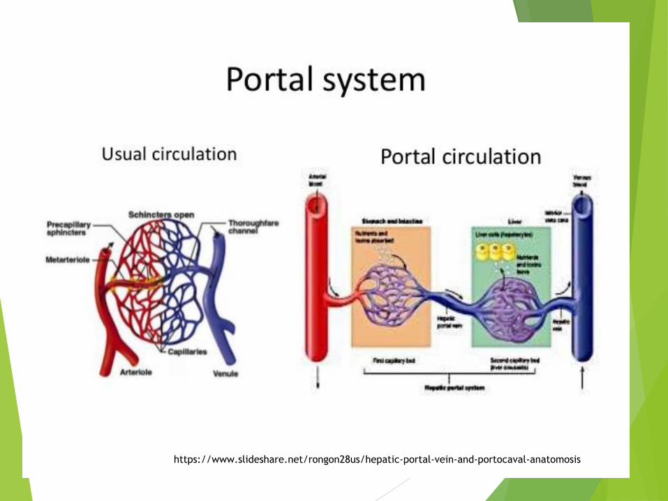
https://www.slideshare.net/rongon28us/hepatic-portal-vein-and-portocaval-anatomosis

https://www.slideshare.net/charslan626/hepatic-anastomosis

Causes of portal hypertension
Cirrhosis is most common
Alcoholic liver disease.
Hepatic schistosomiasis 2nd most common worldwide.
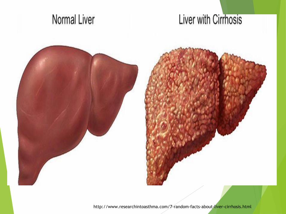
http://www.researchintoasthma.com/7-random-facts-about-liver-cirrhosis.html

Clinical Features
Often asymptomatic.
Rupture leads to massive hematemesis and death.
50% of patients die from the first bleed despite
interventions.
Death due to: hemorrhage, hepatic coma, and
hypovolemic shock
Rebleeding in 20%.
ESOPHAGITIS
Esophageal Lacerations.
Mucosal Injury
Infections
Reflux Esophagitis
Eosinophilic Esophagitis

Esophageal Lacerations
Mallory weiss tears are most common
Due to : severe retching or prolonged
vomiting
Present with hematemesis.
Failure of gastroesophageal musculature
to relax prior to antiperistaltic
contraction associated w/ vomiting >>
stretching >>>tear.
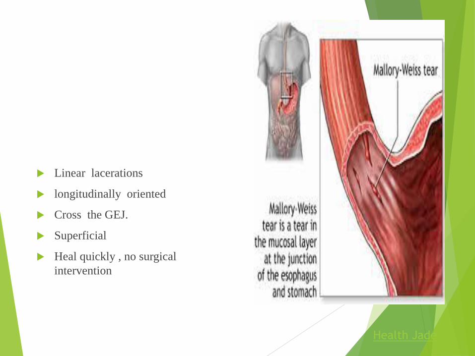
Linear lacerations
longitudinally oriented
Cross the GEJ.
Superficial
Heal quickly , no surgical
intervention
Health Jade

Chemical Esophagitis
Damage to esophageal mucosa by irritants
Alcohol,
Corrosive acids or alkalis
Excessively hot fluids
Heavy smoking
Medicinal pills (doxycycline and bisphosphonates)
Iatragenic (chemotx, radiotx , GVHD)

Clinical symptoms &
morphology
Ulceration and acute inflammation.
Only self-limited pain, odynophagia (pain with
swallowing).
Hemorrhage, stricture, or perforation in severe cases
Infectious esophagitis
Mostly in debilitated or immunosuppressed.
Viral (HSV, CMV)
Fungal (candida >>> mucormycosis & aspergillosis)
Bacterial: 10%.
Candidiasis :
Adherent.
Gray-white pseudomembranes
Composed of matted fungal hyphae and inflammatory
cells

https://www.pinterest.com/pin/374291419013418659/
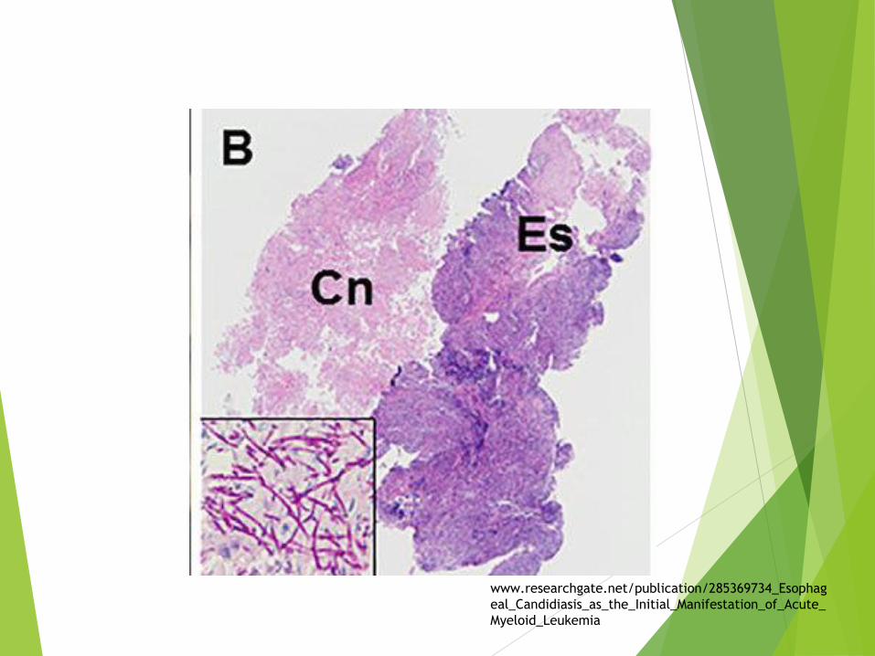
www.researchgate.net/publication/285369734_Esophag
eal_Candidiasis_as_the_Initial_Manifestation_of_Acute_
Myeloid_Leukemia

Herpes viruses
Punched-out ulcers
Histopathologic:
Nuclear viral inclusions
Degenerating epithelial cells ulcer edge
Multinucleated epithelial cells.


Robbins Basic Pathology 10th edition

CMV :
Shallower ulcerations.
Biopsy: nuclear and cytoplasmic inclusions in capillary
endothelium and stromal cells
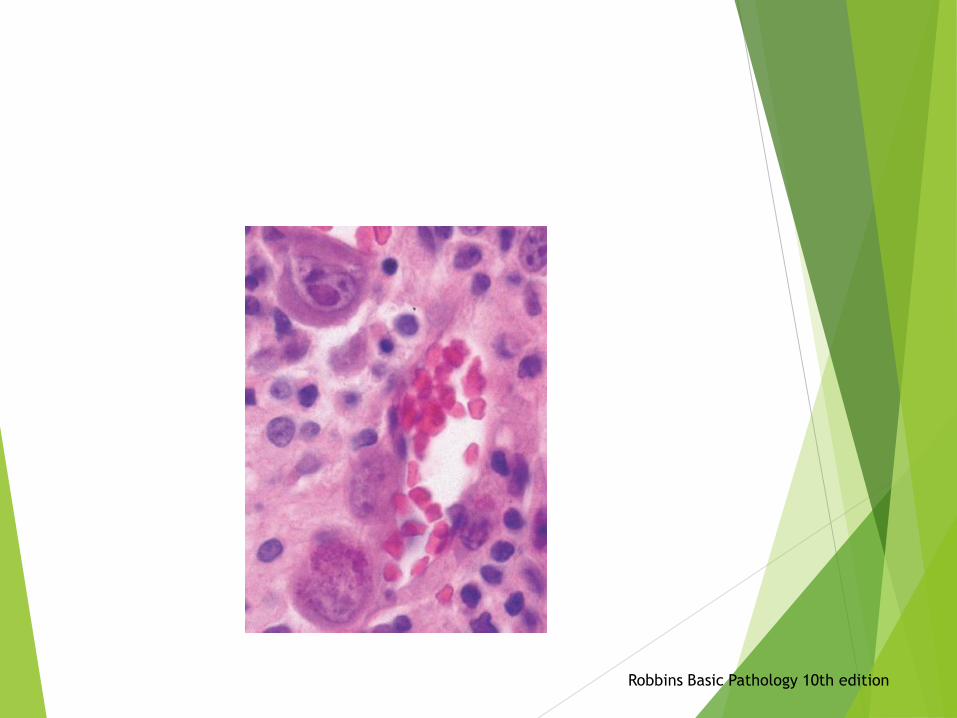
Robbins Basic Pathology 10th edition

Reflux Esophagitis
Reflux of gastric contents into the lower esophagus
Most frequent cause of esophagitis
Most common complaint by patients
Gastroesophageal reflux disease, GERD
Squamous epithelium is sensitive to acids
Protective forces: mucin and bicarbonate, high LES
tone

Pathogenesis
Decreased lower esophageal sphincter
tone
(alcohol, tobacco, CNS depressants)
Increase abdominal pressure
( obesity,, pregnancy, hiatal hernia, delayed
gastric emptying, and increased gastric volume)
Idiopathic!!
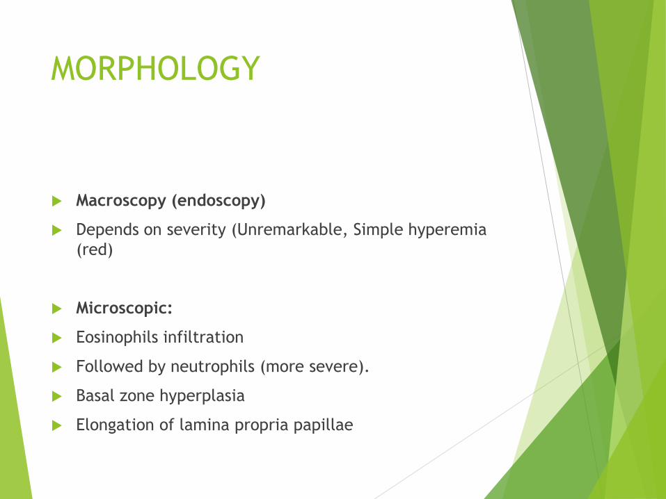
MORPHOLOGY
Macroscopy (endoscopy)
Depends on severity (Unremarkable, Simple hyperemia
(red)
Microscopic:
Eosinophils infiltration
Followed by neutrophils (more severe).
Basal zone hyperplasia
Elongation of lamina propria papillae

nature.comRobbins Basic Pathology 10th edition

Clinical Features
Most common over 40 years.
May occur in infants and children
Heartburn , dysphagia,
Regurgitation of sour-tasting gastric contents
Rarely: Severe chest pain, mistaken for heart disease
Tx: proton pump inhibitors

Complications
Esophageal ulceration
Hematemesis
Melena
Strictures
Barrett esophagus (precursor of Ca.)

Eosinophilic Esophagitis
Chronic immune mediated disorder
Symptoms:
Food impaction and dysphagia in adults
Feeding intolerance or GERD-like symptoms in children
Endoscopy:
Rings in the upper and mid esophagus.
Microscopic:
Numerous eosinophils w/n epithelium
Far from the GEJ.

Robbins Basic Pathology 10th edition

Most patients are: atopic (atopic dermatitis, allergic
rhinitis, asthma) or modest peripheral eosinophilia.
Tx:
Dietary restrictions( cow milk and soy products)
Topical or systemic corticosteroids.
Refractory to PPIs.
Barrett Esophagus
Complication of chronic GERD
Intestinal metaplasia within the esophageal
squamous mucosa.
10% of individuals with symptomatic GERD
Males>>females, 40-60 yrs
Direct precursor of esophageal adenocarcinoma
Metaplasia >> 0.2-1% /year >>dysplasia>>
adenocarcinoma.

MORPHOLOGY
Endoscopy:
Red tongues extending upward from the GEJ.
Histology:
Gastric or intestinal metaplasia
Presence of goblet cells
+-Dysplasia : low-grade or high-grade
Intramucosal carcinoma: invasion into the lamina propria.

Gastroenterology Consultants of San Antonio

Robbins Basic Pathology 10th edition

Baishideng Publishing Group

Management of Barrett
Periodic surveillance endoscopy with biopsy to screen
for dysplasia.
High grade dysplasia & intramucosal carcinoma needs
interventions.
ESOPHAGEAL TUMORS
Squamous cell carcinoma (most common worldwide)
Adenocarcinoma (on the rise, half of cases)
Adenocarcinoma
Background of Barrett esophagus and long-standing
GERD.
Risk factors: dysplasia associated Barrett, smoking,
obesity, radioTx.
Male : female (7:1)
Geographic & racial variation (devloped countries)

Pathogenesis
From Barrett>>dysplasia>>adenocarcinoma
Acquisition of genetic and epigenetic changes.
Chromosomal abnormalities and TP53 mutation.

MORPHOLOGY
Distal third.
Early: flat or raised patches
Later: exophytic infiltrative masses
Microscopy:
Forms glands and mucin.

Robbins Basic Pathology 10th edition


Clinical Features
Pain or difficulty swallowing
Progressive weight loss
Chest pain
Vomiting.
Advanced stage at diagnosis: 5-year survival <25%.
Early stage: 5-year survival 80%

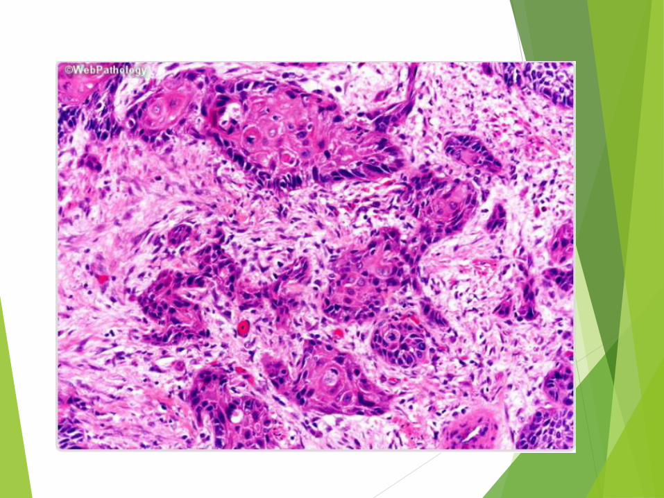
Squamous Cell Carcinoma
Male : female (4:1)
Underdeveloped countries.
Risk factors:
Alcohol
Tobacco use
Poverty
Caustic injury
Achalasia .
Plummer-Vinson syndrome
Frequent consumption of very hot beverages
Previous radiation Tx .

Pathogenesis
In western : alcohol and tobacco use.
Other areas: polycyclic hydrocarbons, nitrosamines,
fungus-contaminated foods
HPV infection implemented in high risk regions.

MORPHOLOGY
Middle third (50% of cases)
Polypoid, ulcerated, or infiltrative.
Wall thickening, lumen narrowing
Invade surrounding structures (bronchi, mediastinum,
pericardium, aorta).

Microscopy:
Pre-invasive: Squamous dysplasia & CIS.
Well to moderately differentiated invasive SCC.
Intramural tumor nodules
Lymph node metastases :
Upper 1/3: cervical LNs
Middle 1/3: mediastinalparatracheal, and
tracheobronchial LNs.
Lower 1/3: gastric and celiac LNs.

Clinical Features
Dysphagia
Odynophagia
Obstruction
Weight loss and debilitation
Impaired nutrition & tumor associated cachexia
Hemorrhage and sepsis if ulcerated.
Aspiration via a tracheoesophageal fistula
Dismal Px: 5 year survival <9%

Robbins Basic Pathology 10th edition