diseases of mulberry silkworm and their control
TRANSCRIPT

1
DISEASES OF MULBERRY SILKWORM AND THEIR CONTROL
Dr.H.B.Mahesha, Yuvaraja’s College, University of Mysore, Mysuru.
Mulberry silkworm Bombyx mori is affected by a number of diseases caused
by viruses, bacteria, fungi and protozoa. These diseases are known to occur in
almost all the silkworm rearing areas of the world causing considerable damage to
the silkworm cocoon crop. A number of measures have been suggested for the
prevention and control of these diseases, but none of them has proved to be fool-
proof with the result that one has always to be careful to eliminate the cause of
primary infection as well as to prevent the cross infection. Care is also needed to be
taken to see that they are not exposed to stress conditions like adverse temperature
and humidity, bad ventilation and nutritional deficiency which may make them
easily susceptible to various diseases.
VIRAL DISEASES
Viral diseases of silkworm pose a major problem to sericulture as they
account for almost 70 per cent of the total loss due to diseases. Viral diseases of
silkworm comprise of inclusion and non-inclusion types. The inclusion virus
diseases form typical inclusion bodies. They are nuclear polyhedrosis and
Cytoplasmic polyhedrosis which can be more easily identified through ordinary
microscopy. The non-inclusion type consists of Infectious flacherie and
Densonucleosis which can be detected only through electron/fluorescent
microscopy and serological tests.
Nuclear Polyhedrosis
It is one of the most serious viral diseases in tropical countries and occurs
throughout the year. This disease is also known as Grasseric, Jaundice, Milky
disease, Fatty degeneration and Hanging disease.
Causes of the disease
This disease is caused by Borrelina bombycis virus belonging to the sub-group
A of the family Baculoviridae. As the name implies, this virus multiplies and forms
polyhedra (Fig. 1) in the nucleus of the tracheal epithelial cells, adipose tissue cells,
dermal cells and blood cells. Occasionally the nucleus of the middle and posterior
portion of silk gland cells are also affected. The viral particles are rod shaped and
the size is around 330 x 80 nm. The size of the polyhedra varies from 3-6µ. The
shape is usually octadecahedral or hexahedral and sometimes tetragon or trigon.
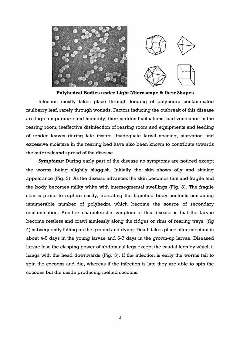
2
Polyhedral Bodies under Light Microscope & their Shapes
Infection mostly takes place through feeding of polyhedra contaminated
mulberry leaf, rarely through wounds. Factors inducing the outbreak of this disease
are high temperature and humidity, their sudden fluctuations, bad ventilation in the
rearing room, ineffective disinfection of rearing room and equipments and feeding
of tender leaves during late instars. Inadequate larval spacing, starvation and
excessive moisture in the rearing bed have also been known to contribute towards
the outbreak and spread of the disease.
Symptoms: During early part of the disease no symptoms are noticed except
the worms being slightly sluggish. Initially the skin shows oily and shining
appearance (Fig. 2). As the disease advances the skin becomes thin and fragile and
the body becomes milky white with intersegmental swellings (Fig. 3). The fragile
skin is prone to rupture easily, liberating the liquefied body contents containing
innumerable number of polyhedra which become the source of secondary
contamination. Another characteristic symptom of this disease is that the larvae
become restless and crawl aimlessly along the ridges or rims of rearing trays, (fig
4) subsequently falling on the ground and dying. Death takes place after infection in
about 4-5 days in the young larvae and 5-7 days in the grown-up larvae. Diseased
larvae lose the clasping power of abdominal legs except the caudal legs by which it
hangs with the head downwards (Fig. 5). If the infection is early the worms fail to
spin the cocoons and die, whereas if the infection is late they are able to spin the
cocoons but die inside producing melted cocoons.

Prevention and control
rearing rooms, mulberry storage rooms, mounting rooms, equipments and rearing
premises should be thoroughly disinfected
essentially surface disinfected. Silkworms should be reared under strict hygienic
conditions. During rearing the diseased and dead larvae form the major source of
infection with the largest quantity of fresh polyhedra av
larvae should be removed carefully without breaking the skin and disposed suitably
by putting them in lime vats
suitable temperature and humidity
fresh air circulation should be ensured by providing cross ventilation. The
silkworms should be fed with
feeding of tender leaf should be avoided. Depending upon the stage of larvae,
optimum spacing and required quantum of leaf should be given. Proper bed drying
is necessary before each feed to avoid accumulation of moisture in the bed.
3
Prevention and control: For effective prevention of this disease, the silkworm
rearing rooms, mulberry storage rooms, mounting rooms, equipments and rearing
premises should be thoroughly disinfected before brushing. The eggs should be
essentially surface disinfected. Silkworms should be reared under strict hygienic
conditions. During rearing the diseased and dead larvae form the major source of
infection with the largest quantity of fresh polyhedra available. Hence, the diseased
larvae should be removed carefully without breaking the skin and disposed suitably
by putting them in lime vats or by burning. Depending upon the
suitable temperature and humidity should be provided. During I
fresh air circulation should be ensured by providing cross ventilation. The
silkworms should be fed with nutritive rich mulberry leaf and during later stages
feeding of tender leaf should be avoided. Depending upon the stage of larvae,
mum spacing and required quantum of leaf should be given. Proper bed drying
each feed to avoid accumulation of moisture in the bed.
For effective prevention of this disease, the silkworm
rearing rooms, mulberry storage rooms, mounting rooms, equipments and rearing
before brushing. The eggs should be
essentially surface disinfected. Silkworms should be reared under strict hygienic
conditions. During rearing the diseased and dead larvae form the major source of
ailable. Hence, the diseased
larvae should be removed carefully without breaking the skin and disposed suitably
stage of silkworm,
should be provided. During IV and V instars
fresh air circulation should be ensured by providing cross ventilation. The
rich mulberry leaf and during later stages
feeding of tender leaf should be avoided. Depending upon the stage of larvae,
mum spacing and required quantum of leaf should be given. Proper bed drying
each feed to avoid accumulation of moisture in the bed.

In addition to the above, use of certain bed disinfectants could also prevent
secondary contamination and spread of the disease. Paraforrnaldehyde compounds
are known to have anti-microbial properties and various formulations involving this
chemical have been prepared like Papazol in Japan and Reshamkeet Oushadh in
India. The latter is a bed disinfectant fo
Trichloromythyl Thio-4-Cyclohexane 1,2
(Tri- oxymethylene) 2% Benzoic acid and 96
protection against grasserie
bed with the help of a thin cloth at the rate of
instars and 4- 5 grams/0.1sq
(Fig. 6) preferably once after each moult, half an hour bef
additional dusting should be done on the 4
The dusting should not be done when the larvae are under moult or preparing for
moult. The quantity of Reshamkeet Oushadh required for 100 disea
(40,000 larvae) is between 3
PROTOZOAN DISEASES
Protozoa which are injurious to silkworm are the parasitic ones belonging to
the class Microsporidia and genera
the major protozoan disease of the silkworm is the pebrine disease, so named due
to the appearance of black peppery patches following infection.
Pebrine
Pebrine is a chronic and disastrous disease of the silkworm
was responsible for the sudden collapse of the silkworm industry of both France
and Italy in 1865. Even though the fight against this disease in all the s
countries is going on since more than 100 years, the disease is not yet eliminated.
4
In addition to the above, use of certain bed disinfectants could also prevent
nd spread of the disease. Paraforrnaldehyde compounds
microbial properties and various formulations involving this
chemical have been prepared like Papazol in Japan and Reshamkeet Oushadh in
India. The latter is a bed disinfectant formulation containing 1 per cent captan (N
Cyclohexane 1,2- Dicarboxymide), 1 % paraformaldehyde
Benzoic acid and 96 % slaked lime powder giving dual
protection against grasserie and muscardine. It should be dusted
of a thin cloth at the rate of 2-3 grams/0. 1 sq m. area during
sq m. during IV and V instars. The dusting should be done
(Fig. 6) preferably once after each moult, half an hour before resumption of feed. An
additional dusting should be done on the 4th day of final instar after bed cleaning.
should not be done when the larvae are under moult or preparing for
moult. The quantity of Reshamkeet Oushadh required for 100 disea
(40,000 larvae) is between 3-3.5 kg.
Protozoa which are injurious to silkworm are the parasitic ones belonging to
and genera Nosema, Pleistophora and Thelohania.
the major protozoan disease of the silkworm is the pebrine disease, so named due
to the appearance of black peppery patches following infection.
Pebrine is a chronic and disastrous disease of the silkworm
sudden collapse of the silkworm industry of both France
65. Even though the fight against this disease in all the s
countries is going on since more than 100 years, the disease is not yet eliminated.
In addition to the above, use of certain bed disinfectants could also prevent
nd spread of the disease. Paraforrnaldehyde compounds
microbial properties and various formulations involving this
chemical have been prepared like Papazol in Japan and Reshamkeet Oushadh in
rmulation containing 1 per cent captan (N-
paraformaldehyde
slaked lime powder giving dual
on the larvae and
m. area during early
m. during IV and V instars. The dusting should be done
ore resumption of feed. An
final instar after bed cleaning.
should not be done when the larvae are under moult or preparing for
moult. The quantity of Reshamkeet Oushadh required for 100 disease free layings
Protozoa which are injurious to silkworm are the parasitic ones belonging to
Thelohania. However,
the major protozoan disease of the silkworm is the pebrine disease, so named due
Pebrine is a chronic and disastrous disease of the silkworm Bombyx mori L. It
sudden collapse of the silkworm industry of both France
65. Even though the fight against this disease in all the sericultural
countries is going on since more than 100 years, the disease is not yet eliminated.

5
However, it has been kept under check by following the techniques of strict mother
moth examination for the supply of disease free silkworm eggs, in addition to
disinfection and hygienic rearings. Though the disease is under reasonable control,
it appears sporadically due to infected seed and persisting secondary
contamination in the rearing house.
Causes of the disease: Pebrine is caused by Nosema bombycis Nageli belonging to
family Nosematidae of order Microsporidia. The pathogen infects the host through
feeding of contaminated mulberry leaf (peros) and also by rearing infected
silkworm eggs (transovarial).
Sources of infection are rather extensive. The main source is the rearing of
transovarially and surface contaminated layings. Infection also results from diseased
and dead larvae, faeces of larvae, moths, diseased egg shells, larval and pupal
exuviae etc. In the rearing bed major source of infection is the faeces of diseased
larvae, contaminated tray, seat paper and dust from infected rearing and leaf
storage rooms. Sometimes infection takes place through contaminated mulberry
leaf from field. The excreta and dead larvae of pebrine infected wild insects may
also form a source of infection.
Symptoms: The symptoms of this disease can be observed in all the stages of
silkworm viz., egg, larva, pupa and adult. These symptoms form an important
criterion for identifying the disease.
In the egg stage, poor egg number, lack of adequate adherence to the
substratum, lack of egg uniformity, more of unfertilized and dead eggs, poor and
irregular hatching are some of the symptoms. Sometimes infected eggs cannot
hatch out and hatched larvae may also die.
Larvae show poor appetite, retarded growth and development leading to un-
uniformity in size (Fig. 10). Larvae moult irregularly and show sluggishness.
Transovarially infected larvae die before third moult but those which are heavily
infected die during first instar itself. The larval body shows wrinkled skin with rustic
brown colour and in the moribund (near death) stage they do not rot but remain
rubbery. The affected gut becomes opaque and the silkgland shows white pustules
in different places along its length. Sometimes black irregular pepper like spots are
noticed on larval skin (Fig. 11).

The infected pupae are flabby and swollen with lusterless and softened
abdomen. Sometimes irregular black spots are
wing and abdominal area. Highly infected pupae fail to metamorphose into adults.
The moth emergence is delayed and improper. They have clubbed wings with
distorted antennae and do not mate properly. The scales from wings a
area easily come off. In infected moths if the accessory glands are infected the moth
may lay eggs with less gluey substance resulting in their detachment from the egg
cards.
Healthy and diseased moths
Prevention and control: The fundamental measure for the prevention and control of
this disease is to produce healthy eggs, so as to avoid em
be achieved by conducting
methods are to conduct effective disinfection of rearing rooms, equipments and
surroundings and maintenance of strict hygienic conditions during rearing. It
essential to surface disinfect the layings in 2
incubation. Such surface disinf
repeated again after release from cold storage as also by farmers. If the eggs are in
6
The infected pupae are flabby and swollen with lusterless and softened
abdomen. Sometimes irregular black spots are noticed near the rudiments of the
wing and abdominal area. Highly infected pupae fail to metamorphose into adults.
The moth emergence is delayed and improper. They have clubbed wings with
distorted antennae and do not mate properly. The scales from wings a
area easily come off. In infected moths if the accessory glands are infected the moth
may lay eggs with less gluey substance resulting in their detachment from the egg
Healthy and diseased moths
The fundamental measure for the prevention and control of
this disease is to produce healthy eggs, so as to avoid embryonic infection. This can
ing systematic mother moth examination. The other
onduct effective disinfection of rearing rooms, equipments and
surroundings and maintenance of strict hygienic conditions during rearing. It
disinfect the layings in 2 % formalin for 10 minutes before
incubation. Such surface disinfection though practiced in grainages should be
repeated again after release from cold storage as also by farmers. If the eggs are in
The infected pupae are flabby and swollen with lusterless and softened
noticed near the rudiments of the
wing and abdominal area. Highly infected pupae fail to metamorphose into adults.
The moth emergence is delayed and improper. They have clubbed wings with
distorted antennae and do not mate properly. The scales from wings and abdominal
area easily come off. In infected moths if the accessory glands are infected the moth
may lay eggs with less gluey substance resulting in their detachment from the egg
The fundamental measure for the prevention and control of
onic infection. This can
systematic mother moth examination. The other
onduct effective disinfection of rearing rooms, equipments and
surroundings and maintenance of strict hygienic conditions during rearing. It is
formalin for 10 minutes before
ection though practiced in grainages should be
repeated again after release from cold storage as also by farmers. If the eggs are in

7
advanced stage of embryonic development surface disinfection is done with 1 per
cent formalin for 5 minutes. The room and equipments must be washed and
disinfected before incubation.
Young silkworms should be reared under hygienic conditions. As a precaution
test examination of unhatched blue eggs, dead eggs, hatched larvae and egg shells
can be done and if pebrine is detected, such eggs should not be brushed and if
brushed the larvae should be destroyed. Similarly predictive examination could be
conducted by utilizing unequal larvae, late moulters, faecal matter and exuviae for
the detection of pebrine spores. These tests may not only minimize the chances of
rearing transovarially infected layings, but also check cross contamination and
spread of the disease. Infected silkworms, faeces and mulberry field pests are
important sources of infection and should be properly disposed of to prevent cross
infection and spread of the disease.
During seed production in addition to mother moth examination, care should
be taken to prevent contamination from other sources. The equipments used for one
lot should not be used for the other till they have been thoroughly cleaned and
disinfected. Eggs after surface disinfection should be dried and stored in a separate
room away from egg production and examination room.
Besides, the above preventive/corrective measures, it has been reported that
immersing of the silkworm eggs in hot water, high temperature treatment of the
pupae, dipping of the eggs in hot hydrochloric acid minimize the incidence of
pebrine. Chemotherapy of Nosema infection has been reported through a number
of antimicrosporidian drugs like fumagillin, benomyl, bengard, bavistin, ethyl and
methyl thiophanate and some of their analogues with positive results, but
preventive methods have always been found to be better than the curative
measures.
FUNGAL DISEASES
Fungal diseases otherwise called mycosis, is caused in the silkworm by a few
parasitic fungi. Two major kinds of such disease are Muscardine and Aspergillosis.
Muscardine appears in various forms and depending upon the colour of spores
which cover the body of the silkworm giving a characteristic colour, they have been
named as white-muscardine, green-muscardine, yellow-muscardine, black-
muscardine, red-muscardine etc. The more common muscardine diseases are,
however, white and green-muscardine. In addition Aspergillosis is also found to

occur. Since the silkworm attacked by a fungal disease in course of time turns hard
and chalky, muscardine disease is also called Calcino.
White Muscardine: It is the most common and widely prevalent fungal disease
found in all sericultural countries. This disease occurs usually during rainy and
winter seasons under moderate to low temperature and high humidity conditions.
Causes of the disease: This disease is caused by different species of
which the most virulent is
Moniliaceae, order Moniliales of class Fungi imperfecti. Infection is mainly by body
contact, rarely through wounds and not by ingestion. Main sources of infection are
the mummified larvae, infected seat paper, tray and dead wild lepidoptero
from the mulberry field. The disease is highly
borne.
The developmental cycle of
namely conidium, vegetativ
The conidium is colorless
when gathered in a mass. Under favourable conditions of temperature and humidity
the conidium germinates within 8
silkworm. On germination the conidium not only sends out its germ tube but also
secretes chitinase which facilitates the germ tube to penetrate the body wall for
further multiplication. The germinating tube of the conidium a
blood of the larvae develops into vegetative
or oval shaped short hyphae develops. These often detach themselves and elongate
to form vegetative hyphae.
The vegetative hypha comes
innumerable conidiophores. These conidiophores give rise to small branches which
bear one or two conidia.
8
occur. Since the silkworm attacked by a fungal disease in course of time turns hard
and chalky, muscardine disease is also called Calcino.
It is the most common and widely prevalent fungal disease
ericultural countries. This disease occurs usually during rainy and
winter seasons under moderate to low temperature and high humidity conditions.
This disease is caused by different species of
t virulent is Beauveria bassiana. This fungus belongs
Moniliaceae, order Moniliales of class Fungi imperfecti. Infection is mainly by body
contact, rarely through wounds and not by ingestion. Main sources of infection are
, infected seat paper, tray and dead wild lepidoptero
from the mulberry field. The disease is highly contagious as the conidia are air
The developmental cycle of Beauveria bassiana consists of three
namely conidium, vegetative mycelium and aerial mycelium.
colorless, globular or rarely oval in shape and porcelain white
when gathered in a mass. Under favourable conditions of temperature and humidity
inates within 8-10 hours of coming in contact with the body of
silkworm. On germination the conidium not only sends out its germ tube but also
secretes chitinase which facilitates the germ tube to penetrate the body wall for
further multiplication. The germinating tube of the conidium a
blood of the larvae develops into vegetative hyphae. At the tip of the hyphae round
or oval shaped short hyphae develops. These often detach themselves and elongate
to form vegetative hyphae.
hypha comes out of the skin to form aerial hyphae bearing
innumerable conidiophores. These conidiophores give rise to small branches which
occur. Since the silkworm attacked by a fungal disease in course of time turns hard
It is the most common and widely prevalent fungal disease
ericultural countries. This disease occurs usually during rainy and
winter seasons under moderate to low temperature and high humidity conditions.
This disease is caused by different species of Beauveria of
This fungus belongs to the family
Moniliaceae, order Moniliales of class Fungi imperfecti. Infection is mainly by body
contact, rarely through wounds and not by ingestion. Main sources of infection are
, infected seat paper, tray and dead wild lepidopteron larvae
as the conidia are air
consists of three distinct stages
, globular or rarely oval in shape and porcelain white
when gathered in a mass. Under favourable conditions of temperature and humidity
ing in contact with the body of
silkworm. On germination the conidium not only sends out its germ tube but also
secretes chitinase which facilitates the germ tube to penetrate the body wall for
further multiplication. The germinating tube of the conidium after invading the
. At the tip of the hyphae round
or oval shaped short hyphae develops. These often detach themselves and elongate
form aerial hyphae bearing
innumerable conidiophores. These conidiophores give rise to small branches which

9
Symptoms: At the early stage of infection symptoms are not distinct, but as the
disease advances, moist specks appear on the skin. At this stage, larvae lose
appetite and become inactive. The body of the larvae becomes limp, loses its skin
elasticity, stops movement and finally they die. Before death, symptoms of diarrhea
and vomiting appear (Fig. A). After death, the body is initially soft, but within 6-8
hours it becomes stiff and hard (Fig. B). At this stage the body is pink in colour. This
is due to the multiplication of Serratia marcescens, a secondary bacterium. One to
two days later, wooly aerial hyphae grow out between inter-segmental membranes.
Subsequently the whole body is covered with white powdery conidia except the
chitinous parts of the head. The larvae, unlike other diseases do not rot or decay but
remains hard (Fig. C) as the fungus secretes double oxalate - crystals of ammonium
and magnesium.
Fig A Fig B Fig C
In case of pupal infection the pupae slowdown their reaction to outside stimuli.
The thorax shrinks and abdomen is wrinkled. The aerial hyphae and conidia grow
up to one third of its ordinary weight inside the cocoons. Such cocoons sound like
dried cocoons when shaken. During moth stage the body is hardened and the wings
fall of easily.
Prevention and control: Before the commencement of silkworm rearing, rooms,
appliances and rearing surroundings must be thoroughly disinfected with 2 percent
formalin or 5 percent bleaching powder solution. This disease can be kept under
check by avoiding low temperature and high humidity during rearing as they are
more ideal for fungal growth. The rearing bed should as much as possible be kept
thin and dry in order to avoid the germination of conidia and spread of the fungus. If
the disease is found during rearing, the trays, seat papers, cleaning nets, foam pads
etc., must be disinfected and replaced. Diseased worms should be removed

10
carefully before they get mummified and should be placed in lime jars or destroyed
by burning or deep burying, with a disinfectant spray. Similarly the bed refuse
along with the faeces should be disposed of properly. Mulberry pests in the garden
should be controlled as they get easily infected with this pathogen, later becoming
an important source for cross contamination to the silkworm.
In addition to the above, anti-muscardine powders can be fruitfully used to
control the outbreak and spread of this disease. A few methods of application of the
same are given below:
Application of formalin chaff:
In this method formalin solution of required concentration depending on the
silkworm instar is mixed with burnt paddy husk and sprinkled on the larval body
and bed (Fig.). The concentration of formalin required is 0.4 per cent during I and II
instars, 0.5 percent in III ins tar, 0.6 percent in IV instar and 0.8 per cent during V ins
tar. The paddy husk is charred or burnt either by burning or roasting in a pan
without making ash. Depending on the instar of larvae, the required strength of
formalin is mixed with the burnt paddy husk in the ratio of 1:10 by volume and
mixed thoroughly. Then it is sprinkled evenly on the larvae and covered with a
paraffin or double fold newspaper. After 1/2 an hour the paper cover is removed
and feed is given. Formalin chaff application should not be done when larvae are
preparing for moult or under moult. Application of formalin chaff can be done
before brushing on the newly hatched larvae and after each moult 1/2 an hour
before the resumption of feeding. The frequency of application of formalin chaff
should be increased depending on the incidence of disease.
Application of Formalin Chaff
Application of Dithane M 45 (Zinc ion Manganese ethylene oxide bisdithio
carbomate) or captan (N-Trichloromethyl Thi0-4-Cyclohexane 1,2-Dicarboximide).
These are the two commonly available fungicides used for the control of

11
muscardine. These fungicides are used at a concentration of 1 per cent during, I, II
and III instars and 2 per cent during IV and V instars in combination with levigated
China clay or Kaolin. The ingredients are thoroughly mixed and tied in a thin cloth
and dusted on newly born larvae and after each moult 1/2 an hour before the
resumption of feed. An additional dusting should be done on the 4th day of final
instar after bed cleaning (Fig.). The quantity required is 2-3 grams per 0.1 sq m.
area during I, II and III instars and 4-5 grams during IV and V instars. The dustings
should not be done when the larvae are preparing for moult or are under moult. The
dusting frequency should be increased if the intensity of infection is high.
Application of "Reshamkeet Oushadh"
It is a bed disinfectant formulation used to prevent both muscardine and
grasserie. Its constitution, method of application and quantity required has already
been indicated in the part covering the control of grasserie disease.
Dusting of Bed disinfectant
BATERIAL DISEASES
Bacterial diseases affecting silkworm are collectively known as flacherie due to
the flaccid nature of the diseased larvae. The incidence of flacherie is high during
hot and humid seasons. In general, massive out-break of these diseases are
uncommon but depending upon poor disinfection, accumulation of faeces in the
rearing trays, feeding of mulberry leaves with contamination, improper handling
and unsafe use of bacterial pesticides, large scale loss in crops sometimes occur.
Bacterial diseases of silkworms are divided into three major types namely bacterial
septicemia, bacterial diseases of the digestive tract and bacterial toxicosis.
Septicemia:
This is a condition where bacteria multiply enormously in the blood
(haemolymph) of the larvae, pupae and moths. Septicemia during the larval stage
leads to larval mortality whereas the infection in pupal and moth stages leads to a

12
large number of melted cocoons affecting the egg production in the grainages.
Causes of the disease: This disease is caused by the multiplication of a large
number of bacteria, bacilli, streptococci and staphylococci in the haemolymph. The
route of infection is through injury or wounds and rarely perorally. Two major types
of bacterial septicemia are generally observed, one is the black thorax septicemia
caused by Bacillus sp. belonging to the family Bacillaceae of the order Eubacteriales
size 1-1.5 x 3 microns, spores subterminal, gram-positive and the other is the red
septicemia or serratia septicemia caused by the bacillus Serratia marcescens Bizio
size 0.6-1.0 x0.5 microns non-sporulating and gram negative. The former is more
resistant to disinfectants than the latter except for lime emulsion.
Symptoms: They have some common symptoms like sluggish movement,
decreased appetite, straightened body, swollen thorax, shrinkage of abdominal
segments, vomiting and bead like faeces and loss of clasping power of legs.
Further, the body becomes soft and discolored and the body wall ruptures easily
emitting foul smelling fluid.
Difference in the symptoms of two diseases are that, in case of the black
thorax septicemia, the blackening starts from the thorax and extends to the dorsal
vessel till the whole body blackens and rots (Figs.) whereas in the latter case the
whole body softens taking a slightly reddish tinge (Fig.). Septicemias are generally
acute diseases, spreading quickly, the time clasping from the time of infection to
death at 28°C being about 10 hours. At higher temperature and under epidemic
conditions they may die within 5-6 hours.
Black Thorax Septicemia Red Septicemia
Prevention and control: High temperature and humidity conditions are most
favourable to the propagation of the bacteria responsible for these diseases and so

13
these diseases occur chiefly in the seasons having high temperature and humidity.
They normally follow wound infection. The bacteria enter generally through the
wound and multiply in the haemolymph, disrupting the normal physiological
functions, causing septicemia. The 5th instar larvae are more prone to injury and
these diseases thus occur mostly in the later part of this stage. An effective means of
control of these diseases can be the maintenance of hygienic condition so that these
bacteria do not occur on the mulberry leaves, in the rearing room and rearing
equipment. Care should be taken to avoid injury to the worms, overcrowding of
trays and accumulation of faeces in the rearing bed.
References:
1. Hand book on pest and disease control of mulberry and silkworm, CSB, India.
2. Internet.
--------------------