diseases of mouth, palate, lips & cheek
TRANSCRIPT

By,
Dr.K.Priyatham,
Final year Post Graduate,
General Surgery.
DISEASES OF MOUTH, PALATE, LIPS, CHEEK

THE EXTERNAL MOUTH• Composes primarily of the
upper and lower lip
• Separated to the skin by vermillion border
• Bounded
• Superiorly – Philtrum
Supero-laterally- Naso-labial fold
Inferiorly-
Mento-labial fold

PARTS OF THE EXTERNAL MOUTH
Philtrum
Vermillion border
Mento-labial fold
Naso-labial fold
Angle of the mouth
Oral fissure
Upper lip
Lower lip

Muscle that supports the external mouth is the Orbicularis Oris Muscle

Functions of mouth1. The first part of digestive
system wherein mastication occur.
2. It receives food (ingestion) and break up the food into smaller pieces (digestion).
3. Takes part in respiration as well as in speech production
4. Lastly it conveys emotion
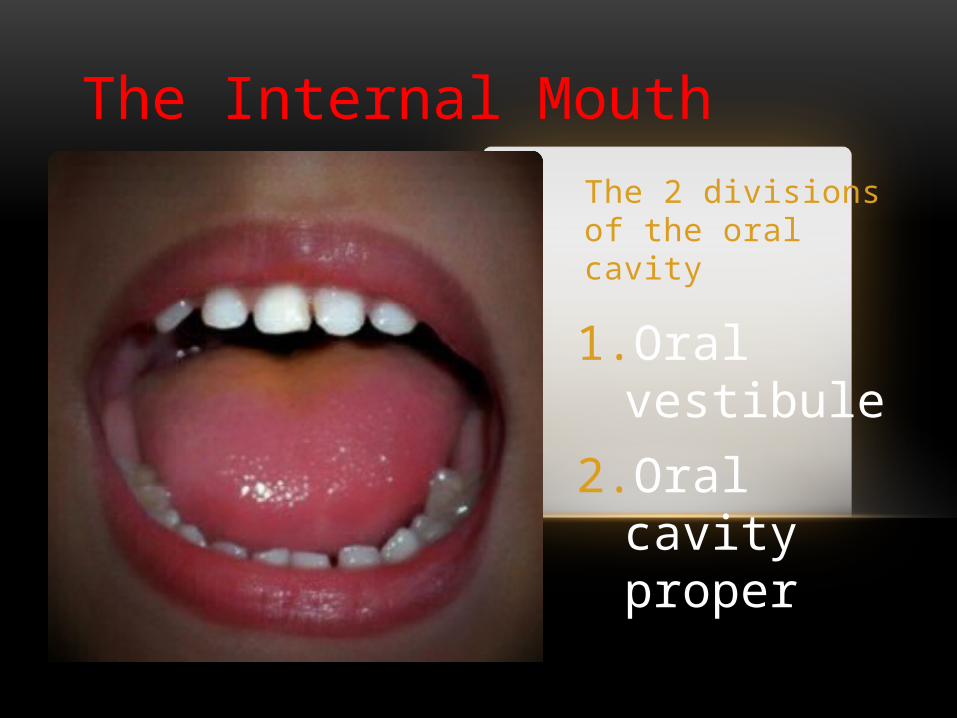
The 2 divisions of the oral cavity
1.Oral vestibule
2.Oral cavity proper
The Internal Mouth

The Oral vestibuleA slit like space bounded
Laterally by cheek and lips
Medially by the buccal and labial surfaces of the upper and lower teeth
Posteriorly by the Retromolar area
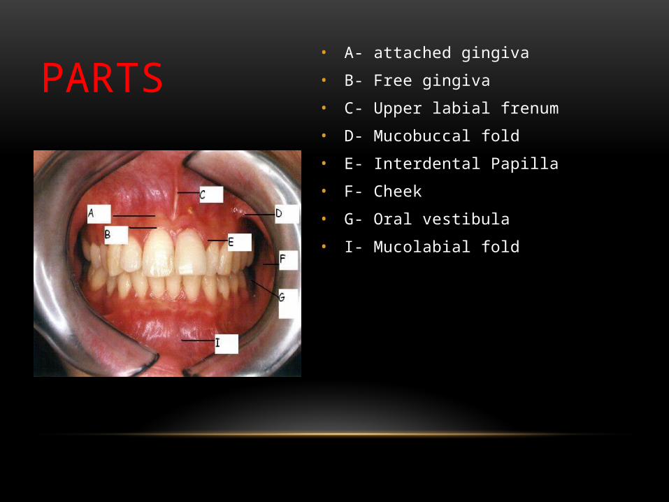
PARTS • A- attached gingiva
• B- Free gingiva
• C- Upper labial frenum
• D- Mucobuccal fold
• E- Interdental Papilla
• F- Cheek
• G- Oral vestibula
• I- Mucolabial fold

ORAL VESTIBULE
Oral vestibule- Left side Oral vestibule- right side

PART
Upper buccal frenum
Lower buccal frenum

• Bounded
• Laterally by palatal and lingual surfaces of the upper and lower teeth
• Superiorly by the palate• (hard & soft)
• Inferiorly by the tongue and or the floor of the mouth
• Posteriorly by the isthmus of fauces
THE ORAL CAVITY PROPER

Part of the Oral cavity proper
Hard Palate
Uvula
Palatoglossal arch
Palatine tonsil
Tongue
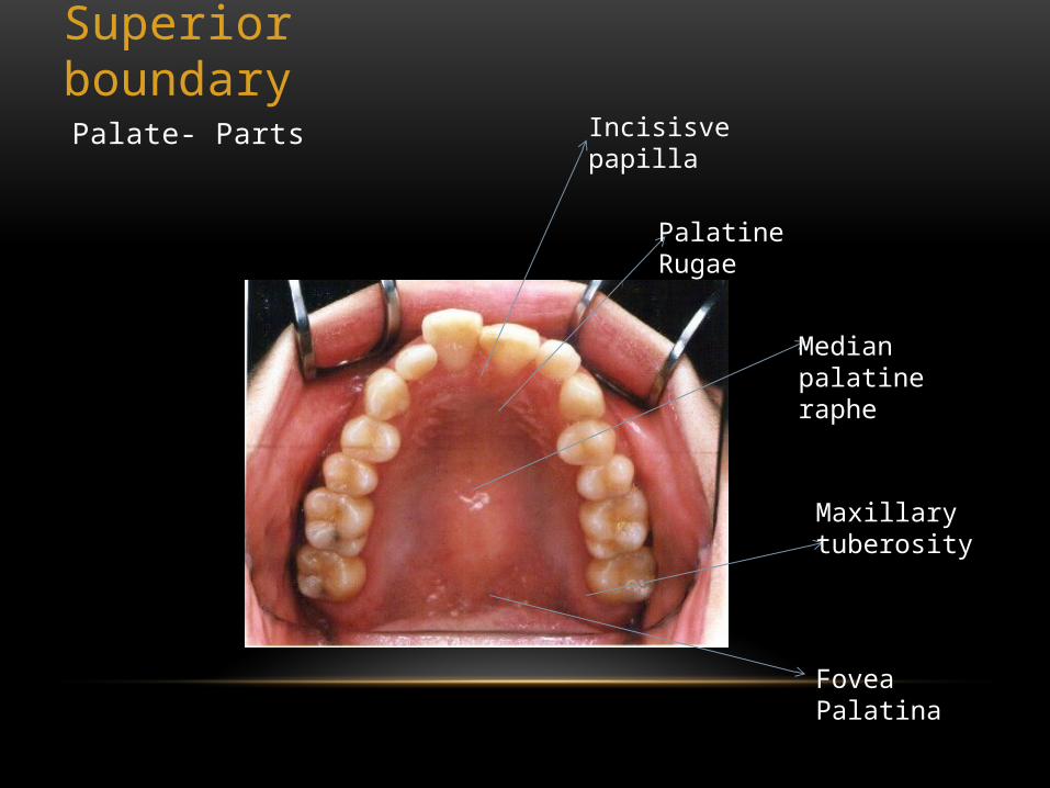
Superior boundaryPalate- Parts Incisisve papilla
Palatine Rugae
Median palatine raphe
Maxillary tuberosity
Fovea Palatina

Bony framework of the hard palate
Palatine process of the maxilla
Horizontal plate of the palatine bone

Inferior boundary
The Floor of the mouth
Parts Ventral surface of the tongue
Lingual Frenulum
Sublingual fold
Sublingual caruncle

ORAL CAVITY (MOUTH) Extends from the lips to
the oropharyngeal isthmus
• The oropharyngeal isthmus:
• Is the junction of mouth and pharynx.
• Is bounded:• Above by the soft
palate and the palatoglossal folds
• Below by the dorsum of the tongue
Subdivided into Vestibule & Oral cavity proper
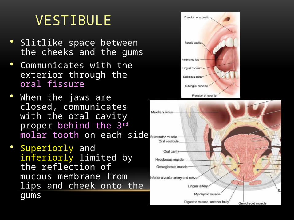
VESTIBULE Slitlike space between
the cheeks and the gums Communicates with the
exterior through the oral fissure
When the jaws are closed, communicates with the oral cavity proper behind the 3rd molar tooth on each side
Superiorly and inferiorly limited by the reflection of mucous membrane from lips and cheek onto the gums
VESTIBULE CONT’D
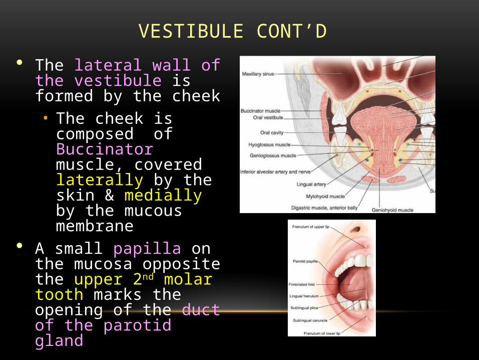
The lateral wall of the vestibule is formed by the cheek• The cheek is
composed of Buccinator muscle, covered laterally by the skin & medially by the mucous membrane
A small papilla on the mucosa opposite the upper 2nd molar tooth marks the opening of the duct of the parotid gland
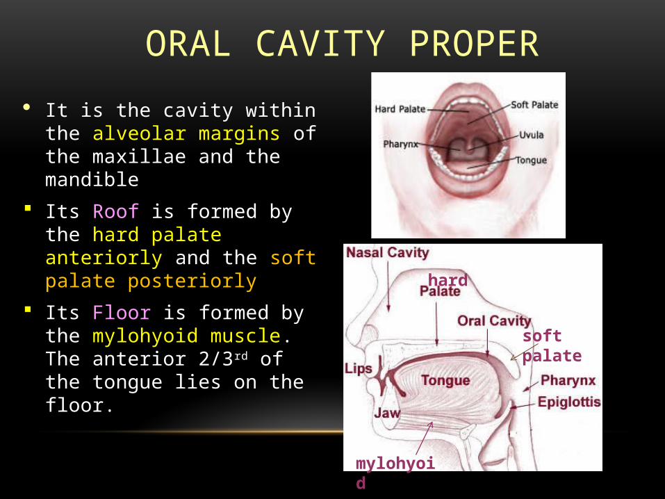
ORAL CAVITY PROPER
It is the cavity within the alveolar margins of the maxillae and the mandible
Its Roof is formed by the hard palate anteriorly and the soft palate posteriorly
Its Floor is formed by the mylohyoid muscle. The anterior 2/3rd of the tongue lies on the floor.
hard
soft palate
mylohyoid
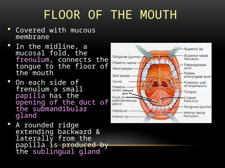
FLOOR OF THE MOUTH Covered with mucous
membrane In the midline, a
mucosal fold, the frenulum, connects the tongue to the floor of the mouth
On each side of frenulum a small papilla has the opening of the duct of the submandibular gland
A rounded ridge extending backward & laterally from the papilla is produced by the sublingual gland

NERVE SUPPLY
o Sensory Roof: by greater palatine and nasopalatine nerves
(branches of maxillary nerve)
Floor: by lingual nerve (branch of mandibular nerve)
Cheek: by buccal nerve (branch of mandibular nerve)
oMotor Muscle in the cheek (buccinator) and the lip
(orbicularis oris) are supplied by the branches of the facial nerve

PALATE Lies in the roof of
the oral cavity Has two parts:
• Hard (bony) palate anteriorly
• Soft (muscular) palate posteriorly
hard
soft palate
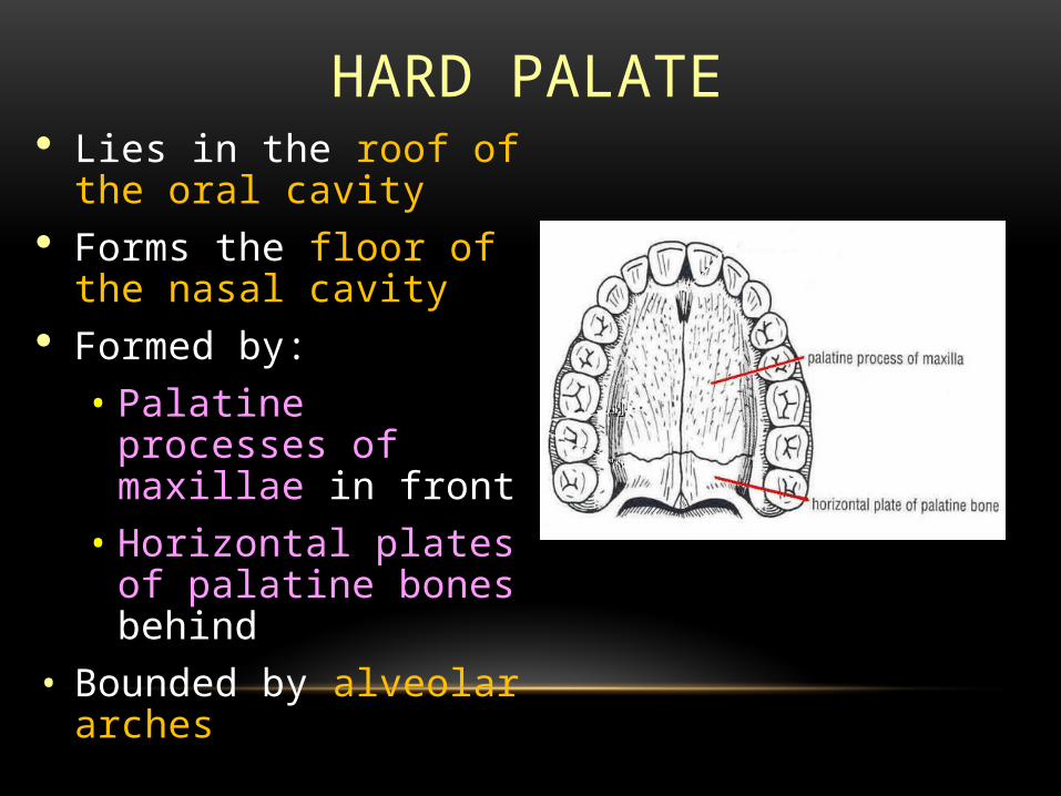
HARD PALATE Lies in the roof of
the oral cavity Forms the floor of
the nasal cavity Formed by:• Palatine processes
of maxillae in front• Horizontal plates
of palatine bones behind
• Bounded by alveolar arches
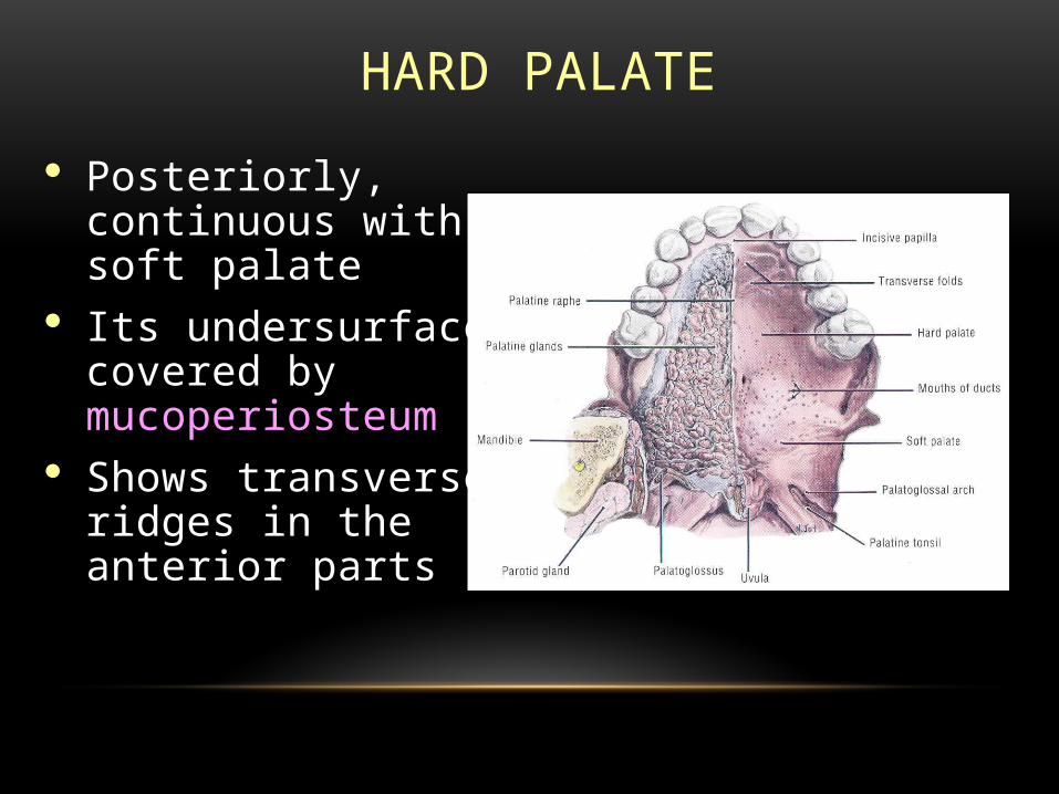
HARD PALATE
Posteriorly, continuous with soft palate
Its undersurface covered by mucoperiosteum
Shows transverse ridges in the anterior parts

SOFT PALATE Attached to the posterior
border of the hard palate
Covered on its upper and lower surfaces by mucous membrane
Composed of:
• Muscle fibers
• An aponeurosis
• Lymphoid tissue
• Glands
• Blood vessels
• Nerves

PALATINE APONEUROSIS Fibrous sheath Attached to
posterior border of hard palate
Is expanded tendon of tensor velli palatini
Splits to enclose musculus uvulae
Gives origin & insertion to palatine muscles

MUSCLES Tensor veli palatini
• Origin: spine of sphenoid; auditory tube
• Insertion: forms palatine aponeurosis
• Action: Tenses soft palate
• Levator veli palatini• Origin:petrous temporal bone,
auditory tube, palatine aponeurosis
• Insertion: palatine aponeurosis
• Action: Raises soft palate
• Musculus uvulae• Origin: posterior border of hard
palate
• Insertion: mucosa of uvula
• Action: Elevates uvula
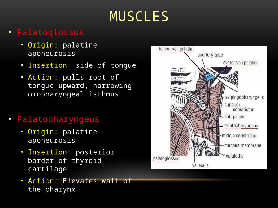
MUSCLES• Palatoglossus
• Origin: palatine aponeurosis
• Insertion: side of tongue
• Action: pulls root of tongue upward, narrowing oropharyngeal isthmus
• Palatopharyngeus• Origin: palatine
aponeurosis
• Insertion: posterior border of thyroid cartilage
• Action: Elevates wall of the pharynx
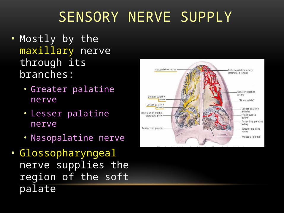
SENSORY NERVE SUPPLY• Mostly by the
maxillary nerve through its branches:• Greater palatine
nerve
• Lesser palatine nerve
• Nasopalatine nerve
• Glossopharyngeal nerve supplies the region of the soft palate

MOTOR NERVE SUPPLY
• All the muscles, except tensor veli palatini, are supplied by the:
• Pharyngeal plexus
• Tensor veli palatini supplied by the:
• Nerve to medial pterygoid, a branch of the mandibular division of the trigeminal nerve

BLOOD SUPPLY• Branches of the
maxillary artery
• Greater palatine
• Lesser palatine
• Sphenopalatine
• Ascending palatine, branch of the facial artery
• Ascending pharyngeal, branch of the external carotid artery
LIP ANATOMY

ANATOMY
• Lips form anterior boundary of oral cavity
• Parts: 2 surface of lip, skin & mucosa become continous with one another round & this margin vermilion
• Vermilion border:
Dry vermilion: pattern of wrinkles has clear cut boundary line between it & skin proper

• Smooth wet vermilion: merges without obvious surface change with mucosa lining of lip.

• Epithelium:
Lip covered with non-keratinised stratified squamous
epithelium which is transparent & contain no hair, sebaceous
glands or pigments. Hence, Red.
On vermilion border, distance between epithelium & muscle is just
2mm.

LIP RECONSTRUCTION
• Anatomy• Motor Innervation
• Facial nerve VII• Buccal
• Elevators of commissures and orbicularis oris
• Marginal mandibular
• Lip depressors
• Sensory innervation • Trigeminal nerve V
• Mental nerve terminal branch of inferior alveolar nerve
• Lower lip
• Infraorbital nerve
• Upper lip

LIP RECONSTRUCTION
• Anatomy• Muscles
• Orbicularis oris• Closes the oral sphincter
• Primarily horizontal fibers - compress lips
• Originate lateral to the commissures
• Mingle with cranial VII muscles at modiolus
• Cross the lip
• Decussate in the midline
• Insert into opposite philtral column
• Oblique fibers - evert lip
• Arise from modiolus
• Travel upward and medial
• Insert at the anterior nasal spine, nasal septum, and anterior nasal floor
LIP RECONSTRUCTION• Anatomy
• Muscles
• Major elevators upper lip
• Levator labii superioris (LLS)
• Originates from orbital margin
• Curves around the alar base
• Inserts into ipsilateral orbicularis oris and philtral column
• Zygomaticus major
• extends from malar eminence inserts in modiolus
• Levator anguli oris
• arises just below the lateral edge of the LLS

MUSCLES
• Sensory innervation
• Trigeminal nerve • Mental nerve terminal branch of inferior alveolar
nerve( mandibular br. )• Lower lip
• Infraorbital nerve (maxillary br.)• Upper lip

• LYMPHATIC DRAINAGE
• Upper lip: drains into preauricular, infraparotid & submandibular nodes
• Lower lip:
Medial portion of lower lip submental nodes
Lateral portion submandibular nodes

LIP FUNCTION
• Oral competence
• Deglutition
• Articulation
• Expression of emotion
• Symbol of beauty

TONGUE ANATOMY

INTRODUCTION
Mass of striated muscles covered with mucous membrane Voluntary muscular structure Length: 3 inches Location: floor of the mouth Shape: triangular Attachement: With mandible and hyoid bone Has an apex , body and root

SURFACES : Two surfaces
• Superior surface
• Inferior surface
Superior surface is divided into three parts
• Anterior 2/3 part called as Oral part
• Posterior 1/3 part called as Pharyngeal part
• Base(root) of tongue
GENERAL FEATURES

GENERAL FEATURES
TERMINAL SULCUS V-shaped sulcus
divides tongue into anterior & posterior parts
Apex of sulcus marked by a pit - FORAMEN CECUM

Foramen cecum ,an embryological remnant, marks the upper end of thyroglossal duct
Sometime a thyroglossal duct persists and connects the foramen cecum with the thyroid gland in neck(thyroglossal cyst)
GENERAL FEATURES
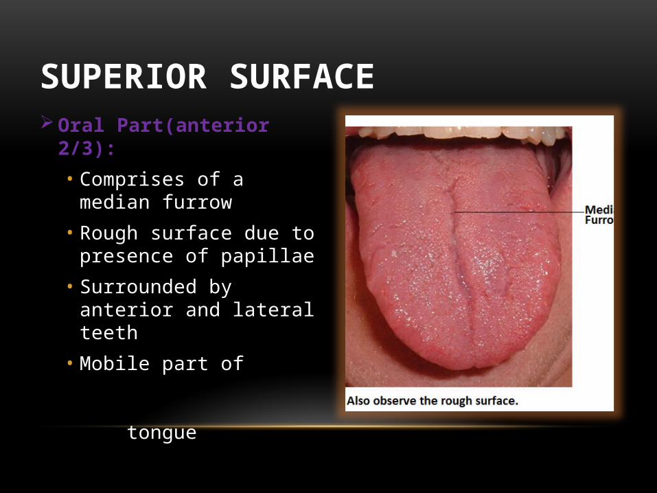
Oral Part(anterior 2/3):
• Comprises of a median furrow
• Rough surface due to presence of papillae
• Surrounded by anterior and lateral teeth
• Mobile part of tongue
SUPERIOR SURFACE
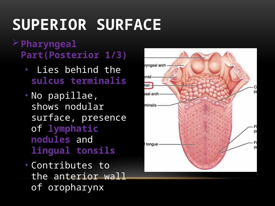
Pharyngeal Part(Posterior 1/3)
• Lies behind the sulcus terminalis
• No papillae, shows nodular surface, presence of lymphatic nodules and lingual tonsils
• Contributes to the anterior wall of oropharynx
SUPERIOR SURFACE

PHARNYGEAL PART

Base of tongue is far back and is bottom of tongue
Contributes to the front wall of pharynx
Movement can affect the diameter of pharynx i.e
• When it push forwards, thereby expanding the pharynx
• When it pull backwards, thereby constricting the pharynx
• Lacks papillae
BASE OF TONGUE
INFERIOR SURFACE
Covered by smooth mucous membrane In the midline , a mucosal fold called Frenulum
connects the tongue with the floor of the mouth Lateral to frenulum, deep lingual vein can be seen
through the mucosa Lateral to the lingual vein , mucosal fold called as plica
fimbriata is present

Frenulum
Lingual veinsPlica fimbriata

PAPILLAE

Indentation of any structure in the overlying epithelium is called papillae
Superior surface of tongue , covered by numerous papillae
Have taste buds on their surfaces
Types of of papillae;
• Vallate/circumvallate
• Filiform
• Fungiform
• Foliate
PAPPILAE

VALLATE PAPILLAE
Largest among papillae SHAPE: Blunt-ended cylindrical NUMBER: 8 to 12 LOCATION: infront of sulcus terminalis ARRANGEMENT: Occur in V shape line

VALLATE PAPILLAE

FILIFORM PAPILLAE SHAPE: Thin, long papillae having pointed ends
‘V’ shaped cones Only papillae having no taste buds NUMBER: numerous These papillae are mechanical and not involved in
gustation
Identified by increased keratinization LOCATION: Present at pre-sulcal area of the tongue
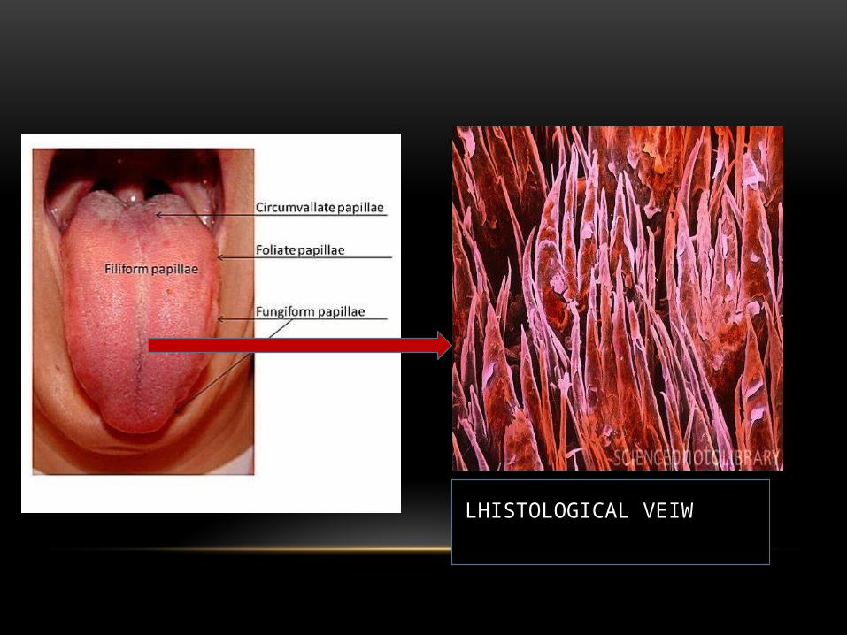
LHISTOLOGICAL VEIW

FUNGIFORM PAPPILAE SHAPE: slightly mushroom-shaped if looked at in
longitudinal section Taste buds on their surfaces LOCATION: apex of the tongue as well as the margins Larger than filiform papillae

FUNGIFORM PAPILLAE

FOLIATE PAPPILAE
SHAPE: Short vertical folds
LOCATION: Present lateral to terminal sulcus and at margins

FOLLIATE PAPILLAE
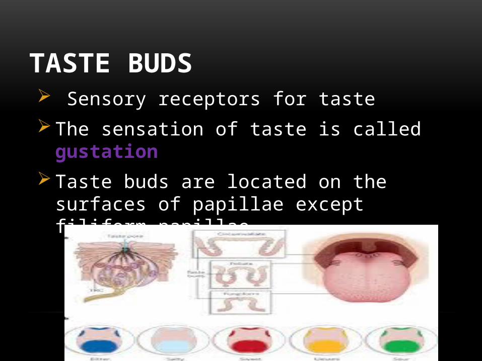
TASTE BUDS Sensory receptors for taste The sensation of taste is called gustation
Taste buds are located on the surfaces of papillae except filiform papillae

ELECTRON MICROSCOPIC STRUCTURE OF TASTE BUDS

TASTE BUDS Four taste sensations, recently a fifth basic taste has
been added: sour, sweet, salty, bitter and the recently added umami
UMAMI
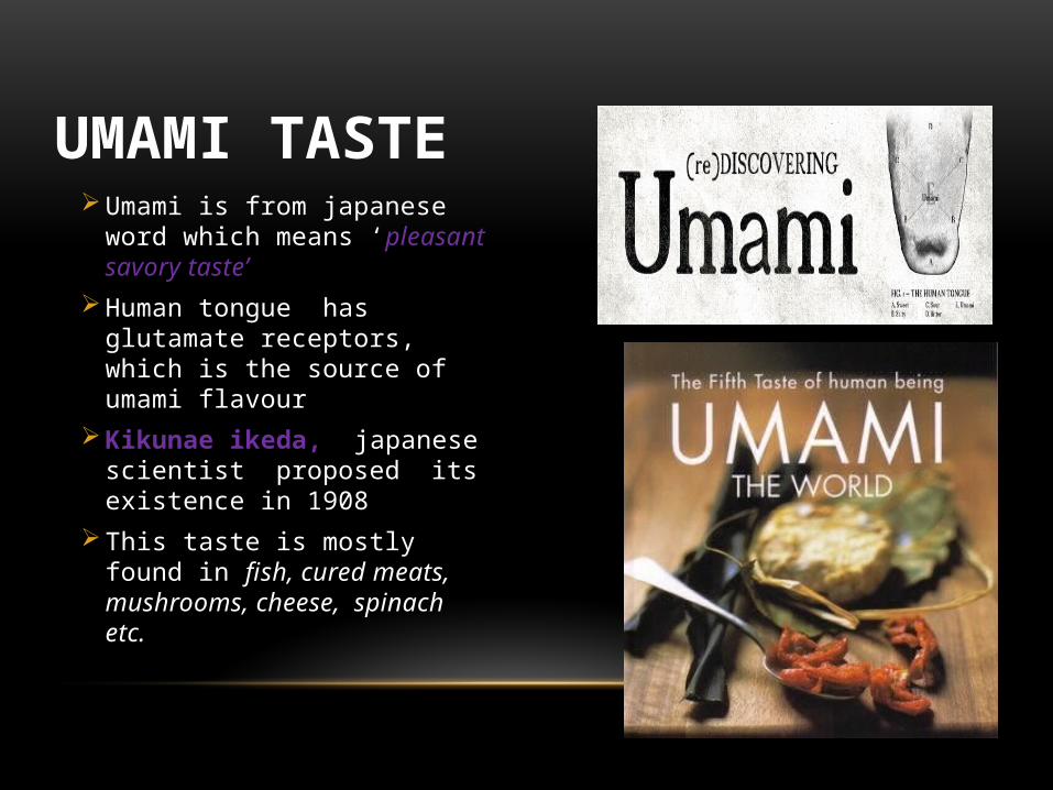
Umami is from japanese word which means ‘pleasant savory taste’
Human tongue has glutamate receptors, which is the source of umami flavour
Kikunae ikeda, japanese scientist proposed its existence in 1908
This taste is mostly found in fish, cured meats, mushrooms, cheese, spinach etc.
UMAMI TASTE

INTERESTING FACTS
• Women have shorter tongue than men.
• Blue whale has the largest tongue in animal kingdom and weighs 5400 lbs
• About half of all bacteria in your mouth live on your tongue
• Tongue heals faster than any part of the body
• Your tongue is germ free only if it is pink. If it is white there is a thin film of bacteria on it.
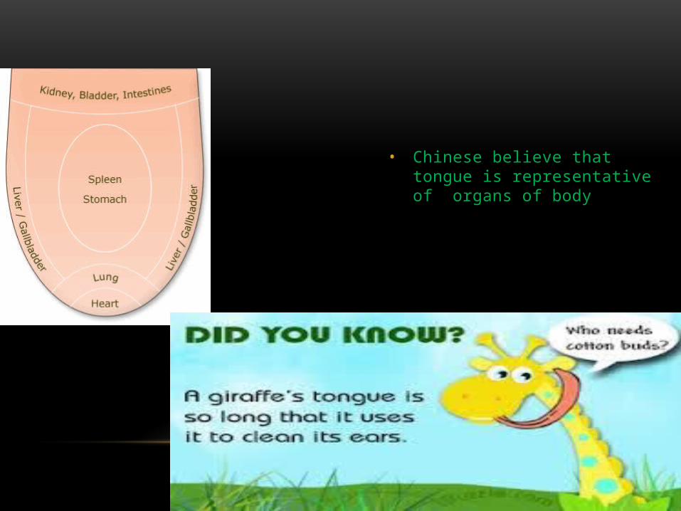
• Chinese believe that tongue is representative of organs of body


MUSCLES OF TONGUE

MUSCLES OF THE TONGUE
MUSCLES OF
TONGUE
EXTRINSIC MUSCLES
INTRINSIC MUSCLES
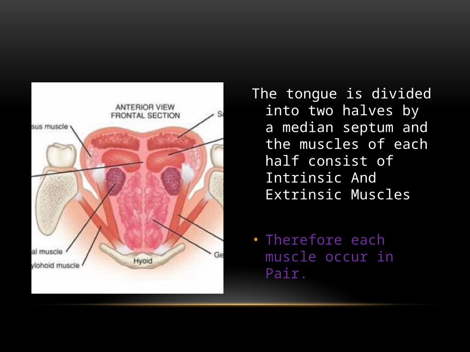
The tongue is divided into two halves by a median septum and the muscles of each half consist of Intrinsic And Extrinsic Muscles
• Therefore each muscle occur in Pair.

INTRINSIC MUSCLES• These muscles are confined to the tongue,
• They originate and inserts within the tongue,
• No bony attachments,
• FUNCTION: They alter the shape of tongue

TYPES OF INTRINSIC MUSCLES
There are four types
Superior Longitudinal,
Inferior Longitudinal,
Vertical muscles,
And
Transverse muscles.

• It lies just beneath the dorsum of the tongue.
• ACTION: It curls the tip upward and rolls it posteriorly
SUPERIOR LONGITUDINAL MUSCLE

SUPERIOR LONGITUDINAL

ACTION OF SUPERIOR LONGITUDINAL MUSCLE
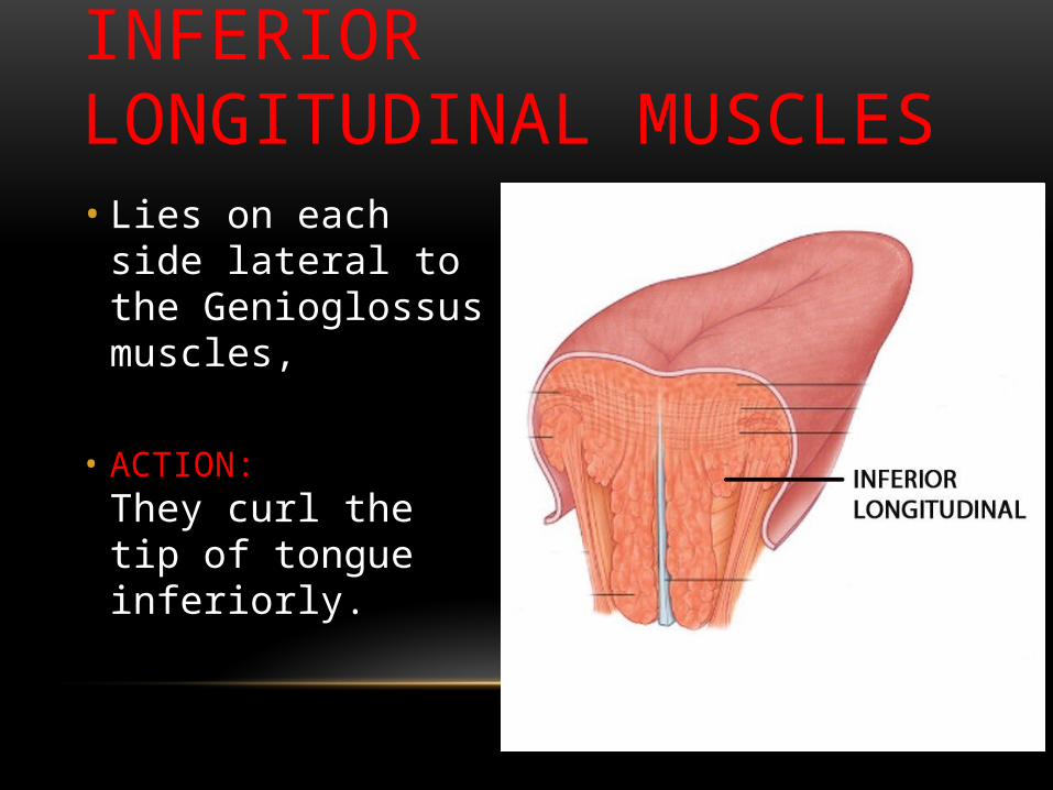
• Lies on each side lateral to the Genioglossus muscles,
• ACTION: They curl the tip of tongue inferiorly.
INFERIOR LONGITUDINAL MUSCLES
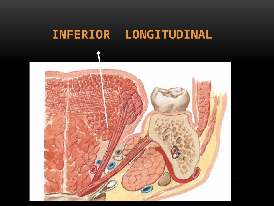
INFERIOR LONGITUDINAL
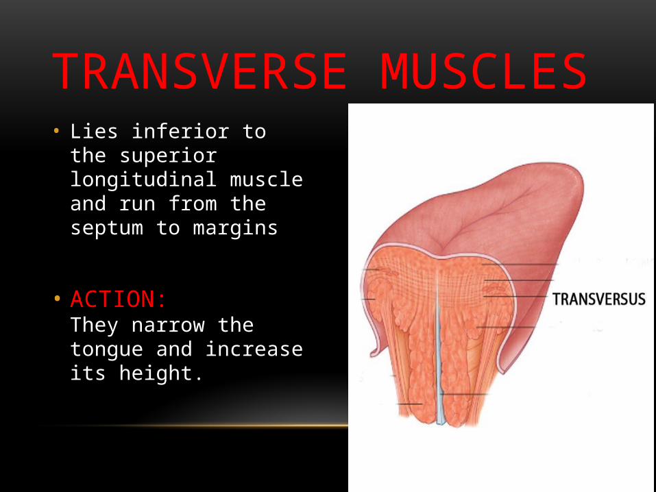
• Lies inferior to the superior longitudinal muscle and run from the septum to margins
• ACTION: They narrow the tongue and increase its height.
TRANSVERSE MUSCLES

VERTICAL MUSCLES• It runs inferolaterally
from the dorsum,
• ACTION:
Flattens the dorsum.

VERTICAL AND TRANSVERSE
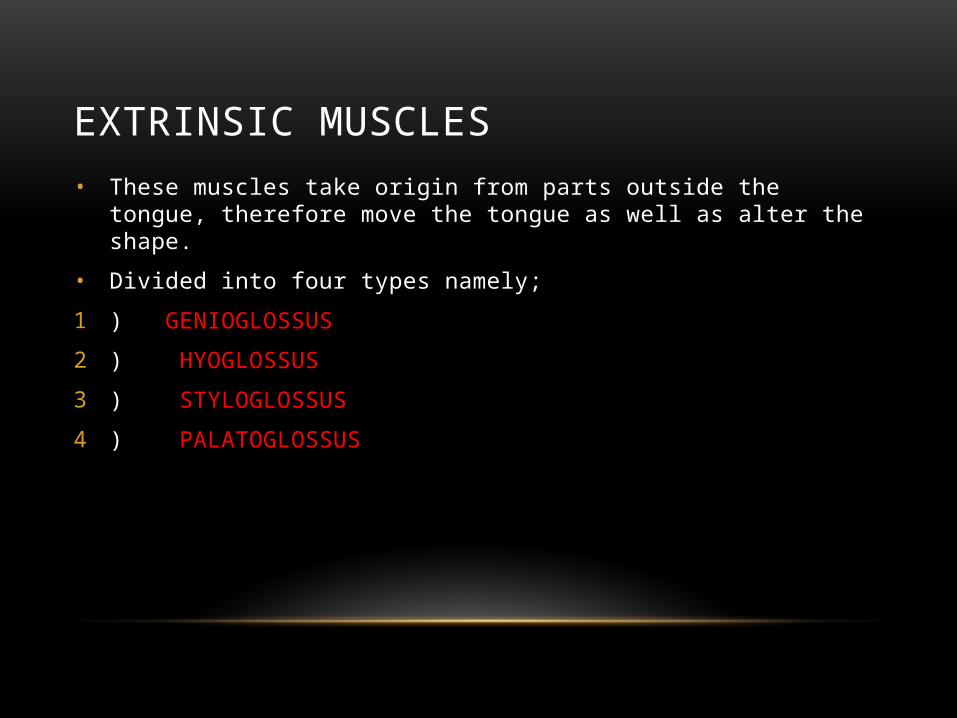
EXTRINSIC MUSCLES• These muscles take origin from parts outside the tongue, therefore move the tongue as
well as alter the shape.
• Divided into four types namely;
1 ) GENIOGLOSSUS
2 ) HYOGLOSSUS
3 ) STYLOGLOSSUS
4 ) PALATOGLOSSUS

STYLOGLOSSUS and PALATOGLOSSUS attach the tongue superiorly, while GENIOGLOSSUS and HYOGLOSSUS attach the tongue inferiorly.

GENIOGLOSSUS• ORIGIN: From superior mental spines,
• INSERTION: Into the mucous membrane of the tongue.
• Action: Protrudes the tongue, depress central part of tongue and increase the volume of mouth as in sucking.
GENIOGLOSSUS

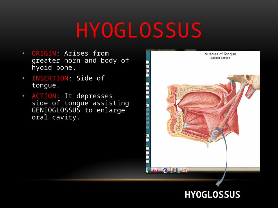
• ORIGIN: Arises from greater horn and body of hyoid bone,
• INSERTION: Side of tongue.
• ACTION: It depresses side of tongue assisting GENIOGLOSSUS to enlarge oral cavity.
HYOGLOSSUS
HYOGLOSSUS
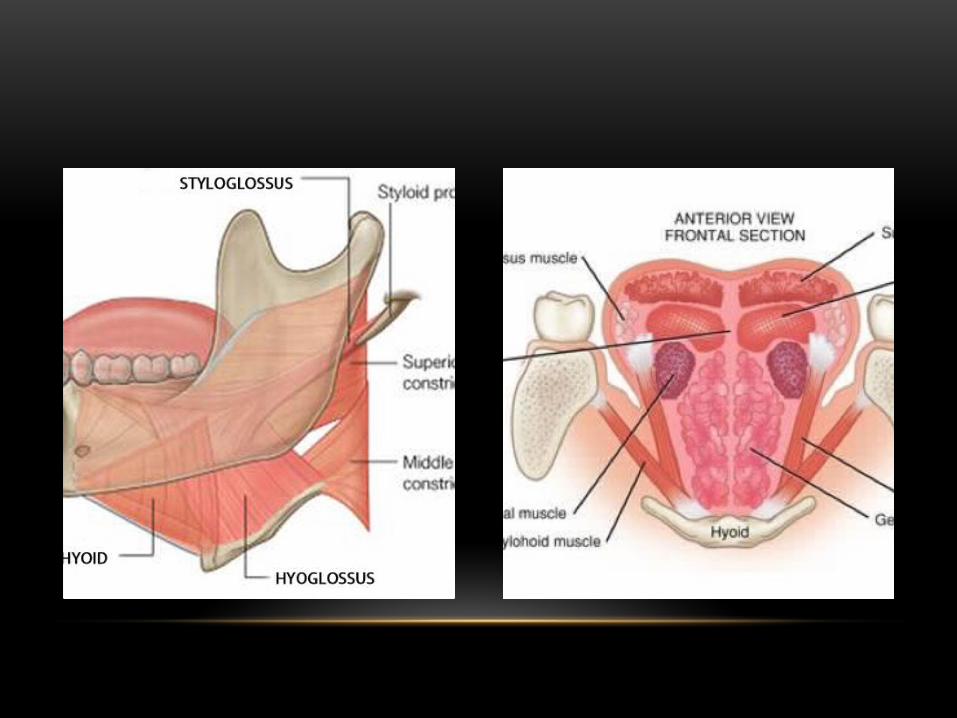
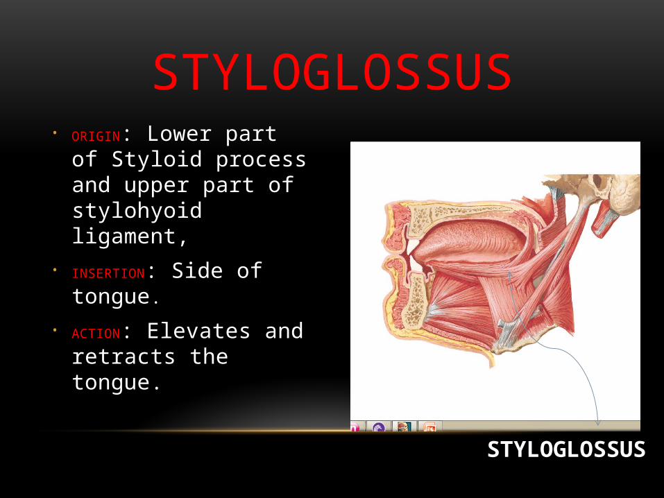
• ORIGIN: Lower part of Styloid process and upper part of stylohyoid ligament,
• INSERTION: Side of tongue.
• ACTION: Elevates and retracts the tongue.
STYLOGLOSSUS
STYLOGLOSSUS

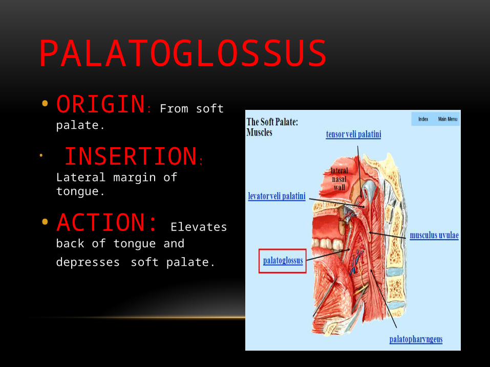
• ORIGIN: From soft palate.
• INSERTION: Lateral margin of tongue.
• ACTION: Elevates back of
tongue and depresses soft palate.
PALATOGLOSSUS

MOVEMENTS• Protrusion:
• Genioglossus on both sides acting together• Retraction:
• Styloglossus and hyoglossus on both sides acting together• Depression:
• Hyoglossus and genioglossus on both sides acting together• Elevation:
• Styloglossus and palatoglossus on both sides acting together
INNERVATION• Both extrinsic and intrinsic muscles are
supplied by HYPOGLOSSAL NERVE except PALATOGLOSSUS muscle which is in turn supplied by VAGUS NERVE.


FACTS1). The tongue is the strongest muscle in the body and the only muscle that is connected only
at one end.
2). Using a tongue scraper to clean your tongue is proven to prevent heart attacks, pneumonia, premature births, diabetes.
3). Your tongue never stops working. Even when you sleep it is pushing saliva into your throat.
VASCULATURE & INNERVATION OF TONGUE

Arterial Supply
• Lingual artery - supplies tongue and floor of the mouth.
• Originates from external carotid artery in neck
• Passes between hyoglossus and genioglossus muscles of tongue
BLOOD SUPPLYILingual artery
ARTERIAL SUPPLY• Lingual artery mainly gives three branches within the
tongue namely
• Dorsal lingual artery
• Deep lingual artery
• Sub lingual artery


ARTERIAL SUPPLY
• Also secondary supply to the tongue by:
• Tonsillar branch of facial artery
• Ascending pharyngeal artery (branch of external carotid artery)

• Drained by dorsal lingual vein and deep lingual veins
• Deep Lingual Veins:
• Begins near tip of tongue and run beneath the mucous membrane
• Visible on the inferior surface of tongue
• Anterior to lingual artery
• Ultimately drains into internal jugular vein
VENOUS DRAINAGE
Deep lingual vein
Dorsal lingual vein

Deep lingual vein

• Dorsal Lingual Veins
• Drain the dorsum and sides of tongue
• Runs along the lingual artery
• Drains into internal jugular vein
VENOUS DRAINAGE
Dorsal lingual vein

LYMPHATICS
Apical Vessels:Drains into Submental nodes & deep cervical nodes
Marginal Vessels:Drains into Submandibular nodes & deep cervical nodes
Basal Vessels:Drains into Deep cervical nodes (jugulodigastric mainly)

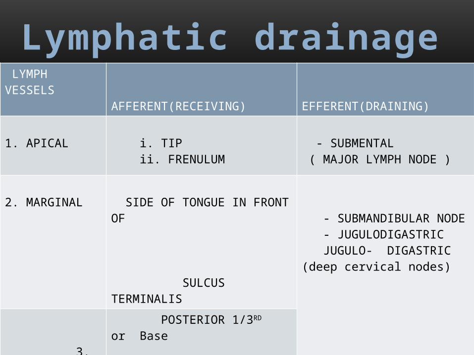
LYMPH VESSELS AFFERENT(RECEIVING) EFFERENT(DRAINING)
1. APICAL i. TIP ii. FRENULUM
- SUBMENTAL ( MAJOR LYMPH NODE )
2. MARGINAL SIDE OF TONGUE IN FRONT OF SULCUS TERMINALIS
- SUBMANDIBULAR NODE - JUGULODIGASTRIC JUGULO- DIGASTRIC (deep cervical nodes)
3. BASAL POSTERIOR 1/3RD or Base
Lymphatic drainage

INNERVATION• Innervation is complex and consists of three different
supplies
• Motor supply
• General sensory supply
• Special sensory supply
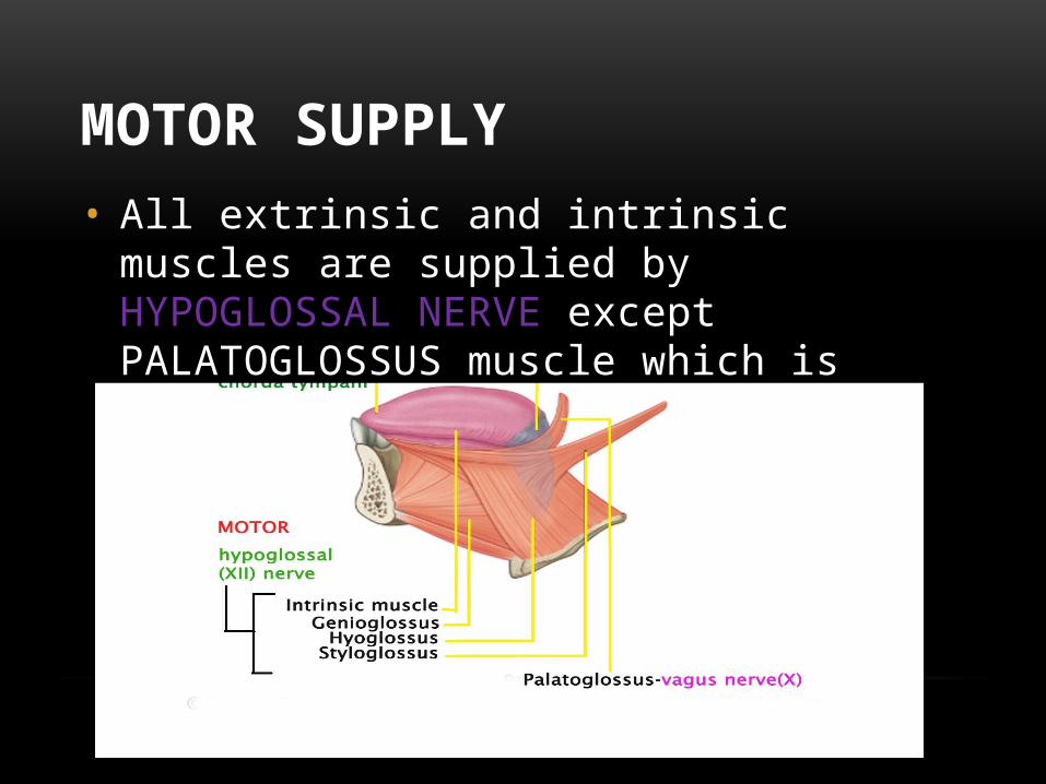
MOTOR SUPPLY• All extrinsic and intrinsic muscles are supplied by
HYPOGLOSSAL NERVE except PALATOGLOSSUS muscle which is supplied by VAGUS NERVE.

• General sensory sensation is by three nerves• Lingual nerve – anterior 2/3rd of
tongue
• Glossopharyngeal nerve – posterior 1/3rd of tongue
• Vagus nerve – posterior most part of tongue
SENSORY SUPPLY

• Supplied by three nerves
• Chorda tympani (facial) – taste sensation of anterior 2/3rd of tongue
• Glossopharyngeal (ix) – taste sensation of posterior 1/3rd of tongue
• Vagus nerve (x) – taste sensation of posterior most part
SPECIAL SENSORY SUPPLY


FUNCTIONS• Deglutition
• Taste sensation
• Speech production
• Breast feeding
• Self cleansing system
• Mastication