diseases of field cash crops - hill...
TRANSCRIPT

Dr. B. R. ThakurPlant Pathologist
CSKHPKV, Palampur-176 062
DISEASES OF FIELD CASH CROPS AND THEIR MANAGEMENT
111
DISEASES OF SUGARCANE (SACCHARUM OFFICINARUM)
• Red rot- Colletotrichum falcatum( or Glomerella tucumanensis)
• Smut-Ustilago scitaminea• Grassy shoot-Phytoplasma• Ratoon stunting-Clavibacter xyli pv.
xyli (Xylem limited fastidious bacteria)

Red Rot of Sugarcane
DISEASE: Red RotPATHOGEN: Colletot
richum falcatum(Glomerellatucumanensis)
HOSTS: Sugarcane3
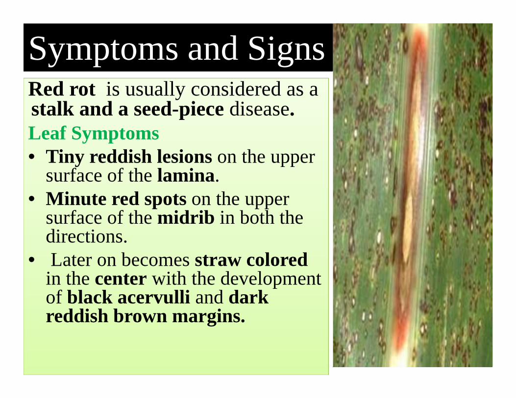
Red rot is usually considered as astalk and a seed-piece disease.Leaf Symptoms• Tiny reddish lesions on the upper
surface of the lamina. • Minute red spots on the upper
surface of the midrib in both the directions.
• Later on becomes straw colored in the center with the development of black acervulli and dark reddish brown margins.
Symptoms and Signs
4
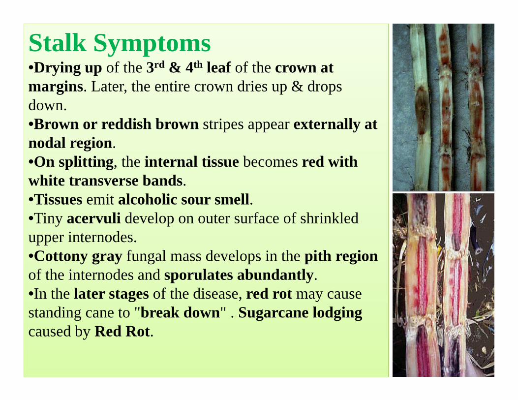
Stalk Symptoms•Drying up of the 3rd & 4th leaf of the crown at margins. Later, the entire crown dries up & drops down. •Brown or reddish brown stripes appear externally at nodal region. •On splitting, the internal tissue becomes red with white transverse bands. •Tissues emit alcoholic sour smell. •Tiny acervuli develop on outer surface of shrinkled upper internodes. •Cottony gray fungal mass develops in the pith region of the internodes and sporulates abundantly.•In the later stages of the disease, red rot may cause standing cane to "break down" . Sugarcane lodging caused by Red Rot.
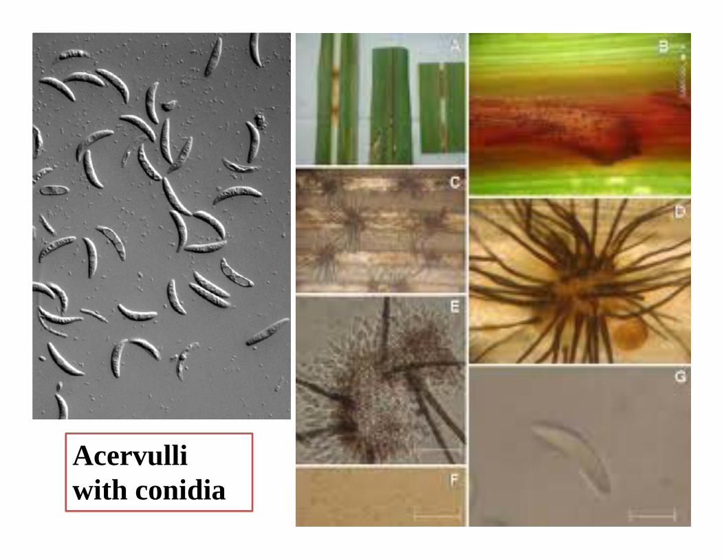
Acervulli with conidia

Pre disposing (environmental) factors
• Warm ‐humid weather conditions with intermittent rains.
• Mono-culturing of sugarcane,• Successive ratoon cropping, • Water logged conditions • Injuries by sugarcane borers and sugarcane
weevils.

Disease cycle
8
The fungus is sett-borne. The fungus also persists in the soil on the diseased clumps and stubbles as acervulli. The primary infection is mainly from infected setts. Secondary spread in the field may be through irrigation water and cultivation tools. The rain splash, air currents and dew drops also help in the spread of conidia from the diseased to healthy plants in the field. The fungus also survives on collateral hosts like Sorghum vulgare, S. halepense and Saccharum spontaneum.

Management strategy• Grow resistant varieties like CO 6907, CO 7219, CO 8013, CO 8021, CO 7706, CO A 7602, CO A 89082, CO A 89085, 87 A 397, CO T 8201, etc.
• Removal and destruction of infected plant debris, stubbles and trash.
• Deep tillage .• Crop rotation.• Use disease free setts.• Avoid ratooning of the diseased crop.• Avoid flood irrigation. • Soak the setts in 0.1% Carbendazim solution for 20 minutes before planting. 9
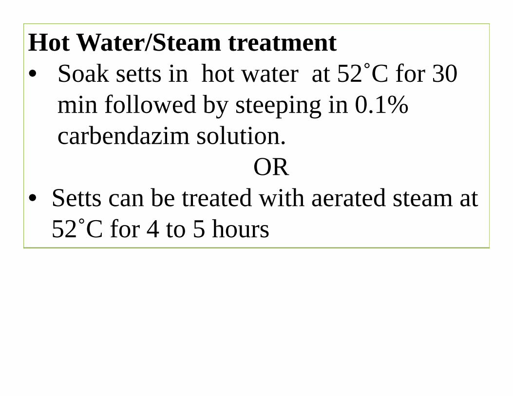
Hot Water/Steam treatment• Soak setts in hot water at 52˚C for 30
min followed by steeping in 0.1% carbendazim solution.
OR• Setts can be treated with aerated steam at
52˚C for 4 to 5 hours
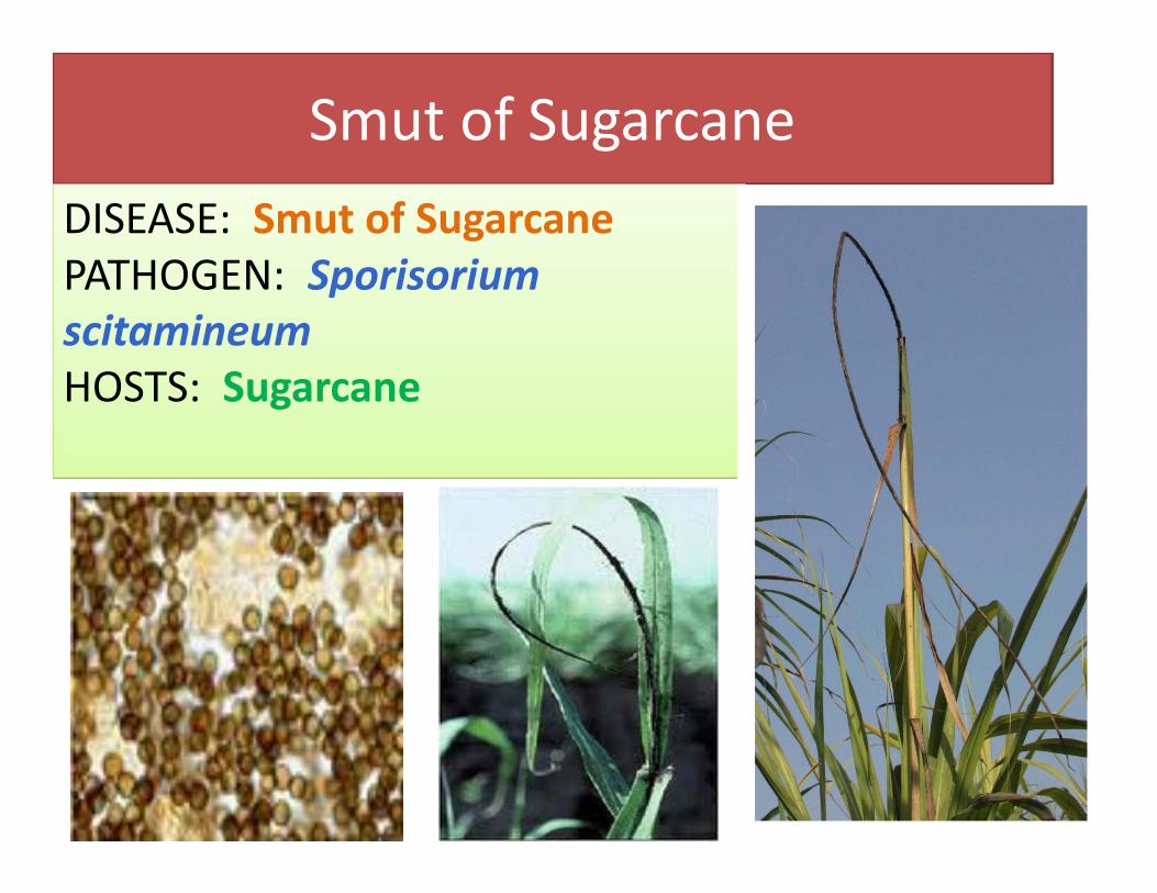
Smut of SugarcaneDISEASE: Smut of SugarcanePATHOGEN: Sporisorium scitamineum HOSTS: Sugarcane
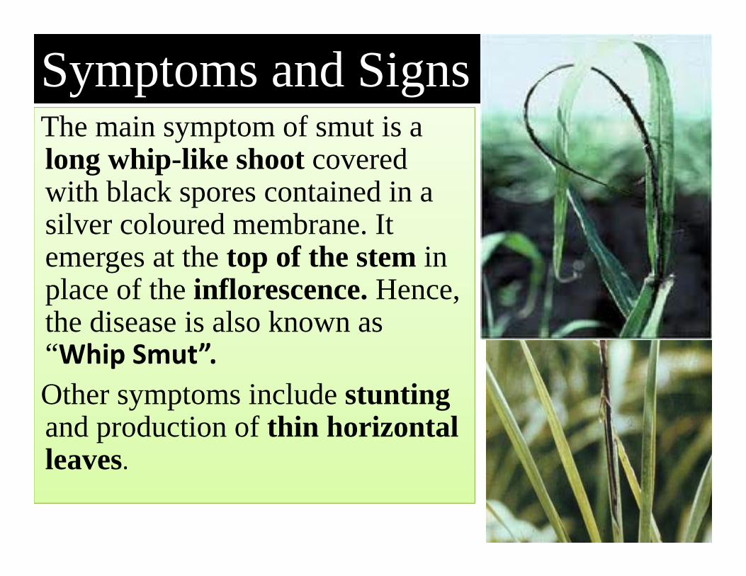
The main symptom of smut is a long whip-like shoot covered with black spores contained in a silver coloured membrane. It emerges at the top of the stem in place of the inflorescence. Hence, the disease is also known as “Whip Smut”.Other symptoms include stunting and production of thin horizontal leaves.
Symptoms and Signs
12
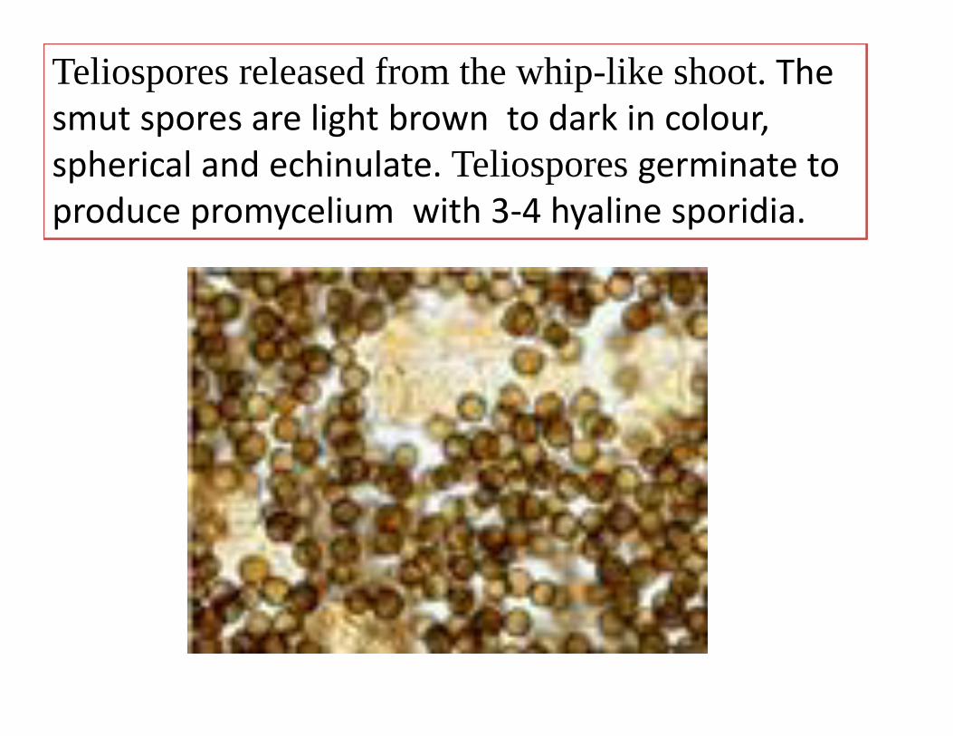
Teliospores released from the whip-like shoot. The smut spores are light brown to dark in colour, spherical and echinulate. Teliospores germinate to produce promycelium with 3‐4 hyaline sporidia.
Pre disposing (environmental) factors
• Mono‐culturing of sugarcane• Continuous ratooning • Dry weather during tillering stage favours the disease.
When mature canes are split open, vascular bundles appear discoloured.
Disease cycle
15
Teliospores may survive in the infected plant debris / soil. The smut spores and dormant mycelium also present in or on the infected setts may serve the source of primary inoculum. Hence, the primary spread of the disease is through diseased seed‐pieces (setts). In addition, sporidia and spores present in the soil also spread through rain and irrigation water and cause soil‐borne infection. The secondary spread in the field is mainly through the smut spores developed in the whips, aided by air currents. The fungus also survives on collateral hosts like Saccharum spontaneum, S. robustum, Sorghum vulgare, Imperata arundinacea and Cyperus dilatatus.
Management strategy• Grow resistant varieties like Co 6806 and
Co 62175 • Plant healthy setts taken from disease free
area.• Remove and destroy the smutted clump.• Avoid ratooning of the diseased crops.• Follow crop rotation.• Treat the setts in hot water at 52˚C for 20
minutes.16

Ratoon stunting of Sugarcane
DISEASE: Ratoon stuntingPATHOGEN: Clavibacterxyli subsp. xyl (Xylem limited fastidious bacteria)HOSTS: Sugarcane

Diseased clumps usually display stunted growth, reduced tillering, thin stalks with shortened internodes and yellowish foliage (mild chlorosis). Coryneform Xylem limited fastidious bacteria infects the vascular bundles of canes . When mature canes are split open, vascular bundles appear discolored. In young canes, pink colour is seen in the form of minute pin head like areas near the nodes. The disease reduces the length, girth and the number of canes per clump
Symptoms and Signs
18

Pre disposing (environmental) factors
• Continuous ratooning• Injuries through implements.

Disease cycle
20
The disease spreads through use of diseased setts. The disease also spreads through cane harvesting implements contaminated with the juice of the diseased canes. Maize, sorghum, Sudan grass and Cynodon are some of the collateral hosts of the pathogen.
Management strategy• Grow setts from disease free field.• Remove and burn the clumps showing the disease.
• Sterilization of cutting knives with spirit or any other antiseptic solution.
• Hot air treatment of setts at 52˚ C for 8 hours or hot water treatment at 52˚ C for 2 0 minutes or aerated steam treatment at 50˚ C for 1 hour.
21
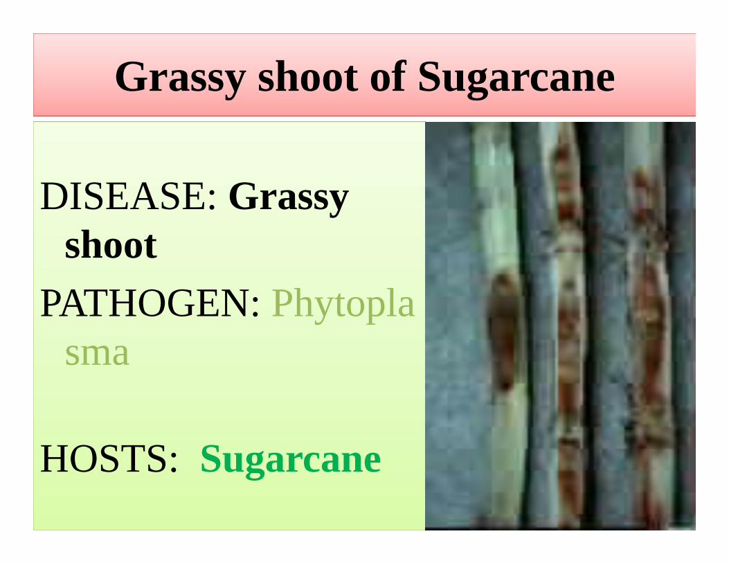
Grassy shoot of Sugarcane
DISEASE: Grassy shoot
PATHOGEN: Phytoplasma
HOSTS: Sugarcane22

Phytoplasma-infected sugarcane plants show a proliferation of tillers, which give it typical grassy appearance, hence the name grassy shoot disease. The plants appear bushy and ‘grass like’ due to reduction in the length of internodes, premature and continuous tillering. The leaves of infected plants do not produce chlorophyll, and therefore appear white or creamy yellow.
Symptoms and Signs
23
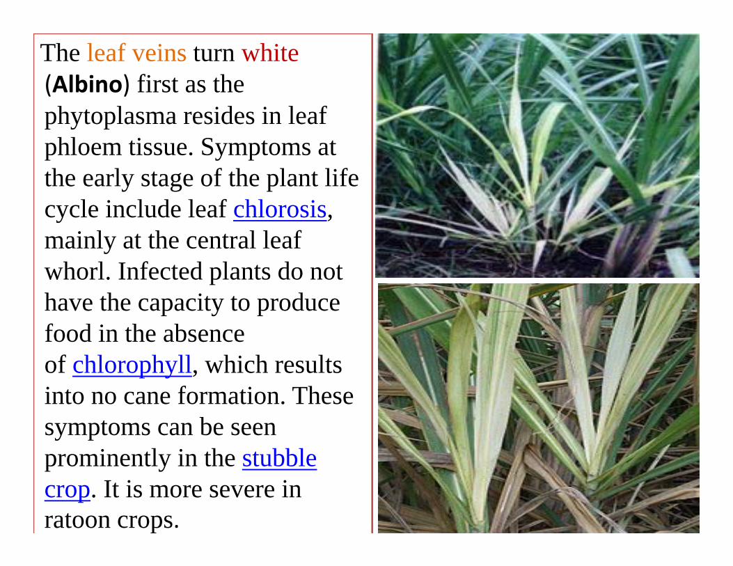
The leaf veins turn white(Albino) first as the phytoplasma resides in leaf phloem tissue. Symptoms at the early stage of the plant life cycle include leaf chlorosis, mainly at the central leaf whorl. Infected plants do not have the capacity to produce food in the absence of chlorophyll, which results into no cane formation. These symptoms can be seen prominently in the stubble crop. It is more severe in ratoon crops.
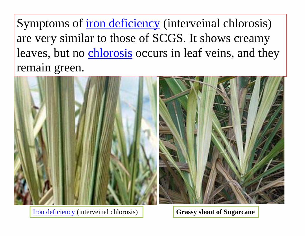
Symptoms of iron deficiency (interveinal chlorosis) are very similar to those of SCGS. It shows creamy leaves, but no chlorosis occurs in leaf veins, and they remain green.
Iron deficiency (interveinal chlorosis) Grassy shoot of Sugarcane
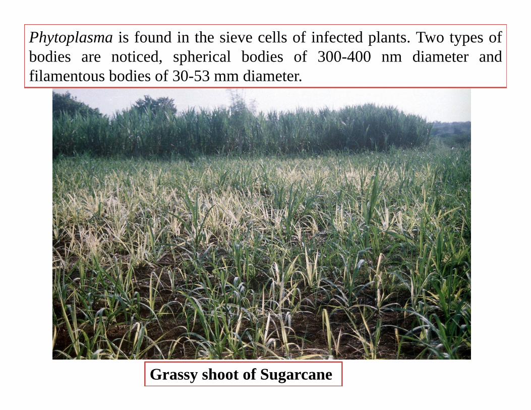
Grassy shoot of Sugarcane
Phytoplasma is found in the sieve cells of infected plants. Two types ofbodies are noticed, spherical bodies of 300-400 nm diameter andfilamentous bodies of 30-53 mm diameter.
Disease cycle
27
Phytoplasmas, formerly called mycoplasma-like organisms (MLOs), are a large group of obligate, intracellular, cell wallless parasites classified within the class Mollicutes. Sugarcane is a vegetativelypropagated crop, so the pathogen is transmitted via seed setts/planting material and by phloemfeeding vectors as aphids, viz.,Aphis maidis, Rhopalosiphum maidis, Longiunguis sacchari, Melanaphis sacchari and M. indosacchari. In addition, leaf hopper, Proutista moesta also involves in phytoplasma transmission in sugarcane. Sorghum serves as a natural collateral host.

Management strategyPhytoplasma infection also spreads through insect vectors; it is therefore important to control them• Control vector by spraying Malathion or Dimethoate @
2ml/lt• Plant disease free setts.• Remove and burn the infected clumps periodically.• Avoid ratooning in problem areas• Hot Water Treatment (HWT) of setts at 52˚ C for 30min or
Aerated Steam Therapy (AST) at 50˚ Cfor 1hrfollowed by steeping in fungicidalsolution of carbendazim @ 0.05% for 15 minutes.
28
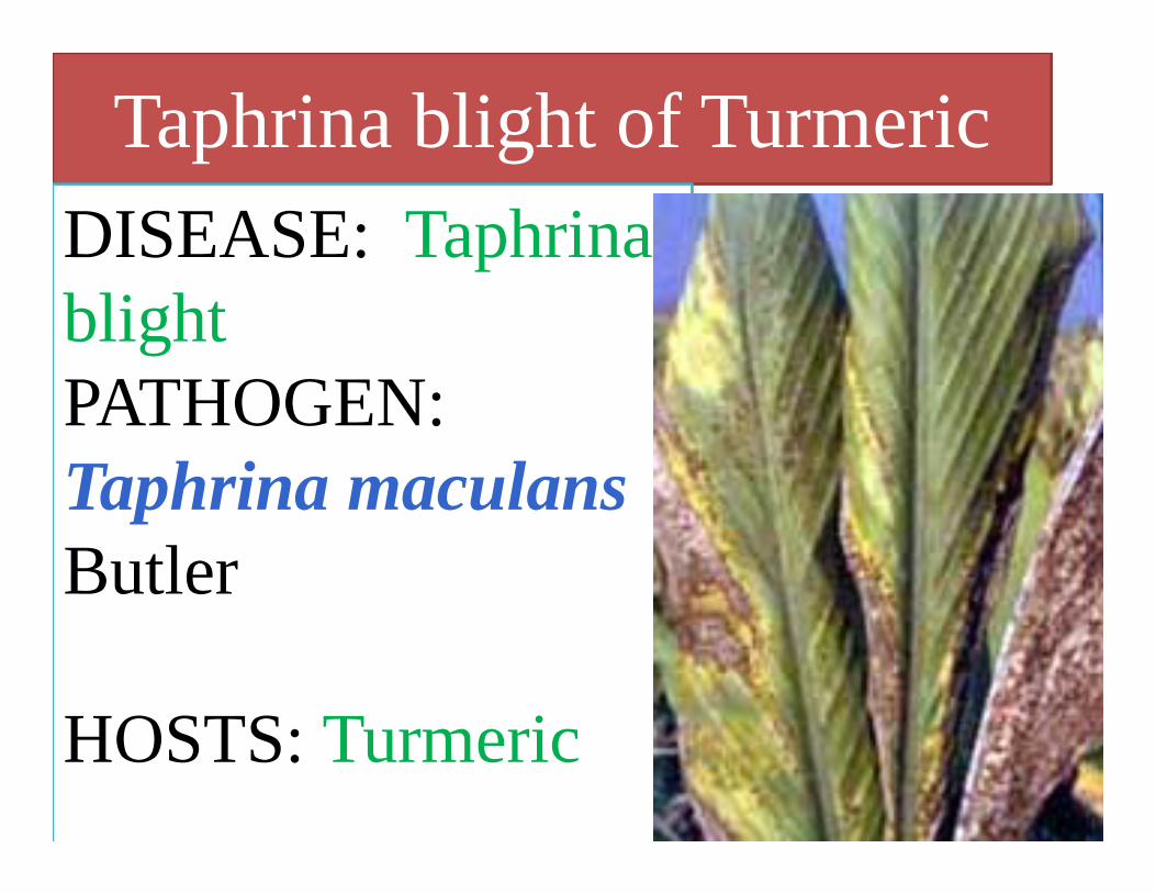
Taphrina blight of TurmericDISEASE: Taphrina blight PATHOGEN:Taphrina maculansButler
HOSTS: Turmeric

Turmeric
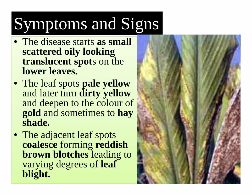
• The disease starts as small scattered oily looking translucent spots on the lower leaves.
• The leaf spots pale yellow and later turn dirty yellow and deepen to the colour of gold and sometimes to hay shade.
• The adjacent leaf spots coalesce forming reddish brown blotches leading to varying degrees of leaf blight.
Symptoms and Signs
31
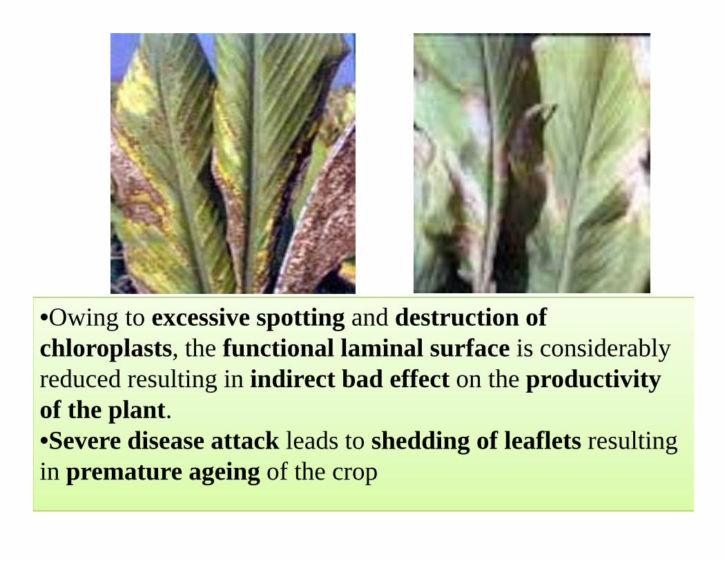
•Owing to excessive spotting and destruction of chloroplasts, the functional laminal surface is considerably reduced resulting in indirect bad effect on the productivity of the plant.•Severe disease attack leads to shedding of leaflets resulting in premature ageing of the crop
Pre disposing factors
Favourable conditions: • Moist cloudy weather with temperature of 21-23 ˚ C
• Relative Humidity 80 %
Disease cycle
34
• The ascospores discharged from asci in dried leaves having spots and lying in the field or Collateral hosts: Curcuma amada, C. angustifolia may function as chief source of primary inoculum.
• The secondary infection is by ascospores discharged from successively maturing asci which grow into octosporous microcolonies and infect fresh leaves without any dormancy.
Management strategy
• Grow resistant varieties• Crop rotation• Field sanitation- Collect and destroy diseased leaves• Spray [email protected]% or COC@ 0.25% at 20 days interval.
35
DISEASES OF TOBACCO (Nicotianatabacum)
Black Shank of Tobacco: Phytophthora nicotianae var. nicotianae)Mosaic of Tobacco : Tobacco mosaic virus (TMV)

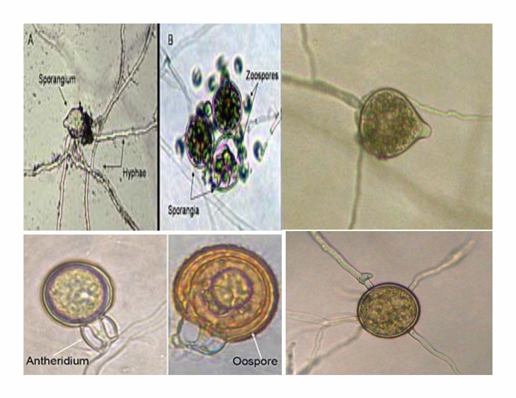
Black Shank of TobaccoDISEASE: Black ShankPATHOGEN: Phytophthora nicotianae var. nicotianae)HOSTS: Tobacco
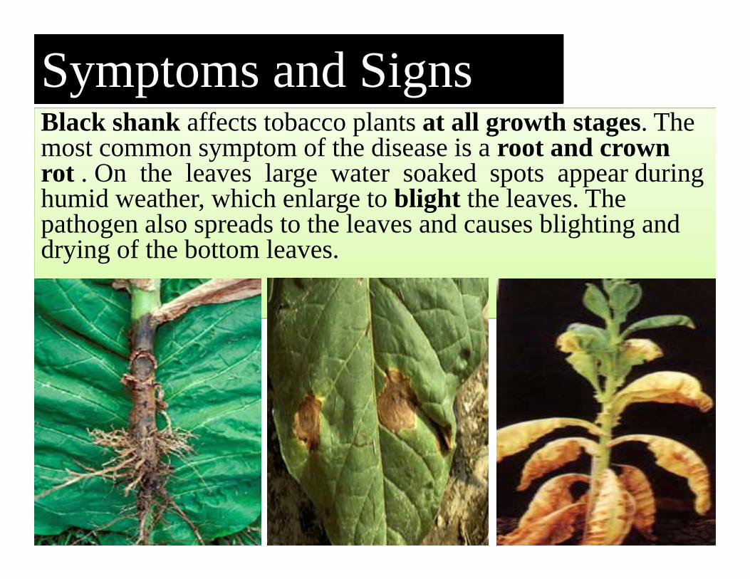
Black shank affects tobacco plants at all growth stages. The most common symptom of the disease is a root and crown rot . On the leaves large water soaked spots appear during humid weather, which enlarge to blight the leaves. The pathogen also spreads to the leaves and causes blighting and drying of the bottom leaves.
Symptoms and Signs
38
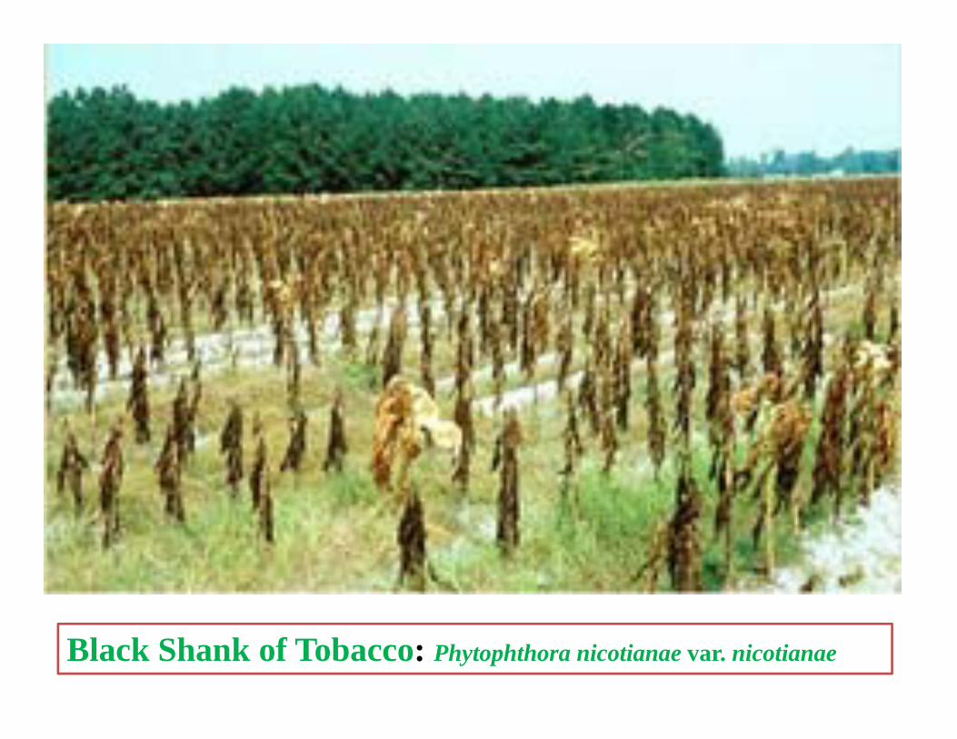
Black Shank of Tobacco: Phytophthora nicotianae var. nicotianae
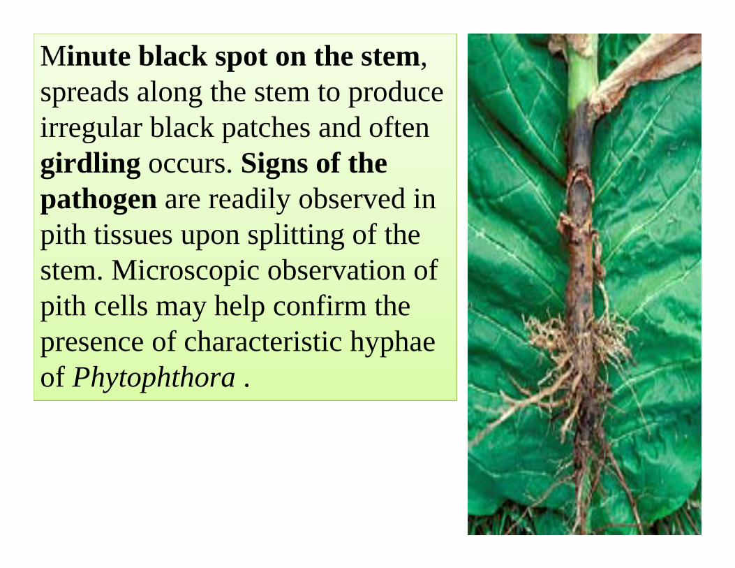
Minute black spot on the stem, spreads along the stem to produce irregular black patches and often girdling occurs. Signs of the pathogen are readily observed in pith tissues upon splitting of the stem. Microscopic observation of pith cells may help confirm the presence of characteristic hyphae of Phytophthora .
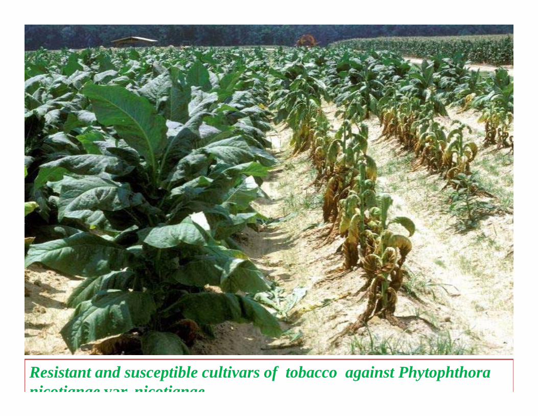
Resistant and susceptible cultivars of tobacco against Phytophthora nicotianae var nicotianae

Pre disposing factors
• Frequent rainfall, • High soil moisture and • High population of root knot
nematodes, i.e. Meloidogyneincognita, favours the disease.
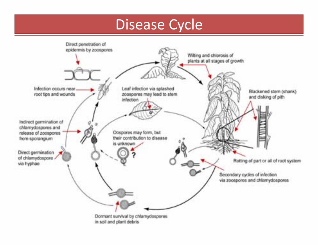
Disease Cycle
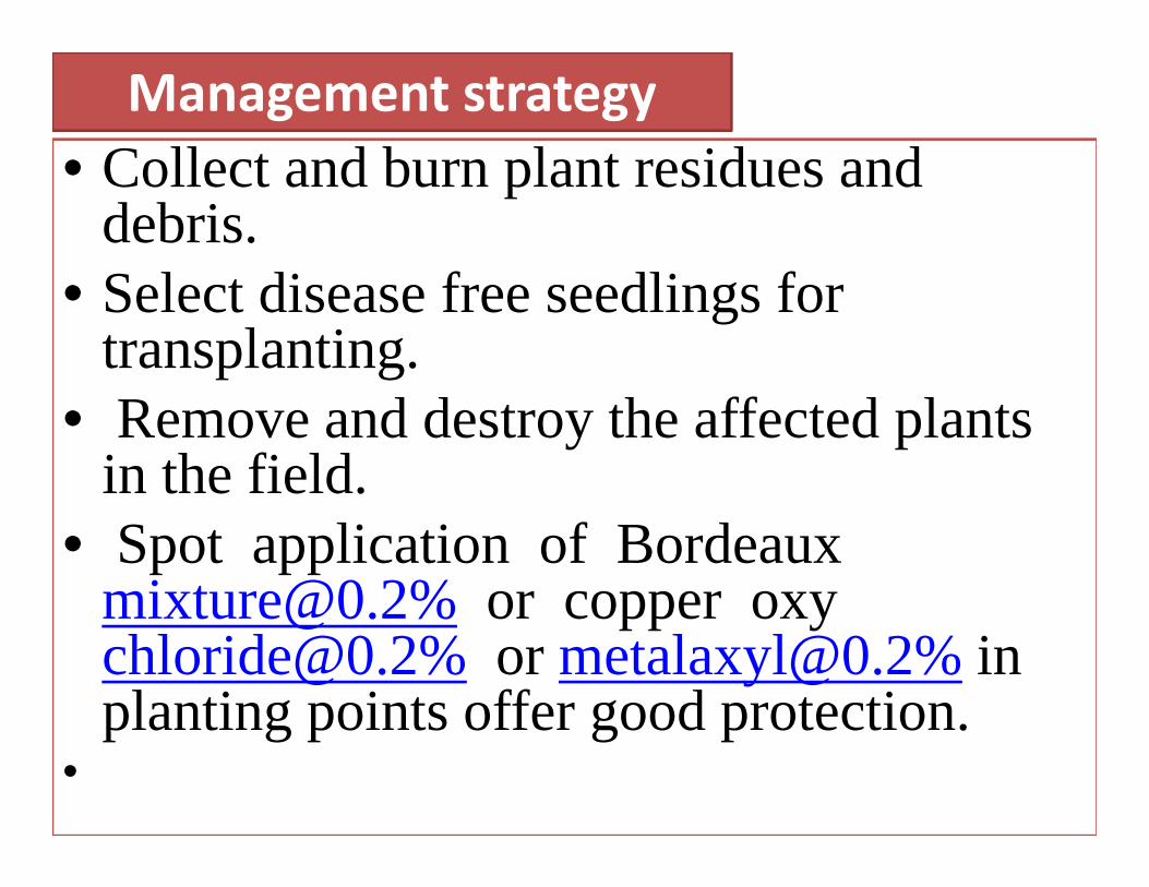
Management strategy• Collect and burn plant residues and
debris.• Select disease free seedlings for
transplanting.• Remove and destroy the affected plants
in the field.• Spot application of Bordeaux
[email protected]% or copper oxy [email protected]% or [email protected]% in planting points offer good protection.
•
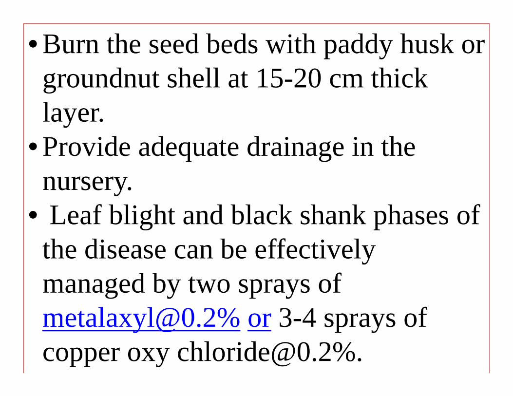
• Burn the seed beds with paddy husk or groundnut shell at 15-20 cm thick layer.
• Provide adequate drainage in the nursery.
• Leaf blight and black shank phases of the disease can be effectively managed by two sprays of [email protected]% or 3-4 sprays of copper oxy [email protected]%.
Mosaic of Tobacco
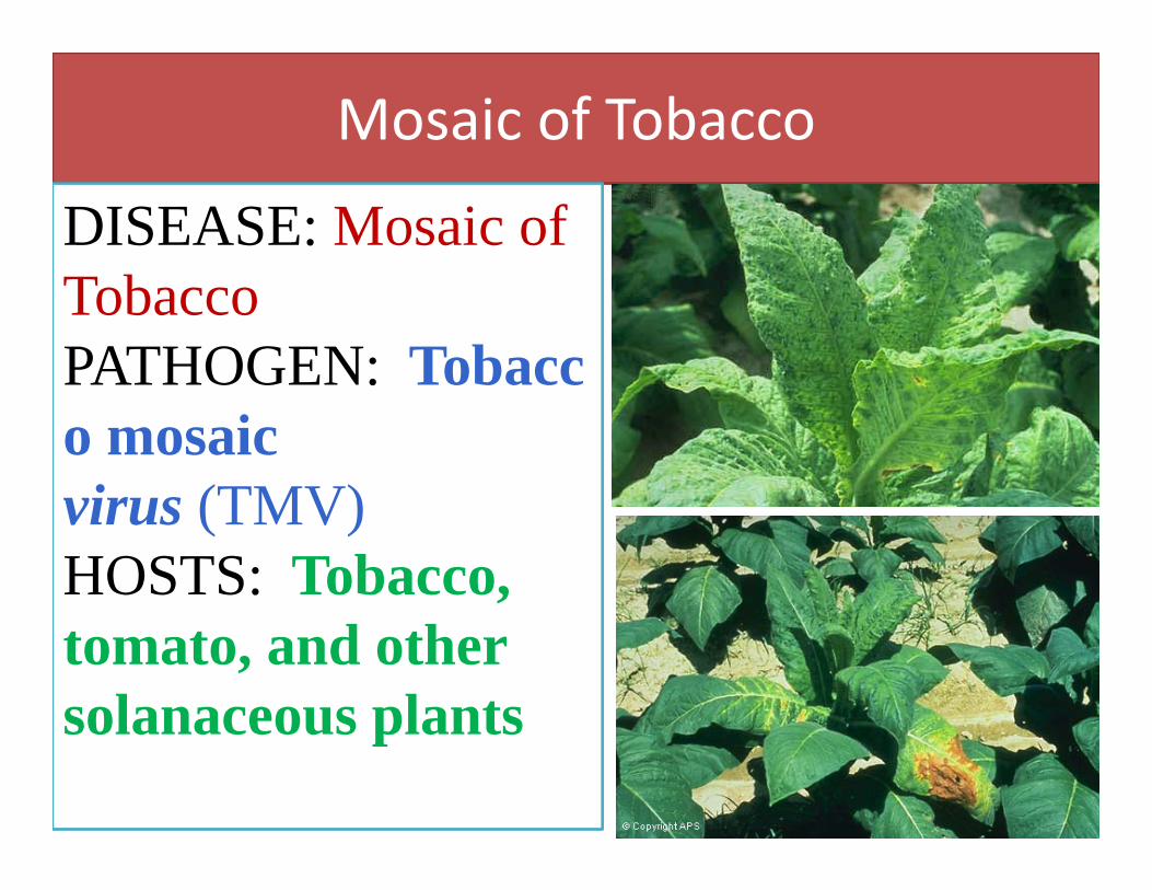
DISEASE: Mosaic of TobaccoPATHOGEN: Tobacco mosaic virus (TMV)HOSTS: Tobacco, tomato, and other solanaceous plants

Symptoms and SignsThe characteristic symptoms of Tobacco mosaic virus (TMV) includes mosaic, mottling , necrosis , stunting, leaf curling, and yellowing of plant tissues. Symptoms vary depending upon virus strains, plant age and growing conditions. A common characteristics is yellow or light green coloration between the veins of young leaves followed by “mosaic” or mottled pattern of light and dark green areas in the leaves. Lower leaves are subjected to “mosaic burns” in hot and dry weather. Infected leaves may be crinkled, puckered, or elongated.
Disease CycleTMV has broad host range and important are tobacco species, peppers, tomatoes and potatoes. There are high concentrations of TMV in cells of tobacco and solanaceous plants. TMV is easily transmissible from crop to crop primarily by mechanical transmission or in the roots/soil from infected plants but is not known to spread by insect vector transmission. TMV can also survive outside the plant in sap that has dried on tools and other surfaces.TMV is so stable that its infectivity can be maintained for about 2 years in the soil that is not exposed to freezing and drying. The virions are stable in plants residues in the soil and in the composts of tomato plants. Tobacco mosaic is one of the most persistent diseases of tomatoes because the virus can remain infectious without a host for many years’ and is able to withstand heat. The hands and clothing of smokers who come in contact with TMV infected tobacco easily transmit TMV to susceptible plants while working in the fields.
Management strategy
• Plant TMV resistant varieties. • Rogue out infected plants. • Crop rotation by growing non-host plants.• Prohibit smoking during working.• Workers hygiene should be maintain.• Destroy all weed hosts of TMV in tobacco
and nearby fields.
DISEASES OF GROUND NUT (Arachis hypogea)
• Tikka disease of ground nut:Cercospora arachidicola(Mycosphaerella arachidis)
• Rust of ground nut:Puccinia arachidis
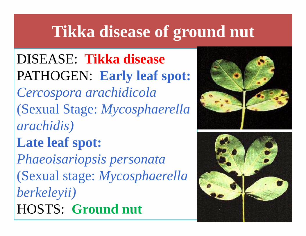
Tikka disease of ground nutDISEASE: Tikka disease PATHOGEN: Early leaf spot: Cercospora arachidicola(Sexual Stage: Mycosphaerellaarachidis)Late leaf spot: Phaeoisariopsis personata(Sexual stage: Mycosphaerellaberkeleyii) HOSTS: Ground nut

The Tikka Disease occurs as two distinct types of leaf spots, caused by two species of Cercosporidium.
Early leaf spot (Cercospora arachidicola):Early Infection leads to circular to irregular reddish brown or dark brown spots with some yellow halo on upper surface of leaves. Symptoms may also appear on rachis, petioles, stipules and stalks etc, as elongated, elliptical spots with definite border.
Symptoms and Signs
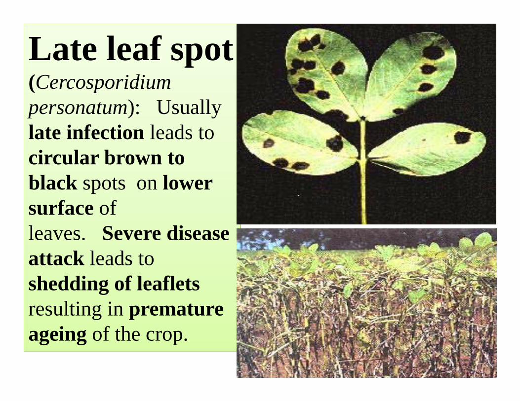
Late leaf spot (Cercosporidiumpersonatum): Usually late infection leads to circular brown to black spots on lower surface of leaves. Severe disease attack leads to shedding of leaflets resulting in premature ageing of the crop.
Pre disposing factors
• Wet weather‐ High relative humidity (above 85 per cent)
• Heavy rainfall• Leaf wetness• Moderate temperature of 22‐25 0C.
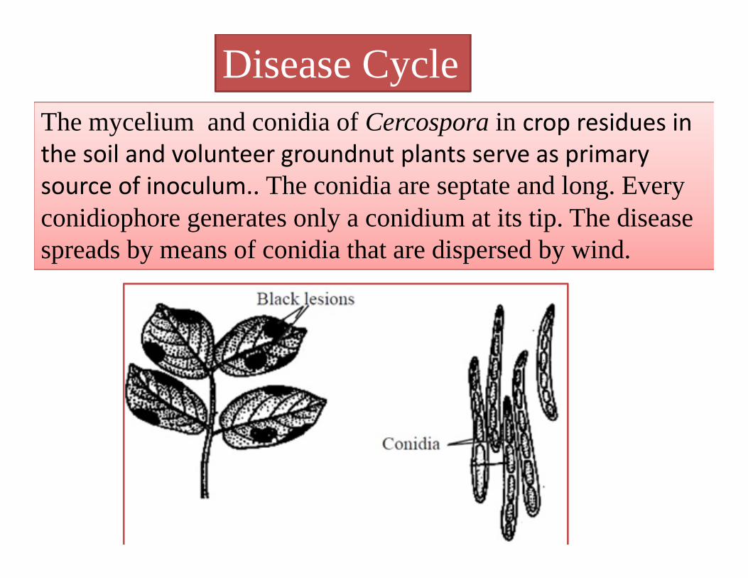
The mycelium and conidia of Cercospora in crop residues in the soil and volunteer groundnut plants serve as primary source of inoculum.. The conidia are septate and long. Every conidiophore generates only a conidium at its tip. The disease spreads by means of conidia that are dispersed by wind.
Disease Cycle
Management strategy• Grow resistant varieties like Vemana (early and late
leaf spots), Naveen, Tirupathi-3 (early leaf spot only). • General Sanitation:
-Remove and destroy the infected plant debris.-Eradicate the volunteer groundnut plants.
• Crop rotation with millets• Seed Treatment with Captan or Thiram at 4g/kg or
[email protected]%• Spray [email protected]% or [email protected]% or
[email protected]% and if necessary, repeat after 15 days.
•
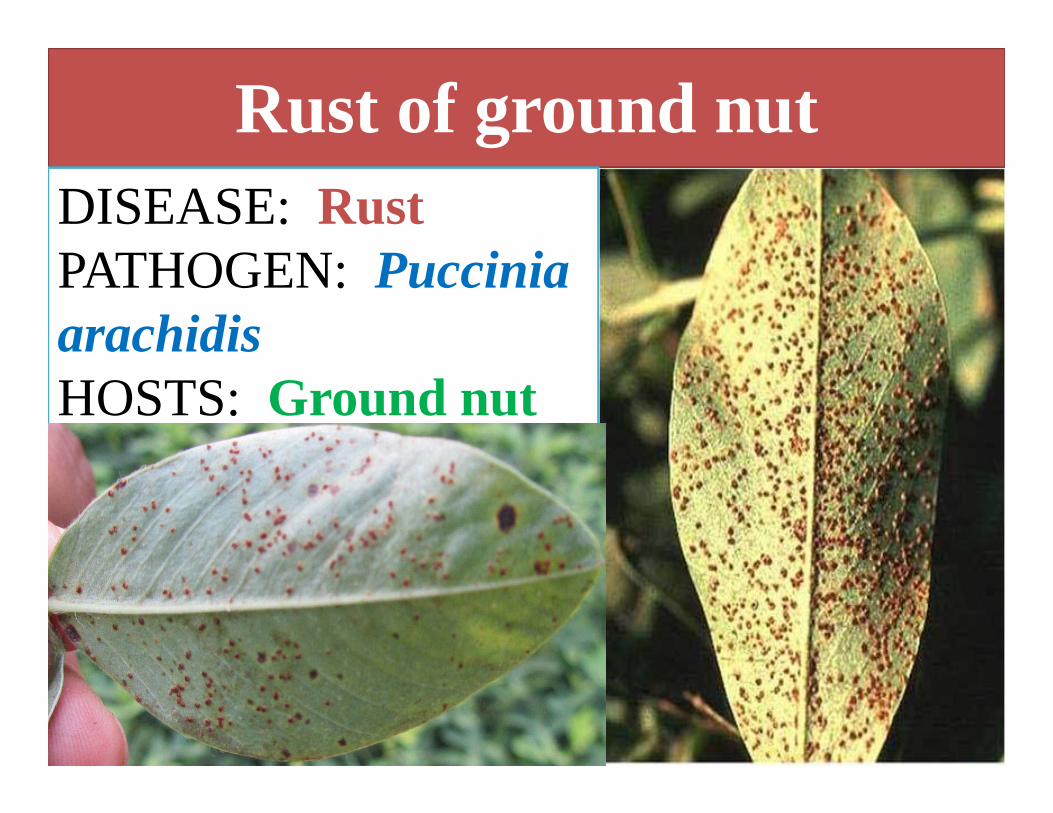
Rust of ground nutDISEASE: Rust PATHOGEN: Puccinia arachidisHOSTS: Ground nut

• The disease attacks all aerial parts of the plant.
• Orange coloured pustules (uredinia) on the lower surface of leaves.
• Corresponding to the sori, small, necrotic, brown spots appear on the upper surface of leaves.
• Rupture epidermis to release mass of reddish brown urediniospores
• Pustules appear first on the lower surface of leaves and then on the upper surface .
• Severely infected leaves turn necrotic and desiccate.
• Late in the season, dark pustules full of teliospores appear .
Symptoms and Signs
59

Pre disposing factors
• Wet weather‐ High relative humidity (above 85 per cent)
• Heavy rainfall• Leaf wetness• Moderate temperature of 22‐250 C.

Disease CycleThe pathogen survives as uredospores on volunteer groundnut plants. The fungus also survives in infected plant debris in soil. The uredospores also spread as contaminants of seeds and pods. The spread is mainly through wind-borne inoculum of uredospores. Rain splash and implements also help in dissemination. The fungus also survives on the collateral hosts like Arachis marginata and A. prostrata.
Management strategy
• Avoid mono‐culturing of groundnut.• Remove volunteer groundnut plants and collateral hosts.
• Spray Chlorothalonil or [email protected]%.• Arachis glabarata can be used in breeding programme.
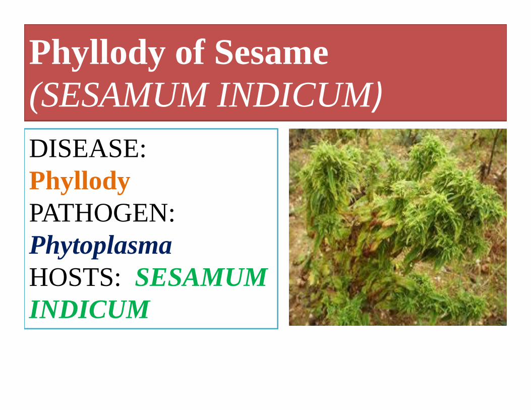
Phyllody of Sesame (SESAMUM INDICUM)DISEASE:Phyllody PATHOGEN:PhytoplasmaHOSTS: SESAMUM INDICUM
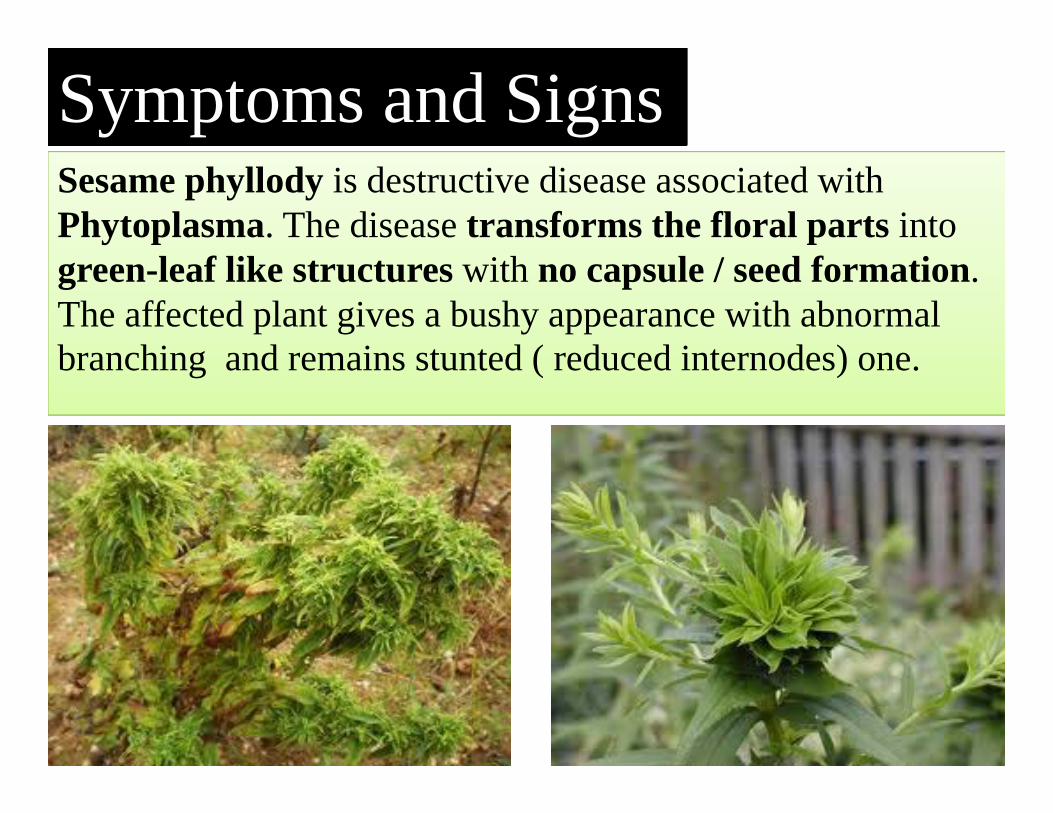
Sesame phyllody is destructive disease associated with Phytoplasma. The disease transforms the floral parts into green-leaf like structures with no capsule / seed formation. The affected plant gives a bushy appearance with abnormal branching and remains stunted ( reduced internodes) one.
Symptoms and Signs
64
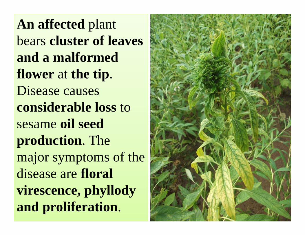
An affected plant bears cluster of leaves and a malformed flower at the tip. Disease causes considerable loss to sesame oil seed production. The major symptoms of the disease are floral virescence, phyllody and proliferation.

Pre disposing factors
• Dry weather,• Moderate temperature (250 C), • Low humidity (65%) and • Minimum rainfall (0.6mm)
Disease cycle
67
Phytoplasmas, formerly called mycoplasma-like organisms (MLOs), are a large group of obligate, intracellular, cell wallless parasites classified within the class Mollicutes. The pathogen has a wide host range and survives on hosts like Brassicacampestris var. toria, B. rapa, Cicerarietinum, Crotalaria sp., Trifolium sp., Arachis hypogea and some weed hosts. The disease is transmitted by jassid, Orosiusalbicinctus in a persistant manner.
Management strategy• Grow resistant varieties.• General sanitation
-Rogue out the infected plants periodically.- Remove all the reservoir and weed hosts.
• Delay sowing to reduce the vector population.• Crop rotation-Avoid growing sesamum near
cotton, groundnut and grain legumes.• Spray 2-3 times with Monocrotophos @
0.03% at flowering stage reduces the vector population.
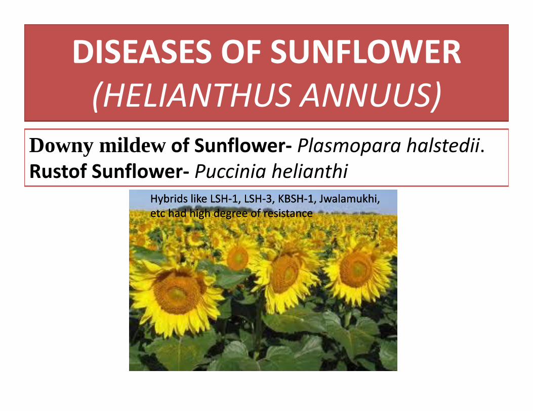
DISEASES OF SUNFLOWER (HELIANTHUS ANNUUS)
Downy mildew of Sunflower‐ Plasmopara halstedii.Rustof Sunflower‐ Puccinia helianthi
Hybrids like LSH‐1, LSH‐3, KBSH‐1, Jwalamukhi, etc had high degree of resistanceHybrids like LSH‐1, LSH‐3, KBSH‐1, Jwalamukhi, etc had high degree of resistance
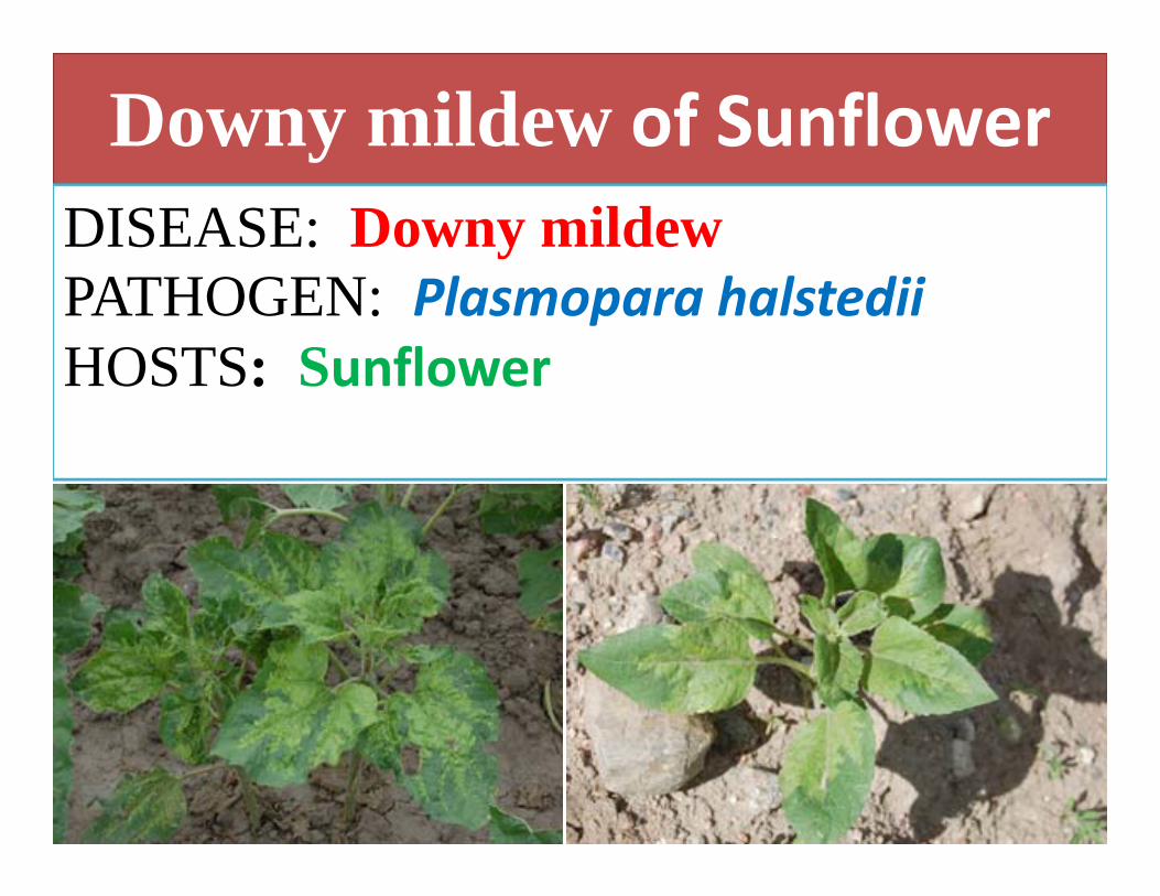
Downy mildew of SunflowerDISEASE: Downy mildew PATHOGEN: Plasmopara halstediiHOSTS: Sunflower

Two different types of symptoms: systemic and localized. Systemic infection is characterized by a thickening and yellowing of midribs of leaves at seedlings stage and affected plants remain severely stunted which ultimately die. Localized secondary infection takes place through windblown zoospores producing small-angular chlorotic lesions on the upper leaf surface. White cottony masses (fungal mycelium and spores) appear on the underside of infected leaves and are a good diagnostic sign of the disease. Flower heads of affected plants remain sterile.
Symptoms and Signs
71
Pre disposing factors
• Cool – humid weather• Intermittent rains.
The pathogen perpetuates in the form of oospores in soil. Oospores are round-to-oblong and thick-walled. They are highly resistant to adverse environmental conditions, and are capable of remaining dormant in soils for up to 10 years. When soils are saturated and cool (54-57°F), oospores can germinate and form zoosporangia that release motile zoospores that swim through soil water to infect seedling roots to cause systemic infections. Those surviving plants will then exhibit signs and symptoms producing inoculum for further spread to cause secondary infections.
Disease Cycle

Management strategy• Grow resistant hybrids like LSH‐1, LSH‐3, KBSH‐1, Jwalamukhi
• Follow spacing of 60x30cm or 45x30cm• Rogue out infected plants and destroy• Cropping sequence of sunflower followed by groundnut reduces the disease.
• Seed treatment with metalaxyl @ 0.6%• Spray Ridomil MZ( metalaxyl +mancozeb) @ 0.2% at 15 days intervals.
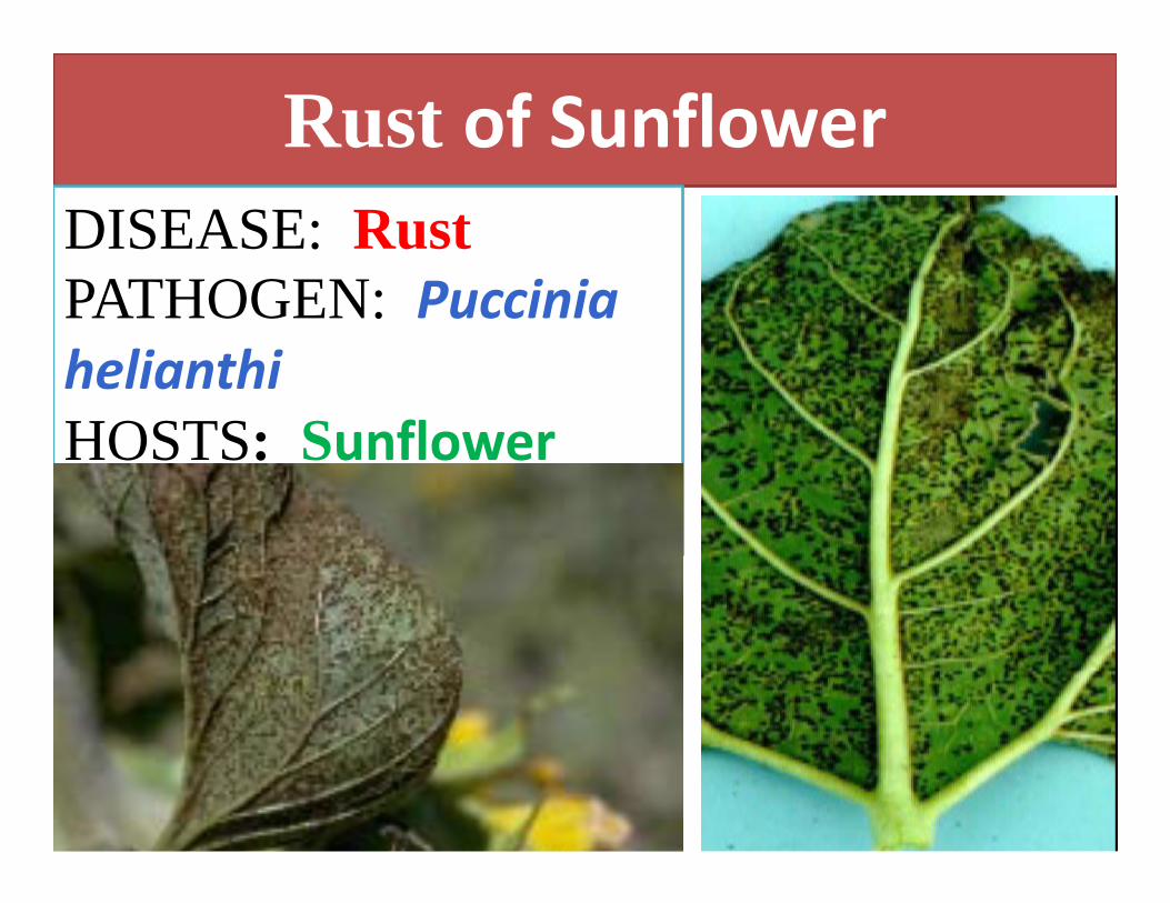
Rust of SunflowerDISEASE: Rust PATHOGEN: Puccinia helianthiHOSTS: Sunflower

Symptoms and Signs
76

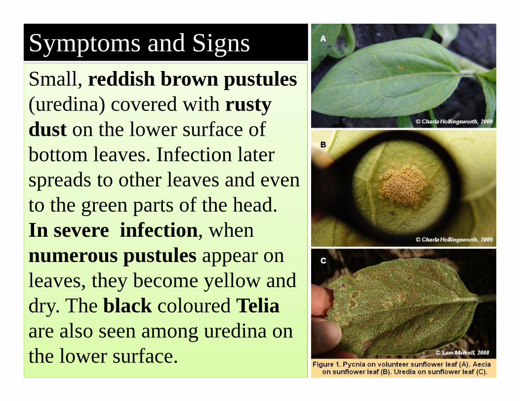
Small, reddish brown pustules (uredina) covered with rusty dust on the lower surface of bottom leaves. Infection later spreads to other leaves and even to the green parts of the head. In severe infection, when numerous pustules appear on leaves, they become yellow and dry. The black coloured Telia are also seen among uredina on the lower surface.
Symptoms and Signs
78
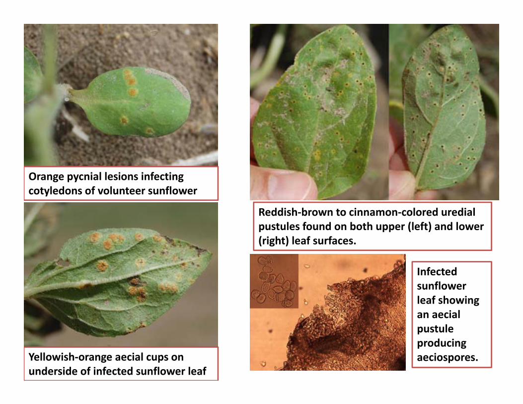
Orange pycnial lesions infecting cotyledons of volunteer sunflower
Yellowish‐orange aecial cups on underside of infected sunflower leaf
Reddish‐brown to cinnamon‐colored uredial pustules found on both upper (left) and lower (right) leaf surfaces.
Infected sunflower leaf showing an aecial pustule producing aeciospores.

Uredinia- urediopustules full ofreddish brown uredospores
Telia with full of teliospores
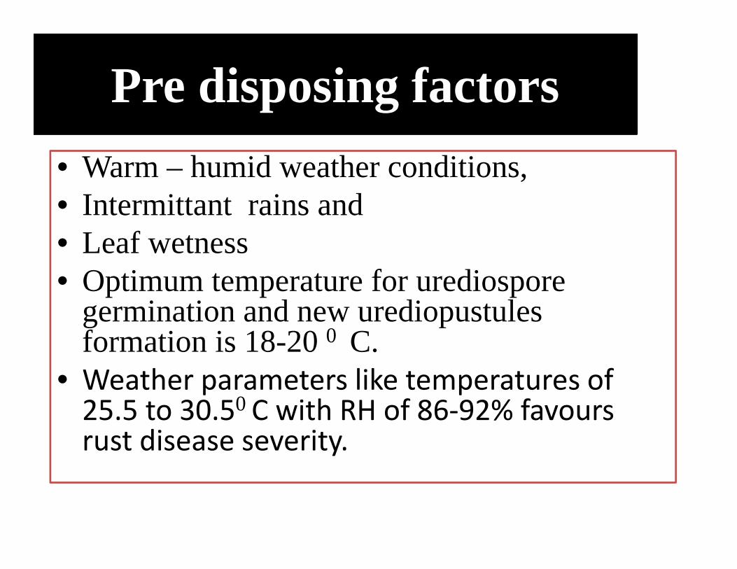
Pre disposing factors• Warm – humid weather conditions, • Intermittant rains and • Leaf wetness• Optimum temperature for urediospore
germination and new urediopustules formation is 18-20 0 C.
• Weather parameters like temperatures of 25.5 to 30.50 C with RH of 86‐92% favours rust disease severity.
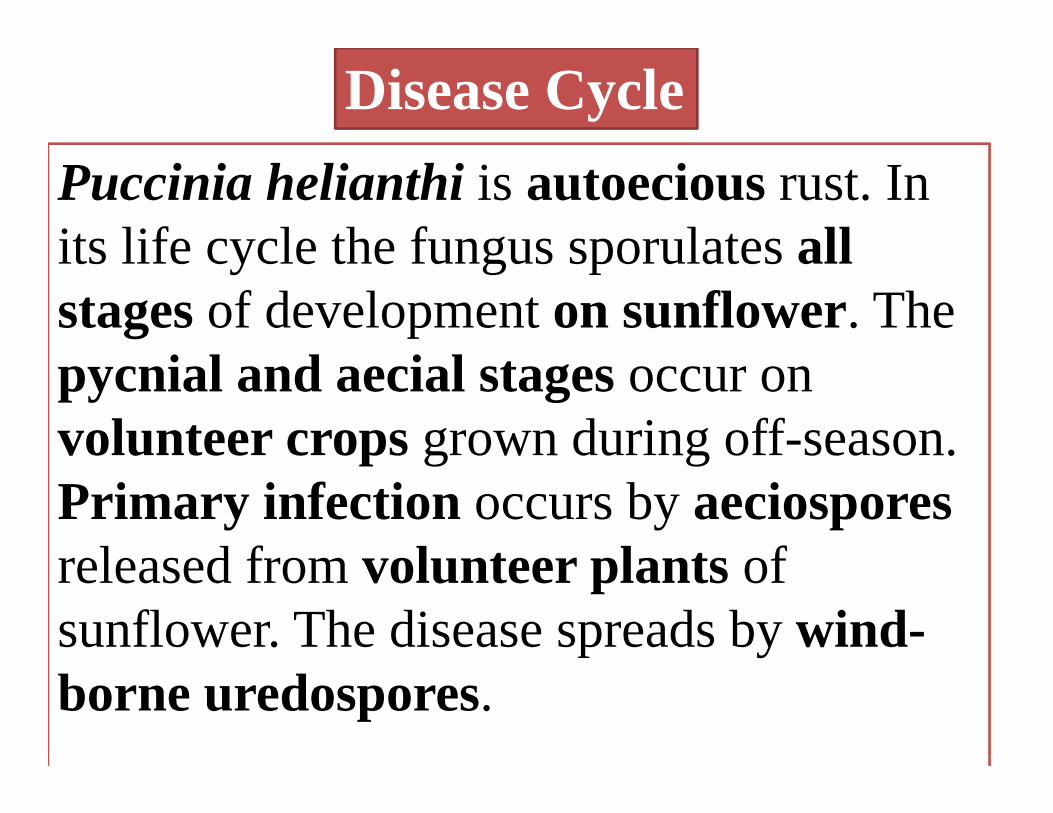
Puccinia helianthi is autoecious rust. In its life cycle the fungus sporulates all stages of development on sunflower. The pycnial and aecial stages occur on volunteer crops grown during off-season. Primary infection occurs by aeciospores released from volunteer plants of sunflower. The disease spreads by wind-borne uredospores.
Disease Cycle

Disease Cycle
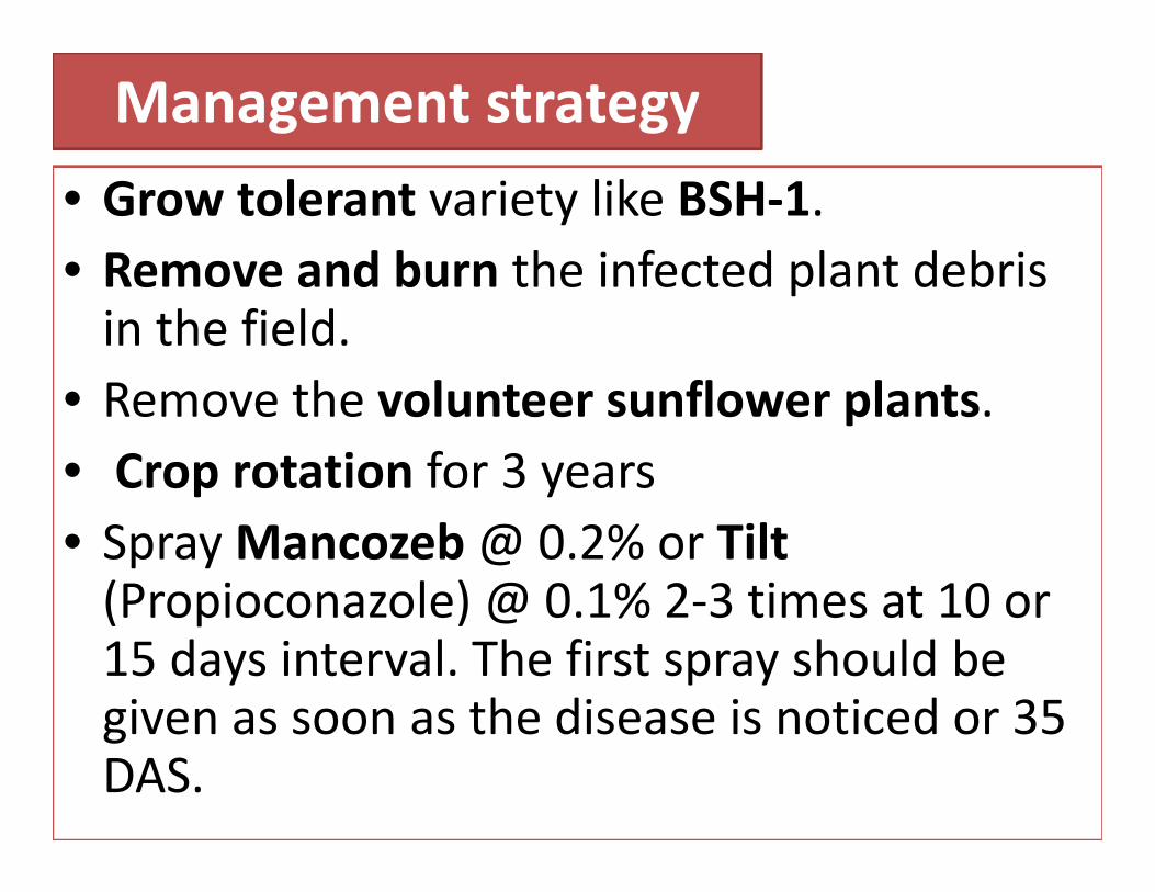
Management strategy• Grow tolerant variety like BSH‐1. • Remove and burn the infected plant debris in the field.
• Remove the volunteer sunflower plants.• Crop rotation for 3 years• Spray Mancozeb@ 0.2% or Tilt (Propioconazole) @ 0.1% 2‐3 times at 10 or 15 days interval. The first spray should be given as soon as the disease is noticed or 35 DAS.

Diseases of Cotton (Gossypiumhirsutum L.)
Angular leaf spot of cotton-Xanthomonas campestris pv. MalvacearumFusarium wilt-Fusarium oxysporum f.sp. vasinfectumVerticillium wiltVerticillium dahliaeLeaf curl of cotton
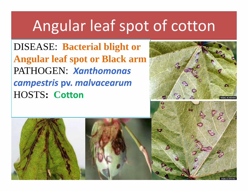
Angular leaf spot of cottonDISEASE: Bacterial blight or Angular leaf spot or Black armPATHOGEN: Xanthomonas campestris pv. malvacearumHOSTS: Cotton
p g g p
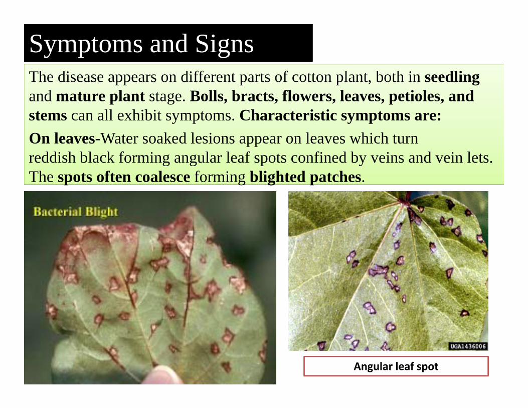
The disease appears on different parts of cotton plant, both in seedling and mature plant stage. Bolls, bracts, flowers, leaves, petioles, and stems can all exhibit symptoms. Characteristic symptoms are:On leaves-Water soaked lesions appear on leaves which turn reddish black forming angular leaf spots confined by veins and vein lets. The spots often coalesce forming blighted patches.
Symptoms and Signs
87Angular leaf spot
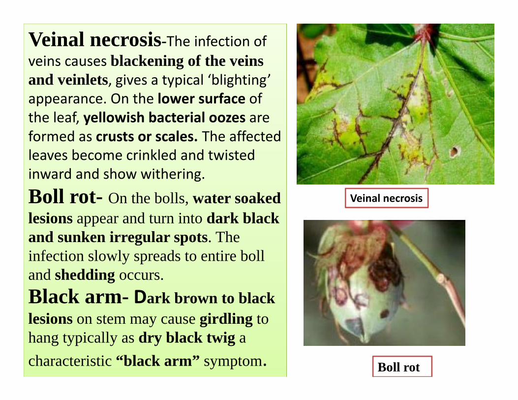
Veinal necrosis
Boll rot
Veinal necrosis-The infection of veins causes blackening of the veins and veinlets, gives a typical ‘blighting’ appearance. On the lower surface of the leaf, yellowish bacterial oozes are formed as crusts or scales. The affected leaves become crinkled and twisted inward and show withering.Boll rot- On the bolls, water soaked lesions appear and turn into dark black and sunken irregular spots. The infection slowly spreads to entire boll and shedding occurs.Black arm- Dark brown to black lesions on stem may cause girdling to hang typically as dry black twig a characteristic “black arm” symptom.
Pre disposing factors
• Optimum soil temperature of 280 C, • High atmospheric temperature of 30‐400 C, • Relative humidity of 85 per cent,• Potassium deficiency in soil. • Rain followed by bright sunshine during the months of October and November are highly favourable.
The bacterium survives on infected plant debris in soil for several years. The bacterium is also seed-borne and remains in the form of slimy mass on seed coat. It multiplies soon after the seed is sown and infects the seedling to cause seedling blights. Volunteer plants also provide a source of primary infection. The bacterium also attacks other hosts like Thurbaria thespesioides, Eriodendronanfructuosum and Jatropha curcas. The primary infection starts mainly from the seed-borne bacterium. The secondary spread of the bacteria may be through wind, wind blown rain splash, irrigation water, insects and other implements. The bacterium enters through natural openings or insect caused wounds.
Disease Cycle
Management strategy• Host resistance-Grow resistant varieties like HG-9, BJA 592, G-
27, Sujatha, 1412 and CRH 71. Gossypium herbaceum and G. arboreum are almost immune. G. barbadense, G. hirsutum, G. herbaceum var typicum and G. herbaceum var acerifoliumhave considerable resistance.
• General sanitation-Remove and destroy the infected plant debris. Also rogue out the volunteer cotton plants and weed hosts.
• Crop rotation -Follow with non-host crops.• Cultural practices-Early thinning, good tillage, early irrigation,
early earthing up and addition of potash to the soil reduces disease incidence.
• Chemical control-Treat the seeds with Carboxin at 2 g/kg seed or soak the seeds in 1000 ppm Streptomycin sulphate overnight or treat the seed with hot water at 52-560 C for 10-15 minutes.
• Spray with Streptomycin sulphate (Agrimycin 100) @ .05% or Copper oxychloride at 0.3%.
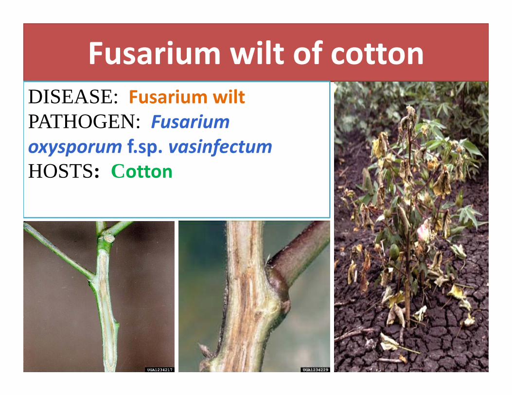
Fusarium wilt of cottonDISEASE: Fusarium wilt PATHOGEN: Fusarium oxysporum f.sp. vasinfectumHOSTS: Cotton
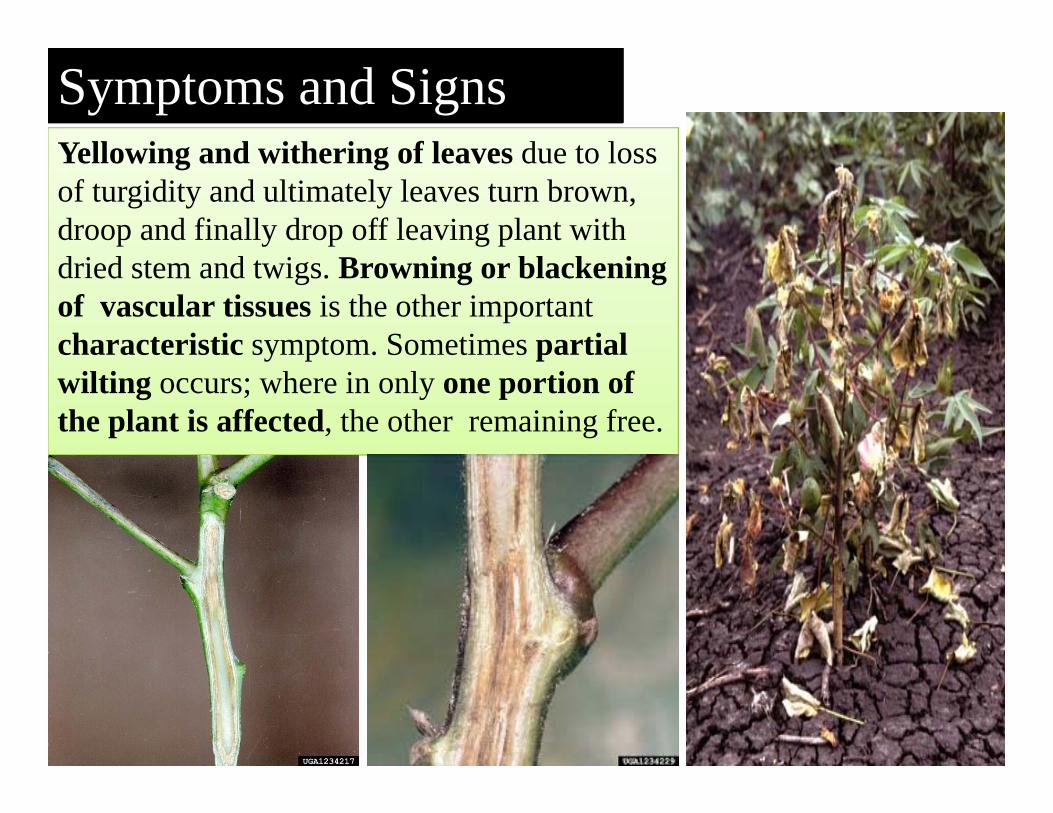
Yellowing and withering of leaves due to loss of turgidity and ultimately leaves turn brown, droop and finally drop off leaving plant with dried stem and twigs. Browning or blackening of vascular tissues is the other important characteristic symptom. Sometimes partial wilting occurs; where in only one portion of the plant is affected, the other remaining free.
Symptoms and Signs
93

Chlamydospores
Culture of Fusarium oxysporum
Macro conidia, micro conidia, and chlamydospores of Fusarium oxysporum
Pre disposing factors• Soil temperature of 20‐300C• Hot and dry periods followed by rains• Heavy black soils with an alkaline reaction, increased doses of nitrogen and phosphaticfertilizers and
• Wounds caused by nematode (Meloidogyne incognita) and grubs of Ashweevil (Myllocerus pustulatus).
The fungus can survive in soil as saprophytefor many years and chlamydospores act as resting spores. The pathogen is both externally and internally seed-borne. The primary infection is mainly from dormant hyphae and chlamydospores in the soil. The secondary spread is through conidia and chlamydospores which are disseminated by irrigation water.
Disease Cycle
Management strategy• Host resistance- Grow disease resistant varieties of G. hirsutum
and G. barbadense like Varalakshmi, Vijaya, Pratap, Jayadhar, Jarila, Jyothi, G 22 and Verum.
• General sanitation- Remove and burn the infected plant debris in the soil after deep summer ploughing.
• Crop rotation -Follow with non-host crops.• Cultural practices- Apply heavy doses of farm yard
manure or other organic manures at 10 t/ha. Follow mixed cropping with non-host plants to lower the soil temperature below 200C by providing shade. Soil amendment with zinc.Apply increased doses of potash with a balanced dose of nitrogenous and phosphatic fertilizers.
• Chemical control-Treat the seeds with Carboxin at 2 g/kg seed or treat the seed with Carboxin or Chlorothalonil at 4 g/kg.

Verticillium wilt of cottonDISEASE: Verticillium wiltPATHOGEN: Verticillium dahliaeHOSTS: Cotton
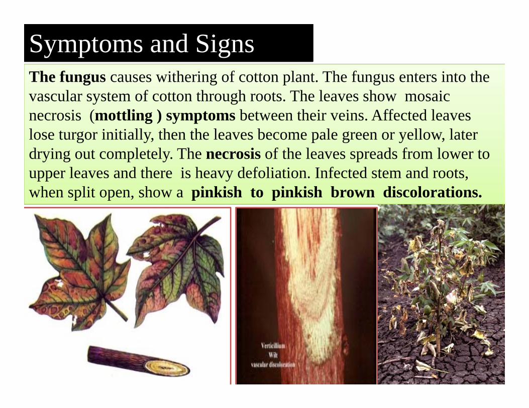
The fungus causes withering of cotton plant. The fungus enters into the vascular system of cotton through roots. The leaves show mosaic necrosis (mottling ) symptoms between their veins. Affected leaves lose turgor initially, then the leaves become pale green or yellow, later drying out completely. The necrosis of the leaves spreads from lower to upper leaves and there is heavy defoliation. Infected stem and roots, when split open, show a pinkish to pinkish brown discolorations.
Symptoms and Signs
99

Verticillium dahliae
Pre disposing factors
• Low temperature of 15‐200C• Low lying and ill‐drained soils• Heavy soils with alkaline reaction and • Heavy doses of nitrogenous fertilizers favours the disease.
The fungus also infects the other hosts like brinjal, chilli, tobacco and bhendi. The fungus can survive in the infected plant debris and in soils as microsclerotia upto 14 years. The seeds also carry the microsclerotia and conidia in the fuzz. The primary spread is through the micro sclerotia or conidia in the soil. The secondary spread is through the contact of diseased roots to healthy ones and through dissemination of infected plant parts through irrigation water and other implements.
Disease Cycle
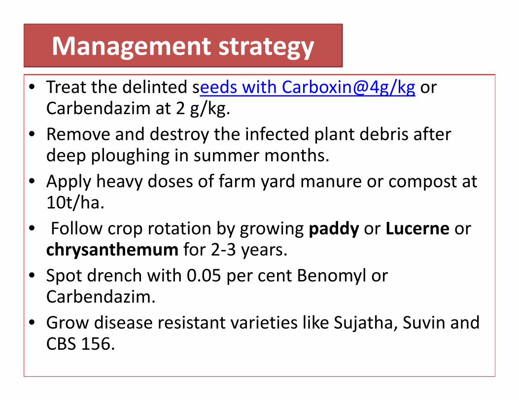
Management strategy• Treat the delinted seeds with Carboxin@4g/kg or Carbendazim at 2 g/kg.
• Remove and destroy the infected plant debris after deep ploughing in summer months.
• Apply heavy doses of farm yard manure or compost at 10t/ha.
• Follow crop rotation by growing paddy or Lucerne or chrysanthemum for 2‐3 years.
• Spot drench with 0.05 per cent Benomyl or Carbendazim.
• Grow disease resistant varieties like Sujatha, Suvin and CBS 156.
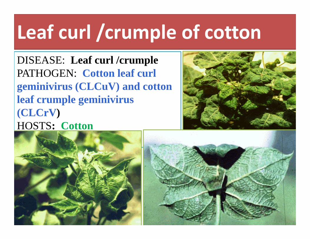
Leaf curl /crumple of cottonDISEASE: Leaf curl /crumplePATHOGEN: Cotton leaf curl geminivirus (CLCuV) and cotton leaf crumple geminivirus (CLCrV)HOSTS: Cotton
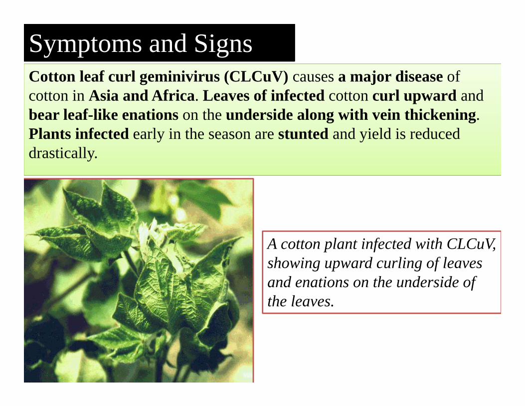
Cotton leaf curl geminivirus (CLCuV) causes a major disease of cotton in Asia and Africa. Leaves of infected cotton curl upward and bear leaf-like enations on the underside along with vein thickening. Plants infected early in the season are stunted and yield is reduced drastically.
Symptoms and Signs
A cotton plant infected with CLCuV, showing upward curling of leaves and enations on the underside of the leaves.
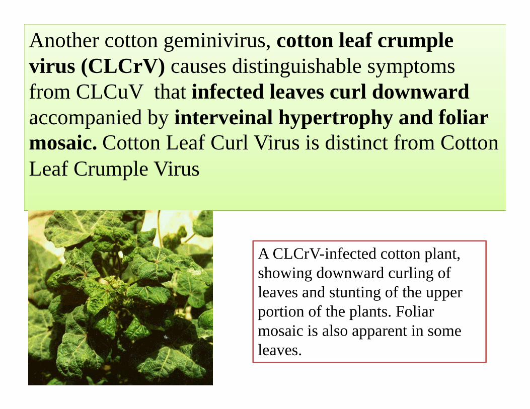
Another cotton geminivirus, cotton leaf crumple virus (CLCrV) causes distinguishable symptoms from CLCuV that infected leaves curl downward accompanied by interveinal hypertrophy and foliar mosaic. Cotton Leaf Curl Virus is distinct from Cotton Leaf Crumple Virus
A CLCrV-infected cotton plant, showing downward curling of leaves and stunting of the upper portion of the plants. Foliar mosaic is also apparent in some leaves.
Pre disposing factors
• Both CLCrV and CLCuV infect dicotyledonous plants and
• Are whitefly‐transmitted.

Management strategy
• Grow resistant varieties.• General sanitation
-Rogue out the infected plants periodically.- Remove all the reservoir and weed hosts
• Spray 2-3 times with Monocrotophos @ 0.03% reduces the vector population.