disease and immunity in caribbean and indo-pacific ... on each of 7 reefs surveyed in the florida...
TRANSCRIPT
MARINE ECOLOGY PROGRESS SERIESMar Ecol Prog Ser
Vol. 266: 273–302, 2004 Published January 30
INTRODUCTION
Coral reefs are in severe decline. The most reliableestimates suggest that worldwide 27% have alreadybeen lost, with another 16% at serious risk of loss(Wilkinson 2002). Coral disease is thought to be amajor cause for this decline (Dustan 1999, Porter et al.2001). Epizootics have been reported for several coralspecies (Goreau et al. 1998, Richardson 1998, Richard-son et al. 1998a,b, Harvell et al. 1999, 2001, Porter et al.2001) and evidence is mounting of substantial declinesin the biodiversity and abundance of reef-buildingcorals worldwide (Hayes & Goreau 1998, Porter &Tougas 2001, Wilkinson 2002). Within the Caribbean,populations of elkhorn and staghorn corals, Acroporapalmata and A. cervicornis, are being decimated bydisease (Gladfelter 1982, Bythell & Sheppard 1993,Aronson & Precht 1997, 2001, Aronson et al. 1998,2002, Greenstein et al. 1998, Miller et al. 2002, Patter-son et al. 2002), with losses of A. palmata in the Florida
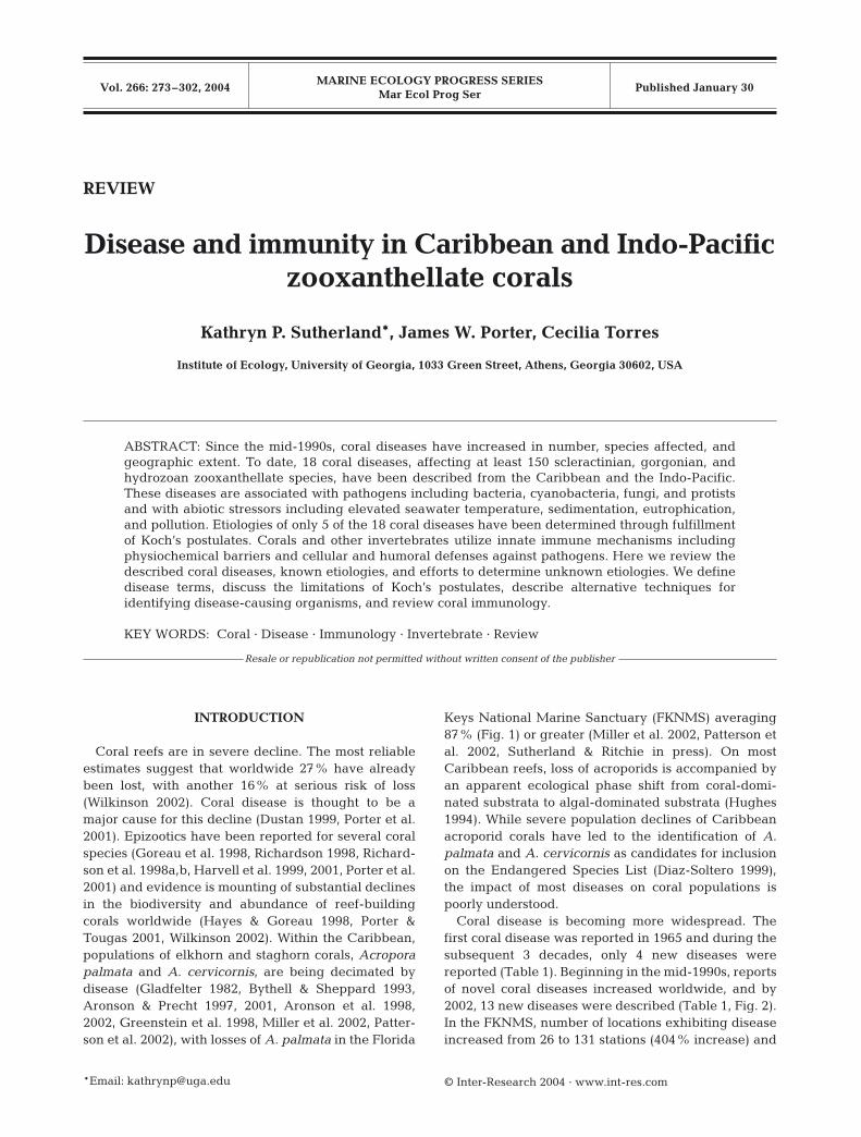
Keys National Marine Sanctuary (FKNMS) averaging87% (Fig. 1) or greater (Miller et al. 2002, Patterson etal. 2002, Sutherland & Ritchie in press). On mostCaribbean reefs, loss of acroporids is accompanied byan apparent ecological phase shift from coral-domi-nated substrata to algal-dominated substrata (Hughes1994). While severe population declines of Caribbeanacroporid corals have led to the identification of A.palmata and A. cervicornis as candidates for inclusionon the Endangered Species List (Diaz-Soltero 1999),the impact of most diseases on coral populations ispoorly understood.
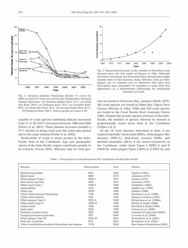
Coral disease is becoming more widespread. Thefirst coral disease was reported in 1965 and during thesubsequent 3 decades, only 4 new diseases werereported (Table 1). Beginning in the mid-1990s, reportsof novel coral diseases increased worldwide, and by2002, 13 new diseases were described (Table 1, Fig. 2).In the FKNMS, number of locations exhibiting diseaseincreased from 26 to 131 stations (404% increase) and
© Inter-Research 2004 · www.int-res.com*Email: [email protected]
REVIEW
Disease and immunity in Caribbean and Indo-Pacificzooxanthellate corals
Kathryn P. Sutherland*, James W. Porter, Cecilia Torres
Institute of Ecology, University of Georgia, 1033 Green Street, Athens, Georgia 30602, USA
ABSTRACT: Since the mid-1990s, coral diseases have increased in number, species affected, andgeographic extent. To date, 18 coral diseases, affecting at least 150 scleractinian, gorgonian, andhydrozoan zooxanthellate species, have been described from the Caribbean and the Indo-Pacific.These diseases are associated with pathogens including bacteria, cyanobacteria, fungi, and protistsand with abiotic stressors including elevated seawater temperature, sedimentation, eutrophication,and pollution. Etiologies of only 5 of the 18 coral diseases have been determined through fulfillmentof Koch’s postulates. Corals and other invertebrates utilize innate immune mechanisms includingphysiochemical barriers and cellular and humoral defenses against pathogens. Here we review thedescribed coral diseases, known etiologies, and efforts to determine unknown etiologies. We definedisease terms, discuss the limitations of Koch’s postulates, describe alternative techniques foridentifying disease-causing organisms, and review coral immunology.
KEY WORDS: Coral · Disease · Immunology · Invertebrate · Review
Resale or republication not permitted without written consent of the publisher
Mar Ecol Prog Ser 266: 273–302, 2004
number of coral species exhibiting disease increasedfrom 11 to 36 (218% increase) between 1996 and 2000(Porter et al. 2001). These disease increases parallel a37% decline in living coral over the same time periodand at the same stations (Porter et al. 2002).
Biodiversity of corals is much greater in the Indo-Pacific than in the Caribbean. Age and geographicextent of the Indo-Pacific region contributes greatly toits richness (Veron 1995). Whereas only 53 coral spe-
cies are found in Discovery Bay, Jamaica (Wells 1973),362 coral species are found in Milne Bay, Papua NewGuinea (Werner & Allen 1998) and 350 coral speciesare found in the Great Barrier Reef, Australia (Veron1985). Despite the greater species richness of the Indo-Pacific, the number of species affected by disease isproportionally much lower than in the Caribbean(Tables 2 & 3).
Of the 18 coral diseases described to date, 4 arereported globally: black band (BBD), white plague-likediseases (WPL-L), shut-down reaction (SDR), andskeletal anomalies (SKA); 9 are found exclusively inthe Caribbean: white band Types I (WBD I) and II(WBD II), white plague Types I (WPL I), II (WPL II), and
274
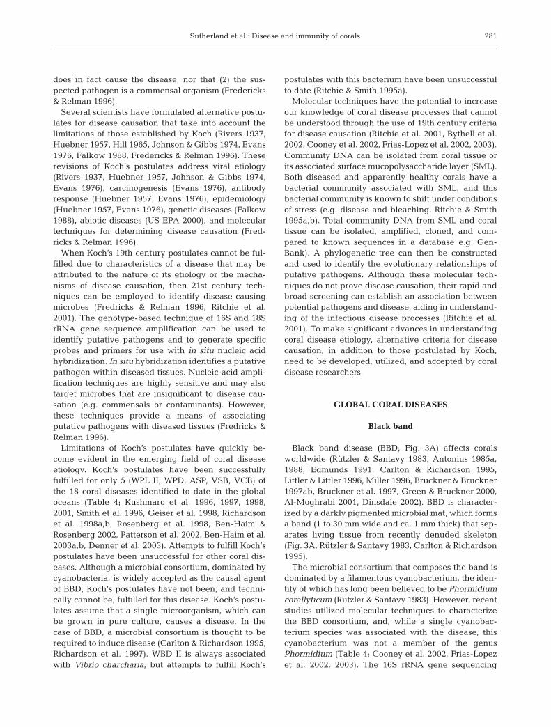
Fig. 1. Acropora palmata. Population decline (% cover) by2002 on each of 7 reefs surveyed in the Florida Keys NationalMarine Sanctuary. (h) Western Sambo Reef: 91%; (s) SandKey Reef: 100%; (n) Molasses Reef: 79%; (e) Carysfort Reef:89%; (s) Rock Key Reef: 58%; (h) Grecian Rocks Reef: 95%;
(y) Sombrero Reef: 100%. Data in graph are mean ± SD
Table 1. First report of coral diseases in the Caribbean and the Indo-Pacific
Disease Abbreviation Year Source
Skeletal anomalies SKA 1965 Squires (1965)Black band BBD 1973 Antonius (1973)White plague Type I WPL I 1977 Dustan (1977)Shut-down reaction SDR 1977 Antonius (1977)White band Type I WBD I 1982 Gladfelter (1982)Aspergillosis ASP 1996 Smith et al. (1996)White pox WPD 1996 Holden (1996)Vibrio shiloi-induced bleaching VSB 1996 Kushmaro et al. (1996)Yellow blotch/band YBL 1997 Santavy & Peters (1997)White plague Type II WPL II 1998 Richardson et al. (1998a)White band Type II WBD II 1998 Ritchie & Smith (1998)Yellow band YBD 1998 Korrûbel & Riegl (1998)Dark spots DSD 1998 Goreau et al. (1998)Skeleton eroding band SEB 2000 Antonius & Lipscomb (2000)Fungal-protozoan syndrome FPS 2000 Cerrano et al. (2000)White plague Type III WPL III 2001 Richardson et al. (2001)Pink-line syndrome PLS 2001 Ravindran et al. (2001)Vibrio coralliilyticus-induced bleaching and disease VCB 2002 Ben-Haim & Rosenberg (2002)
1965 1970 1975 1980 1985 1990 1995 2000 20050
5
10
15
20
Co
ral D
isea
se (n
umb
er)
Year
Fig. 2. Exponential increase in the number of described coraldiseases since the first report of disease in 1965. Althoughincreased awareness and increased observational time mightexplain some of this increase, many diseases, such as whiteplague, are so common and so distinctive that their firstdescription may reasonably be assumed to mark their firstappearance as a phenomenon influencing the population
dynamics of corals

Sutherland et al.: Disease and immunity of corals 275
Table 2. Caribbean scleractinian, hydrozoan, and gorgonian coral species affected by diseases (total number of reported speciesaffected by each disease and total number of reported diseases affecting each species). For WPL and WBD: I = Type I, II = Type II,
III = Type III, X = type not determined. See Table 1 for disease definitions
BBD WPL SDR SKA ASP WBD WPD YBL DSD No. of diseases
ScleractiniansAgaricia agaricites I, II X X 4Agaricia fragilis X 1Agaricia lamarcki II X 2Agaricia tenuifolia X 1Acropora cervicornis X X I,II 4Acropora palmata X X X I X 5Colpophyllia natans X I,II,III X X X 7Cladocora arbuscula X 1Dendrogyra cylindrus II 1Dichocoenia stokesi X II X X 4Diploria clivosa X X 2Diploria labyrinthiformis X I,II X X X 6Diploria strigosa X II X X 4Eusmilia fastigiata II 1Favia fragum X X X X 4Isophyllastrea rigida I X 2Isophyllia sinuosa X 1Leptoseris cucullata X 1Madracis decactis X II 2Madracis formosa X 1Madracis mirabilis X II 2Manicina areolata II X 2Meandrina meandrites X II X 3Montastraea annularis X I,II,III X X X X 8Montastraea cavernosa X I,II X X 5Montastraea faveolata X I X X 4Montastraea franksi X X X X 4Mussa angulosa I 1Mycetophyllia aliciae X 1Mycetophyllia danaana X 1Mycetophyllia ferox I 1Mycetophyllia lamarkiana I 1Oculina diffusa X 1Porites astreoides X I X X 4Porites porites X X 2Scolymia cubensis X 1Siderastrea radians X X X 3Siderastrea siderea X I,II X X X 6Solenastrea bournoni II 1Solenastrea hyades X X X 3Stephanocoenia michelinii X I,II X 4Total 19 38 6 16 0 2 1 9 11
HydrozoansMillepora alcicornis II X 2Millepora complanata X 1Total 0 2 0 1 0 0 0 0 0
GorgoniansGorgonia flabellum X X 2Gorgonia ventalina X X X 3Gorgonia sp. X 1Plexaura flexuosa X 1Plexaura homomalla X 1Plexaura sp. X 1Plexaurella sp. X 1Plexaurella homomalla X 1Plexaurella flexuosa X 1Pseudoplexaura spp. X X 2Pseudoplexaura porosa X 1Pseudopterogorgia acerosa X 1Pseudopterogorgia americana X X 2Total 6 0 0 5 7 0 0 0 0
Total no. of species 25 40 6 22 7 2 1 9 11

Mar Ecol Prog Ser 266: 273–302, 2004276
Table 3. Indo-Pacific scleractinian and gorgonian coral species affected by diseases (total number of reported species affected by each disease and total number of reported diseases affecting each species). See Table 1 for disease definitions
BBD WPL-L SKA VSB VCB SEB YBD PLS FPS No. of diseases
ScleractiniansAcropora aspera X 1Acropora capillaris X 1Acropora clathrata X X X X X 5Acropora cytherea X 1Acropora downingi X X X X 4Acropora florida X X X X 4Acropora formosa X X X 3Acropora gemmifera X 1Acropora hemprichi X 1Acropora humilis X X X 3Acropora hyacinthus X X X 3Acropora intermedia X 1Acropora microclados X 1Acropora microphthalma X 1Acropora millepora X 1Acropora monticulosa X 1Acropora nobilis X X X X 4Acropora palifera X X 2Acropora pharaonis X X X 3Acropora robusta X 1Acropora sarmentosa X 1Acropora squarrosa X 1Acropora tenuis X X 2Acropora valenciennesi X 1Acropora valida X X X X 4Acropora variabilis X 1Acropora virgata X 1Alveopora gigas X 1Astreopora myriophthalma X 1Cladocora caespitosa X 1Coscinarea monile X 1Cyphastrea chalcidicum X 1Cyphastrea microphthalma X 1Cyphastrea serailia X 1Echinophyllia aspera X 1Echinopora gemmacea X 1Enallopsammia rostrata X 1Favia favus X X 2Favia matthaii X 1Favia pallida X X 2Favia stelligera X X X 3Favia valenciennesii X 1Favites abdita X 1Favites pentagona X X 2Goniastrea pectinata X X 2Goniastrea retiformis X X X 3Goniopora columna X 1Goniopora somaliensis X 1Goniopora sp. X 1Hydnophora microconos X X X 3Leptastrea purpurea X 1Leptoria phrygia X X 2Leptoseris explanata X 1Leptoseris glabra X 1Leptoseris mycetoseroides X 1Lobophyllia corymbosa X 1Madrepora kauaiensis X 1Madrepora oculata X 1Montipora aequituberculata X X 2Montipora ehrenbergi X 1Montipora florida X 1Montipora foliosa X 1Montipora informis X 1Montipora monasteriata X 1

Sutherland et al.: Disease and immunity of corals
III (WPL III), aspergillosis (ASP), white pox (WPD), yel-low blotch/band (YBL), dark spots (DSD); and 6 areapparently endemic to the Indo-Pacific: yellow band(YBD), skeleton eroding band (SEB), pink-line syn-drome (PLS), fungal-protozoan syndrome (FPS),Vibrio shiloi-induced bleaching (VSB), V. coralliilyticus-induced bleaching and disease (VCB). WPL-L in theIndo-Pacific may or may not be etiologically related tothe 3 Caribbean WPL diseases. Etiologies and mecha-nisms of tissue death of the majority of coral diseasesare not understood (Richardson 1998).
Tissue loss or damage from predation is often impos-sible to distinguish from tissue loss or damage fromdisease. To the human eye, there are not many ways inwhich coral tissue can exhibit signs of stress. Coralli-vores, including fishes, gastropods, and other inverte-brates, produce predation scars that are easily con-
fused with disease signs. The best method to distin-guish between predation and disease is to observe theprogress of the condition in the absence of predation,via predator exclusion in situ or predator removalunder laboratory conditions. A number of coral abnor-malities, some of which have been described in the lit-erature as coral diseases, are likely associated withpredation rather than disease. For example, predationby the stoplight parrotfish Sparisoma viride, was ini-tially described as a coral disease termed rapid wastingsyndrome (Cervino et al. 1997). Subsequent investiga-tions identified fish predation as the primary cause ofskeletal loss (Bruckner & Bruckner 2002), but the pos-sible role of fungi in the dissolution of the skeleton(Cervino et al. 1997, Hayes & Goreau 1998) has notbeen fully vetted. Further, in some cases, disease signsattributed to coral disease (e.g. WPL, WBD, and WPD)
277
Table 3 (continued)
BBD WPL-L SKA VSB VCB SEB YBD PLS FPS No. of diseases
Scleractinians (continued)Montipora patula X 1Montipora verrucosa X X 2Montipora sp. X X 2Mycedium elephantotus X 1Oculina patagonica X 1Pavona gigantea X 1Pachyseris gemmae X 1Pachyseris rugosa X 1Platygyra daedalea X 1Platygyra lamellina X X 2Platygyra pini X 1Platygyra sinensis X 1Pocillopora damicornis X X X X 4Pocillopora eydouxi X 1Pocillopora verrucosa X X X 3Pocillopora meandrina X 1Podabacia crustacea X 1Porites sp. X 1Porites compressa X X 2Porites harrisoni X 1Porites lichen X 1Porites lobata X 1Porites lutea X X X X X 5Porites nodifera X 1Pratzia mirabilis X 1Stylophora erthyaea X 1Stylophora pistillata X X X 3Symphyllia radians X 1Turbinaria mesenterina X 1Turbinaria reniformis X X 2Verrillofungia concinna X 1Total 45 38 24 1 1 24 12 2 1
GorgoniansCorallium rubrum X 1Eunicella cavolini X 1Eunicella singularis X 1Eunicella verrucosa X 1Leptogorgia sarmentosa X 1Paramuricea clavata X 1Total 0 0 0 0 0 0 0 0 6
Total no. of species 45 38 24 1 1 24 12 2 7
Mar Ecol Prog Ser 266: 273–302, 2004
may be difficult to distinguish from predation scarsproduced by corallivores including the gastropod Coral-liophila abbreviata, and the fire worm Hermodice caru-culata (Patterson et al. 2002).
In order to facilitate an understanding of disease pro-cesses and causation in corals, it is necessary to under-stand general disease terminology and coral immunity.The objectives of this paper are to: (1) define diseaseterminology and relate these terms to coral disease,(2) discuss the process of proving disease causation,(3) review described coral diseases, known etiologies,and efforts to determine unknown etiologies, (4) illus-trate each of the known Caribbean coral diseases, and(5) review coral immunology.
DISEASE TERMINOLOGY
A disease is any impairment (interruption, cessation,proliferation, or other disorder) of vital body functions,systems, or organs (Stedman 2000). The term syn-drome is synonymous with disease (Stedman 2000).Etiology is analysis of causes, development, and conse-quences of a disease (Kinne 1980). Disease causation
(i.e. etiology) may be attributed to pathogens, environ-mental stressors, or a combination of biotic and abioticfactors. Biotic diseases are caused by pathogenicmicroorganisms such as viruses, bacteria, fungi, andprotists and are often species-specific (Peters 1997)and infectious (Kinne 1980). Abiotic diseases resultfrom both natural and human-induced environmentalstressors including change in ambient conditions orexposure to pollutants. Biotic and abiotic diseases areoften closely related. Biotic diseases may be associ-ated with environmental stressors that: (1) hinder theresistence of host organisms, (2) promote growth andvirulence of pathogens, (3) trigger the pathogenicprocess, or (4) increase the rate of disease transmission(Kushmaro et al. 1996, 1998, Peters 1997, Toren etal. 1998, Ben-Haim et al. 1999, 2003b, Alker et al. 2001,Banin et al. 2001a, Israely et al. 2001, Ben-Haim &Rosenberg 2002, Kuta & Richardson 2002, Richardson& Kuta 2003). Abiotic diseases may be exacerbated bysecondary opportunistic infections (Peters 1997).
Etiologies of 11 diseases affecting scleractinian andgorgonian corals may involve pathogens includingbacteria, cyanobacteria, fungi, and protists (Table 4).Ten diseases are associated with abiotic stressors in-
278
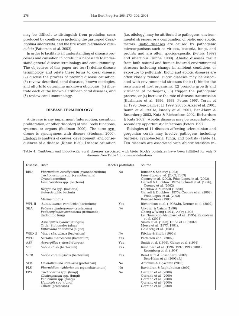
Table 4. Caribbean and Indo-Pacific coral diseases associated with biota. Koch’s postulates have been fullfilled for only 5 diseases. See Table 1 for disease definitions
Disease Biota Koch’s postulates Source
BBD Phormidium corallyticum (cyanobacterium) No Rützler & Santavy (1983)Trichodesmium spp. (cyanobacteria) Frias-Lopez et al. (2002, 2003)Cyanobacterium Cooney et al. (2002), Frias-Lopez et al. (2003)Desulvovibrio spp. (bacteria) Garrett & Ducklow (1975), Schnell et al. (1996),
mCooney et al. (2002)Beggiatoa spp. (bacteria) Ducklow & Mitchell (1979b)Heterotrophic bacteria Garrett & Ducklow (1975), Cooney et al. (2002),
mFrias-Lopez et al. (2002)Marine fungus Ramos-Flores (1983)
WPL II Aurantimonas coralicida (bacterium) Yes Richardson et al. (1998a,b), Denner et al. (2002)SKA Petrarca madreporae (crustacean) No Grygier & Cairns (1996)
Podocotyloides stenometra (trematode) Cheng & Wong (1974), Aeby (1998)Endolithic fungi Le Champion-Alsumard et al. (1995), Ravindran
met al. (2001)Aspergillus sydowii (fungus) Smith et al. (1998), Dube et al. (2002)Order Siphonales (algae) Morse et al. (1977, 1981),Entocladia endozoica (algae) Goldberg et al. (1984)
WBD II Vibrio charcharia (bacterium) No Ritchie & Smith (1995a)WPD Serratia marcescens (bacterium) Yes Patterson et al. (2002)ASP Aspergillus sydowii (fungus) Yes Smith et al. (1996), Geiser et al. (1998)VSB Vibrio shiloi (bacterium) Yes Kushmaro et al. (1996, 1997, 1998, 2001),
mRosenberg et al. (1998)VCB Vibrio coralliilyticus (bacterium) Yes Ben-Haim & Rosenberg (2002),
mBen-Haim et al. (2003a,b)SEB Halofolliculina corallasia (protozoan) No Antonius & Lipscomb (2000)PLS Phormidium valderianum (cyanobacterium) No Ravindran & Raghukumar (2002)FPS Trichoderma spp. (fungi) No Cerrano et al. (2000)
Clodosporium spp. (fungi) Cerrano et al. (2000)Penicillum spp. (fungi) Cerrano et al. (2000)Humicola spp. (fungi) Cerrano et al. (2000)Ciliate (protozoan) Cerrano et al. (2000)
Sutherland et al.: Disease and immunity of corals
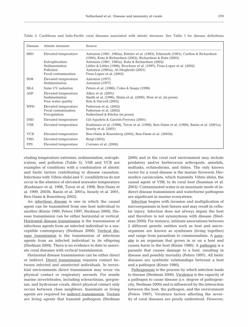
cluding temperature extremes, sedimentation, eutroph-ication, and pollution (Table 5). VSB and VCB areexamples of conditions with a combination of abioticand biotic factors contributing to disease causation.Infections with Vibrio shiloi and V. coralliilyticus do notoccur in the absence of elevated seawater temperature(Kushmaro et al. 1998, Toren et al. 1998, Ben-Haim etal. 1999, 2003b, Banin et al. 2001a, Israely et al. 2001,Ben-Haim & Rosenberg 2002).
An infectious disease is one in which the causalagent can be transmitted from one host individual toanother (Kinne 1980, Peters 1997, Stedman 2000). Dis-ease transmission can be either horizontal or vertical.Horizontal disease transmission is the transmission ofinfectious agents from an infected individual to a sus-ceptible contemporary (Stedman 2000). Vertical dis-ease transmission is the transmission of infectiousagents from an infected individual to its offspring(Stedman 2000). There is no evidence to date to associ-ate coral diseases with vertical transmisssion.
Horizontal disease transmisssion can be either director indirect. Direct transmission requires contact be-tween infected and uninfected individuals. In terres-trial environments direct transmission may occur viaphysical contact or respiratory aerosols. For sessilemarine invertebrates, including scleractinian, gorgon-ian, and hydrozoan corals, direct physical contact onlyoccurs between close neighbors. Inanimate or livingagents are required for indirect transmission. Vectorsare living agents that transmit pathogens (Stedman
2000) and in the coral reef environment may includepredatory and/or herbivorous arthropods, annelids,mollusks, echinoderms, and fishes. The only knownvector for a coral disease is the marine fireworm Her-modice carunculata, which transmits Vibrio shiloi, thecausal agent of VSB, to its coral host (Sussman et al.2003). Contaminated water is an inanimate mode of in-direct disease transmission and waterborne pathogensare significant in marine ecosystems.
Infection begins with invasion and multiplication ofmicroorganisms in host tissues and may result in cellu-lar injury. Infection does not always impair the hostand therefore is not synonymous with disease (Sted-man 2000). For instance, intimate associations between2 different genetic entities such as host and micro-organism are known as symbioses (living together)and range from parasitism to commensalism. A para-site is an organism that grows in or on a host andcauses harm to the host (Kinne 1980). A pathogen is aparasite that causes damage to a host, resulting indisease and possibly mortality (Peters 1997). All bioticdiseases are symbiotic relationships between a hostand a pathogen (Kinne 1980).
Pathogenesis is the process by which infection leadsto disease (Stedman 2000). Virulence is the capacity ofa pathogen to cause disease (i.e. degree of pathogeni-city, Stedman 2000) and is influenced by the interactionbetween the host, the pathogen, and the environment(Peters 1997). Virulence factors affecting the sever-ity of coral diseases are poorly understood. However,
279
Table 5. Caribbean and Indo-Pacific coral diseases associated with abiotic stressors. See Table 1 for disease definitions
Disease Abiotic stressors Source
BBD Elevated temperature Antonius (1981, 1985a), Rützler et al. (1983), Edmunds (1991), Carlton & Richardson(1995), Kuta & Richardson (2002), Richardson & Kuta (2003)
Eutrophication Antonius (1981, 1985a), Kuta & Richardson (2002)Sedimentation Littler & Littler (1996), Bruckner et al. (1997), Frias-Lopez et al. (2002)Pollution Antonius (1985a), Al-Moghrabi (2001)Fecal contamination Frias-Lopez et al. (2002)
SDR Elevated temperature Antonius (1977)Sedimentation Antonius (1977)
SKA Solar UV radiation Peters et al. (1986), Coles & Seapy (1998)
ASP Elevated temperature Alker et al. (2001)Sedimentation Smith et al. (1996), Shinn et al. (2000), Weir et al. (in press)Poor water quality Kim & Harvell (2002)
WPD Elevated temperature Patterson et al. (2002)Fecal contamination Patterson et al. (2002)Precipitation Sutherland & Ritchie (in press)
DSD Elevated temperature Gil-Agudelo & Garzón-Ferreira (2001)
VSB Elevated temperature Kushmaro et al. (1998), Toren et al. (1998), Ben-Haim et al. (1999), Banin et al. (2001a),Israely et al. (2001)
VCB Elevated temperature Ben-Haim & Rosenberg (2002), Ben-Haim et al. (2003b)
YBD Elevated temperature Riegl (2002)
FPS Elevated temperature Cerrano et al. (2000)

Mar Ecol Prog Ser 266: 273–302, 2004
elevated seawater temperature increases the virulenceof Vibrio shiloi (Kushmaro et al. 1998, Banin et al.2001a), V. coralliilyticus (Ben-Haim & Rosenberg2002), Aspergillus sydowii (Alker et al. 2001) and theBBD microbial consortium (Table 5, Kuta & Richardson2002, Richardson & Kuta 2003). Virulence factorsaffecting VSB include both heat-stable and heat-sensitive toxins that target the zooxanthellae and playa role in pathogenesis (Rosenberg et al. 1998, Ben-Haim et al. 1999).
Susceptibility is the capacity of a host to becomeinfected (Kinne 1980). Host susceptibility may varybetween species and within species or individuals andaccording to environmental stressors, nutrition, genet-ics, age, and developmental stage (Kinne 1980, Peters1997). Resistance is a measure of susceptibility of ahost to an invading organism, i.e. the ability of anorganism to maintain its immunity to or to counteract adisease agent (Kinne 1980, Stedman 2000). Resistanceand susceptibility of corals can vary with health status(Kim et al. 2000a) and size class (Dube et al. 2002, Kim& Harvell 2002, Nugues 2002) of the host. Corals arelikely more susceptible to (i.e. less resistant to) diseasewhen they are exposed to environmental stressorsincluding sub- and supra-optimal temperature andsalinity levels, or poor water quality associated withanthropogenic disturbances including eutrophication,sedimentation, and pollution (Mitchell & Chet 1975,Ducklow & Mitchell 1979a, Johnston et al. 1981, Glynnet al. 1984, Peters 1984, Hodgson 1990, Richmond1993, Frias-Lopez et al. 2002). Physical stressors suchas temperature-induced coral bleaching may also pro-mote disease susceptibility (Kushmaro et al. 1997,Harvell et al. 1999).
Epizootiology is the study of the occurrence, distrib-ution, and control of a disease in an animal population(Stedman 2000). The term is synonymous with epi-demiology in human populations (Stedman 2000). Inci-dence is the number of individuals with new cases of adisease during a specified time period in a specifiedpopulation (Stedman 2000). Prevalence is the numberof cases of a disease in a population at a specific time(Stedman 2000). An epizootic is analogous to an epi-demic in human populations and is defined as: (1) adisease occurence with a frequency in excess of theexpected frequency in an animal population during agiven time interval (Stedman 2000) or (2) an outbreakof an infectious animal disease within a localizedregion (Kinne 1980). Epizootics may result from:(1) introduction of a new pathogen into a susceptiblepopulation, (2) increase in pathogen numbers or viru-lence, or (3) lowered resistance of the host population(Peters 1997). Corals and other marine organisms ofthe Caribbean have sustained epizootics in recentyears. Two separate epizootics affecting the sea fans
Gorgonia ventalina and G. flabellum have occurredsince the 1980s (Garzon-Ferreira & Zea 1992, Nagel-kerken et al. 1997b), and both have been attributed toASP (Smith et al. 1996, Geiser et al. 1998). An epizooticaffecting the long-spined sea urchin Diadema antil-larum occurred in 1983 and resulted in catastrophicreductions in urchin populations (Lessios et al. 1984).WBD (Type I) and WPD epizootics, which began in themid-1970s and the mid-1990s, respectively, have deci-mated populations of acroporid corals on Caribbeancoral reefs (Gladfelter 1982, Bythell & Sheppard 1993,Aronson & Precht 1997, 2001, Aronson et al. 1998,Patterson et al. 2002).
PROVING DISEASE CAUSATION
Since the 19th century, it has been generally acceptedthat in order to prove disease causation by a bioticagent, Koch’s postulates must be fulfilled (Koch 1882).Koch’s postulates require that: (1) the putative path-ogen be found in every diseased individual, (2) theputative pathogen be isolated from a diseased indi-vidual and grown in pure culture, and (3) the diseasebe induced in experimental organisms by transferringthe pathogen from the culture (Koch 1882). A fourthpostulate, that: (4) the same pathogen be isolated fromthe experimental organism after the disease develops,was later added to Koch’s list but was not required byKoch himself. Limitations of the postulates were imme-diately apparent, and Koch was never able to fulfill hisown formulation for disease causation for cholera orleprosy. The accepted etiologic agent of leprosy, Myco-bacterium leprae, remains unproven by fulfillment ofKoch’s postulates (Fredericks & Relman 1996).
Although Koch’s postulates have elucidated the eti-ologies of countless diseases, this method of provingdisease causation has numerous limitations. Koch’sthird postulate cannot ethically be fulfilled for fatal dis-eases that exclusively affect humans (e.g. HIV). Koch’spostulates cannot be fulfilled according to the strictdefinition of the procedure for diseases that: (1) arecaused by unculturable bacteria, fungi, or viruses(2) are caused by a consortium of microorganisms,(3) are caused by abiotic stressors, (4) require a vectoror a carrier state, (5) cause subclinical or latent infec-tion, or (6) cause injury through systemic attack viavirulence factors such as toxins (Fredericks & Relman1996, US EPA 2000). In addition, Koch’s postulatesignore the classic paradigm of disease causationthrough pathogen interaction with host and environ-ment. Fulfillment of Koch’s postulates unequivocallydemonstrates disease causation by a specific pathogen;however, failure to fulfill these postulates does noteliminate the possibility that: (1) the putative pathogen
280
Sutherland et al.: Disease and immunity of corals
does in fact cause the disease, nor that (2) the sus-pected pathogen is a commensal organism (Fredericks& Relman 1996).
Several scientists have formulated alternative postu-lates for disease causation that take into account thelimitations of those established by Koch (Rivers 1937,Huebner 1957, Hill 1965, Johnson & Gibbs 1974, Evans1976, Falkow 1988, Fredericks & Relman 1996). Theserevisions of Koch’s postulates address viral etiology(Rivers 1937, Huebner 1957, Johnson & Gibbs 1974,Evans 1976), carcinogenesis (Evans 1976), antibodyresponse (Huebner 1957, Evans 1976), epidemiology(Huebner 1957, Evans 1976), genetic diseases (Falkow1988), abiotic diseases (US EPA 2000), and moleculartechniques for determining disease causation (Fred-ricks & Relman 1996).
When Koch’s 19th century postulates cannot be ful-filled due to characteristics of a disease that may beattributed to the nature of its etiology or the mecha-nisms of disease causation, then 21st century tech-niques can be employed to identify disease-causingmicrobes (Fredricks & Relman 1996, Ritchie et al.2001). The genotype-based technique of 16S and 18SrRNA gene sequence amplification can be used toidentify putative pathogens and to generate specificprobes and primers for use with in situ nucleic acidhybridization. In situ hybridization identifies a putativepathogen within diseased tissues. Nucleic-acid ampli-fication techniques are highly sensitive and may alsotarget microbes that are insignificant to disease cau-sation (e.g. commensals or contaminants). However,these techniques provide a means of associatingputative pathogens with diseased tissues (Fredricks &Relman 1996).
Limitations of Koch’s postulates have quickly be-come evident in the emerging field of coral diseaseetiology. Koch’s postulates have been successfullyfulfilled for only 5 (WPL II, WPD, ASP, VSB, VCB) ofthe 18 coral diseases identified to date in the globaloceans (Table 4; Kushmaro et al. 1996, 1997, 1998,2001, Smith et al. 1996, Geiser et al. 1998, Richardsonet al. 1998a,b, Rosenberg et al. 1998, Ben-Haim &Rosenberg 2002, Patterson et al. 2002, Ben-Haim et al.2003a,b, Denner et al. 2003). Attempts to fulfill Koch’spostulates have been unsuccessful for other coral dis-eases. Although a microbial consortium, dominated bycyanobacteria, is widely accepted as the causal agentof BBD, Koch’s postulates have not been, and techni-cally cannot be, fulfilled for this disease. Koch’s postu-lates assume that a single microorganism, which canbe grown in pure culture, causes a disease. In thecase of BBD, a microbial consortium is thought to berequired to induce disease (Carlton & Richardson 1995,Richardson et al. 1997). WBD II is always associatedwith Vibrio charcharia, but attempts to fulfill Koch’s
postulates with this bacterium have been unsuccessfulto date (Ritchie & Smith 1995a).
Molecular techniques have the potential to increaseour knowledge of coral disease processes that cannotbe understood through the use of 19th century criteriafor disease causation (Ritchie et al. 2001, Bythell et al.2002, Cooney et al. 2002, Frias-Lopez et al. 2002, 2003).Community DNA can be isolated from coral tissue orits associated surface mucopolysaccharide layer (SML).Both diseased and apparently healthy corals have abacterial community associated with SML, and thisbacterial community is known to shift under conditionsof stress (e.g. disease and bleaching, Ritchie & Smith1995a,b). Total community DNA from SML and coraltissue can be isolated, amplified, cloned, and com-pared to known sequences in a database e.g. Gen-Bank). A phylogenetic tree can then be constructedand used to identify the evolutionary relationships ofputative pathogens. Although these molecular tech-niques do not prove disease causation, their rapid andbroad screening can establish an association betweenpotential pathogens and disease, aiding in understand-ing of the infectious disease processes (Ritchie et al.2001). To make significant advances in understandingcoral disease etiology, alternative criteria for diseasecausation, in addition to those postulated by Koch,need to be developed, utilized, and accepted by coraldisease researchers.
GLOBAL CORAL DISEASES
Black band
Black band disease (BBD; Fig. 3A) affects coralsworldwide (Rützler & Santavy 1983, Antonius 1985a,1988, Edmunds 1991, Carlton & Richardson 1995,Littler & Littler 1996, Miller 1996, Bruckner & Bruckner1997ab, Bruckner et al. 1997, Green & Bruckner 2000,Al-Moghrabi 2001, Dinsdale 2002). BBD is character-ized by a darkly pigmented microbial mat, which formsa band (1 to 30 mm wide and ca. 1 mm thick) that sep-arates living tissue from recently denuded skeleton(Fig. 3A, Rützler & Santavy 1983, Carlton & Richardson1995).
The microbial consortium that composes the band isdominated by a filamentous cyanobacterium, the iden-tity of which has long been believed to be Phormidiumcorallyticum (Rützler & Santavy 1983). However, recentstudies utilized molecular techniques to characterizethe BBD consortium, and, while a single cyanobac-terium species was associated with the disease, thiscyanobacterium was not a member of the genusPhormidium (Table 4; Cooney et al. 2002, Frias-Lopezet al. 2002, 2003). The 16S rRNA gene sequencing
281
Mar Ecol Prog Ser 266: 273–302, 2004282

Sutherland et al.: Disease and immunity of corals
identified at least 3 different taxa of cyanobacteriaassociated with BBD and determined that these taxavary between the Caribbean and Indo-Pacific (Frias-Lopez et al. 2003). In the Caribbean, the BBD mat isdominated by an unidentified cyanobacterium mostclosely related to the genus Oscillatoria (Cooney et al.2002, Frias-Lopez et al. 2003). In the Indo-Pacific, theBBD cyanobacterium is most closely related to thegenus Trichodesmium (Frias-Lopez et al. 2003), andthis genus (specifically T. tenue) has also been isolatedfrom BBD mats in the Caribbean (Frias-Lopez et al.2002).
Since 16S rRNA gene sequencing did not conclu-sively identify the cyanobacteria associated with BBD,but rather provided a most homologous match (Frias-Lopez et al. 2003), there is currently a discussion aboutthe identitiy of the BBD cyanobacteria. It is importantto note that the only 2 studies targeting the physiologyof BBD cyanobacteria in the laboratory (Taylor 1983,Richardson & Kuta 2003) both used cultures isolatedfrom BBD that contained cyanobacteria identified asPhormidium corallyticum based on the morphology ofthis species (Rützler & Santavy 1983). Future researchwill determine whether or not P. corallyticum is anessential component of the BBD microbial consortium.
Other microbes identified in the BBD consortiuminclude sulfate-reducing bacteria Desulfovibrio spp.(Garrett & Ducklow 1975, Schnell et al. 1996, Cooneyet al. 2002), sulfide oxidizing bacteria Beggiatoa spp.(Ducklow & Mitchell 1979b), a multitude of heterotro-phic bacteria (Garrett & Ducklow 1975, Cooney et al.2002, Frias-Lopez et al. 2002), and a marine fungus(Ramos-Flores 1983). Two molecular studies carriedout to investigate BBD (Cooney et al. 2002, Frias-Lopezet al. 2002) each found over 50 bacterial species asso-ciated with BBD. These included a wide assortmentof proteobacteria, Cytophega sp., and an α-proteobac-terium closely related to the pathogen that causesjuvenile oyster disease. The disease consortium, andnot a single microorganism alone, is thought to berequired for disease causation (Carlton & Richardson1995, Richardson et al. 1997). Koch’s postulates havenot been fulfilled with any component of the consor-tium (Carlton & Richardson 1995).
BBD affects 19 (of 66) Caribbean shallow-water scle-ractinian species (Table 2; Rützler et al. 1983, Kuta &Richardson 1996, Garzón-Ferreira et al. 2001, Porter etal. 2001, Wheaton et al. 2001) and 45 (of approximately400) Indo-Pacific scleractinian species (Table 3; Anto-nius 1985a,b, Miller 1996, Green & Bruckner 2000, Al-
Moghrabi 2001, Riegl 2002, Frias-Lopez et al. 2003).Six Caribbean gorgonian species are also affected byBBD (Table 2; Antonius 1981, 1985b, Feingold 1988).Caribbean corals most susceptible to BBD includeDiploria strigosa, D. labyrinthiformis, Montastraeaannularis, M. cavernosa, M. faveolata, M. franksi andColpophyllia natans (Antonius 1981, Ramos-Flores1983, Rützler et al. 1983, Kuta & Richardson 1996).These corals are massive species and the dominantreef-builders. Acropora cervicornis, A. prolifera, andPorites porites appear resistant to BBD infections(Antonius 1981). BBD was recently reported affectingA. palmata in the Colombian Caribbean (Garzón-Ferreira et al. 2001). Prior to this report A. palmata wasthought to be resistant to the disease. While BBD israrely reported on acroporids in the Caribbean, thisgenus is among the most susceptible to BBD on theGreat Barrier Reef, Australia (Miller 1996, Dinsdale2002). A BBD epizootic affecting faviid corals occurredat Looe Key Reef, Florida in 1986 (Peters 1993). BBDmost often affects fewer than 1% of coral colonies onany reef area at any one time (Edmunds 1991, Kuta& Richardson 1996), but the susceptibility of majorframe-building species greatly enhances the threatthat BBD poses to the reef community.
Infection with BBD usually begins on upper surfacesof a coral colony (Antonius 1981) as a small darkly-pigmented patch (1 to 2 cm diameter). The patchquickly forms a ring, the circumference of whichrapidly increases as the band migrates horizontallyacross the coral. As the microbial mat migrates, it killsall tissue and leaves behind bare skeleton (Antonius1981, Carlton & Richardson 1995). Horizontal move-ment of the band is greatest at the front of the band(adjacent to living tissue) during the day, and at theback of the band (adjacent to dead skeleton) at night.This migration pattern results in a widening of themicrobial mat during the day and a contraction at night(Richardson 1996).
BBD progresses at an average rate of 3 mm d–1, but iscapable of advancing up to 1 cm d–1 (Antonius 1981,1985a, Edmunds 1991, Carlton & Richardson 1995).This rapid rate of tissue loss, coupled with the slowgrowth rate of scleractinian corals, denudes livingcoral tissue quickly and allows for complete colonymortality. However, BBD may disappear before com-plete colony mortality occurs (Carlton & Richardson1995). This cessation of BBD most often occurs withthe onset of lower seawater temperatures (Carlton& Richardson 1995).
283
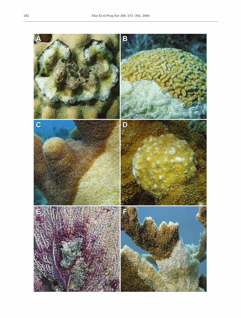
Fig. 3. Caribbean coral diseases: (A) black band (BBD) on Montastraea annularis complex; (B) white plague Type II (WPL II) onDichocoenia stokesi; (C) WPL II on Dendrogyra cylindrus; (D) skeletal anomaly (SKA) on Acropora palmata; (E) aspergillosis
(ASP) on Gorgonia sp.; (F) white band Type I (WBD I) on A. palmata. Photographs by J.W.P. and C.T.
Mar Ecol Prog Ser 266: 273–302, 2004
BBD has been widely reported as a seasonal phe-nomenon, with most active infections occurring duringlate summer and fall, and cessation occurring duringwinter (Antonius 1981, 1985a, Edmunds 1991, Carlton& Richardson 1995). Seasonality of BBD is related tosummer seawater temperatures in excess of 25°C(Rützler et al. 1983, Edmunds 1991, Kuta & Richardson2002, Richardson & Kuta 2003). When BBD disappearsin the winter, remaining living coral tissue survives(Antonius 1981, Carlton & Richardson 1995). However,seasonal reappearance of warm seawater coincideswith observations of reinfection, and reinfection makescomplete colony mortality possible (Carlton & Richard-son 1995, Kuta & Richardson 1996). It is important tonote that BBD has been reported year-round, evenat seawater temperatures as low as 20°C (Kuta &Richardson 1996), and that the cyanobacteria associ-ated with BBD are capable of photosynthesis at tem-peratures as low as 18 and 20°C (Taylor 1983, Richard-son & Kuta 2003).
BBD has been proposed to be correlated with otherenvironmental and physiological stressors, includingterrestrial runoff (Littler & Littler 1996, Bruckner et al.1997, Frias-Lopez et al. 2002), coral overgrowth byalgae (Bruckner et al. 1997), eutrophication (Antonius1981, 1985a, Kuta & Richardson 2002), and pollution(Antonius 1985a, Al-Moghrabi 2001), including humanfecal contamination (Frias-Lopez et al. 2002, Table 5).However, very little quantitative data, and no defini-tive results, have supported a positive correlation. Pol-lution has been implicated in extending depth range,frequency, and severity of BBD (Antonius 1985a). BBDhas been frequently reported from shallow depths(Rützler et al. 1983, Antonius 1985a, Kuta & Richardson2002) and reefs with low coral species diversity (Bruck-ner & Bruckner 1997b, Bruckner et al. 1997, Kuta &Richardson 2002).
The mechanism by which BBD kills coral tissue isdirectly linked to dynamics of the microbial mat com-munity, which produces a vertical zonation of oxygenand sulfide microenvironments that migrates on a dielbasis. The dominant constituent of BBD, the cyano-bacteria, forms the scaffolding of the band and isalways present throughout the band. The cyanobac-teria undergo oxygenic photosynthesis during the day,producing an oxygen supersaturated oxic zone in thetop 1⁄2 to 2⁄3 of the band. The cyanobacteria adapt to thehigh sulfide environment of the disease band by per-forming oxygenic photosynthesis in the presenceof sulfide (Richardson & Kuta 2003). The anoxic base ofthe band is dominated by sulfate-reducing bacteriaDesulfovibrio spp. The oxic/anoxic interface containssulfide-oxidizing bacteria Beggiatoa spp., and thiszone migrates vertically on a diel basis in response tochanges in light intensity (Viehman & Richardson
2002) and photosynthetic activity occurring within theband (Carlton & Richardson 1995, Richardson et al.1997). Under low light conditions (e.g. shade, dark-ness) the surface community of BBD is dominated bycyanobacteria, but when light intensity increases, Beg-giatoa spp. migrate to the band surface (Viehman &Richardson 2002). At night, in absence of oxygen pro-duction, Desulfovibrio spp. undergo sulfate reduction,increasing sulfide concentrations in the band. As aresult, sulfide is present throughout the band at nightand the oxic/anoxic interface migrates to the band sur-face (Carlton & Richardson 1995, Richardson et al. 1997).
Presence of sulfide and anoxia at the base of theband (adjacent to coral tissue) is thought to be thecause of tissue lysis and death (Carlton & Richardson1995). BBD-induced coral mortality presumably re-leases inorganic nutrients (NH4
+ and PO43–) that
support cyanobacterial photosynthesis. Furthermore,because the disease band directly overlies coral tissuethat is being degraded, nutrients supplied by tissuemortality diffuse directly into the band, providing con-centrated nutrients to the microbial consortium, andfueling their growth and reproduction (Carlton &Richardson 1995). This process of tissue death mayserve as the source for elevated nitrite levels associ-ated with seawater immediately surrounding BBD-affected corals (Kuta & Richardson 2002).
The mechanism of BBD transmission remains un-known. However, BBD may be infectious (Edmunds1991, Kuta & Richardson 1996) and transmitted in thewater column (Kuta & Richardson 1996, Bruckner et al.1997). Sediment patches on the surfaces of apparentlyhealthy corals may serve as reservoirs of the cyanobac-teria associated with BBD (Richardson 1997).
Disease signs similar to BBD were reported in 1983(Rützler et al. 1983) and documented as a new diseasetermed red band (Richardson 1993). Red band report-edly affects Gorgonia ventalina in Belize, Puerto Ricoand the Florida Keys (Santavy & Peters 1997), andMontastraea annularis and M. faveolata in the Colom-bian Caribbean (Garzón-Ferreira et al. 2001). Redband is characterized by a red-brown to brown-blackmicrobial mat that forms a migrating band that sepa-rates recently denuded skeleton from living tissue(Santavy & Peters 1997). The condition was considerednot to be BBD because Phormidium corallyticum,accepted at the time to be the dominant component ofthe BBD consortium, was not present in red band (Rüt-zler et al. 1983). Instead, red band was dominanted byother species of cyanobacteria, identified as Schizo-thrix calcicola, S. mexicana (Rützler et al. 1983) andOscillatoria spp. (Richardson 1993). The RBD microbialconsortium may include other cyanobacteria, hetero-trophic bacteria, the sulfur-oxidizing bacterium Beggia-toa sp., and the nematode Araeolaimus sp. (Santavy &
284
Sutherland et al.: Disease and immunity of corals
Peters 1997). The recent discovery that BBD is associ-ated with at least 3 different taxa of cyanobacteria(Frias-Lopez et al. 2003) and the lack of new reports ofred band disease since the early 1990s, suggest thatred band is not a distinct disease, but rather BBD.
White plague
White plague (WPL; Fig. 3B,C) has affected Carib-bean and Indo-Pacific corals since the late 1970s(Dustan 1977, Richardson et al. 1998a) and early 1980s(Antonius 1985a), respectively. In the Caribbean, thedisease reported in the 1970s has been renamed WPL Ito distinguish this condition from 2 new diseases withsimilar disease signs: WPL II (Richardson et al. 1998a)and WPL III (Richardson et al. 2001). WPL II is an infec-tious biotic disease (Richardson et al. 1998a) caused bya new genus and species of bacterium, Aurantimonascoralicida (Table 4; Richardson et al. 1998b, Denner etal. 2003). Pathogenesis and transmission of WPL II arenot understood. Histopathology of WPL I affected tis-sues shows necrosis at lesion boundaries (Peters 1984,Bythell et al. 2002) accompanied by dense clusters ofcoccoid bacteria that do not resemble the rod-shapedA. coralicida from WPL II infections (Bythell et al.2002). Similarly, 16S rDNA sequencing indicates thatA. coralicida is not associated with a WPL-like diseaseaffecting Caribbean corals (Pantos et al. 2003). Thesestudies suggest that there is indeed more than one eti-ologic agent associated with the various WPL diseasesin the Caribbean. The causal agent(s) of WPL I and IIIare unknown.
In the Indo-Pacific, WPL-like (WPL-L) disease signshave been reported. Authors that documented WPL-Ldisease in the Indo-Pacific referred to the disease asWBD (Antonius 1985a, Coles 1994, Riegl 2002). Spe-cies affected and disease signs reported for the Indo-Pacific (Antonius 1985a, Coles 1994) are indicative of aWPL-L disease. In the future, Indo-Pacific researchersmust distinguish between WPL-L and WBD-like dis-eases, which are characterized, respectively, by asharp line of tissue loss that progresses across a coralcolony and by a ring of tissue loss that progresses up ordown acroporid coral branches. WPL-L disease affects38 Indo-Pacific scleractinian species (Table 3; Antonius1985a, Coles 1994, Riegl 2002).
WPL I progresses slowly (3.1 mm d–1 maximum) andis characterized by a sharp line of tissue loss wherehealthy tissue is immediately adjacent to recentlydenuded skeleton (Fig. 3B,C; Dustan 1977). WPL Iaffects at least 13 Caribbean scleractinian species(Table 2; Dustan 1977, Richardson et al. 1998a).
WPL II progresses rapidly (2 cm d–1 maximum) and ischaracterized by a sharp line of disease progression. At
times a narrow band (2 to 3 mm) of bleached tissueseparates healthy tissue and bare skeleton, but morecommonly the disease line appears the same as forWPL I, i.e. healthy tissue is immediately adjacent torecently denuded tissue (Richardson et al. 1998a,b).Another distinguishing characteristic of WPL II is thatinfection most often begins at the base of the coralcolony and progresses upward in a concentric ringaround the entire colony (Richardson et al. 1998a,b).Three major epizootics of WPL II have been reported inSouth Florida since the mid-1990s (Richardson et al.1998a). The number of species affected by WPL II isincreasing. In 1995, Richardson et al. (1998a,b) re-ported 17 species of scleractinian corals affected onreefs in the Florida Keys, and by 2000 this number hadincreased to 32 species (Weil et al. 2002). Highestdisease prevalence of WPL II has been recorded forDichocoenia stokesi, with mortality as high as 38%(Richardson et al. 1998a).
WPL III was first documented in 1999 in the northernFlorida Keys. WPL III appears to exclusively affectlarge colonies (3 to 4 m diameter) of Colpophyllianatans and Montastraea annularis (Table 2). Tissueloss attributed to WPL III is extremely rapid and greatlyexceeds loss rates attributed to WPL I and WPL II(Richardson et al. 2001).
Surveys of WPL prevalence conducted in Puerto Rico(Bruckner & Bruckner 1997a) and St. Lucia, WestIndies (Nugues 2002) did not distinguish type. How-ever, based on slow rate of disease progression(1.3 mm d–1 maximum) measured in St. Lucia (Nugues2002) and rapid disease progression (1.4 cm d–1 maxi-mum) measured in Puerto Rico (Bruckner & Bruckner1997a) it is likely that the diseases surveyed were WPLI and II, respectively. Highest disease prevalence wasrecorded for Diploria labyrinthiformis (47% of coloniesaffected) in Puerto Rico (Bruckner & Bruckner 1997a)and for Montastraea faveolata (19% of coloniesaffected) and Colpophyllia natans (13% of coloniesaffected) in St. Lucia (Nugues 2002).
Nugues (2002) observed diseased individuals of 4scleractinian species, Isophylastrea rigida, Montastraeafaveolata, Mussa angulosa, and Mycetophyllia sp., notyet documented as susceptible to WPL I (Table 2).Surveys conducted within the FKNMS documentedWPL (type not determined) on 14 additional species(Table 2; Porter et al. 2001).
Shut-down reaction
Shut-down reaction (SDR) is a condition that mostoften affects corals contained in aquaria and is rarelyobserved in natural coral reef environments. SDRoccurs only on wounded corals (e.g. predation, diver
285

Mar Ecol Prog Ser 266: 273–302, 2004
contact) and is always associated with abiotic environ-mental stressors (e.g. temperature extremes, sedimen-tation, Table 5; Antonius 1977). SDR begins at andradiates from the interface between wound andhealthy tissue. Tissue is sloughed off the affectedcolony at the rapid rate of 10 cm h–1. Once SDR is trig-gered, complete colony mortality is inevitable. SDR iscontagious, indicating presence of a pathogen, and canbe both directly and indirectly transmitted from aninfected colony to a stressed, but otherwise apparentlyhealthy colony, via physical contact between neigh-boring colonies and water transport, respectively (An-tonius 1977). SDR cannot be transmitted to unstressedcoral colonies (Antonius 1977).
SDR has been experimentally induced in aquaria for6 Caribbean scleractinian species (Table 2; Antonius1977). In the Caribbean, SDR has been reported affect-ing only 3 individual coral colonies in the field; the 2species affected were Montastraea annularis andAcropora cervicornis (Table 2; Antonius 1977). Prior torecent reports of SDR in the Red Sea (species affectednot reported, Antonius & Riegl 1997, 1998), new casesof the condition had not been documented since thefirst report in the late 1970s (Antonius 1977).
Skeletal anomalies
Skeletal anomalies include tumors, galls, nodules,and other abnormalities of coral tissue and skeleton.Skeletal anomalies of scleractinian corals have beenobserved on reefs throughout the world including theFlorida Keys (Peters et al. 1986), Netherlands Antilles(Bak 1983), Hawaii (Squires 1965, Cheng & Wong 1974,Hunter & Peters 1993 Grygier & Cairns 1996, Aeby1998), Guam and Enewetak (Cheney 1975), Oman(Coles & Seapy 1998), Japan (Yamashiro et al. 2000),and the Great Barrier Reef, Australia (Loya et al. 1984).
A tumor is an abnormal tissue proliferation (Sinder-man 1990) and, in corals, is often associated with anabnormal skeletal growth (Yamashiro et al. 2000).Tumors result from neoplasia, hyperplasia, or hyper-trophy. Neoplasia (neoplasm) is uncontrolled cell pro-liferation. Hyperplasia and hypertrophy are nonneo-plastic controlled cell proliferation and nonneoplasticincrease in cell size, respectively (Sinderman 1990).The terms tumor and neoplasia are considered bymany to be synonymous (Stedman 2000).
The first suspected neoplasm of a scleractinian coralwas documented in 1965 on Madrepora kauaiensis inthe Hawaiian Islands (Table 3, Squires 1965). Thiscondition, also observed on M. oculata (Table 3), hasrecently been reinterpreted as a polyp hypertrophycharacterized by gall formation (i.e. a parasite-inducedproliferation of tissues, Grygier & Cairns 1996). These
lesions develop on Madrepora spp. when the crusta-cean Petrarca madreporae, an obligate endoparasite ofcorals, invades a normal coral polyp as a larva andmatures within the polyp, causing development ofan enlarged (hypertrophied) corallite with abnormalseptae (Table 4; Grygier & Cairns 1996).
Other skeletal anomalies have been attributed tointeractions with foreign organisms in the skeleton(Table 4). Porites compressa and P. lobata in KaneoheBay, Oahu, Hawaii develop grossly visible pink nod-ules in response to the encystment of the digenetictrematode Podocotyloides stenometra within the tenta-cles of the coral polyps (Table 3; Cheng & Wong 1974,Aeby 1998). The scleractinian corals P. lobata, P. lutea,Manicina areolata, and Montastraea cavernosa candetect invasion by endolithic fungi and respond by sur-rounding the site of fungal penetration within a layerof thickened calcium carbonate produced by hyper-trophied calicoblasts (Tables 3 & 4; Le Champion-Alsumard et al. 1995, Ravindran et al. 2001, E. C.Peters pers. comm.). However, this defense mechanismfails to hinder fungal advancement, and, as hyphaepenetrate the layer of calcium carbonate repair, thecoral repeats the process, resulting in a calcareousskeletal protuberance composed of a number of car-bonate layers (Le Champion-Alsumard et al. 1995).Nodules on the gorgonian coral Gorgonia ventalinaare attributed to both infection with Aspergillussydowii, the causal agent of aspergillosis (Dube et al.2002), and to infestation with filamentous green algaeof the Order Siphonales (Tables 2 & 4; Morse et al.1977, 1981). The algal nodules are hyperplasias of theaxis epithelial cells that produce the endoskeletal gor-gonin. Amoebocytes infiltrate the associated mesogleaand encapsulate the algal filaments (Morse et al. 1977,1981). Aspergillosis-associated nodule formation maybe a defense mechanism that sequesters fungalhyphae and limits spread of infection (Smith et al.1998). Nodules on the gorgonian corals Pseudoplex-aura spp. are the result of skeletal encapsulation of themarine microalgae Entocladia endozoica (Tables 2 & 4;Goldberg et al. 1984).
Peters et al. (1986) and Coles & Seapy (1998) de-scribed the only known true neoplasms (tumors) ofcorals. Peters et al. (1986) observed neoplasms, termedcalicoblastic epitheliomas, on Acropora palmata in theFlorida Keys (Table 2). These calicoblastic epithe-liomas result from proliferation of calicoblasts andassociated tissues and are characterized by raised (upto 1 cm high), irregularly shaped, smooth, white lumpsthat develop on all parts of the coral colony. Meangrowth rate of tumors is 0.12 mm d–1 or 25 to 44 mmyr–1 (Peters et al. 1986). Similar calicoblastic epithe-liomas affect A. valenciennesi and A. valida in the Gulfof Oman, Indian Ocean (Table 3; Coles & Seapy 1998).
286
Sutherland et al.: Disease and immunity of corals
Coral skeletal anomalies are characterized by: (1) thin-ning of coral tissue covering anomalies (Peters et al.1986, Coles & Seapy 1998), (2) increased porosity ofcoral skeleton (Peters et al. 1986, Coles & Seapy 1998,Yamashiro et al. 2000), (3) loss of mucous secretorycells and nematocysts (Peters et al. 1986, Coles &Seapy 1998), (4) loss of zooxanthellae (Bak 1983, Peterset al. 1986, Coles & Seapy 1998, Yamashiro et al. 2000),(5) loss, reduction, or degeneration of normal polypstructures (Bak 1983, Peters et al. 1986, Coles & Seapy1998, Yamashiro et al. 2000), and (6) reduced fecundity(Yamashiro et al. 2000).
Skeletal anomalies pose a serious threat to affectedcorals. Loss of the primary defense mechanism,mucous secretory cells, inhibits removal of foreignmaterial from the coral surface, contributing to celldeath and increasing susceptibility to invasion byfilamentous algae. Porous skeletons may be moresusceptible to storm-related damage (Peters et al.1986). Loss of zooxanthellae reduces fecundity, skele-tal growth, calcification rates, and nutrition (Bak 1983,Porter et al. 1989, Brown 1997a). Polyp destructionlimits reproductive capacity (Peters et al. 1986, Yama-shiro et al. 2000).
Skeletal anomalies (SKA; Fig. 3D) affect 16 Carib-bean (Table 2) and 24 Indo-Pacific (Table 3) sclerac-tinian species, 1 Caribbean hydrozoan, and at least5 species of Caribbean gorgonians (Table 2; Squires1965, Cheney 1975, Morse et al. 1977, 1981, Bak 1983,Goldberg et al. 1984, Loya et al. 1984, Peters et al.1986, Hunter & Peters 1993, Le Champion-Alsumardet al. 1995, Grygier & Cairns 1996, Coles & Seapy1998, Green & Bruckner 2000, Yamashiro et al. 2000,Ravindran et al. 2001, Dube et al. 2002). Acroporidsappear to be the most susceptible to neoplasia (Peterset al. 1986, Coles & Seapy 1998), and this may be dueto the rapid growth rates of this genus (Peters et al.1986). Acropora palmata is capable of linear extensionrates as high as 47 to 99 mm yr–1 (Gladfelter et al.1978).
With the exception of microorganism-inducednodule or gall formation (Cheng & Wong 1974,Morse et al. 1977, 1981, Goldberg et al. 1984, LeChampion-Alsumard et al. 1995, Grygier & Cairns1996, Aeby 1998, Ravindran et al. 2001, Dube et al.2002), the etiology of coral skeletal anomalies isunknown (Peters et al. 1986, Coles & Seapy 1998,Yamashiro et al. 2000). Parasitic and commensalorganisms have been ruled out as potential causalagents of the skeletal anomalies described by Peterset al. (1986) in the Florida Keys and by Yamashiro etal. (2000) in Japan. Solar UV radiation has beenhypothesized as a possible initiator of neoplasia for-mation (Table 5; Peters et al. 1986, Coles & Seapy1998).
CARIBBEAN CORAL DISEASES
Aspergillosis
Mass mortalities of the sea fans Gorgonia ventalinaand G. flabellum were reported throughout theCaribbean during the 1980s (Garzón-Ferreira & Zea1992). A second epizootic affecting Gorgonia spp.began in 1995 (Nagelkerken 1997ab, Slattery 1999),which was less virulent but more widespread thanthe 1980s epizootic (Nagelkerken 1997a,b).
Both epizootics have been attributed to the fungusAspergillus sydowii (Table 4; Smith et al. 1996, Geiseret al. 1998). Aspergillus disease, termed aspergillosis(ASP; Fig. 3E), destroys living tissue and degradesskeletal framework (Nagelkerken et al. 1997b). Inaddition to Gorgonia ventalina and G. flabellum, ASPsigns have been observed on 6 additional gorgonianspecies from 5 genera (Kim et al. 2000b, Weil et al.2002; Table 2). However, Koch’s postulates have onlybeen fulfilled, establishing A. sydowii as the causativeagent, for disease cases affecting G. ventalina and G.flabellum (Smith et al. 1996).
ASP lesions are characterized by recession of rindtissue (coenenchyme) exposing the internal axialskeleton. Aspergillus sydowii hyphae are embedded inliving tissue at the receding edge of the lesion (Smithet al. 1996). Lesions are often circumscribed by a pur-ple halo indicative of an abundance of purple sclerites(Kim et al. 1997, Smith et al. 1998, Slattery 1999).Purple sclerite-dense nodules often erupt on affectedsea fans (Kim et al. 1997, Smith et al. 1998, Dube et al.2002). Sclerite recruitment and nodule formation maybe methods of defense that sequester fungal hyphaeand limit spread of infection (Smith et al. 1998). Mech-anisms by which A. sydowii produces tissue degra-dation and nodule formation remain unknown, butvirulence of the pathogen is known to increase withelevated seawater temperatures (30°C, Table 5; Alkeret al. 2001).
The genus Aspergillus is not commonly found inmarine environments and most often inhabits terres-trial soils. However, Aspergillus spp. can easily copewith the salinity of seawater (Kendrick et al. 1982) andhave been isolated from marine environments (Mun-tanola-Cvetkovic & Ristanovic 1980, Kendrick et al.1982, Ravindran et al. 2001). A. sydowii has beenshown to bioerode living stony corals (Kendrick et al.1982).
Aspergillus sydowii is a terrestrial fungus. Deliveryof A. sydowii to the marine environment may be asso-ciated with either local sediment runoff (Smith et al.1996) or long distance transport (Table 5, Shinn et al.2000). If local runoff is the source, then ASP may belinked to anthropogenic disturbance. Shinn et al.
287
Mar Ecol Prog Ser 266: 273–302, 2004
(2000) hypothesize that A. sydowii, and perhaps othercoral disease-causing pathogens, are transported tothe western Atlantic in African dust air masses. A.sydowii has been cultured from spores collected in theUS Virgin Islands during African dust storm events.These A. sydowii isolates, when inoculated ontohealthy sea fans, produced ASP signs (Weir et al. inpress).
Terrestrial sources are a possible mode of primarytransmission of Aspergillus sydowii hyphae and/orspores to unaffected sea fans in the marine environ-ment. A. sydowii germinates but does not sporulate onsea fans. Hyphae must break free from an infected gor-gonian and reach the surface of the water to producespores (G. W. Smith pers. comm.). Secondary transmis-sion of ASP from infected to uninfected sea fans mayoccur through: (1) direct physical contact with aninfected individual, i.e. a diseased sea fan may brushagainst a close neighbor (Smith et al. 1996, Jolles et al.2002), (2) transport of fungal hyphae in the water col-umn (Jolles et al. 2002), or (3) transport of fungalspores (produced at the sea surface from hyphaereleased from diseased sea fans) in the water column(G. W. Smith pers. comm.).
Incidence and prevalence of ASP are greater at pro-tected than at exposed sites and increase with depth inareas with low to moderate wave action, indicatingthat the more frequent mechanical swaying of coloniesthat occurs in shallow and more exposed areas maydecrease sea fan susceptibility to disease (Nagel-kerken 1997a). ASP is prevalent in the Florida Keys,affecting 43% of Gorgonia ventalina colonies Keys-wide (Kim & Harvell 2002). Poor water quality (i.e.increased turbidity and increased chl a) may play arole in the impact of ASP on sea fan populations(Table 5). Kim & Harvell (2002) found that severity ofASP in the Florida Keys was greatest near the city ofKey West. With approximately 25500 residents and ahigh seasonal influx of tourists, Key West is by far themost populous town in the Florida Keys (US BOC2000). Runoff of nutrients and pollutants into themarine environment is likely higher near Key Westthan in other less populated areas in the Florida Keys.
White band
White band disease (WBD; Fig. 3F) has affectedCaribbean scleractinian corals since the late 1970s(Antonius 1985a, Bythell & Sheppard 1993). The dis-ease, as first described by Gladfelter (1982), has beenrenamed WBD I to distinguish the condition from WBDII (Ritchie & Smith 1998). WBD I and II exclusivelyaffect branching acroporid corals. While WBD I affectsboth Acropora palmata and A. cervicornis Caribbean-
wide (Gladfelter 1982, Peters 1984), WBD II has beenreported only from the Bahamas, exclusively affectingA. cervicornis (Ritchie & Smith 1998; Table 2).
WBD Type I has been implicated as the principalcause of mass mortalities of Acropora cervicornis andA. palmata that occurred in the 1980s and 1990s (Glad-felter 1982, Bythell & Sheppard 1993, Aronson &Precht 1997, 2001, Aronson et al. 1998, 2002). On mostreefs, loss of acroporids was accompanied by an eco-logical phase shift from a coral-dominated to an algal-dominated reef (Hughes 1994). However, on theBelizean Barrier Reef, the grazing urchin Echinometraviridis consumed fleshy and filamentous macroalgaeand allowed for a wide scale (at least 500 km2) coralcommunity shift (Aronson et al. 2002). Following theWBD epizootic of the late-1980s, the previously domi-nant coral A. cervicornis was replaced by the thin-leaflettuce coral Agaricia tenuifolia (Aronson & Precht1997, Aronson et al. 2002). Examination of the fossilrecord indicates that the wide-scale Acropora-to-Agaricia shift is unprecedented in the last 3800 yr(Aronson & Precht 1997, 2001, Aronson et al. 2002).This evidence suggests that WBD is an emergent dis-ease and not a natural cyclic phenomenon that hasoccurred on Caribbean reefs in the past.
WBD I progresses rapidly (2 cm d–1 maximum) andhas the potential to cause extensive mortality (Anto-nius 1981, Gladfelter 1982, Peters et al. 1983). The dis-ease is characterized by a white band of recentlydenuded skeleton adjacent to a necrotic front of nor-mally pigmented living tissue (Fig. 3F; Gladfelter 1982,Peters et al. 1983). WBD I develops at the base of acoral colony or branch and progresses upward towardbranch tips in a concentric ring (Gladfelter 1982).
WBD II was first documented in 1993 in the Bahamas(Ritchie & Smith 1995a, Ritchie & Smith 1998). WBD IIis distinguished from WBD I by a band (2 to 20 cmwide) of living bleached tissue separating denudedskeleton from normally pigmented tissue. The bleach-ing of the tissue progresses more rapidly than does themargin of necrotic tissue and can arrest, allowing thenecrotic margin to catch up to normal tissue. When thisoccurs, WBD II resembles WBD I (Ritchie & Smith1998) and the 2 diseases cannot be distinguished froma single observation in the field. Like WBD I, WBD IIcan develop at the base of a coral colony and progressupward, but WBD II is also capable of developing attips of branches and progressing downward. When thedisease begins at branch tips, tissue loss can be accom-panied by skeletal degradation, i.e. dissolution andloss of branch tips (Ritchie & Smith 1998).
The causative agents of WBD I and II are unknown,however, efforts have been made to determine etiolo-gies of these conditions. Tissues of WBD I-diseasedand apparently healthy Acropora palmata and A. cer-
288

Sutherland et al.: Disease and immunity of corals
vicornis at St. Croix, US Virgin Islands and at Bonairewere found to contain Gram-negative bacterial aggre-gates. Aggregates were more abundant in diseasedcorals than in apparently healthy corals. However,other apparently healthy and diseased acroporids donot contain aggregates (Peters et al. 1983). Examina-tion of A. cervicornis from the Bahamas and the FloridaKeys revealed WBD I-affected colonies both with andwithout bacterial aggregates (Peters 1984). Thus, therole of bacterial aggregates in WBD I is uncertain.Histopathology of WBD I tissues shows no signs ofnecrosis or clustering of microorganisms (Peters et al.1983, Bythell et al. 2002).
WBD II is always associated with the bacterium Vibriocharcharia (Table 4; Ritchie & Smith 1995a). Attemptsto fulfill Koch’s postulates with V. charcharia were un-successful, and therefore the significance of the bac-terium to the etiology of WBD II remains unknown.
White pox
White pox disease (WPD; Fig. 4A,B), also termedacroporid serratiosis (Patterson et al. 2002) and patchynecrosis (Bruckner & Bruckner 1997a), was first docu-mented in 1996 on reefs off Key West, Florida (Holden1996). WPD has since been observed throughout theCaribbean (Porter et al. 2001, Rodríquez-Martínez etal. 2001, Santavy et al. 2001, Patterson et al. 2002). Thedisease exclusively affects Acropora palmata (Table 2),and is caused by the common fecal enterobacteriumSerratia marcescens (Table 4; Patterson et al. 2002).
Serratia marcescens is a Gram-negative bacteriumclassified as a coliform and a member of the Entero-bacteriaceae family. It is found in feces of humans andother animals and in water and soil (Grimont & Gri-mont 1994). The prevalence of S. marcescens in themarine environment is unknown. However, this bac-terium has been found in the marine environment insewage-polluted estuaries. For example, S. marces-cens has been linked to disease of white perch Moroneamericanus in the sewage-polluted Back River, Mary-land (Baya et al. 1992).
Identification of Serratia marcescens as a coralpathogen marked the first time that a common memberof the human gut microbiota was shown to be a marineinvertebrate pathogen. While S. marcescens is ubiqui-tous, its noted association with human hosts promptsspeculation that improperly treated sewage may beassociated with white pox disease in corals. Humanenteric bacteria and viruses are prevalent in coral SMLand other marine environments of the Florida Keys(Griffin et al. 1999, Lipp et al. 2002). The origin andpathogenic mechanisms of the WPD pathogen areunknown (Patterson et al. 2002).
Coral colonies affected by WPD are characterized byirregularly shaped distinct white patches of recentlyexposed skeleton surrounded by a necrotic front ofnormally pigmented living tissue (Fig. 4B). Lesionsrange in area from a few square centimeters to>80 cm2 and develop simultaneously on all surfaces ofthe coral colony (Fig. 4A; Patterson et al. 2002). Lesionsexhibit tissue loss along the perimeter and increase inarea as tissue is lost from the leading edge of infection.Rate of tissue loss is rapid, averaging 2.5 cm2 d–1, and isgreatest during periods of seasonally elevated temper-ature and rainfall (Table 5). WPD is highly contagious,with nearest neighbors most susceptible to infection(Patterson et al. 2002). The disease spread rapidlywithin and between reefs in the Florida Keys duringthe mid-1990s (Porter et al. 2001, Patterson et al. 2002).
WPD has been implicated as the principal cause ofmass mortality of Acropora palmata within the FKNMS(Patterson et al. 2002). Between 1996 and 2002, aver-age loss of A. palmata Keys-wide was 87% (Fig. 1;Patterson et al. 2002, Sutherland & Ritchie in press).Losses of A. palmata at Eastern Dry Rocks Reef, FL(24° 27.715’ N, 81° 50.801’ W) between 1994 and 2002,and at Looe Key Reef, FL (24° 33’ N, 81° 24’ W) between1983 and 2000, were 97 and 93%, respectively (Milleret al. 2002, Patterson et al. 2002, Sutherland & Ritchiein press). These severe population declines of the coralcommunity’s most important primary producer andshallow water framework builder have led to theidentification of A. palmata as a candidate for inclusionon the Endangered Species List (Diaz-Soltero 1999).Diseases of acroporid corals (WPD and WBD) arechanging the composition, structure, and function ofCaribbean coral reef ecosystems (Hughes 1994, Aron-son & Precht 1997, 2001, Aronson et al. 2002, Pattersonet al. 2002).
Yellow blotch/band
Yellow blotch/band (YBL; Fig. 4C,D) disease hasbeen reported throughout the Caribbean since 1994(Santavy & Peters 1997, Santavy et al. 1999, Cervino etal. 2001, Garzón-Ferreira et al. 2001, Toller et al. 2001,Weil et al. 2002). YBL affects 9 scleractinian coralspecies (Table 2; Garzón-Ferreira et al. 2001), but mostoften affects Montastraea annularis (Cervino et al.2001). The causal agent of YBL is unknown.
YBL lesions are characterized by circular to irregu-larly shaped patches (Fig. 4C) or bands (Fig. 4D) of dis-colored coral tissue. Lesions can develop on all areas ofthe coral colony, but are most common on upper sur-faces (Santavy et al. 1999). A patch of exposed skeletonis often present at the center of each circular to irre-gularly shaped lesion and at the edge of each band-
289

Mar Ecol Prog Ser 266: 273–302, 2004290

Sutherland et al.: Disease and immunity of corals
shaped lesion. This dead zone is surrounded by ringsor bands of yellow translucent tissue, which may inturn be surrounded by rings or bands of pale brown tobright-white bleached tissue. Apparently healthytissue surrounds the outer regions of YBL lesions(Santavy et al. 1999, Toller et al. 2001). Tissue loss rateassociated with YBL is approximately 0.6 cm mo–1
(Cervino et al. 2001).YBL-affected coral tissues have approximately 40%
fewer algal symbionts than do apparently healthy tis-sues, and these zooxanthellae appear vacuolated andlack organelles (Cervino et al. 2001). In San Blas(Panama) zooxanthellae within yellow and normaltissues from YBL-affected Montastraea annularis, M.faveolata, and M. franksi were found to be of differenttaxa of the genus Symbiodinium (Toller et al. 2001).Normal (unaffected) tissues were dominated by Sym-biodinium sp., clade C, the taxon common in healthycorals at depth of collection (1 to 10 m). Yellow(affected) tissues predominately contained Symbio-dinium sp., clade A, the source of the characteristicyellow color of YBL lesions (Toller et al. 2001).
Dark spots
Dark spots disease (DSD; Fig. 4E,F) was first docu-mented in the early 1990s in Colombia and today isfound throughout the Caribbean (Goreau et al. 1998,Cervino et al. 2001, Garzón-Ferreira et al. 2001,Gil-Agudelo & Garzón-Ferreira 2001, Weil et al. 2002).DSD reportedly affects 11 scleractinian species(Table 2; Goreau et al. 1998, Cervino et al. 2001,Garzón-Ferreira et al. 2001, Gil-Agudelo & Garzón-Ferreira 2001).
DSD is characterized by irregularly shaped darkspots of purple, maroon, or brown coloration on normaltissue (Goreau et al. 1998). DSD may be associatedwith tissue necrosis and/or depression of the colonysurface (Cervino et al. 2001). When DSD is associatedwith tissue loss, a spot may expand into a characteristicdark ring separating dead skeleton from living tissue(Goreau et al. 1998).
Dark pigmentation of DSD-affected Stephanocoeniamichelinii extends into, and stains, the skeleton, and maybe attributed to zooxanthellae, which appear darker inpigment in diseased corals of this species. In Siderastreasiderea affected with DSD, skeletal staining and zoo-xanthellae of darker pigmentation are not evident, but
zooxanthellae do appear swollen and necrotic. Theseobservations suggest that similar disease signs observedon S. michelinii (Fig. 4E) and S. siderea (Fig. 4F) mayrepresent 2 different diseases (Cervino et al. 2001).
In the Colombian Caribbean, DSD is correlated withdepth and temperature. DSD is more prevalent atdepths less than 6 m and during summer when sea-water temperature is highest (Table 5). Depth distribu-tion of DSD may be due to high disease prevalence(94%) on 2 coral species that occupy shallow depths,Siderastrea siderea and Montastraea annularis. Distri-bution of DSD on Colombian reefs is clumped, indica-ting that the disease may be contagious and thereforeof biotic origin (Gil-Agudelo & Garzón-Ferreira 2001).The causal agent of DSD is unknown.
INDO-PACIFIC CORAL DISEASES
Vibrio-induced bleaching
Coral bleaching is a global phenomenon that occurswhen symbiotic algae (zooxanthellae) and/or theirpigments are lost from host coral tissues, resulting inpale brown to translucent living tissue. This conditionreduces reproductive output, skeletal growth, calcifi-cation rates, and nutrition (Porter et al. 1989, Brown1997a), and therefore is a sign of disease. Bleaching ismost often correlated with elevated seawater tem-perature, but may be associated with salinity extremes(Coles & Jokiel 1992), light irradiance extremes(Brown et al. 1994, 2000, Brown 1997b, Shick et al.1996), and pollutants (Peters et al. 1981, Harland &Brown 1989). Bleaching may result in coral mortality,but most often the coral regains its algal symbionts,resulting in full recovery (Fitt et al. 1993).
Coral bleaching, once regarded exclusively as an abi-otic condition, has recently been associated with 2 bac-terial pathogens of the genus Vibrio. In the Mediter-ranean Sea, bleaching of the coral Oculina patagonicacan be induced by an infection with the bacterium V.shiloi (Tables 3 & 4; Kushmaro et al. 1996, 1997, 1998,2001, Rosenberg et al. 1998). Bleaching and lysis oftissue of the coral Pocillopora damicornis in Zanzibar,Indian Ocean and Eilat, Red Sea are induced by in-fection with V. coralliilyticus (Tables 3 & 4; Ben-Haim& Rosenberg 2002, Ben-Haim et al. 2003a,b). Vibrio-induced bleaching and lysis are always associated withelevated seawater temperatures in excess of 24.5°C
291
Fig. 4. Caribbean coral diseases: (A) white pox (WPD) on Acropora palmata; (B) WPD lesions on A. palmata; (C) yellowblotch/band (YBL) circular-shaped lesion and (D) YBL band-shaped lesion on Montastraea annularis complex; (E) dark spots(DSD) on Stephanocoenia michelinii; (F) DSD on Siderastrea siderea. Photographs: (A) and (E) by J.W.P., (B) by K.P.S., (C) and (D)
by C. Quirolo, (F) by J.W.P. and C.T.
Mar Ecol Prog Ser 266: 273–302, 2004
(Kushmaro et al. 1998, Toren et al. 1998, Ben-Haim etal. 1999, 2003b, Banin et al. 2001a, Israely et al. 2001),and therefore are examples of diseases with etiologiesassociated with both abiotic and biotic factors.
Vibrio shiloi - induced bleaching
Vibrio shiloi-induced bleaching (VSB) of Oculinapatagonica is arguably the most characterized coraldisease in terms of etiology and mechanism of patho-genesis. Koch’s postulates were fullfilled and showedthat bleaching can be induced by infection with V.shiloi in apparently healthy corals maintained at ele-vated seawater temperatures (25 to 29°C, Kushmaro etal. 1998). Elevated seawater temperatures increasesthe virulence of V. shiloi (Table 5; Kushmaro et al.1998, Toren et al. 1998, Ben-Haim et al. 1999, Baninet al. 2001a, Israely et al. 2001).
Under conditions of elevated seawater temperatures,infection begins with adhesion of Vibrio shiloi to aβ-galactoside-containing receptor in mucus on the sur-face of the host coral (Toren et al. 1998, Banin et al.2001b). Photosynthetically active zooxanthellae insidethe host coral produce the β-galactoside-containingreceptor. Only V. shiloi that are grown at elevated sea-water temperatures are capable of adhesion (Toren etal. 1998). Therefore, adhesion only occurs under condi-tions of elevated temperature and if photosyntheticallyactive zooxanthellae are present (Banin et al. 2001b).
The second step in the infection process is penetra-tion of Vibrio shiloi into the epidermis of the host coral.Once V. shiloi are inside the tissues they multiplyand transform into a viable but non-culturable state(VBNC). In other words, intracellular V. shiloi are notcapable of forming colonies on media that supportgrowth of extracellular V. shiloi. Intracellular bacteriaare protected against effects of treatment with theantibiotic gentamicin. Gentamicin kills extracellular V.shiloi associated with coral mucus, but has no effect onV. shiloi that have successfully penetrated host tissue(Banin et al. 2000). At elevated temperatures, VBNCV. shiloi infect healthy coral and induce bleaching(Israely et al. 2001).
Following penetration into the host coral, Vibrioshiloi produces both heat-stable and heat-sensitivetoxins that target zooxanthellae and play a role inpathogenesis. The heat-sensitive toxin bleaches andlyses symbiotic algal cells isolated from the host coral(Rosenberg et al. 1998, Ben-Haim et al. 1999). Theextracellular heat-stable toxin, termed Toxin P, is aproline-rich dodecapeptide that binds to zooxanthellaeand inhibits photosynthesis in the presence of ammo-nia (Rosenberg et al. 1998, Ben-Haim et al. 1999, Baninet al. 2001a). Inhibition of photosynthesis damages zoo-
xanthellae and contributes to coral bleaching (Banin etal. 2001a). Toxin P is produced only under conditions ofelevated seawater temperature (25 to 30°C, Banin etal. 2001a). V. shiloi require another virulence factor,superoxide dismutase, in order to survive inside thehost coral. High levels of superoxide dismutase areproduced by V. shiloi under conditions of elevatedseawater temperature (30°C, Israely et al. 2001).
During summer in the Mediterranean Sea, Vibrioshiloi are present in all bleached colonies of Oculinapatagonica. However, during winter, when water tem-perature drops to 16°C, intracellular Vibrio shiloi lyseand die, and corals affected by VSB recover. V. shiloioutside the coral survive the temperature shift, indicat-ing that the coral host may play a role in seasonaldemise of the pathogen (Israely et al. 2001). Reinfec-tion of O. patagonica colonies the following summer isfacilitated, at least in part, by the marine firewormHermodice carunculata, which serves as a reservoirand a vector of V. shiloi. During winter, V. shiloi ispresent in the VBNC state inside H. carunculata, andduring summer, when the worm feeds on O. patago-nica, the VSB pathogen is transmitted to a few coralcolonies. This indirect transmission serves to restartthe infection process, and the infectious VSB thenspreads from colony to colony (Sussman et al. 2003).
Vibrio coralliilyticus- induced bleaching and disease
Vibrio coralliilyticus-induced bleaching and disease(VCB) affects the scleractinian coral Pocillopora dami-cornis. VCB is characterized by bleaching (Ben-Haimet al. 2003b) and lysis of coral tissue (Ben-Haim &Rosenberg 2002, Ben-Haim et al. 2003b). Koch’s postu-lates were fulfilled and showed that VCB is caused bya new species of bacterium, V. coralliilyticus (Table 4;Ben-Haim & Rosenberg 2002, Ben-Haim et al. 2003a,b).Bleaching and tissue loss associated with V. coral-liilyticus is temperature dependent. Infection with V.coralliilyticus induces bleaching at seawater tempera-tures of 24.5 and 25.0°C, and tissue lysis and colonydeath at seawater temperatures of 27 and 29°C (Ben-Haim et al. 2003b). Bleaching does not preceed lysisat temperatures greater than 27°C (Ben-Haim et al.2003b). VCB is infectious and can be transmittedthrough direct contact between an infected coralcolony and an un-infected neighbor (Ben-Haim &Rosenberg 2002). V. coralliilyticus produces an extra-cellular protease that may play a role in pathogenesis,and production of this enzyme increases at tempera-tures greater than 24°C (Ben-Haim et al. 2003b). Ele-vated seawater temperature increases either pathogenvirulence or host susceptibility, or both (Ben-Haim &Rosenberg 2002).
292
Sutherland et al.: Disease and immunity of corals
Skeleton eroding band
Skeleton eroding band (SEB) disease was first docu-mented in 1988 in Papua New Guinea (Antonius &Lipscomb 2000). SEB has since been observed in theRed Sea, Indian Ocean, and the Great Barrier Reef,Australia. Attempts to locate the disease in theCaribbean have been unsuccessful. Although Koch’spostulates have not been fulfilled, SEB is believed tobe caused by the protozoan Halofolliculina corallasia(Table 4). SEB affects 24 Indo-Pacific scleractinianspecies (Table 3; Antonius & Lipscomb 2000).
Halofolliculina corallasia is a folliculinid heterotrichciliate that is sessile in a secreted black sac-like testcalled a lorica. Dense clusters of protozoans, with basalportions of their loricae embedded in host coral skele-ton, form the characteristic black band of SEB. SEBadvances across a coral by asexually producing migra-tory larval stages that move ahead of the band inmasses and locate a site to settle and secrete loricae. Acombination of chemicals associated with productionof pseudochitinous loricae, and drilling of loricae intocoral skeleton, result in skeletal erosion and tissuedeath (Antonius & Lipscomb 2000).
SEB resembles BBD in gross disease signs, exceptthat SEB is not associated with a microbial mat thatcan be lifted from the coral surface. SEB affectedcorals are characterized by a black band ranging inwidth from less than 1 mm to 80 cm, depending onspecies affected. The line migrates horizontally acrossaffected coral at a rate that may be as slow as 1 mmwk–1 or as fast as 1 mm d–1, leaving dead skeleton inits wake. Denuded skeleton is flecked with tiny blackspots composed of clusters of empty loricae, a char-acteristic that clearly distinguishes SEB from BBD,which leaves behind bright white skeleton (Antonius& Lipscomb 2000).
SEB is found at depths ranging from 0 to 35 m and ismost common at 0.5 to 3 m. The disease does notappear to be correlated with seasonal seawater tem-peratures. SEB can be directly transmitted from adiseased colony to a healthy colony only via directphysical contact. Attempts to transmit the disease viaseawater in enclosed aquaria were unsuccessful (Anto-nius & Lipscomb 2000).
Yellow band
Yellow band disease (YBD) was first observed in theGulf of Oman, Arabian Sea in the late 1990s. The dis-ease affects 12 Indo-Pacific scleractinian coral species(Table 3; Korrûbel & Riegl 1998, Riegl 2002). YBD isnot the same disease as Caribbean yellow blotch/banddisease (YBL).
YBD is characterized by a broad band of yellow-pigmented tissue (Korrûbel & Riegl 1998) and a yellow-pigmented microbial mat (B. Riegl pers. comm.) thatmigrate horizontally across the coral, producing a mar-gin of decaying tissue adjacent to healthy tissue andleaving behind dead skeleton that often retains a yel-low pigmentation (Korrûbel & Riegl 1998). The pres-ence of a microbial mat suggests that YBD may be avariant of BBD (B. Riegl pers. comm.). Rate of tissueloss is correlated with seasonally elevated temperatureand is greater in summer (19.7 mm wk–1) than inwinter (9.4 mm wk–1, Table 5; Riegl 2002). YBD istransmissible from colony to colony (Riegl 2002).
Pink-line syndrome
Pink-line syndrome (PLS) was first reported in 2001affecting the scleractinian corals Porites compressaand P. lutea of Kavaratti Island, Indian Ocean (Table 3).PLS is characterized by a band of pink-pigmented tis-sue separating dead skeleton from apparently healthytissue. This band may begin as a small ring andprogress outward horizontally across a coral colony.As with other coral diseases, the zone of dead skeletonis bright white in appearance, indicating a relativelyrapid rate of disease progression (Ravindran et al.2001). PLS is associated with the cyanobacteriumPhormidium valderianum, which induces pink col-oration of coral tissue, hypothesized by increasinglevels of pCO2 (Ravindran & Raghukumar 2002).
Fungal-protozoan syndrome
Epizootics affecting 6 gorgonian species, 1 scleractin-ian species (Table 3), 1 zoanthid (Parazoanthus axinel-lae), sponges, and encrusting coralline algae occurredin the Liguarian Sea, north-western Mediterranean, inlate summer 1999. Losses of gorgonian corals were esti-mated to be in the millions (Cerrano et al. 2000).
Gorgonian corals affected by the syndrome, heretermed fungal-protozoan syndrome (FPS), are charac-terized by an increase in mucus production, loss of pig-mentation, and loss of coenenchyme tissue. Spicules inthe coenenchyme lose the thin outer layer of epidermalcells and become disorganized. Coenenchyme tissuesare colonized by fungi and coral polyps are colonizedand consumed by protozoan ciliates. Fungi associatedwith FPS were most commonly of the genus Tricho-derma, but Clodosporium, Penicillum, and Humicolawere also common (Table 4; Cerrano et al. 2000).
Fungi and protozoa associated with FPS may beopportunistic rather than primary pathogens. FPS epi-zootics of 1999 were correlated with elevated seawater
293

Mar Ecol Prog Ser 266: 273–302, 2004
temperature (Table 5). The etiology of FPS may stemfrom a combination of an abiotic stressor (i.e. elevatedseawater temperature) and a biotic attack by oppor-tunistic microorganisms (Cerrano et al. 2000).
CORAL IMMUNITY
Immunity is the protection against infectious diseaseand can be innate or acquired (Stedman 2000). Know-ledge of the immune systems of zooxanthellate coralsand other invertebrates is limited; however, these or-ganisms are known to possess highly efficient defensemechanisms against infection. In order to fully under-stand coral immunity, it is helpful to first briefly reviewthe immune mechanisms of invertebrates in general.
Invertebrates are limited to innate immunity (Roch1999), defined as a non-specific general ability of cer-tain cells to resist most pathogens (Stedman 2000).Vertebrates, on the other hand, utilize both innateimmunity and acquired immunity (Roch 1999, Sted-man 2000), a specific and highly sophisticated mecha-nism for developing resistance to individual pathogens(Stedman 2000). Components of the acquired immunesystem, including antibodies, antigen presenting cells,interactive lymphocytes, and lymphoid organs areabsent in invertebrates (Sinderman 1990).
Invertebrate and vertebrate immune responses aresimilar in that both types of organisms employ physio-chemical barriers, cellular defenses, and humoraldefenses against pathogens (Roch 1999). Physiochemi-cal barriers serve as the first line of defense againstinvaders and include epidermis, mucus, cuticles, tests,shells, and gut barriers (Roitt et al. 1996, Peters 1997).Cellular (whole cell) defenses depend on the ability ofthe organism to distinguish non-self from self, and ininvertebrates include: (1) coagulation and wound heal-ing, (2) hemocytosis, (3) phagocytosis, (4) encapsula-tion, and (5) immunological memory (Kinne 1980,Sinderman 1990, Roitt et al. 1996). Invertebratesemploy both natural and inducible humoral (cell prod-uct) defenses (Sinderman 1990, Roitt et al. 1996) thatprovide the lytic properties of phagocytic cells andhemolymph (blood, Sinderman 1990).
There is evidence for vertebrate defense mechanismsin invertebrates including cytokines, complement, andimmunological memory (Roitt et al. 1996). Invertebratecytokine-like molecules regulate invertebrate host de-fenses by activating phagocytosis and encapsulation.Invertebrates known to produce cytokine-like mole-cules include protozoa, annelids, echinoderms, tuni-cates, and arthropods (Roitt et al. 1996). Complementparticipates in control of inflammation, bacterial lysis,microbial killing, and phagocytosis (Roitt et al. 1996).Phagocytosis is enhanced when complement mole-
cules, termed opsonins, bind to invading microorgan-isms or other foreign material (Bayne 1990, Roitt et al.1996). Opsonin-dependent phagocytosis is found in anumber of invertebrates including echinoderms, crus-taceans, mollusks, annelids, and insects (Bayne 1990,Roitt et al. 1996). Immunological memory is a mecha-nism by which a host can distinguish non-self from selfand resist infection by previously encountered patho-genic microorganisms (Sinderman 1990). Memory hasbeen observed in scleractinian and gorgonian corals(Theodor 1970, Hildemann et al. 1975, 1977a, 1980a,Raison et al. 1976, Johnston et al. 1981, Bak & Criens1982, Neigel & Avise 1983), sponges (Hildemann et al.1979, 1980b, Hildemann & Linthicum 1981, Bigger et al.1982, 1983, Johnston & Hildemann 1983), echinoderms(Karp & Hildemann 1976, Hildemann et al. 1979),mollusks, crustaceans (Anderson 1986, Sinderman1990), tunicates (Lakshma Reddy et al. 1975, Raftoset al. 1987), nemerteans (Langlet & Bierne 1982), andannelids (Hildemann et al. 1979).
Coral physiochemical barriers
Scleractinian and gorgonian corals utilize mucus pro-duction and a protective epidermis as physiochemicalbarriers (Peters 1997, Santavy & Peters 1997, Hayes &Goreau 1998). Sloughing of mucus is an important de-fense mechanism of corals against attachment of poten-tially pathogenic bacteria to surface tissues (Ducklow &Mitchell 1979a, Rublee et al. 1980). However, there areexamples of mucus aiding in attachment of pathogenicmicroorganisms to the surface of corals. Adhesion ofVibrio shiloi, the pathogen that causes VSB, to the coralsurface requires a β-galactoside-containing receptor inthe mucus (Toren et al. 1998). Further, Lipp et al. (2002)demonstrated that potentially pathogenic enteric bac-teria and viruses are concentrated on the surface mucuslayers of scleractinian corals under natural near-shoreconditions in the Florida Keys.
Coral cellular defenses
Cellular defenses depend on the ability of the coral todistinguish non-self (e.g. sediment, pathogens) from self.Cellular defenses documented for scleractinian andgorgonian corals include: (1) wound healing (Bigger &Hildemann 1982, Meszaros & Bigger 1999), (2) phago-cytosis (Bayne 1990, Sinderman 1990, Peters 1997), and(3) immunological memory (Theodor 1970, Hildemann etal. 1975, 1977a, 1980a, Raison et al. 1976, Johnston etal. 1981, Bak & Criens 1982, Neigel & Avise 1983).
Wound healing involves the infiltration of granularamoebocytes to immobilize invading microorganisms
294

Sutherland et al.: Disease and immunity of corals
(Bigger & Hildemann 1982, Sinderman 1990). Coelen-terate invertebrates, including scleractinian and gor-gonian corals, have the ability to repair wounds,i.e. regenerate lost tissue (Patterson & Landolt 1979,Meszaros & Bigger 1999). Wound healing in coelenter-ates is carried out by mobile phagocytic cells, termedamoebocytes, that migrate from uninjured tissue, accu-mulate at the site of injury, and arrange into intercon-nected cell cords that form the healing front (Patterson& Landolt 1979, Meszaros & Bigger 1999). Amoebo-cytes also function to recognize, engulf, and destroymicrobial invaders through phagocytosis (Bigger &Hildemann 1982, Bayne 1990, Sinderman 1990, Mes-zaros & Bigger 1999). For corals and other tropicalmarine invertebrates, phagocytosis is the principalcellular defense (Sinderman 1990, Peters 1997).
Immunological memory in corals is a component ofallogenic recognition, the ability to distinguish selffrom non-self in tissue grafts. Corals accept isogenic(intracolony) grafts through complete fusion of tissueand skeleton (Hildemann et al. 1975, 1977a, Raisonet al. 1976, Bak & Criens 1982, Neigel & Avise 1983,Jokiel & Bigger 1994), but reject allogenic (intra-species) and xenogenic (interspecies) grafts throughcytotoxic interaction and necrosis (Theodor 1970,Hildemann et al. 1975, 1977a,b, 1980a,b, Raison et al.1976, Johnston et al. 1981, Jokiel & Bigger 1994). Thisaggression is accelerated for repeat grafts, indicatingthe presence of specific induced alloimmune memory(Theodor 1970, Raison et al. 1976, Hildemann et al.1977a, 1980a,b, Johnston et al. 1981), an immune re-sponse generally reserved for vertebrates. However, incontrast to long-term vertebrate memory, coral allo-immune memory is short-term, lasting for a period of2 to 4 wk (Raison et al. 1976, Hildemann et al. 1977a,1980a,b). Cytotoxic reactions to both primary and sec-ondary allografts are accelerated under conditions ofseasonally elevated seawater temperatures (27°C, Big-ger & Hildemann 1982, Johnston et al. 1981), suggest-ing that decreased temperatures (21°C) may result inimmunosuppression (Johnston et al. 1981), reducinga coral’s ability to defend itself against pathogenicmicroorganisms.
Corals probably evolved mechanisms for cytotoxicattack due to the constant threat of overgrowth by adja-cent benthic organisms. Scleractinian corals compete forspace on the reef by utilizing a variety of defense mech-anisms including: (1) rapid growth rate to overgrowneighbors (Lang 1973, Porter 1974, Maguire & Porter1977), (2) extrusion of mesenterial filaments to attackneighboring species, killing coral tissue via extracoe-lenteric digestion (Lang 1973, Porter 1974), and (3) alle-lochemical attack, resulting in tissue necrosis (Theodor1970, Hildemann et al. 1975, Raison et al. 1976, Hilde-mann et al. 1977a,b, 1980a,b, Johnston et al. 1981).
Corals that grow rapidly (e.g. acroporids) and there-fore can overgrow and outcompete neighbors forspace, possess less potent mesenterial filaments andallelochemicals than do slower growing corals (Lang1973, Porter 1974, Bak & Criens 1982). Intracolony iso-grafts of Acropora cervicornis (Neigel & Avise 1983)and A. palmata (Bak & Criens 1982) are characterizedby complete fusion of tissue and skeleton. Intercolonyallografts of these 2 species result in overgrowth oftissues of either graft over recipient colony or recipientcolony over graft (Bak & Criens 1982, Neigel & Avise1983), and fusion of the skeleton is evident in A. cervi-cornis allografts (Neigel & Avise 1983). Xenografts ofA. palmata and A. cervicornis are characterized byovergrowth where A. palmata consistently grows overA. cervicornis (Bak & Criens 1982). Fusion of skeletonand overgrowth of tissues of allografts and xenograftsindicate that acroporid corals utilize minimal antago-nistic intraspecific and interspecific defenses (Bak &Criens 1982, Neigel & Avise 1983).
A number of cellular immune responses have beenobserved in scleractinian and gorgonian coralsaffected by coral disease. ASP-affected Caribbean gor-gonian corals Gorgonia spp. utilize sclerite recruitmentand nodule formation to sequester Aspergillus sydowiifungal hyphae and limit spread of infection (Smithet al. 1998). Similarly, Caribbean gorgonian coralsPseudoplexaura spp., with skeletal nodules caused byencapsulation of the algae Entocladia endozoica, pro-duce many more sclerites than do healthy corals, andthese sclerites surround and encapsulate the infectedskeleton (Goldberg et al. 1984). Pseudoplexaura spp.with algal nodules produce large numbers of granularamoebocytes when algal filaments extend beyond theskeletal lesion and invade host mesoglea. These amoe-bocytes coat invading algae with a layer of mesoglea-like material, and coated algae are then encapsulatedby host skeleton (Goldberg et al. 1984). Indo-Pacificscleractinian corals Porites spp. detect invasion byendolithic fungi and respond by surrounding the site offungal penetration within layers of repair calcium car-bonate (Le Champion-Alsumard et al. 1995, Ravindranet al. 2001).
Coral humoral defenses
Corals employ both natural and inducible humoraldefenses. Lysozyme and lysosomal enzymes are nat-ural humoral defenses used by corals (Sinderman1990). Lysozyme is an antimicrobial lysin found inphagocytic cells that destroys susceptible bacteria byelevating levels of lysosomal enzymes (Sinderman1990). Lysosomal enzymes are contained within mem-brane-bound vesicles (lysosomes) and are granules
295
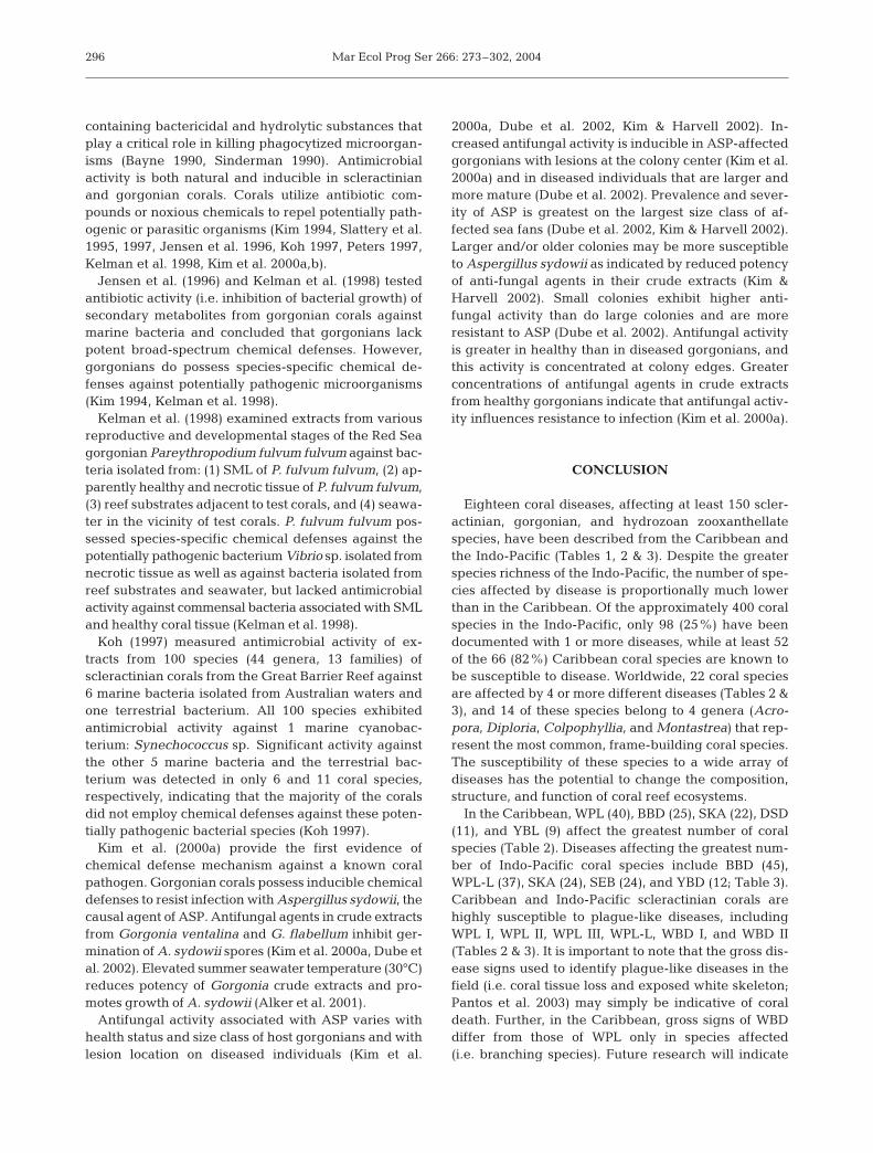
Mar Ecol Prog Ser 266: 273–302, 2004
containing bactericidal and hydrolytic substances thatplay a critical role in killing phagocytized microorgan-isms (Bayne 1990, Sinderman 1990). Antimicrobialactivity is both natural and inducible in scleractinianand gorgonian corals. Corals utilize antibiotic com-pounds or noxious chemicals to repel potentially path-ogenic or parasitic organisms (Kim 1994, Slattery et al.1995, 1997, Jensen et al. 1996, Koh 1997, Peters 1997,Kelman et al. 1998, Kim et al. 2000a,b).
Jensen et al. (1996) and Kelman et al. (1998) testedantibiotic activity (i.e. inhibition of bacterial growth) ofsecondary metabolites from gorgonian corals againstmarine bacteria and concluded that gorgonians lackpotent broad-spectrum chemical defenses. However,gorgonians do possess species-specific chemical de-fenses against potentially pathogenic microorganisms(Kim 1994, Kelman et al. 1998).
Kelman et al. (1998) examined extracts from variousreproductive and developmental stages of the Red Seagorgonian Pareythropodium fulvum fulvum against bac-teria isolated from: (1) SML of P. fulvum fulvum, (2) ap-parently healthy and necrotic tissue of P. fulvum fulvum,(3) reef substrates adjacent to test corals, and (4) seawa-ter in the vicinity of test corals. P. fulvum fulvum pos-sessed species-specific chemical defenses against thepotentially pathogenic bacterium Vibrio sp. isolated fromnecrotic tissue as well as against bacteria isolated fromreef substrates and seawater, but lacked antimicrobialactivity against commensal bacteria associated with SMLand healthy coral tissue (Kelman et al. 1998).
Koh (1997) measured antimicrobial activity of ex-tracts from 100 species (44 genera, 13 families) ofscleractinian corals from the Great Barrier Reef against6 marine bacteria isolated from Australian waters andone terrestrial bacterium. All 100 species exhibitedantimicrobial activity against 1 marine cyanobac-terium: Synechococcus sp. Significant activity againstthe other 5 marine bacteria and the terrestrial bac-terium was detected in only 6 and 11 coral species,respectively, indicating that the majority of the coralsdid not employ chemical defenses against these poten-tially pathogenic bacterial species (Koh 1997).
Kim et al. (2000a) provide the first evidence ofchemical defense mechanism against a known coralpathogen. Gorgonian corals possess inducible chemicaldefenses to resist infection with Aspergillus sydowii, thecausal agent of ASP. Antifungal agents in crude extractsfrom Gorgonia ventalina and G. flabellum inhibit ger-mination of A. sydowii spores (Kim et al. 2000a, Dube etal. 2002). Elevated summer seawater temperature (30°C)reduces potency of Gorgonia crude extracts and pro-motes growth of A. sydowii (Alker et al. 2001).
Antifungal activity associated with ASP varies withhealth status and size class of host gorgonians and withlesion location on diseased individuals (Kim et al.
2000a, Dube et al. 2002, Kim & Harvell 2002). In-creased antifungal activity is inducible in ASP-affectedgorgonians with lesions at the colony center (Kim et al.2000a) and in diseased individuals that are larger andmore mature (Dube et al. 2002). Prevalence and sever-ity of ASP is greatest on the largest size class of af-fected sea fans (Dube et al. 2002, Kim & Harvell 2002).Larger and/or older colonies may be more susceptibleto Aspergillus sydowii as indicated by reduced potencyof anti-fungal agents in their crude extracts (Kim &Harvell 2002). Small colonies exhibit higher anti-fungal activity than do large colonies and are moreresistant to ASP (Dube et al. 2002). Antifungal activityis greater in healthy than in diseased gorgonians, andthis activity is concentrated at colony edges. Greaterconcentrations of antifungal agents in crude extractsfrom healthy gorgonians indicate that antifungal activ-ity influences resistance to infection (Kim et al. 2000a).
CONCLUSION
Eighteen coral diseases, affecting at least 150 scler-actinian, gorgonian, and hydrozoan zooxanthellatespecies, have been described from the Caribbean andthe Indo-Pacific (Tables 1, 2 & 3). Despite the greaterspecies richness of the Indo-Pacific, the number of spe-cies affected by disease is proportionally much lowerthan in the Caribbean. Of the approximately 400 coralspecies in the Indo-Pacific, only 98 (25%) have beendocumented with 1 or more diseases, while at least 52of the 66 (82%) Caribbean coral species are known tobe susceptible to disease. Worldwide, 22 coral speciesare affected by 4 or more different diseases (Tables 2 &3), and 14 of these species belong to 4 genera (Acro-pora, Diploria, Colpophyllia, and Montastrea) that rep-resent the most common, frame-building coral species.The susceptibility of these species to a wide array ofdiseases has the potential to change the composition,structure, and function of coral reef ecosystems.
In the Caribbean, WPL (40), BBD (25), SKA (22), DSD(11), and YBL (9) affect the greatest number of coralspecies (Table 2). Diseases affecting the greatest num-ber of Indo-Pacific coral species include BBD (45),WPL-L (37), SKA (24), SEB (24), and YBD (12; Table 3).Caribbean and Indo-Pacific scleractinian corals arehighly susceptible to plague-like diseases, includingWPL I, WPL II, WPL III, WPL-L, WBD I, and WBD II(Tables 2 & 3). It is important to note that the gross dis-ease signs used to identify plague-like diseases in thefield (i.e. coral tissue loss and exposed white skeleton;Pantos et al. 2003) may simply be indicative of coraldeath. Further, in the Caribbean, gross signs of WBDdiffer from those of WPL only in species affected(i.e. branching species). Future research will indicate
296
Sutherland et al.: Disease and immunity of corals
whether plague-like signs on Caribbean and Indo-Pacific coral species represent a single disease condi-tion, caused by a single pathogen (e.g. Aurantimonascoralicida, Richardson et al. 1998a,b, Denner et al.2003), or if similar disease signs documented for theplague-like diseases represent different diseasescaused by a variety of pathogens.
Accumulating evidence suggests that human activityin the watershed may be causally related to coraldecline. Increases in the number of both new diseases(Fig. 2) and species affected may be directly linked to hu-man-induced alterations in coral reef environments bothin terms of land-based sources of pollution as well asglobal climate change issues such as global warming(Table 5). Elevated seawater temperature may be asso-ciated with the etiologies of at least 9 coral conditionsand is likely the most common abiotic stressor influenc-ing disease pathogenicity (Table 5). Further, 5 coraldisease-causing pathogens, including the BBD cyano-bacteria, Aurantimonas coralicida (WPL II), Aspergillussydowii (ASP), Vibrio shiloi (VSB), and V. coralliilyticus(VCB), are most virulent at seawater temperature at orabove 29°C (Kushmaro et al. 1998, Alker et al. 2001,Banin et al. 2001a, Israely et al. 2001, Ben-Haim & Rosen-berg 2002, Kuta & Richardson 2002, Ben-Haim et al.2003b, Richardson & Kuta 2003). Seawater temperaturenormally increases during late summer, but all currentmodels of global climate change suggest that, on aver-age, ocean temperatures will rise over the next century(Kleypas et al. 1999). Elevated temperature is a stressorin corals, causing thermally induced breakdown in thecoral-zooxanthellae host-symbiont relationship (Porteret al. 1989, Fitt et al. 1993, Brown 1997b). Elevated tem-perature also promotes growth and virulence of path-ogens (Kushmaro et al. 1996, 1998, Toren et al. 1998,Alker et al. 2001, Banin et al. 2001a, Israely et al. 2001,Ben-Haim & Rosenberg 2002, Kuta & Richardson 2002,Ben-Haim et al. 2003b, Richardson & Kuta 2003) andreduces immune response in host corals (Toren et al.1998, Alker et al. 2001).
Nutrient and sediment loading may deliver poten-tially pathogenic organisms to the marine environment.Transport of the terrestrial fungus Aspergillus sydowii,the causal agent of ASP, to the marine environmentmay be associated with local sediment run-off fromland or long-distance atmospheric transport (Smith etal. 1996, Shinn et al. 2000). Increasing evidence sug-gests that the health of reef organisms is affected bysewage pollution. BBD and WPD may be associatedwith fecal contamination of possible human origin(Frias-Lopez et al. 2002, Patterson et al. 2002). Humanenteric bacteria and viruses are prevalent on coralsurfaces and in nearshore, offshore, and canal watersin the Florida Keys (Lapointe et al. 1990, Paul et al.1995a,b, 1997, Griffin et al. 1999, Lipp et al. 2002). The
1983 epizootic affecting the Caribbean long-spined seaurchin Diadema antillarum caused catastrophic reduc-tions in urchin populations (Lessios et al. 1984). It hasbeen suggested that the fecal bacterium Clostridiumperfringens may have been involved in the urchin die-off (Bauer & Agerter 1987, 1994). Since 1996, popula-tions of the sewage consuming reef sponge Clionadelitrix have increased by a factor of 10 on reefs in theFlorida Keys (Ward-Paige 2003) while, concurrently,corals have declined by 37% (Porter et al. 2002).
Etiologies of only 5 coral diseases have been deter-mined through fulfillment of Koch’s postulates, but sev-eral other disease conditions have been linked to specificbiotic organisms (Table 4). The application of Koch’s pos-tulates for the identification of coral disease has severedrawbacks, and in order for the study of coral disease eti-ology to advance, alternative techniques for identifyingdisease-causing pathogens and abiotic stressors must beaccepted and implemented. Knowledge of coral diseasereservoirs, transmission, pathogenesis, and epizootio-logy is limited, and significant advances remain to bemade in the field of coral immunology.
Acknowledgements. We thank C. Beaver, W. Jaap, P. Dustan,V. Kosmynin, and the crews of the RV ‘Tortugas’ and OSV‘Peter W. Anderson’ for field assistance. We thank E. C. Petersand W. K. Fitt for comments on the manuscript, and C. Quirolofor field support and photographs.This research was supportedby a US Environmental Protection Agency/University of WestFlorida Science Training in Ecology Program (STEP) Fel-lowship (to K.P.S.), by a US EPA grant EPA-FDEP X-99-34649-94-0 (to J.W.P.), and by a Florida Department of Envi-ronmental Protection grant SP626 (E. K. Lipp, K.P.S. and J.W.P.).
LITERATURE CITED
Aeby GS (1998) A digenean metacercaria from the reef coral,Porites compressa, experimentally identified as Podocoty-loides stenometra. J Parasitol 84:1259–1261
Alker AP, Smith GW, Kim K (2001) Characterization ofAspergillus sydowii (Thom et Church) a fungal pathogenof Caribbean sea fan corals. Hydrobiologia 460:105–111
Al-Moghrabi SM (2001) Unusual black band disease (BBD)outbreak in the northern tip of the Gulf of Aqaba (Jordan).Coral Reefs 19:330–331
Anderson RM (1986) Genetic variability in resistance to para-sitic invasion: population implications for invertebratehost species. Symp Zool Soc Lond 56:239–274
Antonius A (1973) New observations on coral destruction inreefs. Abs Assoc Isl Mar Lab Caribb 10:3
Antonius A (1977) Coral mortality in reefs: a problem forscience and management. Proc 3rd Int Coral Reef SympMiami 2:617–623
Antonius A (1981) The ‘band’ diseases in coral reefs. Proc 4thInt Coral Reef Symp Philippines 2:7–14
Antonius A (1985a) Coral diseases in the Indo-Pacific: a firstrecord. PSZN I: Mar Ecol 6:197–218
Antonius A (1985b) Black band disease infection experimentson hexacorals and octocorals. Proc 5th Int Coral ReefCongr Moorea 6:155–160
297
Mar Ecol Prog Ser 266: 273–302, 2004
Antonius A (1988) Distribution and dynamics of coral diseasesin the Eastern Red Sea. Proc 6th Int Coral Reef SympTownsville 2:293–298
Antonius AA, Lipscomb D (2000) First protozoan coral-killeridentified in the Indo-Pacific. Atoll Res Bull 481:1–21
Antonius A, Riegl B (1997) A possible link between coral dis-eases and a corallivorous snail (Drupella cornus) outbreakin the Red Sea. Atoll Res Bull 447:1–9
Antonius A, Riegl B (1998) Coral diseases and Drupella cor-nus invasion in the Red Sea. Coral Reefs 17:48
Aronson RB, Precht WF (1997) Stasis biological disturbanceand community structure of a Holocene coral reef. Paleo-biology 23:326–346
Aronson RB, Precht WF (2001) White-band diseases and thechanging face of Caribbean coral reefs. Hydrobiologia460:25–38
Aronson RB, Precht WF, Macintyre IG (1998) Extrinsic controlof species replacement on a Holocene reef in Belize: therole of coral disease. Coral Reefs 17:223–230
Aronson RB, Macintyre IG, Precht WF, Murdoch TJT, Wap-nick CM (2002) The expanding scale of species turnoverevents on coral reefs in Belize. Ecol Monogr 72:233–249
Bak RPM (1983) Neoplasia regeneration and growth in thereef-building coral Acropora palmata. Mar Biol 77:221–227
Bak RPM, Criens SR (1982) Experimental fusion in AtlanticAcropora (scleractinia). Mar Biol Lett 3:67–72
Banin E, Israely T, Kushmaro A, Loya Y, Orr E, Rosenberg E(2000) Penetration of the coral-bleaching bacterium Vibrioshiloi into Oculina patagonica. Appl Environ Microbiol 66:3031–3036
Banin E, Khare SK, Naider F, Rosenberg E (2001a) Proline-rich peptide from the coral pathogen Vibrio shiloi thatinhibits photosynthesis of zooxanthellae. Appl EnvironMicrobiol 67:1536–1541
Banin E, Israely T, Fine M, Loya Y, Rosenberg E (2001b) Roleof endosymbiotic zooxanthellae and coral mucus in theadhesion of the coral-bleaching pathogen Vibrio shiloi toits host. FEMS Microbiol Lett 199:33–37
Bauer JC, Agerter CJ (1987) Isolation of bacteria pathogenicfor the sea urchin Diadema antillarum (Echinodermata:Echinoidea). Bull Mar Sci 40:161–165
Bauer JC, Agerter CJ (1994) Isolation of potentially patho-genic bacterial flora from tropical sea urchins in selectedWest Atlantic and East Pacific sites. Bull Mar Sci 55:142–150
Baya AM, Toranzo AE, Lupiani B, Santos Y, Hetrick FM(1992) Serratia marcescens: a potential pathogen for fish.J Fish Dis 15:15–26
Bayne CJ (1990) Phagocytosis and non-self recognition ininvertebrates. BioScience 40:723–731
Ben-Haim Y, Rosenberg E (2002) A novel Vibrio sp pathogenof the coral Pocillopora damicornis. Mar Biol 141:47–55
Ben-Haim Y, Banin E, Kushmaro A, Loya Y, Rosenberg E(1999) Inhibition of photosynthesis and bleaching of zoo-xanthellae by the coral Vibrio shiloi. Environ Microbiol 1:223–229
Ben-Haim Y, Thompson FL, Thompson CC, Cnockaert MC,Hoste B, Swings J, Rosenberg E (2003a) Vibrio coralliily-ticus sp nov, a temperature-dependent pathogen of thecoral Pocillopora damicornis. Int J Syst Evol Microbiol53:309–315
Ben-Haim Y, Zicherman-Keren M, Rosenberg E (2003b)Temperature-regulated bleaching and lysis of the coralPocillopora damicornis by the novel pathogen Vibriocoralliilyticus. Appl Environ Microbiol 69:4236–4242
Bigger CH, Hildemann WH (1982) Cellular defense systemsof the Coelenterata. In: Cohen N, Sigel MM (eds) The reti-culoendothelial system. Plenum Press, New York, p 59–87
Bigger CH, Jokiel PL, Hildemann WH, Johnston IS (1982)Characterization of alloimmune memory in a sponge.J Immunol 129:1570–1572
Bigger CH, Jokiel PL, Hildemann WH (1983) Cytotoxic trans-plantation immunity in the sponge Toxadocia violacea.Transplantation 35:239–243
Brown BE (1997a) Disturbances to reefs in recent times. In:Birkeland C (ed) Life and death of coral reefs. Chapman &Hall, New York, p 354–379
Brown BE (1997b) Coral bleaching: causes and consequences.Coral Reefs 16:129–138
Brown BE, Dunne RP, Scoffin TP, Le Tissier MDA (1994) Solardamage in intertidal corals. Mar Ecol Prog Ser 105:219–230
Brown BE, Dunne RP, Goodson MS, Douglas AE (2000)Bleaching patterns in reef corals. Nature 404:142–143
Bruckner AW, Bruckner RJ (1997a) Outbreak of coral diseasein Puerto Rico. Coral Reefs 16:260
Bruckner AW, Bruckner RJ (1997b) The persistence of blackband disease in Jamaica: impact on community structure.Proc 8th Int Coral Reef Symp, Panama 1:601–606
Bruckner AW, Bruckner RJ (2002) Coral predation by Spari-sima viride and lack of relationship with coral disease.Proc 9th Int Coral Reef Symp, Bali 2:1245–1249
Bruckner AW, Bruckner RJ, Williams EH Jr (1997) Spread of ablack-band disease epizootic through the coral reef sys-tem in St Ann’s Bay Jamaica. Bull Mar Sci 61:919–928
Bythell J, Sheppard C (1993) Mass mortality of Caribbeanshallow corals. Mar Pollut Bull 26:296–297
Bythell JC, Barer MR, Cooney RP, Guest JR, O’Donnell AG,Pantos O, Le Tissier MDA (2002) Histopathologicalmethods for the investigation of microbial communitiesassociated with disease lesions in reef corals. Lett ApplMicrobiol 34:359–364
Carlton RG, Richardson LL (1995) Oxygen and sulfidedynamics in a horizontally migrating cyanobacterial mat:black band disease of corals. FEMS Microbiol Ecol 18:155–162
Cerrano C, Bavestrello G, Bianchi CN, Cattaneo-vietti R and 8others (2000) A catastrophic mass-mortality episode of gor-gonians and other organisms in the Ligurian Sea (North-western Mediterranean) summer 1999. Ecol Lett 3:284–293
Cervino J, Goreau TJ, Goreau M, Smith G and 11 others(1997) Rapid spread of diseases in Caribbean coral reefs.Proc Assoc Isl Mar Lab Caribb 28:43
Cervino J, Goreau TJ, Nagelkerken I, Smith GW, Hayes R(2001) Yellow band and dark spot syndromes inCaribbean corals: distribution rate of spread cytology andeffects on abundance and division rate of zooxanthellae.Hydrobiologia 460:53–63
Cheney DP (1975) Hard tissue tumors of scleractinian corals.In: Hildemann WH, Benedict AA (ed) Immunologic phylo-geny. Plenum, New York, p 77–87
Cheng TC, Wong AKL (1974) Chemical, histochemical, andhistopathological studies on corals, Porites spp., parasitizedby trematode metacercariae. J Invertebr Pathol 23:303–317
Coles SL (1994) Extensive coral disease outbreak at FahlIsland Gulf of Oman Indian Ocean. Coral Reefs 13:242
Coles SL, Jokiel PL (1992) Effects of salinity on coral reefs. In:Connell DW, Hawker DW (ed) Pollution in tropical aquaticsystems. CRC Press, Boca Raton, FL, p 147–166
Coles SL, Seapy DG (1998) Ultra-violet absorbing compoundsand tumorous growths on acroporid corals from BandarKhayran Gulf of Oman Indian Ocean. Coral Reefs 17:195–198
Cooney RP, Pantos O, Le Tissier MDA, Barer MR, O’DonnellAG, Bythell JC (2002) Characterization of the bacterialconsortium associated with black band disease in coral
298

Sutherland et al.: Disease and immunity of corals
using molecular microbiological techniques. EnvironMicrobiol 47:401–413
Denner EBM, Smith G, Busse HJ, Schumann P, Narzt T, Pol-son SW, Lubitz W, Richardson LL (2003) Aurantimonascoralicida gen. nov., sp. nov., the causative agent of whiteplague type II on Caribbean scleractinian corals. Int J SystEvol Microbiol 53:1115–1122
Diaz-Soltero H (1999) Endangered and threatened species: arevision of candidate species list under the EndangeredSpecies Act. Fed Register 64(210):33 466–33 468
Dinsdale EA (2002) Abundance of black-band disease oncorals from one location on the Great Barrier Reef: a com-parison with abundance in the Caribbean region. Proc 9thInt Coral Reef Symp, Bali 2:1239–1243
Dube D, Kim K, Alker AP, Harvell CD (2002) Size structureand geographic variation in chemical resistance of sea fancorals Gorgonia ventalina to a fungal pathogen. Mar EcolProg Ser 231:139–150
Ducklow H, Mitchell R (1979a) Bacterial populations andadaptations in the mucus layers on living corals. LimnolOceanogr 24:715–725
Ducklow H, Mitchell R (1979b) Observations on naturally andartificially diseased tropical corals: a scanning electronmicroscope study. Microbiol Ecol 5:215–223
Dustan P (1977) Vitality of reef coral populations off Key LargoFlorida: recruitment and mortality. Environ Geol 2:51–58
Dustan P (1999) Coral reefs under stress: sources of mortalityin the Florida Keys. Nat Res Forum 23:147–155
Edmunds PJ (1991) Extent and effect of black band disease ona Caribbean reef. Coral Reefs 10:161–165
Evans AS (1976) Causation and disease: the Henle-Kochpostulates revisited. Yale J Biol Med 49:175–195
Falkow S (1988) Molecular Koch’s postulates applied tomicrobial pathogenicity. Rev Infect Dis 10:S274–S276
Feingold JS (1988) Biological studies of a cyanobacterialinfection on the Caribbean sea plume Pseudopterogorgiaacerosa (Coelenterata: Octocorallia). Proc 6th Int CoralReef Symp, Townsville 3:157–162
Fitt WK, Spero H, Halas J, White M, Porter J (1993) Recovery ofthe coral Montastraea annularis in the Florida Keys after the1987 Caribbean ‘bleaching event.’ Coral Reefs 12:57–64
Fredricks DN, Relman DA (1996) Sequence-based identifica-tion of microbial pathogens: a reconsideration of Koch’spostulates. Clin Microbiol Rev 9:18–33
Frias-Lopez J, Zerkle AL, Bonheyo GT, Fouke BW (2002) Par-titioning of bacterial communities between seawater andhealthy black band diseased and dead coral surfaces.Appl Environ Microbiol 68:2214–2228
Frias-Lopez J, Bonheyo GT, Jin Q, Fouke BW (2003) Cyano-bacteria associated with coral black band disease inCaribbean and Indo-Pacific reefs. Appl Environ Microbiol69:2409–2413
Garrett P, Ducklow H (1975) Coral diseases in Bermuda.Nature 253:349–350
Garzón-Ferreira JM, Zea S (1992) A mass mortality of Gorgo-nia ventalina (Cnidaria: Gorgoniidae) in the Santa Martaarea Caribbean coast of Colombia. Bull Mar Sci 50:522–526
Garzón-Ferreira J, Gil-Agudelo DL, Barrios LM, Zea S (2001)Stony coral diseases observed in southwestern Caribbeanreefs. Hydrobiologia 460:65–69
Geiser DM, Taylor JW, Ritchie KB, Smith GW (1998) Cause ofsea fan death in the West Indies. Nature 394:137–138
Gil-Agudelo DL, Garzón-Ferreira J (2001) Spatial and sea-sonal variation of dark spots disease in coral communitiesof the Santa Marta area (Colombian Caribbean). Bull MarSci 69:619–629
Gladfelter EH, Monahan RK, Gladfelter WB (1978) Growth
rates of five reef-building corals in the northeastern Carib-bean. Bull Mar Sci 28:728–734
Gladfelter WB (1982) White-band disease in Acropora pal-mata: implications for the structure and growth of shallowreefs. Bull Mar Sci 32:639–643
Glynn PW, Howard LS, Corcoran E, Cofer-Shabica SV (1984)The occurrence and toxicity of herbicides in reef buildingcorals. Mar Pollut Bull 15:370–374
Goldberg WM, Makemson JC, Colley SB (1984) Entocladiaendozoica sp nov a pathogenic chlorophyte: structure lifehistory physiology and effect on its coral host. Biol Bull166:368–383
Goreau TJ, Cervino J, Goreau M, Hayes R and 14 others(1998) Rapid spread of diseases in Caribbean coral reefs.Rev Biol Trop 46:157–171
Green EP, Bruckner AW (2000) The significance of coraldisease epizootiology for coral reef conservation. BiolConserv 96:347–361
Greenstein BJ, Curran HA, Pandolfi JM (1998) Shifting eco-logical baselines and the demise of Acropora cervicornisin the western North Atlantic and Caribbean Province: aPleistocene perspective. Coral Reefs 17:249–261
Griffin DW, Gibson III CJ, Lipp EK, Riley K, Paul III JH, RoseJB (1999) Detection of viral pathogens by reverse tran-scriptase PCR and of microbial indicators by standardmethods in the canals of the Florida Keys. Appl EnvironMicrobiol 65:4118–4125
Grimont PA, Grimont F (1994) Genus VIII Serratia Bizio, 1823.In: Holt JG, Kreig NR, Sneath PHA, Staley JT, WilliamsST (eds) Bergey’s manual of determinative bacteriology,Vol 4. Williams & Wilkins, Baltimore, p 477–484
Grygier MJ, Cairns SD (1996) Suspected neoplasms in deep-sea corals (Scleractinia: Oculinidae: Madrepora spp.) re-interpreted as galls caused by Petrarca madreporae n. sp.(Crustacea: Ascothoracida: Petrarcidae). Dis Aquat Org24:61–69
Harland AD, Brown BE (1989) Metal tolerance in the sclerac-tinian coral Porites lutea. Mar Pollut Bull 20:353–357
Harvell CD, Kim K, Burkholder JM, Colwell RR and 9 others(1999) Emerging marine diseases-climate links and anthro-pogenic factors. Science 285:1505–1510
Harvell D, Kim K, Quirolo C, Weir J, Smith G (2001) Coralbleaching and disease: contributors to 1998 mass mortalityin Briareum asbestinum (Octocorallia Gorgonacea). Hydro-biologia 460:97–104
Hayes RL, Goreau TJ (1998) The significance of emerging dis-eases in the tropical coral reef ecosystem. Rev Biol Trop46:173–185
Hildemann WH, Linthicum DS (1981) Transplantation im-munity in the Palaun sponge Xestospongia exigua.Transplantation 32:77–80
Hildemann WH, Linthicum DS, Vann DC (1975) Transplanta-tion and immunoincompatibility reactions among reef-building corals. Immunogenetics 2:269–284
Hildemann WH, Raison RL, Cheung G, Hull CJ, Akaka L,Okamoto J (1977a) Immunological specificity and memoryin a scleractinian coral. Nature 270:219–223
Hildemann WH, Raison RL, Hull CJ, Akaka LK, Okamoto J,Cheung GP (1977b) Tissue transplantation immunity incorals. Proc 3rd Int Coral Reef Symp Miami 1:538–543
Hildemann WH, Johnson IS, Jokiel PL (1979) Immunocompe-tence in the lowest Metazoan phylum: transplantationimmunity in sponges. Science 204:420–422
Hildemann WH, Jokiel PL, Bigger CH, Johnston IS (1980a)Allogeneic polymorphism and alloimmune memory inthe coral Montipora verrucosa. Transplantation 30:297–301
Hildemann WH, Bigger CH, Johnston IS, Jokiel PL (1980b)
299
Mar Ecol Prog Ser 266: 273–302, 2004
Characteristics of transplantation immunity in the spongeCallyspongia diffusa. Transplantation 30:362–367
Hill AB (1965) The environment and disease: association orcausation? Proc R Soc Med 58:295–300
Hodgson G (1990) Tetracycline reduces sedimentation dam-age to corals. Mar Biol 104:493–496
Holden C (1996) Coral disease hot spot in the Florida Keys.Science 274:2017
Huebner RJ (1957) The virologist’s dilemma. Ann NY AcadSci 67:430–438
Hughes TP (1994) Catastrophes phase shifts and large-scale de-gradation of a Caribbean coral reef. Science 265:1547–1551
Hunter CL, Peters EC (1993) Effects of stress-induced necro-sis and ‘tumors’ on the scleractinian coral Porites lobatain Hawaii. Am Zool 33:19A
Israely T, Banin E, Rosenberg E (2001) Growth differentiationand death of Vibrio shiloi in coral tissue as a function ofseawater temperature. Aquat Microb Ecol 24:1–8
Jensen PR, Harvell CD, Wirtz K, Fenical W (1996) Antimicro-bial activity of extracts of Caribbean gorgonian corals.Mar Biol 125:411–419
Johnson RT, Gibbs CJ (1974) Koch’s postulates and slowinfections of the nervous system. Arch Neurol 30:36–38
Johnston IS, Hildemann WH (1983) Morphological correlatesof intraspecific grafting reactions in the marine demo-sponge Callyspongia diffusa. Mar Biol 74:25–33
Johnston IS, Jokiel PL, Bigger CH, Hildemann WH (1981) Theinfluence of temperature on the kinetics of allograft reactionsin a tropical sponge and a reef coral. Biol Bull 160:280–291
Jokiel PL, Bigger CH (1994) Aspects of histocompatibilityand regeneration in the solitary reef coral Fungia scutaria.Biol Bull 186:72–80
Jolles AE, Sullivan P, Alker AP, Harvell CD (2002) Diseasetransmission of aspergillosis in sea fans: inferring processfrom spatial pattern. Ecology 83:2373–2378
Karp RD, Hildemann WH (1976) Specific allograft reactivityin the sea star Dermasterias imbricata. Transplantation 22:434–439
Kelman D, Kushmaro A, Loya Y, Kashman Y, Benayahu Y(1998) Antimicrobial activity of a Red Sea soft coral Par-erythropodium fulvum fulvum: reproductive and develop-mental considerations. Mar Ecol Prog Ser 169:87–95
Kendrick B, Risk MJ, Michaelides J, Bergman K (1982)Amphibious microborers: bioeroding fungi isolated fromlive corals. Bull Mar Sci 32:862–867
Kim K (1994) Antimicrobial activity in gorgonian corals(Coelenterata Octocorallia). Coral Reefs 13:75–80
Kim K, Harvell CD (2002) Aspergillosis of sea fan corals: dis-ease dynamics in the Florida Keys. In: Porter JW, PorterKG (ed) The Everglades Florida Bay and coral reefs ofthe Florida Keys: an ecosystem sourcebook. CRC Press,New York, p 813–824
Kim K, Harvell CD, Smith GW (1997) Mechanisms of seafan resistance to a fungal epidemic. I. Role of sclerites.Am Zool 37:132A
Kim K, Harvell CD, Kim PD, Smith GW, Merkel SM (2000a)Fungal disease resistance of Caribbean sea fan corals(Gorgonia spp). Mar Biol 36:259–267
Kim K, Kim PD, Alker AP, Harvell CD (2000b) Chemical resis-tance of gorgonian corals against fungal infections. MarBiol 137:393–401
Kinne O (1980) Diseases of marine animals: general aspects.In: Kinne O (ed) Diseases of marine animals, Vol 1. Gen-eral aspects, Protozoa to Gastropoda. John Wiley & Sons,New York
Kleypas JA, Buddemeier RW, Archer D, Gattuso JP, LangdonC, Opdyke BN (1999) Geochemical consequences of
increased atmospheric carbon dioxide on coral reefs.Science 284:118–120
Koch R (1882) Die Aetiologie der Tuberculose. Berl KlinischeWochenschrift 19:221–230
Koh EGL (1997) Do scleractinian corals engage in chemicalwarfare against microbes? J Chem Ecol 23:379–398
Korrûbel JL, Riegl B (1998) A new coral disease from theArabian Gulf. Coral Reefs 17:22
Kushmaro A, Loya Y, Fine M, Rosenberg E (1996) Bacterialinfection and coral bleaching. Nature 380:396
Kushmaro A, Rosenberg E, Fine M, Loya Y (1997) Bleachingof the coral Oculina patagonica by Vibrio AK-1. Mar EcolProg Ser 147:159–165
Kushmaro A, Rosenberg E, Fine M, Ben-Haim Y, Loya Y (1998)Effect of temperature on bleaching of the coral Oculina pata-gonica by Vibrio shiloi AK-1. Mar Ecol Prog Ser 171:131–137
Kushmaro A, Banin E, Loya Y, Stackebrandt E, Rosenberg E(2001) Vibrio shiloi sp nov the causative agent of bleach-ing of the coral Oculina patagonica. Int J Syst Evol Micro-biol 51:1383–1388
Kuta KG, Richardson LL (1996) Abundance and distributionof black band disease on coral reefs in the northernFlorida Keys. Coral Reefs 15:219–223
Kuta KG, Richardson LL (2002) Ecological aspects of blackband disease of corals: relationships between disease inci-dence and environmental factors. Coral Reefs 21:393–398
Lakshma Reddy A, Bryan B, Hildemann WH (1975) Integu-mentary allograft versus autograft reactions in Cionaintestinalis: a protochordate species of solitary tunicate.Immunogenetics 1:584–590
Lang J (1973) Interspecific aggression by scleractinian corals. 2.Why the race is not only to the swift. Bull Mar Sci 23:260–279
Langlet C, Bierne J (1982) Immune characteristics of graft re-jection in nemerteans of the genus Lineus. Eur J Immunol12:705–708
Lapointe BE, O’Connell JD, Garrett GS (1990) Nutrient cou-pling between on-site sewage disposal systems ground-waters and nearshore surface waters of the Florida Keys.Biogeochemistry 10:289–307
Le Champion-Alsumard T, Golubic S, Priess K (1995) Fungi incorals: symbiosis or disease? Interaction between polypsand fungi causes pearl-like skeleton biomineralization.Mar Ecol Prog Ser 117:137–147
Lessios HA, Robertson DR, Cubit JD (1984) Spread of Diademamass mortality through the Caribbean. Science 226:335–337
Lipp EK, Jarrell JL, Griffin DW, Lukasik J, Jacukiewicz J,Rose JB (2002) Preliminary evidence for human fecal con-tamination in corals of the Florida Keys, USA. Mar PollBull 44:666–670
Littler MM, Littler DS (1996) Black band disease in the SouthPacific. Coral Reefs 15:20
Loya Y, Bull G, Pichon M (1984) Tumor formations in sclerac-tinian corals. Helgol Wiss Meeresunters 37:99–112
Maguire LA, Porter JW (1977) A spatial model of growth andcompetition strategies in coral communities. Ecol Model 3:249–271
Meszaros A, Bigger C (1999) Qualitative and quantitativestudy of wound healing processes in the coelenteratePlexaurella fusifera: spatial temporal and environmental(light attenuation) influences. J Invertebr Pathol 73:321–331
Miller I (1996) Black band disease on the Great Barrier Reef.Coral Reefs 15:58
Miller MW, Bourgue AS, Bohnsack JA (2002) An analysis ofthe loss of acroporid corals at Looe Key, Florida USA:1983–2000. Coral Reefs 21:179–182
Mitchell R, Chet I (1975) Bacterial attack of corals in pollutedwater. Microb Ecol 2:227–233
300

Sutherland et al.: Disease and immunity of corals
Morse DE, Morse ANC, Duncan H (1977) Algal ‘tumors’ in theCaribbean sea-fan Gorgonia ventalina. Proc 3rd Int CoralReef Symp Miami 1:623–629
Morse DE, Morse A, Duncan H, Trench RK (1981) Algaltumors in the Caribbean octocorallian Gorgonia ventalina:II. Biochemical characterization of the algae and first epi-demiological observations. Bull Mar Sci 31:399–409
Muntanola-Cvetkovic M, Ristanovic B (1980) A mycologicalsurvey of the south Adriatic Sea. J Exp Mar Biol Ecol 43:193–206
Nagelkerken I, Buchan K, Smith GW, Bonair K and 10 others(1997a) Widespread disease in Caribbean sea fans: II. Pat-terns of infection and tissue loss. Mar Ecol Prog Ser 160:255–263
Nagelkerken I, Buchan K, Smith GW, Bonair K and 8 others(1997b) Widespread disease in Caribbean sea fans: I.Spreading and general characteristics. Proc 8th Int CoralReef Symp, Panama 1:679–682
Neigel JE, Avise JC (1983) Clonal diversity and populationstructure in a reef-building coral, Acropora cervicornis:self-recognition analysis and demographic interpretation.Evolution 37:437–453
Nugues MM (2002) Impact of a coral disease outbreak oncoral communities in St Lucia: what and how much hasbeen lost? Mar Ecol Prog Ser 229:61–71
Pantos O, Cooney RP, Le Tissier MDA, Barer MR, O’DonnellAG, Bythell JC (2003) The bacterial ecology of a plague-like disease affecting the Caribbean coral Montastreaannularis. Environ Microbiol 5:370–382
Patterson KL, Porter JW, Ritchie KB, Polson SW, Mueller E,Peters EC, Santavy DL, Smith GW (2002) The etiology ofwhite pox a lethal disease of the Caribbean elkhorn coralAcropora palmata. Proc Natl Acad Sci USA 99:8725–8730
Patterson MJ, Landolt ML (1979) Cellular reaction to injuryin the Anthozoan Anthopleura elegantissima. J InvertebrPathol 33:189–196
Paul JH, Rose JB, Brown J, Shinn EA, Miller S, Farrah SR(1995a) Viral tracer studies indicate contamination ofmarine waters by sewage disposal practices in Key Largo,Florida. Appl Environ Microbiol 61:2230–2234
Paul JH, Rose JB, Jiang S, Kellogg C, Shinn EA (1995b)Occurrence of fecal indicator bacteria in surface watersand the subsurface aquifer in Key Largo, Florida. ApplEnviron Microbiol 61:2235–2241
Paul JH, Rose JB, Jiang SC, Zhou X and 6 others (1997) Evi-dence for groundwater and surface marine water contam-ination by waste disposal wells in the Florida Keys. WaterRes 31:1448–1454
Peters EC (1984) A survey of cellular reactions to environ-mental stress and disease in Caribbean scleractiniancorals. Helgol Meeresunters 37:113–137
Peters EC (1993) Diseases of other invertebrate phyla: Porifera,Cnidaria, Ctenophora, Annelida, Echinodermata. In: CouchJA, Fournie JW (eds) Pathobiology of marine and estuarineorganisms. CRC Press, Boca Raton, FL, p 393–449
Peters EC (1997) Diseases of coral reef organisms. In: Birke-land C (ed) Life and death of coral reefs. Chapman & Hall,New York, p 114–139
Peters EC, Meyers PA, Yevich PP, Blake NJ (1981) Bioaccu-mulation and histopathological effects of oil on a stonycoral. Mar Pollut Bull 12:333–339
Peters EC, Yevich PP, Oprandy JJ (1983) Possible causalagent of ‘white band disease’ in Caribbean acroporidcorals. J Invertebr Pathol 41:394–396
Peters EC, Halas JC, McCarty HB (1986) Calicoblastic neo-plasms in Acropora palmata with a review of reports onanomalies of growth and form in corals. J Natl Cancer Inst
76:895–912Porter JW (1974) Community structure of coral reefs on oppo-
site sides of the Isthmus of Panama. Science 186:543–545Porter JW, Tougas JI (2001) Reef ecosystems: threats to their
biodiversity. Encyclopedia of biodiversity, Vol 5. Acade-mic Press, San Diego, p 73–95
Porter JW, Fitt WK, Spero HJ, Rogers CS, White MW (1989)Bleaching in reef corals: physiological and stable isotopicresponses. Proc Natl Acad Sci USA 86:9342–9346
Porter JW, Dustan P, Jaap WC, Patterson KL, Kosmynin V, MeierOW, Patterson ME, Parsons M (2001) Patterns of spread ofcoral disease in the Florida Keys. Hydrobiologia 460:1–24
Porter JW, Kosmynin V, Patterson KL, Porter KG and 13 oth-ers (2002) Detection of coral reef change by the FloridaKeys Coral Reef Monitoring Project. In: Porter JW, PorterKG (eds) The Everglades Florida Bay and coral reefs of theFlorida Keys. CRC Press, Boca Raton, FL, p 749–769
Raftos DA, Tait NN, Briscoe DA (1987) Allograft rejection andalloimmune memory in the solitary urochordate Styelaplicata. Dev Comp Immunol 11:343–351
Raison RL, Hull CJ, Hildemann WH (1976) Allogenic graftrejection in Montipora verrucosa, a reef-building coral.In: Wright RK, Cooper EL (eds) Phylogeny of thymus andbone marrow-bursa cells. Elsevier/North-Holland Bio-medical Press, Amsterdam, p 3–8
Ramos-Flores T (1983) Lower marine fungus associated withblack line disease in star corals (Montastraea annularisE & S). Biol Bull 165:429–435
Ravindran JC, Raghukumar C (2002) Pink line syndrome (PLS)in the scleractinian coral Porites lutea. Coral Reefs 21:252
Ravindran J, Raghukumar C, Raghukumar S (2001) Fungi inPorites lutea: association with healthy and diseased corals.Dis Aquat Org 47:219–228
Richardson LL (1993) Red band disease: a new cyanobacterialinfestation of corals. Pure Appl Chem 1:153–160
Richardson LL (1996) Horizontal and vertical migration patternsof Phormidium corallyticum and Beggiatoa spp. associatedwith black-band disease of corals. Microb Ecol 32:323–335
Richardson LL (1997) Occurrence of the black band diseasecyanobacterium on healthy corals of the Florida Keys.Bull Mar Sci 61:485–490
Richardson LL (1998) Coral diseases: what is really known?Trends Ecol Evol 13:438–443
Richardson LL, Kuta KG (2003) Ecological physiology of theblack band disease cyanobacterium Phormidium coral-lyticum. FEMS Microbiol Ecol 43:287–298
Richardson LL, Kuta KG, Schnell S, Carlton RG (1997) Eco-logy of the black band disease microbial consortium.Proc 8th Int Coral Reef Symp Balboa 1:597–600
Richardson LL, Goldberg WM, Carlton RG, Halas JC (1998a)Coral disease outbreak in the Florida Keys: plague type II.Rev Biol Trop 46:187–198
Richardson LL, Goldberg WM, Kuta KG, Aronson RB and5 others (1998b) Florida’s mystery coral killer identified.Nature 392:557–558
Richardson LL, Smith GW, Ritchie KB, Carlton RG (2001) Inte-grating microbiological microsensor molecular and physi-ologic techniques in the study of coral disease pathogene-sis. Hydrobiologia 460:71–89
Richmond RH (1993) Coral reefs: present problems and futureconcerns resulting from anthropogenic disturbance. AmZool 33:524–536
Riegl B (2002) Effects of the 1996 and 1998 positive sea-sur-face temperature anomolies on corals, coral diseases andfish in the Arabian Gulf (Dubai, UAE). Mar Biol 140:29–40
Ritchie KB, Smith GW (1995a) Carbon-source utilizationpatterns of coral associated marine heterotrophs. J Mar
301

Mar Ecol Prog Ser 266: 273–302, 2004
Biotechnol 3:105–107Ritchie KB, Smith GW (1995b) Preferential carbon utilization
bysurface bacterial communities from water mass normaland white-band diseased Acropora cervicornis. Mol MarBiol Biotechnol 4:345–352
Ritchie KB, Smith GW (1998) Type II white-band disease.Rev Biol Trop 46:199–203
Ritchie KB, Polson SW, Smith GW (2001) Microbial diseasecausation in marine invertebrates: problems, practices,and future prospects. Hydrobiologia 460:131–139
Rivers TM (1937) Viruses and Koch’s postulates. J Bacteriol33:1–12
Roch P (1999) Defense mechanisms and disease prevention infarmed marine invertebrates. Aquaculture 172:125–145
Rodríquez-Martínez RE, Banaszak AT, Jordán-Dahlgren E(2001) Necrotic patches affect Acropora palmata (Sclerac-tinia: Acroporidae) in the Mexican Caribbean. Dis AquatOrg 47:229–234
Roitt I, Brostoff J, Male D (1996) Immunology, 4th edn. Mosby,New York
Rosenberg E, Ben-Haim Y, Toren A, Banin E, Kushmaro A, FineM, Loya Y (1998) Effect of temperature on bacterial bleach-ing of corals. In: Rosenberg E (ed) Current perspectives inmicrobial ecology. ASM Press, Washington, DC, p 242–254
Rublee PA, Lasker HR, Gottfried M, Roman MR (1980) Pro-duction and bacterial colonization of mucus from the softcoral Briarium asbestinum. Bull Mar Sci 30:888–893
Rützler K, Santavy D (1983) The black band disease of At-lantic reef corals. I. Description of a cyanophyte pathogen.PSZN I: Mar Ecol 4:301–319
Rützler K, Santavy DL, Antonius A (1983) The black band dis-ease of Atlantic reef corals. III. Distribution ecology anddevelopment. PSZN I: Mar Ecol 4:329–358
Santavy DL, Peters EC (1997) Microbial pests: coral diseaseresearch in the western Atlantic. Proc 8th Int Coral ReefSymp Balboa 1:607–612
Santavy DL, Peters EC, Quirolo C, Porter JW, Bianchi CN(1999) Yellow-blotch disease outbreak on reefs of the SanBlas Islands Panama. Coral Reefs 18:97
Santavy DL, Mueller E, Peters EC, MacLaughlin L, Porter JW,Patterson KL, Campbell J (2001) Quantitative assessmentof coral diseases in the Florida Keys: strategy and method-ology. Hydrobiologia 460:39–52
Schnell S, Assmus B, Richardson LL (1996) Role of sulfate-re-ducing bacteria in the black band disease of corals. AnnualMeeting of the VAAM (Vereinigung fuer Allgemeine undAngewandte Mikrobiologie) and GBCH (Gesellschaft fuerBiologische Chemie). Biospektrum, p 116
Shick JM, Lesser MP, Jokiel PL (1996) Effects of ultravioletradiation on corals and other coral reef organisms. GlobChang Biol 2:527–545
Shinn EA, Smith GW, Prospero JM, Betzer P, Hayes ML, Gar-rison V, Barber RT (2000) African dust and the demise ofCaribbean coral reefs. Geophys Res Lett 27:3029–3032
Sinderman CJ (1990) Principal diseases of marine fish andshellfish, Vol 2, 2nd edn. Academic Press, New York
Slattery M (1999) Fungal pathogenesis of the sea fan Gorgo-nia ventalina: direct and indirect consequences. Chemo-ecology 9:97–104
Slattery M, McClintock JB, Heine JN (1995) Chemicaldefenses in Antarctic soft corals: evidence for antifoulingcompounds. J Exp Mar Biol Ecol 190:61–77
Slattery M, Hamann MT, McClintock JB, Perry TL, PuglisiMP, Yoshida WY (1997) Ecological roles for water-bornemetabolites from Antarctic soft corals. Mar Ecol Prog Ser161:133–144
Smith GW, Ives LD, Nagelkerken IA, Ritchie KB (1996)Caribbean sea-fan mortalities. Nature 383:487
Smith GW, Harvell CD, Kim K (1998) Response of sea fans toinfection with Aspergillus sp. (Fungi). Rev Biol Trop 46:205–208
Squires DF (1965) Neoplasia in a coral? Science 148:503–505Stedman TL (2000) Stedman’s medical dictionary, 27th edn.
Lippincott Williams & Wilkins, BaltimoreSussman M, Loya Y, Fine M, Rosenberg E (2003) The marine
fireworm Hermodice carunculata is a winter reservoir andspring-summer vector for the coral-bleaching pathogenVibrio shiloi. Environ Microbiol 5:250–255
Sutherland KP, Ritchie KB (in press) White pox disease of theCaribbean elkhorn coral Acropora palmata. In: RosenbergE, Loya Y (eds) Coral health and disease. Springer-Verlag,Heidelberg
Taylor DL (1983) The black band disease of Atlantic reefcorals. PSZN I: Mar Ecol 4:321–328
Theodor JL (1970) Distinction between ‘self’ and ‘not-self’ inlower invertebrates. Nature 227:690–692
Toller WW, Rowan R, Knowlton N (2001) Repopulation ofzooxanthellae in the Caribbean corals Montastraea annu-laris and M. faveolata following experimental and dis-ease-associated bleaching. Biol Bull 201:360–373
Toren A, Landau L, Kushmaro A, Loya Y, Rosenberg E (1998)Effect of temperature on adhesion of Vibrio strain AK-1 toOculina patagonica and on coral bleaching. Appl EnvironMicrobiol 64:1379–1384
US BOC (US Bureau of the Census) (2000) 2000 Census ofPopulation. US Department of Commerce, Economics andStatistics Division, US BOC, Washington, DC
US EPA (US Environmental Protection Agency) (2000)Stressor identification document. Office of Water, Officeof Research and Development, US EPA, Washington, DC
Veron J (1985) Aspects of the biogeography of hermatypiccorals. Proc 5th Int Coral Reef Cong Tahiti 4:83–88
Veron J (1995) Corals in space and time. The biogeographyand evolution of the Scleractinia. Comstock-Cornell Press,Ithaca, NY
Viehman TS, Richardson LL (2002) Motility patterns of Beg-giatoa and Phormidium corallyticum in black band dis-ease. Proc 9th Int Coral Reef Symp, Bali 2:1251–1255
Ward-Paige CA (2003) Bioerosion surveys on the Florida reeftract suggest widespread land-based stress on reefs. MSthesis, McMaster Univ, Ontario
Weil E, Urreiztieta I, Garzón-Ferreira J (2002) Geographicvariability in the incidence of coral and octocoral diseasesin the wider Caribbean. Proc 9th Int Coral Reef Symp Bali2:1231–1237
Weir JW, Garrison V, Shinn EA, Smith GW (in press) The rela-tionship between gorgonian coral (Cnidaria: Gorgonacea)diseases and African dust storms. Aerobiology
Wells J (1973) New and old scleractinian corals from Jamaica.Bull Mar Sci 23:16–58
Werner T, Allen G (ed) (1998) A rapid biodiversity assessmentof the coral reefs of Milne Bay Province, Papua NewGuinea. Conservation International, Washington, DC
Wheaton J, Jaap WC, Porter JW, Kosmynin V and 7 others(2001) EPA/FKNMS Coral Reef Monitoring Project Execu-tive Summary. FKNMS Symposium: an ecosystem reportcard. Florida Mar Res Inst, St. Petersburg, FL
Wilkinson C (ed) (2002) Status of coral reefs of the world: 2002.Australian Institute of Marine Science, Townsville
Yamashiro H, Yamamoto M, van Woesik R (2000) Tumorformation on the coral Montipora informis. Dis Aquat Org41:211–217
302
Editorial responsibility: Charles Birkeland (ContributingEditor), Honolulu, Hawaii, USA
Submitted: June 17, 2002; Accepted: November 6, 2003Proofs received from author(s): January 9, 2004