discrimination learning alters the distribution of protein ... · reach the sp within that...
TRANSCRIPT

The Journal of Neuroscience, November 1990, IO(11): 37073713
Discrimination Learning Alters the Distribution of Protein Kinase C in the Hippocampus of Rats
James L. Olds,’ Stephanie Golski,* Donna L. McPhie,’ David OKon,* Mot-timer Mishkin,3 and Daniel L. Alkonl
Section on Neural Systems, Laboratory of Cellular and Molecular Neurobiology, NINDS, National Institutes of Health, Bethesda, Maryland 20892, *Neuromnemonics Laboratory, Department of Psychology, Johns Hopkins University, Baltimore, Maryland 21218, and 3National Institute of Mental Health, Bethesda, Maryland 20892
Protein kinase C (PKC), an enzyme that plays an essential role in eukaryotic cell regulation (Nishizuka, 1988; Huang et al., 1989), is critical to memory storage processes both in the marine snail Hermissenda crassicornis and in the rabbit (Alkon et al., 1988; Bank et al., 1988; Olds et al., 1989). Specifically, activation of PKC mimics neurobiological cor- relates of classical conditioning in both Hermissenda and the rabbit, and the distribution of the enzyme within the rabbit hippocampus changes after Pavlovian conditioning. Here, we report that the amount of PKC, as assayed by specific binding of 3H-phorbol- 12,13-dibutyrate (‘H-PDBU), de- creased significantly within the hippocampal CA3 cell region in rats trained to solve a water maze task either by cognitive mapping or by visual discrimination strategies, but not in control rats. Furthermore, hippocampal lesions interfered with acquisition of both of these tasks. We interpret these find- ings to support the conclusion that distributional changes of PKC within the mammalian hippocampus play a crucial role in memory storage processes.
The hippocampus is highly enriched in protein kinase C (PKC; Saito et al., 1988) and shows steady-state learning-specific bio- physical alterations that can be mimicked by activation of PKC (Coulter et al., 1989). Microdissected CA1 regions from clas- sically conditioned rabbits had increased membrane-associated PKC activity compared with pseudoconditioned rabbits given the same but unpaired sensory stimuli (Bank et al., 1988). Sub- sequently, quantitative autoradiographic methods showed changes in the distribution of the membrane-associated PKC over the hippocampal cytoarchitecture after classical condition- ing but not after control experience (Olds et al., 1989). The results from these previous experiments implicate PKC in mne- monic processes within the hippocampus and suggest that neu- robiological processes related to memory formation might be assessed in more complex behavioral tasks using 3H-phorbol- 12,13-dibutyrate (‘H-PDBU) as an autoradiographic probe for PKC.
The present study introduces variations of a behavioral task (Morris, 1984) that allow the application of autoradiographic
Received Apr. 13, 1990; revised July 20, 1990; accepted July 23, 1990. The authors gratefully acknowledge the input of B. W. Shreurs, M. E. Olds, and
A. M. Scharenberg. Correspondence should he addressed to James L. Olds, Ph.D., Rm lW125
Building 9, NSS,LMCN,NINDS, National Institutes of Health, Bethesda, MD 20892. Copyright 0 1990 Society for Neuroscience 0270-6474/90/l 13707-07$03.00/O
analyses of cognitively relevant learning. Procedures were se- lected to meet the following criteria: (1) that correct mnemonic performance depend on the integrity of the hippocampus (O’Keefe and Nadel, 1978; Morris et al., 1982), and (2) that control procedures can be used to assess the extent to which changes in the hippocampal distribution of membrane-associ- ated PKC are related to the hippocampus-dependent mnemonic processes.
The distribution of membrane-associated PKC in the brain has been described in the mouse, rat, and rabbit (Worley et al., 1986a,b) using quantitative film autoradiographic methods (Pan et al., 1983). With appropriate procedures, the radioligand 3H- PDBU is highly specific and measures mainly PKC associated with the membrane (Olds et al., 1989). In the present study, the use of this radioligand was combined with computerized im- aging techniques to assess behaviorally induced alterations in the amount and distribution of this enzyme within the hippo- campus. The results indicate that this distribution changed with- in the hippocampal CA3 area after the learning of discrimination tasks that depended upon the integrity of the hippocampus.
Materials and Methods
Experiment 1: PKC distributional analysis Subjects The subjects were 32 male Long-Evans rats (Charles River) weighing 300-350 gm at the start of the experiment. Each rat was randomly assigned to 1 of 4 groups (N = 8 in each group): Cage, Swim No Stimulus (SNS), Spatial Discrimination (SD), or Cue Discrimination (CD).
Apparatus
The tank was circular, 1.8 m in diameter x 0.6 m high. The tank was filled to a depth of 0.3 m with water. Water temperature was maintained at 22°C with a 300-W tank heater (Visithenn).
A stable platform (SP) of transparent plastic (Plexiglas; 14 cm2) was submerged so that the top of the platform was 1 cm below the surface of the water. Attached to the top of the stable platform was a Styrofoam pad (3 cm high) that protruded 2 cm above the surface of the water. An unstable platform (UP) was a second Styrofoam pad anchored by a line and weight so that it also protruded 2 cm above the surface of the water. The UP did not support a rat and did not permit the rat to escape from the water. One pad was white, the other had black and white stripes, and both were 14 cm2.
Twelve platform locations were specified. Each of 3 concentric circles, 25 cm, 50 cm, and 75 cm from the edge of the tank, respectively, had 4 equally spaced locations around it. The locations on each circle were in register so that they were along 2 perpendicular lines passing through the center of the tank; each line had 6 locations along it.
A black fabric curtain was suspended around the tank to reduce the salience of stimuli outside the tank. The tank stimuli were selected for their distinct properties, such as shape, size, and color, and included a

3708 Olds et al. . Learning-Specific PKC Distributional Changes in Rat Hippocampus
green plastic soda bottle (2 liter), a white baseball cap, a bunch of multicolored silk flowers, and 3 large geometric shapes composed of white cardboard. Six locations spaced equidistantly around the tank were used to attach the tank stimuli to the tank inside the curtain. One stimulus was placed in each location. Consequently, a given topographic relationship (TR) among the tank stimuli could be placed in 6 different locations (rotations) around the tank.
Four equally spaced points along the tank’s edge were used as starting positions. A transparent plastic (Plexiglas) start platform was used to lower the rat into the water. The start platform, 18 cm long x 8.5 cm wide, was mounted on a piece of transparent plastic 31 cm long x 18 cm wide.
General procedure
For each trial, the tank and platform stimuli were arranged around the tank in the appropriate TR, the actual arrangement for a trial was chosen according to the specifications for that group, as described below.
For the first day of testing, the rat was manually placed into the tank; for subsequent days, the start platform was used for this purpose. The start platform was hung on the inside of the tank at 1 of the 4 starting locations. The rat was gently placed on the platform and lowered into the water when timing with a stopwatch began. If the rat reached the SP within 60 set, it was allowed to climb onto it. If the rat failed to reach the SP within that interval, the experimenter guided the rat man- ually to the SP, where it was allowed to stay for 10 sec. The rat was then removed and returned to its home cage for the intertrial interval.
For each block of 4 trials in the 2 discrimination groups, each starting location was used once. The order of the starting locations was changed from block to block. In each block of 12 trials, the 2 platforms were placed so that, relative to the start location, the SP was farther away than the UP for 4 trials, closer than the UP for 4 trials, and equidistant for 4 trials.
Performance was measured by counting errors, defined as touching the UP with the nose or paws. A maximum of 1 error was counted for each trial. Additionally, response time was measured for each trial. This measurement was defined as the time necessary to go from the start location to the stable platform and was recorded by the experimenter with a stopwatch. Response time was used to determine swim times for yoked SNS controls (see below) and to record the 60-set maximum for each trial. Response time was not an appropriate performance measure because a quick, inaccurate rat could swim to the UP first and then to the SP in the same time that a slow, accurate rat could reach the SP alone.
The first day of testing consisted of 1 block of 12 trials with an intertrial interval (ITI) of approximately 8 min. Each of the next 4 consecutive days had 2 blocks of 12 trials for a total of 108 trials.
Specific groups
Cage. The rats in this group remained in their cages and indicated baseline levels of ‘H-PDBU binding to PKC. Each rat was handled several times on each of 3 consecutive days, then remained in its cage for the duration of the experiment.
Spatial Discrimination (SD). The rats in this group learned a cognitive mapping (spatial) discrimination. One TR among the tank stimuli was chosen randomly and used for all SD rats throughout testing. The TR among the tank stimuli and between the tank stimuli and the SP re- mained constant throughout testing. The platform stimulus (white or striped) on the SP varied from trial to trial. Thus, only the tank stimuli were relevant for identifying the stable platform; the platform stimuli were irrelevant. The tank stimuli were arranged on the tank in 1 of the 6 possible rotations, and the grid of possible platform locations was rotated to maintain a constant TR between the SP and the tank stimuli. The 6 rotations were randomized between trials, with the constraint that the same rotation was never used on consecutive trials. The SP was placed in the tank so that the TR between the tank stimuli and the SP was identical for all trials. The other platform stimulus was placed in 1 of the 11 remaining positions in the tank.
Cue Discrimination (CD). The rats in this group learned a cue dis- crimination. For this group, the TR among the tank stimuli and between the tank stimuli and the SP was varied from trial to trial. The platform stimulus on the SP was constant. Thus, only the platform stimuli were relevant for identifying the SP; the tank stimuli were irrelevant. Five specified TRs among the tank stimuli were counterbalanced so that each TR appeared once in every block of 5 trials.
For each trial, the tank stimuli were arranged according to the specified TR, independent of the location of the SP. The white platform stimulus was attached to the top of the SP, which was placed in 1 of the 12 locations, and the striped platform stimulus was placed in 1 of the 11 remaining positions.
Swim No Stimuli (SNS). The rats in this group had the same motor experience as rats in the 2 discrimination groups (see above), but did not have a discrimination to learn. For this group, the platforms, plat- form stimuli. and tank stimuli were removed from the tank. Each SNS rat was randomly yoked to a rat in the SD (N = 4) or CD (N = 4) condition. The swim time for each trial was determined by the response time of the rat in the SD or CD condition to which the SNS rat was yoked. Thus, each SNS rat had a gradual reduction in swim time across trials equivalent to that of a rat in the appropriate (SD or CD) group. For each trial, the rat was released into the water from the appropriate start location and allowed to swim in the tank. When the predesignated amount of time had passed, the experimenter removed the rat from the tank. Each rat was aiven 9 blocks of 12 trials for a total of 108 trials with an IT1 of 8 mm.
Table 1 summarizes the experimental conditions for all groups.
Quantitative autoradiography for 3H-PDBU binding
Each rat was anesthetized with secobarbital(40 mg/kg, i.p.) and killed by decapitation within 1 hr after the last trial. The brain was rapidly removed from the skull and frozen onto cryostat blocks with powdered dry ice. Cryostat sections (20 rm) were mounted on gelatin-coated slides and stored at - 70°C until use. The sections were incubated in ‘H-PDBU (2.5 nm; specific activity, 20 Ci/mM; New England Nuclear) as described earlier (Olds et al., 1989). Nonspecific binding was assessed for each animal by incubating a neighboring section with lOOO-fold excess cold PDBU. Nonspecific binding was never more than 8% of total binding. After the sections were dried, autoradiograms were produced by si- multaneously exposing LKB Ultrofilm (Pharmacia-LKB) to brain sec- tions and radioactive plastic standards that had previously been cali- brated to radioactive brain paste (American Radiolabelled Chemicals) for 10 d. Films were developed in D-19 (Eastman Kodak Inc.) and analyzed on an MCID Image Processing System (Imaging Research Inc.) for specifically bound radioligands. Sections were analyzed every 100 pm throughout each region of interest (ROI). In all sections, ROIs were determined by using the rat brain atlas of Paxinos and Watson (1986). For each coronal section through the dorsal hippocampus, 7 ROIs were produced without knowledge of the experimental groups (see Table 3, note) around the CAl, CA3, and dentate gyrus cell fields and, addi- tionally, around occipital cortex area 1 binocular and monocular fields of the neocortex, the caudate-putamen, areas 1 and 2 of the cingulate cortex, and the medial and lateral habenula (Paxinos and Watson, 1986). Measurements of multiple coronal brain image slices from each rat produced an overall mean ‘H-PDBU binding for each ROI in that rat.
Statistical analysis
The behavioral scores (mean number of correct choices) were evaluated for statistical significance with an analysis of variance (ANOVA) with repeated measures. While the term “choice accuracy” (the number of correct responses divided by the total number of trials) was used throughout the Results section to describe the learning curves, all be- havioral data statistics were performed using only the mean number of correct choices as the dependent variable. For the 3H-PDBU binding data, group means for each ROI were calculated from ROIs for indi- vidual rats. The means for each ROI for different groups were then compared using a l-way ANOVA with planned post hoc Sheffe con- trasts.
Experiment 2: hippocampal lesions
Subjects The subjects were 12 male Long-Evans rats (Charles River) and weighed 300-350 gm at the start of the experiment. The rats were housed as described in Experiment 1.
Apparatus
The tank, platforms, and stimuli were the same as those used in Ex- periment 1.

The Journal of Neuroscience, November 1990, 70(11) 3709
Table 1. Summary of groups and experiences Table 2. Coordinates of hippocampal lesions’
Grout Swim Tank stimuli
Discrimi- nation Svatial Cued Placement
Posterior to breama
Lateral to brenma Ventral”
Cage SNS x
SD X X X X
CD X X X X
Procedure The general procedures and behavioral protocols were also the same as those of Experiment 1, with the exception that the total number of trials was 216 during 10 d (18 blocks of 12 trials each).
1
are presented in mm.
1.4
h Distance. is from the surface of the cortex.
1.3 3.2 2 2.4 1.9 2.8 3 2.4 4.6 8.3 4 3.6 3.0 3.3 5 3.6 5.3 5.5 6 3.6 5.3 5.5 7 3.6 5.3 1.5
8 4.8 4.5 4.0 9 4.8 5.5 5.5
17 Stereotaxic coordinates are. with the incisor bar 0.5 mm above the ear bars and
Specific groups Each rat was randomly assigned to 1 of 2 surgical procedures and 1 of the 2 previously described discrimination tasks (see Experiment l), with the constraint that each of the 4 groups had an equal number of rats (N = 3, each group). The surgical procedures were either a hippocampal lesion (HPC) or a control operation (CON). The resulting 4 groups were given the abbreviations HPC-SD, CON-SD, HPC-CD, and CON-CD.
Surgery Each rat was anesthetized with secobarbital (40 mg/kg, i.p.), injected with methyl atropine (0.2 mp/kg, i.p.) to control secretions, and placed in a stereotaxic apparatus on an isothermal pad at 38°C.
For the CON rats receiving sham lesions, the scalp was cut and re- sected. Following surgery, the scalp was sutured, and the rat was mon- itored until it recovered from anesthesia.
For the HPC rats, bilateral hippocampal lesions were produced by passing radio frequency current (Grass LM4 Lesion Maker) through a stainless-steel electrode, insulated except for 1.0 mm at the tip. The current was 16 mA for 18 set at each location (see Table 2). Behavioral testing began between 10 and 14 d after surgery for all rats.
Histology Following behavioral testing, each rat was deeply anesthetized and per- fused intracardially with 0.9% saline followed by 10% formalin solution. The brain was removed and stored in 10% formalin for 1 week. Each brain was frozen, and cryosections (20 pm) were taken. Every fifth section throughout the area containing the lesion was mounted and stained with Cresyl violet. The stained sections were examined under a light microscope for histological verification of the lesion placement and determination of extent of tissue damage.
Statistical analysis
Swim No Stimuli (SNS). Rats did not demonstrate learned helplessness or other stress-related behavior. Some rats did fol- low the same swim path for the duration of the testing (e.g., swim to the left, swim to the right, swim straight out from edge), but all rats traversed the tank in a pattern similar to rats seen in the SD and CD groups.
Quantitative autoradiography Overall group means and summary statistics for each ROI are presented in Table 3. Whereas there is an evident trend towards a decrease in ‘H-PDBU binding specific to the SD group relative to the other groups in all ROIs, only the CA3 region showed a statistically significant decrease in both the SD and CD groups compared to both the Cage and the SNS groups: SD, 19% less than Cage, 20% less than SNS, F = 6.452, df = 1,28, p < 0.017, one-way ANOVA; CD, 16% less than Cage, 17% less than SNS, F = 4.922, df = 1,28, p < 0.035, one-way ANOVA. Neither the SD and CD groups nor the Cage and SNS groups differed significantly from one another Cp = 0.78 and p = 0.88, respec- tively; post hoc Sheffe contrasts).
Table 3. Average binding of 3H-PDBU to ROI as a function of behavioral group
The behavioral scores (mean number of correct choices) were evaluated for statistical significance with an ANOVA with repeated measures. ROI CAGE SNS SD CD
Results Experiment 1 Behavior Spatial Discrimination (SD). During initial training, all rats per- formed at chance (Fig. 1). The mean choice accuracy (the num- ber of correct responses divided by the total number of trials) during the first block of 12 trials was 57%. Mean choice accuracy improved with training, rising to 94% in the final 12 trials.
Cued Discrimination (CD). Scores of these rats were similar to those in the SD group (Fig. 1). During the first block of 12 trials, the mean choice accuracy was 6 1%. During the final block of 12 trials, the mean choice accuracy was 85%.
Two-way ANOVA yielded significant effects for blocks of trials [F(8,112) = 17.29, p < 0.011, but not for groups. Thus, the SD and CD groups were statistically indistinguishable in their rates of acquisition.
Hippocampus CA1 28.2 k 2.1 30.0 t- 1.8 26.0 k 1.6 27.0 + 2.0 CA3 25.0 z!z 2.0 25.3 +- 1.4 20.3 f 1.6** 20.9 t l.l* DEN 21.4 +- 1.9 22.8 k 1.3 19.4 + 2.2 20.1 + 2.2
Other brain regions occ 21.2 + 2.0 23.8 + 1.4 20.7 + 2.0 20.0 + 1.7 CPU 19.5 + 0.7 18.3 ? 1.5 15.7 + 1.1 19.7 + 1.8 CIN 21.5 + 1.9 20.7 k 1.6 18.6 5 1.5 20.0 iz 1.8
HAB 8.0 + 0.7 10.7 It 0.9 10.8 k 4.1 9.9 k 2.5
All values are in nCi/gm of tissue + SEM (N= 8) for each group. Groups are described in text. In all sections, the ROIs were determined (in a manner blind to the experimental groups) according to the rat brain atlas of Paxinos and Watson (1986). Abbreviations: CA1 and CA3, cell fields of the hippocampus according to Lorente de No; DEN, dentate gyrus cell field ofthe hippocampus; OCC, occipital cortex area 1 binocular and monocular fields; CPU, caudate-putamen; CIN, areas 1 and 2 of cingulate cortex; HAB, medial and lateral habenula. * p < 0.05, 1 -way ANOVA with post hoc Sheffe contrasts.
** p i 0.02, l-way ANOVA with post hoc Sheffe contrasts.

3710 Olds et al. * Learrung-Specific PKC Dlstrlbubonal Changes In Rat Hippocampus
12
ll- g
-OSD
-0 CD
1 2 3 4 5 6 7 8 9 BLOCKS OF TWELVE TRIALS
Fzgure 1. Behavioral performance of SD and CD rats for each block of 12 trials in Experiment 1. Each point is the mean choice accuracy (the mean number of correct responses in a block of 12 trials). The vertmzl lzne at each point is the SEM. The rate of acquisition was similar in both procedures (see Results).
Figure 2 shows representative digitized images of ‘H-PDBU distribution within the hippocampus from each experimental group. These representative examples show depression of mem- brane-associated PKC in the CA3 region of the rats from both the SD and CD groups as compared to the level in the control groups.
Experiment 2
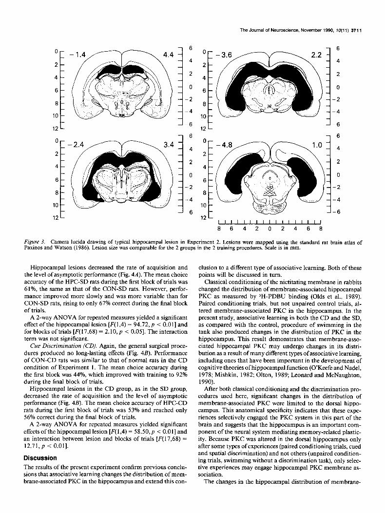
Histology The hippocampal lesions produced complete destruction of the dorsal and ventral hippocampus and little damage to the ad- jacent structures. A typical lesion is depicted in Figure 3.
Behavior
Spatial Discrimination (SD). General surgical procedures pro- duced no long-lasting effects (Fig. 4.4). Performance of CON- SD rats (N = 3) was similar to that of normal rats in the SD condition described above. The mean choice accuracy (the num- ber of correct responses divided by the total number of trials) during the first block of trials was 61%. This improved with training to 97% during the final block of trials.
Figure 2. Computer-generated pseudocolor images of membrane-associated PKC distribution in representative CA3 regions from dorsal hippo- campi from 4 groups (left to right): Cage, SNS, SD, and CD. The inset illustrates how this region of interest was selected in each digitized autoradiographic brain section. Each dorsal hippocampus was individually magnified by a factor of 2 and subjected to a low-frequency 3 x 3 convolution matrix filter operation to reduce high-frequency autoradiographic artifact prior to sampling. Values for each CA3 region of interest were averaged for all sections from an animal, and these average values were then compared between the groups using 1 -way ANOVA performed with the Systat statistical analysis system (Systat Inc., La Jolla, CA). Both the SD and CD groups had significantly less binding in the CA3 ROI than both the Cage and SNS groups (see Results, Table 3). The color bar in the lower right provides quantitative calibration for all 4 images.
The Journal of Neuroscience, November 1990, fO(11) 3711
O-
2-
4-
6-
8-
10 -
12 -
‘t -2.4 m 3.4 2
4
6
8
10
12 i
:
6
4
2
0
-2
-4
-6
6
4
2
0
-2
-4
-6
0
2
4
6
8
10
12
-6
- 4
- 2
- 0
--2
--4
--6
0
2
4
6
8
10
*A
lllllllllllllllJ
864202468
Figure 3. Camera lucida drawing of typical hippocampal lesion in Experiment 2. Lesions were mapped using the standard rat brain atlas of Paxinos and Watson (1986). Lesion size was comparable for the 2 groups in the 2 training procedures. Scale is in mm.
Hippocampal lesions decreased the rate of acquisition and the level of asymptotic performance (Fig. 4A). The mean choice accuracy of the HPC-SD rats during the first block of trials was 61%, the same as that of the CON-SD rats. However, perfor- mance improved more slowly and was more variable than for CON-SD rats, rising to only 67% correct during the final block of trials.
A 2-way ANOVA for repeated measures yielded a significant effect of the hippocampal lesion [F( 1,4) = 94.72, p < 0.0 I] and for blocks of trials [F( 17,68) = 2.10, p < 0.051. The interaction term was not significant.
Cue Discrimination (CD). Again, the general surgical proce- dures produced no long-lasting effects (Fig. 4B). Performance of CON-CD rats was similar to that of normal rats in the CD condition of Experiment 1. The mean choice accuracy during the first block was 44%, which improved with training to 92% during the final block of trials.
Hippocampal lesions in the CD group, as in the SD group, decreased the rate of acquisition and the level of asymptotic performance (Fig. 4B). The mean choice accuracy of HPC-CD rats during the first block of trials was 53% and reached only 56% correct during the final block of trials.
A 2-way ANOVA for repeated measures yielded significant effects of the hippocampal lesion [F( 1,4) = 58.50, p < 0.011 and an interaction between lesion and blocks of trials [F(17,68) = 12.71, p -c 0.011.
Discussion The results of the present experiment confirm previous conclu- sions that associative learning changes the distribution of mem- brane-associated PKC in the hippocampus and extend this con-
elusion to a different type of associative learning. Both of these points will be discussed in turn.
Classical conditioning of the nictitating membrane in rabbits changed the distribution of membrane-associated hippocampal PKC as measured by 3H-PDBU binding (Olds et al., 1989). Paired conditioning trials, but not unpaired control trials, al- tered membrane-associated PKC in the hippocampus. In the present study, associative learning in both the CD and the SD, as compared with the control, procedure of swimming in the tank also produced changes in the distribution of PKC in the hippocampus. This result demonstrates that membrane-asso- ciated hippocampal PKC may undergo changes in its distri- bution as a result of many different types of associative learning, including ones that have been important in the development of cognitive theories of hippocampal function (O’Keefe and Nadel, 1978; Mishkin, 1982; Olton, 1989; Leonard and McNaughton, 1990).
After both classical conditioning and the discrimination pro- cedures used here, significant changes in the distribution of membrane-associated PKC were limited to the dorsal hippo- campus. This anatomical specificity indicates that these expe- riences selectively engaged the PKC system in this part of the brain and suggests that the hippocampus is an important com- ponent of the neural system mediating memory-related plastic- ity. Because PKC was altered in the dorsal hippocampus only after some types of experiences (paired conditioning trials, cued and spatial discrimination) and not others (unpaired condition- ing trials, swimming without a discrimination task), only selec- tive experiences may engage hippocampal PKC membrane as- sociation.
The changes in the hippocampal distribution of membrane-

3712 Olds et al. * Learning-Specific PKC Distributional Changes in Rat Hippocampus
PLACE DISCRIMINATION
O-O CON TP
0 -0 HPC
I t 1 I 1 1 I , 1
2 4 6 8 10 12 14 16 18
BLOCKS OF 12 TRIALS
CUED DISCRIMINATION
4’ I I I I I 1 1 I I - 0 2 4 6 8 10 12 14 16 18
BLOCKS OF 12 TRIALS
Figure 4. Effect of hippocampal lesions. A, The effect of hippocampal lesions on choice accuracy in the SD procedure. The format is the same as in Figure 1. The lesioned rats (HPC-SD) had significantly lower choice accuracy than the control rats (CON-SD) as assessed by a 2-way ANO- VA for repeated measures (see Results). B, The effect of hippocampal lesions on choice accuracy in the CD procedure. The format is the same as in A. The lesioned animals (HPC-CD) had significantly lower choice accuracy than the control rats (CON-CD) as assessed by a 2-way ANO- VA for repeated measures (see Results).
associated PKC differ in the conditioning and the discrimination procedures. After classical conditioning, membrane-associated PKC was increased (Olds et al., 1989) whereas after both dis- criminations, it was decreased. The present study was not de- signed to identify the experimental variables responsible for these differences, and many factors, individually or in combi- nation, may have been responsible. The decrease in the present study may reflect downregulation of PKC in the membrane after activation and subsequent association with the membrane. PKC activation is often followed by a rapid degradation of PKC enzyme activity or 3H-PDBU binding associated with cell mem- branes (Easom et al., 1989, Price et al., 1989). If such a mech- anism is in operation here, the decrease in membrane-associated PKC after the extensive behavioral testing in the present ex- periments may be the consequence of a substantial increase in membrane-associated PKC earlier in training. This possibility can be tested by measuring PKC after different amounts of training; if the reduction of membrane-associated PKC ob- served after extensive training is in reaction to a preceding in-
crease of membrane-associated PKC, then PKC activation should be present after a shorter period of testing.
An alternative possibility is that the time point at which mem- brane-associated PKC was measured (within 1 hr after the final one of many training sessions, as compared with 24 and 72 hr in the rabbit-conditioning studies) affected the anatomical lo- calization and direction of the PKC changes. This possibility is supported by the clear break in the learning curves for both the CD and SD rats between trial blocks 3 and 4 (Fig. 1). Therefore, animals killed at this point might have manifested a leaming- specific increase in membrane-associated PKC. This empirical aspect of the data is additionally supported by the recent finding that rabbits killed immediately after the 80th classical condi- tioning trial show a learning-specific increase in 3H-PDBU bind- ing within the stratum oriens layer of CA3 and not in CA1 (A. M. Scharenberg, J. L. Olds, B. G. Schreurs, A. M. Craig, and D. L. Alkon, unpublished observations). Furthermore, in rabbits killed 3 hr after 3 d of 80 conditioning trials, 3H-PDBU binding was significantly depressed in CA3 and not in CA1 (J. L. Olds, D. L. McPhie, M. Stultz, and D. L. Alkon, unpublished observations).
The results from the present lesion experiment suggest that the hippocampus is necessary for normal performance in both types of discriminations. Future experiments should determine whether selective blockade of the CA3 PKC second-messenger system alone would produce a similar impairment.
Modifications in the hippocampal distribution of membrane- associated PKC in both of the discrimination procedures used here may not be too surprising given that performance in both of these procedures, as in Pavlovian conditioning of the nicti- tating membrane, was found to be impaired by hippocampal lesions (Morris et al., 1982; Akase et al., 1989). The involvement of this anatomical structure in memory storage is further sup- ported by the findings that the activity of hippocampal neurons is closely correlated with the development of the conditioned behavioral response (Olds et al., 1972; Segal and Olds, 1972, 1973; Segal et al., 1972; Berger et al., 1983) in both the rat and the rabbit. Moreover, long-term steady-state biophysical changes that occur in CA1 pyramidal cells, and which accompany as- sociative learning, are mimicked by activation of PKC (Alkon et al., 1988).
The learning-specific changes in membrane-associated PKC may depend upon coactivation of either excitatory amino acid (EAA) receptors, cholinergic receptors, GABA-B receptors, or some combination, all of which are present on hippocampal pyramidal cells, and all of which might activate phospholipase C through one of their receptor subtypes (McPhie et al., 1989). While the resolution inherent in the film autoradiographic meth- od does not permit the actual visualization of these distribu- tional changes in PKC to the level of individual neurons, the results of this study taken together with results of other ana- tomical localization experiments (Worley et al., 1986a,b) suggest that these changes may well have occurred within the hippo- campal pyramidal cells. Cotemporal receptor activations (pos- sibly representing converging sensory information channels) would create the conditions for enhanced PKC membrane as- sociation at specific postsynaptic spines. Subsequent to such membrane association, PKC would catalyze the phosphoryla- tion of specific proteins (such as the recently discovered cp20; Nelson et al., 1990) which in turn could subserve leaming- specific modifications in ionic channels. After this activation step, membrane-associated PKC might be rapidly degraded by

The Journal of Neuroscience, November 1990, IO(11) 3713
specific proteases, creating a “PKC deficit” localized to the area of previous activation. The processes involved in long-term potentiation (LTP) may also be important in understanding the present results because a shift in the phosphorylation of B50, a PKC substrate, has been found to be associated with mainte- nance of LTP (Lovinger and Routtenberg, 1988).
The mystery of why so many cells appear to be engaged into a given memory storage process has been addressed previously (Berger et al., 1983; Mamounas et al., 1984; Disterhoft et al., 1986; Alkon, 1989; Olds et al., 1989). We speculate that, in the water maze procedure used here, initial dendritic membrane association of PKC may result in local inactivation of K+ chan- nels followed by rapid downregulation of PKC. This down- regulation could be accompanied by a rapid retrograde message to the cell soma, which would in turn lead to long-lasting in- creases (on the order of days after the memory storage process- ing) in membrane-associated PKC at the same dendritic spines as the initial activation.
PKC, of course, is only 1 molecule in a complex chain of events that leads from sensory input to behavioral changes as- sociated with discrimination learning. Nevertheless, the present evidence supports the view that this enzyme is at a nexus in the memory storage process, able to play a vital role both in the initial laying down of the memory trace and in its later con- solidation.
References
Akase E, Alkon DL, Disterhoft JF (1989) Hippocampal lesions impair memory of short-delay conditioned eye blink in rabbits. Behav Neu- rosci 103:935-943.
Alkon DL (1989) Memory storage and neural systems. Sci Am 261: 42-50.
Alkon DL, Naito S, Kubota M, Chen C, Bank B, Smallwood J, Gallant P, Rasmussen H (1988) Regulation of Hermissendu K+ channels by cytoplasmic and membrane-associated C-kinase. J Neurochem 5 1: 903-917.
Bank B, DeWeer A, Kuzirian AM, Rasmussen H, Alkon DL (1988) Classical conditioning induces long-term translocation of protein ki- nase C in rabbit hippocampal CA1 cells [published erratum, Proc Nat1 Acad Sci USA 8553441 Proc Nat1 Acad Sci USA 85:1988-1992.
Berger TW, Rinaldi PC, Weisz DJ, Thompson RF (1983) Single-unit analysis of different hippocampal cell types during classical condi- tioning of rabbit nictitating membrane response. J Neurophysiol 50: 1197-1219.
Coulter DA, LoTurco JJ, Kubota M, Disterhoft JF, Moore JW, Alkon DL (1989) Classical conditioning reduces amplitude and duration of calcium-dependent afterhyperpolarization in rabbit hippocampal pyramidal cells. J Neurophysiol 6 1:97 l-98 1.
Disterhoft JF, Coulter DA, Alkon DL (1986) Conditioning-specific membrane changes of rabbit hippocampal neurons measured in vitro. Proc Nat1 Acad Sci USA 8312733-2737.
Easom RA, Hughes JH, Landt M, Wolf BA, Turk J, McDaniel ML (1989) Comparison ofeffects of phorbol esters and glucose on protein kinase C activation and insulin secretion in pancreatic islets. Biochem J 264127-33.
Leonard B, McNaughton BL (1990) Spatial representation in the rat: conceptual, behavioral, and neurophysiological perspectives. In: Neu-
robiology of comparative cognition (Kesner RP, Olton DS, eds), pp 363422. Hillsdale, NJ: Lawrence Erlbaum.
Lovinger DM, Routtenberg A (1988) Synapse-specific protein kinase C activation enhances maintenance of long term potentiation in rat hippocampus. J Physiol (Lond) 400:321-333.
Mamounas LA, Thompson RF, Lynch G, Baudry M (1984) Classical conditioning ofthe rabbit eyelid response increases glutamate receptor binding in hippocampal synaptic membranes. Proc Nat1 Acad Sci USA 8 1:2548-2552.
McPhie DL, Olds JL, Sanchez-Andres JV, Alkon DL (1989) Isolated pharmacological activation of three neurotransmitter systems pro- duces differential translocation of PKC in the rabbit hippocampus. Sot Neurosci Abstr 15:505.
Mishkin M (1982) A memory system in the memory. Philos Trans R Sot Lond [Ser B] 298:85-95.
Morris RG (1984) Development of a water maze procedure for study- ing spatial learning in the rat. J Neurosci Meth 11:47-60.
Morris RG, Garmd P, Rawlins JN, O’Keefe J (1982) Place navigation impaired in rats with hippocampal lesions. Nature 297:68 1-683.
Nelson TJ. Collin C. Alkon DL (1990) Isolation of a G nrotein that I \ ,
is modified by learning and reduces potassium currents-in Hermis- senda. Science 247~1479-1483.
Nishizuka Y (1988) The molecular heterogeneity of protein kinase C and its implications for cellular regulation. Nature 334:661-665.
O’Keefe J, Nadel L (1978) The hippocampus as a cognitive map. New York: Academic.
Olds J, Disterhoft JF, Segal M, Komblith CL, Hirsh R (1972) Learning centers of rat brain mapped by measuring latencies of conditioned unit responses. J Neurophysiol 35:202-2 19.
Olds JL, Anderson ML, McPhie DL, Staten LD, Alkon DL (1989) Imaging of memory-specific changes in the distribution of protein kinase C in the hippocampus. Science 245:866-869.
Olton DS (1989) Mneumonic functions of the hippocampus: single unit analyses in rats. In: The hippocampus: new vistas (Chan-Palay V, Kohler C, eds), pp 41 l-424. New York: Liss.
Pan H, Frey S, Kirk A, Young AB, Penney JB Jr (1983) Changes in ‘H-muscimol binding in substantia nigra, entopeduncular nucleus, globus pallidus, and thalamus after striatal lesions as demonstrated by quantitative autoradiography. J Neurosci 3: 1189-l 198.
Paxinos G, Watson C (1986) The rat brain in stereotaxic coordinates. 2d ed. San Diego: Academic.
Price BD, Morris JD, Hall A (1989) Stimulation of phosphatidylcho- line breakdown and diacyglycerol production by growth factors in Swiss-3T3 cells. Biochem J 264:509-515.
Saito N, Kikkawa U, Nishizuka Y, Tanaka C (1988) Distribution of protein kinase C-like immunoreactive neurons in rat brain. J Neurosci 8:369-382.
Segal M, Olds J (1972) Behavior of units in hippocampal circuit of the rat during learning. J Neurophysiol 35:680-690.
Segal M, Olds J (1973) Activity of units in the hippocampal circuit of the rat during differential classical conditioning. J Comp Physiol Psycho1 82: 195-204.
Segal M, Disterhoft JF, Olds J (1972) Hippocampal unit activity dur- ing classical aversive and appetitive conditioning. Science 175:792- 794.
Worley PF, Baraban JM, Snyder SH (1986a) Heterogeneous localiza- tion of protein kinase C in rat brain: autoradiographic analysis of phorbol-ester receptor binding. J Neurosci 6: 199L207. -
Worlev PF. Baraban JM. De Souza EB. Snvder SH (1986b) MaDDin!
second messenger systems in the brain:-differential locahzations 07 adenylate cyclase and protein kinase C. Proc Nat1 Acad Sci USA 83: 4053-4057.