discovery of an unusual biosynthetic origin for circular ... · discovery of an unusual...
TRANSCRIPT

Discovery of an unusual biosynthetic originfor circular proteins in legumesAaron G. Potha,b, Michelle L. Colgraveb, Russell E. Lyonsb, Norelle L. Dalya, and David J. Craika,1
aInstitute for Molecular Bioscience, University of Queensland, Brisbane, Queensland 4072, Australia; and bCommonwealth Scientific and IndustrialResearch Organisation Division of Livestock Industries, St. Lucia, Queensland 4067, Australia
Edited by Sharon R. Long, Stanford University, Stanford, CA, and approved April 18, 2011 (received for review March 6, 2011)
Cyclotides are plant-derived proteins that have a unique cycliccystine knot topology and are remarkably stable. Their naturalfunction is host defense, but they have a diverse range of pharma-ceutically important activities, including uterotonic activity andanti-HIV activity, and have also attracted recent interest as tem-plates in drug design. Here we report an unusual biosyntheticorigin of a precursor protein of a cyclotide from the butterflypea, Clitoria ternatea, a representative member of the Fabaceaeplant family. Unlike all previously reported cyclotides, the domaincorresponding to the mature cyclotide from this Fabaceae plant isembedded within an albumin precursor protein. We confirmed theexpression and correct processing of the cyclotide encoded bythe Cter M precursor gene transcript following extraction fromC. ternatea leaf and sequencing by tandem mass spectrometry.The sequence was verified by direct chemical synthesis and thepeptide was found to adopt a classic knotted cyclotide fold asdetermined by NMR spectroscopy. Seven additional cyclotidesequences were also identified from C. ternatea leaf and flower,five of which were unique. Cter M displayed insecticidal activityagainst the cotton budworm Helicoverpa armigera and bound tophospholipid membranes, suggesting its activity is modulated bymembrane disruption. The Fabaceae is the third largest family offlowering plants and many Fabaceous plants are of huge signifi-cance for human nutrition. Knowledge of Fabaceae cyclotide genetranscripts should enable the production of modified cyclotides incrop plants for a variety of agricultural or pharmaceutical applica-tions, including plant-produced designer peptide drugs.
cyclic peptides ∣ structure ∣ cystine knot ∣ kalata B1
Cyclotides (1) are topologically unique plant proteins that areexceptionally stable. They comprise approximately 30 amino
acids arranged in a head-to-tail cyclized peptide backbone thatadditionally is restrained by a cystine knot motif. The cystine knotis built from two disulfide bonds, and their connecting backbonesegments form an internal ring in the structure that is threaded bya third disulfide bond to form an interlocked and cross-bracedstructure (Fig. 1). Superimposed on this cystine knot core area β-sheet and a series of turns displaying surface-exposed loops.
Cyclotides express a diversity of peptide sequences within theirbackbone loops and have a broad range of biological activities,including uterotonic (2), anti-HIV (3), antimicrobial (4), and an-ticancer activities (5). Accordingly, they are of great interest forpharmaceutical applications. Some plants from which they arederived are used in indigenous medicines, including kalata-kalata,a tea from the plant Oldenlandia affinis, which is used for accel-erating childbirth in Africa and contains the prototypic cyclotidekalata B1, kB1 (2). This ethnobotanical use and recent biophy-sical studies (6) illustrate the remarkable stability of cyclotides;i.e., they survive boiling and ingestion, observations unprece-dented for conventional peptides. Their exceptional stability hasled to their use as templates in peptide-based drug design appli-cations (7), where the grafting of bioactive peptide sequences intoa cyclotide framework offers a new approach to stabilize peptide-based therapeutics, thereby overcoming one of the major limita-tions of peptides as drugs. Chemical (8, 9), chemo-enzymatic
(10), and recombinant (11) approaches to the synthesis of cyclo-tides have been developed to facilitate these pharmaceuticalapplications.
The natural function of cyclotides appears to be in plant de-fense, based on their pesticidal activities, including insecticidal(12, 13), nematocidal (14), and molluscicidal (15) activities.These activities appear to be mediated by selective membranebinding and disruption (12, 16) that occurs as a result of cyclo-tides having a surface-exposed patch of hydrophobic residues.Individual plants typically contain dozens of cyclotides, expressedin multiple tissues, including flowers, leaf, and seeds, leading totheir description as a natural combinatorial template (17). Plantspresumably use this combinatorial strategy to target multiplepests or to reduce the possibility of an individual pest species de-veloping resistance to the protective cyclotide armory. More than170 cyclotides have been sequenced (18), although it is estimated
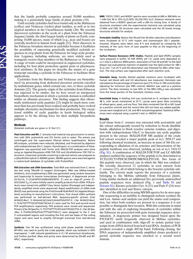
Fig. 1. Structures and sequences of cyclotides. The structure of the proto-typical cyclotide kB1 from O. affinis is illustrated. The conserved Cys residuesare labeled with Roman numerals and various loops in the backbone be-tween them are labeled loops 1–6. The sequences of kB1 (PDB code 1NB1)(38), cycloviolacin O2 (1), MCoTI-II (39), and Cter A (21) represent examplesof cyclotides isolated from the Rubiaceae, Violaceae, Cucurbitaceae, andFabaceae plant families. The conserved cysteines are boxed and their locationon the structure is indicated by the solid arrows. The putative processingpoints by which mature cyclotides are excised from precursor proteins areindicated and correspond to an N-terminal Gly and a C-terminal Asn (N)or Asp (D) residue (indicated by dashed arrows). The novel cyclotides identi-fied in this study (Cter M-R) are aligned with representative cyclotides. Psyle F(24) was also identified in C. ternatea flower.
Author contributions: D.J.C. designed research; A.G.P., M.L.C., R.E.L., and N.L.D. performedresearch; A.G.P., M.L.C., R.E.L., N.L.D., and D.J.C. analyzed data; and A.G.P., M.L.C., andD.J.C. wrote the paper.
The authors declare no conflict of interest.
This article is a PNAS Direct Submission.
Data deposition: NMR, atomic coordinates, chemical shifts, and restraints. The ProteinData Bank (www.pdb.org) ID code for Cter M is 2LAM. The GenBank accession no. for CterM is JF501210. The UniProtKB accession codes for C. ternatea cyclotides are: P86899 forCyclotide Cter M, P86900 for Cyclotide Cter N, P86901 for Cyclotide Cter O, P86902 forCyclotide Cter P, P86904 for Cyclotide Cter Q, and P86903 for Cyclotide Cter R.
See Commentary on page 10025.1To whom correspondence should be addressed. E-mail: [email protected].
This article contains supporting information online at www.pnas.org/lookup/suppl/doi:10.1073/pnas.1103660108/-/DCSupplemental.
www.pnas.org/cgi/doi/10.1073/pnas.1103660108 PNAS ∣ June 21, 2011 ∣ vol. 108 ∣ no. 25 ∣ 10127–10132
BIOCH
EMISTR
YSE
ECO
MMEN
TARY
that the family probably comprises around 50,000 members,making it a particularly large family of plant proteins (19).
Until recently cyclotides had been found only in the Rubiaceae(coffee) and Violaceae (violet) plant families, as well as in twoatypical members in the Cucurbitaceae family (20). We recentlydiscovered cyclotides in the seeds of a plant from the Fabaceae(legume) family, the third largest family of plants on Earth, com-prising 18,000 species, many of which (e.g., peas and beans) arecentrally involved in human food supply (21). Their discovery inthe Fabaceae broadens interest in cyclotides because it facilitatesthe possibility of expressing genetically modified cyclotide se-quences in crop plants from the Fabaceae for large-scale produc-tion. Legumes, including soybeans, are more widely used astransgenic vectors than members of the Rubiaceae or Violaceae.A range of traits could be incorporated in engineered cyclotides,including for host plant protection against pests or pharmaceu-tical attributes. In this paper we describe the nature of a genetranscript encoding a cyclotide in the Fabaceae to facilitate theseapplications.
Cyclotides from the Rubiaceae and Violaceae are biosynthe-sized via processing from dedicated precursor proteins encodedby multidomain genes, which contain one, two, or three cyclotidedomains (22). The genetic origin of the cyclotides from Fabaceaewas expected to be similar, but we here reveal an unexpectedbiosynthetic mechanism in which a cyclotide domain is embeddedwithin an albumin precursor. This finding suggests that riboso-mally synthesized cyclic peptides (23) might be much more com-mon than has previously been realized and probably have evolvedmultiple alternative mechanisms for their production. The excep-tional stability of cyclic peptides in harsh biological milieuappears to be the driving force for their multiple biosyntheticpathways.
Methods(Detailed methods are given in SI Text S1.)
Plant Extraction and MS. C. ternatea leaf material was ground prior to extrac-tion with 50% acetonitrile and 2% formic acid in water. The extract wascentrifuged and the supernatant filtered before lyophilization. BeforeMS analyses, cyclotides were reduced, alkylated, and linearized by digestionwith endoproteinase Glu-C, trypsin, chymotrypsin, or a combination of these.Digestion was quenched with formic acid. MALDI-TOF analyses were doneusing an Applied Biosystems 4700 TOF-TOF and UltrafleXtreme TOF-TOF in-strument. Linearized cyclotide-containing crude leaf extract was analyzed ona QStar® Elite hybrid LC-MS/MS system.MS/MS spectra were searched againsta custom-built database of cyclotides with ProteinPilot.
RNA Extraction and cDNA Generation. Total RNA was extracted from C. terna-tea leaf using TRIzol® LS reagent (Invitrogen). RNA was DNAse-treated(Ambion), and complementary DNA was generated using random hexamersand Superscript III reverse transcriptase (Invitrogen). A degenerate primer(Ct-For1A, 5′-CCiACNTGYGGNGARACNTG- 3′) and an oligo-dT primer (5′-GCCCGGG T20-3′) were initially used to amplify products from cDNA. PCR pro-ducts were cloned into pGEM-T Easy Vector System (Promega) and indepen-dently amplified clones were sequenced. Rapid amplification of cDNA ends(RACE) was performed using the FirstChoice® RLM-RACE kit (Applied Biosys-tems) according to manufacturer’s instructions. First strand cDNA synthesiswas performed on leaf-derived RNA. Sequence-specific primers (CterM-RACE-Rev1, 5′-GGAAACACCAACCAAAATGGATGT-3′; Cter M-RACE-Rev2,5′-TCACTGTTTTTGCATTAGCTGCAA-3′) were used for first and second roundPCR amplifications respectively. PCR products were cloned and sequenced.Primers (CterM-SpecFor, 5′-TCCTTATTTTCATCAACTATGGCTTA-3′; CterM-Spe-cRev, 5′-TCATACATGATCACTTTTAGTTGG-3′) were designed to the 5′ and3′-untranslated regions and including the first and last bases of the codingregion and were used to amplify full-length transcript from leaf-derivedcDNA.
Synthesis. Cter M was synthesized using solid phase peptide chemistry.RP-HPLC was used to purify the crude peptide, which was oxidized in 50%isopropanol, 1 mM reduced glutathione in 0.1 M ammonium bicarbonate,and further purified. Mass and purity were determined by ESI-MS and RP-HPLC, respectively.
NMR. TOCSY, COSY, and NOESY spectra were recorded at 600 or 900 MHz on1 mM Cter M in 10% D2O∕20% CD3CN∕70% H2O. Distance restraints wereobtained from a NOESY spectrum with a 200 ms mixing time. A family ofstructures consistent with the experimental restraints was calculated usingCYANA and CNS; 50 structures were calculated and the 20 lowest energystructures selected for analysis.
Proteolytic Stability. Native Cter M and Cter M after reduction and alkylationwere incubated at 37 °C with trypsin and chymotrypsin (20∶1 peptide∶enzyme). Aliquots were taken over 8 h and analyzed by comparing theintensity of the cyclic form of each peptide to that at the beginning ofthe incubation.
Surface Plasmon Resonance (SPR) Studies. Peptide and lipid (POPC) sampleswere prepared in buffer 10 mM HEPES, pH 7.4. Lipids were deposited ona L1 chip in a BIAcore 3000 system. Association of Cter M and kB1 to the lipidbilayer was evaluated by injection of peptide over the lipid surface for 180 s(5 μL min−1) at 25 °C and the dissociation was followed for 600 s after injec-tion. The chip surface was regenerated after each injection cycle.
Hemolytic Assay. Serially diluted peptide solutions were incubated withhuman red blood cells and UV absorbance was measured. Hemolysis wascalculated as the percentage ofmaximum lysis (1% Triton X-100 control) afteradjusting for minimum lysis (PBS control). Melittin was used as a positivecontrol. The dose necessary to lyse 50% of the RBCs (HD50) was calculatedfrom the linear portion of the hemolytic titration curve.
Insecticidal Assay. A feeding trial was conducted on H. armigera larvae for48 h, with larvae maintained at 25 °C. Larvae were given diets consistingof wheat germ, yeast, and soy flour. Test diets contained Cter M or kB1 (usedas a positive control) and the control diet did not have any added peptide.Larvae were weighed at 0, 24, and 48 h and photographed. Statistical differ-ences were analyzed using a paired t-test or ANOVA test.
ResultsLeaf tissue from C. ternatea was extracted with acetonitrile andthe extract subsequently treated by reduction to break disulfidebonds, alkylation to block reactive cysteine residues, and diges-tion with endoproteinase Glu-C to linearize any cyclic peptidespresent in the extract. MALDI-TOF MS analysis of the crudeleaf extract revealed a major peptide peak at m∕z 3058.57 andfollowing chemical treatment, an increase in mass of 366 Da cor-responding to alkylation of six cysteines and linearization of thepeptide backbone was observed, yielding an ion at m∕z 3424.33(Fig. S1). A combination of MALDI-TOF/TOF and LC-MS/MSanalyses enabled the sequence of the peptide to be determined asTCTLGTCYVPDCSCSWPICMKNGLPTCGE. Two forms ofthis peptide were observed, one in which the Met was oxidized.We recently discovered 12 cyclotides in seed extracts fromC. ternatea (21), all of which belong to the bracelet cyclotide sub-family. The current study reports the presence of a cyclotidebelonging to the Möbius subfamily from Fabaceous plants.Using similar methods an additional five previously undescribedpeptide sequences were deduced (Fig. 1 and Table S1 andDataset S1). Known cyclotides Cter A (21) and Psyle F (24) werealso identified in leaf and flower extracts.
One of the difficulties in usingMS/MS spectra for de novo pep-tide sequencing is an inability to distinguish isobaric residues Ileand Leu. Amino acid analysis can yield the amino acid composi-tion, but when both residues are present in a sequence it is notpossible to distinguish their location. With this constraint in mindand with the aim of exploring biosynthesis of cyclotides withinthe Fabaceae we proceeded with gene transcript sequence deter-mination. A degenerate primer was designed based upon thePTCGETC motif frequently observed in Möbius cyclotides,and used in combination with oligo-dT to isolate partial tran-scripts from cDNA derived from leaf total RNA. Analysis of PCRproducts revealed a single 402-bp band. Following cloning, theDNA sequences of independently amplified clones predicted apartial cyclotide sequence was attached to a second noveldomain.
10128 ∣ www.pnas.org/cgi/doi/10.1073/pnas.1103660108 Poth et al.

Cyclotide gene transcripts elucidated to date predict maturecyclotide domains followed by a C-terminal region (CTR) of3–11 amino acids comprising a small amino acid (Gly or Ala)in the first position and a conserved Leu in the second position.This Leu has been postulated to play a critical role in docking to abinding pocket of asparaginyl endoprotease (AEP) during pep-tide excision and ligation (25). In the case of the C. ternatea-de-rived sequence, the mature peptide is flanked on the C terminusby a 74 amino acid tail, in which the Gly and the “critical” Leu arenotably absent. BLAST searching of this C-terminal tail regionrevealed that it had high sequence homology to the C-terminalportion of albumin-1 proteins from a variety of Fabaceousspecies.
To ensure that this unusual sequence was not an artifact ofcDNA synthesis or PCR, we undertook 5′ RACE studies. Follow-ing 5′ RACE amplification and alignment to previous sequences,a 514-bp consensus sequence was obtained. To confirm that thissequence represented a single mRNA expressed in C. ternatealeaf, primers were designed within the 5′ and 3′ untranslated re-gions, and a single 418 bp PCR product was amplified. Sequenceanalysis revealed this product, as anticipated, encoded a pre-dicted protein of 127 amino acids (Fig. 2 and Fig. S2). The fullprotein sequence of the novel Fabaceae cyclotide precursor wasaligned to the homologous albumin proteins identified in theinitial BLAST search (Fig. S3). In the precursor protein fromOldenlandia affinis that encompasses the prototypic cyclotide,kB1, the mature peptide sequence is flanked by an endoplasmicreticulum (ER) signal and prodomains of 65aa at the N terminusand 7aa at the C terminus, with all of the six cysteines in the pre-cursor located within the mature kB1 sequence (13). In contrast,the Cter M precursor protein has a typical ER signal sequencethat immediately precedes the N terminus of the mature cyclo-tide. The cyclotide peptide sequence is then linked via 10aa toan albumin a-domain (Fig. 2).
Cter M was chemically synthesized and was identical to thenative form by MS and HPLC (Fig. S4) and, like the native pep-tide, had low solubility in water. The addition of 20% acetonitrileimproved solubility for NMR analysis. NMR spectra of native andsynthetic Cter M were identical. Cter M is extremely stable,as indicated by its resistance to heat denaturation; spectra wererecorded before and after heating the peptide to 95 °C and nochanges were observed upon return to ambient temperature
(Fig. 3A). As for kB1, Cter M in its native oxidized form was im-pervious to trypsin and chymotrypsin, with no hydrolysis detectedafter 8 h incubation at 37 °C. By contrast the disulfide-reducedform of Cter M was susceptible to proteolysis (Fig. S5).
The 3D structure of Cter M was determined based on 398NMR-derived distance restraints and 14 angle restraints. The de-rived family of structures had excellent geometric and energeticstatistics (Table S2). An ensemble and ribbon representation isshown in Fig. 3B,C along with a comparison with PA1b (Fig. 3 Dand E), a pea albumin whose precursor shares high homologywith the Cter M precursor protein. Although variation in the loopregions of the two peptides is apparent, the eight-membered ringformed between loops 1 and 4 and their interconnecting disulfidebonds at the core of the cystine knot shows striking similarities.
Analysis of the structure of Cter M with PROMOTIF identi-fied a type I β-turn at residues 9–12, a type II β-turn at residues16–19, and a type VIa1 β-turn at residues 22–25. A β-hairpin oc-curs over residues 20–27, as typically seen in cystine knot proteins(26). Further examination of Cter M reveals the surface exposureof hydrophobic residues to be 51% of the solvent accessible sur-face (Fig. S6), which might explain why addition of acetonitrileis required for its solubilization in water, whereas at similarconcentrations kB1 does not require extra solubilization (38%surface exposure).
The properties and functions of Cter M were assessed usingbiophysical and biological assays. First, we undertook SPR stu-dies to determine if the surface-exposed hydrophobic patch ofresidues identified in the structural study predisposed Cter Mto membrane binding. Indeed, Cter M interacted with phospho-lipid vesicles in a concentration-dependent manner, similar tokB1 (Fig. 4 A and B). The calculated peptide-to-lipid ratio at100 μMpeptide concentration was 0.055 for Cter M and 0.032 forkB1. Second, the effect of Cter M on larval growth was assessed ina feeding trial where the peptide was incorporated into the diet ofL3 larvae of H. armigera, a highly polyphagous species that is apest of cotton, tomato, maize, chickpea, peanuts, and tobacco(27). Control larvae increased in weight in a monotonic mannerwith time whereas retardation of growth occurred in a dose-de-pendentmanner for insects fed peptide-containing diets (Fig. 4C).At the highest concentration tested (1 μmol peptide g−1 diet), lar-val mortality occurred. This concentration is in a physiologicallyrelevant range, as Cter M peptide was detected in C. ternatea atlevels up to 5 μmol g−1 of leaf tissue. The insecticidal activity ofCter M was similar to kB1, which was tested in parallel (Fig. S6)and had a potency consistent with previous results (28). Finally,the hemolytic activity of Cter M was compared with kB1 and thepore-forming agent from bee venom, melittin. Cter M was onlymildly hemolytic to human erythrocytes; the HD50 was 1.4 μM formelittin, 7.8 μM for kB1 and >100 μM for Cter M (Fig. 4D).Thus, despite the increase in hydrophobicity of Cter M comparedto kB1, a lower hemolytic activity was observed, indicating that anincrease in surface-exposed hydrophobic residues in cyclotidesdoes not necessarily correlate with increased hemolytic activity.
DiscussionHere we report a precursor transcript sequence encoding a pre-viously undescribed cyclotide from the Fabaceae plant family andshow that the peptide comprises a CCK motif, is ultrastable andinsecticidal like other cyclotides, but has an unexpected biosyn-thetic origin in that it is embedded within an albumin precursor. Itrepresents one of a suite of cyclotides in this plant that appear tohave evolved differently from previously known cyclotides. Faba-ceae plants are economically important in agriculture as nitrogensources and in human nutrition. The discovery of an alternativebiosynthetic origin for cyclotides in legumes potentially enables awide range of new applications of cyclotides in agriculture andmedicine.
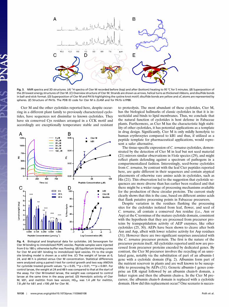
Fig. 2. Schematic of Cter M precursor protein (middle) alongside a typicalFabaceae albumin precursor (top), and a typical two-domain cyclotide pre-cursor (bottom). Violaceae and Rubiaceae cyclotide mRNAs encode an ERsignal peptide, an N-terminal Pro region, the N-terminal repeat (NTR), themature cyclotide domain, and a C-terminal flanking region (CTR). Theremay be up to three repeats of the NTR, cyclotide domain and CTR withina typical cyclotide gene. In contrast, the CterM transcript shows an ER signalpeptide immediately followed by the cyclotide domain and is flanked at the Cterminus by a linking peptide and the albumin a-chain. The Cter M cyclotidedomain replaces the PA1b subunit-b present in typical albumin-1 genes.The sequences of the precursor proteins are illustrated (bottom) using thecolor scheme from the schematic representation to indicate the locationof the domains.
Poth et al. PNAS ∣ June 21, 2011 ∣ vol. 108 ∣ no. 25 ∣ 10129
BIOCH
EMISTR
YSE
ECO
MMEN
TARY
Cter M and the other cyclotides reported here, despite occur-ring in a different plant family to previously characterized cyclo-tides, have sequences not dissimilar to known cyclotides. Theyhave six conserved Cys residues arranged in a CCK motif andaccordingly are exceptionally temperature stable and resistant
to proteolysis. The most abundant of these cyclotides, Cter M,has the biological hallmarks of classic cyclotides in that it is in-secticidal and binds to lipid membranes. Thus, we conclude thatthe natural function of cyclotides is host defense in Fabaceaeplants. Furthermore, as Cter M has the characteristic high stabi-lity of other cyclotides, it has potential applications as a templatein drug design. Significantly, Cter M is only mildly hemolytic tohuman erythrocytes compared to kB1 and thus, if utilized as apeptide template for pharmaceutical applications, would repre-sent a safer alternative.
The tissue-specific expression of C. ternatea cyclotides, demon-strated by the detection of Cter M in leaf but not seed material(21) mirrors similar observations in Viola species (29), and mightreflect plants defending against a spectrum of pathogens in acompartmentalized fashion. Interestingly, seed-borne cyclotidesfrom C. ternatea, by contrast with the leaf Cter peptides reportedhere, are quite different in their sequences and contain atypicalplacements of otherwise rare amino acids in cyclotides, such asHis (21). This observation led to the suggestion that cyclotide se-quences are more diverse than has earlier been realized and thatthere might be a wider range of processing mechanisms availablefor the production of these circular proteins. The current studyclearly shows that this is the case, based on different amino acidsthat flank putative processing points in Fabaceae precursors.
Despite variation in the residues flanking the processingsites for the cyclotides isolated from leaf, flower, and seed ofC. ternatea, all contain a conserved Asx residue (i.e., Asn orAsp) at the C terminus of the mature cyclotide domain, consistentwith the hypothesis that they are processed from precursor pro-teins by transpeptidation activity of AEP enzymes, like othercyclotides (25, 30). AEPs have been shown to cleave after bothAsn and Asp, albeit with lower relative activity for Asp residues(31). However there are two significant surprises associated withthe C. ternatea precursor protein. The first is the nature of theprecursor protein itself. All cyclotides reported until now are pro-cessed from precursor proteins encoded by dedicated genes. Bycontrast, the Cter M precursor involves the recycling of an unre-lated gene, notably via the substitution of part of an albumin-1gene with a cyclotide domain (Fig. 2). Albumins form part ofthe nutrient reservoir in plants and are well known to be suscep-tible to processing by AEPs (31). Generic albumin-1 genes com-prise an ER signal followed by an albumin chain-b domain, alinker region and then the albumin chain-a. In the Cter M pre-cursor, the albumin chain-b domain is replaced with a cyclotidedomain. How did this replacement occur? One scenario is succes-
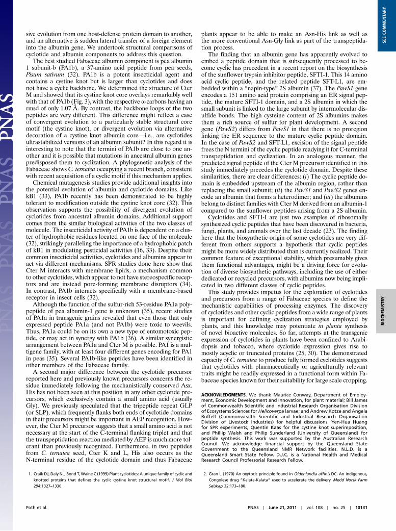
Fig. 3. NMR spectra and 3D structures. (A) 1H spectra of Cter M recorded before (top) and after (bottom) heating to 95 °C for 5 minutes. (B) Superposition ofthe 20 lowest energy structures of Cter M. (C) Overview structure of Cter M. Strands are shown as arrows, helical turns as thickened ribbons, and disulfide bondsin ball-and-stick format. (D) Superposition of Cter M and PA1b highlighting the cystine knot motif; disulfide bonds are yellow and αC atoms are represented byspheres. (E) Structure of PA1b. The PDB ID code for Cter M is 2LAM and for PA1b is1P8B.
Fig. 4. Biological and biophysical data for cyclotides. (A) Sensorgram forCter M binding to immobilized POPC vesicles. Peptide samples were injectedfrom 0 to 180 s; otherwise buffer was flowing. (B) Equilibrium binding curvesfor Cter M and kB1 binding to immobilized lipid vesicles. Fit to the singlesite binding model is shown as a solid line. (C) The weight of larvae at 0,24, and 48 h is plotted versus Cter M concentration. Statistical differenceswere analyzed using a paired t-test for control growth and two way ANOVAfor cyclotide treated growth values: *p < 0.05, **p < 0.01, ***p < 0.001. Forcontrol larvae, the weight at 24 and 48 h was compared to that at the start ofthe assay. For Cter M-treated larvae, the weight was compared to controllarvae at the same time in the assay period. (D) Hemolytic activity of CterM, kB1, and melittin from bee venom; HD50 was 1.4 μM for melittin,7.8 μM for kB1 and >100 μM for Cter M.
10130 ∣ www.pnas.org/cgi/doi/10.1073/pnas.1103660108 Poth et al.
sive evolution from one host-defense protein domain to another,and an alternative is sudden lateral transfer of a foreign elementinto the albumin gene. We undertook structural comparisons ofcyclotide and albumin components to address this question.
The best studied Fabaceae albumin component is pea albumin1 subunit-b (PA1b), a 37-amino acid peptide from pea seeds,Pisum sativum (32). PA1b is a potent insecticidal agent andcontains a cystine knot but is larger than cyclotides and doesnot have a cyclic backbone. We determined the structure of CterM and showed that its cystine knot core overlays remarkably wellwith that of PA1b (Fig. 3), with the respective α-carbons having anrmsd of only 1.07 Å. By contrast, the backbone loops of the twopeptides are very different. This difference might reflect a caseof convergent evolution to a particularly stable structural coremotif (the cystine knot), or divergent evolution via alternativedecoration of a cystine knot albumin core—i.e., are cyclotidesultrastabilized versions of an albumin subunit? In this regard it isinteresting to note that the termini of PA1b are close to one an-other and it is possible that mutations in ancestral albumin genespredisposed them to cyclization. A phylogenetic analysis of theFabaceae shows C. ternatea occupying a recent branch, consistentwith recent acquisition of a cyclic motif if this mechanism applies.
Chemical mutagenesis studies provide additional insights intothe potential evolution of albumin and cyclotide domains. LikekB1 (33), PA1b recently has been demonstrated to be highlytolerant to modification outside the cystine knot core (32). Thisobservation supports the possibility of divergent evolution ofcyclotides from ancestral albumin domains. Additional supportcomes from the similar biological activities of the two classes ofmolecule. The insecticidal activity of PA1b is dependent on a clus-ter of hydrophobic residues located on one face of the molecule(32), strikingly paralleling the importance of a hydrophobic patchof kB1 in modulating pesticidal activities (16, 33). Despite theircommon insecticidal activities, cyclotides and albumins appear toact via different mechanisms. SPR studies done here show thatCter M interacts with membrane lipids, a mechanism commonto other cyclotides, which appear to not have stereospecific recep-tors and are instead pore-forming membrane disruptors (34).In contrast, PA1b interacts specifically with a membrane-basedreceptor in insect cells (32).
Although the function of the sulfur-rich 53-residue PA1a poly-peptide of pea albumin-1 gene is unknown (35), recent studiesof PA1a in transgenic grains revealed that even those that onlyexpressed peptide PA1a (and not PA1b) were toxic to weevils.Thus, PA1a could be on its own a new type of entomotoxic pep-tide, or may act in synergy with PA1b (36). A similar synergisticarrangement between PA1a and Cter M is possible. PA1 is a mul-tigene family, with at least four different genes encoding for PA1in peas (35). Several PA1b-like peptides have been identified inother members of the Fabaceae family.
A second major difference between the cyclotide precursorreported here and previously known precursors concerns the re-sidue immediately following the mechanistically conserved Asx.His has not been seen at this position in any other cyclotide pre-cursors, which exclusively contain a small amino acid (usuallyGly). We previously speculated that the tripeptide repeat GLP(or SLP), which frequently flanks both ends of cyclotide domainsin their precursors might be important in AEP recognition. How-ever, the Cter M precursor suggests that a small amino acid is notnecessary at the start of the C-terminal flanking triplet and thatthe transpeptidation reaction mediated by AEP is much more tol-erant than previously recognized. Furthermore, in two peptidesfrom C. ternatea seed, Cter K and L, His also occurs as theN-terminal residue of the cyclotide domain and thus Fabaceae
plants appear to be able to make an Asn-His link as well asthe more conventional Asn-Gly link as part of the transpeptida-tion process.
The finding that an albumin gene has apparently evolved toembed a peptide domain that is subsequently processed to be-come cyclic has precedent in a recent report on the biosynthesisof the sunflower trypsin inhibitor peptide, SFTI-1. This 14 aminoacid cyclic peptide, and the related peptide SFT-L1, are em-bedded within a “napin-type” 2S albumin (37). The PawS1 geneencodes a 151 amino acid protein comprising an ER signal pep-tide, the mature SFTI-1 domain, and a 2S albumin in which thesmall subunit is linked to the large subunit by intermolecular dis-ulfide bonds. The high cysteine content of 2S albumins makesthem a rich source of sulfur for plant development. A secondgene (PawS2) differs from PawS1 in that there is no proregionlinking the ER sequence to the mature cyclic peptide domain.In the case of PawS2 and SFT-L1, excision of the signal peptidefrees the N termini of the cyclic peptide readying it for C-terminaltranspeptidation and cyclization. In an analogous manner, thepredicted signal peptide of the Cter M precursor identified in thisstudy immediately precedes the cyclotide domain. Despite thesesimilarities, there are clear differences: (i) The cyclic peptide do-main is embedded upstream of the albumin region, rather thanreplacing the small subunit; (ii) the PawS1 and PawS2 genes en-code an albumin that forms a heterodimer; and (iii) the albuminsbelong to distinct families with Cter M derived from an albumin-1compared to the sunflower peptides arising from a 2S-albumin.
Cyclotides and SFTI-I are just two examples of ribosomallysynthesized cyclic peptides that have been discovered in bacteria,fungi, plants, and animals over the last decade (23). The findinghere that the biosynthetic origin of some cyclotides are very dif-ferent from others supports a hypothesis that cyclic peptidesmight be more widely distributed than is currently realized. Theircommon feature of exceptional stability, which presumably givesthem functional advantages, might be a driving force for evolu-tion of diverse biosynthetic pathways, including the use of eitherdedicated or recycled precursors, with albumins now being impli-cated in two different classes of cyclic peptides.
This study provides impetus for the exploration of cyclotidesand precursors from a range of Fabaceae species to define themechanistic capabilities of processing enzymes. The discoveryof cyclotides and other cyclic peptides from a wide range of plantsis important for defining cyclization strategies employed byplants, and this knowledge may potentiate in planta synthesisof novel bioactive molecules. So far, attempts at the transgenicexpression of cyclotides in plants have been confined to Arabi-dopsis and tobacco, where cyclotide expression gives rise tomostly acyclic or truncated proteins (25, 30). The demonstratedcapacity of C. ternatea to produce fully formed cyclotides suggeststhat cyclotides with pharmaceutically or agriculturally relevanttraits might be readily expressed in a functional form within Fa-baceae species known for their suitability for large scale cropping.
ACKNOWLEDGMENTS. We thank Maurice Conway, Department of Employ-ment, Economic Development and Innovation, for plant material; Bill Jamesof Commonwealth Scientific and Industrial Research Organisation Divisionof Ecosystems Sciences for Helicoverpa larvae; and Andrew Kotze and AngelaRuffell (Commonwealth Scientific and Industrial Research OrganisationDivision of Livestock Industries) for helpful discussions. Yen-Hua Huangfor SPR experiments, Quentin Kaas for the cystine knot superimposition,and Phillip Walsh and Philip Sunderland (University of Queensland) forpeptide synthesis. This work was supported by the Australian ResearchCouncil. We acknowledge financial support by the Queensland StateGovernment to the Queensland NMR Network facilities. N.L.D. is aQueensland Smart State Fellow. D.J.C. is a National Health and MedicalResearch Council Professorial Research Fellow.
1. Craik DJ, Daly NL, Bond T, Waine C (1999) Plant cyclotides: A unique family of cyclic and
knotted proteins that defines the cyclic cystine knot structural motif. J Mol Biol
294:1327–1336.
2. Gran L (1970) An oxytocic principle found in Oldenlandia affinis DC. An indigenous,
Congolese drug “Kalata-Kalata” used to accelerate the delivery. Medd Norsk Farm
Selskap 32:173–180.
Poth et al. PNAS ∣ June 21, 2011 ∣ vol. 108 ∣ no. 25 ∣ 10131
BIOCH
EMISTR
YSE
ECO
MMEN
TARY

3. Gustafson KR, et al. (1994) Circulins A and B: Novel HIV-inhibitory macrocyclic peptidesfrom the tropical tree Chassalia parvifolia. J Am Chem Soc 116:9337–9338.
4. Tam JP, Lu Y-A, Yang J-L, Chiu K-W (1999) An unusual structural motif of antimicrobialpeptides containing end-to-endmacrocycle and cystine-knot disulfides. Proc Natl AcadSci USA 96:8913–8918.
5. Svangård E, et al. (2007) Mechanism of action of cytotoxic cyclotides: Cycloviolacin O2disrupts lipid membranes. J Nat Prod 70:643–647.
6. Colgrave ML, Craik DJ (2004) Thermal, chemical, and enzymatic stability of the cyclo-tide kalata B1: The importance of the cyclic cystine knot. Biochemistry 43:5965–5975.
7. Gunasekera S, et al. (2008) Engineering stabilized vascular endothelial growthfactor-A antagonists: Synthesis, structural characterization, and bioactivity of graftedanalogues of cyclotides. J Med Chem 51:7697–7704.
8. Daly NL, Love S, Alewood PF, Craik DJ (1999) Chemical synthesis and folding pathwaysof large cyclic polypeptides: Studies of the cystine knot polypeptide kalata B1.Biochemistry 38:10606–10614.
9. Tam JP, Lu YA (1998) A biomimetic strategy in the synthesis and fragmentation of cyclicprotein. Protein Sci 7:1583–1592.
10. Thongyoo P, Roque-Rosell N, Leatherbarrow RJ, Tate EW (2008) Chemical and biomi-metic total syntheses of natural and engineered MCoTI cyclotides. Org Biomol Chem6:1462–1470.
11. Camarero JA, Kimura RH, Woo YH, Shekhtman A, Cantor J (2007) Biosynthesis of afully functional cyclotide inside living bacterial cells. ChemBioChem 8:1363–1366.
12. Barbeta BL, Marshall AT, Gillon AD, Craik DJ, Anderson MA (2008) Plant cyclotidesdisrupt epithelial cells in the midgut of lepidopteran larvae. Proc Natl Acad SciUSA 105:1221–1225.
13. Jennings C, West J, Waine C, Craik D, Anderson M (2001) Biosynthesis and insecticidalproperties of plant cyclotides: The cyclic knotted proteins from Oldenlandia affinis.Proc Natl Acad Sci USA 98:10614–10619.
14. Colgrave ML, et al. (2008) Cyclotides: Natural, circular plant peptides that possess sig-nificant activity against gastrointestinal nematode parasites of sheep. Biochemistry47:5581–5589.
15. Plan MR, Saska I, Cagauan AG, Craik DJ (2008) Backbone cyclised peptides from plantsshow molluscicidal activity against the rice pest Pomacea canaliculata (golden applesnail). J Agric Food Chem 56:5237–5241.
16. Huang YH, et al. (2009) The biological activity of the prototypic cyclotide kalata B1 ismodulated by the formation of multimeric pores. J Biol Chem 284:20699–20707.
17. Craik DJ, Cemazar M, Wang CK, Daly NL (2006) The cyclotide family of circular mini-proteins: Nature’s combinatorial peptide template. Biopolymers 84:250–266.
18. Wang CKL, Kaas Q, Chiche L, Craik DJ (2008) CyBase: A database of cyclic proteinsequences and structures, with applications in protein discovery and engineering.Nucleic Acids Res 36:D206–D210.
19. Gruber CW, et al. (2008) Distribution and evolution of circular miniproteins in flower-ing plants. Plant Cell 20:2471–2483.
20. Chiche L, et al. (2004) Squash inhibitors: From structural motifs to macrocyclic knottins.Curr Protein Pept Sci 5:341–349.
21. Poth AG, et al. (2011) Discovery of cyclotides in the Fabaceae plant family providesnew insights into the cyclization, evolution and distribution of circular proteins.ACS Chem Biol, 10.1021/cb100388j.
22. Dutton JL, et al. (2004) Conserved structural and sequence elements implicated in theprocessing of gene-encoded circular proteins. J Biol Chem 279:46858–46867.
23. Craik DJ (2006) Seamless proteins tie up their loose ends. Science 311:1563–1564.24. Gerlach SL, Burman R, Bohlin L, Mondal D, Göransson U (2010) Isolation, characteriza-
tion, and bioactivity of cyclotides from the Micronesian plant Psychotria leptothyrsa.J Nat Prod 73:1207–1213.
25. Gillon AD, et al. (2008) Biosynthesis of circular proteins in plants. Plant J 53:505–515.26. Craik DJ, Daly NL, Waine C (2001) The cystine knot motif in toxins and implications for
drug design. Toxicon 39:43–60.27. Wu KM, Lu YH, Feng HQ, Jiang YY, Zhao JZ (2008) Suppression of cotton bollworm
in multiple crops in China in areas with Bt toxin-containing cotton. Science321:1676–1678.
28. Jennings CV, et al. (2005) Isolation, solution structure, and insecticidal activity of KalataB2, a circular protein with a twist: Do Möbius strips exist in nature? Biochemistry44:851–860.
29. Trabi M, Craik DJ (2004) Tissue-specific expression of head-to-tail cyclized miniproteinsin Violaceae and structure determination of the root cyclotide Viola hederacea rootcyclotide 1. Plant Cell 16:2204–2216.
30. Saska I, et al. (2007) An asparaginyl endopeptidase mediates in vivo protein backbonecyclization. J Biol Chem 282:29721–29728.
31. Hiraiwa N, Nishimura M, Hara-Nishimura I (1999) Vacuolar processing enzyme isself-catalytically activated by sequential removal of the C-terminal and N-terminalpropeptides. FEBS Lett 447:213–216.
32. Da Silva P, et al. (2010) Molecular requirements for the insecticidal activity of the plantpeptide Pea Albumin 1 subunit b (PA1b). J Biol Chem 285:32689–32694.
33. Simonsen SM, et al. (2008) Alanine scanning mutagenesis of the prototypic cyclotidereveals a cluster of residues essential for bioactivity. J Biol Chem 283:9805–9813.
34. Huang Y-H, Colgrave ML, Clark RJ, Kotze AC, Craik DJ (2010) Lysine-scanning muta-genesis reveals an amendable face of the cyclotide kalata B1 for the optimisationof nematocidal activity. J Biol Chem 285:10797–10805.
35. Higgins TJ, et al. (1986) Gene structure, protein structure, and regulation of the synth-esis of a sulfur-rich protein in pea seeds. J Biol Chem 261:11124–11130.
36. Petit J, et al. (2009) Bureau IP WO/2009/056689 (07/05/2009).37. Mylne JS, et al. (2011) Albumins and their processing machinery are hijacked for cyclic
peptides in sunflower. Nat Chem Biol, (in press) 10.1038/nchembio.542.38. Saether O, et al. (1995) Elucidation of the primary and three-dimensional structure of
the uterotonic polypeptide kalata B1. Biochemistry 34:4147–4158.39. Hernandez J-F, et al. (2000) Squash trypsin inhibitors fromMomordica cochinchinensis
exhibit an atypical macrocyclic structure. Biochemistry 39:5722–5730.
10132 ∣ www.pnas.org/cgi/doi/10.1073/pnas.1103660108 Poth et al.