discovery of an orally active small-molecule irreversible ... of an orally active small-molecule...
TRANSCRIPT

Discovery of an orally active small-moleculeirreversible inhibitor of protein disulfideisomerase for ovarian cancer treatmentShili Xua, Alexey N. Butkevichb, Roppei Yamadaa, Yu Zhouc, Bikash Debnatha, Roger Duncana, Ebrahim Zandic,d,Nicos A. Petasisb,c,1, and Nouri Neamatia,c,1
aDepartment of Pharmacology and Pharmaceutical Sciences, School of Pharmacy, University of Southern California, Los Angeles, CA 90033; bDepartmentof Chemistry and Loker Hydrocarbon Research Institute, Dornsife College, University of Southern California, Los Angeles, CA 90089; cNorris ComprehensiveCancer Center, Keck School of Medicine, University of Southern California, Los Angeles, CA 90033; and dDepartment of Molecular Microbiology andImmunology, Keck School of Medicine, University of Southern California, Los Angeles, CA 90033
Edited by Dennis A. Carson, University of California at San Diego, La Jolla, CA, and approved August 16, 2012 (received for review March 29, 2012)
Protein disulfide isomerase (PDI), an endoplasmic reticulum chaper-one protein, catalyzes disulfide bond breakage, formation, and re-arrangement. The effect of PDI inhibition on ovarian cancer pro-gression is not yet clear, and there is a need for potent, selective,and safe small-molecule inhibitors of PDI. Here, we report a class ofpropynoic acid carbamoyl methyl amides (PACMAs) that are activeagainst a panel of human ovarian cancer cell lines. Using fluorescentderivatives, 2D gel electrophoresis, and MS, we established thatPACMA 31, one of the most active analogs, acts as an irreversiblesmall-molecule inhibitor of PDI, forming a covalent bond with theactive site cysteines of PDI. We also showed that PDI activity isessential for the survival and proliferation of human ovarian cancercells. In vivo, PACMA 31 showed tumor targeting ability and signif-icantly suppressed ovarian tumor growth without causing toxicityto normal tissues. These irreversible small-molecule PDI inhibitorsrepresent an important approach for the development of targetedanticancer agents for ovarian cancer therapy, and they can alsoserve as useful probes for investigating the biology of PDI-implicated pathways.
oral bioavailability | drug resistance | BODIPY-conjugation
Ovarian cancer is one of the leading causes of death in womenwith gynecological cancers in the United States. About 70%
of ovarian cancer cases are diagnosed at a late stage and therefore,poorly treatable (1). Although the current standard treatment forovarian cancer involving the use of paclitaxel and carboplatin afteraggressive surgical cytoreduction usually results in multiyear sur-vival, prolonged use of platinum-based chemotherapy often inducesdrug resistance, which causes ovarian cancer relapse and eventually,death of patients (2). In this context, there is an urgent medicalneed for breakthrough drugs with effective therapeutic impact onovarian cancer.Protein disulfide isomerase (PDI) is a 57-kDa chaperone protein
located in the endoplasmic reticulum (ER) (3). Acting as a thioloxidoreductase, PDI catalyzes the formation, breakage, and rear-rangement of disulfide bonds and therefore, regulates oxidativeprotein folding as well as cell viability (4, 5). Located in the two PDIactive sites in the α- and α′-domains are two conserved cysteineresidues within the CGHCmotif, which are essential for the activityof PDI and the cycle between oxidized (disulfide) and reduced(dithiol) states (6). It has been reported that ER stress and unfoldedprotein response can activate PDI expression (7). Increased PDIlevels have been documented in a variety of human cancers, in-cluding ovarian (8), prostate (9), and lung cancers (10, 11) as well aslymphoma (11), glioma (12, 13), acute myeloid leukemia (7), andmelanoma (5, 14). Inhibition of PDI activity leads to apoptosis incancer (5), suggesting that PDI is a promising druggable target.Moreover, small-molecule PDI inhibitors have been reported toinhibit HIV-1 entry into cells (15). Several small molecules werepreviously reported as selective irreversible PDI inhibitors that
suppressed apoptosis caused by misfolded proteins in a model ofHuntington disease (4). A peptide antibiotic, bacitracin, interactswith and inhibits PDI through disulfide bond formation with activityin the high micromolar range (16). Although bacitracin is widelyused as a PDI inhibitor in research, its clinical use is hampered by itsnephrotoxicity and lowmembrane permeability (17–19). Therefore,the development of safer and more effective small-molecule PDIinhibitors remains an attractive approach for cancer treatment.Previously, we reported that a class of propynoic acid carbamoyl
methyl amides (PACMAs) showed a broad spectrum of cytotoxicityin a panel of human cancer cell lines, with relatively selective po-tency in ovarian cancer cells resistant to doxorubicin and paclitaxel(20). Herein, we designed and synthesized a series of PACMA de-rivatives exhibiting significant cytotoxicity in human ovarian cancer.We also established that these small molecules act as potent irre-versible PDI inhibitors. Among these molecules, PACMA 31 (boldnumbers are used to indicate compounds) exhibited in vivo activitywith oral bioavailability in a mouse xenograft model of humanovarian cancer. PACMA 31 is an orally active small-molecule PDIinhibitor with desirable pharmacological properties for cancertreatment. Most importantly, this study shows that PDI is a drug-gable target for cancer therapy, and it opens a promising area ofresearch to develop treatments with a unique mechanism of action.
ResultsPACMAs Show Cytotoxicity in a Panel of Ovarian Cancer Cell Lines. Toestablish more informative structure–activity relationships and gainkey insights on the likely protein targets of PACMAs in cancer,we designed and synthesized a series of PACMA derivatives (SIMaterials and Methods, Scheme S1, and Fig. S1). The compoundswere tested in human ovarian cancer cell lines OVCAR-8, NCI/ADR-RES, HEY, and OVCAR-3. Many of these compounds ex-hibited cytotoxicity with IC50 values below 10 μM (Table S1). It isimportant to note that the NCI/ADR-RES cell line shares a largenumber of karyotypic abnormalities with OVCAR-8 (21) butexpresses high levels of MDR1 (multidrug resistance protein 1)/P-glycoprotein (22), resulting in resistance to multiple anticancerdrugs in clinical use, including paclitaxel and doxorubicin. Inaddition, the human ovarian cancer cell line HEY is naturallyresistant to cisplatin. Therefore, these results implicate our
Author contributions: S.X., N.A.P., and N.N. designed research; S.X., R.Y., Y.Z., B.D., R.D.,and E.Z. performed research; A.N.B. contributed new reagents/analytic tools; S.X., Y.Z., B.D.,N.A.P., and N.N. analyzed data; and S.X., N.A.P., and N.N. wrote the paper.
The authors declare no conflict of interest.
This article is a PNAS Direct Submission.1To whom correspondence may be addressed. E-mail: [email protected] or [email protected].
This article contains supporting information online at www.pnas.org/lookup/suppl/doi:10.1073/pnas.1205226109/-/DCSupplemental.
16348–16353 | PNAS | October 2, 2012 | vol. 109 | no. 40 www.pnas.org/cgi/doi/10.1073/pnas.1205226109

PACMA compounds’ potential ability to overcome the currentdrug resistance issue in ovarian cancer therapy.
Active PACMA Analogs Covalently Bind to Their Cellular TargetProtein in Human Ovarian Cancer Cells. Based on the electron-de-ficient nature of the propynoic acid amidemoiety and confirmed bythe structure–activity analysis (SI Results), we anticipated that theactive PACMAs would be able to react irreversibly with certainnucleophilic groups, such as the thiol groups of cysteine side chains,to form covalent adducts. This property can be used to identify theprotein target responsible for their activity and selectivity. To testthis hypothesis, we first conjugated one of the most active analogs,31, to the fluorescent dye BODIPY (boron-dipyrromethene),resulting in 57. We also synthesized 58, a close analog of 57 thatlacks the propynoyl group and is expected to be inactive, as well asthe BODIPY compound 59 with acylated linker that can serve asthe control (Fig. 1A). Fluorolog was used to determine the fluo-rescent properties of 57 (ex = 490 nm, em=537 nm) (Fig. S2). Theability of 31, 57, 58, and 59 to inhibit ovarian cancer cell growthwas compared. PACMAs 31 and 57 exhibited similar potency(Fig. 1B), indicating that the conjugation of BODIPY to 31 didnot affect the cytotoxic activity of 31. No considerable cytotoxicitywas observed with 58 or 59, showing that the electrophilic alkyneis essential for potency and that the BODIPY moiety does notcontribute to cytotoxicity. In addition, 57 and 58 displayed com-parable fluorescent activity and were slightly less fluorescent than59 (Fig. 1C), suggesting that the conjugations quenched thefluorescence of BODIPY only to a small extent.To examine whether the active analog 57 covalently binds to
its target protein, we treated OVCAR-8 cells with 57, 58, 59, orequal amounts of DMSO. Cells were lysed after treatment andsubjected to SDS/PAGE. A fluorescent band (∼57 kDa) was
only observed in the lane with 57-treated samples (Fig. 1D). Theinteraction of 57 with its cellular protein target of ∼57 kDa iscovalent, because it was preserved under the denaturing con-ditions of the SDS/PAGE.
Identification of PDI as the Target of PACMAs. To identify the 57-kDa protein, we performed 2D gel electrophoresis with whole-celllysates from 57-treated OVCAR-8 cells (Fig. 2A). Using MS, PDIwas identified as a protein target of compound 57 (Fig. S3).To confirm PDI as the target, we treated OVCAR-8 cells with
57–59 or DMSO. Whole-cell lysates were subjected to immu-noprecipitation with anti-PDI antibody. A strong fluorescentband of ∼57 kDa was detected only in the lane with 57-treatedOVCAR-8 cells (Fig. 2B), indicating that 57 covalently bound toPDI. In addition, subcellular colocalization of PDI and 57 wasdetermined in OVCAR-8 cells using confocal microscopy(Fig. 2C). To evaluate whether the parent PACMA 31 binds tothe same site in PDI as its fluorescent analog 57, we performeda competition assay using purified recombinant PDI protein; 31pretreatment blocked recombinant PDI protein from binding 57(Fig. 2D), showing that the conjugation with BODIPY moietydoes not change the target site of 31. Fig. 2E shows that 57 binds toPDI protein in a time-dependent manner. The presence of DTTconsiderably increased this interaction (comparing lane 8 withlane 2), indicating that 57 targets free cysteine residues. Addi-tionally, the binding of 57 to PDI is temperature-dependent (Fig.S4). We also evaluated the selectivity of the active analogs forPDI. After incubating 57 with an equal amount of PDI, BSA, orGRP78 (78-kDa glucose-regulated protein) core domain (anotherimportant molecular chaperone within the ER), we showed that57 selectively binds to PDI, whereas no detectable fluorescencewas observed from 57-treated BSA or GRP78 core domain (Fig.2F and Fig. S5). Together, these results indicate that the activePACMAs selectively target and covalently bind to PDI.To identify the precise cysteine residues in PDI that form co-
valent bonds with 31, online LC-Orbitrap CID (collision induceddissociation) and ETD (electron transfer dissociation) MS/MSwere used (technical details are described in SI Materials andMethods and workflow for sample preparation and data analysisis shown in Fig. S6A). The addition of 31 led to a defined masschange of one peptide derived from the digestion of recombinantPDI protein that was directlymeasured by high-resolutionMSwithhigh confidence (Fig. S6B). This peptide fragment contains PDI’sactive site cysteines: C(397)GHC(400). Themass shift suggests thatrecombinant PDI was modified by 31 at either Cys397 orCys400. Interestingly, Cys397 and Cys400 were not modified si-multaneously, presumably because of steric hindrance caused bythe binding of a PACMA 31 molecule. CID and ETD fragmenta-tion MS/MS were also used to localize the modification site withsingle amino acid resolution. Detection of precursor ions at highresolution and a nearly complete series of fragmentation ions fromboth CID (Fig. S6C) and ETD (Fig. S6D) allowed the accuratesequencing and assignment of the modification site to Cys397/Cys400. Integrating all of the CID and ETD results, Protein Dis-coverer 1.3 automatically assigned potential modification sites ateither Cys397 or Cys400 with high confidence (Fig. S6 E and F).When 31 was docked on Cys397 of PDI using Protein Data
Bank structure ID code 3UEM, we obtained a fitness score of40.73, forming a covalent bond between the terminal carbon atomof the propynoic moiety of 31 and the sulfur atom of Cys397. Wealso observed two additional π–π interactions: one between 31’sphenyl ring in R1 and PDI’s Trp396, and the other between 31’sthienyl ring (R2) and PDI’s Phe304 (Fig. 3A). When 31was dockedonCys400 of PDI, we obtained a fitness score of 33.53. PACMA 31bound PDI through a covalent bond with Cys400 on the oppositeside of the Cys397 site (Fig. 3B). In addition, 31’s amide (-NH)formed a hydrogen bond with an oxygen within Pro395. We also
A
31 57
Cel
l Gro
wth
Inhi
bitio
n (%
)
Concentration ( M)
D
− 170 kDa −− 130 kDa −
− 95 kDa −
− 72 kDa −
− 55 kDa −
− 43 kDa −
− 34 kDa −
Fluorescence scan SYPRO Ruby stain
Fluo
resc
ence
C
B
Concentration ( M)
CO2EtHN
ON
S
MeO
OMe
O
NB
N
FF
OHN
NH
HN
O
ON
S
MeO
OMe
O
NB
N
FF
OHN
NH
HN
O
ON
S
MeO
OMe
O
NB
N
FF
OHN
NH
O
0
20
40
60
80
100
0.1 1 10
31575859
0.E+00
1.E+06
2.E+06
3.E+06
4.E+06
5.E+06
6.E+06
0.1 10 1000
575859
58 59
Fig. 1. BODIPY conjugation of PACMA 31. (A) Structures of 31, 57, 58, and 59;(B) 31 and 57but not 58 and 59 inhibited growth of OVCAR-8 cells asmeasuredby MTT assay after 72 h treatment. Curves were generated from mean values(BAR, SEM). (C) BODIPY labeling of 57, 58, and 59 displays significant con-centration-dependent fluorescence (λex = 492 nm, λem = 535 nm). (D) PACMA57 covalently binds to specific cellular proteins. Whole-cell lysates of OVCAR-8cells treatedwith 57, 58, and 59 at 2 μMfor 30minwere subjected to SDS/PAGEfollowed by fluorescence scan of BODIPY (Left) and SYPRO Ruby (Right). Ar-row indicates a fluorescent band in 57-treated cells. One of three represen-tative experiments is shown.
Xu et al. PNAS | October 2, 2012 | vol. 109 | no. 40 | 16349
PHARM
ACO
LOGY

observed an extra π-cation interaction between 31’s phenyl ring inR1 and a side chain nitrogen of Lys401.
Active PACMAs Affect PDI Secondary Structure and Inhibit PDI Activity.Circular dichroism spectroscopy was performed to examinewhether covalent binding of the active PACMAs to PDI wouldaffect its secondary structure (23). Based on the circular di-chroism data analysis using the K2D2 web server (www.ogic.ca/projects/k2d2/) (24), PACMAs 57 and 31 affected the secondarystructure of PDI, whereas no substantial difference was observedbetween the spectra of recombinant PDI treated with vehicle
control DMSO and the inactive analog 56 (Table 1 and Fig. S7A,representative curves). No substantial changes were observed inthe secondary structure of the control protein BSA treated with31, 56, or 57 (Table 1 and Fig. S7B, representative curves), in-dicating that covalent binding of active PACMAs to PDI affectsits secondary structure.Changes in protein structure are usually associated with var-
iations in protein activity. PDI in the ER of mammalian cells is inthe reduced state, allowing PDI to reduce and isomerize non-native disulfide bonds of target proteins (25). We, therefore,examined the reductase activity of PDI with or without PACMAtreatment in the insulin aggregation assay, a well-establishedassay for evaluating the activity of PDI (15); 31 significantlyinhibited the activity of PDI in a dose- and time-dependentmanner, producing complete inhibition at 100 μM (Fig. 4A).Direct comparison of 31 and phenylarsine oxide (PAO; a pre-viously reported small-molecule PDI inhibitor) (26) (Fig. 4B)showed that 31 (IC50 of 10 μM) is a more potent PDI inhibitorthan PAO (IC50 of 85 μM). However, 56 was inactive as expected(Fig. 4C). These results show that covalent binding of the activePACMAs to PDI inhibits its enzymatic activity.Although PDI has been reported to play an important role in
cancer progression (5, 12, 23, 27), it may be cancer- and cell type-specific. Therefore, we evaluated the viability of human ovariancancer cells by silencing PDI. PDI siRNA substantially down-regulated PDI expression in OVCAR-8 cells between 24 and 96h (Fig. 5A). This finding was consistent with the significant in-hibition of OVCAR-8 cell growth in the 3-(4,5-dimethylthiazol-2-yl)-2,5-diphenyltetrazolium bromide (MTT) assay (Fig. 5B).
A
B
55 kDa
72 kDa
55 kDa
72 kDa
IP: anti-PDI
IB: anti-PDI
Fluorescence scan:(ex: 488; em 523)
An�-PDI 57 Merge
C
72 kDa -
55 kDa -
D
72 kDa -
55 kDa -
1 2 3 4 5 6 7 8 +
+ + + + + + +
0 180 0 15 30 60 90 180
Lane:
DTT:
57:
Reac�on �me (min):
E90 kDa
72 kDa
55 kDa
43 kDa
90 kDa
72 kDa
55 kDa
43 kDa
Lane: 1 2 3F
Fluorescence scan Silver stain Merge
Fig. 2. PACMA 57 covalently binds to PDI. (A)Identification of PDI as the cellular protein targetfor 57. OVCAR-8 cells were incubated with 2 μM 57for 30 min. Whole-cell lysates were prepared asdescribed for 2D gel electrophoresis (SI Materialsand Methods), analyzed by isoelectric focusing/SDS/PAGE, scanned for BODIPY fluorescence (λex = 488nm, λem = 526 nm), and silver stained. The fluo-rescently tagged protein spot (arrow; ∼57 kDa) wasexcised from the gel and analyzed by MS. (B)Immunoprecipitated cellular PDI was selectively andcovalently bound by 57. OVCAR-8 cells were in-cubated with 57, 58, and 59 at 2 μM for 30 min.Whole-cell lysates were subjected to immunopre-cipitation using monoclonal anti-PDI antibody.Precipitated proteins were analyzed by SDS/PAGEand scanned for BODIPY fluorescence (Upper) fol-lowed by Western blotting with anti-PDI antibody(Lower). (C) Subcellular colocalization of PDI and57. OVCAR-8 cells were treated with 2 μM 57 for 30min followed by fixation and permeabilization.Cellular PDI was stained with anti-PDI mAb. Sub-cellular localization of PDI and 57 was analyzedusing confocal fluorescence microscopy. Red, PDI;green, 57; yellow, merge. One of five representa-tive microscope fields is shown. (D) Competitionbetween 57 and 31 on PDI; 100 ng/μL recombinantPDI protein was incubated with 100 μM 31 or DMSOin sodium phosphate buffer (pH 7.0) for 1 h at 37 °Cfollowed by 1 h incubation with 20 μM of 57. Sol-utions were mixed with 5× SDS sample buffer andanalyzed by SDS/PAGE, fluorescence scanning (Up-per), and Coomassie Blue stain (Lower). (E) Kineticsstudy of covalent interaction between PDI and 57;100 ng/μL recombinant PDI was incubated with 20μM 57 and/or 100 μM DTT for the indicated time at37 °C followed by analysis using SDS/PAGE, fluo-rescence scanning (Upper), and Coomassie Bluestaining (Lower). (F) PACMA 57 selectively bound to PDI; 20 μM 57 was incubated with 100 ng/μL recombinant PDI (lane 1; with 100 ng/μL BSA as a carrierprotein), BSA (lane 2), or the core domain of GRP78 (lane 3) for 30 min at 37 °C followed by analysis using SDS/PAGE, fluorescence scan (Upper), and CoomassieBlue stain (Lower).
BA
F249
F440
F304
R300
W396
C397
C400
D408
K424Y393
P395 A404
K401C400
E391
Fig. 3. PACMA 31 covalently binds to Cys397/Cys400 in PDI active site. Covalentdocking of 31 (red) against PDI (blue; Protein Data Bank ID code 3UEM) witha covalent bond between the terminal carbon atom of 31’s propynoic moietyand the sulfur atom of (A) Cys397 or (B) Cys400. Genetic Optimization for LigandDocking fitness values are 40.73 and 33.53 for Cys397 and Cys400, respectively.
16350 | www.pnas.org/cgi/doi/10.1073/pnas.1205226109 Xu et al.

Additionally, silencing of PDI significantly inhibited colony for-mation by OVCAR-8 cells (Fig. 5C). Similarly, 31 significantlyinhibited colony formation in OVCAR-8 cells in a dose-dependentmanner (Fig. 5D). These results indicate that silencing of PDI issufficient to cause considerable cytotoxicity in ovarian cancer cells.
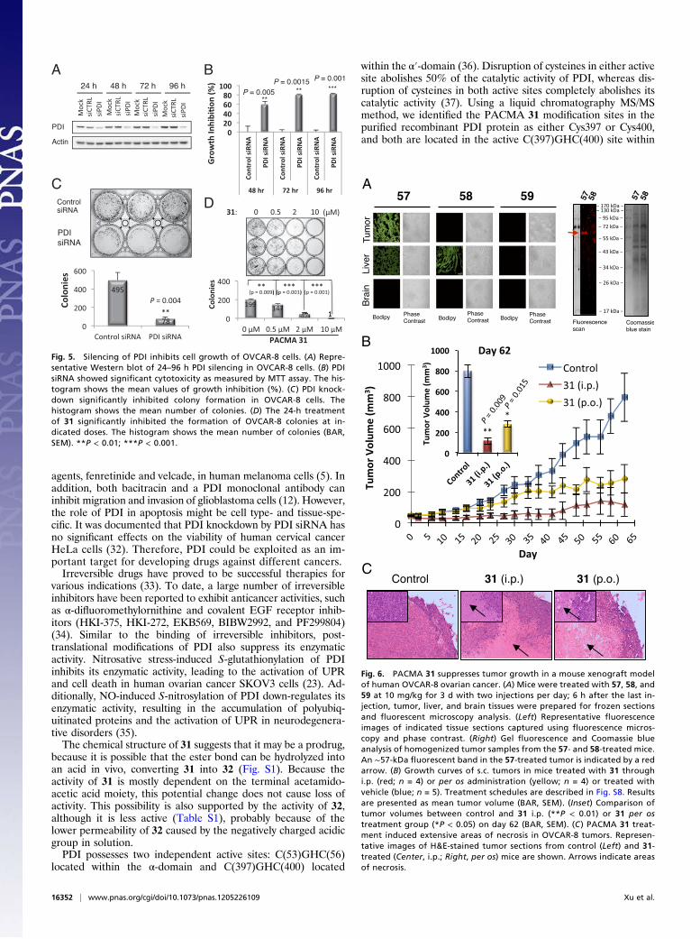
PACMA 31 Suppresses Tumor Growth in Human Ovarian Cancer MouseXenografts. To evaluate the tumor targeting ability of activePACMAs in vivo, we tested 57–59 in a mouse xenograft model ofhuman OVCAR-8 ovarian cancer. After 3 d of continuous i.p.administration, tumor, liver, and brain tissues were collected andprepared for frozen sections that were analyzed using fluores-cent microscopy. PACMA 57-treated tumor sections exhibitedstrong fluorescence intensity, whereas no fluorescence was de-tected in 58- or 59-treated tumor sections (Fig. 6A). Consistentwith our in vitro data, one fluorescent band at ∼57 kDa wasobserved only in the 57-treated tumor sample (Fig. 6A), con-firming that PDI is covalently modified by 57 in the tumors.Because the liver is the major organ for drug metabolism, we alsoexamined the fluorescent intensity in the liver of 57–59-treatedanimals by fluorescence microscopy using the same settings andconditions used to examine the tumor sections. Compared with57-treated tumor sections, the liver sections of 57-treated animalsexhibited substantially lower fluorescent intensity (Fig. 6A). Incomparison, liver sections from 58-treated animals exhibitedhigher fluorescent intensity than liver sections from 57-treatedanimals. No fluorescence emission was detected in 59-treatedliver sections as expected. In addition, no fluorescence emissionwas detected in brain sections with treatments of active or in-active compounds. Taken together, these data show that the ac-tive PACMAs selectively target and accumulate in ovarian tu-mors in vivo, and they do not cross the blood–brain barrier.To determine the in vivo efficacy of 31, we tested its effect on
established tumors from OVCAR-8 cells through i.p. or per osadministration; 31 was given i.p. at 20 mg/kg per day for the first3 wk, with 5-d on and 2-d off treatment cycles. The dose wasescalated to 40 mg/kg per day for the next 7 d. After 30-d i.p.treatment, the mouse xenografts were left untreated for anadditional 32 d (Fig. S8). A second xenograft study was
conducted to evaluate per os administration of 31. Treatmentwas initiated with a dose of 20 mg/kg per day, and it was graduallyincreased by 20 mg/kg per day with each dose for 3 d before itwas orally dosed at 200 mg/kg per day for an additional 32 d. Thetotal duration of the oral treatment was 62 d (Fig. S8). Comparedwith the control group, i.p. or per os administration of 31 signif-icantly inhibited tumor growth by 85% (from 796.6 to 117.0 mm3,P = 0.009) and 65% (from 796.6 to 280.1 mm3, P = 0.015) at day62, respectively (Fig. 6B). Thus, 31 not only suppresses tumorgrowth in vivo but also is orally bioavailable. One mouse wasfound dead on day 30 in the i.p. administration group, but noobvious abnormalities were observed in other 31-treated mice.The tumors in the i.p. treatment group did not aggressivelygrow after treatment was stopped on day 30. This finding maybe at least partially because of the prolonged duration of drugaction of 31, a common feature of irreversible inhibitors.Compared with mice in the control group, no substantial bodyweight loss was detected in i.p. or per os groups during the study(Fig. S9A), indicating that 31 did not exert severe adverseeffects on the mice at its effective anticancer dose. All micewere dissected at the end of the study (day 62). H&E staining oftumor sections showed extensive areas of necrosis in both 31treatment groups (i.p. and per os) compared with the controlgroup (Fig. 6C). In addition, no detectable abnormalities wereobserved in the organs examined, including liver, kidney,spleen, heart, lung, and pancreas (Fig. S9B), further showingthe safety of PACMA 31.
DiscussionIn this study, we designed and synthesized a series of PACMAsthat showed in vitro and in vivo anticancer activity in humanovarian cancer by targeting PDI. As a prototype of the ER pro-tein disulfide isomerase family, PDI catalyzes the formation,cleavage, and rearrangement of disulfide bonds and facilitates oxi-dative protein folding by acting as a molecular chaperone (28).Therefore, PDI inhibition causes accumulation of unfolded ormisfolded proteins that leads to ER stress and the unfolded pro-tein response (UPR) that results in cell death (5). Compared withnormal tissues, PDI is overexpressed in ovarian tumors (8). In ad-dition, estrogen has been reported to increase PDI expression(29), bind PDI (30), and promote ovarian cancer progression(31). Based on these evidences, PDI is a promising drug targetfor ovarian cancer therapy.We showed that inhibition of PDI by either small-molecule
compounds or PDI siRNA resulted in substantial cytotoxicity inhuman ovarian cancer cells. Previously, it was shown that PDIknockdown results in apoptosis in human breast cancer MCF-7cells and human neuroblastoma SH-SY5Y cells (32). Moreover,a PDI inhibitor, bacitracin, abrogated survival responses to ERstress and enhanced apoptosis caused by ER stress-inducing
Table 1. Active PMCMAs affected PDI secondary structure
DMSO 31 57 56
PDIα (%) 42.60 47.81 36.77 42.60β (%) 9.77 8.18 12.31 9.77
BSAα (%) 84.27 84.27 84.27 84.27β (%) 1.24 1.24 1.24 1.24
A B C
Fig. 4. Active PACMAs inhibit PDI activity. (A) PACMA 31 significantly inhibited the activity of PDI in a dose- and time-dependent manner in the insulinaggregation assay. Curves were generated from mean values (BAR, SEM). *P < 0.05; **P < 0.01; ***P < 0.001. (B) Comparison of the inhibitory activity of 31and PAO. (C) PACMA 56 did not exhibit significant effects on the enzymatic activity of PDI. Experiments were performed in triplicate.
Xu et al. PNAS | October 2, 2012 | vol. 109 | no. 40 | 16351
PHARM
ACO
LOGY
agents, fenretinide and velcade, in human melanoma cells (5). Inaddition, both bacitracin and a PDI monoclonal antibody caninhibit migration and invasion of glioblastoma cells (12). However,the role of PDI in apoptosis might be cell type- and tissue-spe-cific. It was documented that PDI knockdown by PDI siRNA hasno significant effects on the viability of human cervical cancerHeLa cells (32). Therefore, PDI could be exploited as an im-portant target for developing drugs against different cancers.Irreversible drugs have proved to be successful therapies for
various indications (33). To date, a large number of irreversibleinhibitors have been reported to exhibit anticancer activities, suchas α-difluoromethylornithine and covalent EGF receptor inhib-itors (HKI-375, HKI-272, EKB569, BIBW2992, and PF299804)(34). Similar to the binding of irreversible inhibitors, post-translational modifications of PDI also suppress its enzymaticactivity. Nitrosative stress-induced S-glutathionylation of PDIinhibits its enzymatic activity, leading to the activation of UPRand cell death in human ovarian cancer SKOV3 cells (23). Ad-ditionally, NO-induced S-nitrosylation of PDI down-regulates itsenzymatic activity, resulting in the accumulation of polyubiq-uitinated proteins and the activation of UPR in neurodegenera-tive disorders (35).The chemical structure of 31 suggests that it may be a prodrug,
because it is possible that the ester bond can be hydrolyzed intoan acid in vivo, converting 31 into 32 (Fig. S1). Because theactivity of 31 is mostly dependent on the terminal acetamido-acetic acid moiety, this potential change does not cause loss ofactivity. This possibility is also supported by the activity of 32,although it is less active (Table S1), probably because of thelower permeability of 32 caused by the negatively charged acidicgroup in solution.PDI possesses two independent active sites: C(53)GHC(56)
located within the α-domain and C(397)GHC(400) located
within the α′-domain (36). Disruption of cysteines in either activesite abolishes 50% of the catalytic activity of PDI, whereas dis-ruption of cysteines in both active sites completely abolishes itscatalytic activity (37). Using a liquid chromatography MS/MSmethod, we identified the PACMA 31 modification sites in thepurified recombinant PDI protein as either Cys397 or Cys400,and both are located in the active C(397)GHC(400) site within
A B
CD
Fig. 5. Silencing of PDI inhibits cell growth of OVCAR-8 cells. (A) Repre-sentative Western blot of 24–96 h PDI silencing in OVCAR-8 cells. (B) PDIsiRNA showed significant cytotoxicity as measured by MTT assay. The his-togram shows the mean values of growth inhibition (%). (C ) PDI knock-down significantly inhibited colony formation in OVCAR-8 cells. Thehistogram shows the mean number of colonies. (D) The 24-h treatmentof 31 significantly inhibited the formation of OVCAR-8 colonies at in-dicated doses. The histogram shows the mean number of colonies (BAR,SEM). **P < 0.01; ***P < 0.001.
A
B
C
Fig. 6. PACMA 31 suppresses tumor growth in a mouse xenograft modelof human OVCAR-8 ovarian cancer. (A) Mice were treated with 57, 58, and59 at 10 mg/kg for 3 d with two injections per day; 6 h after the last in-jection, tumor, liver, and brain tissues were prepared for frozen sectionsand fluorescent microscopy analysis. (Left) Representative fluorescenceimages of indicated tissue sections captured using fluorescence micros-copy and phase contrast. (Right) Gel fluorescence and Coomassie blueanalysis of homogenized tumor samples from the 57- and 58-treated mice.An ∼57-kDa fluorescent band in the 57-treated tumor is indicated by a redarrow. (B) Growth curves of s.c. tumors in mice treated with 31 throughi.p. (red; n = 4) or per os administration (yellow; n = 4) or treated withvehicle (blue; n = 5). Treatment schedules are described in Fig. S8. Resultsare presented as mean tumor volume (BAR, SEM). (Inset) Comparison oftumor volumes between control and 31 i.p. (**P < 0.01) or 31 per ostreatment group (*P < 0.05) on day 62 (BAR, SEM). (C ) PACMA 31 treat-ment induced extensive areas of necrosis in OVCAR-8 tumors. Represen-tative images of H&E-stained tumor sections from control (Left) and 31-treated (Center, i.p.; Right, per os) mice are shown. Arrows indicate areasof necrosis.
16352 | www.pnas.org/cgi/doi/10.1073/pnas.1205226109 Xu et al.

the α′-domain (Fig. S6 B–F). The location of these modificationsites is consistent with the ability of 31 to inhibit the enzymaticactivity of PDI. Additional modifications at Cys53 and Cys56may occur in vivo because of the different redox environment inthe ER, where these four cysteines should be in a reduced statethat is required for full catalytic activity. In the recombinant PDIprotein produced in Escherichia coli, a disulfide bond is probablyformed between Cys53 and Cys56, which was shown in a PDIstructure (Protein Data Bank ID code 1MEK) solved from arecombinant protein also produced in E. coli (38). Such a disul-fide bond can protect Cys53/56 from PACMA 31 modificationin vitro.In summary, we identified PDI as a cellular protein target for
PACMAs. We also showed that PDI knockdown in humanovarian cancer cells was cytotoxic and that our irreversible PDIinhibitors exhibited both in vitro and in vivo anticancer activity inhuman ovarian cancer models with tumor targeting ability and nosubstantial toxicity to normal tissues. Moreover, PACMAs wereeffective on human ovarian cancer cell lines resistant to con-ventional chemotherapy. Resistance to first-line therapy occurs inall ovarian cancer patients and is a major cause of mortality.Therefore, development of effective and safe PDI inhibitors asanticancer agents may overcome the current treatment failure inovarian cancer therapy.
Materials and MethodsMeasurement of PDI Activity. PDI activity was assayed by measuring the PDI-catalyzed reduction of insulin in the presence of DTT, thus measuring theaggregation of reduced insulin B chains at 620 nm as described previously(15). Briefly, recombinant PDI protein (0.4 μM) was incubated with indicatedcompounds at 37 °C for 1 h in sodium phosphate buffer (100 mM sodium
phosphate, 2 mM EDTA, 8 μM DTT, pH 7.0). After this incubation, themodified recombinant PDI protein was added to the reaction mixtureconsisting of DTT (500 μM) and bovine insulin (130 μM; Sigma). The re-duction reaction was catalyzed by PDI at room temperature, and theresulting aggregation of reduced insulin B chains was measured at 620 nm.
In Vivo Tumor Xenograft Studies. OVCAR-8 cells in logarithmic growth phasefrom cell culture were implanted in athymic mice (5 × 10⁵ cells in 100 μL PBS/mouse) under aseptic conditions. Tumor growth was assessed by biweeklymeasurement of tumor diameters with a Vernier caliper (length × width).Tumor volume was calculated according to the formula tumor volume (inmillimeters cubed) = D × d2/2, where D and d are the longest and shortestdiameters, respectively. For i.p. administration, tumors were allowed to growto an average volume of 50 mm3. Mice were then randomly assigned intothree groups: vehicle control (n = 5), i.p. treatment with 31 (n = 4; 20mg/kg perday for the first 3 wk with 5-d on and 2-d off treatment cycles, and dose wasescalated to 40 mg/kg per day for the next 7 d), and per os treatment of 31(n = 4; the initial dose of 20 mg/kg per daywas gradually increased by 20mg/kgper day with each dose for 3 d before it was orally dosed at 200 mg/kg perday for an additional 32 d, increasing the dose from 20 to 200 mg/kg).Treatment schedules are described in Fig. S8. Treatment of each animal wasbased on individual body weight. The body weights and tumor volumes ineach group were measured two times per week. The percentage of tumorgrowth inhibition was calculated as T/C% = 100 × (mean TW of treatedgroup)/(mean TW of control group).
Additional reagents and procedures used in this report are described indetail in SI Materials and Methods.
ACKNOWLEDGMENTS. This work was supported in part by the Universityof Southern California Zumberge Research and Innovation Fund, the ArthurC. Cope Scholar Fund administered by the American Chemical Society(N.A.P.), and the Sharon and William Cockrell Endowed Cancer ResearchFund (N.N.) and Department of Defense Ovarian Cancer Program (W81XWH-07-1-0414) Idea Award (N.N).
1. Siegel R, Naishadham D, Jemal A (2012) Cancer statistics, 2012. CA Cancer J Clin 62:10–29.
2. Petty R, Evans A, Duncan I, Kurbacher C, Cree I (1998) Drug resistance in ovariancancer—the role of p53. Pathol Oncol Res 4:97–102.
3. Hatahet F, Ruddock LW (2009) Protein disulfide isomerase: A critical evaluation of itsfunction in disulfide bond formation. Antioxid Redox Signal 11:2807–2850.
4. Hoffstrom BG, et al. (2010) Inhibitors of protein disulfide isomerase suppress apo-ptosis induced by misfolded proteins. Nat Chem Biol 6:900–906.
5. Lovat PE, et al. (2008) Increasing melanoma cell death using inhibitors of proteindisulfide isomerases to abrogate survival responses to endoplasmic reticulum stress.Cancer Res 68:5363–5369.
6. Gruber CW, Cemazar M, Heras B, Martin JL, Craik DJ (2006) Protein disulfide isom-erase: The structure of oxidative folding. Trends Biochem Sci 31:455–464.
7. Haefliger S, et al. (2011) Protein disulfide isomerase blocks CEBPA translation and isup-regulated during the unfolded protein response in AML. Blood 117:5931–5940.
8. Bonome T, et al. (2008) A gene signature predicting for survival in suboptimally de-bulked patients with ovarian cancer. Cancer Res 68:5478–5486.
9. Welsh JB, et al. (2001) Analysis of gene expression identifies candidate markers andpharmacological targets in prostate cancer. Cancer Res 61:5974–5978.
10. Beer DG, et al. (2002) Gene-expression profiles predict survival of patients with lungadenocarcinoma. Nat Med 8:816–824.
11. Basso K, et al. (2005) Reverse engineering of regulatory networks in human B cells.Nat Genet 37:382–390.
12. Goplen D, et al. (2006) Protein disulfide isomerase expression is related to the invasiveproperties of malignant glioma. Cancer Res 66:9895–9902.
13. Rickman DS, et al. (2001) Distinctive molecular profiles of high-grade and low-gradegliomas based on oligonucleotide microarray analysis. Cancer Res 61:6885–6891.
14. Talantov D, et al. (2005) Novel genes associated with malignant melanoma but notbenign melanocytic lesions. Clin Cancer Res 11:7234–7242.
15. Khan MM, et al. (2011) Discovery of a small molecule PDI inhibitor that inhibits re-duction of HIV-1 envelope glycoprotein gp120. ACS Chem Biol 6:245–251.
16. Dickerhof N, Kleffmann T, Jack R, McCormick S (2011) Bacitracin inhibits the reductiveactivity of protein disulfide isomerase by disulfide bond formation with free cysteinesin the substrate-binding domain. FEBS J 278:2034–2043.
17. Wang EJ, Snyder RD, Fielden MR, Smith RJ, Gu YZ (2008) Validation of putative ge-nomic biomarkers of nephrotoxicity in rats. Toxicology 246:91–100.
18. Weston BS, Wahab NA, Roberts T, Mason RM (2001) Bacitracin inhibits fibronectinmatrix assembly by mesangial cells in high glucose. Kidney Int 60:1756–1764.
19. Godin B, Touitou E (2004) Mechanism of bacitracin permeation enhancementthrough the skin and cellular membranes from an ethosomal carrier. J Control Re-lease 94:365–379.
20. Yamada R, et al. (2011) Discovery and preclinical evaluation of a novel class of cytotoxicpropynoic acid carbamoyl methyl amides (PACMAs). J Med Chem 54:2902–2914.
21. Roschke AV, et al. (2003) Karyotypic complexity of the NCI-60 drug-screening panel.Cancer Res 63:8634–8647.
22. Alvarez M, et al. (1995) Generation of a drug resistance profile by quantitation ofmdr-1/P-glycoprotein in the cell lines of the National Cancer Institute Anticancer DrugScreen. J Clin Invest 95:2205–2214.
23. Townsend DM, et al. (2009) Nitrosative stress-induced s-glutathionylation of proteindisulfide isomerase leads to activation of the unfolded protein response. Cancer Res69:7626–7634.
24. Perez-Iratxeta C, Andrade-Navarro MA (2008) K2D2: Estimation of protein secondarystructure from circular dichroism spectra. BMC Struct Biol 8:25.
25. Feige MJ, Hendershot LM (2011) Disulfide bonds in ER protein folding and homeo-stasis. Curr Opin Cell Biol 23:167–175.
26. Root P, Sliskovic I, Mutus B (2004) Platelet cell-surface protein disulphide-isomerasemediated S-nitrosoglutathione consumption. Biochem J 382:575–580.
27. Fonseca C, et al. (2009) Protein disulfide isomerases are antibody targets during im-mune-mediated tumor destruction. Blood 113:1681–1688.
28. Noiva R (1999) Protein disulfide isomerase: The multifunctional redox chaperone ofthe endoplasmic reticulum. Semin Cell Dev Biol 10:481–493.
29. Ejima K, et al. (1999) 17beta-estradiol induces protein thiol/disulfide oxidoreductasesand protects cultured bovine aortic endothelial cells from oxidative stress. Eur J En-docrinol 140:608–613.
30. Primm TP, Gilbert HF (2001) Hormone binding by protein disulfide isomerase, a highcapacity hormone reservoir of the endoplasmic reticulum. J Biol Chem 276:281–286.
31. Spillman MA, et al. (2010) Tissue-specific pathways for estrogen regulation of ovariancancer growth and metastasis. Cancer Res 70:8927–8936.
32. Hashida T, Kotake Y, Ohta S (2011) Protein disulfide isomerase knockdown-induced celldeath is cell-line-dependent and involves apoptosis in MCF-7 cells. J Toxicol Sci 36:1–7.
33. Singh J, Petter RC, Baillie TA, Whitty A (2011) The resurgence of covalent drugs. NatRev Drug Discov 10:307–317.
34. Jänne PA, et al. (2011) Phase I dose-escalation study of the pan-HER inhibitor,PF299804, in patients with advanced malignant solid tumors. Clin Cancer Res 17:1131–1139.
35. Uehara T, et al. (2006) S-nitrosylated protein-disulphide isomerase links protein mis-folding to neurodegeneration. Nature 441:513–517.
36. LaMantia ML, Lennarz WJ (1993) The essential function of yeast protein disulfideisomerase does not reside in its isomerase activity. Cell 74:899–908.
37. Vuori K, Myllylä R, Pihlajaniemi T, Kivirikko KI (1992) Expression and site-directedmutagenesis of human protein disulfide isomerase in Escherichia coli. This multi-functional polypeptide has two independently acting catalytic sites for the isomeraseactivity. J Biol Chem 267:7211–7214.
38. Kemmink J, Darby NJ, Dijkstra K, Nilges M, Creighton TE (1996) Structure de-termination of the N-terminal thioredoxin-like domain of protein disulfide isomeraseusing multidimensional heteronuclear 13C/15N NMR spectroscopy. Biochemistry 35:7684–7691.
Xu et al. PNAS | October 2, 2012 | vol. 109 | no. 40 | 16353
PHARM
ACO
LOGY