discovery and optimization of inhibitors of stat3 ...ccc.chem.pitt.edu/wipf/topics/zhang.pdf ·...
TRANSCRIPT

Discovery and Optimization of Inhibitors of STAT3 Activation for the Treatment of Squamous
Cell Carcinoma of the Head and Neck
Feng Zhang
Wipf Group Research Topic Seminar
02-09-2013
1
Feng Zhang @ Wipf Group Page 1 of 12 2/23/2013
Squamous Cell Carcinoma of the Head and Neck (SCCHN)
§ Squamous Cell Carcinoma of the Head and Neck (SCCHN) is the sixth most common form of cancer and accounts for ~500,000 new cancer cases per year worldwide.
§ Traditional therapies, including surgery, radiation therapy, and chemotherapy are able to eradicate head and neck cancer in only 50% of cases.
§ Treatments incorporating radiation or conventional chemotherapy drugs, such as cisplatin, may result in a host of negative side effects, some permanent.
§ As a result, there has been continuing investigation into potential alternative and less toxic therapies for head and neck cancer, with the aim of achieving a more favorable clinical outcome while reducing treatment morbidity.
§ Patient-derived primary SCCHN cells and SCCHN cell lines have been shown to overexpress a number of key signaling proteins that contribute to the enhanced growth and survival properties of these cells.
§ SCCHN cells commonly exhibit overexpression and/or hyperactivation of EGFR, signal transducer and activator of transcription 3 (STAT3), STAT3 is a key downstream target of EGFR
2
Feng Zhang @ Wipf Group Page 2 of 12 2/23/2013

Signal Transducers and Activators of Transcription
v Seven members: u STAT1 (α/β), STAT2, STAT3 (α/β/γ),
STAT4, STAT5a, STAT5b & STAT6 u Cytoplasmic transcription factors
regulating cytokine gene expression u STATs are activated via the tyrosine
phosphorylation cascade after ligand binding and stimulation of the Cytokine Receptor–Kinase complex and Growth Factor-Receptor complex
§ EGF (Epidermal Growth Factor), FGF (Fibroblast Growth Factor), PDGF (Platelet-Derived Growth Factor), IL-6 (Interleukin-6), OSM (Oncostatin-M), CSF1R (Colony Stimulating Factor-1 Receptor), c-kit, Insulin receptor, c-Met and GPCRs (G-Protein Coupled Receptors), etc.
v Conserved tyrosine residue-‐ Y701, Y705 or Y695
Crystal structure of STAT3 dimer bound to DNA
Becker et al., Nature 1998, 394, 145-151
Nature © Macmillan Publishers Ltd 1998
8
The STAT homodimer grips the DNA like a pair of pliers. Themonomers are held together by the C-terminal SH2 domains, andthe large 4-helix-bundle domains form the ‘handles’ of the pliers.The DNA is almost entirely enclosed by the protein dimer, incontact with loops from the �-barrel and the connector domains.
Structure of the proteinThe N-terminal domain, a bundle of four antiparallel helicesconnected by short loops, is very elongated (�80 A long). Helices�1 (44 residues) and �2 (54 residues) span the entire domain;helices �3 and �4 are considerably shorter (32 and 25 residues).
Helix �2, which has a kink at residue 234, packs against strand �aand helices �5 and �6, linking the 4-helix bundle with the �-barreland the connector domain. The central hydrophobic core of thebundle contains Val, Leu and Ile residues. The underlying heptadspacing in this region was recognized by Fu et al.15, who correctlypredicted a coiled-coil motif.
The 4-helix bundle is immediately followed by the eight-stranded�-barrel. Strands a, b and e form one sheet of the barrel; strands c, fand g form the other. Strands x/c� connect the two sheets byhydrogen bonding, first to strand e of sheet abe, then switchingover to pair with strand c of sheet cfg. Strand extensions a� and g�
form a small two-stranded sheet closing the �-barrel at the end nextto the DNA, where three loops of this domain participate in DNAbinding (Figs 2, 3a, b).
The �-barrel domain is linked to the SH2 domain by a smallhelical domain, formed by two helix–loop–helix modules (helices�5 to �8), which we call the ‘connector’ domain. This domain showsstructural similarity to calcium-binding domains, as found introponin C16,17, for example. In common with Ca2+-bindingdomains, the connector domain has an identical arrangement ofhelices, a pseudodyad between the �5/�6 and �7/�8 helix–loop–helix motifs (r:m:s:34C�-atoms ¼ 1:9 A), and a small two-stranded �-pleated sheet, formed by strands h and i, pairing these two motifs.However, the loops of the connector domain are longer, and thepolar residues that chelate the metal in calcium-binding domainsare replaced by hydrophobic residues which pack against each other.The connector domain shows no structural similarity to SH3domains, as was previously suggested on the basis of sequencehomology.
The SH2 domain of Stat3 shares with other SH2 domains acentral three-stranded �-pleated sheet (strands B, C and D) flankedby helix �A and strands �A and �G. (Secondary-structure elementsand residues are denoted according to established nomenclature18.)
articles
NATURE | VOL 394 | 9 JULY 1998 147
Figure 2 Ribbon diagram of the Stat3� homodimer–DNA complex. The N-
terminal 4-helix bundle is shown in blue, the �-barrel domain in red, the connector
domain in green, and the SH2 domain and phosphotyrosine-containing region in
yellow. Disordered regions between helices �1 and �2 and residues 689 to 701
have been modelled in grey. This figure and Fig. 3b were produced with program
RIBBONS45. Views are shown a, along the DNA axis (the dyad of the complex
running vertically); b, from the side, with monomer 2 depicted in grey; and c, from
the top. Phosphotyrosines are indicated by a Y in c.
SH2 domain and pY-containing region
Connector domain
N-terminal 4-helix bundle
DNA
β-barrel domain
3
Feng Zhang @ Wipf Group Page 3 of 12 2/23/2013
§ STAT3 is one of seven members of the signal transducer and activator of transcription (STAT) family of proteins whose function is to relay signals from the cell surface receptors to the nucleus and initiate transcription.
§ STAT3 plays a vital role in regulating cell growth and survival. In response to growth factor and cytokine stimulation, STAT3 is phosphorylated, dimerizes, and translocates into the nucleus to up-regulate transcription of a wide spectrum of genes.
§ STAT3 is a key tumor promoting transcription factor, constitutively activated in many types of cancer including SCCHN, prostate, breast and colorectal cancers.
§ Activated STAT3 promotes tumor cell proliferation/survival and tumor metastasis, while suppressing the anti-tumor immune response. Inhibition of STAT3 signaling has been shown to inhibit tumor growth in vitro and in vivo.
What is STAT3
4
Feng Zhang @ Wipf Group Page 4 of 12 2/23/2013

§ STAT1 has significant sequence and functional similarity to STAT3, however, whereas STAT3 is oncogenic, STAT1 is associated with tumor suppression.
§ STAT1 can be activated by various ligands including interferon-alpha, interferon-gamma, EGF, PDGF and IL-6.
§ Activated STAT1 plays a critical role in promoting an effective anti-tumor immune response.
§ STAT3 inhibitors identified through target-based screening strategies (e.g. SH2-targeting molecules) often exhibit poor cell permeability, efficacy, selectivity (vs STAT1) and/or stability, and have not progressed into the clinic.
§ STAT3 pathway-specificity (i.e. without STAT1 signaling inhibition) is highly critical when developing anticancer agents designed to block STAT3 activation.
§ The ultimate goal is to discover and develop selective inhibitors of the STAT3 pathway for the treatment of SCCHN tumors.
The Opposing Roles of STAT3 & STAT1 in Cancer
5
Feng Zhang @ Wipf Group Page 5 of 12 2/23/2013

The Opposing Roles of STAT3 & STAT1 in Cancer
• STAT3 is a proto-oncogene
• STAT1 is a tumor suppressor
G. Regis et al. Semin. Cell Dev. Biol. 2008, 19, 351–359. 6
Feng Zhang @ Wipf Group Page 6 of 12 2/23/2013

STAT3 Pathway 2360 Current Medicinal Chemistry, 2011 Vol. 18, No. 16 Lavecchia et al.
Fig. (1). STAT proteins are activated by receptor and non-receptor tyrosine kinases via several mechanisms. (A) Receptor-associated JAKs are activated upon cytokine receptor binding, through the classic cross-phosphorylation pathway. These activated JAKs phosphorylate Y residues on the receptor, which thereby become docking elements for cytoplasmic STAT proteins. STATs are subsequently phosphorylated on a single Y residue in the C-terminal portion of the receptor, and then form homo- or heterodimers through reciprocal interaction between the pY of one STAT and the Src-SH2 domain of another. Dimerized STATs are carried into the nucleus via importins, where they then induce target gene transcription by binding to specific regulatory elements. (B) Receptors with intrinsic tyrosine kinase activities, including platelet-derived growth factor receptor (PDGFR), epidermal growth factor receptor (EGFR), and FMS-related (formerly McDonough feline sarcoma viral oncogene homologue) tyrosine kinase 3 (FLT3), may directly activate STATs without JAK involvement. (C) STATs can be phosphorylated by constitutively active non-receptor protein tyrosine kinases (PTKs), such as Src and breakpoint cluster region/Abelson tyrosine kinase (Bcr/Abl). (D) Unphosphorylated STATs can independently enter the nucleus to mediate gene transcription, possibly by acting as transcriptional co-regulators in DNA binding.
Fig. (2). Topology of STAT protein structures with the most important conserved domains involved in specific functions. Y and S are phosphorylation sites in tyrosine and in serine.
Lavecchia, A. et al. Curr. Med. Chem. 2011, 18, 2359-2375. 7
Feng Zhang @ Wipf Group Page 7 of 12 2/23/2013

STAT3-based Anticancer Strategies
Introduction
Signal transducer and activator of transcription 3 (STAT3) is a member of a family of seven proteins (STATs 1, 2, 3, 4, 5a, 5b, and 6) that relay signals from activated cytokine and growth factor receptors in the plasma membrane to the nucleus, where they regulate gene transcription (1–3). STAT3 modulates the transcrip-tion of responsive genes involved in the regulation of a variety of critical functions, including cell differentiation, proliferation, apoptosis, angiogenesis, metastasis, and immune responses (2, 4–6). Multiple lines of evidence place STAT3 at a central node in the development, progression, and maintenance of many human tumors, validating STAT3 as an anticancer target (Table 1) (2, 4–7). Cellular transformation by the viral oncogene v-src requires activated STAT3 (2, 7–10), and the transfection and expression of a constitutively activated form of STAT3 is sufficient to transform immortalized fibroblasts and normal epithelial cells derived from breast and prostate tissue (8). Most of the major human malig-nancies manifest elevated levels of constitutively activated STAT3 (Table 1) as well as transcriptional profiles that are consistent with STAT3-regulated gene expression (2, 4, 6, 7). For many cancers, elevated levels of activated STAT3 have been associated with a poor prognosis (Table 1) (2, 4, 5, 11). STAT3-activated genes block apoptosis, favor cell proliferation and survival, promote angiogen-esis and metastasis, and inhibit antitumor immune responses (2, 4–6, 12). Tumor cell lines bearing constitutively activated STAT3 require continued STAT3 activation, a phenotype that has been termed “oncogene addiction” (4). In contrast, approaches that dis-rupt STAT3 signaling lead to growth inhibition and apoptosis in tumor cell lines and can impair tumor growth in mouse xenograft cancer models (Table 1) (2, 5, 6, 9, 13–19). Although knockout of STAT3 leads to embryonic lethality in mice, the cumulative data
in postnatal mice (conditional knockouts) indicates that STAT3 may be dispensable for the function of normal cells and tissues (2, 4). The ability of nontransformed cells to withstand inhibition of STAT3 signaling suggests that a potential anticancer drug that tar-gets STAT3 might be well tolerated (2, 4).
STAT3 and STAT1 have great sequence similarity and function as transcription factors that bind to very similar DNA sequences, but in many physiological contexts, they are regulated in a recipro-cal manner to produce opposing effects (12, 17, 20). In the cancer context, activated STAT3 is oncogenic, whereas activated STAT1 behaves as a tumor suppressor (12). Therefore, the selective inhibi-tion of STAT3, without interference in STAT1 signaling, might be proposed as the basis for anti-cancer drug development. However, in experimental situations, STAT1 and STAT3 may be functionally interchangeable in promoting cytokine-induced programs of gene expression [e.g., STAT1 on its own may affect such gene expres-sion via the interleukin-6 receptor, whereas STAT3 may operate via the interferon-gamma receptor (2, 12)]. It should also be noted that constitutively activated STAT5, which shows little sequence similarity with STAT3, has also been implicated in oncogenesis (2, 21). Here, we review approaches that have been pursued to target STAT3, and we highlight some of the challenges associated with developing an anticancer drug that might therapeutically inhibit the STAT3 signaling pathway.
STAT3 Domain Structure, Signaling, and Regulation
STAT3 is structurally typical of the STAT family: an N-terminal coiled-coiled domain involved in protein–protein interactions; a DNA binding domain; a Src homology-2 (SH-2) domain; and a C-terminal transactivation domain (5, 22). In the canonical STAT3
Table 1. STAT3 in the Context of Various Cancers: Validation as an Anticancer Target
Cancers Characterized by Elevated STAT3 Expression or Activity
Poor Prognosis Linked to High STAT3 Levels
Upstream/Downstream Abnormalities of STAT3 Signaling
Xenograft Models Responsive to Inhibition of STAT3
STAT3 Signaling Pathway as a Therapeutic Target in Cancer�
Johnston PA, Grandis JR, Mol Interv. 2011 Feb; 11 (1): 18-26
8
Feng Zhang @ Wipf Group Page 8 of 12 2/23/2013
Inhibitors of IL-‐6 receptor complex pSTAT3 AcGvaGon: Mechanisms of AcGon
9
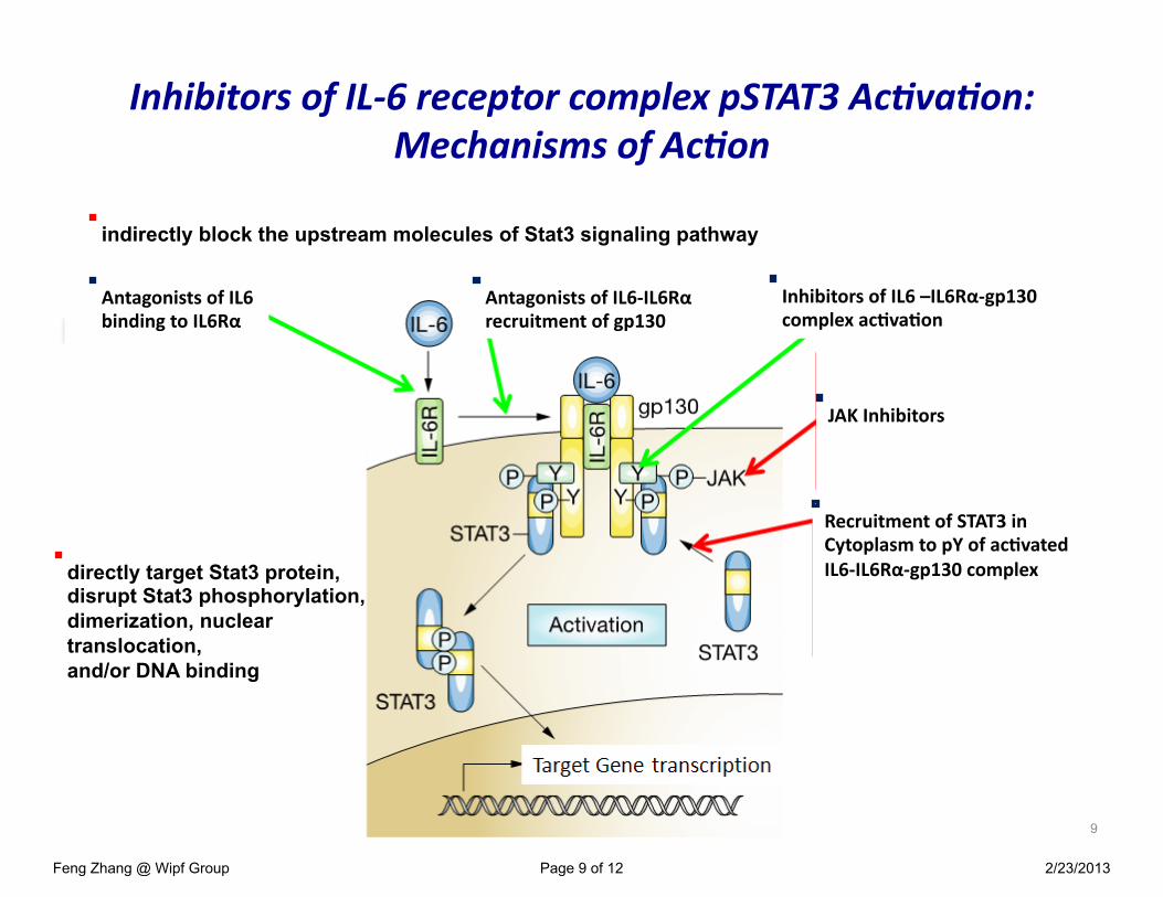
Antagonists of IL6 binding to IL6Rα
Antagonists of IL6-‐IL6Rα recruitment of gp130
Inhibitors of IL6 –IL6Rα-‐gp130 complex ac@va@on
JAK Inhibitors
Recruitment of STAT3 in Cytoplasm to pY of ac@vated IL6-‐IL6Rα-‐gp130 complex
indirectly block the upstream molecules of Stat3 signaling pathway�
directly target Stat3 protein, disrupt Stat3 phosphorylation, dimerization, nuclear translocation, and/or DNA binding�
Feng Zhang @ Wipf Group Page 9 of 12 2/23/2013

v Direct target Stat3 protein
§ STA-21: inhibits luciferase activity at 20 µM; inhibit STAT3 dimerization, nuclear translocation, STAT3-DNA binding in MDA-MB-435s cells at 20 or 30 µM; inhibit Bcl-XL and cyclin D1 in MDA-MB-468 breast carcinoma cells.
§ S31-201: selectively inhibits STAT3 DNA binding (STAT3:STAT3 IC50 = 86 µM, STAT1:STAT3 IC50 = 160 µM, STAT1:STAT1 IC50 > 300 µM) in EGF-stimulated mouse fibroblasts NIH 3T3/hEGFR.
§ Stattic: inhibits luciferase activity at IC50 = 5.1 µM; inhibit STAT3 DNA binding at 10 µM in the nuclear extracts form EGF-stimulated cells; selectively inhibit IL-6 induced pSTAT3 over IFN-γ induced pSTAT1 in HepG2 liver carcinoma cells.
Current Status of STAT3 Inhibitors
O
O
O
OH
STA-21
HN
HOOC OH
O
OSOO
S31-201
SO O
O2N
Stattic
10
Feng Zhang @ Wipf Group Page 10 of 12 2/23/2013

v indirectly block the upstream molecules of Stat3 signaling pathway�
v Curcumin: inhibits JAK2, Src and Erb2, and epidermal growth factor receptor; no selectivity between pSTAT3 and pSTAT1; inhibition of pSTAT3 is reversible.
v Capsaicin: preferentially inhibited constitutive Stat3 phosphorylation in multiple myeloma cells through the inhibition of JAK1 and c-Src.
v Emodin: inhibited Stat3 phosphorylation by targeting JAK2.
Debnath, B.; Xu, S.; Neamati, N. J. Med. Chem. 2012, 55, 6645-6668.
HOO
O O
OHO
Curcumin Capsaicin
O
HO
NH
OO
O
OH
OH
OH
H3C
Emodin
Current Status of STAT3 Inhibitors
11
The goal of the STAT3 project is to identify small molecules that selectively inhibit STAT3 over STAT1 signaling and that can be developed into clinical compounds for the treatment of squamous cell carcinoma of the head and neck. �
Feng Zhang @ Wipf Group Page 11 of 12 2/23/2013

Acknowledgement�
v Dr. Peter Wipf. v Dr. Donna Huryn, v Dr. Paul A Johnston
(HCS bioassay), Dr. Lynn Resnick, Dr. Matthew G LaPorte Dr. Erin Skoda,
v Mary Liang v Shelby Anderson v Mr. Pete Chambers (ELS, LC-MS), v Wipf group members past & present. v Funding: NCI/SAIC−Frederick 29XS127.
12
Feng Zhang @ Wipf Group Page 12 of 12 2/23/2013