discospondylitis in dogs: a review - vet times
TRANSCRIPT

Vet TimesThe website for the veterinary professionhttps://www.vettimes.co.uk
Discospondylitis in dogs: a review
Author : Luca Motta
Categories : Vets
Date : August 1, 2009
Luca Motta discusses possible causes, diagnosis and treatment of this spinal disease affectingdogs
DISCOSPONDYLITIS is an infectious inflammatory disease centred on the intervertebraldisc that extends to involve the adjacent endplates and vertebral bodies (Kornegay et al,1980).
The resulting erosive lesions of the intervertebral disc and the vertebral body endplates may resultin instability, extradural spinal cord or cauda equina compression due to granulation tissue,collapse of the intervertebral space and/or pathological vertebral fractures or luxation.
Any level of the vertebral column may be affected by discospondylitis, and multiple lesions may beseen, although caudalcervical, thoracolumbar and lumbosacral intervertebral spaces of thevertebral column are most often involved (Gage, 1975; Gilmore, 1987). Haematogenous spread ofbacteria or fungi from foci elsewhere in the body, or by direct inoculation from tracking foreignbodies, is probably the most common cause of discospondylitis (Moore, 1992). Frequently, isolatedorganisms include Staphylococcus intermedius, Brucella canis, Escherichia coli, Staphylococcusaureus, Pseudomonas aeruginosa, Staphylococcus epidermidis, Bordetella species, Streptococcusspecies, Aspergillus species, Enterococcus faecalis, Fusarium species, Mucor species and Paecilomyces variotii, (Hurov et al, 1978; Johnson et al, 1982; Adamo et al, 2001; Cherubini et al,2004; Braund et al, 1987; Kornegay et al, 1990; Kornegay 1993). Sources of infection includebacterial endocarditis, dental diseases, sites of dental extraction, penetrating wounds, plantmaterial migration, urinary tract infections and skin infections (Gage, 1975; Moore 1992; Kornegay,1993). However, in many cases, the primary source of the infection may be not identified (Turnwald
1 / 23

et al, 1986; Cherubini et al, 2004). Discospondylitis may occur with increased frequency inimmunocompromised animals due to factors, such as diabetes mellitus and hyperadrenocorticism(Barta et al, 1985). Young German shepherd bitches seem to be predisposed to aspergillosis(Berry et al, 1996), whereas young basset hounds contract discospondylitis due to systemictuberculosis caused by Mycobacterium avium (Carpenter et al, 1988).
Pathophysiology
The pathophysiology of discospondylitis is unclear. It has been hypothesised that the presence ofsubchondral vascular loops in the vertebral epiphysis slows circulation, allowing colonisation byblood-borne bacteria, which then diffuse through the cartilaginous endplate of the vertebral body toreach the disc. Infection is further disseminated to adjacent vertebrae through freelycommunicating venous sinuses (Trueta, 1959; Kornegay et al, 1990).
Several theories exist to explain migration of grass awns to the vertebral column. These foreignbodies may be swallowed and migrate through the bowel wall (possibly at the caudal duodenalflexure), through the mesentery to the attachment to ventral epaxial muscles and, consequently, tothe vertebral column. As dogs with discospondylitis – thought to be due to plant awn migration –have lesions most commonly in the cranial lumbar spine (L2-L4), it has been suggested that awnsmay be inhaled and migrate through the lungs to the diaphragm, and lodge at the crural insertionon the lumbar vertebrae. Plant awns may also migrate through skin and paravertebral or abdominalmuscles to the vertebral column. Grass seeds are able to travel long distances, owing to thedirection of the barbs. Forward progress may be aided by muscle movements (Case, 1983;LeCouter et al, 1989).
Clinical signs
Clinical findings depend on the location of the affected vertebra or vertebrae. The typical clinicalsigns are:
• weight loss;
• anorexia;
• depression;
• fever;
• reluctance to run or jump; and
• apparent spinal pain (which may be severe).
2 / 23

Hyperesthesia may be present only over the site of the lesion or may be poorly localised, especiallywith involvement of multiple sites. Various degrees of neurologic deficits may also be evident. Theyrange from lumbosacral pain without neurological deficits to nonambulatory paraparesis (Gilmore,1987). Discospondylitis should always be considered in dogs with neurological signs associatedwith increased body temperature and systemic illness, particularly in large, male, middle-aged dogs(Kornegay, 1993).
Diagnosis
When discospondylitis is suspected, urine and blood culture should be performed, as they arepositive in up to 50 per cent and 75 per cent respectively; serology for brucellosis should also beperformed in view of its zoonotic potential (Thomas, 2000). Collection of cerebrospinal fluid (CSF)is indicated in animals with neurologic deficits. The CSF white blood cell count may be normal, ormay be elevated, with an increase in polymorphonucleated neutrophils in CSF from animals withmeningitis or myelitis (LeCouter, 2006). Presumptive diagnosis of discospondylitis is frequentlymade before radiographic signs appear, based on the clinical findings. Definitive diagnosis isusually made on evaluation of discospondylitis signs on the survey radiographs. However,radiographic changes have been reported to lag behind the onset of clinical signs by four to sixweeks (Kornegay et al, 1990; Moore 1992). Moreover, most of the time the radiographic featuresassociated with discospondylitis are nonspecific, and differentials, such as neoplasia or severespondylosis, should always be considered (Gonzalo-Orden et al, 2000). Radiographic findings onwhich the diagnosis of discospondylitis is based include rarefaction of bone, collapse of disc space,proliferative bony changes adjacent to the intervertebral disc space and sclerosis at the margins ofbone rarefaction (Figure 1). Reaction to infection of the vertebral bodies and disc spaces is evidencedas osteolysis, sclerosis and vertebral spur formation.
The first radiographic signs of discospondylitis are usually a collapsed intervertebral disc space,with or without subtle vertebral endplate erosion. Sclerosis of bone and ventral spur formation arecommon signs in chronic infections; these may be accompanied by marked osteolysis and newbone formation. Chronic lesions have a mixture of bone lysis and extensive new bone production,with osteophytes bridging adjacent vertebrae containing a central destructive focus. Affectedvertebral bodies may be shortened, and bony proliferation may result in the fusion of one or morevertebrae (Hurou et al, 1978; Johnson et al, 1983; Jimenez et al, 1995).
Radiography can also be used to monitor the response to treatment or the progression to thedisease. Radiographic follow-up should be evaluated with caution – because radiographs of matureand older dogs may look worse for an additional three to nine weeks – despite successfultreatment (Shamir et al, 2001). Computed tomography (CT) can identify subtle endplate erosionand paravertebral soft tissue swelling. Magnetic resonance imaging (MRI) provides uniqueinformation for those patients where axial skeletal radiographic lesions are not definitive, or wherediagnosis is needed to make management decisions prior to the time period when confirmatorysurvey radiographic lesions have developed (Kraft et al, 1998). In human medicine, MRI is
3 / 23
considered to be the most sensitive and specific imaging modality for inflammatory and infectiousdiseases of the spine (Hovi et al, 1994).
Discospondylitis is diagnosed in people as well as in animals by MRI criteria, which includes:
• narrowed intervertebral disc space;
• loss of endplate clarity, signifying cortical bone erosion;
• decreased signal o n T1-weighted images, increased signal on T2-weighted images of the bonemarrow and endplates of affected vertebrae; and
• abnormal paraspinal and epidural soft tissue (Figures 2, 4, 5, 7 and 9).
Contrast medium administration may help to define paravertebral and epidural extension of thedisease, and may produce areas of enhancement within the affected area of the vertebral bodies(Thrush et al, 1990; Hovi et al, 1994; Cherubini et al, 2004) (Figures 3, 6 and 10).
Fat suppression techniques are very useful for detecting infectious or neoplastic lesions by makingthose areas look hyperintense to fat in the bone marrow or epidural space (Tien et al, 1992) (Figure 8
).
Gradient echo sequences are useful to determine whether endplate irregularity is present (Dennis,2005). Another diagnostic imaging option in dogs with discospondylitis is scintigraphy, which maybe helpful as early as three days after the onset of clinical signs when radiographic signs areequivocal (Lamb, 1987). Scintigraphy may show different degrees of radiopharmaceutical uptake atthe affected disc spaces and endplates (Stern et al, 2007). However, care must be taken wheninterpreting this finding, as spondylosis also causes some increase in uptake (Kornegay, 1991).Surgical biopsy may be indicated in affected dogs in which a causative organism has not beenisolated from blood or urine, and/or in patients unresponsive to initial broad-spectrum antibiotics.Fluoroscopically guided percutaneous needle aspiration of the disc space has been described, andthe procedure has been documented to be up to 75 per cent sensitive (Fischer et al, 1997). Openbiopsy of the vertebrae may be considered if needle aspiration is unrewarding (Kornegay et al,1980).
Treatment
Medical treatment, consisting of antibiotic therapy against the common potential pathogens andrestriction of exercise, is usually undertaken first (Johnson, 1995). Use of analgesics may behelpful during the first five to seven days of treatment, but should not be necessary after five daysand should be discontinued to allow clinical assessment of the patient. Results of blood and urinecultures may require alteration of the antibiotic’s class.
4 / 23

Antibiotics should be administered intravenously if severe neurological compromise is present. Ifclinical improvement is seen within two weeks of starting antibiotic therapy – such as resolution offever, improved appetite or reduction of apparent spinal pain – medical treatment should becontinued for at least six weeks, and vertebral radiographs should be performed every four to sixweeks to monitor progression/regression of a lesion, and to monitor for development of newlesions.
Antibiotic administration may be necessary for up to six months before radiographic evidence ofresolution of lesions is seen. Clinical signs may recur if the infection is not completely eliminatedprior to cessation of antibiotic therapy, and repeated cultures of blood and urine and ongoingtreatment with an appropriate antibiotic may be necessary (Gilmore, 1987).
Surgical treatment is indicated when clinical signs do not improve after one to two weeks ofappropriate antibiotic therapy, when neurological deficits are present, or when spinal instability,associated with bone destruction, is evident radiographically (Kornegay, 1993; Gilmore, 1987; andOzuna et al, 1996). Reports on the surgical management of discospondylitis in dogs mentiondecompressive procedures (laminectomy, hemilaminectomy), stabilisation, curettage, bonegrafting, and internal fixation as possible treatments.
The objectives of surgery are debridement of necrotic tissue, decompression, and spinalstabilisation by use of various fixation devices and bone grafting. These techniques aim ateradicating infection and interbody fusion with neurological recovery and minimal resultingdeformities (Ozuna et al, 1996; Jeanneret et al, 1994; Graziano et al, 1993; Eysel et al, 1997 andRath et al, 1996). A distraction-fusion technique similar to one described in humans for treatment ofcauda equina compression has been used successfully in dogs affected by discospondylitis(McKee et al, 1990). It seems that discospondylitis of L7-S1 complicated by instability, regardlessof whether neurological deficits are present, carries an excellent prognosis when treated with adistraction-fusion technique.
Dramatic improvement of the condition shortly after surgery is likely to be due to the opening ofcollapsed L7-S1 foramina with resolution of L7 spinal nerve entrapment, alleviation of cauda equinacompression, and stabilisation of the lumbosacral segment throughout the healing process.Debridement and cancellous bone grafting of the intervertebral space appear beneficial to obtaininterbody fusion (McKee et al, 1990; Auger et al, 2000).
Prognosis
The prognosis for animals with discospondylitis depends on the ability to eliminate the causativeorganism and on the degree of neurologic dysfunction. Animals with severe neurologic deficits andfungal infections have a guarded-to-poor prognosis (LeCouter, 2006). The potential for recurrenceshould be considered, especially if brucellosis has been diagnosed or an underlyingimmunosuppressive condition is present (Thomas, 2000).
5 / 23

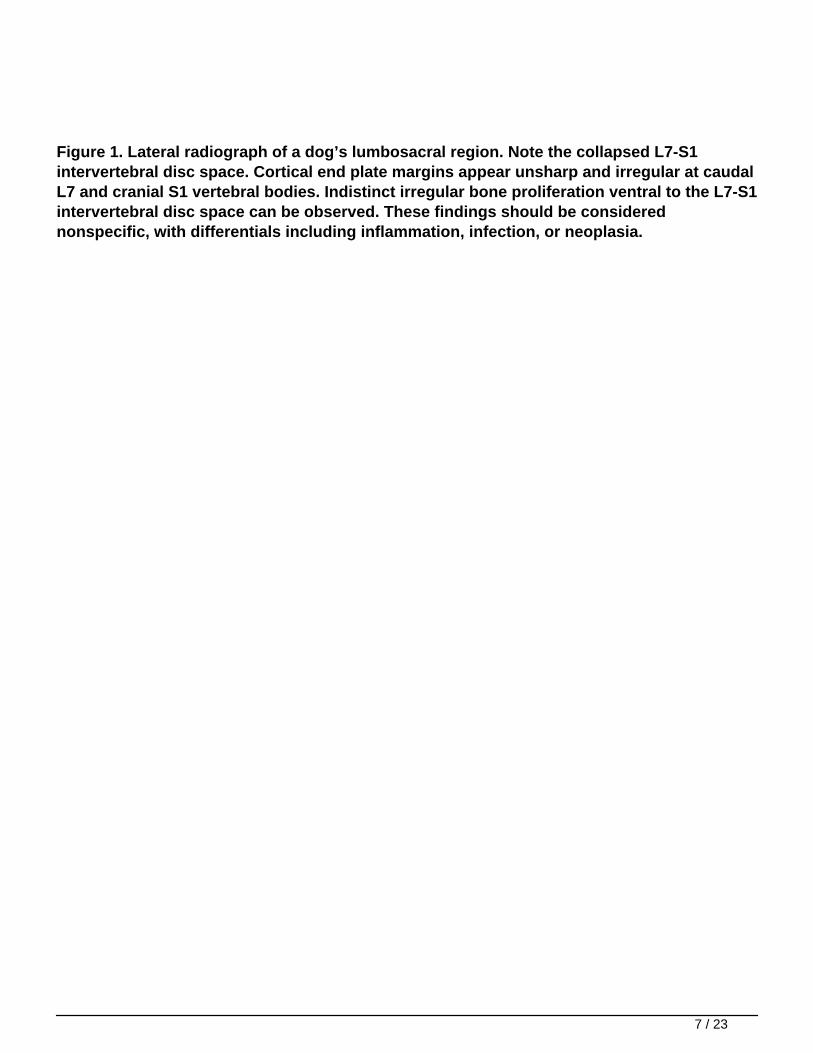
Figure 1. Lateral radiograph of a dog’s lumbosacral region. Note the collapsed L7-S1intervertebral disc space. Cortical end plate margins appear unsharp and irregular at caudalL7 and cranial S1 vertebral bodies. Indistinct irregular bone proliferation ventral to the L7-S1intervertebral disc space can be observed. These findings should be considerednonspecific, with differentials including inflammation, infection, or neoplasia.
7 / 23
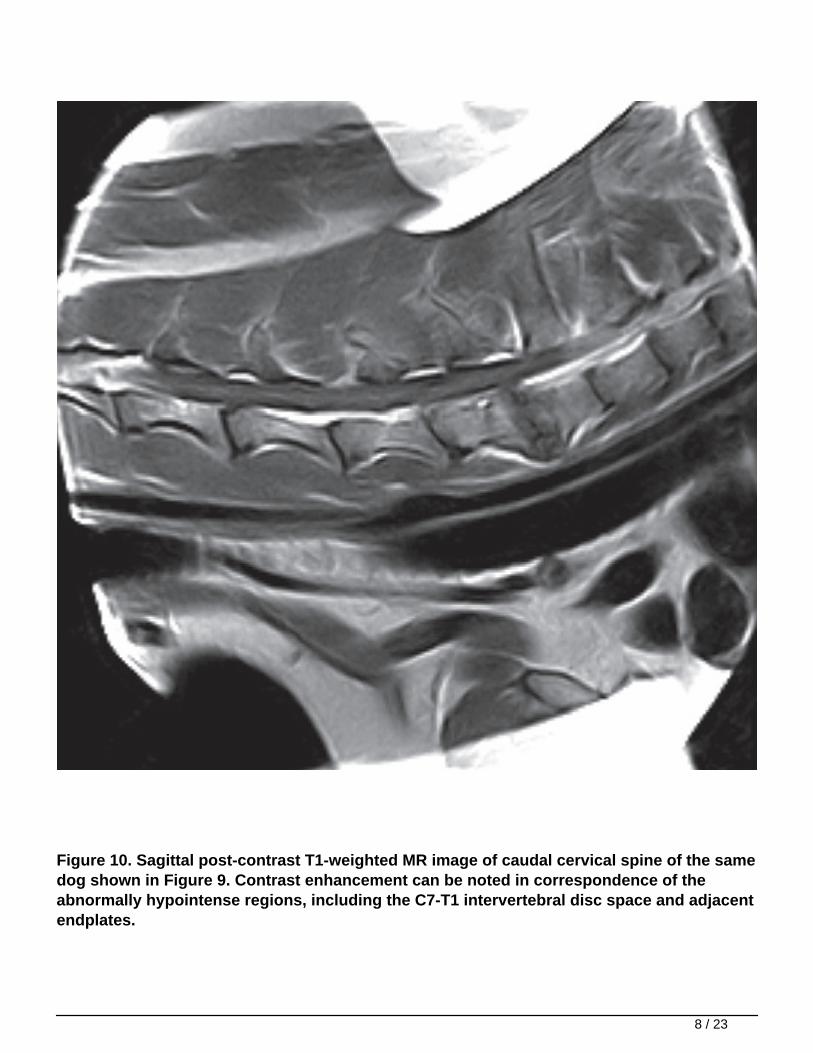
Figure 10. Sagittal post-contrast T1-weighted MR image of caudal cervical spine of the samedog shown in Figure 9. Contrast enhancement can be noted in correspondence of theabnormally hypointense regions, including the C7-T1 intervertebral disc space and adjacentendplates.
8 / 23
Figure 2. Sagittal pre-contrast T1-weighted MR image of caudal lumbar and lumbosacralspine in a dog with confirmed discospondylitis caused by Staphylococcus intermedius. L7
9 / 23
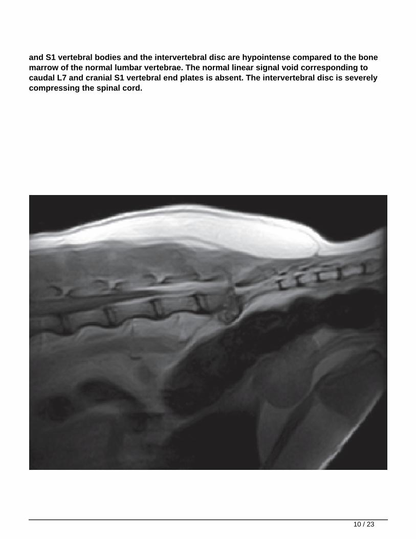
and S1 vertebral bodies and the intervertebral disc are hypointense compared to the bonemarrow of the normal lumbar vertebrae. The normal linear signal void corresponding tocaudal L7 and cranial S1 vertebral end plates is absent. The intervertebral disc is severelycompressing the spinal cord.
10 / 23

Figure 3. Sagittal post-contrast T1-weighted MR image of caudal lumbar and lumbosacralspine of the same dog shown in Figure 2. Contrast enhancement has led to increased signalintensity of the abnormally hypointense regions, including the L7-S1 intervertebral discspace and adjacent endplates.
11 / 23

Figure 4. Sagittal T2-weighted MR image of the caudal lumbar and lumbosacral spine of thesame dog shown in Figure 2. The caudal aspect of L7 and the cranial aspect of S1 vertebralbodies cannot be distinguished from material within the L7-S1 intervertebral disc space.The intervertebral disc is severely compressing the spinal cord.
12 / 23

13 / 23

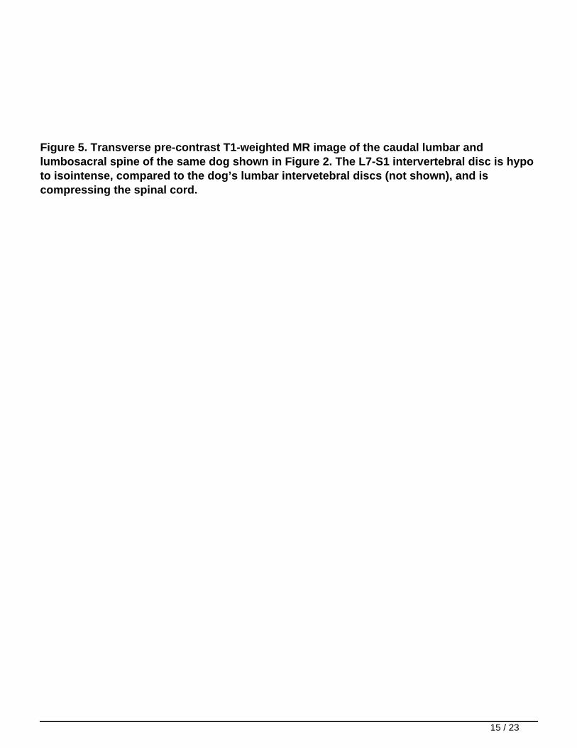
Figure 5. Transverse pre-contrast T1-weighted MR image of the caudal lumbar andlumbosacral spine of the same dog shown in Figure 2. The L7-S1 intervertebral disc is hypoto isointense, compared to the dog’s lumbar intervetebral discs (not shown), and iscompressing the spinal cord.
15 / 23


Figure 6. Transverse post-contrast T1-weighted MR image at the level of the lumbosacralintervertebral disc of the same dog shown in Figure 2. Contrast enhancement has led tononuniform increased signal intensity through the L7-S1 intervertebral disc.
17 / 23

Figure 7. Sagittal pre-contrast T1-weighted MR image of the caudal lumbar and lumbosacralspine of the same dog shown in Figure 2 displaying the paravertebral extension of thedisease.
18 / 23

19 / 23


Figure 8. Dorsal STIR MR image of the caudal lumbar and lumbosacral spine of the samedog shown in Figure 2. A marked hyperintensity in correspondence of L7-S1 intervertebraldisc space can be seen. This MRI finding could be supportive of both infectious/inflammatory and neoplastic lesions.
21 / 23
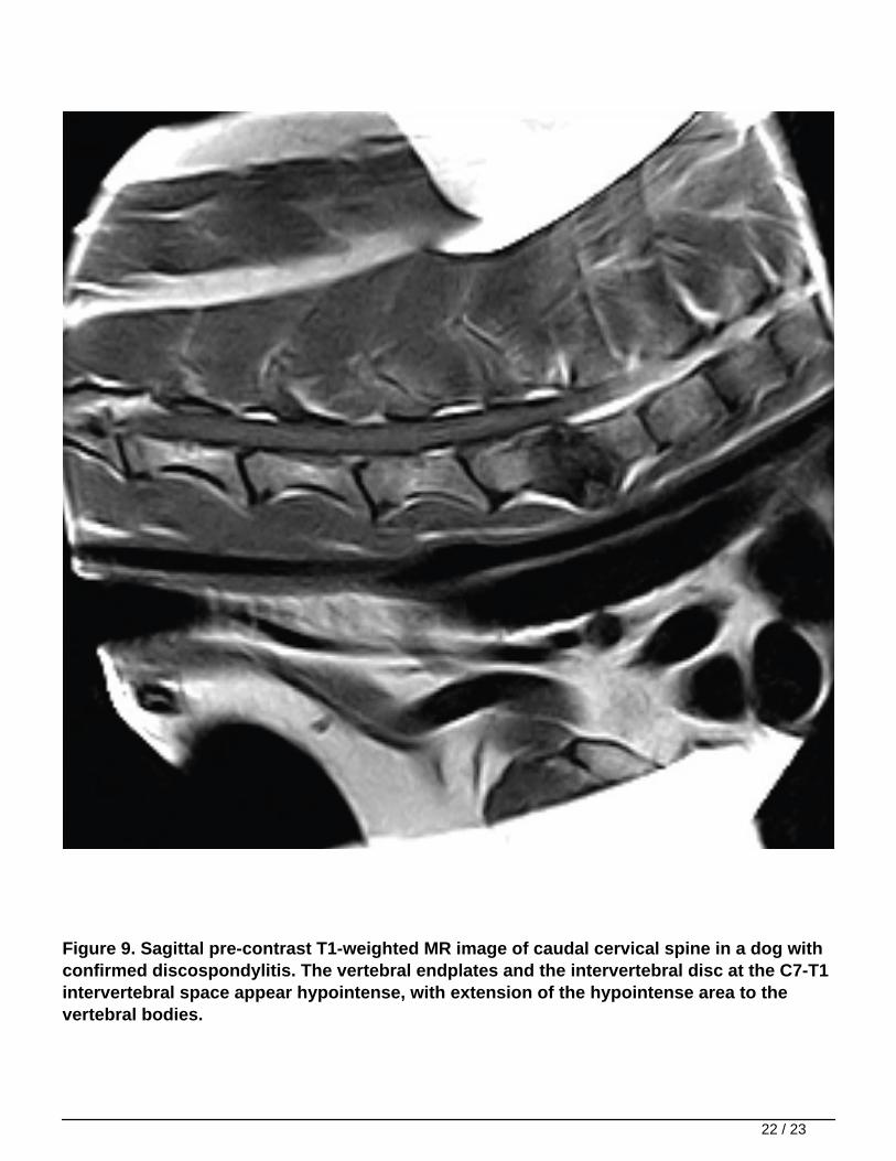
Figure 9. Sagittal pre-contrast T1-weighted MR image of caudal cervical spine in a dog withconfirmed discospondylitis. The vertebral endplates and the intervertebral disc at the C7-T1intervertebral space appear hypointense, with extension of the hypointense area to thevertebral bodies.
22 / 23
