dilation assisted stone extraction technique spotlight (dase)
TRANSCRIPT

Figure 1Cholangiogram with Large Filling Defects
(Stones)
Figure 2Endoscopic View of CRE
Dilating Balloon
CRE™ Wireguided Balloon and SpyGlass® Direct Visualization System
Dilation Assisted Stone Extraction (DASE)
technique spot l ight
Figure 3 Endoscopic View of Bile Duct Through
Dilating Balloon
History
A 63 year old male status post cholecystectomy presented to the hospital with fever and altered mental status. On further evaluation he was noted to have elevated liver function testing. His transabdominal ultrasound showed a dilated common bile duct with an internal 2cm shadowing stone. Following medical treatment of cholangitis with antibiotics, the patient was referred for ERCP for definitive management of choledocholithiasis.
Procedure
At the time of ERCP a cholangiogram showed a dilated common bile duct (up to 2.5cm) with several large filling defects (measuring more than 2 cm). A biliary sphincterotomy was performed. Given the distal narrowing of the bile duct and the size discrepancy of the bile duct stone and the biliary orifice, a decision was made to perform Dilation Assisted Stone Extraction (DASE). This was accomplished by incremental wire guided hydrostatic balloon dilation using the CRE Wireguided Balloon beginning at 15mm up to 20mm. An initial waist seen on fluoroscopy was obliterated at the end of dilation. Following this maneuver, attempts were made to extract the large stones using a 3cm wire guided basket. However, once captured, the large stone was unable to be pulled out of the bile duct, resulting in a trapped basket and stone. Attempts at mechanical lithotripsy failed, resulting in fracture of the basket wire and retained basket and stone. The trapped basket was subsequently removed using another wire guided basket, however the stone remained within the bile duct. A decision was made to proceed with per oral cholangioscopy (Spyglass System) for mechanical lithotripsy under direct visualization. Once mounted, the Spyglass Scope was inserted into the bile duct and the large pigmented gallstone was identified. Then, using a Holmium laser probe, laser lithotripsy was performed (1J, 10 pulses per second; 10 Watts) with resultant fragmentation of the stone. The stone fragments were then extracted using wire guided baskets and biliary extraction balloons. However, despite this some stone material remained. Therefore, the decision was made to stent the bile duct to facilitate drainage and further facilitate stone fragmentation.
Abhitabh Patil, MDAssistant Professor Rush University Medical CenterChicago, IL
Boston Scientific CorporationOne Boston Scientific PlaceNatick, MA 01760-1537www.bostonscientific.com/endoscopy
Ordering Information 1.800.225.3226
© 2012 Boston Scientific Corporation or its affiliates. All rights reserved.
ENDO-71204-AA April 2012
technique spot l ight CRE™ Wireguided Balloon and SpyGlass® Direct Visualization System
CRE and SpyGlass are trademarks of Boston Scientific Corporation or its affiliates.
Indications, Contraindications, Warnings and Instructions for Use can be found in the product labeling supplied with each device. Caution: Federal (uSa) law restricts this device to sale by or on the order of a physician.
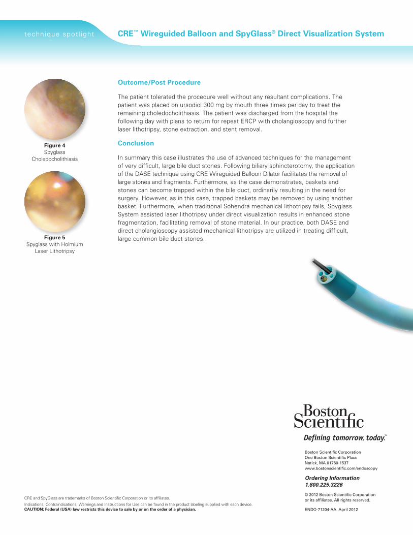
Figure 4Spyglass
Choledocholithiasis
Figure 5 Spyglass with Holmium
Laser Lithotripsy
outcome/Post Procedure
The patient tolerated the procedure well without any resultant complications. The patient was placed on ursodiol 300 mg by mouth three times per day to treat the remaining choledocholithiasis. The patient was discharged from the hospital the following day with plans to return for repeat ERCP with cholangioscopy and further laser lithotripsy, stone extraction, and stent removal.
Conclusion
In summary this case illustrates the use of advanced techniques for the management of very difficult, large bile duct stones. Following biliary sphincterotomy, the application of the DASE technique using CRE Wireguided Balloon Dilator facilitates the removal of large stones and fragments. Furthermore, as the case demonstrates, baskets and stones can become trapped within the bile duct, ordinarily resulting in the need for surgery. However, as in this case, trapped baskets may be removed by using another basket. Furthermore, when traditional Sohendra mechanical lithotripsy fails, Spyglass System assisted laser lithotripsy under direct visualization results in enhanced stone fragmentation, facilitating removal of stone material. In our practice, both DASE and direct cholangioscopy assisted mechanical lithotripsy are utilized in treating difficult, large common bile duct stones.