digital slide and virtual microscopy-based routine and
TRANSCRIPT
5
Digital Slide and Virtual Microscopy-Based Routine and Telepathology Evaluation of Gastrointestinal Biopsy Specimen
Bela Molnar, Lajos Berczi, Levente Ficsor, Viktor Varga, Attila Tagscherer, and Zsolt Tulassay
2.1Introduction
Medical digital image analysis, following the rapid developments in the information technology industry, is getting more and more an accepted method [21]. Digital techniques and laboratories [7] already exist for the radiology applications. New standalone automated microscope systems and recently fully automated digital slide scanners were developed for the histo/cytology applications in the last decade and years as well.
The automated rescreening of cervical smears is now available for routine [14, 17]. Several new semiautomatic microscopes were developed for the quality control of the cytotechnologists’ work [1, 2, 12, 13]. These systems notify the scanned area and images, and additional electronic recording of selected images is also available. The automated histological analysis is important research project for several years, but the results were unsatisfactory for routine applica-tions [3, 19]. However, the histological diagnosis can be supported already today by new electronic techniques like the TV image cytometry and teleconsultation based on histological images rather than entire samples [6].
Telepathology services were built in the last decade around two technology platforms. The dynamic telepathology includes remote-controlled microscope systems with high-throughput online image transport channels [8, 27]. This method has the advantage of entire slide access and lacks the error source of preselected microscopic frames; however, the costs of the system and working are relatively high. The application of static preselected images for teleconsulta-tion needs much less hardware investment; however, a sampling error can occur [25, 26]. Using Internet as a telecommunication pathway for static images is a low-cost widely available alternative [9, 11].
2
6 Bela Molnar et al.
The application of a digital slide should be considerable as a source and tar-get of the telepathology and automated histological analysis. In the last years, attempts to use an electronic or digital slide for education, teleconsultation, and immunohistochemical analysis are growing [4, 5, 15, 23]. The storage capacity and speed of personal computers became applicable only recently for handling the huge amount of image information stored on a slide.
Low-cost, commercial, motorized microscopes were marketed by several manufacturers in late 1990s. Recently, several automated digital slide scanning systems became widely available and used for transmitted light and multichannel fluorescence. They are using either optical elements of the microscopy with a CCD camera or a line sensor. A special alternative could be the multilens slide scanning systems.
Working on digital slides should require virtual or digital microscopy. Pre-liminary positive results on a limited number of mosaicked microscopic images have been reported recently [22].
The aim of the study was the evaluation of a digital slide scanning system and a virtual microscope (VM) on gastrointestinal biopsy specimen in a routine environment. A comparison was performed between the optical microscope (OM) and digital microscope evaluation in local and remote mode.
2.2Materials and Methods
2.2.1Gastric Routine Biopsy Specimen Analysis
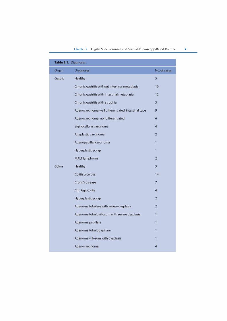
Biopsy specimens were placed in buffered formalin, routinely processed and stained with hematoxylin and eosin. Altogether 103 specimens were selected from the files of cases seen at the Ist Department of Pathology. Single representative slide was evaluated from each case. The distribution of the cases is listed in Table 2.1.
The histological sections were evaluated in separate setting on the VM and OM without the knowledge of the previous analysis by two independent, board-certified histologists. First, the evaluation of the glass slides on optical micro-scopy was done. The paper copies of the clinical histories and the evaluation report were collected. In several weeks, the slides were digitized by our system described in later sections by an assistant.
The slide digitization was done in the Digital Microscopy Laboratory. The scanning computer was used as a digital slide server too. The virtual microscopy evaluation was done on a separate local area network (LAN) workstation in the
Chapter 2 Digital Slide Scanning and Virtual Microscopy-Based Routine 7
Table 2.1. Diagnoses
Organ Diagnoses No. of cases
Gastric Healthy 5
Chronic gastritis without intestinal metaplasia 16
Chronic gastritis with intestinal metaplasia 12
Chronic gastritis with atrophia 3
Adenocarcinoma well differentiated, intestinal type 9
Adenocarcinoma, nondifferentiated 6
Sigillocellular carcinoma 4
Anaplastic carcinoma 2
Adenopapillar carcinoma 1
Hyperplastic polyp 1
MALT lymphoma 2
Colon Healthy 5
Colitis ulcerosa 14
Crohn’s disease 7
Chr. Asp. colitis 4
Hyperplastic polyp 2
Adenoma tubulare with severe dysplasia 2
Adenoma tubulovillosum with severe dysplasia 1
Adenoma papillare 1
Adenoma tubulopapillare 1
Adenoma villosum with dysplasia 1
Adenocarcinoma 4
8 Bela Molnar et al.
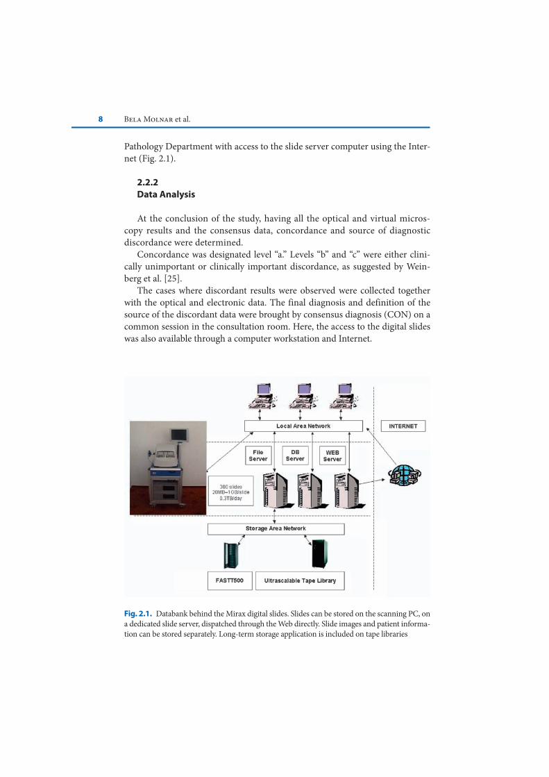
Fig. 2.1. Databank behind the Mirax digital slides. Slides can be stored on the scanning PC, on a dedicated slide server, dispatched through the Web directly. Slide images and patient informa-tion can be stored separately. Long-term storage application is included on tape libraries
Pathology Department with access to the slide server computer using the Inter-net (Fig. 2.1).
2.2.2Data Analysis
At the conclusion of the study, having all the optical and virtual micros-copy results and the consensus data, concordance and source of diagnostic discordance were determined.
Concordance was designated level “a.” Levels “b” and “c” were either clini-cally unimportant or clinically important discordance, as suggested by Wein-berg et al. [25].
The cases where discordant results were observed were collected together with the optical and electronic data. The final diagnosis and definition of the source of the discordant data were brought by consensus diagnosis (CON) on a common session in the consultation room. Here, the access to the digital slides was also available through a computer workstation and Internet.

Chapter 2 Digital Slide Scanning and Virtual Microscopy-Based Routine 9
The reasons for discordance were classified by the consensus meeting as to inadequate image quality (class I), interpretation (class II), and insufficient clin-ical information (class III). The significance of the concordance was determined using Kendall’s concordance coefficient determined by the Statistica program package (V.4.3, Statsoft, USA).
2.2.3Slide Digitization and the VM System
2.2.3.1Used Hardware Tools
We used a Mirax Scan digital slide scanner (developed and produced by 3DHISTECH Ltd, Budapest; distributed worldwide by Carl Zeiss, Jena, Ger-many) (Fig. 2.2). This scanner allows the fully automated scanning of up to 300 slides in a batch, using slide magazines each for 50 slides. The automated identi-fication of the slides is done through the barcode label. Automated identification of area of interest is done through a preview camera. The microscope functions (objectives, stage, focus, illumination, and filters) can be controlled and changed
Fig. 2.2. The Mirax scanner developed for high-throughput automated scanning of large-volume slides. (a) Frontal view with the stage and control computers. (b) Side view: view of the camera, fluorescent illumination. Note the slide magazines on the loading rack

10 Bela Molnar et al.
through the RS232 interface from an application program. The mechanic accu-racy of the motorized scanning stage for X/Y and Z directions was 0.3 μm.
In our work, we used the Allied Vision Technologies Marlin 1,4 MP one-chip CCD camera with 1,380 × 1,030 pixels. The integration time of the camera can be controlled through the computer interface RS485. The programs were running on computers with a double-core Intel processor, 128 MB RAM, and 2 GB hard disk. The scanning time for a biopsy, including loading, barcode identification, scanning, and slide repositioning into the slide holding magazine, is between 2 and 4 min.
2.2.3.2Features of the Digital Slide Scanning Program
1. Calibration of the microscope, scanning stage, camera resolution.2. Slide loading.3. Barcode reading and identification.4. Scanning area determination after preview scanning.5. Autoscanning was started after setting the stage at zero position. In the scan-
ning process, autofocusing was done only at each 3–5 field of view. Each of the images was compressed in JPEG and stored in the slide databank in the corresponding position. Autofocusing was done using Brenner’s algorithm [10]. At 40× objective, 125,856 frames would have to be stored for the entire slide. However, a threshold filter was used to store only frames with image content. This way only the area of a biopsy was stored, which means 160–700 frames per slide.
6. First, the mosaicking of the field of views was done using mathematical algorithms [20]; however, the error of the stage between the required and real position was proved to be less than +0.5 μm. A high-precision software mosaicking alignment was used for stitching of the single field of views (Fig. 2.3).
2.2.3.3Features of the VM Program
1. Slide selection. After scanning, the slides are stored in subdirectories called projects for a higher ordering. After selection, an electronically minimized slide image is shown on the screen (Fig. 2.4).
2. Slide orientation maps. This map represents the whole slide, where one pixel on the screen corresponds to one field of view. The recorded segments are labeled with white pixels on a gray background.

Chapter 2 Digital Slide Scanning and Virtual Microscopy-Based Routine 11
Fig. 2.3. (a) Scanning principle of the Mirax system: the sample on the slide is recorded in stitched mosaic of single field of views. (b) Digital magnification of a digital slide
Fig. 2.4. User interface of the MIRAX viewer software. Image of the slide, intermedier mag-nification of the slide, and virtual microscopy are available for perfect orientation. Parallel viewing of the same specimen is available with different sections and staining in oriented and multiple windows
12 Bela Molnar et al.
3. Applicable electronic magnifications. After finding the interested area, the user has several options for magnification of the selected segments. One can use the prepared magnification steps of 100× and 200× or special “+” and “−” mouse arrows to change the magnification.
4. Moving and scrolling the slide. If the interested area is not on the screen in the required magnification on the slide, then the moving arrows (up, down, left, and right) can be used to move the slide into any directions.
5. Labelling of interested frames for re-evaluation, consultation, and reporting. Up to ten colored labels can be placed on the digital slide in the software. This option can also be used for reconsultation by experts via LAN or in specific cases via Internet.
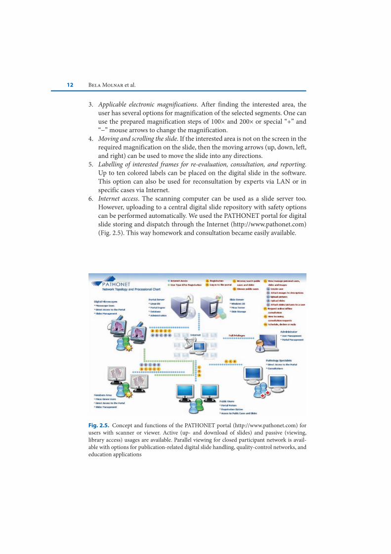
6. Internet access. The scanning computer can be used as a slide server too. However, uploading to a central digital slide repository with safety options can be performed automatically. We used the PATHONET portal for digital slide storing and dispatch through the Internet (http://www.pathonet.com) (Fig. 2.5). This way homework and consultation became easily available.
Fig. 2.5. Concept and functions of the PATHONET portal (http://www.pathonet.com) for users with scanner or viewer. Active (up- and download of slides) and passive (viewing, library access) usages are available. Parallel viewing for closed participant network is avail-able with options for publication-related digital slide handling, quality-control networks, and education applications
Chapter 2 Digital Slide Scanning and Virtual Microscopy-Based Routine 13
In the case of direct access to the slide server, the slide server’s IP address has to be defined on the remote workstation. After connecting to the server computer, the list of the available slides and their information is transported to the client workstation. After selecting a slide, its electronically compressed low-resolution image is transferred to the workstation. Every image that is transported during the evaluation will be stored on the local machine for safety reasons.
2.3Results
2.3.1Concordance
In altogether seven cases (6.7%), discordance was found between VM or OM and the CON. In four cases the OM and in three cases the VM yielded the correct diagnosis.
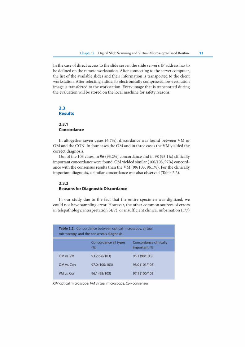
Out of the 103 cases, in 96 (93.2%) concordance and in 98 (95.1%) clinically important concordance were found. OM yielded similar (100/103, 97%) concord-ance with the consensus results than the VM (99/103, 96.1%). For the clinically important diagnosis, a similar concordance was also observed (Table 2.2).
2.3.2Reasons for Diagnostic Discordance
In our study due to the fact that the entire specimen was digitized, we could not have sampling error. However, the other common sources of errors in telepathology, interpretation (4/7), or insufficient clinical information (3/7)
Table 2.2. Concordance between optical microscopy, virtual
microscopy, and the consensus diagnosis
Concordance all types (%)
Concordance clinically important (%)
OM vs. VM 93.2 (96/103) 95.1 (98/103)
OM vs. Con 97.0 (100/103) 98.0 (101/103)
VM vs. Con 96.1 (98/103) 97.1 (100/103)
OM optical microscope, VM virtual microscope, Con consensus

14 Bela Molnar et al.
Table 2.3. Reasons of diagnostic discordance
Level discordance
Reason of discordance
VM diagnoses
Optical micro-scopy diagnosis
Review consensus diagnosis
B II Chronic gastritis
Acute and chronic gastritis
VM
C III Gastric adenoma
Gastric hyper-plastic polyp
VM
C II Chronic atrophic gastritis
Chronic gastritis with low-grade dysplasia
OM
B II Hyperplastic polyp
Hyperplastic mucosa
OM
C II Mild colitis Normal colon OM
C III Colitis ulcerosa with dysplasia
Colitis ulcerosa VM
C III Adenoma papillare
Hyperplastic polyp
OM
Discordance: B clinically not important, C clinically importantReason of discordance: I image quality, II interpretation, III insufficient clinical information
could be observed (Table 2.3). With the advances in optical imaging (higher-resolution, multiple focus levels), no image quality errors were found.
2.3.3Technical and Practical Data at the Application of VM
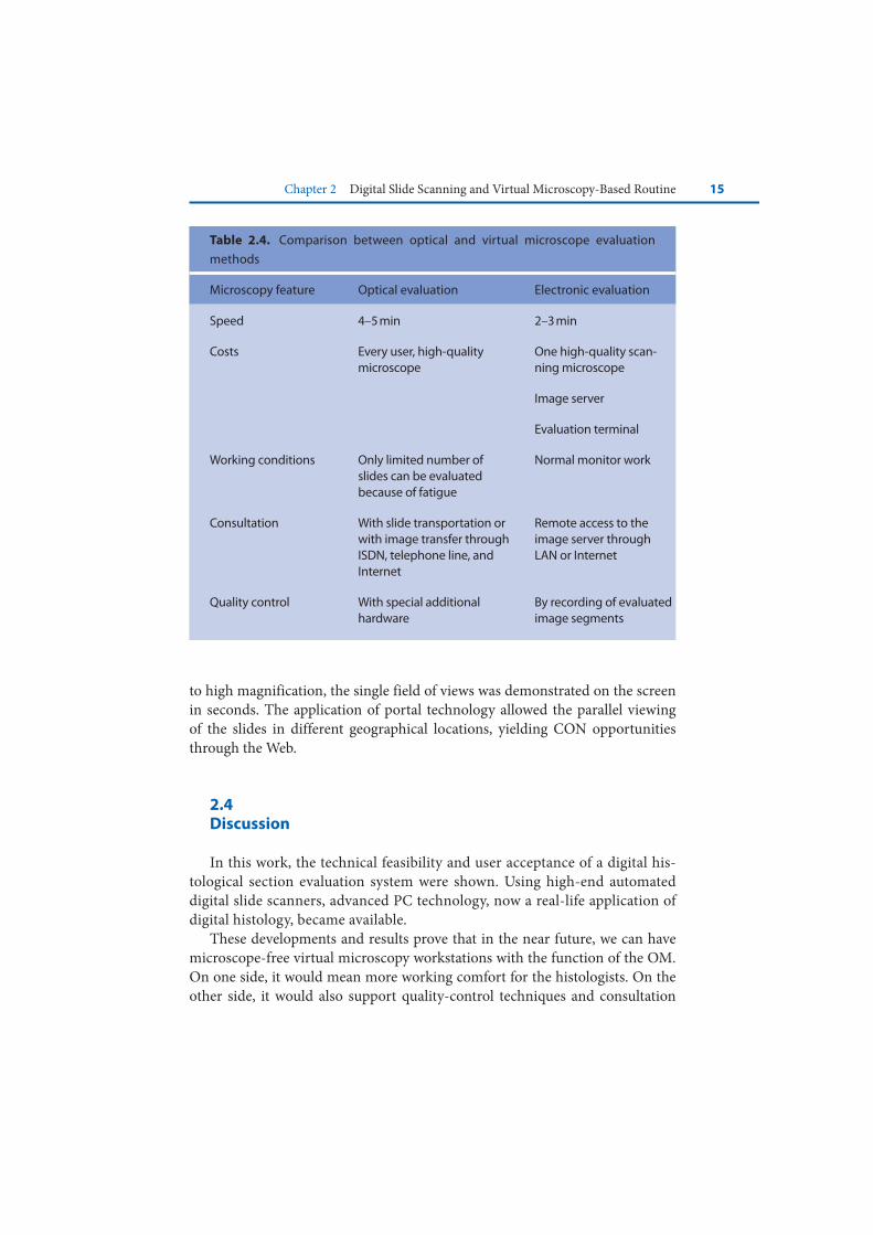
The hard-disk volume of a microscopic field of view is between 60 and 100 KB after JPEG compression. The overall hard-disk place for a gastric biopsy is between 30 and 50 MB, and the scanning time is between 20 and 40 min, depending on the number and area of sections on a slide. The evaluation of the specimen on the monitor is more comfortable and more reproducible and docu-mentable, as compared to the optical way (Table 2.4).
In the Internet remote access, the speed of the transfer was relatively fast. The first minimized image was uploaded in the real time. As the user switched over
Chapter 2 Digital Slide Scanning and Virtual Microscopy-Based Routine 15
to high magnification, the single field of views was demonstrated on the screen in seconds. The application of portal technology allowed the parallel viewing of the slides in different geographical locations, yielding CON opportunities through the Web.
2.4Discussion
In this work, the technical feasibility and user acceptance of a digital his-tological section evaluation system were shown. Using high-end automated digital slide scanners, advanced PC technology, now a real-life application of digital histology, became available.
These developments and results prove that in the near future, we can have microscope-free virtual microscopy workstations with the function of the OM. On one side, it would mean more working comfort for the histologists. On the other side, it would also support quality-control techniques and consultation
Table 2.4. Comparison between optical and virtual microscope evaluation
methods
Microscopy feature Optical evaluation Electronic evaluation
Speed 4–5 min 2–3 min
Costs Every user, high-quality microscope
One high-quality scan-ning microscope
Image server
Evaluation terminal
Working conditions Only limited number of slides can be evaluated because of fatigue
Normal monitor work
Consultation With slide transportation or with image transfer through ISDN, telephone line, and Internet
Remote access to the image server through LAN or Internet
Quality control With special additional hardware
By recording of evaluated image segments
16 Bela Molnar et al.
with remote experts as well. The primary application of digital slides and virtual microscopy should be now the routine sign-out and not only the consultation.
Remote evaluation of the slides through Internet includes the advantages of the previously used static or dynamic telepathology methods (entire slide is available in high magnification, microscope, and remote assistance-free evalu-ation) [24].
The observed concordance with optical microscopy was higher than in pre-vious static image-based telepathology studies [25–27]. This can be explained by the fact that this technology eliminates the sampling error. Evaluation of cer-tainty, time constraints, and fatigue was not done. It is considered to be done in a larger interlaboratory study.
The digitization of the routine specimen for Intranet or telediagnostics could be performed directly in the H/E sample preparation laboratory. The Mirax scanner has slide magazines with 50 slides each. In the routine workflow in the biopsy laboratory, the coverslipping is done also in batches of 50–60 slides. In our laboratory, the first set of H/E-stained, coverslipped slides are ready at 11:00 a.m. Using the high-speed scanning, the first set of slides are on the workstations monitor at between 11:20 and 11:30. The routine sign-out can happen directly, and an electronic report could be prepared on the monitor and sent out even in an email in seconds.
O’Brien and Sotnikov [18] defined in their 1996 review article about the computerization of the histology laboratory as a desirable but far future. Leong and McGee [16] published recently that automated complete slide digitization has influence on all levels of the clinical practice and education. They empha-sized the importance of a dedicated software technology for the practical use.
The results of our study showed that the digital slide and virtual microscopy technology can be used in selected cases for telepathology consultation, but not for everyday routine use.
2.5Summary
Automated digital slide scanning became available in the last years through digital slide scanners.
Their pilot application on gastrointestinal specimen is rational due to the volume of the biopsy sections.
Teleconsultation through Intra- and Internet are both applicable alternatives. The concordance between optical and virtual microscopy results to concord-
ance diagnosis is similarly high due to image quality developments in the digital slide scanning technology.
Chapter 2 Digital Slide Scanning and Virtual Microscopy-Based Routine 17
The application of virtual microscopy with digital slide scanners for gastro-intestinal biopsy specimen telepathology is now supported by the results of our study.
References
1. Anderson TL, Nelson AC (1995) Quality control and proficiency testing of cytological smear screening: an integrated approach using automation. In: Wied GL, Keebler CM, Rosenthal DL, Schenck U, Somrak TM, Vooijs GP (eds) Compendium on quality assur-ance, proficiency testing and workload limitations on clinical cytology. Tutorials of Cytol-ogy, Chicago, IL, pp. 83–287
2. Baker RW, Wadsworth J, Brugal G, Coleman DV (1998) An evaluation of ‘rapid review’ as a method of quality control of cervical smears using the AxioHOME microscope. Cytopa-thology 8:85–95
3. Belhomme P, Elmoataz A, Herlin P, Bloyet D (1996) Generalised region growing operator with optimal scanning: application to segmentation of breast cancer images. J Microsc 886:41–50
4. Burns BF (1997) Creating low power photomicrographs using a 35 mm digital slide scan-ner. Am J Surg Pathol 21:865–866
5. Dee FR (2006) Virtual microscopy for comparative pathology. Toxicol Pathol 34:966–973 6. Dictor M (1997) The surgical pathologist in a client/server computer network: work
support, quality assurance, and the graphical user interface. Mod Pathol 10:259–266 7. Dooley RL, Engel C, Muller ME (1997) Automated scanning and digitizing of roentgeno-
graphs for documentation and research. Clin Orthop 274:113–119 8. Dunn BE, Almagro UA, Choi H, et al (1997) Dynamic-robotic telepathology: department
of Veteran Affairs feasibility. Hum Pathol 28:8–12 9. Eusebi V, Foschini L, Erde S, et al (1997) Transcontinental consults in surgical pathology
via the Internet. Hum Pathol 28:13–1610. Firestone L, Cook K, Culp K (1991) Comparison of autofocus methods for automated
microscopy. Cytometry 12:195–20611. Gombas P, Szende B, Stotz G (1996) Support by telecommunications in diagnostic pathology.
Experience with the first telepathology system in Hungary. Orv Hetilap 137:2299–230312. Grohs DH, Gombrich PP, Domanik RA (1996) AccuMed International, Inc. Meeting the
challenges in cervical cancer screening: the AcCell Series 2000 automated slide handling and data management system. Acta Cytol 40:26–30
13. Grohs DH, Dadeshidze VV, Domanik RA, Gombrich PP, Olsson LJ, Pressman NJ (1997) Utility of the TracCell system in mapping Papanicolaou-stained cytologic material. Acta Cytol 41:144–152
14. Hailey DM, Lea R (1995) Prospects for newer technologies in cervical cancer screening programmes. J Qual Clin Pract 15:139–145
15. Helin HO, Lundin ME, Laakso M, Lundin J, Helin HJ, Isola J (2006) Virtual microscopy in prostate histopathology: simultaneous viewing of biopsies stained sequentially with hematoxylin and eosin, and alpha-methylacyl-coenzyme A racemase/p63 immunohisto-chemistry. J Urol 175:459–504
16. Leong FJWM, McGee O’D (2001) Automated complete slide digitization: a medium for simultaneous viewing by multiple pathologists. J Pathol 195:508–514

18 Bela Molnar et al.
17. Mango LJ, Ivasauskas EZ (1995) Computer assisted cervical cytology using the PAPNET testing. In: Wied GL, Keebler CM, Rosenthal DL, Schenck U, Somrak TM, Vooijs GP (eds) Compendium on quality assurance, proficiency testing and workload limitations on clinical cytology. Tutorials of Cytology, Chicago, IL, pp. 155–167
18. O’Brien MJ, Sotnikov AV (1996) Digital imaging in anatomic pathology. Am J Clin Pathol 106:25–32
19. Ong SH, Jin XC, Sinniah R (1996) Image analysis of tissue sections. Comput Biol Med 26:269–279
20. Ott SR (1997) Acquisition of high-resolution digital images in video microscopy: auto-mated image mosaicking on a desktop microcomputer. Microsc Res Tech 38:335–343
21. Shotton DM (1995) Robert Feulgen Prize Lecture 1995. Electronic light microscopy: present capabilities and future prospects. Histochem Cell Biol 104:97–137
22. Singson RPC, Natarajan S, Greenson JK, Marchevsky AM (1999) Virtual microscopy and the Internet as telepathology consultation tools. A study of gastrointestinal biopsy specimens. Am J Pathol 111:792–795
23. Stewart J III, Myazaki K, Bevans-Wilkins K (2007) Virtual microscopy for cytology profi-ciency testing: are we there yet? Cancer 111:203–212
24. Weinberg DS (1996) How is telepathology being used to improve patient care. Clin Chem 42:831–835
25. Weinberg DS, Allaert FA, Dusserre P, et al (1996) Telepathology diagnosis by means of digital still images: an international validation study. Hum Pathol 27:111–118
26. Weinstein RS (1996) Prospects for telepathology. Hum Pathol 17:433–43427. Weinstein RS, Battacharayya AK, Graham AR, et al (1997) Telepathology a ten-year
progress report. Hum Pathol 28:1–7