digestive system (1)
DESCRIPTION
A presentation on the organs in the digestive system and its functions, how the nerves and blood vessels run through the system.TRANSCRIPT

DIGESTIVE SYSTEMDIGESTIVE SYSTEM
http://images.google.com.sg/imgres?imgurl=http://training.seer.cancer.gov/module_anatomy

INTRODUCTION
DIGESTIVE SYSTEM Digestive system is a system which involve in digesting
or broken down the food and liquid portion that we ate into smaller molecules of nutrients
Digestive system is made up of the digestive tract or gastrointestinal tract and accessory digestive organs.
Digestive tract : mouth-pharynx-esophagus-stomach-small intestine-large intestine-rectum and anal canal-anus
Accessory organs : salivary glands, exocrine glands, liver, pancreas, gallbladder.
There are 4 primary layers in digestive system: mucosa, submucosa, muscularis, serosa

MOUTH ( ORAL CAVITY ) Covered by stratified squamous epithelium and mucous membrane Produce saliva and digest food become bolus Oral cavity includes:
Lip3 layer: outer skin layer, middle muscle layer and inner mucosa layer
Hard and soft palate Hard lined by keratinized type of
stratified squamous epithelium Soft lined by non-keratinized type of
stratified squamous epithelium Tongue
Covered by non-keratinized type of stratified squamous epithelium
Uvula Teeth
http://images.google.com.sg/imgres?imgurl=http://embryology

PHARYNX Lympatic tissue area present
called tonsils such as palatine tonsile, lingual tonsile and pharyngeal
Divided into 3:1. Nasopharynx Pseudostratified columnar
epithelium2. Oropharynx Stratified columnar epithelial3. Laryngopharynx Stratified squamous epithelium
http://en.academic.ru/pictures/enwiki/71/Gray994-adenoid.png
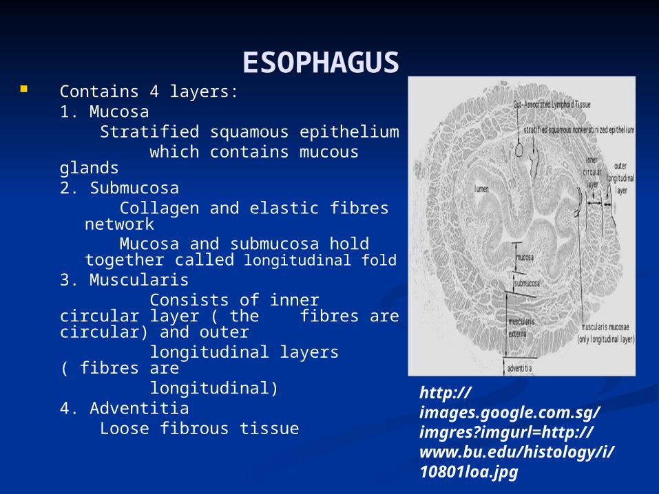
ESOPHAGUS Contains 4 layers:
1. Mucosa Stratified squamous epithelium
which contains mucous glands2. Submucosa
Collagen and elastic fibres network Mucosa and submucosa hold together
called longitudinal fold3. Muscularis
Consists of inner circular layer ( the fibres are circular) and outer
longitudinal layers ( fibres are longitudinal)
4. Adventitia Loose fibrous tissue http://
images.google.com.sg/imgres?imgurl=http://www.bu.edu/histology/i/10801loa.jpg

STOMACH Contains 4 layers:
1. Mucosa -lamina propia ( loose CT)
inside lamina propia : colagen and reticular fibres, cells such as fibroblasts, lymphocytes, mast cells, eosinophils and a few plasma cells
2. Submucosa Dense irregular connective tissue
with abundant of collagen and elastic fibres.
3. Muscularis Smooth muscle tissue which is divided into 3 coats such as circular, oblique, and longitudinal muscle layer.
4. Serosa http://images.google.com.sg/imgres?imgurl=http://www.bu.edu/histology/i/10801loa.jpg
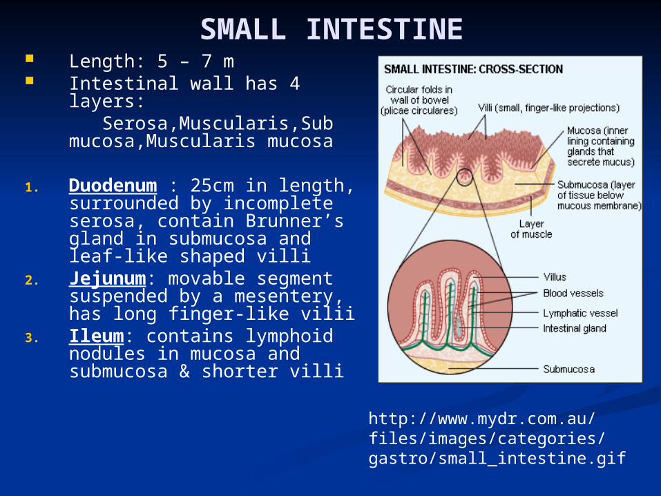
SMALL INTESTINE Length: 5 – 7 m Intestinal wall has 4 layers: Serosa,Muscularis,Sub
mucosa,Muscularis mucosa
1. Duodenum : 25cm in length, surrounded by incomplete serosa, contain Brunner’s gland in submucosa and leaf-like shaped villi
2. Jejunum: movable segment suspended by a mesentery, has long finger-like vilii
3. Ileum: contains lymphoid nodules in mucosa and submucosa & shorter villi
http://www.mydr.com.au/files/images/categories/gastro/small_intestine.gif

Length : 1.5 – 2 m Width : 6.5 cmContains 3 segments: Caecum with appendix
attached : 1st part located in the right lower abdomen
Ascending, descending, transverse, sigmoid colon
Rectum consists of 2parts : rectum proper and anal canal which is controlled by sphincters
LARGE INTESTINE
http://catalog.nucleusinc.com/imagescooked/406W.jpg

ACCESSORY ORGANS Salivary gland: connected with
oral cavity through excretory ducts; consists of minor gland and 3 main parts : parotid, sublingual, submandibular glands
Liver: largest gland surrounded by a collagen elastic fiber and made up of hepatocytes,
Gall bladder: ovoid sac covered by muscular wall _tall columnar epithelium for absorption
Pancreas: located behind stomach and contains pancreatic endocrine and exocrine cells
http://www.cs.odu.edu/~twt/resume/Photoshop/medical/DigestionAccessoryOrgans.gif

Functions of Digestive System The primary function is to break down the materials into
smaller parts so the body can use them to build and nourish cells and provide energy
Can be viewed as a series of integrated steps that consists of Ingestion, Mechanical processing, Digestion, Secretion, Absorption, and Excretion
It begins in the mouth, following with pharynx, esophagus, stomach, small intestines, large intestines, anus
Functions of Digestive System
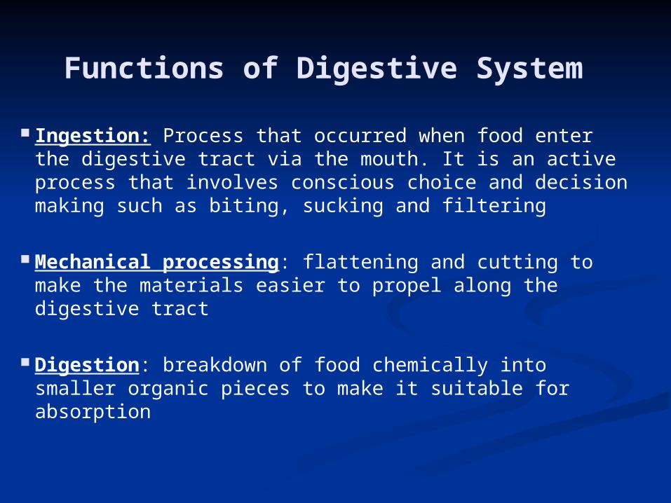
Ingestion: Process that occurred when food enter the digestive tract via the mouth. It is an active process that involves conscious choice and decision making such as biting, sucking and filtering
Mechanical processing: flattening and cutting to make the materials easier to propel along the digestive tract
Digestion: breakdown of food chemically into smaller organic pieces to make it suitable for absorption

Functions of Digestive System
Secretion: the released of a substance, like water, acids, enzymes, buffers, and salts by the epithelium of the digestive tract and by glandular organs
Absorption: movement of organic substrates, electrolytes (inorganic ions), vitamins, and water across the digestive epithelium and into the adjacent blood and lymphatic vessel of the digestive tract
Excretion: removal of waste products from body fluids into the anus.

Specific functions in digestive system
Mouth/Oral cavityMechanical processing, moistening, and mixing with saliva
Salivary glandSecretes saliva to break down carbohydrates
Pharynx/ThroatTransfer food from mouth to esophagus

Specific functions in digestive system Esophagus
Connects pharynx to stomach
Liversecretes bile for lipid digestion, storing nutrient, detoxifying drugs
StomachChemical processing and breaking down of protein by using hydrochloric acid and enzymes

Specific functions in digestive system
GallbladderStores and concentrates bile
PancreasExocrine tissues: synthesizes and secretes pancreatic juices that contains digestive enzymesendocrine tissues: secretes hormones
Small intestineDigests by enzymes and absorbing nutrients

Specific Specific functions in digestive system in digestive system
Large intestine
Absorbs the remaining water from the waste and preparing it for elimination
Anus
Excretes the waste out from the body

Nerves in Digestive system
1. Mouth
• Parasympathetic nerves releases acetylcholine which increases salivary secretion and vasoactive intestinal polypeptide which expands the salivary gland blood vessels
• Sympathetic nerves causes secretion of small amounts of saliva, rich in protein and glycoprotein
2. Esophagus
• Between two smooth muscles lies the nerves and nerve cells of the myenteric nerve plexus which controls peristalsis movements

Nerves in Digestive system
3. Swallowing Cranial nerves : excites muscles Intercostals nerves : inhibits diaphragm and intercostals
muscles Vagus nerves to myenteric nerves : peristalsis waves of
contraction to move the food down to stomach
4. Stomach Once messages from hypothalamic feeding centre from
brain reaches medulla, autonomic nervous system activates reaction
5. Pancreas
• Vagus nerves releases small quantity of gastrin which stimulates functions within
6. Gall bladder
• Vagus nerves causes bile to be expelled out into small intestine
7. Small intestine•Myenteric nerve plexus found in outer Serosa of
peritoneum controls peristalsis movements•Extrinsic nerve causes parasympathetic stimulation to
increase contractions and sympathetic stimulation to decrease motility
•Submucous nerves plexus, stimulated by intestinal hormones, causes movements of villi, to absorb nutrients
Nerves in Digestive system
8. Gut wall
• Myenteric nerves in smooth muscle layers coordinates secretory and muscular activities
9. Intestinal secretions• Intrinsic nerve plexuses causes stimuli to secrete intestinal juice• Cholinergic nerves stimulates secretion of mucus but it is inhibited by sympathetic nerves
Nerves in Digestive system

10.Gut wall of large intestine
• Intrinsic nervous system : myenteric and sub mucous plexus for controlling gut movement
• Types of neurons found : postganglionic parasympathetic, secretory, sensory ( chemoreceptor and mechanoreceptors), interneuron
• Extrinsic autonomic nerves : sympathetic decreases and parasympathetic (pelvic and pudendal) increases gut movement and secretion
• Acetylcholine released by parasympathetic stimulation contracts gut muscles and relaxes sphincters
Nerves in Digestive system

Blood system in digestive system
1. Stomach / pancreas
Humoral control: chemical messages are carried via blood stream to gastric glands
• In gastric phase, gastrin from G cells are released when blood leaves stomach / pancreas
• Arterial blood returns to stomach / pancreas to stimulate secretion of juices rich in acid and pepsinogen (enzyme digesting protein)
• In intestinal phase, chyme is broken down by (GIP) ,leaves through blood circulation
• Arterial blood returns to stomach / pancreas to inhibit formation of bile and decrease acidity and motility
http://www.medical-look.com/systems_images/Circulatory_System.gif

2. Gall bladder
•Humoral intestinal phase : products of fat disgestion stimulate duodenal (small intestine) mucosa to secrete cholecystokinin (CCK)
• This enters venous blood and via portal circulation, arterial blood causes contraction of gall bladder
3. Liver
•It receives 1/3 arterial blood from hepatic artery
•Remainder venous blood from hepatic portal vein which begins from capillaries of esophagus, stomach ,small intestine and most from large intestine
•Blood leaving liver re enters via inferior vena cava
Blood system in digestive system

What happens when we age?
1.1. The rate of epthelial stem cell division declinesThe rate of epthelial stem cell division declines Digestive cells more prone to damage and stomach ulcers Digestive cells more prone to damage and stomach ulcers
occurs most likelyoccurs most likely
2.2. Smooth muscles tone decreasesSmooth muscles tone decreases Motility decreases and pancreatic contraction weakensMotility decreases and pancreatic contraction weakens
3.3. Cancer rate increasesCancer rate increases
4.4. Changes in other systems have direct or indirect effects on Changes in other systems have direct or indirect effects on digestive systemdigestive system
For e.g. : reduce in bone mass and calcium content of For e.g. : reduce in bone mass and calcium content of skeleton can cause tooth lossskeleton can cause tooth loss
Decline in olfactory and gustatory sensitivity can lead to Decline in olfactory and gustatory sensitivity can lead to dietary changesdietary changes

References
Fox, S. I.,1999. Human physiology. 6th ed. U.S.A: The McGraw Hill Companies.
Kierszbaum, A. L., 2002. Histology and cell biology : an introduction to pathology. USA: Mosby.
Mackenna, B. R., 1997. Illustrated physiology. 6th ed. Singapore: Longman Singapore Publisher.
Martini, F. H., 1998. Fundamentals of anatomy and physiology. 4th ed. New Jersey: Prentice Hall International.
Martini, F.H., 2006. Fundamental of anatomy & physiology. 7th ed. San Francisco: Daryl Fox.
MDIS. 2005. Anatomy course study booklet. Singapore: MDIS.

~The End~~The End~
THANK YOU ALL FOR YOUR THANK YOU ALL FOR YOUR KIND ATTENTION!!KIND ATTENTION!!
DONE BY:
Farhath Jabien, Nishachand Vohreh, Felly, Ensy Caroline, Melisa and Ton Nu Thien An