diffusion glucose, insulin, inulin, evans blue protein ... · journal of clinical investigation...
TRANSCRIPT
Journal of Clinical InvestigationVol. 46, No. 6, 1967
Diffusion of Glucose, Insulin, Inulin, and Evans Blue Proteininto Thoracic Duct Lymph of Man *
EUGENIOA. RASIO,t CONSTANTINEL. HAMPERS,J. STUARTSOELDNER,ANDGEORGEF. CAHILL, JR.4
(From the Elliott P. Joslin Research Laboratory and the Renal Transplant Unit, Departmentof Medicine, Peter Bent Brigham Hospital and Harvard Medical School, and the
Diabetes Foundation, Inc., Boston, Mass.)
Summary. Immunoreactive insulin, like inulin, quickly equilibrates withinterstitial fluid, as evidenced by recovery in thoracic duct lymph in man.
Insulin-like activity not accounted for by immunoreactive insulin behavesas a large protein and is confined to the vascular compartment.
Introduction
Although insulin has been extracted, purified incrystalline form, and recently synthesized, insulinas it exists in body fluids, particularly in man, hasnot been isolated or chemically characterized.Therefore, we can only refer to circulating insulinas "the activity in plasma that mimics the action ofcrystalline pancreatic insulin in one or more spe-cific systems" (1).
Pancreatic insulin can be bound to a specificantibody; the plasma activity that exhibits a simi-lar specific binding is called immunoreactive insu-lin (IRI). Extracted pancreatic insulin also en-hances glucose metabolism in a variety of in vivoand in zitro biological systems, but because theylack specificity the plasma activities detected withthese biological techniques are called insulin-likeactivity (ILA). In some bioassays, one or morecirculating ILA that are not immunochemicallyreactive can be measured; these components ofplasma ILA, not accounted for by IRI, have been
* Submitted for publication December 1, 1966; acceptedFebruary 10, 1967.
Supported in part by U. S. Public Health Servicegrants T1 AM-5077-11, AM-09584-02, AM-09748-02, and8 M01-FR-31-05, and the John A. Hartford Foundation,Inc., New York, and the Adler Foundation, Inc., Rye,N. Y.
t International postdoctoral fellow of the National In-stitutes of Health.
t Investigator, Howard Hughes Medical Institute. Ad-dress requests for reprints to Dr. George F. Cahill, Jr.,Diabetes Foundation, Inc., 170 Pilgrim Rd., Boston, Mass.02215.
called "bound," "atypical," or "nonsuppressible"insulin. A critical review of the state of insulinin blood has recently been made by Berson andYalow (1).
Few in vitro investigations have attempted toelucidate the nature of plasma insulin; there issome indirect evidence that most of the circulatingIRI exists as a relatively small molecule (1-3),whereas the fraction of plasma ILA immunologi-cally inactive may be composed of one or morespecies of larger molecules, obtained either by poly-merization of a smaller unit (4) or by binding toa carrier protein (5).
In this study, an attempt has been made to char-acterize in vivo some physiochemical properties ofcirculating insulin. Diffusion of molecules throughblood capillaries is dependent upon molecularweight, protein interactions, and other factors.Therefore, the ability of intravascular insulin toequilibrate with thoracic duct lymph in man wascompared to the diffusion of glucose (mol wt 180),inulin (mol wt approximately 5,500), and Evansblue-stained plasma protein (mol wt approxi-mately 69,000). Intravascular insulin was eitherendogenous hormone released after a rapid glu-cose load or exogenous crystalline pork hormoneinjected intravenously.
MethodsSix patients with chronic renal failure and bilateral
nephrectomy were investigated. They were maintainedin good nutritional and metabolic balance by intermit-tent hemodialysis and transfusions (Table I). There
903
RASIO, HAMPERS,SOELDNER,AND CAHILL
TABLE I
Metabolic data in the six patients during testing periods
Serum Lymphprotein protein
Hemato- albumin/ albumin/Patient Sex Age Weight BUN* K C02 crit total total
years kg mg/ mEqIL mmoles/L % g/100 ml100 ml
1 M 32 60 86 5.7 24 33 3.0/6.2 2.0/3.72 M 26 81 42 4.1 24 22.5 3.2/5.1 1.8/2.43 F 39 54 50 5.1 24 25 2.7/5.5 1.7/3.14 M 25 65 42 3.8 29 29 3.7/5.75 M 24 60 52 4.6 28 32 3.0/5.2 1.6/2.06 M 30 52 31 4.7 26 25 3.9/6.5 2.6/4.2
* Blood urea nitrogen.
_300
00o200[
100L_
fi40 _-
:L 30[w-21
cnoo: 20
0
0
:zzPe
z
z
10L
6050-40-30
20
10L0
600E _-.
400
200
0 lo 20 30
TIME (minutes)
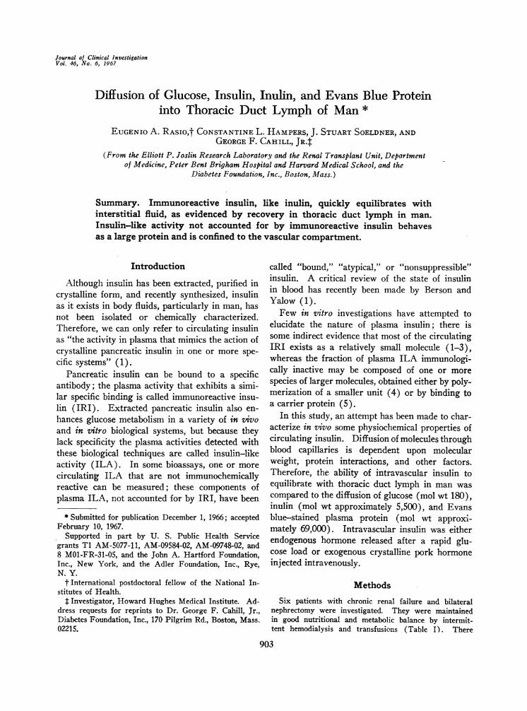
FIG. 1. CONCENTRATIONOF GLUCOSE, IMINSULIN (IRI), INULIN, AND INSULIN-
(ILA) IN LYMPHAND SERUMAFTER RAPII
ADMINISTRATION OF GLUCOSEAND INULIN TC
SUBJECTS. No correction was made forlymph collection via the thoracic duct c
explains the dissociation from an ideal pre
relationship.
was no clinical evidence of edema or congestive heartfailure; blood urea nitrogen never achieved the concen-
trations known to increase capillary permeability (6).Each patient as part of his treatment before renal trans-plant had a thoracic duct cannulation and a forearm ar-
teriovenous bypass and was scheduled for glucose andinsulin tolerance tests. During testing periods these pa-tients were afebrile, consuming daily a diet of at least
LYMPH 200 g carbohydrate and 2,000 calories, and not receiving
SERUM steroids, immunosuppressive drugs, or thiazides. Two
tests were performed; the first was an intravascular in-jection of a mixture of 50%o glucose in water and 10%oinulin 1 in water, for a total amount of 0.5 g per kg body
j- weight of glucose and 5 g of inulin. The second was an
intravascular injection of a mixture of 80 U per ml ofcrystalline pork insulin 2 and 10% inulin in water. Dos-age was 0.1 U per kg body weight of insulin and 2.5 g
of inulin. In four patients, 25 mg of a 5% aqueous solu-tion of Evans blue' was added to the solution to beinjected.
These tests were performed on each patient on consecu-
----____I tive days after an overnight fast. The solutions were in-* jected intravenously via the shunt in approximately 30
seconds.Simultaneous samples of thoracic duct lymph and ar-
terial blood were timed after the end of the injection.When lymph flow did not allow a collection of adequateamounts of lymph, samples were obtained within + 30seconds of the alleged time. Furthermore, in timing the
- Hi lymph samples, the changes in flow and the dead spaceof the thoracic duct catheter were not taken into account.The relative diffusion rates of glucose, insulin, inulin,
f---~ 120 and Evans blue protein were compared within the same60" 120
patient. The samples of blood and lymph were allowedto clot at room temperature for 4 hours; after centrifu-
IMUNOREACTIVE gation, the supernatant serum and cell-free lymph were
-LIKE ACTIVITY collected and stored at -200 C. Glucose was measuredD INTRAVENOUS by the glucose oxidase method; its disappearance rates
SIX ANEPHRIC after the glucose load (K) or after insulin injection
the delay in (i =net glucose disappearance rate between 5 and 30
-annula, which 1 Warner-Chilcott, Morris Plains, N. J.
cursor-product 2 Eli Lilly Co., Indianapolis, Ind., courtesy of Dr.W. R. Kirtley.
904

DIFFUSION OF INSULIN INTO EXTRAVASCULARFLUID OF MAN
minutes) were estimated by the slope of time against thelogarithm of concentration and expressed as the per centdisappearing per minute (7, 8).
Inulin was determined with resorcinol after the sam-ples were preincubated with purified glucose oxidase(9). Evans blue was measured by colorimetry after ex-traction from the carrier protein (10). Immunoreactiveinsulin was assayed in duplicate, at a 1:10 dilution, bya double-antibody technique (11). The results wereexpressed in microunits per milliliter of equivalent hu-man crystalline insulin; in this immunoassay the affini-ties of the antibody for crystalline pork and crystallinehuman insulin were found to be identical. The half-timeof IRI in serum and lymph after injection of exogenousinsulin was computed on the basis of changes in the in-crement of IRI over fasting levels after equilibration ofIRI in serum and lymph was achieved. The volumes ofdistribution of glucose, inulin, IRI, and Evans blue pro-tein were determined by V = Q/(CQ - Cf), where Q isthe amount of the substance injected, Cf the initial fast-ing concentration in serum or lymph, and C the theo-retical value obtained by extrapolating to time 0 the slopeof disappearance after equilibration. Insulin-like ac-tivity was measured by the conversion of glucose-l-"4C to"4CO2 by the rat epididymal fat pad (12). Each sampleof serum or lymph was assayed in triplicate at the dilu-tion of 1: 4, and the results were expressed in microunitsper milliliter of equivalent crystalline pork insulin inwhole serum or lymph. In this bioassay crystalline hu-man and pork insulins exhibited the same activity.
Results
For ease of presentation, Figures 1, 2, and 3 con-tain the data expressed as a mean of all individualstudies. However, the patterns and temporal re-lationships among the parameters in the pooleddata are representative of each individual study.
Intravascular injection of glucose and inulin.This test allows a comparison of the diffusion ofglucose, inulin, and endogenous insulin. Theresults are illustrated in Figure 1 and Table II.
Glucose was consistently the first substance toappear and peak in the thoracic duct lymph; a sig-nificant rise in lymph glucose was observed within1 to 5 minutes after the intravascular injection,with a peak occurring between 7 and 15 minutes.After equilibration in vascular and extravascularcompartments, glucose disappearance rates weresimilar in serum (mean K = 1.2 ± 0.2) and lymph(mean K = 1.2 + 0.1). Glucose space calculatedfrom glucose distribution in serum and lymph av-eraged, respectively, 33.6 ± 2.8 and 29.5 + 1.9%oof body weight. Inulin and endogenous immuno-reactive insulin had a similar pattern of diffusion
wL 100 -
fco E-C~o 75
400200
D 50E
30
400 _
200
100F
10
_70E 60-o 50-- 40-E 300Z 20
z 10Lv
600-E
X400
_200--
INSULIN INJECTION
0 10 20 30TIME (minutes)
(1
60 "
120
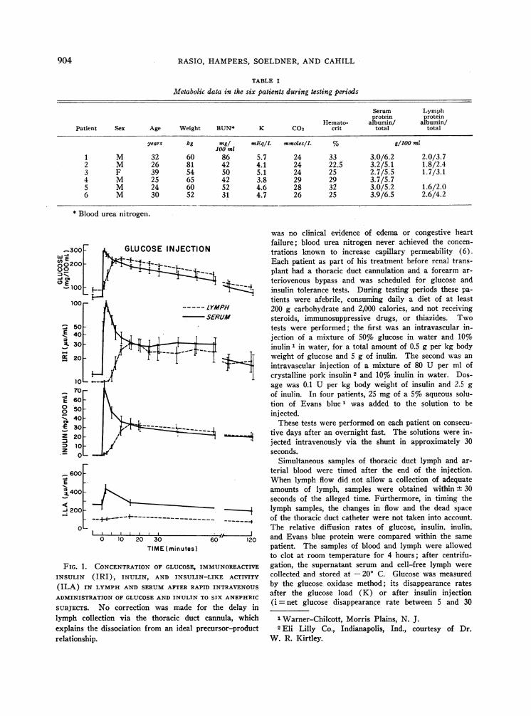
FIG. 2. CONCENTRATIONOF GLUCOSE, IRI, INULIN, ANDILA IN LYMPHAND SERUMAFTER RAPID ADMINISTRATIONOF INSULIN (0.1 U PER KG BODY WEIGHT) AND INULINTO THE SAMESIX ANEPHRIC SUBJECTS. As in Figure 1, nocorrection was made for the delay in lymph collection viathe thoracic duct cannula, but these data can be directlycompared to those in Figure 1, assuming no change in therate of lymph flow and no change in the dead spacevolume.
during the time required for their equilibration inserum and lymph. In each patient, their appear-ance and peak in lymph were similarly delayedwhen compared with glucose; both IRI and inulinincreased in lymph within 3 to 7 minutes andpeaked simultaneously within 15 to 20 minutes.Inulin was thereafter cleared slowly but signifi-cantly from serum and lymph, despite the absenceof kidneys. The low disappearance rate of inulinin serum and lymph allowed the estimate of inulinvolume, which represented, respectively, 24.5 ±2.1 and 23.7 ± 1.2% of the body weight.
After the diffusion in lymph of the initial peakof serum insulin, the concentrations of immuno-
905

RASIO, HAMPERS,SOELDNER,AND CAHILL
TABLE II
Volumes of distribution of glucose, inulin, insulin,of injected glucose (K), endogenous glucose (i),
Inulin space Insulin space Insulin disappearance rate
Patient Serum %BWt Lymph %BW Serum %BW Lymph %BW Serum t4 Lymph t
L L L L minutes1 16.1 26.8 15.2 25.3
16.7 27.8 15.6 26.0 15,0 25.0 15.0 25.0 7.5 14.5
2 22.7 26.2 22.7 26.221.3 24.6 20.0 23.1 22.7 26.2 26.1 30.2 10.0 13.0
3 9.1 16.6 11.1 20.08.1 14.7 7.4 13.5 12.0 21.8 12.0 21.8 7.0 10.5
4 20.8 32.0 17.2 26.520.8 32.0 20.8 32.0 18.6 28.6 21.3 32.8 7.0 13.5
5 14.7 24.5 13.5 22.516.6 27.6 16.7 24.5 15.8 26.3 15.8 26.3 5.5 9.5
6 10.8 21.2 10.0 19.78.3 16.7 8.3 16.7 13.0 25.5 13.8 27.0 6.0 12.5
24.5 i 2.1t 23.3 4 1.323.9 4- 2.7 22.6 4t 2.7 25.7 i 0.9 27.2 i 1.6 7.2 ± 0.6 12.2 i 0.8
* In each patient, the upper figures were obtained after injection of glucose (first test) and the lower figures afterinjection of insulin (second test).
t Per cent of body weight.t Mean i standard error of the mean.
reactive insulin in both compartmenttained at a relatively constant level aling value throughout the duration of
Insulin-like activity increased insecond minute, when concentrationsactive insulin were maximal, then dec120 minutes was below the fasting Inificant change in lymph ILA was ol
Intravascular injection of inulin,Evans blue. The data obtained wit]test are shown in Figures 2 and 3 ani
E .75E0
0
E s
axD.25
zC,)
--LYMPH
- SERUM
I I I .
0 5 10 20 30TIME (minutes)
FIG. 3. POOLED DATA FROM FOUR SUBJCEIVED EVANS BLUE DYE INTRAVENOUSLY:INSULIN AND INULIN, SHOWINGCONCENTIIN LYMPHAND SERUMAS A FUNCTION OF
:s were main- After the injection of insulin, glucose disap-bove the fast- pearance rates in serum and lymph were closely
the test. related during the first 30 minutes; i = 2.9 + 0.2serum at the in serum and i = 3.1 + 0.4 in lymph. Inulinof immunore- peaked in lymph simultaneously with immunore-:reased and at active insulin; the characteristics of diffusion werelevel; no sig- similar to those obtained during the iv glucosebserved. tolerance test. Inulin spaces were 23.9 + 2.7 and
insulin, and 22.6 ± 2.7% of body weight. Insulin spaces wereh this second 25.6 ± 0.9 and 27.2 ± 1.6%o of body weight whend Table II. calculations were performed as described in
Methods.The half-time of serum IRI after equilibration
with lymph was 7.2 ± 0.6 minutes and of lymphI j; IRI after the peak, 12.2 + 0.8, a significant differ-
ence. The appearance and equilibration of Evansblue protein complex in lymph were considerably
-4 delayed compared with inulin and IRI (Figure 3).The volume of distribution of Evans blue protein
60 120 averaged 6.2 ± 0.7%o of body weight.ILA in serum and lymph followed a pattern
JECTS WHORE- similar to IRI. As observed during the first test,
RATIONS OF DYE basal levels of ILA in lymph were considerablyTIME. lower than in serum.
906
____

DIFFUSION OF INSULIN INTO EXTRAVASCULARFLUID OF MAN
TABLE II
and Evans blue protein. Disappearance rateand exogenous crystalline pork insulin (tj)*
Glucose disappearance rateEvans blue
K i Glucose space protein space
Serum Lymph Serum Lymph Serum %BW Lymph %BW %BW
%/minue L L L0.9 0.9 13.3 22.2 13.3 22.2
3.6 4.7 4.0 6.7
1.5 1.5 36.1 39.6 29.2 33.83.3 3.3 7.2 8.9
2.1 1.7 16.9 30.2 14.8 26.43.2 3.7 3.1 5.7
0.8 0.8 27.0 41.0 23.0 35.02.1 2.1
1.1 1.1 21.7 36.2 18.4 30.72.6 2.6 3.5 5.8
0.9 1.0 16.6 32.6 14.7 28.82.3 2.3
1.2 -=- 0.2 1.2 it 0.1 33.6 4- 2.8 29.5 4- 1.92.9 ±- 0.2 3.1 -- 0.4 6.8 At 0.7
Discussion
The exchange of compounds between the bloodand the extravascular fluid occurs mostly by dif-fusion; the rate of diffusion is determined by thesize of the compound, a possible binding to othersubstances, and by the permeability of the capil-lary wall (13). Some characteristics of insulintransport in body fluids can therefore be investi-gated in vivo by comparing the ability of circu-lating insulin and tracer molecules to equilibratewith thoracic duct lymph. As capillary perme-ability is not uniform, varying amounts of lymphof different compositions are drained into the mainlymphatic channels. In conditions of bed rest,thoracic duct lymph is mostly a mixture of liverand gastrointestinal lymph in unknown propor-tions. These viscera have a high turnover rate ofproteins and highly permeable capillaries. There-fore, this study does not provide information forinsulin equilibration through all body fluids, par-ticularly those in muscle, fat, and brain. Withthese limitations in mind, the results indicate thatthe permeability of capillaries to glucose was thegreatest of the compounds investigated. Disap-
pearance rates of injected glucose and of endoge-nous glucose after insulin injection were similarin serum and lymph, suggesting a rapid diffusionof glucose back and forth across the capillary mem-brane; thus, the glucose assimilation by the cellsdepleted the vascular and extravascular pools si-multaneously, as if they were a single pool.
In these nephrectomized patients, as in healthynormal subjects (11), a rapid infusion of glucoseinduced an immediate release of endogenous in-sulin, which peaked in blood, similar to that aftera rapid intravascular injection of the hormone.This almost instantaneous release of insulin hasalso been reported with isolated perfused pancreaspreparations (14). When inulin diffusion wascompared with the diffusion of peak endogenousinsulin, similar patterns were observed within thesame individual, the equilibration time averaging10 to 15 minutes. Thereafter, inulin and IRI werecleared slowly from both compartments, the slowinulin clearance due to the absence of kidneys andthe apparent slow fall in insulin due to a continu-ous pancreatic secretion during the test.
For these data on endogenous insulin transport
907

RASIO, HAMPERS,SOELDNER,AND CAHILL
to be valid, no direct insulin secretion into thelymphatics of the pancreas should occur after aglucose load. If it did, some of the insulin in thethoracic duct lymph would have bypassed the bloodcapillary membrane. Although the possibility ofa "lymphocrinie" has been suggested in the thyroidgland (15), the endometrial crypts (16), and thekidney (17), it seems unlikely for the endocrinepancreas; no lymphatics have been found in theislets of Langerhans (18), and no immediate andimportant rise in insulin concentration was ob-served in lymph collected from the cysterna chyliof rats after an intravenous load of glucose (19).
Further information can be obtained from thesecond test, when crystalline pork insulin, inulin,and Evans blue were simultaneously injected. Inu-lin and exogenous insulin again equilibrated si-multaneously with thoracic duct lymph. There-fore, pancreatic extracted insulin and endogenousinsulin released by glucose stimulation behave simi-larly to each other and to inulin in their ability tocross vascular membranes. The sensitivity of thisin vivo procedure does not warrant the conclusionthat endogenous and exogenous insulin are identi-cal and that their molecular weight approximates5,500, as it is possible that small but significantdifferences in molecular weight could not be dis-criminated, although there were clear-cut differ-ences among diffusion of glucose, inulin, andEvans blue protein. Furthermore, a similar dif-fusion of inulin and insulin does not necessarilyimply that circulating insulin is a relatively smallmolecule; it is known, for instance, that free fattyacids in blood are strongly bound to albumin butvery rapidly separated from their carrier proteinas they move into extravascular fluids, the unbind-ing occurring at a capillary level (20). An analo-gous situation could be possible for insulin. De-spite their limitations, the techniques employedin this study provide the only information availableon the nature of insulin transport in vivo. Thatcirculating immunoreactive insulin crosses thecapillary membrane as a molecule smaller thanEvans blue protein is in agreement with obser-vations by Berson and Yalow, obtained with invitro techniques; after ultracentrifugation ofplasma, 60 to 80% of the IRI present in the un-centrifuged samples was found in the supernatantsolution above the sedimentary boundary of thealbumin (3). On starch gel electrophoresis,
plasma insulin migrated in the immediate preal-bumin zone (1). We concluded from these ex-periments that most of the plasma insulin is pres-ent as a small molecule unbound to serum protein.
Circulating immunoreactive insulin is clearedrapidly from the blood; the half-life of endogenousor exogenous insulin has been found to vary from5 to 78 minutes (1, 21-24). In this investigation,the half-life of the injected insulin was measuredafter 10 to 15 minutes required for equilibration,the fasting value was subtracted, and pancreaticoutput of insulin was assumed to remain constantduring the test. Our values of insulin half-lifewere consistently faster in serum than in corre-sponding lymph. This is in keeping with the hy-pothesis that insulin, after crossing capillary mem-branes, does not re-enter the vascular compartmentat the same level but is carried away into the lym-phatics; a similar phenomenon has been shown forother molecules of molecular weight greater than6,000 (13, 25).
From the slope of disappearance of insulin inserum and lymph, a mean volume of insulin dis-tribution was calculated; it approximates 26%oof body weight for an inulin space of 23%. Thesedata are similar to a recent report in which in-jected insulin was shown to be distributed withinthe extracellular fluid (22).
A different volume of distribution would applyto the insulin-like activity not accounted for byIRI; this activity was indeed consistently lowerin thoracic duct lymph than in serum after eitheran overnight fast or a glucose load. Similar re-sults were obtained with rats (19). No insulininhibitor, which might have lowered the glucosemetabolism of the rat adipose tissue during thebioassay, was present in lymph, as shown by thegood recovery of the activity of immunoreactive in-sulin as measured by lymph ILA after insulin in-jection (Figure 2) or insulin addition in vitro(19).
In our patients, after an overnight fast, lymphILA represented only one-third of serum ILA,whereas the concentrations of IRI were similar inboth compartments. This suggests that ILA notaccounted for by IRI is a large molecule, mostlyrestricted to the vascular compartment, which mayaffect the metabolism of organs and tissues ac-cording to local capillary permeability. In inter-stitial fluids of tissues with low capillary perme-
908

DIFFUSION OF INSULIN INTO EXTRAVASCULARFLUID OF MAN
ability, low levels of ILA might be expected. Inspinal fluid, for instance, where protein concentra-tion is minimal, ILA is undetectable (26). Instriated muscle and in adipose tissue, the capillaryendothelium as examined by the electron micro-scope has no recognizable interstices, either in-tracellular, as in the intestinal villus, or inter-cellular, as in liver, spleen, and bone marrow sinus-oids (27). Most likely, these tissues are littleaffected by serum ILA under in vivo physiologicalconditions. During bioassay using striated muscleor adipose tissue in vitro for ILA determinations,a nonphysiological situation is created as high con-centrations of serum proteins, never achieved invivo, are present in the incubation medium and di-rectly in contact with the cellular surface.
After the intravascular injection of glucose,lymph ILA remained unchanged. If the early risein serum ILA were a hormonal secretion distinctfrom the pancreatic secretion of IRI, our dataagain would indicate a high molecular weight forthis hormone, as it would diffuse in thoracic ductlymph slower than Evans blue protein.
A few in vitro observations have been reportedabout the nature of nonimmunoreactive insulin.During dialysis in Visking tubing, atypical insulinbehaved like a molecule of molecular weightgreater than 40,000 (28); gel filtration of serumon Sephadex G-200 at pH 7.2 revealed a molecu-lar weight of nonsuppressible insulin-like activitybetween 100,000 and 200,000 (4). Chromatog-raphy on Sephadex G-75 and G-100 indicated amolecular weight for bound insulin between 60,000and 100,000 (29). Thus, our in vivo investiga-tion has provided information on ILA transportthat is in agreement with in vitro estimates of ILAmolecular weight.
AcknowledgmentsWe gratefully acknowledge the cooperation of Drs.
J. E. Murray and J. P. Merrill in allowing us to studytheir patients. The technical assistance of Mrs. DzidraRumba is greatly appreciated.
References
1. Berson, S. A., and R. S. Yalow. Insulin in bloodand insulin antibodies. Amer. J. Med. 1966, 40,676.
2. Hall, J. L. Moving boundary electrophoretic studyof insulin. J. biol. Chem. 1941, 139, 175.
3. Berson, S. A., and R. S. Yalow. Immunoassay ofplasma insulin in Immunoassay of Hormones.Ciba Foundation Colloquia on Endocrinology,G. E. W. Wolstenholme and M. P. Cameron, Eds.London, J. & A. Churchill, 1962, vol. 14, p. 182.
4. Burgi, H., W. A. Muller, R. E. Humbel, A. Labhart,and E. R. Froesch. Non-suppressible insulin-likeactivity of human serum. I. Physicochemical prop-erties, extractions and partial purification. Bio-chim. biophys. Acta (Amst.) 1966, 121, 349.
5. Antoniades, H. N., J. A. Bougas, R. Camerini-Davalos, and H. M. Pyle. Insulin regulatorymechanisms and diabetes mellitus. Diabetes 1964,13, 230.
6. Mayerson, H. S. The physiologic importance oflymph in Handbook of Physiology, Circulation II,W. F. Hamilton and P. Dow, Eds. Washington,American Physiological Society, 1963, p. 1064.
7. Conard, V. Mesure de l'assimilation du glucose.Acta med. belg. (Bruxelles) 1956, 37.
8. Franckson, J. R. M. Mesure de l'activite de l'in-suline chez l'homme. Acta med. belg. (Brux-elles) 1958, 23.
9. Froesch, E. R., J. B. Reardon, and A. E. Renold.The determination of inulin in blood and urineusing glucose oxidase for the removal of interferingglucose. J. Lab. clin. Med. 1957, 50, 918.
10. Constable, B. J. Estimation of Evans blue dye inblood plasma and its application to blood volumedetermination. Clin. Sci. 1958, 17, 597.
11. Soeldner, J. S., and D. Slone. Critical variables inthe radioimmunoassay of serum insulin using thedouble antibody technic. Diabetes 1965, 14, 771.
12. Renold, A. E., D. B. Martin, Y. M. Dagenais, J.Steinke, R. J. Nickerson, and M. C. Sheps. Mea-surement of small quantities of insulin-like activityusing rat adipose tissue. I. A proposed procedure.J. clin. Invest. 1960, 39, 1487.
13. Yoffey, J. M., and F. C. Courtice. The formationof lymph in Lymphatics, Lymph and LymphoidTissue, 2nd ed. Cambridge, Harvard UniversityPress, 1956, p. 53.
14. Grodsky, G. M., and L. L. Bennet. Time sequence inthe release of insulin: the effect of -glucose, glu-cagon and potassium. Diabetes 1966, 15, 521.
15. Daniel, P. M., M. M. Gale, and 0. E. Pratt. Theconcentration of radioactive iodine in the thyroidlymph, thyroid venous blood and peripheral bloodof primates. J. Physiol. (Lond.) 1963, 169, 330.
16. Amoroso, E. C. The biology of the placenta inGestation, Transaction of the Fifth Conference,C. A. Villee, Ed. New York, Macy Foundation,1958, p. 15.
17. Skinner, S. L., J. W. McCubbin, and I. H. Page.Angiotensin in blood and lymph following reduc-tion in renal arterial perfusion pressure in dogs.Circulat. Res. 1963, 13, 336.
18. Rusznyak, I., M. Foldi, and G. Szabo. The specialanatomy of lymphatic system in Lymphatics andLymph Circulation. Oxford, Pergamon, 1960, p. 79.
909

RASIO, HAMPERS,SOELDNER,AND CAHILL
19. Rasio, E. A., J. S. Soeldner, and G. F. Cahill, Jr.Insulin and insulin-like activity in serum and ex-travascular fluid. Diabetologia 1965, 1, 125.
20. Frederickson, D. S., and R. S. Gordon, Jr. Trans-port of fatty acids. Physiol. Rev. 1958, 38, 585.
21. Samols, E., and V. Marks. Disappearance-rate ofendogenous insulin in man. Lancet 1966, 2, 700.
22. 0rskov, H., and N. J. Christensen. Disappearance-rate of exogenous human insulin. Lancet 1966,2, 701.
23. Vinnick, L., and N. Freinkel. Basal insulin turn-over. Clin. Res. 1966, 14, 290.
24. Cerasi, E., and R. Luft. Insulin response to glucoseloading in acromegaly. Lancet 1964, 2, 769.
25. Rusznyak, I., M. F6ldi, and G. Szabo. Origin oflymph in Lymphatics and Lymph Circulation.Oxford, Pergamon, 1960, p. 175.
26. Mahon, W. A., J. Steinke, G. M. McKhann, andM. L. Mitchell. Measurement of I'-insulin andof insulin-like activity in cerebrospinal fluid ofman. Metabolism 1962, 11, 416.
27. Majno, G. Ultrastructure of the vascular membranein Handbook of Physiology, Circulation III, W. F.Hamilton and P. Dowv, Eds. Washington, Ameri-can Physiological Society, 1965, p. 2293.
28. Samaan, N., R. Fraser, and W. J. Dempster. The"typical" and "atypical" forms of serum insulin.Diabetes 1963, 12, 339.
29. Antoniades, H. N., A. M. Huber, B. R. Boshell,C. A. Saravis, and S. N. Gershoff. Studies on thestate of insulin in blood: properties of circulating"free" and "bound" insulin. Endocrinology 1965,76, 709.
NOTICE TO SUBSCRIBERS
Post Offices will not forward the JOURNALwhen you move.
Please notify THE JOURNALOF CLINICAL INVESTIGATION, TheRockefeller University Press, Box 261, New York, N. Y. 10021,when your address changes. Include your zip code number.
910