differentiation in colonies of aspergillus niger · differentiation in colonies of aspergillus...
TRANSCRIPT

Differentiation in colonies of Aspergillus niger
Ana M. Levin Chucrel 2007


Differentiation in colonies of Aspergillus niger
Differentiatie in kolonies van Aspergillus niger (met een samenvatting in het Nederlands)
Diferenciación en colonias de Aspergillus niger
(con un resumen en Castellano)
PROEFSCHRIFT ter verkrijging van de graad van doctor aan de Universiteit Utrecht op gezag van de rector magnificus, prof. dr. W.H.
Gispen, ingevolge het besluit van het college voor promoties in het openbaar te verdedigen op donderdag 15 maart 2007 des
middags te 4.15 uur
door
Ana Marcela Levin Chucrel
geboren op 17 januari 1976 te Buenos Aires (Argentinië)

Promotor: Prof. Dr. H.A.B. Wösten
Co-promotor: Dr. Ir. R.P. de Vries
This work was supported by The Netherlands Technology Foundation (STW) Pionier project UGC.5683.

Satisfaction lies in the effort, not in the attainment, full effort is full victory.
M. Gandhi
Bad times have a scientific value. These are occasions a good learner would not miss.
R.W. Emerson
To my parents

Good Use Right "It is strictly prohibited to use, to investigate or to develop, in a direct or indirect way, any of the scientific contributions of the author contained in this work by any army or armed group in the world, for military purposes and for any other use which is against human rights or the environment, unless a written consent of all the persons in the world is obtained" ISBN: 90-393-44897 Printed by Digital Print Partners Utrecht, Houten Cover design: www.gonzalocervello.com Printing of this Thesis has been financially supported by Utrecht University.

Contents
Contents Chapter 1 General Introduction 11 Chapter 2 Localization of protein secretion in fungal 31
colonies using a novel culturing technique; the ring-plate system
Chapter 3 Colonies from the filamentous fungus 39
Aspergillus niger are highly differentiated in spite of cytoplasmic continuity
Chapter 4 Spatial differentiation in colonies of 63
Aspergillus niger Chapter 5 The exploitation of the whole mycelium for 79
secretion; production of laccase in colonies of Aspergillus niger
Chapter 6 General Discussion 95 Samenvatting 105 Resumen 111 Acknowledgements 117 List of Publications 123 Curriculum Vitae 125
7


Chapter 1
General Introduction


General introduction
Introduction to Aspergillus and its use by the industry The genus Aspergillus comprises about 175 species that are among the most abundant fungi worldwide. They can be found in soil and decaying organic matter or can be associated with sponges in marine environments (Hiort et al., 2004). Aspergilli can also be abundant in man made habitats like buildings (e.g. hospitals, libraries and breweries), stored crops and dried food (Samson et al., 2000). Aspergillus species are not only saprophytic but can also be pathogens of animals and humans with a compromised immune system (Brakhage et al., 2005; Pitt et al., 1994). For instance, Aspergillus fumigatus is one of the most common fungal pathogens, causing both non-invasive aspergilloma as well as invasive aspergillosis.
Aspergilli secrete a wide variety and large amounts of enzymes. These enzymes liberate small molecules from polymers within the substrate that can be taken up to serve as nutrients. Apart from these enzymes, Aspergilli secrete high amounts of organic acids (e.g. citric acid, itaconic acid, gluconic acid and tartaric acid) (Nout et al., 2000). This secretory capacity in combination with the established fermentation technology and molecular biology makes Aspergillus species such as A. niger and A. oryzae attractive cell factories. The fact that the fermentation products are generally recognized as safe (mycotoxins are known not to be produced during the controlled fermentation conditions) adds to the attractiveness of these fungi. Commercial citric acid production with the use of A. niger was already initiated at the starting of the twentieth century (Bennett et al., 1998), while partial purified amylase of A. oryzae was patented in 1894 as a substitute for malting enzyme in beer production and as a digestive aid for the treatment of dyspepsia (Takamine,1894; Underkofler et al., 1954). Nowadays, glucoamylase is produced at 30 grams per litre (Finkelstein et al., 1989). This enzyme, which is used for the production of glucose syrups, is only one of the examples of enzymes from Aspergilli that are used in the food and feed industry (Table 1).
Fungal growth and fermentation Less than 1% of the identified fungi have been isolated from marine habitats and of these fungi 98% were isolated from solid substrates within the aqueous environment (Kelecom et al., 2002).
11

Chapter 1
Table 1: Examples of enzymes of Aspergilli and their application in the food and feed industry (Adapted from Wösten et al., 2006).
Enzyme Source Application(s) References α-Amylase
A. niger
A. oryzae
Preparation of starch syrup and dextrose; preparation of alcohol and beer.
(Wong and Robertson, 2003)
Catalase A. niger
Preservation of colour, texture, flavour, taste and aroma of frozen foods.
(Diehl et al., 1936)
Cellulase A. niger
A. aculeatus
Brewing and baking; wine and juice production; improvement of digestibility of feed.
(Tenkanen et al., 2003)
Feruloyl-esterase A. niger,
A.tubingensis,
A. oryzae
Release of ferulic acid for vanillin production.
Williamson, 1998.
Glucoamylase A. niger
A. oryzae
Saccharification of steamed rice and potato; preparation of glucose syrup.
(Reilly, 2003)
Glucose oxidase A. niger
Removal of residual glucose or oxygen to increase shelf life; flavor and color stability; reduction of alcohol percentage in wine.
(Frederick et al., 1990)
Lipase A. niger A. oryzae
Manufacturing of cheese, cheese flavours and other dairy products.
(Wong, 2003)
Phytase A. niger, A. oryzae, A. melleus,
Degradation of phytate in animal feed; starch processing.
Misset, 2003
Proteases A. niger
Softening of dough; improvement of texture, elasticity and volume of bread; brewing; production of miso and tofu; flavour development in cheese; improving digestibility of animal feeds; preparation of soy bean milk and dehydrated soups; clarification of wine.
(Whitaker, 2003)
Xylanases A. niger
Production of food-additives; improvement of digestibility of feed; preparation of baking products; clarification of fruit juices.
(Biely, 2003)
12
General introduction
Most filamentous fungi thus grow in terrestrial ecosystems and they have particularly evolved to grow on. and in solid substrates They do so by forming a mycelium, which consists of a network of interconnected hyphae. The interconnectedness is the result of subapical branching and hyphal fusion and is best illustrated by the continuity of the cytoplasm. This is due to the fact that the septa that divide hyphae in compartments are highly porous, allowing streaming of water, nutrients and even organelles from one compartment to the other. Streaming is particularly functional when the mycelium colonizes a substrate in which nutrients and water are not uniformly distributed (Jennings et al., 1974).
In the industrial setting, fungi are grown either in solid state or
submerged fermentation. The former mode of growth is most similar to the way fungi grow in nature, yet the latter is most frequently used by the industry. Submerged fermentations A submerged fermentation is the cultivation of microorganisms in a liquid nutrient medium ranging from a small-scale system, as a liquid shaking flask, to a hundred thousand liter scale industrial fermentor. A submerged fermentation is a complicated multi-phase, multi-component system, in which growth and production are affected by a wide range of parameters. The composition of the cultivation medium has a strong effect on growth and product formation. For instance, carbon and nitrogen sources may have repressing or inducing effects on the gene encoding the protein of interest. The fungal morphology also affects productivity (Wang et al., 2005). Two main types of morphology are distinguished during submerged fermentation; i.e. pellets and dispersed growth. Clumps are aggregated hyphae that are considered as an intermediate state between pelleted and dispersed growth. The impact of morphology on productivity is illustrated by the fact that dispersed mycelium is required for production of penicillin in Penicillium chrysogenum, whereas pellets are required for the production of citric acid by A. niger (Vecht-Lifshitz et al., 1990). It is not clear how morphology exactly affects productivity. Some studies indicate that this is due to its effect on the viscosity of the medium (Bhargaba et al., 2003). Viscosity correlates with the extent of dispersed growth; large pellets thus result in the lowest viscosity. However, the center of large pellets may experience oxygen starvation and other nutrients may also become limiting in this part of the mycelium. Parameters that influence the size of the pellet
13

Chapter 1
are initial pH, agitation, medium composition and inoculum size (Metz and Kossen, 1977). Three different types of submerged fermentations are distinguished; batch, fed-batch and continuous fermentation. Batch fermentation is a closed system, in which nutrients become limited during cultivation. The culture undergoes a lag phase, a logarithmic phase and a stationary phase. During the latter phase viability decreases and autolysis begins. Batch fermentation does not result in high product yield due to substrate limitation, but can be very useful for non-growth associated product formation. Moreover, it has a relatively low risk of contamination (Shuler and Kargi, 1992). During continuous fermentations, the culture is kept in a steady state in optimal conditions for the production of the desired product. The carbon source is normally kept at a constant limiting concentration. These systems generally have high productivity but the product is at a low concentration in the extra cellular medium due to the continuous flux of medium into the system. Moreover, it takes a long time to reach the steady state and, therefore, the risk of genetic instability of the strain and of contaminations is relatively high. Continuous systems are mainly used for research objectives and have not found general application in the industry. In fed-batch fermentations, an initial batch phase is used to obtain biomass. At a certain point, nutrients are added to maintain or to create optimal conditions for the production of the desired metabolite. Normally, the carbon source is limited to maintain a pseudo steady state condition with a desired growth rate and to alleviate carbon catabolite repression (Szabo et al., 1996; Spohr et al., 1998). In fed-batch fermentations, cells and products will remain in the bioreactor until the end of the fermentation. This is the industrially preferred way of submerged fermentation since volumetric productivity is the best of the three systems. Furthermore, fermentation times are short compared to continuous systems and also the risk of genetic instability and contamination is less. However, accumulation of by-products, CO2 and proteases as well as low solubility of medium components might be factors that limit the optimal production. Solid state fermentations Solid state fermentations are defined as the cultivation of microorganisms in moist supports either on inert carriers or on insoluble substrates. The fermentation takes place at very low level of free water. Generally, the mycelium secretes more protein in solid state than in submerged
14

General introduction
fermentations (Hölker et al., 2004), which may be partly due to reduced catabolite repression in the former way of fermentation (Nandakumar et al., 1999; Solis-Pereira et al., 1993). The lower catabolite repression also promotes growth of the fungus (Favela-Torres et al., 1998; Viniegra-Gonzalez et al., 2003). Additional advantages of solid state fermentation are a reduced extracellular protease activity (Solis-Pereira et al., 1993; Maldonado et al., 1998), lower water demand, higher end concentration of products, lower demands on sterility and the use of cheap substrates (Hölker et al., 2004). Not surprisingly, enzyme production in solid state fermentations is estimated to be much cheaper (Tengerdy et al., 1996). For instance, cellulase production was estimated to be hundred fold cheaper in solid state compared to submerged fermentations. Despite this, solid state fermentations are not used on a large scale due to engineering problems. Gradients in temperature, pH, pO2, moisture, and substrate concentration are being built up during up-scaling of the fermentation. These gradients, which are difficult to control due to the low water activity, severely affect productivity (Hölker et al., 2004). When these problems would be solved, solid state fermentation would be a very good alternative for the submerged fermentation technology. The fungal secretion pathway Remarkably little is known about mechanisms that underlie the high secretion capacity of Aspergilli. It is generally accepted that the secretion pathway of filamentous fungi (Figure 1), including that of Aspergillus, shares many characteristics with those of the yeast Saccharomyces cerevisae. In higher eukaryotes proteins are co-translationally translocated over the ER membrane. In contrast, both co-translational and post-translational translocation occurs in S. cerevisae. At present, it is not known whether both pathways also operate in filamentous fungi.
During co-translational translocation, the N-terminal signal peptide emerging from the ribosome is recognized by the signal recognition particle (SRP). As a result, translation is stopped and the SRP-ribosome complex is targeted to the ER membrane where it interacts with the SRP-receptor. The SRP dissociates from the complex and the nascent polypeptide enters the ER through the Sec61 complex (Wiertz et al., 1996). In case of posttranslational translocation, the mRNA is first fully translated followed by translocation of the encoded protein over the ER membrane. The HSP70
15

Chapter 1
protein keeps the protein translocation-competent. Post-translational translocation also depends on the signal sequence but is independent of the SRP and the SRP receptor. Instead, the Sec62-Sec63 protein complex somehow targets the protein to the Sec61 channel (Ng et al., 1996)). In both co- and post-translational translocation, the N-terminal signal peptide is removed from the newly synthesized protein.
Folding of proteins occurs in the ER lumen. This involves the combined action of chaperones and foldases and also N-glycosylation has been shown to be instrumental in this process (van Anken and Braakman, 2005).
Fig 1. The secretion pathway in filamentous fungi and ways to improve production of extracellular proteins. Transcription of the gene encoding the secreted protein has been increased by introducing multiple copies in the host (1). Targeting of transgenes to highly active transcriptional regions and use of strong promoters have also been used to improve transcription. Premature termination of transcripts (2) has been overcome by removing polyadenylation signals within coding sequences, while stability of mRNA could be increased in some cases by introducing introns in cDNA sequences (3). Fusing the protein of interest to a well secreted protein improved translocation across the ER membrane (4) as well as folding in the ER lumen (5). Folding was also facilitated in some cases by introduction of N-glycosylation sites, or by over-expressing the unfolded protein response pathway or its components such as foldases or chaperones. Reduced activity of proteases in the medium has been reported to improve production levels of secreted proteins (6). N; Nucleus, ER; endoplasmic reticulum, V; vacuole, G; golgi, SV; secretory vesicles. (Adapted from Wösten et al., 2006 and Conesa et al., 2001)
16

General introduction
Foldases (e.g. protein disulphide isomerase and peptidyl prolyl cis-trans isomerases) catalyse covalent changes, which are essential to obtain a functional conformation. Chaperones (e.g. binding protein (BiP), calreticulin and calnexin) are not involved in catalysis. By non-covalent interaction, they assist proteins to obtain their correct folding. The ER has a quality control to ensure that only correctly folded proteins are translocated to the next cellular compartment. Two control mechanisms are involved: the ER associated degradation (ERAD), which involves translocation of misfolded proteins to the cytosol and their subsequent degradation, and the unfolded protein response (UPR), which detects the presence of unfolded proteins in the ER and induces the synthesis of folding enzymes. These systems are highly conserved in eukaryots (van Anken and Braakman, 2005) and are interconnected to maintain a functional secretory pathway (Travers et al., 2000). It is generally believed that the ER communicates with other parts of the secretion pathway, since this is the only part of the pathway with a quality control. Indeed, ribosome synthesis was down-regulated when temperature sensitive mutants affected in secretion were grown at the non-permissive temperature (Mizuta and Warner, 1994).
The ERAD system is activated when chaperones like BiP or calnexin detect misfolded proteins in the ER. As a result, these misfolded proteins are retro-translocated to the cytoplasm, most likely through the Sec61 channel. The misfolded proteins are ubiquitinated in the cytoplasm and subsequently degraded by the proteasome (Meusser et al., 2005). UPR is induced by perturbations in the ER. Common causes are protein degradation, inhibition of glycosylation or disulphide bond formation. Depletion of Ca2+ in the ER, the presence of unfolded proteins or overexpression of proteins that accumulate into the ER can also induce UPR (Chapman et al., 1998). In S. cerevisiae the transmembrane serine/threonine kinase IRE1 senses the accumulation of misfolded proteins in the ER. As a result, its RNAse activity is activated and a non-conventional intron is spliced from the HAC1 mRNA. This alleviates a block of translation of this mRNA molecule. Thus, by activation of IRE1 the Hac1p transcription factor is formed that travels to the nucleus to bind to Unfolded Protein Response Elements (UPRE’s) in promoters of genes involved in UPR (Chapman et al., 1998). As a result, not only genes encoding ER chaperones and foldases are up-regulated, but also genes involved in secretion, glycosylation, lipid biosynthesis and ERAD. It
17

Chapter 1
should be noted that other transcription factors, like Gcn4p, also upregulate genes involved in UPR (van Anken and Braakman, 2005).
Correctly folded proteins are transported from the ER to the Golgi. The typical Golgi stacks as observed in animal cells are not present in most fungi, but Golgi associated functions, like Kex2 and glycosyl transferase activities, are found. Glycosylation of proteins is modified in the Golgi system. After this, proteins are packed in vesicles and targeted to the plasma membrane via microtubili. The exocyst is a conserved complex that has been implicated in the tethering of secretory vesicles to exocytic places in the plasma membrane. Components of the exocyst are concentrated at sites of active membrane expansion (Guo et al., 2000). Thus, proteins carried by vesicles will mainly fuse with the plasma membrane at the hyphal apex. Once extruded into the cell wall, proteins migrate together with the newly synthesized cell wall polymers from the inner to the outer part of the wall. This migration is driven by the turgor pressure in the hyphae and the apposition of newly synthesized polymers at the inner part of the wall (Wessels, 1993). At the outside of the cell wall the proteins diffuse into the medium. This so-called bulk-flow theory predicts that proteins will not be secreted by non-growing hyphae because they will not extend their plasma membrane and do not apposite new cell wall material. Indeed, only growing hyphae were shown to secrete glucoamylase (Wösten et al., 1991).
Limits of protein production in Aspergillus and strategies to improve it. Protein production in Aspergilli can be limited at various levels, including transcription, translation, secretion and extracellular stability. Genetic approaches have been used to overcome the bottlenecks in protein production, especially for heterologous proteins (i.e. proteins encoded by genes that do not originate from the host). In general, problems related to the transcriptional level can be easily overcome. In contrast, those acting at the post-transcriptional level are relatively poorly understood and are thus more difficult to solve. So far, nobody addressed whether protein production can be improved by increasing the number of hyphae in the mycelium that are engaged in secretion. In the next three sections an overview of strategies to improve protein production is presented.
18

General introduction
Improvement of protein production at the transcriptional level The gene encoding the protein of interest is usually expressed from a strong promoter. These promoters can be inducible like that of the A. niger glucoamylase gene glaA (Smith et al., 1990) or constitutive like that of the A. nidulans glyceraldehyde-3-phosphate dehydrogenase gene gpdA (Punt et al., 1991). This approach is often combined with a multi-copy integration of the gene. Glucoamylase production correlated with the copy number when up to 20 copies were introduced. At higher copy numbers, the increase in production leveled off probably due to titration of trans-acting regulatory proteins (Verdoes et al., 1994). Limitation in the regulatory protein level could be overcome in Aspergillus nidulans by increasing the expression level of the regulatory gene (Burger et al., 1991; Beri et al., 1990; Kelly and Hynes, 1987).
The site of integration also affects the production level (Verdoes et al., 1993). Production has been increased by targeting the expression cassette to a highly transcriptional active region like that of the cellobiohydrolase gene CBH1 in the case of Trichoderma reesei (Harkki et al., 1991). Finally, premature termination of mRNA’s can cause limitations of protein production at the transcriptional level. Truncation of mRNA of a heterologous aglA gene in A. niger was overcome by replacing an AT-rich sequence with a more GC-rich sequence (Gouka et al., 1996).
Improvement of protein production at (post)-translational level High production levels will not be obtained simply by adding a signal sequence of a highly secreted protein to the protein of interest. For instance, only 15 mg L-1 of bovin chymosin was found in the medium when the encoding gene was fused to the glaA promoter and signal sequence (Ward et al., 1989ab). In contrast, high level of secretion was obtained by fusing the chymosin cDNA to the last codon of glaA of A. niger (Ward et al., 1990). In this case the chymosin was cleaved autocatalytically from the fusion protein and extracellular levels of 150 mg L-1 of the heterologous enzyme were obtained. The homologous carrier protein has been suggested not only to translocate the heterologous protein into the ER but also would allow proper folding, thereby protecting the heterologous protein from degradation. By introducing a KEX2 recognition site, the carrier and the protein of interest are cleaved in the Golgi, allowing them to be secreted as individual entities (Contreras et al., 1991; Broekhuijsen et al., 1993). Upon over-expression of native or engineered proteins, foldases and chaperones may become limiting
19

Chapter 1
in the ER to ensure correct folding. Protein production in A. niger has been improved by over-expression of the BiP chaperone (Punt et al., 1998) but this approach has not been effective for every protein. As an alternative to over-expression of individual chaperones or foldases, protein production in T. reesei was improved by constitutive over-expression of the UPR response (Valkonen et al., 2003). Introduction of an N-glycosylation site into the protein of interest has also been used to improve folding of the protein in the ER (Sagt et al., 2000). In this example, the introduced glycosylation prevented aggregation of a hydrophobic cutinase in the ER.
Low production levels of proteins can also be caused by the action of intracellular or extracellular proteases (Berka et al., 1990; Archer et al., 1992; Broekhuijsen et al., 1993). Aspergillus strains deficient in extracellular proteases have been isolated by random mutagenesis (Mattern et al., 1992; van den Hombergh et al., 1995) or molecular genetic approaches (Berka et al., 1990; van den Hombergh et al., 1997). The use of these protease deficient strains has resulted in improvement of production levels (Berka et al., 1991; Broekhuijsen et al., 1993; Dunn-Coleman et al., 1991). For instance, production levels of chymosin were increased from 150 mg L-1 in the wild-type to 250 mg L-1 (Dunn-Coleman et al., 1991) by introducing the glucoamylase-chymosin fusion into an A. niger strain in which an aspartyl protease gene was deleted (Berka et al., 1990).
Recently, a novel Aspergillus species (A. vadensis) has been identified that is closely related to A. niger (de Vries et al., 2005). This species hardly produces extracellular proteases and does not acidify the culture medium. It is thus an interesting host for protein production (de Vries et al., 2004). Improvement of protein production at the level of the mycelium Proteins are secreted at tips of growing hyphae (Wösten et al., 1991; Moukha et al. 1993). However, not all growing hyphae secrete a particular protein. In 5-day old colonies of A. niger two growth zones can be distinguished (Wösten et al., 1991). One zone is located at the outer part of the colony, the other within the colony centre. Glucoamylase is secreted in the outer growth zone but not in the inner zone. Which proteins are secreted in the latter zone is not known but the amount of protein secreted is significant (Wösten et al., 1991). Similarly, different growth zones were observed in 4-day old colonies of the basidiomycete Phanerochaete chrysosporium. Also in this case both growth zones secreted proteins but
20

General introduction
secretion of lignin peroxidase was shown to occur exclusively in the inner zone (Moukha et al., 1993). It is not known which mechanisms determine that a protein is secreted in a particular growth zone. It may be that it is regulated at the transcriptional level, for instance due to gradients in nutrients that occur in the medium under the colony. It may also be that regulation acts at the post-transcriptional level. For instance, specific foldases or chaperones may only be present in a certain growth zone of the colony. Consequently, secretion of a protein may be inefficient due to the absence of such specific components. This could explain why homologous production of lignin- and manganese peroxidases in P. chrysosporium was low when the encoding genes were placed under control of the strong constitutive GPD promoter (Mayfield et al., 1994; Sollewijn-Gelpke et al., 1999) OUTLINE OF THE THESIS The aim of this Thesis was to study differentiation in colonies of A. niger and to relate this to nutrient availability, growth, and secretion. Maltose and xylose were chosen as carbon sources for this study. These compounds result in different sets of extracellular enzymes. Genes encoding starch-degrading enzymes (glucoamylase, α-amylase, α-glucosidase) are induced on maltose via the transcriptional activator AmyR (Petersen et al., 1999) On the other hand, XlnR is activated on xylose and induces genes encoding xylanolytic and cellulolytic enzymes (de Vries, 2003). In Chapter 2 a novel method is described, the ring plate system, enabling simultaneous detection of protein secretion and environmental conditions (e.g. pH and nutrient concentration) in different parts of the colony. Colonies are grown on a porous polycarbonate membrane that is placed on a plate with 6 ring-shaped wells filled with liquid medium. Pulse labelling with radioactive amino acids showed that secretion of proteins, among which xylanase B, occurs mainly at the periphery of the colony when xylose was used as carbon source. Centre specific protein secretion could not be detected. Chapter 3 describes genome wide expression of 5 concentrical zones (i.e. from centre to periphery) of colonies grown in a thin agarose layer in between two polycarbonate membranes (sandwiched colonies) that had been placed on a nutrient agar medium containing xylose or maltose as carbon source. Protein secretion and growth mainly occurred in the outer
21

Chapter 1
zone of colonies. This correlated with carbon source availability but nitrate, sulphate and phosphate were not limited under the colony. Expression in each zone of the colony was strongly affected by the nature of the carbon source. However, the position within the colony also had a strong impact. The finding that expression profiles at the periphery of the colony changes in time, despite the fact that nutrient availability within this zone did not change in time, supports the hypothesis that developmental processes independently from the carbon source affect gene expression in a colony. Taken together, the results show that colonies are highly differentiated despite the fact that the cytoplasm is a continuous system.
Expression profiles of A. niger were also determined in liquid shaken cultures and compared to the sandwiched culture (Chapter 3). To this end, liquid shaken cultures were grown until the nutrient conditions mimicked those observed at the periphery of the sandwiched colony. Genome wide analysis of the mRNA obtained from the liquid culture revealed two components within the expression profile; one component shows similarity to expression at the periphery of sandwiched colonies, the other to that within the central zones. The sandwiched colony thus seems to be a good model system to study expression in liquid shaken culture. Sandwiched colonies were transferred to a fresh medium for 24 h to address whether the centre of colonies can be reactivated to secrete proteins (Chapter 4). Indeed, secretion was observed in two central zones of the colony after colonies had been transferred. Suprisingly, protein secretion in the central zones did not correlate with growth as determined by the incorporation of N-acetylglucosamine. After transfer, two zones remained inactive in secretion. These zones were shown to sporulate after the upper polycarbonate membrane was removed. Apparently, these zones were programmed to sporulate and not to secrete. These findings also give evidences for differentiation within the colony.
Transfer of colonies to fresh xylose medium also enabled me to establish whether the effect of the spatial position within the colony on global expression can be solely attributed to the availability of the carbon source (Chapter 4). Interestingly, genome wide expression analysis showed that both the availability of the carbon source and the position in the colony affect expression of genes in the colony. The latter implies the existence of non-carbon source related developmental processes. Chapter 5 describes strategies to improve protein production by involving a larger part of the colony in the process of secretion. 6-day-old
22

General introduction
wild-type colonies only produced laccase in the centre of the colony. However, laccase activity was also observed at the periphery when supplements (leucine and/or arginine) were added to the medium. Increasing the part of the mycelium secreting laccase was also accomplished by transforming A. niger with a construct encompassing the laccase gene lccA under regulation of the glaA or the gpdA promoter. Transformants not only showed laccase activity in the centre but also in middle zones of the colony. However, the spatial laccase activity did not correlate with the expression of the glaA and gpdA promoters. Results indicate that young colonies are not able to produce laccase (at least lccA). Furthermore, LccA has affinity for the cell wall resulting in a slow release into the culture medium. Results are summarized and discussed in Chapter 6.
23

Chapter 1
REFERENCES Archer, D.B., D.A. MacKenzie, D.J. Jeenes, and I.N. Roberts. 1992. Proteolytic
degradation of heterologous proteins expressed in Aspergillus niger. Biotechnol Lett 14: 357-362.
Bennett, J.W. 1998. Mycotechnology: the role of fungi in biotechnology. J Biotechnol 66: 101-107.
Beri, R.K., S. Grant, C.F. Roberts, M. Smith, and A.R. Hawkins. 1990. Selective overexpression of the QUTE gene encoding catabolic 3-dehydroquinase in multicopy transformants of Aspergillus nidulans. Biochem J 265: 337-342.
Berka, R., F.T. Bayliss, P. Bloembaum, D. Cullen, N. Dunn-Coleman, K.H. Kodama, L.P. Carlomagno, and S.A. Thompson. 1991. Aspergillus var. awamori as a host for the expression of heterologous genes. In Applications of Enzyme Biotechnology (ed. J.M., Kelly and T.O. Baldwing). pp. 273-292, Plenum, New York.
Berka, R.M., M. Ward, L.J. Wilson, K.J. Hayenga, K.H. Kodama, L.P. Carlomagno, and S.A. Thompson. 1990. Molecular cloning and deletion of the gene encoding aspergillopepsin A from Aspergillus awamori. Gene 86: 153-162.
Bhargaba, S., K. Wenger, and M. Marten. 2003. Pulsed addition of limiting-carbon during Aspergillus oryzae fermentation leads to improved productivity of a recombinant enzyme. Biotechnol Bioeng 82: 111-117.
Biely, P. 2003. Xylanolytic enzymes. In Handbook of Food Enzymology (ed. J.R. Whitaker, A.G.J. Voragen and D.W.S. Wong). pp. 879-915, Marcel Dekker Inc., New York.
Brakhage, A.A. 2005. Systemic fungal infections caused by Aspergillus species: epidemiology, infection process and virulence determinants. Curr Drug Targets 6: 875-886.
Broekhuijsen, M.P., I.E. Mattern, R. Contreras, J.R. Kinghorn, and C.A.M.J.J. van den Hondel. 1993. Secretion of heterologous proteins by Aspergillus niger: production of active human interleukin-6 in a protease-deficient mutant by KEX2-like processing of a glucoamylase-hIL6 fusion protein. J Biotechnol 31: 135-145.
Burger, G., J. Tilburn, and C. Scazzocchio. 1991. Molecular cloning and functional characterization of the pathway-specific regulatory gene nirA, which controls nitrate assimilation in Aspergillus nidulans. Mol Cell Biol 11: 795-802.
Chapman, R., C. Sidrauski, and P. Walter. 1998. Intracellular signaling from the endoplasmic reticulum to the nucleus. Annu Rev Cell Dev Biol 14: 459-485.
Conesa, A., P.J. Punt, N. van Luijk, and C.A.M.J.J. van den Hondel. 2001. The secretion pathway in filamentous fungi: a biotechnological view. Fungal Genet Biol 33: 155-171.
Contreras, R., D. Carrez, J.R. Kinghorn, C.A.M.J.J. van den Hondel, and W. Fiers. 1991. Efficient KEX-like processing of glucoamylase-interleukin-6 fusion protein by Aspergillus nidulans and secretion of mature interleukin-6. BioTechnology 9: 378-381.
de Vries, R.P. 2003. Regulation of Aspergillus genes encoding plant cell wall polysaccharide degrading enzymes; relevance for industrial production. Appl Microbiol Biotechnol 61: 10-20.
de Vries, R.P., K. Burgers, P.J. van de Vondervoort, J.C. Frisvad, R.A. Samson, and J. Visser. 2004. A new black Aspergillus species, A. vadensis, is a promising host for
24

General introduction
homologous and heterologous protein production. Appl Environ Microbiol 70: 3954-3959.
de Vries, R.P., J. Frisvad, P. van der Vondervoort, K. Burgers, A. Kuijpers, R.A Samson, and J. Visser. 2005. Aspergillus vadensis, a new species of the group of black Aspergilli. Antonie Van Leeuwenhoek 87: 195-203.
Diehl, H., H. Campbell, and J. Berry. 1936. Freezing of Alderman peas. Food Res. 1: 61-71.
Dunn-Coleman, N.S., P. Bloebaum, R.M. Berka, E. Bodie, N. Robinson, G. Armstrong, M. Ward, M. Przetak, G.L. Carter and R. LaCost. 1991. Commercial levels of chymosin production by Aspergillus. J Biotechnol 9: 976-981.
Favela-Torres, E., J. Cordova-Lopez, M. Garcia-Ribero, and M. Gutierrez-Rojas. 1998. Kinetics of growth of Aspergillus niger during submerged, agar surface and solid state fermentations. Process Biochem. 33:103-107.
Finkelstein, D.B., J. Rambosek, M.S.Crawford, C.L. Soliday, P.C. McAda, and J. Leach. 1989. Protein secretion in Aspergillus niger. In Genetics and Molecular Biology of Industrial Microorganisms (ed. C.L. Hershberger, S.W. Queener and G. Hegeman). pp. 295-300, American Society of Microbiology, Washington DC.
Frederick, K., J. Tung, R. Emerick, F. Masiarz, S. Chamberlain, A. Vasavada, S. Rosemberg, S. Chakraborty, L. Schopter, and V. Massey. 1990. Glucose oxidase of Aspergillus niger: Cloning, gene sequence, secretion from Sacharomyces cerevisiae and kinetic analysis of a yeast-derived enzyme. J Biol Chem 265: 3793-3802.
Gouka, R.J., P.J. Punt, J.G. Hessing, and C.A.M.J.J. van den Hondel. 1996. Analysis of heterologous protein production in defined recombinant Aspergillus awamori strains. Appl Environ Microbiol 62: 1951-1957.
Guo, W., M. Sacher, J. Barrowman, S. Ferro-Novick, and P. Novick. 2000. Protein complexes in transport vesicle targeting. Trends Cell Biol 10: 251-255.
Harkki, A., A. Mantyla, M. Penttilä, S. Muttilainen, R. Buhler, P. Suominen, J. Knowles, and H. Nevalainen. 1991. Genetic engineering of Trichoderma to produce strains with novel cellulase profiles. Enzyme Microb Technol 13: 227-233.
Hiort, J., K. Maksimenka, M. Reichert, S. Perovic-Ottstadt, W.H. Lin, V. Wray, K. Steube, K. Schaumann, H. Weber, P. Proksch, R. Ebel, W.E. Muller, and G. Bringmann. 2004. New natural products from the sponge-derived fungus Aspergillus niger. J Nat Prod 67: 1532-1543.
Hölker, U., M. Hofer, and J. Lenz. 2004. Biotechnological advantages of laboratory-scale solid-state fermentation with fungi. Appl Microbiol Biotechnol 64: 175-186.
Jennings, D.H., J.D. Thornton, M.F. Galpin, and C.R. Coggins. 1974. Translocation in fungi. Symp Soc Exp Biol 28: 139-156.
Kelecom, A. 2002. Secondary metabolites from marine microorganisms. Ann Acad Bras Cienc 74: 151-170.
Kelly, J.M. and M.J. Hynes. 1987. Multiple copies of the amdS gene of Aspergillus nidulans cause titration of trans-acting regulatory proteins. Curr Genet 12: 21-31.
Maldonado, M.C. and A.M. Strasser de Saad. 1998. Production of pectin esterase and polygalacturonase by Aspergillus niger in submerged and solid state systems. J Industr Microbiol Biotechnol 20: 34-38.
25

Chapter 1
Mattern, I.E., J.M. van Noort, P. van den Berg, D.B. Archer, I.N. Roberts, and C.A.M.J.J. van den Hondel. 1992. Isolation and characterization of mutants of Aspergillus niger deficient in extracellular proteases. Mol Gen Genet 234: 332-336.
Metz, B., and N. Kossen. 1977. Biotechnology review: The growth of molds in the form of pellets- a literature review. Biotechnol Bioeng 19: 781-799.
Meusser, B., C. Hirsch, E. Jarosch, and T. Sommer. 2005. ERAD: the long road to destruction. Nat Cell Biol 7: 766-772.
Misset O. 2003. Phytase. In: Handbook of Food Enzymology (Whitaker JR, Voragen AGJ & Wong DWS, eds.). Marcel Dekker, Inc., New York, pp. 687-706.
Mizuta, K., and J.R. Warner. 1994. Continued functioning of the secretory pathway is essential for ribosome synthesis. Mol Cell Biol 14: 2493-2502.
Moukha, S.M., H.A.B. Wösten, M. Asther, and J.G.H. Wessels. 1993. In situ localization of the secretion of lignin peroxidases in colonies of Phanerochaete chrysosporium using a sandwiched mode of culture. J Gen Microbiol 139: 969-978.
Nandakumar, M., M. Thakur, K. Raghavaro, and N. Ghildyal. 1999. Studies on catabolite repression in solid state fermentation for biosynthesis of fungal amylases. Let Appl Microbiol 29: 380-384.
Ng, D.T., J.D. Brown, and P. Walter. 1996. Signal sequences specify the targeting route to the endoplasmic reticulum membrane. J Cell Biol 134: 269-278.
Nout, M.J.R. 2000. Useful role of fungi in food processing. In Introduction to food and airborne fungi (ed. Samson R.A., Hoekstra E.S., Frisvad J.C. and Filtenborg O.). pp. 364-375, Centraalbureau voor Schimmelcultures, Utrecht.
Petersen, K.L., J. Lehmbeck, and T. Christensen. 1999. A new transcriptional activator for amylase genes in Aspergillus. Mol Gen Genet 262: 668-676.
Pitt, J.I. 1994. The current role of Aspergillus and Penicillium in human and animal health. J Med Vet Mycol 32: 17-32.
Punt, P.J., I.A. van Gemeren, J. Drint-Kuijvenhoven, J.G. Hessing, G.M. van Muijlwijk-Harteveld, A. Beijersbergen, C.T. Verrips, and C.A.M.J.J. van den Hondel. 1998. Analysis of the role of the gene bipA, encoding the major endoplasmic reticulum chaperone protein in the secretion of homologous and heterologous proteins in black Aspergilli. Appl Microbiol Biotechnol 50: 447-454.
Punt, P.J., N.D. Zegers, M. Busscher, P.H. Pouwels, and C.A.M.J.J. van den Hondel. 1991. Intracellular and extracellular production of proteins in Aspergillus under the control of expression signals of the highly expressed Aspergillus nidulans gpdA gene. J Biotechnol 17: 19-33.
Reilly, J. 2003. Glucoamylase. In Handbook of Food Enzymology (ed. J.R. Whitaker, A.G.J. Voragen, and D.W.S. Wong). pp. 727-738, Marcel Dekker, Inc., New York.
Sagt, C.M., B. Kleizen, R. Verwaal, M.D. de Jong, W.H. Muller, A. Smits, C. Visser, J. Boonstra, A.J. Verkleij, and C.T. Verrips. 2000. Introduction of an N-glycosylation site increases secretion of heterologous proteins in yeasts. Appl Environ Microbiol 66: 4940-4944.
Samson R.A., Hoekstra E.S., Frisvad J.C. and Filtenborg O. 2000. Introduction to food- and airborne fungi. Centraalbureau voor schimmelcultures, Utrecht.
Shuler, M., and F. Kargi. 1992. Basic concepts. In Bioprocess Engineering (Ed. E. Cliffs). pp. 148-234, Prentice-Hall, New Jersey.
26

General introduction
Smith, T.L., J. Gaskell, R.M. Berka, M. Yang, D.J. Henner, and D. Cullen. 1990. The promoter of the glucoamylase-encoding gene of Aspergillus niger functions in Ustilago maydis. Gene 88: 259-262.
Solis-Pereira, S., E. Favela-Torres, G. Viniegra-Gonzalez, and M. Gutierrez-Rojas. 1993. Effects of different carbon sources on the synthesis of pectinase by Aspergillus niger in submerged and solid state fermentations. Appl Microbiol Biotechnol 39: 36-41.
Sollewijn-Gelpke, M.D., M. Mayfield-Gambill, G.P. Lin-Cereghino, and M.H. Gold. 1999. Homologous expression of recombinant lignin peroxidase in Phanerochaete chrysosporium. Appl Environ Microbiol 65: 1670-1674.
Spohr, A., M. Carlsen, J. Nielsen, and J. Villadsen. 1998. Alpha-amylase production in recombinant Aspergillus oryzae during fed batch and continuous cultivations. J Ferment Bioeng 86: 46-56.
Szabo, I., G. Johansson, and G. Petterson. 1996. Optimized cellulase production by Phanerochaete chrysosporium: control of catabolite repression by fed-batch cultivation. J Biotechnol 48: 221-230.
Takamine, J. 1894. United States patents 525,820 and 525,823. Tengerdy, R. 1996. Cellulase production by solid substrate fermentation. J Sci Ind res 55:
313-316. Tenkanen, M., M.-J. Niku-Paavola, M.B. Linder, and L. Viikari. 2003. Cellulases in food
processing. In Handbook of Food Enzymology (ed. J.R. Whitaker, A.G.J. Voragen, and D.W.S. Wong). pp. 771-789. Marcel Dekker, Inc., New York.
Travers, K.J., C.K. Patil, L. Wodicka, D.J. Lockhart, J.S. Weissman, and P. Walter. 2000. Functional and genomic analyses reveal an essential coordination between the unfolded protein response and ER-associated degradation. Cell 101: 249-258.
Underkofler, L.A. 1954. Fungal amylolytic enzymes. In Industrial Fermentations (ed. L.A. Underkofler, and Hickey, R.J.). pp. 94-118, Chemical Publishing Company, NY.
Valkonen, M., M. Ward, H. Wang, M. Penttilä, and M. Saloheimo. 2003. Improvement of foreign-protein production in Aspergillus niger var. awamori by constitutive induction of the unfolded-protein response. Appl Environ Microbiol 69: 6979-6986.
van Anken, E., and I. Braakman. 2005. Endoplasmic reticulum stress and the making of a professional secretory cell. Crit Rev Biochem Mol Biol 40: 269-283.
van den Hombergh, J.P.T.W., M.D. Sollewijn-Gelpke, P.J.I. van de Vondervoort, F.P. Buxton, and J. Visser. 1997. Disruption of three acid proteases spectrum, intracellular proteolysis, and degradation of target proteins. Eur J Biochem 247: 605-613.
van den Hombergh, J.P.T.W., P.J.I. van de Vondervoort, N.C.B.A. van der Heijden, and J. Visser. 1995. New protease mutants in Aspergillus niger result in strongly reduced in vitro degradation of target proteins; genetic and biochemical characterization of seven complementation groups. Curr. Genet. 28: 299-308.
Vecht-Lifshitz, S., S. Magdassi, and S. Braun. 1990. Pellet formation and cellular aggregation in Streptomyces tendae. Biotechnol Bioeng 35: 890-896.
Verdoes, J.C., P.J. Punt, J.M. Schrickx, H.W. van Verseveld, A.H. Stouthamer, and C.A.M.J.J. van den Hondel. 1993. Glucoamylase overexpression in Aspergillus
27

Chapter 1
niger: molecular genetic analysis of strains containing multiple copies of the glaA gene. Transgenic Res 2: 84-92.
Verdoes, J.C., A.D. van Diepeningen, P.J. Punt, A.J. Debets, A.H. Stouthamer, and C.A.M.J.J. van den Hondel. 1994. Evaluation of molecular and genetic approaches to generate glucoamylase overproducing strains of Aspergillus niger. J Biotechnol 36: 165-175.
Viniegra-Gonzalez, G., E. Favela-Torres, C. Aguilar, S. Romero-Gomez, G. Diaz-Godinez, and C. Augur. 2003. Advantages of fungal enzyme production in solid state over liquid fermentation systems. Biochem Eng J 13: 157-167.
Wang, L., D. Ridgway, T. Gu, and M. Moo-Young. 2005. Bioprocessing strategies to improve heterologous protein production in filamentous fungal fermentations. Biotechnol Adv 23: 115-129.
Ward, M. 1989a. Heterologous gene expression in Aspergillus. In Proceedings of the EMBO-Alko Workshop on Molecular Biology of Filamentous Fungi (ed. H. Nevalainen, and M. Penttilä) vol 6, pp. 119-128, Foundation for Biotechnical and Industrial Fermentation Research.
Ward, M 1989b. Production of calf chymosin by Aspergillus awamori. In Genetics and Molecular Biology of Industrial Microorganisms. (ed. C. Hershberger, S.W. Queener, and G. Hageman), pp. 288-294, American Society for Microbiology, Wasington DC.
Ward, M., L.J. Wilson, K.H. Kodama, M.W. Rey, and R.M. Berka. 1990. Improved production of chymosin in Aspergillus by expression as a glucoamylase-chymosin fusion. Biotechnology 8: 435-440.
Wessels, J.G.H. 1993. Wall growth, protein excretion and morphogenesis in fungi. New Phytologist 123: 397-413.
Whitaker, J.R. 2003. Proteolytic enzymes. In Handbook of Food Enzymology (ed. J.R. Whitaker, A.G.J. Voragen, and D.W.S. Wong). pp. 993-1018, Marcel Dekker Inc., New York.
Wiertz, E.J., D. Tortorella, M. Bogyo, J. Yu, W. Mothes, T.R. Jones, T.A. Rapoport, and H.L. Ploegh. 1996. Sec61-mediated transfer of a membrane protein from the endoplasmic reticulum to the proteasome for destruction. Nature 384: 432-438.
Williamson G, Kroon PA & Faulds CB. 1998. Hairy plant polysaccharides: a close shave with microbial esterases. Microbiol. 144:2011-2023.
Wong, D.W.S. 2003. Lipase. In Handbook of Food Enzymology (ed. J.R. Whitaker, A.G.J. Voragen D.W.S. Wong). pp. 667-680. Marcel Dekker, Inc., New York.
Wong, D.W.S. and G. Robertson. 2003. Alpha-amylases. In Handbook of Food Enzymology (ed. J.R. Whitaker, A.G.J. Voragen D., and W.S. Wong). pp. 707-718, Marcel Dekker Inc., New York.
Wösten, H.A.B., K. Scholtmeijer, and R.P. de Vries. 2007. Hyper production of enzymes by fungi. In New challenges in Food Mycology (eds. R.A. Samson and J. Dijksterhuis). Marcel Dekker Inc. (in press).
28

Chapter 2
Localization of protein secretion in fungal colonies using a novel
culturing technique; the ring-plate system
Ana M. Levin, Ronald P. de Vries and Han A.B. Wösten Journal of Microbiological Methods In press


Localization of preotein secretion in fungal colonies using the ring plate system
ABSTRACT A novel culturing technique, called the ring-plate system, is described. Fungal colonies are grown on a polycarbonate membrane placed on a plate with 6 ring-shaped wells that are filled with liquid medium. This culturing technique enables monitoring of environmental conditions and secretion in different parts of the fungal colony. Using this method it was shown that secretion of proteins, among which xylanase B, occurs mainly at the periphery of 10-day-old sporulating colonies of Aspergillus niger. INTRODUCTION Filamentous fungi colonize moist substrates by means of hyphae that grow at their tips and that branch subapically. The resulting mycelium secretes a wide variety and large amounts of proteins that degrade the substrate into molecules that can be taken up to serve as nutrients. Their enormous secretory capacity make fungi interesting cell factories for production of homologous and heterologous proteins (Punt et al., 2002). Within the mycelium not all hyphae grow and are active in protein secretion. It was shown that these processes are confined to two zones in colonies of Aspergillus niger and Phanerochaete chrysosporium (Wösten et al., 1991; Moukha et al., 1993). Glucoamylase (GlaA) is secreted in the outer growth zone of A. niger colonies, whereas lignin peroxidase (LiPH8) is released in the central growth zone of colonies of P. chrysosporium. In previous studies protein secretion was localized by growing fungal colonies in between two polycarbonate membranes that had been placed on a protein binding polyvinylidenedifluoride (PVDF) membrane that itself was positioned on a nutrient agar medium (Wösten et al., 1991; Moukha et al., 1993). In this experimental set-up secreted proteins are immobilized by the PVDF membrane shortly after their secretion. Immuno-labeling on the PVDF membrane allows the localization of specific proteins but the protein profile released at each part of the colony cannot be easily assessed. Moreover, determining local medium conditions such as pH and the concentration of nutrients is hampered by lateral diffusion. We here describe a new culturing method, called the ring plate system that allows us to determine the secretome at each part of the colony and to correlate it with environmental conditions. As an example, secretion was studied in colonies of A. niger.
31

Chapter 2
MATERIAL AND METHODS Strain and culture conditions A. niger strain N402 (Bos et al., 1988) was grown at 30 °C on a porous polycarbonate membrane (diameter 76 mm, thickness 5-10 μm, 6 108 pores cm-2, pore size 0.1 μm; GE Osmonics, USA) placed on top of a ring plate, which consists of a polycarbonate disc (9 cm in diameter, 1.2 cm thick) with 6 ring-shaped wells (Fig. 1A). The inner two rings are collectively called ring 1 because of their small volume and the outer ring is called ring 5. The wells, which are separated by 0.1 cm, and which are 0.5 cm deep and 0.5 cm in width, were filled with minimal medium (MM) (de Vries et al., 2004) with 25 mM xylose as carbon source. Cultures were inoculated at the center of the polycarbonate membrane with 1.5 μl of spore suspension (108 spores μl-1).
Fig. 1. The ring plate consists of apolycarbonate disc with 6 ring-shapedwells that are 0.1 cm apart from each other(A). The inner two rings are collectivelycalled ring 1 because of their small volumeand the outer ring is called ring 5. Coloniesare grown on ring plates on top of a porouspolycarbonate membrane (B).
Protein labeling and SDS PAGE Ring-plate cultures were labeled for 4 h with 185 KBq of a mixture of 14C-labelled amino acids (specific activity 189 GBq milliatom-1, Amersham Bioscience UK). The labeled amino acids were distributed proportionally to the volume of each well . SDS PAGE was performed using 12.5% polyacrylamide gels
32

Localization of preotein secretion in fungal colonies using the ring plate system
(Laemli,1970). Non-radioactively labelled medium proteins were concentrated with 10% trichloroacetic acid prior to electrophoresis. Gels were fixed with 45% methanol, 10% acetic acid and stained with Coomassie Brilliant Blue or incubated with AmplifyTM (Amersham Biosciences, UK), dried and exposed at –80 °C to Kodak X-OMAT film (Eastman Kodak, NY, USA). RESULTS A.niger was grown on a polycarbonate membrane placed on top of a ring plate. The periphery of the sporulating colony had reached ring 2 after 3 days and it took 10 days to reach ring 5. At this time point, the pH of the medium had decreased from pH 6 to 5 in all wells. SDS PAGE followed by staining with coomassie brilliant blue showed similar protein profiles for each ring, except for a protein with an estimated MW of 22 kDa that was abundantly present in the three outer rings (ring 3-5) (Fig. 2A). N-terminal sequencing of this protein band revealed the sequence STPSSTGEN-NGFYYS corresponding to the N-terminal sequence of β-1,4-endo xylanase B (XlnB; accession number D38071).
Fig. 2. SDS PAGE of the culturemedium of a 10-day-old A. nigercolony grown on a ring plate withxylose as carbon source. Lane 1represents proteins contained in thetwo most central wells, while lane 5represents the proteins in the mostouter zone of the colony. Proteinswere visualized by coomassiebrilliant blue staining (A) andautoradiography (B). Arrow indicatesXlnB.
Pulse labeling with 14C-labeled amino acids followed by SDS PAGE revealed that a 10-day-old A. niger culture
33

Chapter 2
secretes most proteins in the outer two zones (ring 4 and 5). Labeled proteins were virtually absent in the medium of the central zones (ring 1 to 3) (Fig. 2B). DISCUSSION Protein secretion was studied in 10 day-old sporulating cultures of A. niger using a novel culturing technique, called the ring plate system. Coomassie brilliant blue staining revealed that proteins were present in each of the 5 rings underlying the colony. In contrast, 14C-labelled proteins were mainly detected in the two outer rings after pulse labeling the 10 day-old culture. It can thus be concluded that the proteins that were detected in the inner rings with coomassie brilliant blue had been secreted when the periphery of the colony had reached these wells. These proteins are thus stable for several days, which is explained by the absence of extracelular proteolytic activity after 10 days of growth (results not shown). The β-1,4-endoxylanase B (XlnB; accession number D38071) was shown to be exclusively present in the medium of the outer three rings of 10-day-old cultures of A. niger. The absence of XlnB in the central rings is probably not due to extracelular proteolytic degradation (see above) but may be explained by induction of the encoding gene in mature colonies. Indeed, xlnB is at least ten fold higher expressed in 7-day-old sandwiched colonies compared to 3-day-old xylose grown cultures (A.M. Levin, unpublished data). This is an interesting observation because the composition of the medium underlying the periphery of 3 and 7-day-old cultures is identical. Genome wide expression analysis (Chapter 3) has indicated that differentiation processes take place at the periphery of the A. niger colony. xlnB may be an example of a gene that is subject to this differentiation. The finding that proteins were secreted only at the periphery of the ring plate culture contrasts previous findings with sandwiched colonies. In the latter case secretion occurred both at the periphery and in the central zone (Wösten et al., 1991). The latter zone correlated with the presence of conidiophore-like structures that could not grow into the air due to the upper polycarbonate membrane. These structures may secrete proteins that go undetected if aerial growth is allowed as was done in this study by not covering the colony with a polycarbonate membrane. The results described in this chapter show that the ring plate system is a useful method to localize secretion of proteins and that it can be used to
34

Localization of preotein secretion in fungal colonies using the ring plate system
determine local environmental conditions such as pH and proteolytic activity. The ring plate system could also be used to study gene expression in the confined zone of the colony secreting a particular protein and to relate this to the local environmental conditions. Without doubt this approach is preferred over the common practice to use the whole fungal colony for analysis of gene expression and the total spent medium to assess the environmental conditions. It should be noted that the ring plate system could also contribute to our understanding of the secretion of other fungal metabolites such as antibiotics and mycotoxins. AKNOWLEDGEMENTS The authors acknowledge Cristian Fleer for technical support. This work was supported by The Netherlands Technology Foundation (STW) Pionier project UGC.5683.
35

Chapter 2
REFERENCES Bos, C.J., A.J.M. Debets, K. Swart, A. Huybers, G. Kobus, and S.M. Slakhorst. 1988. Genetic analysis and the construction of master strains for assignment of genes to six linkage groups in Aspergillus niger. Curr Genet 14: 437-443. de Vries, R.P., K. Burgers, P.J.I. van de Vondervoort, J.C. Frisvad, R.A. Samson, and J. Visser. 2004. A new black Aspergillus species, A. vadensis, is a promising host for homologous and heterologous protein production. Appl Environ Microbiol 70: 3954-3959. Laemmli, U.K., 1970. Cleavage of structural proteins during the assembly of the head of bacteriophage T4. Nature 227: 680-685. Moukha, S.M., H.A.B. Wösten, M. Asther, and J.G.H. Wessels. 1993. In situ localization of the secretion of lignin peroxidases in colonies of Phanerochaete chrysosporium using a sandwiched mode of culture. J Gen Microbiol 139: 969-978. Punt, P.J., N. van Biezen, A. Conesa, A. Albers, J. Magnus, and C.A.M.J.J. van den Hondel. 2002. Filamentous fungi as cell factories for heterologous protein production. Trends Biotechnol 20: 200-206. Wösten, H.A.B., S.M. Moukha, J.H. Sietsma, and J.G.H. Wessels. 1991. Localization of growth and excretion of proteins in Aspergillus niger. J Gen Microbiol 137: 2017-2023.
36

Chapter 3
Colonies from the filamentous fungus Aspergillus niger are highly
differentiated in spite of cytoplasmic continuity
Ronald P. de Vries*, Ana M. Levin*, Ana Conesa, Hildegard H. Menke, Manuel Talon, Noël N.M.E. van Peij & Han A.B. Wösten * Both authors equally contributed to this work


Colonies of A. niger are highly differentiated in spyte of cytoplasmic continuity
ABSTRACT Filamentous fungi play an important role in element cycling by degrading organic material. They colonize their substrates by forming a mycelium that consists of interconnected hyphae that share a common cytoplasm. Despite the cytoplasmic continuity significant differences were observed between the RNA profiles of concentric zones of 7-day-old maltose and xylose-grown colonies of the filamentous fungus Aspergillus niger. For instance, only about 30% of the genes were expressed in all 5 concentric zones of the xylose and maltose grown colonies, whereas 3% of the genes were uniquely expressed in one of the zones. These differences could be attributed to the carbon source as well as to the position in the colony. The latter can be explained by the substrate availability since the carbon source was depleted in the center and still available at the periphery. However, also developmental processes seem to play a role as evidenced from the temporal changes in gene expression at the periphery of maltose-grown colonies. Approximately 9% of the genes demonstrated differential expression in time despite the fact that availability of the C-source was unaffected. We can thus conclude that the A. niger mycelium is highly differentiated. Moreover, we present evidence that the colony represents a good model system to study gene expression in liquid cultures. INTRODUCTION Filamentous fungi fulfill an essential role in global carbon cycling by degrading organic material. These fungi form mycelia that consist of interconnected hyphae. Hyphae are compartmentalized by septa that have large pores, allowing streaming of water, nutrients and even organelles. The cytoplasm within a mycelium can thus be considered a continuous system. Hyphae within fungal mycelia are exposed to a heterogenic substrate. The periphery of the colony is exposed to unexplored organic material, whereas its center is confronted with a substrate that has been (partly) utilized. Little is known how this heterogeneity affects cellular processes and gene expression in different parts of the colonizing mycelium. However, previous studies suggest that the mycelium is highly differentiated (Wösten et al., 1991; Moukha et al., 1993; Vinck et al., 2005). For instance, glucoamylase was shown to be solely secreted by a subset of hyphae at the periphery of A. niger colonies (Wösten et al., 1991; Vinck et al., 2005). In this study a genome wide expression analysis has been performed in different zones of
39

Chapter 3
colonies of A. niger. Variation in gene expression in peripheral and central zones of the colony is explained by the nature and availability of the carbon source. However, developmental processes also seem to play a role as evidenced from the temporal changes in gene expression at the periphery of the colony. MATERIAL AND METHODS Growth conditions A. niger N402 (cspA1) (Bos et al., 1988) was grown at 30 ºC as sandwiched cultures (Wösten et al., 1991) or in liquid shaken cultures in minimal medium (de Vries et al., 2004) with 25 mM maltose or xylose as carbon source. A. niger was grown as a sandwiched culture in a 0.2 mm thin layer of 1.25% agarose in between two perforated polycarbonate membranes (diameter 76 mm, pore size 0.1 μm; Osmonics, GE Water Technologies, Trevose, PA, USA) placed on top of solidified (1.5% agar) minimal medium. Cultures were inoculated with 1.5 μl of spore suspension (108 spores μl-1). A. niger was grown in 50 ml liquid medium in 250 ml Erlenmeyers at 250 rpm. Cultures were inoculated with 104 spores mL-1. Cultures were harvested at the moment maltose concentration had dropped to 10 mM (about 5 days of growth). Detection of growth, protein synthesis and protein secretion Growth, protein synthesis and protein secretion were monitored as described (Wösten et al., 1991). Sandwiched colonies were labeled with 185 KBq of 14C-N-acetylglucosamine (specific activity 2.04 GBq mmol-1, Amersham Biosciences, UK) for 10 min to detect growth. Protein synthesis and secretion were monitored by labeling sandwiched cultures, under which a protein binding polyvinylidenedifluoride (PVDF) membrane was placed, for 4h with 185 KBq of a mixture of 14C-labeled amino acids (specific activity 189 GBq milliatom-1, Amersham Biosciences, UK). Label was absorbed to a piece of rice paper with the size of the colony, which was placed on top of the sandwiched culture. After labeling, colonies were fixed with 4% formaldehyde. Fixed colonies as well as PVDF membranes that had been placed under colonies during labeling were washed three times for 60 min with either 0.44 mM N-acetylglucosamine (colonies) or 1% casamino acids (colonies and PVDF membranes). After drying, colonies and PVDF
40

Colonies of A. niger are highly differentiated in spyte of cytoplasmic continuity
membranes were exposed to Kodak Biomax XAR film (Kodak Industrie, France). Analysis of macronutrients in the medium Agar medium was collected from underneath each zone of sandwiched cultures. 5 zones were distinguished; zone 1 consisted of the inner 1.5 cm of the colony, whereas zones 2 to 5 were concentrical rings with a width of 5 mm each. Samples were boiled for 5 min after adding 2 volumes of water. Half of the sample was used to measure phosphate, nitrate and sulphate and the other half to determine the sugar content. The sample was diluted 50 times in water to determine the phosphate, nitrate and sulphate. Samples were filtered through a 0.45 μm pore size filter (OXOID LTD, London, UK) and a 0.22 μm pore size PES membrane (Millex GP, Milipore, Cork, Ireland), respectively. Phosphate and nitrate were determined photospectrometrically in an autoanalyzer AA3 (Bran and Luebbe, Hamburg, Germany) according to the instructions of the manufacturer. Sulphate content was analysed by High-pH anion-exchange chromatography (HPAEC fractionation) using a AS14 column with 1mM NaHCO3 / 3.5 mM Na2CO3 solution as eluent at a flow of 1.20 ml minute-1. Sugar concentration was measured after centrifuging 10 fold-diluted samples for 5 minutes at maximal speed and filtering the supernatant through a polyacrylamide Biogel P2 fine column (45-90 μm wet particle size, Bio-Rad laboratories, CA, USA). Fractions were screened qualitatively for sugar content by thin layer chromatography using silicagel 60 HPTLC plates (VWR International, The Netherlands) and with orcinol/ sulfuric acid as the staining method. Sugar-containing fractions were analysed by HPAEC fractionation using a CarboPac PA-1 (250 x 4 mm) column with a linear gradient resulting from 100mM NaOH and 500mM NaOAc in 100mM NaOH as eluent. Flow rate was 1.0 ml min-1 and 1 mM isomaltose was used as the internal standard. RNA isolation Mycelium was ground using a microdismembrator (B.Braun GmBh, Melsungen, Germany) and RNA was extracted using TRIzol reagent (Invitrogen, Carlsbad, CA, USA) according to the instructions of the manufacturer. The RNA was purified using Nucleospin RNA clean up (Macherey-Nagel GmBh, Düren, Germany). Concentration of RNA was measured at A260. Quality of the RNA was analyzed on a Agilent 2100
41

Chapter 3
BioAnalyzer using the RNA6000 labchip kit (Agilent Technologie, Palo Alto, CA, USA). Northern and Micro array analysis RNA was blotted onto Hybond N+ membranes (Amersham Biosciences, UK) in 10xSSC after separation in 1.5% agarose gels. Hybridisations were done overnight with α-32P-CTP labeled probes of xlnB and 18S rRNA according to (de Vries et al., 2002). Blots were exposed to X-OMAT AR films (Eastman Kodak, New York, USA) with intensifying screens at -80°C. For microarray analysis, biotin-labeled antisense cRNA was generated by labeling 20 or 2 μg of total RNA with the BioArray High Yield RNA transcription labeling kit (ENZO) or the Affymetrix Eukaryotic One-Cycle Target Labeling and Control Reagent package, respectively. The quality of the cRNA was checked using the Agilent 2100 bioanalyzer. The labeled cRNA was hybridized to Affymetrix A. niger Genechips. Absolute values of expression were calculated from the scanned array using the Affymetrix GCOS software after an automated process of washing and staining. Microarray Suite Affymetrix v5.1 (Affymetrix Inc., Santa Clara, CA), Spotfire DecisionSite (Spotfire, Inc. Somerville, MA), GeneData Expressionist Analyst V Pro 2.0.18 (GeneData, Basel, Switzerland) and the R statistical environment (www.r-cran.org) were used for data analyses. Arrays were hybridised with three independently obtained RNA samples of the periphery of 7-day-old sandwiched cultures grown on maltose. Since the correlation between the samples was 0.982 and the average SLR (signal log ratio) was found to be –0.044 it was decided to do all other hybridizations with biological duplicates. Affymetrix DAT files were processed using Affymetrix-Gene Chip Operating System (GCOS). The CHP files were generated from CEL files using Affymetrix Global scaling normalisation to a Target Intensity Value of 100 (TGT-100). Hierarchical clustering analysis is the partitioning of a data set into subsets (clusters), so that the data in each subset share some common trait. In this case this measure was computed in Spotfire using the WPGMA algorithm with correlation as dissimilarity measure. Principal Component Analysis was computed in R applying centering across samples and genes. PCA is a statistical tool in which an orthogonal coordinate system, with axes that are ordered in terms of the amount of variance in a dataset, is produced. This allows the separation of individuals on the basis of differences in their properties and can also be used to evaluate the
42

Colonies of A. niger are highly differentiated in spyte of cytoplasmic continuity
properties that contribute the most to these separations. For both HCA and PCA analysis data was log2 transformed after replacing any value under 10 by 10 to avoid extreme low log values. Genes that had no P assignment along any of the different samples were removed from the dataset. Fisher’s Exact test is a statistical significance test used in the analysis of categorical data where sample sizes are small. This analysis was performed in Gene Data Expressionist selecting only the groups with p-value ≤0.01 Statistical assessment of differential expression between liquid media and periphery samples were performed with the R package Limma (Linear Models in Microarray Analysis (Smyth, 2005) using the 3 liquid media replicates and samples obtained from the periphery at day 3 to 10. Significant variations of gene expression along the three different series of solid media samples (maltose, xylose and periphery in time) were tested using the R package maSigPro (Conesa et al., 2006), designed for the analysis of low-replicated serial data. In both analysis, a significance level of 0.05 was used and Benjamini (Reiner et al., 2003) multiple testing correction was applied. RESULTS Localization of growth, protein synthesis and protein secretion in relation to the spatial availability of nutrients Sandwiched colonies (see Material and Methods) of A. niger were grown on minimal medium using 25 mM maltose or xylose as carbon source. After 7 days, maximally 16%, 44% and 12% of the nitrogen, phosphor and sulfur source, respectively, had been taken up from the medium underlying 5 concentric zones of the colony (see Fig. 1; ring 1 and 5 representing the most central and peripheral part of the colony, respectively) (Table 1).
Fig. 1. Growth (A), protein synthesis (B)and protein secretion (C) in an A. nigercolony grown on maltose. Growth andprotein synthesis were visualised byincorporation of 14C-labelled N-acetylglucosamine and 14C-labelled aminoacids, respectively. Localization of proteinsecretion was monitored by placing aprotein binding PVDF membrane under thecolony. The rings indicate the concentriczones of the colony.
43
Chapter 3
In contrast, xylose and maltose had been exhausted in ring 1-4 and ring 1-3, respectively. The maltose concentration in ring 4 was less than 1 mM and maximally 5 and 10 mM xylose and maltose, respectively, were detected at the periphery. By labeling with a mixture of 14C-amino acids it was shown that proteins were formed throughout the mycelium. However, they were only secreted at the periphery of the colony (ring 5) (Fig.1). Growth took place at the periphery (zone 5) but also in the center (zone 1) as was shown by labeling with 14C-N-acetylglucosamine. These data and those published previously (Wösten et al., 1991) show that cellular processes like growth and protein secretion can take place in the centre of A. niger colonies despite the absence of carbon source in the underlying mycelium. Table 1: Residual levels of nutrients in the medium underlying the different zones of sandwiched colonies of A. niger. Zone 1 represent the most central part and 5 the peripheryof the colony. Minimal medium contains 71, 2, 11, and 25 mM of the N, S, P, and C-source, respectively. ND = not detected.
Zone Nitrate (mM) Sulphate (mM) Phosphate (mM)
Xylose/maltose (mM)
Xylose-grown colonies 1 56.6 ± 21,1 1.49 ± 0.33 12.2 ± 2.9 ND 2 67.0 ± 7,3 1.58 ± 0.16 12.5 ± 2.9 ND 3 70.8 ± 5,5 1.86 ± 0.47 12.8 ± 3.6 ND 4 65,0 ± 7,3 1.59 ± 0.35 11,2 ± 2.3 ND 5 77,2 ± 6,1 1.81 ± 0.34 13,6 ± 2.1 3.33 ± 1.15 Maltose-grown colonies 1 75.7 ± 2.5 2.6 ± 0.5 12.5 ± 0.2 ND 2 71.8 ± 4.3 2.2 ± 0.1 12.00 ± 0.4 ND 3 70.1 ± 2.1 2.3 ± 0.4 11.1 ± 0.9 ND 4 60.5 ± 15.2 1.8 ± 0.5 13.7 ± 1.7 0.33 ± 0.23 5 62.8 ± 18.8 1.9 ± 0.6 11.2. ± 3.6 4.3 ± 3.1
mRNA composition in central and peripheral zones of the colony RNA isolated from the different zones of sandwiched cultures grown on 25 mM maltose or xylose were hybridized to whole genome Affymetrix micro arrays representing 14420 unique A. niger ORFs. Approximately 50% of the genes were not expressed in any of the zones of xylose or maltose grown colonies, whereas 29.5% of the genes were expressed throughout the mycelium at both carbon sources. 3.1% of the genes were active in only a single zone of either xylose or maltose grown colonies. Similar percentages
44

Colonies of A. niger are highly differentiated in spyte of cytoplasmic continuity
were found when only xylose or maltose cultures were taken into account (Table 2, Suppl. Fig. 1 ). The gene expression data were analyzed by Hierarchical Cluster Analysis (HCA) and Principal Component Analysis (PCA). PCA assigned 41% of the variability to the nature of the carbon source (Fig. 2A). This major effect of the carbon source was also indicated by HCA (Suppl. Fig. 2). Apart from the carbon source also the position in the colony affects gene expression. Both HCA and PCA showed that the inner three zones (ring 1-3) and to a lesser extent the outer two zones (ring 4 and 5) of the colony clustered (Fig 2A, Suppl. Fig. 2). Table 2. Percentages of genes that are non-expressed, always expressed and differentially expressed in zones of colonies grown on solid medium and in liquid shaken cultures. *Liquid cultures were performed in triplicate with one labeling method and can therefore only be compared to a single data set of the zonal samples.
Carbon source, culture condition, mycelial zone
Non-expressed
Always expressed
Differentially expressed
Maltose, solid medium, zones 1-5 (a) 51.4 ± 3.4 34.2 ± 5.7 14.4 ± 3.3 Xylose, solid medium, zones 1-5 (b) 52.5 ± 0.2 33.6 ± 1.4 13.9 ± 1.2 (a) + (b) 46.5 ± 2.5 29.5 ± 3.0 24.0 ± 0.5 Maltose, solid medium, zone 5, day 3-10 (c) 55.9 ± 2.7 35.2 ± 2.8 9.0 ± 0.2 (a) + (c) 47.6 ± 2.6 31.1 ± 4.0 21.4 ± 1.5 (a) + (b) + (c) 46.8 ± 2.2 25.6 ± 3.3 27.6 ± 1.8 (a) + (c) + liquid maltose culture* 49.6 25.8 24.6 (a) + (b) + (c) + liquid culture * 45.9 24.5 29.6
Moreover, PCA shows a gradient in gene expression along the colony position, indicative for a transition in the molecular state of the colony from periphery to center. This was in agreement with an analysis of the number of genes whose expression was at least two or four-fold increased or decreased when zones in the colonies were pair-wise compared (Fig. 3). In general, differential expression increased with increasing distance between the zones. The peripheral zone ring 5 showed the highest number of genes that were up or down regulated in comparison to the other zones and differences between zones 5 and 4 were more pronounced than between any of the other neighboring zones. Thus, the gradient in gene expression is not linear. The large number of genes that are differentially expressed between zone 1 and 5 of xylose and maltose grown colonies (779 and 763 genes, respectively, that are at least 2 fold increased and 1077 and 1166 genes that
45

Chapter 3
Fig. 2. PCA analysis of (A) differentzones of 7-day-old A. niger coloniesgrown on maltose (M) or xylose (X);(B) different zones of 7-day-old A.niger colonies grown on maltose (M)and of the most peripheral zone of 3-10 day old maltose grown colonies;(C) different zones of 7-day-old A.niger colonies grown on maltose (M),the most peripheral zone of 3-10 dayold maltose grown colonies and liquidcultures (LC) harvested at the momentthe maltose concentration had droppedto 10 mm maltose. Ring 1 representsthe centre of the colony, ring 5 theperiphery. Samples have been plottedaccording to their loading values onthe first and second PC obtained ineach analysis. For more informationabout PCA see material and methods.
are at least 2 fold decreased; Fig. 3) shows that differential expression is not restricted to a small number of genes. The fact that the 50 most periphery specific and the 50 most central specific genes encode regulators, proteins involved in metabolism, stress response, morphology as well as extracellular enzymes, transporters and other membrane proteins (Suppl. Table 1) shows that differentiation is also not restricted to particular gene sets. This was also indicated by the Fisher´s Exact Test, which was used to evaluate significant changes in expression for functional FunCat classes of genes. This test showed
46

Colonies of A. niger are highly differentiated in spyte of cytoplasmic continuity
differences in expression in gene classes related to metabolism and protein fate between the most peripheral and central zones of maltose grown colonies (p ≤ 0.01) (Table 3). When xylose was used as a carbon source there were also differences in gene classes related to cell cycle, cell communication, homeostasis of cations, cell fate and control of cellular organization.
Fig. 3. Count map of differential gene expression between zones of 7-day-old sandwiched colonies grown on maltose or xylose. Cells depict the differences between colony zones denoted as ring 1 to 5 (ring 1 being the most central zone and ring 5 the most peripheral zone of the colony). Colour intensity is proportional to the number of genes with a 2 or more fold increase (up right, marked I) or decrease (down left, marked D). Temporal expression of genes in the outer zone of maltose-grown colonies Temporal changes in gene expression were determined at the periphery of maltose grown colonies. No significant differences in carbon source concentration were detected in the medium underlying the periphery of 3-10 day-old colonies (data not shown). However, differential gene expression was observed (Table 2, Suppl. Fig. 1). Approximately 9% of the genes were differentially expressed in time, whereas 35.2% of the genes were expressed throughout culturing. HCA and PCA demonstrated that the expression profiles at the periphery of 3 and 5-day-old colonies were most similar and that they are closer to the periphery at day 7 (Fig. 2B and Suppl. Fig. 2). The periphery at day 10 is
47

Chapter 3
more similar to zone 4 (the zone directly behind the periphery of the colony) of 7-day-old maltose grown colonies. The Fisher’s exact test showed that the expression profiles at the periphery at day 3 and day 10 were significantly different for the FunCat gene categories related to metabolism, protein fate, cell rescue and defense, DNA synthesis and replication and transport facilitation (Table 3). Comparison of expression in sandwiched colonies and submerged cultures. A. niger was grown in liquid shaken cultures until the maltose concentration had dropped from 25 to 10 mM maltose. Expression profiles of these cultures were compared to those of the zones of colonies grown on solid medium. Out of 5990 genes that were active in liquid medium, 177 were found not to be active on solid medium. PCA analysis clustered liquid medium samples with the periphery of maltose grown colonies (first principal component consisting of 50% of the variability) (Fig. 2C). This implies that for an important number of genes, expression in liquid medium is more similar to the expression at the periphery of the colony than to that in the center. On the other hand, the second most important direction of variability (second principal component that explains 28% of de total data variability) clusters liquid medium samples with the central zones of solid-medium grown colonies. In agreement with these data was that almost 50% and 10% of the periphery- and center-specific genes, respectively, were also expressed in liquid shaken cultures. Statistical analysis applying Linear Models was in agreement with previous observations: most differences were found between liquid media and central zone grown samples (2735 differentially expressed genes). Expression of 722 genes was significantly different the liquid culture and the hyphae at the periphery of the colony, whereas about 1550 genes were differentially expressed when the center and the periphery of colonies were compared. Table 3. Fisher’s exact test of the peripheral and most central zone of 7-day-old maltose (M1 vs M5) and xylose (X1 vs X5) grown sandwiched colonies of A. niger, as well as the peripheries of 3- and 10 day-old maltose grown colonies (P3 vs P10), and maltose-grown liquid cultures and the periphery of 7-day-old maltose grown sandwiched colonies (L10 vs M5). U and D indicate up- and down-regulation of functional FunCat categories, respectively. SPDFPA:secondary products derived from primary aminoacids
48

Colonies of A. niger are highly differentiated in spyte of cytoplasmic continuity
Category M1vsM5 X1vsX5 P3vsP10 L10vsM5 01 METABOLISM 01.01.01 Aminoacid biosynthesis D 01.01.10 Aminoacid degradation (catabolism) U U 01.02.01 N and S utilization U D 01.03.19 Nucleotide transport U U 01.05.01 C-compound utilisation U U U U 01.05.07 C-compound transport U U U 01.06.01 Lipid biosynthesis U U 01.06.04 Lipid breakdown U U 01.06.13 Lipid transport U 01.20.17 Biosynthesis of SPDFPA U 03 CELL CYCLE AND DNA PROCESSING 03.01.03 DNA synthesis and replication D 03.03.01 Mitotic cell cycle and cell cycle control U 04 TRANSCRIPTION 04.05.05 mRNA processing D 06 PROTEIN FATE 06.07.03 Phosphorilation/dephosphorilation U U U 06.07.05 Ubiquitination/deubiquitination U U 06.13 Proteolitic degradation U 06.13.04 Lysosomal and vacuolar degradation U U U 06.13.99 Other proteolitic degradation U 10 CELL COMMUNICATION/SIGNAL TRANSDUCTION U 10.01 Intracellular signaling D 11 CELL RESCUE, DEFENCE AND VIRULENCE 11.07 Detoxification U 11.10.07 Degradation of exogenous polysaccharides U 13 REGULATION OF/INTERACTION WITH CELLULAR ENVIROMENT 13.01.01 Homeostasis of cations U 14 CELL FATE D 14.01.03 Directional growth (morphogenesis) U 30 CONTROL OF CELLULAR ORGANIZATION U 67 TRANSPORT FACILITATION U
49

Chapter 3
Expression of specific gene groups AmyR and XlnR regulated genes Maltose and xylose induce the transcriptional activators AmyR and XlnR, respectively (Petersen et al., 1999; van Peij et al., 1998). These two regulators each activate a specific gene set mainly encoding extracellular enzymes (de Vries and Visser, 2001; Tsukagoshi et al., 2001; de Vries, 2003). Both amyR and xlnR were found to be higher expressed at the periphery of xylose-grown colonies but this was not observed in maltose-grown colonies. The AmyR regulated genes are most highly expressed at the periphery of both maltose and xylose grown colonies, although expression levels were much lower on the latter carbon source (Fig. 4A, C). The expression profile of XlnR regulated genes followed the expression of xlnR when xylose was used as the carbon source. They showed the highest expression in the two outer zones of the colonies (Fig. 4D). On maltose, expression was 5-10-fold lower and profiles of the genes were not uniform, some were higher at the periphery and some in the center suggesting additional regulation of these genes apart from XlnR (Fig. 4C). Gene amyA was the only AmyR regulated gene whose expression significantly changed in time at the periphery of maltose grown colonies (Fig. 4E). In contrast, about half of the XlnR regulated genes increased their expression in time on this carbon source (Fig. 4F). A similar result was obtained during growth on xylose for one of the XlnR regulated genes, xlnB, as shown by Northern analysis (Fig. 4G). In the submerged culture on maltose the expression of the AmyR regulated genes was most similar to that observed at the periphery at day 3, while the XlnR regulated genes were overall lower expressed than on solid medium (data not shown). Nitrate, phosphate and sulphate metabolism The conversion of extracellular nitrate to intracellular ammonium involves in Aspergillus 3 genes: crnA (encoding a nitrate transporter), niaD (encoding nitrate reductase) and niiA (encoding nitrite reductase) (Johnstone et al., 1990). Surprisingly, although nitrate was present in similar concentrations in the medium underlying all zones of the colony (see Table 1), the expression of these genes was only detected at the periphery of maltose and xylose (Fig.5A, B) grown colonies.
50

Colonies of A. niger are highly differentiated in spyte of cytoplasmic continuity
Fig. 4. Expression profiles of AmyR (A, C, E) and XlnR (B, D, F) regulated genes in colonies of A. niger grown for 7 days on maltose (A, B, E, F) or xylose (C, D) containing medium. Ring 1 and 5 represent the centre and the periphery of the colony, respectively. (E) and (F) show the expression profiles at the periphery at days 3, 5, 7 and 10. Only genes that showed significantly changed expression have been plotted. G shows Northern analysis of xlnB in zones of xylose-grown colonies at different time points. A fragment of the A. niger 18S gene (Melchers et al. 1994) was used as a control for RNA loading.
51

Chapter 3
On the other hand, the nitrogen metabolic repressor hNmrr was shown to be expressed throughout the colony except for the periphery in xylose-grown colonies. Expression of the nitrate catabolic genes was also high in liquid cultures when harvested at 10 mM. hNmrr was not expressed under this condition (Fig. 5C).Phosphate and sulphate metabolism did not appear to be specific for the periphery of the colony. Some genes were expressed throughout the colony, while others were either periphery or centre specific (data not shown). Cell wall synthesis and modification The genome of A. niger contains 13 putative chitin synthases (chsA-M) and 14 putative chitinases (chiA-N) (Pel et al., 2007). These genes have highly varying expression levels and profiles. For both gene classes, genes with high and low expression were detected. While most genes have a similar expression throughout the colony, some are center specific (chsL, chiE) and others are specific for the periphery of the colony (chsF, chiF). The chiE is the highest expressed chitinase gene and peaks on ring 2, in the center of the colony(Fig. 5 D, E). Expression of chsC at the periphery increases in time, while expression of chsL and chsM peaked already at day 3 (Fig. 5F). In submerged cultures 3 genes stand out: while the expression levels of chsC and chsD are 2-fold decreased and increased, respectively, compared to solid cultures, the expression of chiD is 10-fold increased (data not shown). The A. niger genome contains 4 putative β-glucan synthases (bgsA-D), two of which (bgsB, bgsC) are constitutively expressed, whereas no expression was detected for the other two β-glucan synthase genes (data not shown). Diverse expression profiles were observed for the 5 α-glucan synthases (agsA-E) (Damveld et al., 2005). Expression of agsE was center specific on xylose grown colonies. In contrast, agsC was periphery specific for both carbon sources (Fig. 5 D, E). Expresion of the α-glucan synthases did not change in time. In the submerged culture, all genes were expressed. The expression levels of agsB and agsE were 3-fold higher than on solid media, while the expression level of agsC was reduced by half (data not shown). Five putative hydrophobin genes have been identified in the A. niger genome (Pel et al., 2007). hfbE (homologue of T. reesei hfb1) and hfbD (C. cinereus COH1 homologue) were expressed at significant levels throughout maltose and xylose grown colonies (Fig. 5 G, H). Highest expression of these genes was found at the periphery and the centre, respectively. On xylose, hfbA (A. nidulans dewA homologue) and hfbB (A. nidulans dewB
52

Colonies of A. niger are highly differentiated in spyte of cytoplasmic continuity
Fig. 5. Expression profiles of genes involved in nitrate metabolism (A-C), genes involved in cell wall synthesis and modification (D-F), genes encoding hydrophobins (G-I), genes encoding components of the secretion machinery (J-L) and genes encoding proteases (M-O). Ring 1 and 5 represent the centre and the periphery of the colony, respectively. P3 to P10 represent the periphery at days 3, 5, 7 and 10 and LC represents liquid cultures with a residual maltose concentration of 10mM. Only significantly changed genes have been plotted.
53

Chapter 3
homologue) were also expressed. Expression of these genes, and clearly of hfbD, is center specific and peaks in ring 2 (Fig. 5H). Expression of hfbE was only significantly changed in time at the periphery of maltose-grown colonies (fig. 5I). Hydrophobin gene expression in maltose grown mycelium was similar in solid and liquid cultures (Data not shown). Protein secretion and proteases Most genes of A. niger involved in protein secretion were expressed throughout xylose and maltose grown colonies. However, expression of cnx1, cypB, tigA, sec61a and b and hacA were significantly higher at the periphery (Fig. 5 J, K). Similar expression levels were observed in liquid cultures and at the periphery. Interestingly, the expression of a set of genes was affected by the age of the culture. While expression of pdiA, cnx1, tigA and bipA was higher at early time points, expression of cypB, sec61a and dpm1 peaked at day 10 (Fig. 5L) Most genes encoding (putative) proteases (Pel et al., 2007) were evenly expressed in the mycelium. However, some genes were differentially expressed (Fig. 5 M, N) with highest expression at the periphery for protA (similar to serine peptidase of O. sativa), protC (homologue to X-Pro dipeptidyl-peptidase IV of X. maltophilia), protD (precursor of acid proteinase A), prot F (homologue of A. nidulans carboxypeptidase I), protG (very similar to aspartic protease pr1 of Phaffia rhodozyma) and protI (aspergillopepsin II ). In contrast, protE (very similar to the leucyl aminopeptidase Ape2 of S. cerevisiae) and protH (another homologue of carboxypeptidase I of A. nidulans) were higher expressed in the centre of the colony. Temporal expression of some protease genes changed at the periphery of maltose-grown colonies, some peaked at early times (protA, protF) and some at late times ( protD, protI, protH) (Fig. 5 O). The low expression of three of the periphery specific genes (prot D, protE, protI) in liquid cultures harvested at 10 mM maltose was the most notable difference between solid and liquid cultures. DISCUSSION Fungal mycelia are exposed to heterogenic substrates. The substrate at the periphery of the colony is unexplored whereas that in the centre is at least partially degraded. The substrate heterogeneity is expected to affect local
54

Colonies of A. niger are highly differentiated in spyte of cytoplasmic continuity
gene expression in the mycelium. However, the continuity of the cytoplasm and the phenomenon of cytoplasmic streaming raised the question whether there are spatial differences in the RNA composition within the mycelium. This question was addressed by analyzing the RNA composition of 5 concentric zones of 7-day-old maltose and xylose grown sandwiched colonies of A. niger. At the moment of harvesting the nitrogen, phosphor and sulphur were still available in the medium underlying the colony. However, the carbon source had been depleted with the exception of the most outer zone, where maximally 5 and 10 mM of the carbon source (20-40% of the original amount) was still available. The hyphal RNA composition was shown to strongly depend on the carbon source and its location within the mycelium. Data indicate that the spatial differences are due to the availability of the carbon source as well as to developmental processes. Our results thus show that cytoplasmic continuity and streaming do not compensate, at least completely, the spatial differences in gene expression. They also show that colonies grown on a solid medium may be a good model system to understand gene expression in liquid grown mycelium, as performed in industrial fermentations. Statistical analysis showed that RNA profiles of liquid cultures harvested at 10 mM maltose had a periphery- and a center-specific expression component. This is in agreement with the pellet-like morphology of the mycelium, in which both a center and a peripheral part can be distinguished. Approximately 50% of the genes of A. niger were shown to be expressed on solid or liquid medium with either maltose or xylose as a carbon source. Of these genes, half of them were expressed on solid medium, irrespective of the zone in the colony or the carbon source. 20-30 % of the expressed genes were active in all zones of a given sugar medium and 10% of the genes were zone-specific. These results indicate that a moderate percentage of genes constitute the basic pool for colony establishment on solid medium and that there is a significant amount of functions that are regulated by the type of carbon source. The extracellular enzymes and the metabolic pathways needed for utilization of maltose and xylose partly explain the differences in the expression profiles. Interestingly, also genes that are not directly related to the energy source are affected. For instance, genes involved in cell wall formation and modification react differently to the carbon source. The reason for this is not yet known. A gradient in gene expression was shown along the colony radius. The three central zones were most similar in their expression profile. Only
55

Chapter 3
4% of the expressed genes (~ 300 genes) were two-fold up or down regulated between zone 1 and 3 of xylose or maltose grown colonies. The outer zone (zone 5) showed the most unique expression profile with about 1000 differentially expressed genes between this zone and its neighboring zone. These results show that the gradient in expression along the colony radius is not linear. Zonal differentiation of gene expression was not restricted to specific gene groups. For instance, the 50 most periphery-specific genes and the 50 most center-specific genes included members of many different gene groups. These data contrast a recent expression study performed with Aspergillus oryzae (Masai et al., 2006). Genes encoding extra cellular functions were only found to be active at the periphery, whereas transporters were most active in the central part of the colony. This difference could be due to a different experimental approach. In the study of Masai et al. expression was monitored with micro-arrays containing 3000 cDNA’s. These cDNA’s were selected on basis of an EST-sequencing project, which may well have created a bias in and between specific gene sets. In addition, the culture method used for A. oryzae included rich medium and the fungus was forced to grow in one direction. In our study, minimal medium was used and the fungus was allowed to grow radially as occurs in nature. Zonal gene expression profiles are partly attributable to carbon availability in the medium. By transferring 7-day-old xylose grown colonies to fresh medium for 24 h, it was shown that 37% of the zonal variability could be assigned to the availability of the carbon source (Chapter 4). However, 55 % of the variability in gene expression was not related to the C-source. The involvement of non-carbon source related developmental processes was also indicated by the fact that RNA profiles changed at the periphery of maltose grown colonies in time, in spite of the fact that this zone of the colony is constantly exposed to an unexplored medium. About 9% of the genes was subject to a changed temporal expression, among which genes that are controlled by AmyR and XlnR (see below). At the moment it is not yet known what the underlying mechanism is of the temporal changes of gene expression at the periphery of the colony. Possibly, quorum sensing like mechanisms are involved. Non-carbon related differences in gene expression were also observed when the periphery of maltose grown colonies were compared with liquid cultures that were harvested at the moment the maltose concentration had dropped to 10 mM. The expression profile of the liquid culture was most similar to the
56

Colonies of A. niger are highly differentiated in spyte of cytoplasmic continuity
periphery when compared to the other zones. In fact, it was most similar to the periphery of 3-day-old cultures. About 900 genes were two-fold increased or decreased between the liquid culture and the periphery at day 7. Cell wall related genes (e.g. the chitin synthase genes chsC, chsD, the chitinase gene chiD, the α-glucan synthase genes agsB, agsC, agsE) were among the genes that were differentially expressed. This suggests that cell wall composition may differ between solid and liquid cultures. The number of genes with a two-fold changed accumulation between the periphery at day 7 and the liquid culture harvested at a maltose concentration of 10 mM is similar to the difference observed between zone 4 and 5 of maltose grown colonies. This difference can be explained by the fact that hyphae collected at the periphery of the colony (zone 5) are maximally 24 h old, whereas those of a liquid culture are a collection of 1-5 day old hyphae. PCA revealed that expression profile of the liquid culture contained both a periphery and a centre component of sandwiched colonies grown on solid agar medium. Protein synthesis and growth occurred both at the periphery and in the colony center (This study; Wösten et al., 1991). In agreement, genes involved in carbon, sulphate and phosphate utilization were found to be expressed in the different zones of the mycelium. Likewise chitin and glucan synthase genes as well as chitinase genes were active in both zones. Some of these genes were differentially expressed, either at the periphery or the center, suggesting that the cell wall composition may vary between different parts of the colony. It should be noted that growth was less in zones 2, 3 and 4. This did not correlate with differential expression of genes involved in cell wall synthesis. Apparently, post-transcriptional regulatory processes are involved. This is in agreement with a study in Paracoccidioides brasilensis (Nino-Vega et al., 2000), where it was shown that transcript levels of chitin synthase genes did not correlate with chitin content in the cell wall during temperature induced dimorphic transition. Although both the central and the outer part of the colony were shown to be metabolically active, the carbon source was shown to be exhausted in the medium underlying the inner part of the mycelium. The carbon needed to synthesize polymers may be derived from autolysis in the colony center or from degradation of extracellular proteins. It may also be transported from the periphery by cytoplasmic streaming (Jennings, 1987). Interestingly, genes related to nitrate metabolism were found only to be expressed at the periphery of the colony. Yet, nitrate concentration was high throughout the
57

Chapter 3
medium. Possibly, nitrate is taken up at the periphery and transported to the colony center before or after it is converted to ammonium or other nitrogen compounds. Alternatively, regions behind the periphery may use nitrogen sources other than nitrate, such as secreted proteins. This hypothesis is strengthened by the finding that a number of amino acid transporters are specifically expressed in central zones of the colony (data not shown). 7-day-old maltose and xylose-grown colonies of A. niger only secreted proteins at the periphery of the colony. Most genes encoding components of the secretory pathway were expressed throughout the colony. However, expression of 5 genes was significantly increased at the periphery. Could this explain why secretion is most pronounced at the periphery? It has been reported that secretion of manganese peroxidase (MnP) can be increased 4 to 5-fold by overexpression of calnexin (cnx1) (Conesa et al., 2002), whereas over-expression of bipA resulted in increased levels of unprocessed fusion protein (Punt et al., 1998). These examples suggest that an increase in expression of five genes may explain the difference in protein secretion observed in the colony. In summary, we have shown that colonies of A. niger are highly differentiated despite the continuity of the mycelium. Differences are due to the nature and the availability of the carbon source as well as to non-carbon source developmental pathways. Future studies will focus on the underlying mechanisms as well as on differentiation in liquid cultures. ACKNOWLEDGEMENTS The authors would like to thank Charissa de Bekker, Roeland Berendsen and Sander van Leeuwen for excellent technical assistance. This work was supported by The Netherlands Technology Foundation (STW) Pionier project UGC.5683. Supplemental figures are available at http://www.bio.uu.nl/microbiology/fung/PhD%20theses/AMLevin/index.html
58

Colonies of A. niger are highly differentiated in spyte of cytoplasmic continuity
REFERENCES Bos, C.J., A.J.M. Debets, K. Swart, A. Huybers, G. Kobus, and S.M. Slakhorst. 1988.
Genetic analysis and the construction of master strains for assignment of genes to six linkage groups in Aspergillus niger. Curr Genet 14: 437-443.
Conesa, A., D. Jeenes, D.B. Archer, C.A.M.J.J. van den Hondel, and P.J Punt.. 2002. Calnexin overexpression increases manganese peroxidase production in Aspergillus niger. Appl Environ Microbiol 68: 846-851.
Conesa, A., M.J. Nueda, A. Ferrer, and M. Talon 2006. maSigPro: a method to identify significantly differential expression profiles in time-course microarray experiments. Bioinformatics 22: 1096-1102.
Damveld, R.A., P.A. vanKuyk, M. Arentshorst, F.M. Klis, C.A.M.J.J. van den Hondel, and A.F. Ram. 2005. Expression of agsA, one of five 1,3-alpha-D-glucan synthase-encoding genes in Aspergillus niger, is induced in response to cell wall stress. Fung Genet Biol 42: 165-177.
de Vries, R.P. 2003. Regulation of Aspergillus genes encoding plant cell wall polysaccharide degrading enzymes; relevance for industrial production. Appl Microbiol Biotechnol 61: 10-20.
de Vries, R.P., K. Burgers, P.J.I. van de Vondervoort, J.C. Frisvad, R.A. Samson, and J. Visser. 2004. A new black Aspergillus species, A. vadensis, is a promising host for homologous and heterologous protein production. Appl Environ Microbiol 70: 3954-3959.
de Vries, R.P., P.J.I. van de Vondervoort, L. Hendriks, M. van de Belt, J. and Visser. 2002. Regulation of the α-glucuronidase encoding gene (aguA) from Aspergillus niger. Mol Gen Genet 268: 96-102.
de Vries, R.P. and J. Visser. 2001. Aspergillus enzymes involved in degradation of plant cell wall polysaccharides. Microb Mol Biol Rev 65: 497-522.
Jennings, D.H. 1987. Translocation of solutes in fungi. Biol Rev 62: 215–243. Johnstone, I.L., P.C. McCabe, P. Greaves, S.J. Gurr, G.E. Cole, M.A. Brow, S.E. Unkles,
A.J. Clutterbuck, J.R. Kinghorn, and M.A. Innis. 1990. Isolation and characterisation of the crnA-niiA-niaD gene cluster for nitrate assimilation in Aspergillus nidulans. Gene 90: 181-192.
Masai ,K., J. Maruyama, K. Sakamoto, H. Nakajima, O. Akita, K. Kitamoto. 2006. Square-plate culture method allows detection of differential gene expression and screening of novel, region-specific genes in Aspergillus oryzae. Appl Microbiol Biotechnol. 71: 881-891.
Melchers, W.J.G., P.E. Verweij, P. van den Hurk, A. van Belkum, B.E. de Pauw, A.A. Hoogkamp-Korstanje, and J.F.G.M. Meis. 1994. General primer-mediated PCR for detection of Aspergillus species. J Clin Microbiol 32: 1710-1717.
Moukha, S.M., H.A.B. Wösten, M. Asther, and J.G.H. Wessels. 1993. In situ localization of the secretion of lignin peroxidases in colonies of Phanerochaete chrysosporium using a sandwiched mode of culture. J Gen Microbiol 139: 969-978.
Nino-Vega, G.A., C.A. Munro, G. San-Blas, G.W. Gooday, and N.A. Gow. 2000. Differential expression of chitin synthase genes during temperature-induced dimorphic transitions in Paracoccidioides brasiliensis. Med Mycol 38: 31-39.
59

Chapter 3
Pel, H.J., J.H. de Winde, D.B. Archer, P.S. Dyer, G. Hofmann, P.J. Schaap, G. Turner, R.P. de Vries, R. Albang, K. Albermann, M.R. Andersen, J.D. Bendtsen, J.A.E. Benen, M. van den Berg, S. Breestraat, M.X. Caddick, R. Contreras, M. Cornell, P.M. Coutinho, E.G.J. Danchin, A.J.M. Debets, P. Dekker, P.W.M. van Dijck, A. van Dijk, L. Dijkhuizen, A.J.M. Driessen, C. d'Enfert, S. Geysens, C. Goosen, G.S.P. Groot, P.W.J. de Groot, T. Guillemette, B. Henrissat, M. Herweijer, J.P.T.W. van den Hombergh, C.A.M.J.J. van den Hondel, R.T.J.M. van der Heijden, R.M. van der Kaaij, F.M. Klis, H.J. Kools, C.P. Kubicek, P.A. van Kuyk, J. Lauber, X. Lu, M.J.E.C. van der Maarel, R. Meulenberg, H. Menke, A.M. Mortimer, J. Nielsen, S.G. Oliver, M. Olsthoorn, K. Pal, N.N.M.E. van Peij, A.F.J. Ram, U. Rinas, J.A. Roubos, C.M.J. Sagt, M. Schmoll, J. Sun, D. Ussery, J. Varga, W. Vervecken, P.J.I. van de Vondervoort, H. Wedler, H.A.B. Wösten, A.P. Zeng, A.J.J. van Ooyen, J. Visser, and H. Stam (2006). Genome sequence of Aspergillus niger strain CBS 513.88: a versatile cell factory. Nature Biotechnol (in press).
Petersen, K.L., J. Lehmbeck, and T.Christensen. 1999. A new transcriptional activator for amylase genes in Aspergillus. Mol Gen Genet. 262: 668-676.
Punt, P.J., I.A. van Gemeren, J. Drint-Kuijvenhoven, J.G. Hessing, G.M. van Muijlwijk-Harteveld, A. Beijersbergen, C.T. Verrips, and C.A.M.J.J. van den Hondel. 1998. Analysis of the role of the gene bipA, encoding the major endoplasmic reticulum chaperone protein in the secretion of homologous and heterologous proteins in black Aspergilli. Appl Microbiol Biotechnol 50: 447-454.
Reiner, A., D. Yekutieli, and Y. Benjamini. 2003. Identifying differentially expressed genes using false discovery rate controlling procedures. Bioinformatics 19: 368-375.
Smyth, G.K. 2005. Limma: Linear Models on Microarray Data. in Bioinformatics and computational biology solutions using R and bioconductor (ed. R. Gentleman, V. Carey, S. Dudoit, R. Irizarry, and W. Huber). Springer, New York.
Tsukagoshi, N., T. Kobayashi, and M. Kato. 2001. Regulation of the amylolytic and (hemi-)cellulolytic genes in Aspergilli. J Gen Appl Microbiol 47: 1-19.
van Peij, N.N.M.E., J. Visser, and L.H. de Graaff. 1998. Isolation and analysis of xlnR, encoding a transcriptional activator coordinating xylanolytic expression in Aspergillus niger. Mol Microbiol 27: 131-142.
Vinck, A., M. Terlou, W.R. Pestman, E.P. Martens, A.F. Ram, C.A.M.J.J. van den Hondel, and H.A.B. Wösten. 2005. Hyphal differentiation in the exploring mycelium of Aspergillus niger. Mol Microbiol 58: 693-699.
Wösten, H.A.B., S.M. Moukha, J.H. Sietsma, and J.G.H. Wessels. 1991. Localization of growth and excretion of proteins in Aspergillus niger. J Gen Microbiol 137: 2017-2023.
60

Chapter 4
Spatial differentiation in colonies of Aspergillus niger
Ana M. Levin, Ana Conesa, Hildegard H. Menke, Manuel Talon, Noël N.M.E. van Peij, Ronald P. de Vries & Han A.B. Wösten


Spatial differentiation in colonies of Aspergillus niger
ABSTRACT 6-day-old xylose-grown colonies of A. niger were either or not transferred to fresh medium for 24 h. Expression profiles in the centre and at the periphery of such colonies were significantly different irrespective of the transfer. 55% of the variability in gene expression could be attributed to the spatial position within the colony, whereas availability of the carbon source accounted for only 37% of the variability in both zones of the colony. These data show that the fungal mycelium has a highly differentiated spatial organization. This was also indicated by the fact that a restricted part of the middle and central parts of xylose grown colonies started to secrete proteins after transfer to a fresh medium. The non-secreting zones were shown to have the capacity to sporulate. An inverse correlation between the capacity to sporulate and the ability to secrete was also observed when colonies were grown on maltose medium. Of interest, labeling with radioactively labeled N-acetyl glucosamine and whole genome expression analysis indicated that secretion in the middle and central parts of the colony after transfer to a fresh medium is independent of growth. This is the first evidence for efficient secretion of proteins in the absence of growth. INTRODUCTION Hyphae within fungal mycelia are exposed to a heterogenic environment. The periphery of the colony is exposed to an unexplored substrate, whereas its center is confronted with a substrate that has already been (partly) utilized. For instance, the carbon source was exhausted in the center of the colony but not at the periphery when Aspergillus niger was grown for 7 days on 25 mM maltose or xylose (Chapter 3). Moreover, it was shown that expression profiles of such colonies differed significantly at the periphery and at the center. Taken together, the gradient in expression in the colony may be due to a higher extent to carbon source availability. However, global gene expression studies also indicated that developmental processes are involved in differential gene expression. Temporal expression of approximately 9% of the genes changed at the most outer zone of the colony, despite the fact that this zone was exposed continuously to a fresh medium.
Differential expression in the colony is accompanied by the secretion of different proteins in different zones of the colony. For instance,
63

Chapter 4
glucoamylase is secreted at the periphery of A. niger colonies (Wösten et al., 1991), whereas laccase is secreted in the colony center (Chapter 5). In both cases secretion correlated with growth, the latter being monitored by incorporation of radioactively labeled N-acetyl-glucosamine. We here transferred 6-day-old xylose grown colonies to fresh medium to address whether differences in spatial expression and secretion are due to the medium composition. Our results show that the spatial position in the colony is much more important than the composition of the medium. MATERIALS AND METHODS Growth conditions A. niger N402 (cspA1) (Bos et al., 1988) was grown at 30 ºC on minimal medium (de Vries et al., 2004) with 25 mM maltose or xylose as carbon source. A. niger was grown as sandwiched cultures (Wösten et al., 1991) in a 0.2 mm thin layer of 1.25% agarose in between two perforated polycarbonate membranes (diameter 76 mm, pore size 0.1 μm; Osmonics, GE Water Technologies, Trevose, PA, USA) placed on top of solidified (1.5% agar) minimal medium. Cultures were inoculated with 1.5 μl of spore solution (108 spores μl-1). Sandwiched cultures were either or not transferred to fresh solid medium or to liquid minimal medium contained in a ring plate (Chapter 2). Detection of growth, protein synthesis and protein secretion Growth, protein synthesis and protein secretion was monitored as described (Wösten et al., 1991). Sandwiched colonies were labeled with 185 KBq of 14C-N-acetylglucosamine (specific activity 2.04 GBq mmol-1, Amersham Biosciences, UK) for 10 min to detect growth. Protein synthesis and secretion were monitored by labeling sandwiched cultures with 185 KBq of a mixture of 14C-labelled amino acids (specific activity 189 GBq milliatom-
1, Amersham Biosciences, UK) for 4 h. When colonies were labeled on solid medium, a protein binding polyvinylidenedifluoride (PVDF) membrane was placed under the sandwiched culture to immobilize the secreted proteins. Label was absorbed to a piece of rice paper with the size of the colony and placed on top of the sandwiched culture. In case of colonies placed on a ring plate, label was applied directly in the medium or absorbed to rice paper as described above. After labeling, colonies were fixed with 4% formaldehyde.
64

Spatial differentiation in colonies of Aspergillus niger
Fixed colonies and PVDF membranes were washed three times for 60 min with either 0.44 mM N-acetylglucosamine or 1% casamino acids, dried and exposed to Kodak Biomax XAR film (Kodak Industrie, France). Labelled Proteins in the culture medium of ring plates were separated on 10% SDS PAA gels and fixed with 45 % methanol, 10 % acetic acid. After enhancing the gel with AmplifyTM (Amersham Biosciences, UK), it was dried and exposed to Kodak Biomax XAR film (Kodak Industrie, France). RNA isolation RNA was extracted from mycelium ground in a microdismembrator (B.Braun GmBh, Melsungen, Germany) using TRIzol reagent (Invitrogen, Carlsbad, CA, USA) according to the instructions of the manufacturer. The RNA was purified using Nucleospin RNA clean up (Macherey-Nagel GmBh, Düren, Germany). RNA concentration was determined by its A260 and its quality was analyzed on an Agilent 2100 BioAnalyzer using the RNA6000 labchip kit (Agilent Technologies, Palo Alto, CA, USA). Micro array analysis Biotin-labeled antisense cRNA was generated by labeling 2 μg of total RNA with the Affymetrix Eukaryotic One-Cycle Target Labeling and Control Reagent package. The quality of the cRNA was checked using the Agilent 2100 bioanalyzer. The labeled cRNA was hybridized to Affymetrix A. niger Genechips (Affymetrix, Santa Clara, CA, USA). Absolute values of expression were calculated from the scanned array using the Affymetrix GCOS software after an automated process of washing and staining. Spotfire Decision Site (Spotfire, Inc. Somerville, MA), GeneData Expressionist Analyst V Pro 2.0.18 (GeneData, Basel, Switzerland) and the R statistical environment (www.r-cran.org) were used for data analyses. Arrays were hybridised with three independently obtained RNA samples, when quality criteria was applied to the samples, one repetition did not fit the requirements and were removed from the set. Affymetrix DAT files were processed using Affymetrix-Gene Chip Operating System (GCOS). The CHP files were generated from CEL files using Affymetrix Global scaling normalization to a Target Intensity Value of 100 (TGT-100). Principal Component Analysis was computed in R applying centering across samples and genes. Data was log2 transformed after replacing any value under 10 by 10 to avoid extreme low log values. Genes that had no P assignment along any of the different samples were
65

Chapter 4
removed from the dataset. Fisher’s exact test analyses were performed in Gene Data Expressionist, the significantly changed groups had a p-value ≤0.01.
Statistical assessment of differential expression between samples were performed with the R package Limma (Linear Models in Microarray Analysis (Smyth, 2005) using the replicates that passed the quality control. In both analysis, a significance level of 0.05 was required and Benjamini multiple testing correction was applied (Reiner et al., 2003). RESULTS Localization of growth, protein synthesis and protein secretion before and after transfer. Sandwiched colonies of A. niger were grown for 7 days on minimal medium with 25 mM maltose or xylose as a carbon source. 5 concentric zones were distinguished in such colonies. The most central and peripheral zones are called ring 1 and ring 5, respectively (Fig 1). Under these growth conditions, the nitrogen, phosphor and sulphur source are not limited throughout the colony, whereas the carbon source is exhausted except for the peripheral zone ring 5 (Chapter 3).
By labeling 7-day-old maltose or xylose grown colonies with a mixture of 14C-amino acids it was shown that proteins were formed throughout the mycelium (Chapter 3; Fig 1). However, they were only secreted at the periphery of the colony (ring 5) (Chapter 3; Fig 1). On the other hand, growth took place in zone 5 but also in the central zone 1 as was shown by labeling with 14C-N-acetylglucosamine. Spatial growth and protein production in maltose and xylose grown colonies was not affected by transferring these colonies to a fresh medium plate for 24 h (Fig 1). In contrast, secretion was observed not only at the periphery (ring 5) but also in central parts of the mycelium (ring 2 and 3). This was also observed when maltose was used as the carbon source but the effect was less clear. These data show that central parts of the mycelium secrete proteins upon transfer to fresh medium. Nevertheless, not all zones secreted proteins and secretion did not correlate with growth.
Interestingly, the non-secreting zones (ring 1 and 4) of xylose-grown colonies started to sporulate when the upper polycarbonate membrane was removed from the sandwiched culture (Fig 1G). This was irrespective of the fact whether the colony was transferred to fresh medium or not.
66

Spatial differentiation in colonies of Aspergillus niger
Yet, sporulation was more abundant in transferred colonies. Transferred and non-transferred maltose grown colonies sporulated in zones 1-4. Also in this case, sporulation was more pronounced in transferred colonies (Data not shown), but the density of conidiophores was still less compared to that in zone 1 and 4 of xylose grown colonies. Taken together, we here show an inverse correlation between the capacity to sporulate and the ability to secrete.
Fig. 1. Growth (A, D), proteinsynthesis (B, E) and proteinsecretion (C, F) in a 7-day-oldxylose grown sandwiched colony ofA. niger before (A-C) and aftertransfer (D-F) to fresh medium.Growth was monitored by labellingwith 14C-labelled N-acetylglucosamine; whereas proteinsynthesis and protein secretion weredetected by incorporation of 14C-labelled amino acids. Secretedproteins were detected by placing aprotein binding PVDF membraneunder the colony. When the upperpolycarbonate membrane of thesandwiched culture was removed atday 6, spores were formed indistinct zones of the colony (G). Therings indicate the zones in thecolony.
To assess the complexity of the profile of secreted proteins, 6-day-old maltose and xylose grown sandwiched colonies were transferred to a ring plate (Chapter 2) filled with fresh
67
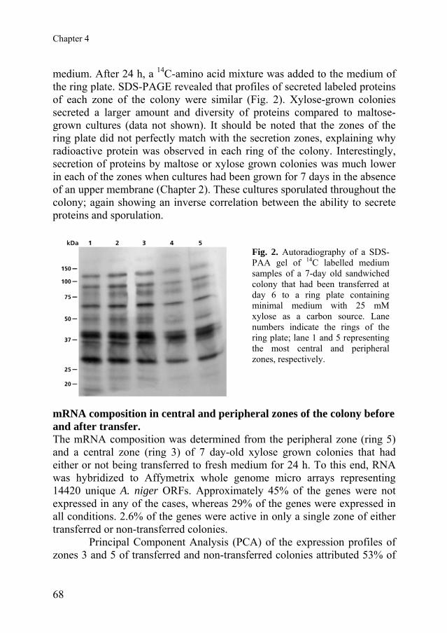
Chapter 4
medium. After 24 h, a 14C-amino acid mixture was added to the medium of the ring plate. SDS-PAGE revealed that profiles of secreted labeled proteins of each zone of the colony were similar (Fig. 2). Xylose-grown colonies secreted a larger amount and diversity of proteins compared to maltose-grown cultures (data not shown). It should be noted that the zones of the ring plate did not perfectly match with the secretion zones, explaining why radioactive protein was observed in each ring of the colony. Interestingly, secretion of proteins by maltose or xylose grown colonies was much lower in each of the zones when cultures had been grown for 7 days in the absence of an upper membrane (Chapter 2). These cultures sporulated throughout the colony; again showing an inverse correlation between the ability to secrete proteins and sporulation.
Fig. 2. Autoradiography of a SDS-PAA gel of 14C labelled mediumsamples of a 7-day old sandwichedcolony that had been transferred atday 6 to a ring plate containingminimal medium with 25 mMxylose as a carbon source. Lanenumbers indicate the rings of thering plate; lane 1 and 5 representingthe most central and peripheralzones, respectively.
mRNA composition in central and peripheral zones of the colony before and after transfer. The mRNA composition was determined from the peripheral zone (ring 5) and a central zone (ring 3) of 7 day-old xylose grown colonies that had either or not being transferred to fresh medium for 24 h. To this end, RNA was hybridized to Affymetrix whole genome micro arrays representing 14420 unique A. niger ORFs. Approximately 45% of the genes were not expressed in any of the cases, whereas 29% of the genes were expressed in all conditions. 2.6% of the genes were active in only a single zone of either transferred or non-transferred colonies.
Principal Component Analysis (PCA) of the expression profiles of zones 3 and 5 of transferred and non-transferred colonies attributed 53% of
68

Spatial differentiation in colonies of Aspergillus niger
the data variability to the position in the colony (i.e. zone 3 versus zone 5, irrespective of transfer) (Fig 3A). The second and third principal component (21% and 16% of the variability, respectively) were attributed to transfer within the zones and transfer independent of the zonal position (Fig 3B). These data show that the position within the colony has a higher influence on gene expression than medium composition.
Fig. 3. PCA analysis of 2 sample sets of the peripheral zone ring 5 and the central zone ring 3 of 7-day-old xylose-grown A. niger colonies; (A) First and second component; (B) second and third component. T and NT indicate transferred and non-transferred colonies, respectively, and the number indicates the zone of the colony.
The number of genes that were 2 and 4 fold changed between ring 5 and 3 of transferred colonies was similar to that in non-transferred colonies (Table 1) and between 2 to 3-fold higher than the number of differentially expressed genes between transfer and no transfer samples of the same zone (Table 1), which is in agreement with the distribution of variability showed by PCA. Analysis of the functional categories represented in the set of differentially expressed genes by Fisher´s exact test showed that expression of FunCat gene classes related to C-compound and carbohydrate metabolism, energy, cell rescue and defense, sub-cellular localization and protein modification were decreased at the periphery upon transfer to fresh medium (Table 2; p ≤ 0.01). Changes were less pronounced in the central zone ring 3. Genes related to sub cellular localization were increased, whereas some groups involved in metabolism were decreased.
69

Chapter 4
A comparison between ring 3 and 5 with and without transfer was also made. In non-transferred colonies, genes related to metabolism, protein fate and transport facilitation were higher expressed at the periphery, whereas expression of genes involved in cell cycle was reduced. Upon transfer, differences in expression in metabolic genes were reduced but became more pronounced for functional classes related to energy, protein fate, cell rescue and defense, cell fate and sub cellular localization. This is quite remarkable considering the fact that after transfer both zones were exposed to identical nutrient conditions.
Table 1. Number of genes that are at least 2 or 4 times increased (I) or decreased (D) between the periphery (ring 5) and a central zone (ring 3) of 7-day-old xylose grown sandwiched colonies. T indicates 24 h transfer to fresh medium. The analysis was performed on a normalized pool of independent triplicates.
2 x I 4 x I 2 x D 4 x D Ring 5 vs Ring 3 643 137 374 101 Ring 5T vs Ring 3T 419 96 591 85 Ring 5 vs Ring 5T 182 37 345 56 Ring 3 vs Ring 3T 167 73 394 34
Expression of specific gene groups XlnR regulated genes Xylose induces the transcriptional activator XlnR, which activates a set of genes mainly encoding extracellular enzymes (de Vries and Visser, 2001). In non-transferred colonies, 6 out of 19 XlnR regulated genes were significantly higher expressed at the periphery compared to the center (note that all 19 genes showed a similar spatial expression profile). However, after transfer only xyrA was still significantly higher expressed at the periphery (Fig. 4A). This shows that XlnR related genes mainly react to carbon source and not by the differentiation of the mycelium. Nitrate, phosphate and sulphate metabolism Genes involved in nitrate uptake and metabolism (crnA encoding a nitrate transporter, niaD encoding nitrate reductase, and niiA encoding nitrite reductase) (Johnstone et al., 1990) are periphery specific despite the fact that nitrate concentration in the medium underlying the colony center is similar to that at the periphery (Chapter 3; Fig. 4B). As expected, the transfer hardly affected spatial expression of these genes (Fig. 4B). Phosphate and sulphate
70

Spatial differentiation in colonies of Aspergillus niger
metabolism did not appear to be specific for the periphery of the colony neither before nor after transfer to fresh media. Some of the genes that have homology to sulphate and phosphate transporters and metabolic functions were expressed throughout the colony; others were specific for the periphery or center (data not shown). Table 2. Fisher’s exact test of the peripheral and a central zone of 7-day-old xylose grown sandwiched colonies of A. niger with (T) or without transfer to fresh medium at day 6. U and D indicate up- and down-regulation of functional FunCat categories, respectively. Category X5vsXT5 X3vsXT3 X5vsX3 XT5vsXT3
01.01.01 amino acid biosynthesis U U01.01.10 amino acid degradation U U U01.02.01 N and S utilization U01.05.01 C-compound and carbohydrate utilisation U U U U01.05.07 C-compound and carbohydrate transport U U U U01.06.01 lipid, fatty acid and isoprenoids biosinthesys U U01.06.04 lipid, fatty acid and isoprenoids breakdown U U01.06.99 other lipid, fatty acid and isoprenoids metabolism U
02.11.05 accessory proteins of electron transport and membrane-associated energy conservation U02.11.99 other electron transport and membrane-associated energy conservation proteins U02.13.03 aerobic respiration U
03.01.05 DNA recombination and DNA repair D03.03.01 mitotic cell cycle and cell cycle control D D04 TRANSCRIPTION04.05.01 mRNA synthesis D04.05.05 mRNA processing D
06.07.99 other protein modifications U U06.13 proteolytic degradation U06.13.04 lysosomal and vacuolar degradation U U06.13.99 other proteolitic degradation U
11.07.01 detoxification involving cytochrome P450 U11.07.99 other detoxification U U11.10.07 degradation of exogenous polysaccharides U U
14.04.03 fungal cell differentiation U40 SUBCELLULAR LOCALISATION U D D67 TRANSPORT FACILITATION U U
14 CELL FATE
02 ENERGY
01 METABOLISM
03 CELL CYCLE AND DNA PROCESSING
06 PROTEIN FATE
11 CELL RESCUE, DEFENCE AND VIRULENCE
71

Chapter 4
Cell wall synthesis and modification The genome of A. niger contains 13 putative chitin synthases (chsA-M), 14 putative chitinases (chiA-N) (Pel et al., 2007) and 5 α-glucan synthases (agsA-E) (Damveld et al., 2005). For all these gene classes, genes with high and low expression were detected. After transfer, expression of two chitinase genes (chiF and chiD), two chitin synthase genes (chsM and chsF), one α-glucan synthase gene (agsE) and one hydrophobin gene (similar to HYP1 of Aspergillus fumigatus) were significantly higher expressed at the periphery (Fig 4C). However, none of the genes showed an altered expression in the central zone after transfer. β-glucan synthases (Pel et al., 2007) were not differentially expressed irrespective of transfer to fresh medium. Protein secretion and proteases 8 out of 12 genes related to protein secretion are periphery specific in a non-transferred colony (Chapter 3; Fig. 4D). After transfer, the expression level of 9 out of these 12 genes increased in the central zone. As a result, only cnx1, rab6 and sec61b are still periphery-specific after transfer (Fig. 4D).
Some genes encoding putative proteases (prot A-I) (Pel et al., 2007) were differentially expressed in non-transferred colonies (Chapter 3; Fig. 4E). For instance, protE and protD (Aspergillopepsin II and its precursor) are more highly expressed at the periphery, whereas protF (encoding a protease similar to Ape2 of S. cerevisiae) is more highly expressed in the centre. After transfer, expression of the protease genes in the centre is similar to that at the periphery before transfer. As a result, expression of protE and protD turned around and they became centre-specific genes.
It can be concluded that genes encoding proteins involved in secretion and protein degradation react to a large extent to the composition of the medium.
Fig. 4. Expression profiles of XlnR regulated genes (A), genes involved in nitrate utilisation (B), cell wall formation and modification (C), secretion (D) and genes encoding (putative) proteases (E). Genes labelled with a star are significant after transfer to fresh medium. A triangle indicates specific significance for ring 5 and a square specific significance for ring 3.
72

Spatial differentiation in colonies of Aspergillus niger Spatial differentiation in colonies of Aspergillus niger
73
73

Chapter 4
DISCUSSION Genome wide expression analysis showed that colonies of A. niger are highly differentiated and that this can only be partly attributed to the heterogenicity of the medium underlying the colony. The spatial differentiation is also indicated by the fact that distinct zones in the colony have the capacity to secrete proteins or to sporulate upon transfer to a fresh medium. Previously it was shown that the RNA composition changes from the periphery to the center of 7-day-old colonies of A. niger (Chapter 3). The outer zone had the most distinct RNA profile, which correlated with the availability of the carbon source. In contrast to the N-, S- and P-source, the C-source was exhausted in the 4 inner zones of xylose-grown colonies, whereas up to 40% of the C-source was still available at the periphery. The importance of the C-source in gene expression was indicated when expression of xylose grown colonies was compared to maltose grown colonies. PCA analysis showed that 41% of the variability could be attributed to the difference in carbon source whereas the position of a zone within the colony accounted for 37%. The latter was proposed to be (partly) due to the availability of the C-source. Indeed, by transferring colonies to a fresh medium we here showed that genes induced by the xylanolitic regulator XlnR, genes encoding proteases and genes of the secretion machinery respond to a fresh medium. However, the transfer did not result in a reduction in the number of genes whose expression was significantly different between the outer zone of a colony and the central zone ring 3. About 1200 genes were differentially expressed between these zones with and without transfer. The number of genes that were differentially expressed within a zone before and after transfer to a fresh medium was also similar. In both cases more than 600 genes were found to have at least a two-fold changed expression level. This indicates that developmental processes independent of the medium composition also strongly affect the zonal gene expression. Indeed, PCA analysis showed that 55% of the variability could be attributed to the position in the colony. The availability of the fresh medium accounted for about 37% of the variability (adding up the second and the third principal component). These data show that differentiation not only occurs in time, as was shown by the temporal changes in gene expression at the periphery of the colony (Chapter 3) but that it also occurs in space. Since the central zone ring 3 was formed at day 4 and the
74

Spatial differentiation in colonies of Aspergillus niger
periphery at day 7, we cannot exclude that the spatial differentiation is in fact the result of temporal differentiation. Spatial differentiation was also reflected in the capacity to secrete or to form spores. The colonies that were used in this study were grown for 7 days on xylose or maltose medium in between two polycarbonate membranes, allowing us to easily label the culture and to transfer the colony to fresh medium. Non-transferred colonies exclusively secreted proteins at the periphery. In contrast, secretion also occurred in two central zones (ring 2 and 3) upon transfer to fresh medium. This correlated with an increased expression of genes involved in secretion. 9 out of 12 genes showed a higher expression in zone 3 upon transfer, which may explain the efficient secretion in this zone of the colony. Surprisingly, secretion in the central zones, as observed after transfer, did not correlate with the incorporation of radioactively labeled N-acetylglucosamine. Genome wide expression analysis did not indicate changes in expression of genes related to growth either (e.g. genes encoding chitin or glucan synthases). Taken together, this is the first evidence for growth independent secretion in fungal colonies. It should be noted that the secretion due to lysis is unlikely, since the secreted proteins contained radioactive amino acids that had been available in the medium only during the last 4 h of the cultivation. The non-secreting zones ring 1 and 4 started to sporulate when the upper polycarbonate membrane of transferred xylose-grown colonies was removed. It seems that the sporulation program was already activated in these parts of the colony before the PC membrane was removed. The results indicate that this program down-regulates secretion by the submerged feeding hyphae. Indeed, secretion was low throughout the mycelium when colonies were grown from day 1 onwards in the absence of an upper polycarbonate membrane (allowing sporulation over the whole colony surface). A similar phenomenon was observed when sandwiched colonies were grown on maltose-containing medium. Spores were formed in a large part of the colony (ring 1-4) when the upper polycarbonate membrane was removed, which correlated with a reduced amount of proteins that were released into the medium when compared to xylose-grown colonies. Taken together, data indicate that not only signaling pathways involved in sporulation and growth interact (Adams and Timberlake, 1990) but also those involved in growth-independent secretion.
75

Chapter 4
REFERENCES Adams, T.H. and W.E. Timberlake. 1990. Developmental repression of growth and gene
expression in Aspergillus. Proc Natl Acad Sci U S A 87: 5405-5409. Bos, C.J., A.J.M. Debets, K. Swart, A. Huybers, G. Kobus, and S.M. Slakhorst. 1988.
Genetic analysis and the construction of master strains for assignment of genes to six linkage groups in Aspergillus niger. Curr Genet 14: 437-443.
Damveld, R.A., P.A. vanKuyk, M. Arentshorst, F.M. Klis, C.A.M.J.J. van den Hondel, and A.F. Ram. 2005. Expression of agsA, one of five 1,3-alpha-D-glucan synthase-encoding genes in Aspergillus niger, is induced in response to cell wall stress. Fungal Genet Biol 42: 165-177.
de Vries, R.P., K. Burgers, P.J. van de Vondervoort, J.C. Frisvad, R.A. Samson, and J. Visser. 2004. A new black Aspergillus species, A. vadensis, is a promising host for homologous and heterologous protein production. Appl Environ Microbiol 70: 3954-3959.
de Vries, R.P. and J. Visser. 2001. Aspergillus enzymes involved in degradation of plant cell wall polysaccharides. Microb. Mol. Biol. Rev. 65: 497-522.
Johnstone, I.L., P.C. McCabe, P. Greaves, S.J. Gurr, G.E. Cole, M.A. Brow, S.E. Unkles, A.J. Clutterbuck, J.R. Kinghorn, and M.A. Innis. 1990. Isolation and characterisation of the crnA-niiA-niaD gene cluster for nitrate assimilation in Aspergillus nidulans. Gene 90: 181-192.
Pel, H.J., J.H. de Winde, D.B. Archer, P.S. Dyer, G. Hofmann, P.J. Schaap, G. Turner, R.P. de Vries, R. Albang, K. Albermann, M.R. Andersen, J.D. Bendtsen, J.A.E. Benen, M. van den Berg, S. Breestraat, M.X. Caddick, R. Contreras, M. Cornell, P.M. Coutinho, E.G.J. Danchin, A.J.M. Debets, P. Dekker, P.W.M. van Dijck, A. van Dijk, L. Dijkhuizen, A.J.M. Driessen, C. d'Enfert, S. Geysens, C. Goosen, G.S.P. Groot, P.W.J. de Groot, T. Guillemette, B. Henrissat, M. Herweijer, J.P.T.W. van den Hombergh, C.A.M.J.J. van den Hondel, R.T.J.M. van der Heijden, R.M. van der Kaaij, F.M. Klis, H.J. Kools, C.P. Kubicek, P.A. van Kuyk, J. Lauber, X. Lu, M.J.E.C. van der Maarel, R. Meulenberg, H. Menke, A.M. Mortimer, J. Nielsen, S.G. Oliver, M. Olsthoorn, K. Pal, N.N.M.E. van Peij, A.F.J. Ram, U. Rinas, J.A. Roubos, C.M.J. Sagt, M. Schmoll, J. Sun, D. Ussery, J. Varga, W. Vervecken, P.J.I. van de Vondervoort, H. Wedler, H.A.B. Wösten, A.P. Zeng, A.J.J. van Ooyen, J. Visser, and H. Stam (2006). Genome sequence of Aspergillus niger strain CBS 513.88: a versatile cell factory. Nature Biotechnol (in press).
Reiner, A., D. Yekutieli, and Y. Benjamini. 2003. Identifying differentially expressed genes using false discovery rate controlling procedures. Bioinformatics 19: 368-375.
Smyth, G.K. 2005. Limma: linear models for microarray data. In Bioinformatics and Computational Biology Solutions using R and Bioconductor (ed. R. Gentleman, V. Carey, S. Dudoit, R. Irizarry, and W. Huber), pp. 397-420. Springer, New York.
Wösten, H.A.B., S.M. Moukha, J.H. Sietsma, and J.G.H. Wessels. 1991. Localization of growth and excretion of proteins in Aspergillus niger. J Gen Microbiol 137: 2017-2023.
76

Chapter 5
The exploitation of the whole mycelium for secretion; production of
laccase in colonies of
Aspergillus niger Ana M. Levin, Robin A. Ohm, Charissa de Bekker, Ronald P. de Vries & Han A.B. Wösten


Production of laccase in colonies of Aspergillus niger
ABSTRACT Wild-type colonies of Aspergillus niger secreted laccase in the medium after 6 days of growth on maltose or xylose containing minimal medium. The activity of this metallo-enzyme, monitored by the oxidization of 2-2’-azinobis(3-ethylbenzthiazoline-6-sulfonic acid) (ABTS), was only visible in the centre of the colony, even after prolonged cultivation. Activity was also observed at the periphery of 6 day-old wild-type colonies by adding leucine or arginine to the medium. The part of the mycelium secreting laccase could also be increased by expressing the lccA laccase gene of A. niger behind the promoter of the glyceraldehyde-3-phosphate dehydrogenase gene gpdA of Aspergillus nidulans or that of the glucoamylase gene glaA of A. niger. In both cases laccase activity was detected in the centre and the middle zone of the colony, but not at the periphery. Notably, expression of lccA in the recombinant colonies was mainly observed at the periphery and the middle zone of the colony. This discrepancy is explained by slow release of the enzyme in the medium due to its affinity to the cell wall. A discordance was also observed between the temporal expression of the lccA reporter constructs and the activity of the encoded laccase. Although the promoters were active throughout culturing, LccA activity was only observed after 5 days. This indicates that post-transcriptional processes are involved. INTRODUCTION Filamentous fungi are attractive cell factories for the production of homologous and heterologous proteins that are of industrial or medical interest. Production of secreted proteins in filamentous fungi has been improved using classical and molecular genetic approaches as well as optimization of large-scale fermentation (Conesa et al., 2001). However, so far differentiation of the mycelium has received little attention. Glucoamylase was shown to be produced and secreted at the periphery of colonies of Aspergillus niger (Wösten et al., 1991). In contrast, the metallo-enzymes lignin peroxidase (LipH8) and manganese peroxidase (MnP1) of Phanerochaete chrysosporium were secreted in the colony center (Moukha et al., 1993a). Northern analysis revealed that the accumulation of LipH8 and MnP1 mRNA started in the center and moved towards the periphery in time (Moukha et al., 1993b). The finding that proteins are only locally secreted
79
Chapter 5
suggests that higher production levels can be reached by increasing the part of the colony that contributes to protein secretion.
Laccases (p-diphenol:dioxygen oxidoreductase EC 1.10.3.2) are multicopper proteins that oxidize substrates similar to p-diphenol. Natural functions of fungal laccases include pigmentation of spores, control of virulence and delignification of wood (Mayer and Staples, 2002). Laccases are also used for industrial applications; the enzyme has been applied in the delignification of wood tissue in the pulp and paper industry, chlorine-free bleaching of paper pulp, wine clarification, drug analysis, production of ethanol and bioremediation (Mayer and Staples, 2002). Recently, laccase was used as a reporter for gene expression in filamentous fungi (Mander et al., 2006).
We here show that a larger part of the mycelium can be involved in secretion of a certain protein in two ways: by adding supplements to the medium or by expressing genes from the glucoamylase glaA promoter or that of the glyceraldehyde-3-phosphate dehydrogenase gene gpdA. MATERIAL AND METHODS Strains used Strain N402 (cspA1) (Bos et al., 1988) was used as a wild-type Aspergillus niger, while its derivative NW249 (leuA1, pyrA6, nicA1, ΔargB, cspA1) (P.J.I. van de Vondervoort and Y. Muller, unpublished data) was used for transformation. Recombinant A. niger strains used in this study are listed in Table 1. Escherichia coli DH5α was used for cloning purposes. Table 1. Derivatives of NW249 used in this study Strain Transforming
construct(s) Description of construct
UU-A016.1 pGW635 pGW635 (Kusters-van Someren et al., 1991) contains the pyrA gene conferring uridine prototrophy.
UU-A102.10 UU-A102.11
pGW635 & pAL025 pAL025 is a derivative of pAN52-10 (Siedenberg et al.,1999) containing the lccA gene behind the glaA promoter.
UU-A103.03 UU-A103.11
pGW635 & pAL026 pAL026 is a derivative of pGPDGFP (Lagopodi et al., 2002) containing the lccA gene behind the gpdA promoter.
80

Production of laccase in colonies of Aspergillus niger
Media and culture conditions A. niger was grown at 30 °C in a humid environment as sandwiched cultures (Wösten et al., 1991) in a 0.2 mm thin layer of 1.25% agarose in between two perforated polycarbonate membranes (diameter 76 mm, pore size 0.1 μm; Osmonics, GE Water Technologies, Trevose, PA, USA) placed on top of solidified (1.5% agar) minimal medium (MM) (de Vries et al., 2004) with either 25 mM maltose or xylose as a carbon source. NW249 was grown in the presence of nicotinamide (1 mg l-1), leucine (0.2 g l-1), arginine (0.2 g l-1) and uridine (0.2 g l-1), while uridine was omitted in case of its derivatives. 0.2 mM ABTS (Sigma-Aldrich Chemie GmbH, Steinheim, Germany) and 0.1 mM CuSO4 were added to the medium to monitor laccase secretion. Cultures were inoculated with 1.5 μl of spore solution (108 spores ml-1). Expression cassettes and vectors The lccA coding sequence was amplified by PCR with primers LccA-ATG-NcoI (CCATGGATCCCTTTCAATTCGGAC) and LccA-3’-SpeI (ACTAGTCCACGTGCATCTCACC) using genomic DNA as a template. The 3348bp lccA fragment containing NcoI and SpeI linkers was cloned in pGEMT-easy (Promega Corp., WI, USA), resulting in plasmid pAL021. To obtain pAL025, the lccA coding sequence was excised from pAL021 by NcoI/SpeI digestion and cloned in pAN52-10 (Siedenberg et al., 1999) digested with NcoI and XbaI. Similarly, the lccA fragment was cloned in pGPDGFP (Lagopodi et al., 2002) to obtain pAL026. Constructs pAL025 and pAL026 express lccA from the glaA and the gpdA promoter, respectively. Transformation of A. niger Fungal strains were transformed as described (Kusters-van Someren et al., 1991). Transformants were selected by co-transforming with pGW635 (containing the A. niger pyrA gene) and purified by repeated streaking of conidia. Co-transformation with pAL025 or pAL026 was confirmed by Southern analysis. Southern analysis Genomic DNA was isolated as described (de Graaff et al., 1988), digested with SphI and hybridised with an lccA probe (Sambrook et al., 1989). Copy number was determined by signal intensity using a phosphoimager (model 810-UNV, Molecular Dynamics, Amersham Pharmacia Biotech, CA, USA).
81

Chapter 5
Northern analysis Concentrical rings of mycelium of sandwiched colonies (5-7 mm in width each) were isolated with a ring cutter or a scalpel. The mycelium was ground using a microdismembrator (B.Braun GmbH, Melsungen, Germany) and RNA was extracted with TRIzol reagent (Invitrogen, Carlsbad, CA, USA) according to the instructions of the manufacturer. After separation in 1.5% agarose gels, the RNA was blotted onto Hybond N+ membranes (Amersham Biosciences, UK) using 10xSSC. Hybridisations were done overnight with α-32P-CTP labeled probes of lccA, glaA and 18S rRNA according to de Vries et al. (2002). Blots were exposed to X-OMAT AR films (Eastman Kodak, New York, USA) with intensifying screens at -80°C. Detection of laccase activity Samples of mycelium and the agar underlying the colony were homogenized in a dismembrator and resuspended in 4 volumes of Na-citrate buffer pH 5.0 with CompleteTM protease inhibitor (Roche Diagnostics GmbH, Manheim, Germany). The soluble and insoluble fractions of the mycelium were separated by centrifugation at 4.000 rpm for 10 min. The latter fraction was resuspended in the same volume of buffer as the soluble fraction. Laccase activity was measured in a total volume of 200 μl in microtiter plates at 420 nm by the oxidation of 5 mM freshly prepared ABTS in Na-citrate buffer pH 5.0. Activity was monitored in a Bio-Rad 550 microplate reader (Bio-Rad laboratories, CA, USA) by determining the activity every 30 minutes during a 12 h period with 5 sec shaking before every read-out. RESULTS ABTS oxidation in wild-type colonies of A. niger. Sandwiched colonies of the wild-type strain N402 were grown on MM in the presence of 0.2 mM ABTS and 0.1 mM CuSO4. Oxidation of ABTS was not observed during the first 5 days of cultivation when either maltose or xylose was used as the carbon source. However, oxidation of the substrate was observed in the colony centre after 6 days of growth (Figure 1A). The activity did not spread to the outer zones of the colony during prolonged cultivation. Interestingly, ABTS oxidizing activity was also observed at the periphery when either leucine (Figure 1B) or arginine (data not shown) was
82

Production of laccase in colonies of Aspergillus niger
added to the medium. This was also observed when medium was supplemented with arginine, leucine, and nicotinamide (Figure 1C) but not when nicotinamide alone was added. The pattern of laccase secretion of the glucoamylase transformant UU-A102.11 (see below) was similar to that of the wild-type grown with all the supplements (Figure 1D). Notably, laccase activity in colonies of the reference strain UU-A016.1 that was only transformed with the construct encompassing the selection marker and that requires the same supplements in the medium as UU-A102.11 was similar to that observed in colonies of the wild-type in the absence of these supplements (Figure 2A, D).
Fig. 1. ABTS-oxidizingactivity, recognized bypresence of green or redcolor, in the mediumunderlying 7 day-oldmaltose-grown sandwichedcolonies of wild-type A. nigerstrain N402 (A, B, C) andtransformant UU-A102.11expressing lccA from theglaA promoter (D) in theabsence (A) or presence ofleucine (B), and acombination of leucine,nicotinamide and arginine(C,D).
Using lccA as a reporter to monitor changes in spatial laccase production in colonies of A. niger Blasting the genomic database of A. niger (strain CBS 513.88) with the laccase gene tilA of Aspergillus nidulans (Scherer and Fischer, 2001) revealed a highly homologous gene (An00g07173; score 717 and an E-value of 0), which we designated lccA. Micro array analysis (Chapter 3) and Northern hybridization (Figure 3) showed that lccA was not expressed in xylose or maltose grown sandwiched colonies of the wild-type strain N402. In contrast, gpdA was found to be expressed both in central and peripheral
83

Chapter 5
Fig. 2. Laccase activity in the medium underlying 7-day-old sandwiched colonies of strain UU-A016.1 (A, D), UU-A102.11 (B, E) and UU-A103.11 (C, F) grown on minimal medium with maltose (A-C) or xylose (D-F) as the carbon source. Strain UU-A016.1 is the reference strain for the transformation with the selection marker, whereas strains UU-A102.11 and UU-A103.11 express lccA behind the glaA and gpdA promoter, respectively. zones of 7 day-old maltose or xylose grown colonies (Figure 3). As expected (Fowler et al., 1990), glaA was highly expressed on maltose, while expression on xylose was low. On both carbon sources glaA mRNA was detected throughout 3-5-day old colonies (data not shown), while it was only detected at the outer zones of 7-day-old cultures (Figure 3).
The fact that lccA is not expressed in xylose or maltose grown sandwiched cultures makes it a good reporter to monitor changes in spatial laccase production due to glaA and gpdA-driven expression, as previously reported by Mander et al. (2006). The coding sequence of lccA was cloned behind the glaA and the gpdA promoter and the resulting constructs pAL025 and pAL026, respectively, were introduced in A. niger NW249. Ten transformants of each construct were analysed for laccase activity using ABTS as a substrate. About 70% of the transformants showed altered laccase secretion patterns when compared to the reference strain UU-A016.1 that had only been transformed with construct pGW635 containing the selectable marker
84

Production of laccase in colonies of Aspergillus niger
Fig. 3. Accumulation of lccA, glaA, and gpdA mRNA in zones of 7-day-old maltose or xylose grown sandwiched colonies of the wild-type A. niger strain N402. Concentrical rings of mycelium were harvested that were 5 mm in width. Ring 1 and ring 5 represent the most central and peripheral zones of the colonies, respectively. Hybridization with 18S rDNA served as a loading controlchosen for further analysis: Two representative transformants of each construct were UU-A102.10 and UU-A102.11 (transformed with pAL025 containing lccA behind the glaA promoter), and UU-A103.03 and UU-A103.11 (transformed with pAL026 containing lccA behind the gpdA promoter). Southern analysis revealed that strains UU-A102.10 and UU-A102.11 contained 5 and 6 copies of pAL025, respectively, whereas 5 and 8 copies of pAL026 had integrated, respectively, in UU-A103.03 and UU-A103.11 (data not shown).
ABTS-oxidation was observed in the medium underlying colonies expressing lccA from the gpdA and the glaA promoter after 6 days of growth. The activity started in the centre and had extended to the middle part of the colony after 7 days of growth (Figure 2). The activity did not correlate with the spatial and temporal expression of the reporter gene. Both the gpdA or glaA promoter were active throughout culturing (data not shown). Moreover, gpdA-driven lccA expression was observed throughout 7-day-old colonies, although levels in the most central part were lower (Figure 4). glaA-driven expression resulted in high levels of lccA mRNA in
85

Chapter 5
the two outer zones of 7-day-old maltose-grown colonies (zones 3 and 4; Figure 4). Lower levels were observed in the outer central zone (zone 2), whereas lccA mRNA was not detectable in the most central part (zone 1). A similar profile was observed in xylose grown colonies but levels of lccA mRNA were lower.
Fig. 4. Accumulation of lccA mRNA in zones of 7-day-old colonies of strains UU-A016.1 (A), UU-A102.11 (B) and UU-A103.11 (C) that had been grown on maltose or xylose containing medium. Strain UU-A016.1 is the reference strain, whereas strains UU-A102.11 and UU-A103.11 express lccA from the glaA and gpdA promoter, respectively. Zone 1 represents the centre and zone 4 the periphery of the colony. Localization of laccase activity Laccase activity was determined in the soluble fraction (containing the cytoplasmic components), the insoluble fraction (i.e. the cell wall) and the agar underneath 3, 5, and 7-day old maltose grown colonies of strains UU-A102.11 and the reference strain UU-A016.1 (Figure 5). The reference strain did not show any activity in any of the fractions tested. Fractions of 3-day-old colonies of UU-A102.11 (expressing lccA behind the glaA promoter) were also inactive. However, after 5 days activity could be detected; 70% was associated with the cell wall and 5% was detected in the medium. Similar results were obtained with 7-day old colonies. In this case the mycelium was split in two fractions; one part represented the zone where activity was observed in the medium; the other part represented the inactive zone. The cell wall fraction of the mycelium of the non-active and active zones represented 80 and 71% of the activity, respectively, whereas 0 and 3% were detected in the medium.
86

Production of laccase in colonies of Aspergillus niger
Fig. 5. Laccase activity of 3, 5 and 7 day-old colonies of wild type A. niger N402 as measured by oxidation of ABTS. The detection was performed in duplo. SF= Soluble Fraction; IF= Insoluble Fraction; M= Medium; NA= Non-active mycelium; A= Active mycelium.
DISCUSSION A. niger is an excellent host for the production of homologous and heterologous proteins. Several strategies have been used in order to improve yields of secretion of industrially relevant enzymes, but so far nobody addressed whether mobilizing a larger part of the mycelium in the secretion process can increase production. Results presented here indicate that central, middle and peripheral zones of the colony have the ability to secrete laccase and that a larger part of the mycelium can be involved in laccase production by changing the composition of the medium or by expressing a laccase gene from a glaA or gpdA promoter. 6-day-old sandwiched cultures of a wild-type A. niger strain released laccase into the medium from the colony centre. The activity was also observed at day 6 in recombinant strains expressing lccA from the glaA and
87

Chapter 5
the gpdA promoter. The zone of wild-type colonies exhibiting laccase activity remained restricted to the colony centre when growth was prolonged. In contrast, the activity zone had extended in 7-day-old maltose- and xylose-grown colonies to the middle zone in strains expressing lccA under control of the gpdA promoter. A similar pattern was observed in maltose grown colonies expressing lccA from the glaA promoter. However, when xylose was used as a carbon source no laccase activity attributable to LccA could be observed.
The appearance of LccA activity did not correlate with the temporal expression of lccA in the colony. The gpdA and glaA promoters are both active throughout culturing. However both the plate assay and the activity assays of mycelial and medium fractions did not show any activity before day 5. In contrast, GFP is formed in young sandwiched colonies (Vinck et al., 2005) when its encoding gene is expressed from these promoters. This suggests that young sandwiched colonies of A. niger may not have the ability to form active laccase.
Like the temporal activity, also the spatial activity did not correlate with lccA expression. The glaA promoter was shown to be mainly active in the two most outer zones of 7-day-old colonies, whereas the gpdA promoter was quite evenly expressed. The discrepancy between the spatial activity and expression is probably due to the affinity of the LccA laccase for the cell wall. Up to 80% of the enzyme was found to be associated to the cell wall of 7-day-old colonies. The enzyme is probably slowly released, causing a delay in the detection of the activity of the protein in the medium. It has been reported that laccase of Cryptococcus neoformans and manganese peroxidase of P. chrysosporium also interact with the cell wall (Zhu et al., 2001; Moukha et al., 1993b) and are slowly released in the medium (Moukha et al., 1993b). Although glaA is much lower expressed on xylose than on maltose (Fowler et al., 1990) expression at the former carbon source still equals that of gpdA. However, in contrast to gpdA driven expression, glaA-driven expression of lccA did not result in LccA activity on xylose. There are two possible explanations for this discrepancy. First, glaA expressing hyphae may not be capable of producing laccase efficiently. Vinck et al. (2005) showed that within the outer zone of the A. niger colony two types of hyphae can be distinguished; those that highly and those that lowly express the glaA gene. It was thus concluded that differentiation in the fungal colony is much more pronounced than previously acknowledged. Extending this
88

Production of laccase in colonies of Aspergillus niger
conclusion to the results described here, it cannot be excluded that high expressing glaA hyphae are not equipped to produce laccase. For instance, copper uptake may be low in these hyphae. The second explanation why LccA activity was not observed on xylose is that the 5’ untranslated region of the mRNA that originates from the glaA promoter may somehow block translation when the fungus is grown on xylose. This is supported by the finding that glaA driven expression of GFP did not result in detectable GFP fluorescence on xylose (Santerre Henriksen et al., 1999; Vinck et al., 2005) despite the fact that the activity of the promoter on this carbon source should be more than sufficient to yield GFP. Spatial distribution of laccase activity in the medium could not only be altered by expressing lccA from the gpdA or glaA promoter, it could also be changed by adding supplements to the medium. Laccase activity was found both in the centre and at the outer periphery of wild-type colonies when leucine or arginine was added to the medium. Again, activity was only observed after 6 days. In contrast, no peripheral laccase activity was observed when the strains expressing lccA behind the glaA or gpdA promoter were grown in the presence of leucine or arginine. These recombinant strains need these amino acids for growth, which may somehow affect the peripheral laccase activity. Introduction of the expression constructs in the wild-type strain using an antibiotic resistance marker for selection may reveal whether leucine and arginine somehow activate the release of laccase in the medium or whether these amino acids activate a specific gene(s) encoding an enzyme that uses ABTS as a substrate.
Laccases have been recently reported as versatile reporter proteins to localize expression of genes (Mander et al., 2006). Data shown in this study does not support this claim. LccA is not suitable as a reporter, at least in A. niger, because of its affinity for the cell wall and its discrepancy in expression and activity. In the future we will test the heterologous Stachybotrys chartarum laccase, which was used by Mander et al. This laccase may be function as a reporter.
89

Chapter 5
ACKNOWLEDGEMENTS The authors want to thank Dr. Alexandra Alves for valuable advice. This work was supported by The Netherlands Technology Foundation (STW) Pionier project UGC.5683. For color pictures see http://www.bio.uu.nl/microbiology/fung/PhD%20theses/AMLevin/index.html REFERENCES Bos, C.J., A.J.M. Debets, K. Swart, A. Huybers, G. Kobus, and S.M. Slakhorst. 1988.
Genetic analysis and the construction of master strains for assignment of genes to six linkage groups in Aspergillus niger. Curr Genet 14: 437-443.
Conesa, A., P.J. Punt, N. van Luijk, and C.A.M.J.J. van den Hondel. 2001. The secretion pathway in filamentous fungi: a biotechnological view. Fungal Genet Biol 33: 155-171.
de Graaff, L.H., H.W.J. van den Broek, and J. Visser. 1988. Isolation and transformation of the pyruvate kinase gene of Aspergillus nidulans. Curr Genet 13: 315-321.
de Vries, R.P., K. Burgers, P.J. van de Vondervoort, J.C. Frisvad, R.A. Samson, and J. Visser. 2004. A new black Aspergillus species, A. vadensis, is a promising host for homologous and heterologous protein production. Appl Environ Microbiol 70: 3954-3959.
de Vries, R.P., P.J.I. van de Vondervoort, L. Hendriks, M. van de Belt, and J. Visser. 2002. Regulation of the α-glucuronidase encoding gene (aguA) from Aspergillus niger. Molec Gen Genet 268: 96-102.
Fowler, T., R.M. Berka, and M. Ward. 1990. Regulation of the glaA gene of Aspergillus niger. Curr Genet 18: 537-545.
Kusters-van Someren, M.A., J.A.M. Harmsen, H.C.M. Kester, and J. Visser. 1991. The structure of the Aspergillus niger pelA gene and its expression in Aspergillus niger and Aspergillus nidulans. Curr Genet 20: 293-299.
Lagopodi, A.L., A.F. Ram, G.E. Lamers, P.J. Punt, C.A.M.J.J. Van den Hondel, B.J. Lugtenberg, and G.V. Bloemberg. 2002. Novel aspects of tomato root colonization and infection by Fusarium oxysporum f. sp. radicis-lycopersici revealed by confocal laser scanning microscopic analysis using the green fluorescent protein as a marker. Mol Plant Microbe Interact 15: 172-179.
Mander, G.J., H. Wang, E. Bodie, J. Wagner, K. Vienken, C. Vinuesa, C. Foster, A.C. Leeder, G. Allen, V. Hamill, G.G. Janssen, N. Dunn-Coleman, M. Karos, H.G. Lemaire, T. Subkowski, C. Bollschweiler, G. Turner, B. Nusslein, and R. Fischer.
90

Production of laccase in colonies of Aspergillus niger
2006. Use of laccase as a novel, versatile reporter system in filamentous fungi. Appl Environ Microbiol 72: 5020-5026.
Mayer, A.M. and R.C. Staples. 2002. Laccase: new functions for an old enzyme. Phytochemistry 60: 551-565.
Moukha, S.M., H.A.B. Wösten, M. Asther, and J.G.H. Wessels. 1993a. In situ localization of the secretion of lignin peroxidases in colonies of Phanerochaete chrysosporium using a sandwiched mode of culture. J Gen Microbiol 139: 969-978.
Moukha, S.M., H.A.B. Wösten, E.J. Mylius, M. Asther, and J.G.H. Wessels. 1993b. Spatial and temporal accumulation of mRNAs encoding two common lignin peroxidases in Phanerochaete chrysosporium. J Bacteriol 175: 3672-3678.
Sambrook, J., E.F. Fritsch, and T. Maniatis. 1989. Molecular cloning -a laboratory manual. Cold Spring Harbour Laboratory, Cold Spring Harbour, N.Y.
Santerre Henriksen, A.L., S. Even, C. Muller, P.J. Punt, C.A.M.J.J. van den Hondel, and J. Nielsen. 1999. Study of the glucoamylase promoter in Aspergillus niger using green fluorescent protein. Microbiology 145: 729-734.
Scherer, M., and R. Fischer. 2001. Molecular characterization of a blue-copper laccase, TILA, of Aspergillus nidulans. FEMS Microb. Lett. 199: 207-213.
Siedenberg, D., S. Mestric, M. Ganzlin, M. Schmidt, P.J. Punt, C.A.M.J.J. van den Hondel, and U. Rinas. 1999. GlaA promoter controlled production of a mutant green fluorescent protein (S65T) by recombinant Aspergillus niger during growth on defined medium in batch and fed-batch cultures. Biotechnol Prog 15: 43-50.
Vinck, A., M. Terlou, W.R. Pestman, E.P. Martens, A.F. Ram, C.A.M.J.J. van den Hondel, and H.A.B. Wösten. 2005. Hyphal differentiation in the exploring mycelium of Aspergillus niger. Mol Microbiol 58: 693-699.
Wösten, H.A.B., S.M. Moukha, J.H. Sietsma, and J.G.H. Wessels. 1991. Localization of growth and excretion of proteins in Aspergillus niger. J Gen Microbiol 137: 2017-2023.
Zhu, X., J. Gibbons, J. Garcia-Rivera, A. Casadevall, and P.R. Williamson. 2001. Laccase of Cryptococcus neoformans is a cell wall-associated virulence factor. Infect Immun 69: 5589-5596.
91


Chapter 6
General Discussion


General discussion
Filamentous fungi, among which A. niger, grow by means of hyphae that extend at their tips and that branch subapically. As a result, a network of interconnected hyphae is formed, called a mycelium. When grown on solid medium, the mycelium is called a colony. The center of the colony represents the oldest part of the mycelium due to radial extension of the network. The cytoplasm within the mycelium is continuous due to the presence of porous septa that divide hyphae in compartments. These porous septa allow streaming of water, nutrients (Jennings et al., 1974) and organelles throughout the mycelium (Shepherd et al., 1993; Cole et al., 1998) as well as antifungals (Oh et al., 1995) and particular proteins (Vinck et al., 2005).
The mycelium of filamentous fungi secrete large amounts of enzymes into the environment. These enzymes degrade the substrate into small molecules that can be taken up by the fungus to serve as nutrients. Aspergillus niger is an example of a fungus with an enormous secretion capacity. For instance, some strains secrete up to 30 grams per liter of glucoamylase (Finkelstein, 1989). This secretory capacity, as well as the established fermentation technology and molecular biology makes A. niger a very suitable cell factory for the production of proteins. The fact that the fermentation products of this fungus are generally regarded as safe adds to the attractiveness of this system.
Protein production has been improved in A. niger using different strategies; e.g. use of strong promoters, increasing the copy number of the gene of interest, optimization of the protein folding machinery and the use of protease deficient strains (Conesa et al., 2001). So far, nobody addressed whether protein production can be improved by increasing the number of hyphae in the mycelium that contributes to protein secretion. Previously, it was shown that proteins are only secreted by the limited number of growing hyphae within a colony. Over and above this, not every growing hypha secretes a particular protein. For instance, glucoamylase was secreted at the periphery of the growing mycelium but not in the central growth zone (Wösten et al., 1991). It can therefore be concluded that a fungal mycelium is not a mass of identical hyphae.
The aim of this Thesis was to study the extent of differentiation in the fungal mycelium and the mechanisms underlying it. This is particularly interesting considering the cytoplasmic continuity in the fungal mycelium. This Thesis shows that expression profiles in the center and the periphery of the mycelium are quite distinct. These differences can be explained by the
95

Chapter 6
availability of the carbon source but also by spatial and temporal differentiation. The high degree of differentiation in the colony was also indicated by the fact that secretion and sporulation occurred in distinct zones of the colony upon transfer to fresh medium. Finally, first evidence was obtained that production of secreted proteins can be increased by expressing the gene of interest from promoters that are active in different zones of the colony.
Culturing methods to study secretion of proteins in fungal colonies Maltose and xylose were used as carbon sources in this study. These sugars are known to activate the transcriptional activators AmyR (Petersen et al., 1999) and XlnR (van Peij et al., 1998), respectively. These two regulators each induce the expression of specific gene sets. The AmyR- regulated gene set includes glaA encoding glucoamylase. This enzyme was shown to be differentially secreted (Wösten et al., 1991). The gene set regulated by XlnR includes XlnB, which was also shown to be differentially secreted in colonies of A. niger (Chapter 2).
Previously, the sandwiched culturing technique was developed to study growth and secretion in colonies of filamentous fungi (Wösten et al., 1991). Colonies were grown on a nutrient agar medium in between two porous polycarbonate membranes in a thin agarose layer. The sandwich prevents sporulation and can be easily transferred e.g. to a fresh medium. Total protein secretion in different parts of the colony can be monitored by labeling with radioactive amino-acids and by placing a protein binding PVDF membrane under the sandwich. However, detection of secretion of specific proteins was limited at that time to those proteins for which an antibody was available. In Chapter 2 a new culturing method was developed, called the ring plate system, which simplifies identification of the proteins secreted at each part of the colony and which also allows monitoring of the local environmental conditions. In the ring plate system, colonies are grown on a polycarbonate membrane placed on a plate with 5 ring-shaped wells that are filled with liquid medium. The experimental set-up is not affected by diffusion of molecules (e.g. proteins, metabolites and nutrients) as occurs in agar medium. Moreover, the secretion profile at each part of the colony can be easily assessed by separating the proteins contained in the medium of each ring by SDS-PAGE.
96

General discussion
Differentiation in colonies of A. niger as assessed by global expression analysis Expression profiles were analysed from 5 concentric zones of 7-day-old maltose or xylose grown sandwiched colonies of A. niger. Ring 1 and 5 represented the most central and most peripheral zone, respectively. Approximately 50% of the genes were expressed in the colonies. Of these, about 60% were expressed in all zones of the colonies and on both carbon sources. These data show that a considerable part of the genes is expressed only in particular zones or on a specific carbon source. Indeed, 3% of the genes was uniquely expressed in a singly zone on one of the carbon sources. Hierarchical cluster analysis (HCA) and principal component analysis (PCA) revealed that the differences in gene expression within the colony result from medium-dependent and medium-independent mechanisms. Medium related differentiation The nature of the carbon source was shown to be a main determinant of the expression profile in the colony (Chapter 3). PCA attributed 41% of the variability to the nature of the carbon source when expression profiles of the different zones of xylose and maltose grown colonies were compared. This variability is partly explained by the metabolic pathways that are needed to metabolize these carbon sources. Maltose is converted to glucose that enters the central metabolism directly through glycolysis. Alternatively, xylose enters the central metabolism through the pentose catabolic pathway followed by the pentose phospate pathway, which serves as a link to glycolisis. However, the carbon source also affected genes that are not directly related to carbon source metabolism. For instance, the Fisher´s test showed that functional gene classes related to control of cellular organization, morphogenesis and cell cycle control are up-regulated at the periphery of xylose grown colonies but not in colonies grown on maltose. Changes in the medium composition during growth also affected global gene expression. This was shown by comparing expression profiles of 7-day-old xylose grown colonies that had either or not been transferred for 24 h to a fresh medium (Chapter 4). PCA analysis indicated that 21% of the variability could be attributed to this composition. Most probably, the availability of the carbon source is the determining factor. Maltose and xylose were exhausted in the central zones but they were still available at the periphery. In contrast, the N-, S-, and P-source were not limited in the medium underlying each zone of the colony (Chapter 3).
97

Chapter 6
The XlnR regulated genes strongly responded to the medium composition (Chapter 4). 1 and 6 out of 19 genes known to be regulated by XlnR were significantly higher expressed at the periphery with and without a transfer to fresh medium, respectively. a similar response was observed for genes related to protein secretion and genes encoding putative proteases. Non-medium related differentiation The nutrient conditions at the periphery of the sandwiched colonies hardly, if at all, change since the hyphae of this zone continuously extend in an unexplored medium. It was therefore of great interest to find that gene profiles at the periphery did change in time (Chapter 3). Expression of approximately 9% of the genes, belonging to different functional gene classes, changed at the periphery of 3-10 day old maltose grown colonies. Among the genes that were subject to temporal changes in expression were half of the XlnR regulated genes including xlnB. Taken together, I suggest that the periphery goes through a developmental program that is regulated by the age of the colony. Apart from temporal changes in the expression profiles, also spatial changes were observed that were independent of the medium composition. This was indicated from the analysis of the expression profiles of colonies that were either or not transferred to a fresh medium for 24 h (Chapter 4). In fact, the spatial position and not the nutrient availability was shown to be the main determinant of the variability in gene expression. PCA analysis attributed 53% and 37% of the variability to the spatial position and the nutrient availability, respectively. The gene cluster involved in the conversion of extracellular nitrate to intracellular ammonium was the most remarkable example of the positional differentiation. These genes were only expressed at the periphery, irrespective of the fact that the nitrate concentration was not limited in the central zone of the colony.
How can the spatial differentiation be explained? It might be that quorum sensing mechanisms are involved. The density of the hyphae is much higher in the center than at the periphery. Transfer to fresh medium would dilute the signaling molecules tenfold (the volume ratios between the sandwich and the agar medium is 1:10), but this may have been insufficient to switch off the regulation. The spatial differentiation may also result from the temporal differentiation. The hyphae in the center are older than those at the periphery and may have changed their expression accordingly.
98

General discussion
Growth and secretion in 7-day-old colonies grown on a xylose or maltose medium Previously it was shown that protein synthesis occurs throughout the mycelium of 5-day-old sandwiched colonies, whereas secretion and growth were restricted to a central zone and the periphery of the colony (Wösten et al., 1991). Similar patterns of protein synthesis and growth were observed in 7-day-old sandwiched colonies (Chapter 3, 4). However, protein secretion was restricted to the periphery as assessed by labeling with 14C amino acids. Yet, laccase activity in sandwiched colonies did correlate with the central growth zone (Chapter 5), showing that proteins are also secreted in this growth zone but the amounts seem to be small.
The finding that a protein is only secreted in a particular region of the colony (Wösten et al., 1991; Moukha et al., 1993; Chapter 2, 5) raised the question whether it is possible to increase protein production by involving other zones of the colony. To study this, laccase was used as a reporter. Activity of this enzyme was observed after 6 days of growth in the center of sandwiched colonies grown on minimal medium with xylose or maltose as a carbon source (Chapter 5). Activity was also observed at peripheral parts when leucine or arginine were added to the culture medium. This showed that both central and peripheral zones of the colony are able to secrete laccase. Increased participation of the mycelium in laccase production was also obtained by expressing the lccA gene of A. niger behind the glucoamylase (glaA) or the glyceraldehyde-3-phospate dehydrogenase (gpdA) promoter. Laccase activity in the medium started in the center of 6-day-old recombinant colonies, but in contrast to the wild-type, extended to the zone behind the periphery. Temporal and spatial activity of laccase in the recombinant colonies did not correlate with the activity of the glaA and gpdA promoters. These promoters are active from day 1 onwards and are mainly active at the periphery of 6-day-old colonies (Chapter 5). The spatial difference in expression and laccase activity can be explained by the affinity of the recombinant enzyme for the cell wall from which it is slowly released. However, the difference in temporal expression and activity is not clear yet. It may be that young colonies do not have the capacity to produce laccase.
99

Chapter 6
Growth and secretion in 7-day-old xylose-grown colonies transferred for 24 h to a fresh medium The spatial growth pattern did not change when 7-day-old colonies were transferred to fresh medium for 24 h (Chapter 4). This was concluded from labeling experiments with 14C N-acetylglucosamine as well as from whole genome expression analysis. In contrast, the spatial secretion pattern did change. Labeling with 14C amino acids did not only reveal secretion at the periphery of the colony but also in an outer central zone. Secretion in this central zone did not correlate with growth. This came to a surprise since it is generally accepted that only growing hyphae secrete proteins (Wessels 1993). So far, experiments showed a strict correlation between growth and secretion. Moreover, the steady state growth theory and the bulk flow theory predict such a correlation. The steady state (Wessels, 1988) describes cell wall synthesis in a growing hypha. Individual cell wall polymers are extruded at the apex of the growing hypha and are pushed outwards and to subapical regions by the intracellular turgor pressure and by the apposition of new cell wall polymers. The cell wall polymers become more and more cross-linked during their transit to the outer and subapical part of the cell wall. As a result, the subapical cell wall is very rigid, whereas it is plastic at the apex. When growth stops the apical cell wall also becomes rigid due to the fact that cross-linking continues in the absence of the flow of cell wall polymers. Why does this model of cell wall synthesis have implications for secretion of proteins? Experimental data indicate that the pores in the rigidified cell wall are too small to allow diffusion of proteins from the inner to the outer part of the cell wall (Money, 1990). The bulk flow theory describes that proteins transit the cell wall by co-migrating with the flow of cell wall polymers at the growing apex (Wessels, 1993), which is a pore-independent process. Taken this into account, how can we explain a growth-independent secretion? Possibly, pores in the cell walls are locally increased by the action of cell wall modifying enzymes. Are sandwiched colonies a good model system for industrial fermentations? In the industry, fungi are grown in solid state or submerged fermentation. In the latter system fungi can grow dispersed or in pellets (Chapter 1). Solid state fermentations are most similar to the way fungi grow in nature. The sandwiched colony seems to be a good model system to optimize such fermentations. However, it should be noted that in the sandwiched colony
100

General discussion
the fungal hyphae are not in direct contact with the substrate and this may affect the extrapolation of the results with solid state fermentations. Intuitively, the sandwiched colony may not seem a suitable model for submerged fermentations. To address this, RNA was isolated from liquid shaking flask cultures that were harvested at the moment the medium mimicked the conditions at the periphery of 7 day-old maltose grown sandwiched colonies (Chapter 3). PCA analysis showed that the expression profile of the liquid culture was most similar to that at the periphery of the sandwiched colony. However, it showed also a component that clustered with the expression profile of the center of 7-day-old maltose grown sandwiched colonies. This is interesting since the mycelium grew as pellets in the liquid culture. Such pellets also have a central and a peripheral part. Thus, sandwiched colonies do seem to be a good model system, especially since zones in a pellet are difficult to dissect. It should be noted that there are differences in liquid and solid cultures. For instance, 3% of the genes were expressed in liquid culture but not in the sandwiched colony. Future research should address to which extent the 3 dimensional expression profile of the mycelium of the sandwiched colony equals that of pellets in a liquid culture. References Cole, L., D.A. Orlovich, and A.E. Ashford. 1998. Structure, function, and motility of
vacuoles in filamentous fungi. Fungal Genetics and Biology 24: 86-100. Conesa, A., P.J. Punt, N. van Luijk, and C.A.M.J.J. van den Hondel. 2001. The secretion
pathway in filamentous fungi: a biotechnological view. Fungal Genet Biol 33: 155-171.
Finkelstein, D.B., J. Rambosek, M.S. Crawford, C.L. Soliday, P.C. McAda, and J. Leach. 1989. Protein secretion in Aspergillus niger. In Genetics and Molecular Biology of Industrial Microorganisms (ed. C.L. Hershberger, S.W. Queener and G. Hegeman), pp. 295-300, American Society of Microbiology, Washington DC.
Jennings, D.H., J.D. Thornton, M.F. Galpin, and C.R. Coggins. 1974. Translocation in fungi. Symp Soc Exp Biol 28: 139-156.
Money, N.P. 1990. Measurement of pore size in the hyphal cell wall of Achlya bisexualis. Exp Mycol 14: 234-242.
Oh, K.B., H. Matsuoka, W. Jing, A. Yamamoto, and H. Kurata. 1995. Transmission of the effect of an antifungal agent within a single hypha. Appl Microbiol Biotechnol 44: 473-478.
Petersen, K.L., J. Lehmbeck, and T. Christensen. 1999. A new transcriptional activator for amylase genes in Aspergillus. Mol Gen Genet 262: 668-676.
101

Chapter 6
Shepherd, V., D.A. Orlovich, and A.E. Ashford. 1993. Cell-to-cell transport via motile tubules in growing hyphae of a fungus. J Cell Science 105: 1173-1178.
van Peij, N.N.M.E., J. Visser, and L.H. de Graaff. 1998. Isolation and analysis of xlnR, encoding a transcriptional activator coordinating xylanolytic expression in Aspergillus niger. Mol Microbiol 27: 131-142.
Vinck, A., M. Terlou, W.R. Pestman, E.P. Martens, A.F. Ram, C.A.M.J.J. van den Hondel, and H.A.B. Wösten. 2005. Hyphal differentiation in the exploring mycelium of Aspergillus niger. Mol Microbiol 58: 693-699.
Wessels, J.G.H. 1988. A steady-state model for apical wall growth in fungi. Acta Bot Neerl 37: 3-16.
Wessels, J.G.H. 1993. Wall growth, protein excretion and morphogenesis in fungi. New Phytologist 123: 397-413.
Wösten, H.A.B., S.M. Mouhka, J.H. Sietsma, and J.G.H. Wessels. 1991. Localization of growth and excretion of proteins in Aspergillus niger. J Gen Microbio. 137: 2017-2023.
102

Samenvatting


Samenvatting
Schimmels vormen een mycelium dat bestaat uit een netwerk van draden (hyfen). Deze hyfen scheiden grote hoeveelheden enzymen uit om dood en levend organisch materiaal af te breken tot kleine moleculen die kunnen worden opgenomen om als voedsel te dienen. Deze enorme uitscheidingscapaciteit maakt dat schimmels, met name Aspergillus niger, aantrekkelijk zijn als productiesysteem voor industriële en farmaceutische eiwitten.
In het verleden is aangetoond dat in kolonies (een mycelium dat groeit op een vast substraat) van A. niger een bepaald eiwit wordt uitgescheiden door een beperkt aantal groeiende hyfen. Zo wordt glucoamylase uitgescheiden aan de rand van de kolonie maar niet in het centrum. Dit werpt de vraag op of het mogelijk is om schimmels dusdanig genetisch te modificeren dat meer hyfen eiwitten uitscheiden, waardoor de totale opbrengst toeneemt. Echter, er is slechts weinig bekend over de verschillen tussen de rand en het centrum van kolonies. Het doel van mijn proefschrift was na te gaan wat de verschillen zijn in genexpressie in verschillende delen van de kolonie en deze te relateren aan groei en uitscheiding van eiwitten.
“Sandwiched” kolonies van A. niger werden gebruikt als modelsysteem. Hiertoe werd de schimmel gegroeid in een dunne laag agarose tussen twee geperforeerde membranen die geplaatst waren op een vast minimaal medium met xylose of maltose als koolstofbron. De membranen voorkomen groei van de hyfen in het medium, wat het overbrengen van de kolonie naar een ander medium mogelijk maakt, alsmede eenvoudige isolatie van RNA. Om te bevestigen dat de sandwiched kolonies een goed model systeem zijn voor de industrieel gebruikte manier van submerse fermentatie, werd gen expressie in beide systemen vergeleken. Vloeistof cultures werden hiertoe gegroeid in minimaal medium totdat de condities identiek waren aan de nutrient omstandigheden aan de rand van een zeven-dagen oude kolonie. Statistische analyse liet zien dat het expressieprofiel van de vloeistofculture grote overeenkomst vertoonde met de rand van de kolonie. Echter, er werd ook overeenkomst waargenomen met het centrum van de kolonie. Dit kan verklaard worden door het feit dat de schimmel als een pellet (een bolvorm) in de vloeistof groeide. Deze pellet heeft net als een kolonie een rand en een centrum. De sandwiched kolonie is dus een goed modelsysteem voor industriele fermentaties met als voordeel dat het centrum en de rand van het mycelium eenvoudig van elkaar gescheiden kunnen worden in tegenstelling tot die in een vloeistofculture. Er
105

Samenvatting
moet echter wel opgemerkt worden dat genexpressie niet volledig overeenkwam, waardoor studies in vloeistofcultures geverifieerd zullen moeten worden.
Groei en uitscheiding vonden slechts plaats aan de uiterste rand en het binnenste centrum van zeven-dagen oude sandwiched kolonies. Analyse van het medium liet zien dat de fosfaat-, zwavel- en stikstof-bron niet gelimiteerd waren in het centrum, dit in tegenstelling tot de koolstofbron (in dit geval xylose of maltose). De lokatie van de groei veranderde niet wanneer kolonies de laatste 24 uur op een vers medium hadden gegroeid. Echter, het uitscheidingspatroon was wel veranderd. Uitscheiding werd namelijk ook waargenomen in het midden van de kolonie. Dit werd bevestigd door gebruik te maken van een nieuwe kweekmethode, die de ringplaatculture wordt genoemd. Hiertoe wordt een sandwiched kolonie gegroeid op een plaat waarin 5 concentrische ringen zijn uitgeslepen. De wanden van de ringen voorkomen dat het medium onder elke zone van de kolonie wordt gemengd. Het medium kan eenvoudig worden geanalyseerd middels SDS PAGE of activiteitsmetingen.
Uitscheiding in het midden van de kolonie correleerde niet met groei, welke werd gevolgd door de inbouw van de radioactieve bouwsteen van chitine in de celwand. Deze vinding is opmerkelijk aangezien experimenten in het verleden hebben gesuggereerd dat de poriën in de celwand van een niet groeiende hyfe te klein zijn om diffusie van eiwitten van het binnenste van de cel naar het medium mogelijk te maken. In de toekomst zal het mechanisme van groei-onafhankelijke uitscheiding uitgezocht worden. Mogelijk zijn er enzymen die lokaal de poriën in de celwand vergroten waardoor eiwitten nu wel kunnen diffunderen.
Genoom-brede expressie analyse van vijf zones (van centrum naar de rand) van maltose en xylose gegroeide zeven-dagen oude kolonies liet zien dat een deel van de genen slechts in bepaalde zones tot expressie komt. Zo kwamen 3% van de genen slechts op maltose of xylose tot expressie en wel in een enkele zone van de kolonie. Statistische analyse liet zien dat de verschillen in gen expressie binnen de kolonie het resultaat is van medium-afhankelijke en medium-onafhankelijke mechanismen.
Door het overbrengen naar een vers medium veranderde het expressieprofiel in de kolonie. Hieruit kan geconcludeerd worden dat de beschikbaarheid aan koolstofbron waarschijnlijk het expressieprofiel in belangrijke mate bepaalt. Zo is de expressie van genen die betrokken zijn bij het uitscheidingsproces hiervoor gevoelig. Deze vinding kan gebruikt
106

Samenvatting
worden om eiwituitscheiding in het centrum van de kolonie te verhogen. De aard van de koolstofbron bepaalt ook de genexpressie in de verschillende delen van de kolonie. De metabole routes die nodig zijn om deze bron te gebruiken verklaart een deel van het verschil in genexpressie. Echter, ook genen die niet direct bij dit metabolisme betrokken zijn bleken te reageren op de aard van de aangeboden koolstofbron. Het bestaan van medium-onafhankelijke mechanismen werd ontdekt bij de analyse van de expressieprofielen van de rand van 3 tot 10 dagen oude kolonies. De expressie van ongeveer 9% van de genen bleek in de tijd te veranderen ondanks het feit dat de rand van de kolonie continu geëxposeerd is aan één bepaalde mediumsamenstelling. Dit suggereert dat een ontwikkelingsprogramma actief is dat mogelijk gereguleerd wordt door de ouderdom van de kolonie. Naast temporele veranderingen werden ook spatiele veranderingen in de kolonie waargenomen die onafhankelijk waren van de mediumsamenstelling. Statistische analyse van expressieprofielen van kolonies die al dan niet waren overgebracht naar een nieuw medium toonde zelfs aan dat het medium minder invloed had op de expressie dan een mechanisme die onafhankelijk hiervan is. Hoe kan dit worden verklaard? Mogelijk spelen “quorum sensing”-achtige mechanismen een rol. Het kan echter ook zijn dat de spatiele differentiatie het resultaat is van de temporele differentiatie. Immers, de hyfen in het centrum van de kolonie zijn ouder dan die aan de rand.
Het feit dat eiwitten door bepaalde delen van kolonies worden uitgescheiden en de vinding dat een kolonie hoog gedifferentieerd is wierp de vraag op of het mogelijk is om eiwitproductie te verhogen door een gen niet alleen in zijn eigen zone maar ook in een ander deel van de kolonie tot expressie te brengen. Op deze wijze zou een groter deel van het mycelium betrokken zijn bij de uitscheiding. Het laccase gen lccA werd gebruikt als een reporter, omdat activiteit van laccase simpelweg aangetoond kan worden door de omzetting van een substraat in een gekleurd product. In wild-type kolonies wordt laccase alleen in het centrum gevormd. Echter, activiteit werd ook meer naar de rand waargenomen indien lccA tot expressie werd gebracht gebruikmakend van de promoter van het glucoamylase gen glaA of dat van het glyceraldehyde-3-fosfaat dehydrogenase gen gpdA. Deze studies toonden aan dat het in principe mogelijk is om laccase in verschillende delen van de kolonie te laten produceren.
107


Resumen


Resumen
Los hongos forman un micelio que consiste en una red de hifas interconectadas. Estas hifas segregan grandes cantidades de enzimas en el medio capaces de degradar la materia orgánica, viva o muerta, convirtiéndola en pequeñas moléculas que pueden ser absorbidas y usadas como nutrientes. Esta enorme capacidad secretora hace de los hongos filamentosos, y en particular de Aspergillus niger, atractivas factorías celulares para producir proteínas de relevancia industrial y farmacéutica.
Estudios anteriores demostraron que dentro de una colonia de A. niger (entendiendo por esto el micelio que crece sobre un substrato sólido) ciertas proteínas son secretadas por un número limitado de hifas en crecimiento. Por ejemplo, sólo en la periferia de la colonia, y no en el centro, se detecta secreción de la enzima glucoamilasa. Esta observación plantea la cuestión de si sería posible la modificación genética del hongo de tal modo que haya mas hifas que contribuyan a la secreción de esa proteína. En elcaso concreto de glucoamilasa, ello implicaría dotar de capacidad secretora a las hifas del centro de la colonia. El conocimiento, sin embargo, de las diferencias entre las hifas del borde y del centro de la colonia es muy limitado, y los procesos celulares específicos en los que habría que incidir para conseguir esta capacidad de secreción son desconocidos. El objetivo de mi Tesis ha sido caracterizar las diferencias en los mecanismos moleculares presentes en las distintas partes de la colonia ( lo cual he llamado diferenciación zonal) mediante el estudio de la expresión génica diferencial, y establecer su relación con la disponibilidad de nutrientes, el crecimiento y la secreción de proteínas.
El sistema modelo para estudiar la diferenciación zonal ha sido la colonia en “sándwich”. En este sistema el hongo se crece en una fina capa de agarosa entre dos membranas de Policarbonato perforadas. Este sándwich se coloca encima del medio de cultivo solidificado y se suple con los nutrientes mínimos y con maltosa o xilosa como fuente de carbono. Las membranas permiten el intercambio de gases, nutrientes y proteínas con el medio, pero previenen el crecimiento del hongo dentro de éste. Esto es especialmente útil para transferir fácilmente la colonia a un medio fresco o para aislar ARN de las distintas zonas de la colonia.
El primer paso del estudio fue confirmar la validez del modelo experimental propuesto mediante su comparación con las fermentaciones usadas en la industria, en las que el hongo crece sumergido en el medio líquido formando micelio disperso o pequeñas bolas. Las condiciones de fermentación se simularon en cultivos en frascos con medio mínimo liquido
111

Resumen
y en agitación constante. Como fuente de carbono se usó maltosa y se permitió el crecimiento hasta que las condiciones del cultivo imitaran a aquellas que encontramos en la periferia de un sándwich de 7 días de edad. El análisis estadístico de los perfiles de expresión génica de los cultivos en medio líquido, demostró que en efecto, éstos son mas parecidos a la periferia de la colonia que al centro, aunque también se encontraron similitudes con esta parte de la colonia. La explicación que se postula para este resultado es que los cultivos en medio líquido crecen en pequeñas bolas miceliares, que, como las colonias, consisten de una parte central y una periférica. Estas características hacen que los sándwiches puedan ser considerados buenos sistemas modelo para el estudio de las fermentaciones industriales de medio liquido, lo cual es muy conveniente teniendo en cuenta que es muy difícil diseccionar las partes internas y externas de estas pequeñas bolas. Sin embargo, aproximadamente un 3% de los genes expresados en medio líquido no se encontraron en los sándwiches, con lo que también se concluye que, aunque son un buen modelo de estudio, los dos métodos no son idénticos.
El crecimiento y la secreción se restringen a la periferia de la colonia y a una pequeña zona central cuando se estudian colonias de 7 días crecidas en sándwich. El análisis del medio que hay debajo de la colonia mostró que no había limitación alguna en las fuentes de Nitrógeno, Azufre y Fósforo en el centro de la colonia. Sin embargo la fuente de carbono ( xilosa o maltosa) si había sido consumida en esta zona. Cuando las colonias de 6 días de edad se trasladaron a medio de cultivo fresco durante 24 horas, el patrón de crecimiento espacial no se vio afectado, aunque si lo hizo el patrón de secreción protéica. No sólo se podía observar secreción en la periferia de la colonia, también se pudo detectar en una zona intermedia, entre centro y periferia. Este fenómeno se confirmo mediante el uso de un nuevo sistema de cultivo, que denominé placa anillada ( por “ring plate system”). En este método de cultivo, el sándwich crece o es transferido a una placa en la que se han excavado 5 anillos concéntricos. Los tabiques de estos anillos previenen que el medio de cultivo bajo las diferentes zonas de la colonia se mezcle, y que pueda ser analizado independientemente para detectar proteínas con ensayos de actividad o por simple SDS-PAGE. Ha de remarcarse que la secreción de la zona intermedia no se correlacionó con la incorporación de N-acetil glucosamina radiactiva en la quitina de la pared celular, determinación que se utiliza como medida del crecimiento del hongo. Este es un dato de gran interés si se considera que en la literatura se
112

Resumen
encuentran experimentos que indican que los poros contenidos en las paredes celulares de hifas que no se encuentran en estado de crecimiento, son demasiado pequeños para permitir el paso de proteínas desde el interior de la célula al medio de cultivo. La teoría del “bulk-flow” o flujo global (Wessels, 1993) presenta un tránsito de proteínas por la pared celular independiente de poros, pero es sólo aplicable a hifas en crecimiento. En futuros estudios se deberá investigar el mecanismo de liberación de proteínas en el medio a través de las paredes celulares de hifas que no crecen. Una posibilidad es la existencia de enzimas modificadores de la pared celular que, actuando de una forma local, incrementan el tamaño de los poros de la misma.
El análisis de la expresión génica de cinco zonas concéntricas (del centro a la periferia) de colonias en sándwich que crecieron en xilosa o maltosa mostró que una parte considerable de genes (aproximadamente el 3%) se expresa sólamente en una zona precisa de la colonia y con una fuente de carbono particular. El análisis estadístico de los datos también mostró que estas diferencias en la expresión génica de las distintas partes de la colonia son el resultado de dos tipos de mecanismos de diferente origen. Unos dependen de la composición del medio, mientras que los otros no..
La transferencia de la colonia a un medio de cultivo fresco alteró los perfiles de expresión en cada parte de la colonia, lo cual demuestra que la disponibilidad de la fuente de carbono es un factor determinante de la expresión génica en la colonia. Los genes relacionados con la secreción protéica también se mostraron afectados por la disponibilidad de la fuente de carbono. Este descubrimiento podría aplicarse para aumentar la producción de proteínas en el centro de la colonia. Los resultados también señalan la naturaleza de la fuente de carbono como factor importante para la expresión génica en una colonia. Parte de la explicación para esta diferencia entre zonas, reside en las diferentes rutas metabólicas necesarias para el metabolismo de la fuente de carbono. No obstante, otros genes que no están directamente relacionados con este metabolismo también se vieron afectados.
Los mecanismos de diferenciación independientes de la fuente de carbono se ponen de manifiesto con el análisis de los perfiles génicos de la periferia de colonias de 3 a 10 días de edad. La expresión de aproximadamente un 9% de los genes varió, aunque la periferia de la colonia es la única parte que está expuesta constantemente a un medio con la misma composición. Esto sugiere la existencia de un programa de
113

Resumen
desarrollo regulado probablemente por la edad de la colonia. También se han detectado cambios espaciales en los perfiles de expresión que eran independientes de la composición del medio. De hecho, el análisis estadístico de los perfiles de expresión de colonias que fueron (o no) transferidas durante 24 horas a un medio de cultivo fresco demostró que el mayor determinante de la variabilidad de la expresión génica era la posición en el espacio y no la disponibilidad de la fuente de carbono. Por lo tanto, ¿Cómo podemos explicar esta diferenciación espacial? Una posibilidad es la existencia de mecanismos de detección de quórum (quorum-sensing) activados, pero también puede explicarse por el hecho de que la diferenciación espacial sea simplemente el resultado de una diferenciación temporal ya que las hifas en el centro son mas viejas que aquéllas en la periferia.
El hecho de que las proteínas se secretan en zonas específicas de la colonia y el descubrimiento del alto grado de diferenciación de la colonia planteó la pregunta sobre si es posible aumentar la producción proteica simplemente expresando el gen codificante de la proteína de interés bajo la regulación de un promotor ( secuencia que determina la expresión de un gen) que se expresa en otra zona diferente de la colonia. De este modo se involucraría una mayor parte del micelio en la secreción de esa proteína en particular. El gen del enzima Lacasa, lccA, se usó a tal fín como gen indicador o “reporter gene”, ya que estos enzimas son fácilmente detectables en reacciones de conversión de un substrato que deriva en un producto coloreado. En colonias del genotipo silvestre, la actividad de la lacasa sólo pudo detectarse en el centro de la colonia. En cambio, cuando se expresó el gen lccA bajo la regulación de promotores de otros genes de expresión periférica, como la glucoamilasa glaA o el del gliceraldehido-3-fosfato deshidrogenasa gpdA, la actividad pudo observarse también en zonas más periféricas de la colonia. Esto demuestra que la zonalidad en la expresión de una proteína puede ser modificada genéticamente.
114

Acknowledgements


Acknowledgements
So, finally the last bit of my Thesis. The part that will not be corrected; yet the only one that everybody reads. The last six-and-a-bit years of my life have been full of learning and teachers, not only did I learn about protein secretion in filamentous fungi, but also about life and people. It would never have been possible to complete this Thesis without the valuable help, support, encouragement and love of many, many people. To all of you my deepest gratitude! I owe special thanks to my promotor, Professor Han Wösten. Dear Han, you were the central fulcrum of this whole Thesis, the motor who kept me running in the moments I did not have the energy to do it by myself. I cannot find words to express how delightful has been to work with you, not only for your bright vision of things, your great fungal knowledge, dedication and modesty, but also for your interest in the human being that lies behind the scientist. Thanks for believing in me and for being always available for corrections, discussions or motivations at any time and day of the week. For all this and much more: Gracias maestro. To my co-promotor Dr. Ronald de Vries, thanks for all your patience when teaching me how to manage in this fungal world, and the priceless help you gave when I became ill. For your determined efforts and valuable help, all my gratitude. This Thesis would have not been possible without the help of all my colleagues. It was a pleasure to be part of the Microbiology group at Utrecht University and share knowledge, fun and interests with so many great people. In the fungal group, I have to start with Jan Grijpstra, my dearest paranymph. Always helpful, funny and keeping the rhythm of his drums with any instrument that reached his hands, many of them, of course, stolen from my desk (or should I say our desk?). Jan dJ, the relaxed man, thanks for all the computer help, corrections and nice talks. And you know we were the best and the first party commission!! Wieke, thank you for all the good moments shared in the hotel rooms during the congresses and the de-stressing talks and walks every time I freaked out (that were many). Arend, thanks for all the good conversations about mental and corporal strength (maybe you want to bring Barend to my farewell party?). Dear Arman, thank you for the nice chats and moments we shared. Keep the good work going! Also Heine, I do not know whether you will read this, but anyway thanks for all the really valuable help and good moments we had. I hope we can meet somewhere, somehow, and have that drink we spoke about last time. Luis, querido compatriota, thanks for all the scientific help and great conversations about life. We may not have always shared the same points of
117

Acknowledgements
view but they were always fun, interesting and above all, made the others sooo curious…
Special mention to my beloved students who are right now my colleagues. I like to think that after all I was not so bad! Robin, thanks a lot for all the help with the laccase story (work collected in Chapter 5) and the nice dinners we had. I hope you learned as much from me as I did from you. Dear Charissa, you were like a present to me, without your work (included in Chapters 3 and 5) this Thesis would not have been possible. I am sorry that my disease took all the energy I wanted to spend with you. To you both my best wishes with your Thesis. Of course I cannot forget Cristian Fleer. Thank you for daring to be the first student under my supervision. Despite the fact you realized science was not your thing, I enjoyed very much working with you. Thanks to the rest of the people in the department: Hans dC, always ready to sing some carnival songs, Evy, Kenneth, Stephanie, Roeland, who made the nutrient determination something funny, Mayken new-old colleague, I hope you find your way in between so many biologists and that your Thesis will be ready soon. Marc Roelofs, always in the mood for a beer at any moment and place, loads of success this year! And also to the bacterial group: Jan T, Martine, Peter, Margot for the great moments during the UCU courses and the fruitful science and life discussions we had, Ria T, Ria K, Frank for all the times I could not find certain chemicals and you pointed it out, just in front of my face with a big smile, Elena, Virginie, Freya and Jeroen who will also be finished soon with great success, I expect. And of course Jorik, who for sure is reading this at the same time I am reading his words, 15th March will be a great day for both of us! Thanks also to all the people who visited us or left us already: Peter B, Boris, Jeroen K, Hendrik, Manuela, Katarina, Sylvia and Mirka. Also within the University I must thank the Genetics department, especially to Mark and Juan for their great advice about the use of the Bioanalyzer and RNA techniques. And the whole group of Bio-Organic Chemistry of Prof. Dr. J.P. Kamerling for sharing with me knowledge and facilities, especially Sander for all the precious help in using and understanding the HPAEC and to Inma, thanks for being there, not only for the nice chat but also when I needed to unburden myself in Spanish. I would also like to thank the people of the bacteriological kitchen, Ton and Cor and the people of the secretary who were always kind and willing to help me with my last minute requirements. Also to Frouke, from Beeldverwerking & Vormgeving who made all the figures of this book with endless patience and humor.
118

Acknowledgements
During my PhD I had to spend quite some time doing microarray analysis at the Bio IT department at DSM Food Specialties. I am thankful to the whole group for sharing their space and knowledge with me, especially to Noël for his very crucial and useful remarks about experiments and analysis, and to Hilly for all the teaching, practising and fun you offered me in this never-ending analysis. We still have to meet for a real Surinam/Spanish dinner. And of course to Cees, my Ayatollah Rockandrollah, ex-colleague, great chauffeur, better scientist and always friend. The only person disappointed when I quitted smoking.
I am very grateful to Ana Conesa. You have been always a reference to me, since we met in my first interview at TNO, when I was a graduate student looking after my dream and you were busy with yours, your PhD. You have always guided and supported me. I am so glad that our conversations finally developed into great scientific collaborations. Also to all the people of the AMGT department at TNO Voeding for giving me the opportunity to work with them and to show me the way in the fungal world and in The Netherlands.
But life does not end with work and I have met during all these years some people that made my life in Utrecht much more pleasant. First of all, my deepest gratitude to my Dutch friends who helped me unconditionally from the beginning and have always accepted our conversations in English without any complain. Marieke, thank you sweetie for sharing your house with me and all the help you have always provided with paperwork and difficult stuff, I could have never managed by myself! Raymond, who kept my life full of music, concerts and always enjoyed to be part of the Spanish meetings, even when he was the only one who was not from Spain. You know you became somebody really special to me. Special thanks also to Martin, my master of deejaying, excellent cooker, concert advisor, sponsor and companion. And together with Ingmar and Friso, the Elephant Soundsystem, thank you for providing the Original Soundtrack to this Thesis. Keep the fire burning! And last but not least Jefke, who shared with me great moments and has never given up this friendship even in my worst moments. I want to thank all the international people I have met during all these years, many are still living here but most of them have spread around the world: The list is huge so you have to forgive me if I do not mention everybody. Thank you all for creating such a free, fun and familiar atmosphere.
Special thanks to Susana and Diego, my first “family” in the Netherlands, I never cried so much when somebody left. Thank you for all
119

Acknowledgements
the love and support, you both know that this Thesis is almost completely your fault. I hope that now that I move back to Valencia we can continue our friendship in a closer way. I would like to thank also all the Pishas, the Andalucian community, Miguel, Alejandro, Mochi, Inda and all the people who formed that unforgettable “Los Yesterday” for all those parties and guitar nights that lasted till the police came. Also David and Marta for the nights of nice talks and relief around a nice dinner and a good bottle of wine. David, thanks for the corrections of my Spanglish in the resume and for allowing me to choose the wine. Pachi, thanks for all those concerts and fun, but also thanks for the handy tips and moral support that you gave me in the final moments of my Thesis, I finally made it, just as you predicted, gracias niño! Thanks also to mis queridos Ana, yRicardo, Javi Escue, Orlando, la prima Deborah, Paco in the night, Fafo, Luismi, Pablo mi amol, Marian, Rebeca, Romain, Alex, Steve, Tjerk, Nacho, Sergio, Laura, Michael, Leyre, Andrea, Pierre, Carlota and all the people that I might be forgetting.
My deepest gratitude is for my four pillars, Sara, Lucas, Martha and Mónica. How lucky I am with such travelling companions that make every single moment special. You helped me with the heavy weight of my luggage and I did what I could with yours. You showed me the marvellous landscape when my eyes were not able to see. You made a big rock in my way a great place to sit, relax and get energy enough to continue walking. Thank you for walking this piece of the way together, I hope we can continue walking for a very long time, however, wherever I am you will always be with me.
Now is time to move to Spanish, otherwise my people do not understand me. Nuri, mi mejor y más querida hermana y para colmo paraninfa. Gracias por tu apoyo, tu amor, tus críticas constructivas y en general, gracias por ser como eres, auténtica. Te quiero Xer. Gracias también a mis amigos que han quedado en España, por seguir mi vida a pesar de las distancias, por no dejar nunca de visitarme ( aunque si me hubiera ido a Noruega a ver si hubierais venido tanto!!), por ayudarme y por recordarme quien soy cada vez que he vuelto. Gracias Mabel, compañera de viajes y fiestas, penas y alegrías por estar siempre ahí, te quiero un montón. Gracias Carlos porque sabes que aunque vivamos cada uno en una punta del mundo siempre tienes tu espacio en mi corazón. Paco, por todas esas llamadas, sms y e-mails echándome de menos a horas intempestivas que me recordaban que yo a ti también, siempre. Gracias a toda la gente de la Ronda de los Llanos que me han tenido horas practicando castañuelas mientras miraba como llovía por la ventana, y al Arroyo los Cagaos, especialmente a
120

Acknowledgements
Cthuchi que un día sabiamente me aconsejo que no institucionalizaran mi saber¡ Pues no lo han conseguido! Gracias a Al, Balles, Oscareta, Juanillo, Xavi y Lidia, Chema, Noe, Juan Toledo, Niko, Patri, Zergiote, Ro, Antonio Guardapapo, a Gon por el superfavorazo de la portada que ha quedado increíble, a la gente de la facultad y por supuesto toda la gente de Escuela 2, profes y alumnos, que efectivamente me enseñaron a aprender ¡Sino nunca me hubiera tirado estudiando hasta los 31! Seguro que me dejo a alguien pero espero que no me lo tengáis en cuenta. También gracias infinitas a Salva, por ser la sonrisa al otro lado de la pantalla, por quererme como soy y dejarse querer, por ayudarme a relativizar y reírme de mi misma de una forma muy sutil, pero sobretodo por mostrarme que la vida es del color que tu la quieras mirar. Por esto y mil cosas más: ¡Gracias bonito! Y finalmente me gustaría agradecer a mi familia, a todos: A Mis maravillosas abuelas Carmen, Lily y Sara, a mis tios Kike, Dani, Laura, Beto, Lily y Carmen, y a todos mis primos, especialmente a mi hermano del alma Ricky y a la Palo, mi primity buena. Porque sin vuestro amor nunca lo hubiera conseguido. A mis padres tengo que agradecerles todo lo que tengo, sin los valores, la responsabilidad, la libertad y el amor que me habéis dado no podría ser la persona que ahora soy. Papá, el que me hayas inculcado desde chiquitita tu imaginación sin límites y tu genuino interés por lo que te rodea, son las causas de que yo haya acabado siendo científica. Mami, gracias por soportar tan bien esta distancia que se nos hacia interminable y por ser la persona que, desde siempre, con tu infinito amor, comprensión y tiernas palabras me recuerda en cada momento quien soy yo. Gracias por haber creado esta relación tan especial.
Mis últimas palabras van dedicadas a mis abuelos Juan y Roberto y a mi padre biológico, Ricardo. ¡Cuánto me gustaría teneros aquí compartiendo este momento! Aunque no haya podido ser asi, seguireís siendo mis referencias y os llevare siempre dentro. Ana
121


List of Publications te Biesebeke, R., G. Ruijter, Y.S. Rahardjo, M.J. Hoogschagen, M. Heerikhuisen, A. Levin, K.G. van Driel, M.A. Schutyser, J. Dijksterhuis, Y. Zhu, F.J. Weber, W.M. de Vos, C.A.M.J.J. van den Hondel, A. Rinzema and P.J. Punt. 2002. Aspergillus oryzae in solid-state and submerged fermentations. Progress report on a multi-disciplinary project. FEMS Yeast Res.;2 (2):245-248. te Biesebeke, R., A. Levin. C. Sagt, J. Bartels., T. Goosen, A. Ram, C.A.M.J.J. van den Hondel and P.J. Punt. 2005. Identification of growth phenotype-related genes in Aspergillus oryzae by heterologous macroarray and suppression subtractive hybridization. Mol Genet Genomics. 273 (1):33-42 Levin, A.M., R. P. de Vries and H. A.B. Wösten.2007. Localization of protein secretion in fungal colonies using a novel culturing technique; the ring-plate system. Journal of Microbiological Methods. In press de Vries*,R.P., A. M. Levin*, A. Conesa, H. H. Menke, M. Talon, N.M.E. van Peij and H.A.B. Wösten. 2007. Colonies from the filamentous fungus Aspergillus niger are highly differentiated in spite of cytoplasmic continuity. Subbmitted. Levin, A.M., A. Conesa, H. H. Menke, M. Talon,N.M.E. van Peij and H. A.B. Wösten . 2007. Spatial differentiation in colonies of Aspergillus niger. Submitted. Levin,A.M., R. A. Ohm, C. de Bekker, R. P. de Vries and H. A.B. Wösten. The exploitation of the whole mycelium for secretion; production of laccase in colonies of Aspergillus niger. Article in preparation.
123


Curriculum Vitae Ana M. Levin Chucrel was born in Buenos Aires, Argentina, on the 17th of January 1976. She obtained her MsC in biology, specialty Genetics by the University of Valencia (Spain) in 1999. As undergraduated student she collaborated in the Departament of Genetics in Genetics of Development of Drosophila melanogaster under the supervision of Dr. Manuel Perez-Alonso. In March 2000 she was granted with an E.U. Leonardo Da Vinci-traineeship to work at TNO Voeding (Zeist, The Netherlands) in the Applied Microbiology and Gene Technology department under the supervision of Dr. Frank Schuren. During this period she was involved in the development of new techniques for the recently formed Microarray laboratory. After the grant, she was appointed Junior Scientist at WCFS (Wageningen Centre for Food Sciences) in the framework of the project “ Solid State fermentations in A. oryzae “ under the supervision of Dr. Peter J. Punt. From March 2002 she started her PhD, which is described in this Thesis, in the Group of Molecular Microbiology, Faculty of Beta-Sciences, Biology Department from Utrecht University. In her research, mainly focused in Genetics of Industrial Microorganisms, she has collaborated with companies like DSM, Greenomics or Biomed as well as a short stay at IVIA (Instituto Valenciano de Investigaciones Agrarias). She has been member and active collaborator of the Institute of Biomenbranes since 2002. This PhD was promoted by Prof. Dr H.A.B. Wösten, co-promoted by Dr. Ir. Ronald P. de Vries and economically supported by The Netherlands Technology Foundation (STW) Pionier project UGC.5683. .
125
