differentiated roles of the periaqueductal gray and … roles of the periaqueductal gray and the...
TRANSCRIPT
Differentiated roles of the periaqueductal gray and
the paralemniscal area on vocalization in the new
world bat Phyllostomus discolor.
Thomas Fenzl
an d
der Ludwig-M
Dissertation
er Fakultät für Biologie
aximilians-Universität München
Differentiated roles of the periaqueductal gray and
the paralemniscal area on vocalization in the new
world bat Phyllostomus discolor.
Dissertation
an der Fakultät für Biologie
der Ludwig-Maximilians-Universität München
Vorgelegt von
Thomas Fenzl
aus Regensburg
München 2003
1. Gutachter: Prof. Dr. Gerd Schuller
2. Gutachter: Prof. Dr. Gerhard Neuweiler
Tag der mündlichen Prüfung: 30. September 2003

Die Arbeit wurde von mir selbstständig und unter Verwendung der
angegebenen Hilfsmittel durchgeführt.

TABLE OF CONTENTS
Table of Contents I: Abstract 1
II: Zusammenfassung 3
III: Introduction 5
1. Communication systems 5
1.1 Vocal communication – different levels of complexity 5
1.2 The special position of speech 6
1.3 Vocalizations of animals and nonverbal human utterances
- homologous behaviors 10
2. On the search of vocalization eliciting substrates 10
2.1 Vocalizations as secondary reactions to stimuli 13
2.2 The anterior cingulate cortex – its role on vocalization 14
2.3 The periaqueductal gray 14
2.3.1 The vocalization controlling system is
organized hierarchically 15
2.3.2 The periaqueductal gray as a pattern generator
of vocal patterns 18
2.3.3 The final common pathway for vocalization 20
3. Role of the periaqueductal gray – a brief summary 23
4. Vocalization in bats 26
4.1 The anterior cingulate cortex triggers echolocation calls 26
4.2 The periaqueductal gray in bats 27
4.3 Other neural substrates in bats yielding echolocation calls 28
4.4 The paralemniscal area 29
4.5 The paralemniscal area – a brief summary 32
5. Central questions of this Thesis 33
IV: Aims and Achievements of this Thesis 34
V: Discussion 37
VI: Summary and Conclusion 40
VII: Things to do 41
References 43
TABLE OF CONTENTS
Paper 1: Periaqueductal gray and the region of the paralemniscal
area have different functions in the control of vocalization
in the neotropical bat, Phyllostomus discolor 50
Paper 2: Echolocation calls and communication calls are processed
differentially in the brainstem of the bat Phyllostomus discolor 51
Thanks to 52
Curriculum vitae 53
Foreword
The main goal of this project was to obtain information at systemic level on differentiated
neural control of communication calls on the one hand and echolocation calls on the
other hand in the bat, as a mammalian animal model.
Why study neural control of vocalization in animals?
Vocal behavior can be subdivided into three levels of complexity. The most complex
vocal behavior is represented by human speech. The next, less complex behavior would
be vocal imitation, in which, besides initiation of a vocal pattern also the acoustic
structure of the pattern is voluntarily controlled. This can be described as vocal plasticity
(Jürgens, 2002). An example for vocal imitation is song learning in songbirds or the
songs of whales. The lowest level of vocal behavior is represented by the genetically
determined vocal reaction. Laughing or shrieking, crying or shouting in humans, i.e., the
so called nonverbal emotional utterances, as well as monkey`s or bat`s calls for example
belong to this group of vocal behavior. Since animal calls and nonverbal emotional
utterances, and in addition emotional intonations during affective speaking most
probably represent homologous vocal behaviors (Jürgens, 1998), animal models are
important to study neural control mechanisms of this type of vocalization. New results
could not only contribute to principle questions about the neural components of the vocal
system but, could also help curing human disorders such as dysarthry or soothing the
effects of a stroke which hit vocalization related brain areas.

ABSTRACT 1
I. Abstract
The interaction of neural components contributing to vocal behavior in mammals is far
from being understood. Above that, ongoing research indicates that the role of neural
components involved in the process of vocalization is not resolved in detail in many
cases. One brain area turned out to play a crucial role on nonverbal vocal behavior,
namely the periaqueductal gray matter (PAG). Research was done mostly in non-human
primates, non-primate monkeys and cats. From bats there is evidence that the
paralemniscal area (PLA) plays a similar crucial role for the control of echolocation calls
as the periaqueductal gray does for communication calls.
Up to date the neural mechanisms for the production of communication calls and the
production of echolocation calls have not been investigated together in a single animal.
The neotropical bat, Phyllostomus discolor with its rich repertoire of communication calls
and its ability to echolocate lends itself to such a combined study of periaqueductal and
paralemniscal control of vocalizations.
Electrical microstimulation elicits several types of communication calls, as well as
echolocation calls at distinct regions within the periaqueductal gray of the bat. Both
classes of calls are not distinguishable from spontaneously emitted calls. Microdialysed
kainic acid (GLU agonist) into this regions demonstrates that activity of neurons and not
fibers of passage are responsible for the vocal responses. Respiration is generally
synchronized with electrically and pharmacologically induced vocalizations.
This indicates that the periaqueductal gray is involved in vocal pathways for the control
of both communication calls and echolocation calls.
In Phyllostomus discolor, the paralemniscal area has similar properties as found
in other bats. Echolocation calls which resemble to natural echolocation calls can be
elicited in a sharply delimited area with electrical microstimulation at very low thresholds.
Communication calls can not be triggered in the paralemniscal area. The activated
elements in the paralemniscal area are again the neurons, not fibers of passage as
demonstrated with microdialysed kainic acid.

ABSTRACT 2
The paralemniscal area seems to be involved in the control of echolocation calls,
exclusively.
Elicitability of communication calls and echolocation calls via chronically
implanted microstimulation electrodes into the periaqueductal gray is differently affected
by kynurenic acid (GLU antagonist), which was simultaneously applied for reversible
inactivation of the paralemniscal area. When applied iontophoretically into the
contralateral paralemniscal area periaqueductally triggered echolocation calls are
selectively and reversibly blocked, whereas periaqueductally triggered communication
calls remain unaffected. Ipsilateral application of kynurenic acid has no effect on neither
communication calls nor echolocation calls triggered in the periaqueductal gray. The
results indicate that echolocation calls and communication calls must be controlled via at
least partly separated vocal pathways below the level of the periaqueductal gray.
Tracer injections of WGA-HRP into vocally active sites within the periaqueductal
gray give rise to projections towards the region of the nucleus ambiguus/retroambiguus
complex (NA/NRA-complex). The nucleus ambiguus in this bat could be identified by
AChE-staining.
These preliminary data could support a direct PAG-NRA pathway in Phyllostomus
discolor as one implementation of vocal control. Other control pathways for vocalization
must exist as the results on the production of echolocation calls and its suppression by
PLA blockades suggest. However, no direct PAG-PLA projection could be demonstrated
to date.

ZUSAMMENFASSUNG 3
II. Zusammenfassung
Das Zusammenspiel neuronaler Komponenten, die zu vokalem Verhalten beisteuern, ist
bei weitem nicht verstanden. Darüber hinaus verdeutlicht die aktuelle Forschung, dass
in vielen Fällen nicht einmal die Rolle neuronaler Bereiche, beteiligt am Prozess der
Vokalisation, im Detail geklärt ist. Ein Gebiet des zentralen Nervensystems, das zentrale
Höhlengrau (PAG), nimmt jedoch, und das zeigte sich bei zahlreichen Untersuchungen,
eine zentrale Rolle bei nichtverbalen vokalem Verhalten ein. Forschung hierzu wurde
vor allem an Primaten, niederen Affen und Katzen durchgeführt. Bei Fledermäusen gibt
es Hinweise, dass dem paralemniscalen Gebiet (PLA) eine ähnlich wichtige Rolle bei
der Kontrolle von Ortungslauten zukommt, wie die des zentralen Höhlengraus bei der
Kontrolle von Kommunikationslauten.
Elektrische Mikrostimulation löst in abgrenzbaren Bereichen des zentralen
Höhlengraus der Fledermaus unterschiedliche Kommunikationslaute und Ortungslaute
aus. Beide Lautklassen sind nicht von spontan geäußerten Lauten zu unterscheiden.
Durch Mikrodialyse in diese vokal aktiven Bereiche appliziertes Kainat (GLU Agonist)
belegt die Beteiligung von Neuronen und nicht von vorbeiziehenden Faserbündeln an
vokalen Antworten. Die Atmung ist generell mit elektrisch als auch pharmakologisch
induzierten Vokalisationen synchronisiert.
Dies zeigt, dass das zentrale Höhlengrau in vokale Pfade eingebunden ist, welche
sowohl Kommunikationslaute als auch Ortungslaute kontrollieren.
Das paralemniscale Gebiet hat in Phyllostomus discolor Eigenschaften, wie sie
ähnlich auch bei anderen Fledermäusen gefunden werden. Ortungsrufe, die natürlichen
Rufen entsprechen, können mit elektrischer Mikrostimulation unter Verwendung sehr
geringer Reizströme in einem scharf abgegrenztem Gebiet ausgelöst werden.
Kommunikationslaute hingegen können in diesem Gebiet nicht ausgelöst werden.
Hierbei handelt es sich wiederum um Neurone und nicht um vorbeiziehende
Faserbündel, wie sich durch Applikation von Kainat zeigt.
Das paralemniscale Gebiet scheint ausschließlich an der Kontrolle von Ortungslauten
beteiligt zu sein.

ZUSAMMENFASSUNG 4
Die Auslösbarkeit von Kommunikationslauten und Ortungslauten über chronisch
in das zentrale Höhlengrau implantierte Reizelektroden wird durch Kynurensäure (GLU
Antagonist), das zur reversiblen Blockade des paralemniscalen Gebietes simultan
appliziert wird, unterschiedlich beeinflusst. Ortungslaute, ausgelöst im zentralen
Höhlengrau, können selektiv und reversibel durch Mikrodialyse des Antagonisten im
kontralteralen paralemniscalen Bereich blockiert werden. Kommunikationslaute, die
ebenfalls im zentralen Höhlengrau induziert werden, lassen sich durch paralemnsicale
Blockaden nicht unterbinden. Ipsilaterale Blockaden haben weder eine Auswirkung auf
die Erzeugung von Kommunikationslauten, noch auf die Erzeugung von Ortungslauten.
Diese Befunde deuten darauf hin, dass Ortungslaute und Kommunikationslaute
zumindest durch teilweise separierte vokale Pfade unterhalb der Ebene des zentralen
Höhlengraus kontrolliert werden müssen.
In vokal aktive Bereiche des zentralen Höhlengraus applizierte Tracer (WGA-
HRP) zeigen Projektionen in Richtung des Nucleus ambiguus/retroambiguus Komplexes
(NA/NRA-Komplex) auf. Der Nucleus ambiguus konnte in Phyllostomus discolor durch
AChE-Färbung identifiziert werden.
Diese vorläufigen Daten könnten einen direkten PAG-NRA Pfad zur Verwirklichung
vokaler Kontrolle bei Phyllostomus discolor implementieren. Wie die Ergebnisse über die
Erzeugung von Ortungslauten und deren Unterdrückung durch PLA-Blockaden zeigen
müssen weitere vokale Pfade existieren. Bis jetzt konnte jedoch keine direkte PAG-PLA
Projektion aufgezeigt werden.

INTRODUCTION 5
III. Introduction
1. Communication systems
Behavioral interactions, used by the transmitter to influence the behavior of the receiver
require effective communication systems. To do so, animals make use of several
channels for sensory communication. Pheromones used as a chemical signal are
probably the oldest means of communication. The aggregation of Dictyostelium for
example, an amoeba with a partly social life cycle is controlled through species-specific
pheromones. In short range interactions, touch is another form of communication.
Grooming in monkeys not only helps to control ectoparasites but also commits
individuals to a social group. In courtship-bound communication, touch is used in
invertebrata as well as vertebrata in a similar manner. Visual signals, e.g. color patterns
of the skin and postures of the body or body parts are wide spread communicative
means. The most adaptive communication system however is represented by vocal
behavior. Here the auditory signals can serve in short, medium or long range
communication, either via airborne sound waves in terrestrial animals, water-borne
sounds in aquatic animals such as some pisces and cetacea or via substrate vibrations
produced by some terrestrial animals.
1.1 Vocal communication – different levels of complexity
Recognizing sound requires detecting waves of alternating pressure, with wave length
ranging upwards from about 20 Hz, lower frequencies are usually felt as vibrations and
not perceived as sound (Barnes et al., 1991). The pressure of the sound wave gives the
loudness, proportional to the amplitude while the sound`s pitch is determined by the
frequency. By modifying these parameters vocal structures become the vocal repertoire
of an animal and can serve as a vocal communication system.

INTRODUCTION 6
This communication system, represented in vocal behavior is organized at three
different levels of complexity:
The lowest level of vocal behavior is represented by a completely genetically determined
vocal reaction. A strong grasp at a monkey`s limb generates a stereotyped vocal
reaction and a heavy blow against the body of an infant, for instance, elicits shrieking
from birth on. The infant’s shrieking reaction and the monkey call may be considered as
a reflex behavior (Jürgens, 2002).
Another, higher-level vocal behavior is vocal imitation as observed in songs performed
by whales (Payne and Payne, 1985). In this case, the initiation process of an (innate)
vocal pattern, as well as the acoustic structure of the pattern are voluntarily controlled
meaning that there is vocal plasticity (Jürgens, 2002).
The most complex level of vocal behavior is represented in human speech. There is not
only voluntary control of initiation and of acoustic structure of the vocal utterances, but
also attribution of specific meaning to these utterances (Jürgens, 2002).
1.2 The special position of speech
Although the ability for acoustic communication is present throughout the class of
vertebrata (and invertebrata in a broader sense) the ability to speak is shared with no
other living creature, it is species-specific to Homo sapiens (Ploog, 1988). This basic
differentiation in vocal communication, on the one hand human speech, on the other
hand vocal imitation and genetically determined vocal reaction can not only be linked to
particular behavioral aspects but can also be traced back to differentiated
neuroanatomical (pre-)adaptations. The central nervous control of monkey calls for
example and the control of human speech, emphasizing the crucial role of cortical
structures, differs in several aspects.
The supplementary motor area and the anterior cingulate gyrus are both vocally active
on electrical microstimulation. But only in humans microstimulation of the supplementary
motor area leads to the utterance of vocalizations while stimulation within the anterior
cingulate gyrus elicits vocalizations only in non-human mammals and not in humans
(Erickson & Woolsey, 1951; Jürgens & Ploog, 1970). As a consequence to findings from

INTRODUCTION 7
several other authors Jürgens (2002) suggests that the anterior cingulate gyrus is
involved in volitional control of emotional states and that of innate motor patterns, while
the supplementary motor area is involved in the volitional control of learned motor
patterns [speech]. Both areas have in common that they control the initiation rather than
the pattern generation of vocal utterances.
FTliIm
supplementary motor areaBroca area
periaqueductal gray
phonatory motoneurones
reticular formationnucleus retroambiguussolitary tract nucleus
anterior cingulate cortex
pontine gray
substantia nigra
putamen
motor face area
lateral prefrontal cortex
cerebellum
ventrolateral thalamus
sensory face cortex
inferior parietal cortex
superior temporal cortex
auditory
input
propriocept.
input
IG. 1: Central control of speech production. he reticular formation, the nucleus retroambiguus and the nucleus of the solitary tract represent the
owest level of this hierarchically organized control system of vocal activity. At this level, the network ntegrates laryngeal, respiratory and articulatory activity using direct access to phonatory motoneurons. nput to this level of control follows two main streams. One main input originates from the cortico-edullary descending motor pathways to transmit information from the motor cortex for motor

INTRODUCTION 8
coordination of learned motor patterns. The other input probably exerts a gating function controlling the release of vocal utterances and has its origin in the periaqueductal gray. Arrows indicate direct anatomic connections, structures within a box are directly connected. Adapted, from Jürgens (2002).
Another difference in neural organization can be found in the direct motor pathway from
the laryngeal representation in the primary motor cortex to the laryngeal motoneurons in
the medulla (FIG. 1). Cortico-spinal pathways, intimately associated with the motor
systems of the spinal cord for the control of voluntary movements, are not only
represented in humans. However the face area in humans for example, which is used in
tasks demanding great precision and fine control, is represented disproportionately large
in the motor area of the cortex when compared with the area in the monkey. The above
mentioned laryngeal projection is realized in man only (Jürgens & Ploog, 1976; Kuypers,
1958) representing the neuronal base for the voluntary control of the vocal folds. While
man is able to produce a wide variety of acoustic patterns at will, vocal operant
conditioning experiments in macaques have shown that acoustic structures of their calls
can only be shaped in a very limited way (Sutton et al, 1973). Although monkeys clearly
have some voluntary control on the initiation of species-specific calls, they can control
the frequency and amplitude modulation only to a very limited extend (Jürgens, 1988).
The vocal folds usually vibrate during vocalizations, this is true for animals as well as for
humans. But in speech, and this is a fundamental difference to vocalizations produced
by animals, articulation is used to produce different speech sounds such as vowels and
consonants. This is achieved by shaping the vocal tract (parts of the human vocal
organs above the level of the larynx) with movements of the soft palate, tongue, lips and
jaw, collectively called the articulators.
The face area within the motor cortex controls these movements used for articulation.
Electrical microstimulation produces movements of vocal folds, tongue, jaw and lips
(Jürgens, 2002). If this area is lesioned bilaterally in humans, a syndrome called
pseudobulbar palsy can be observed, expressed by complete loss of voluntary control
over the speech apparatus (Groswasser er al., 1988) used for articulation, while in
nonhuman primates bilateral distortion has no effects on the production of species-
specific calls (Jürgens et al, 1982).

INTRODUCTION 9
BOX 1: Origins of speech
“ With the domination over Nature beginning with labor… with every new advance toward the human horizon… the evolving human beings came to the point where they had something
to say to each other. The necessity created the organ: The undeveloped larynx of the apes transformed itself slowly but surely through modulation by steadily increasing modulation, and the organ of the mouth gradually learned to pronounce one articulated letter [sic] after another”
(Engels, F.; 1876; taken from Hewes, 1977)
Friedrich Engels had discussed the role of labor in the “hominization” of apes in 1876 and had placed the origin of articulate speech in the lower status of savagery (Hewes, 1977). Social writers like Engels, who found in the linkage of cooperative labor and language a congenial theory somehow glorifying the working man, based their discussions on Noirè, who wrote a thoughtful treatise on the role of tools and tool making in human evolution in 1880 (Hewes, 1977). The late 19th and early 20th post-Darwinian century came up with a few more theories on the evolution of language. According to Hewes, in the “Ding-Dong Theory”, which was much to the contribution of F. Max Müller, man had an innate propensity for associating certain sounds with certain kinds of objects and actions, responding to them in a manner analogous to the way an object resonates when struck (Hewes, 1977). Another interesting theory on the origins of language, as outlined by Hewes, was proposed by Donavan in his “Festal Origin” of language in 1893/95. Donavan saw the beginnings of language in dance, song, and related expressive sound making (Hewes, 1977). Among several other theories the “Gestural Theory of the Origin of Language” (Hewes, 1973, Hewes, 1973) became very prominent. Hewes argues that man’s originally uttered language must have been primarily gestural, carried on with hand and arm signals rather than with vocal sounds. This theory is supported by a distinguished line of scholars, who’s work can be traced back from the presence towards the first half of the 18th century (Hewes, 1973). He also mentions that the gestural theory seems to be the most attractive and advanced theory among the many glottogonic hypotheses and it receives support from recent studies carried out on chimpanzees and other primates. Additional support comes from an recent essay, where findings from behavioral psychology and neuroscience were incorporated leading the author to the statement that language evolved from referential pointing which, when combined with mimed movement, leads to a language of gesture and from imitating the calls of animals (Place, 2000).

INTRODUCTION 10
1.3 Vocalizations of animals and nonverbal human utterances – homologous behaviors
Nonverbal emotional utterances of humans, such as laughing, shouting, crying as well
as emotional intonations superimposed on the verbal component during affective
speaking bear a strong genetic component (Jürgens, 1988). Support for this genetical
determination comes from findings that even children born deaf produce nonverbal
emotional utterances (Eibl-Eibesfeldt, 1973) and emotional vocal expressions are almost
identical all over the world (Beier & Zautra, 1972). This stands in contrast to human
speech with its learned motor patterns, resulting in an immense variety of languages.
Animal models are of limited use to study central nervous processes of speech
production. But this is not true for human utterances. Kasper-Hauser studies in squirrel
monkeys (Winter et al., 1973) have demonstrated that monkeys do not need to learn
species-specific vocalizations from conspecifics (Jürgens, 2002), the acoustic structure
of individual call types is genetically determined. With the assumption that nonhuman
vocalizations (e.g. monkey calls) and nonverbal emotional utterances (human
vocalizations) together with emotional intonations may be considered as homologous
behaviors, it might be suitable to consider monkey calls as an appropriate model for
investigations on the central mechanisms underlying emotional vocal expression
(Jürgens, 1998).
2. On the search of vocalization-eliciting substrates
Exploration of vocally active sites using electrical brain stimulation can be traced back
into the beginning of the 20th century: In the chimpanzee by Brown (1915) and others, in
the cat by Gibbs and Gibbs (1936) and others, in the rhesus monkey by Magoun and
coworkers (1937) and others and in man by Brickner (1940) and others. After world war
II research was carried on in birds by von Holst and von Saint Paul (1960) and others, in
frogs by Schmidt (1966) and others, in the squirrel monkey by Jürgens and coworkers
(1967) and others and in lizards by Kennedy (1975). But it took until 1967 that Robinson
(1967) made a more systematic study on the elicitability of vocalizations in the brain of
INTRODUCTION 11
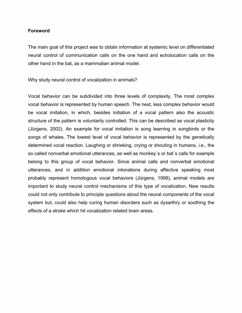
Macaca mulatta using electrical stimulation. Jürgens and Ploog carried out another very
detailed and systematic brain-stimulation study in which the entire brain of the squirrel
monkey was explored for sites where electrical microstimulation yield vocal responses
(Jürgens & Ploog, 1970). They found that the vocalization-eliciting substrate was not
restricted to a small discrete area but rather occupied a widely branching system,
reaching from the forebrain down to the lower brainstem (FIG. 2).
PAG
FTr
cs
PAG
an
ccgc
fr cb
f
aa
hip
cackle
a b c d
kHz12
4
growl
a b
4
12
kHz
csfr
st
aa
cc
gccb
st
anf
4
12
kHz
a b c d
500 msshriek
caw groan
PAG
fr
stf
cc
gc
gpo
aa
cs
cacb
a b a b c d
trill chirpkHz
4
12
cc
grPAG
gc
f
gpopy
oi
cb
cs
IG. 2: The neural substrate for vocalizations in the squirrel monkey. he vocally active sites are not organized in few delimited vocalization centers but rather are wide spread
eaching from the forebrain to the lower brainstem. However, the majority of the active sites belong to the

INTRODUCTION 12
limbic system. The calls, represented as spectrograms (a-d) below each drawing can be elicited in the black areas of the drawings. It can be seen that different types of calls are linked to different brain areas, with the exception of the periaqueductal gray in which virtually all call types can be triggered. Abbreviations: aa, anterior amygdala; an, nucleus anterior thalami; ca, commissura anterior; cb, cerebellum; cc, corpus callosum; cs, colliculus superior; f, fornix; fr, formatio reticularis; gc, cingulate gyrus; gpo, griseum pontis; gr, gyrus rectus, hip, hippocampus; oi, inferior olive; PAG, periaqueductal gray; py, tractus corticospinalis; st, stria terminalis. Adapted, from Jürgens (1998).
According to the authors, the majority of the vocalization-eliciting structures was found
within the limbic system (see also BOX II, this page): septum, nucleus acumbens,
preoptic area, hypothalamus, midline thalamus, amygdala and the bed nucleus of the
stria terminalis, where different call types could be elicited. Only within the
periaqueductal gray of the midbrain (see also BOX III, page 19) all types of calls could
be triggered (Jürgens & Ploog, 1970).
BOX II: What is the limbic system? (findings in humans) The majority of the structures which belong to the limbic system are paleocortical and archicortical. The notion was first created to describe areas covering the corpus callosum, nucleus caudatus, putamen, nucleus subthalamicus and substantia nigra (basal ganglia) and the diencephalon like a “limbus”. The modern anatomy describes a few more areas for the limbic system: hippocampus with fornix, gyrus cinguli, gyrus parahippocampalis with regio entorhinalis, gyrus dentalis, gyrus dentatus, corpus amygdaloideum, corpus mamillare. The definition of the limbic system represents neither a functionally combined central region nor topographically linked areas within the brain. The classic and still widespread assumption that the limbic system serves as a center for “self-maintenance” and reproduction is insufficient. Although the limbic system influences the “self maintenance” by controlling psychic parameters as well as reproduction through modulation of sexual and vegetative parameters, it is incorporated in numerous emotional, intellectual and instinct-bound performances without being misunderstood as the one and only origin of these abilities. Interestingly, vocalizations elicited within the anterior cingulate cortex in squirrel monkeys can be abolished by procaine and kynurenic acid injections into the PAG, leading Jürgens to the statement that there exists a limbic vocal control pathway at midbrain level from the cingulate cortex via the PAG for nonverbal emotional vocal utterances (Jürgens & Zwirner, 1996).
INTRODUCTION 13
2.1 Vocalizations as primary and secondary reactions to stimuli
The limbic vocalization-eliciting sites are characterized by relatively long latencies of
vocal responses and fast habituation to repetitive stimulation. This suggests that the
elicited vocalizations are rather secondary reactions to stimulus-induced motivational
changes than direct motor responses (Jürgens, 1998). This hypothesis was tested by
electrical self-stimulation experiments. It was assumed that there is a high probability
that electrically-induced motivational changes strong enough to induce vocalization as a
secondary reaction do have positive or negative reinforcing qualities (Jürgens, 1976). An
increase in motivation without the possibility of performing the adequate consummatory
act (no goal object existing) would be negatively reinforcing, a decrease in motivation
(drive reduction) would be positively reinforcing. The self-stimulation experiments in
squirrel monkeys showed two types of vocalization-producing brain areas, i.e. those
where electrically induced vocalizations were independent of the accompanying
reinforcement and brain areas where vocalizations and reinforcement were correlated.
The first group included the anterior cingulate gyrus, the adjacent supplementary motor
area, gyrus rectus, ventro-medial edge of the capsula interna and the caudal
periaqueductal gray with the adjacent parabrachial region. The second group included
the caudatum, septum, substantia innominata, amygdala, inferior thalamic peduncle,
stria terminalis, midline thalamus, ventral and periventricular hypothalamus, substantia
nigra, rostral periaqueductal gray, dorso-lateral midbrain tegmentum and lateral medulla.
The author interpreted vocalization areas which did not show a correlation between the
elicited call and accompanying reinforcement as primary vocalization areas and areas
which show such a correlation as secondary vocalization areas. The anterior cingulate
gyrus with a dorsal extension into the supplementary motor area and the caudal
periaqueductal gray including the adjacent parabrachial region fulfilled best the criteria
for primary vocalization areas (Jürgens, 1976).
INTRODUCTION 14
2.2 The anterior cingulate cortex – its role in vocalization Electrical stimulation of the anterior cingulate cortex produces vocalizations in non-
human mammals such as squirrel monkey (Jürgens & Ploog, 1970) but not in man. In a
vocal operant conditioning study in macaques, in which the animals had to perform a
specific call in order to obtain food, the animals could no longer execute the task after
bilateral removal of the anterior cingulate cortex (Sutton et al., 1974). Interestingly this
operation did not interfere with a slightly altered conditioning task where the animals had
to activate a lever without vocalizing to receive food. The involvement of the anterior
cingulate cortex in the performance of such volitionally controlled vocalizations is
supported by electrophysiological studies. Recording of field potentials in the anterior
cingulate cortex was only possible while monkeys vocalized as an operant response to
obtain food while vocal protest reaction after withdrawal of the food could not be
connected with activity in this cortical area (Gemba et al., 1995). Single-unit recording
experiments in macaques yield activity changes of neurons in this area during
vocalization in a vocal operant conditioning experiment (West & Larson, 1995).
The anterior cingulate cortex seems to be involved in voluntary initiation and
suppression of emotional utterances (Jürgens, 2002), at least in monkeys, although the
role of this area does not seem to be limited to nonhuman primates (Jürgens, 1998).
After a bilateral lesion in the anterior cingulate cortex due to occlusion of the ascending
branches of both anterior cerebral arteries a human patient showed akinetic mutism.
This patient recovered to normal with respect to speech (articulation, phonation,
semantics and grammar) but remained deficient in emotional intonation (Jürgens & von
Cramon, 1982; Jürgens, 1998).
2.3 The periaqueductal gray The caudal periaqueductal gray has been probed in different species and demonstrated
that vocalizations can be triggered within these regions: fish (Magoun et al., 1937), cat
(Hunsberger, 1956), bird (Brown, 1971; Delius, 1965), frog (Demski & Gerald, 1974;
Schmidt, 1966), lizard (Kennedy, 1975), squirrel monkey (Jürgens, 1979; Jürgens & Lu,

INTRODUCTION 15
1993), rhesus monkey (Larson & Kistler, 1984), rat (Yajima et al., 1980), guinea pig
(Martin, 1976) and bat (Suga and Yajima, 1988; Valentine et al., 2002).
Reasons to attribute an exceptional role to the periaqueductal gray among the vocally-
active brain structures for the control of vocalizations were:
• The shortest latencies for vocalizations are found in the periaqueductal gray.
• The highest number of different vocalization types can be elicited in the
periaqueductal gray.
• All vocalizations persist until the end of stimulation without habituation to the
stimulus.
• There are several reports of transitory or even permanent mutism after
periaqueductal lesions (Adametz & O´Leary, 1959; Hundsberger, 1956; Skultety,
1965).
Mammalian vocalizations in general incorporate a precise coordination of respiratory
and laryngeal movements. The motoneurons responsible for control of respiratory
movements are located in the anterior horn of the cervical, thoracic and upper lumbar
spinal cord, those controlling the laryngeal muscles are found in the nucleus ambiguus
and neurons responsible for the control of articulatory movements are localized in the
trigeminal motor nucleus, facial nucleus, rostral nucleus ambiguus, hypoglossal nucleus
and anterior horn of the upper cervical spinal cord (Jürgens & Ploog, 1981). At midbrain
level, transection experiments in cats and monkeys have shown that animals become
mute by lesions including the periaqueductal gray and the adjacent tegmentum (Jürgens
& Ploog, 1981) while destruction of other midbrain structures had no effect on
vocalization.
2.3.1 The vocalization controlling system is organized hierarchically
Further findings suggested a hierarchically organization of the vocalization controlling
system including the periaqueductal gray. Vocalizations elicited from the periaqueductal
gray where not affected by bilateral lesions in vocalization-eliciting areas rostral to it, but
where abolished by lesions in the dorso-lateral pons and the ventro-lateral medulla
(Jürgens, 1979).

INTRODUCTION 16
gc
cc
lnabna
f
anlth
sensory input PAG
cs
ci
csp
cics
PAG
csp
ntnf
oinc na
nhns
FIG. 3: Hierarchic central control of vocalizations in monkeys. The anterior cingulate cortex represents the highest level of this organization. The periaqueductal gray receives input not only from this cortical site but also from motivation-controlling brain sites such as hypothalamus, amygdala and thalamus and from sensory pathways. The level of motor coordination (lowest level) is represented by phonatory motoneurons and interneurons in the lower brainstem and the spinal cord. Abbreviations: anl, ansa lenticularis; PAG, periaqueductal gray; bna, nucleus basalis amygdalae; ci, colliculus inferior; cs, colliculus superior; csp, pyramidal tract; gc, cingulate gyrus; lna nucleus lateralis amygdalae; na, nucleus ambiguus, nc, nucleus cochlearis; nf, nucleus facialis; nh, nucleus hypoglossus, ns; nucleus solitarius; nt, motor nucleus of trigeminus; oi, inferior olive; th, thalamus. Adapted, from Jürgens (Jürgens, 1988).
INTRODUCTION 17
The periaqueductal gray receives input from several regions: Visual input reaches the
region from the deep layers of the superior colliculus (Meller & Dennis, 1986), acoustic
input comes from the inferior colliculus (Jürgens & Ploog, 1981; Mantyh, 1982), visceral
input comes from the solitary tract nuclei (Bandler & Tork, 1987) and somatosensory
input comes from the dorsal horn of the spinal cord and the spinal trigeminal nucleus
(Harmann, 1988). These inputs may induce vocalization rather directly through external
stimuli, e.g. pain-shrieking as response to a noxious stimulus (Jürgens, 1991).
In addition to sensory input, there is input from motivation-controlling limbic structures
such as the amygdala, septum, hypothalamus and midline thalamus serving probably to
modulate the vocal reactivity to external stimuli according to prior experience and
momentary emotional state (Jürgens, 1991; Jürgens & Ploog, 1981). Connections are
also found between the anterior cingulate cortex representing the highest level of the
vocalizing system and the periaqueductal gray (FIG. 3). While this cortex site is
unnecessary for (non-spontaneous) vocal reactions, it is needed for the voluntary control
of vocalization (Jürgens & Ploog, 1981).
On the output side, the periaqueductal gray vocalization center is connected to the
reticular formation around the nucleus retroambiguus which itself is connected with all
phonatory motor nuclei (Holstege, 1989; Jürgens & Pratt, 1979; Thoms & Jürgens,
1987). More specifically, the periambigual reticular formation projects to the trigeminal
motor nucleus responsible for jaw control, to the facial nucleus responsible for lip
movements, to the hypoglossal nucleus responsible for tongue movements, to the
nucleus ambiguus controlling the vocal folds and to the thoracic and upper lumbar
ventral horn containing the expiratory motoneurons (Jürgens, 1994).
A potential pathway where the periaqueductal gray might exert its vocalization control on
lower brainstem regions was traced in a combined stimulation/lesion study (Jürgens &
Pratt, 1979). Lesions capable of blocking periaqueductally elicited vocalization could be
traced up from the caudo-lateral periaqueductal gray into the lateral tegmentum
underneath the inferior colliculus, from here, along the medial edge of the lateral
lemniscus into the ventro-lateral pons and from there into the periambigual reticular
formation (Jürgens, 2002).

INTRODUCTION 18
2.3.2 The periaqueductal gray as a pattern generator of vocal patterns
Two subdivisions of the reticular formation, namely the parvocellular and the central
nuclei of the reticular formation are thought to be involved in vocal pattern generation
(Dusterhoft et al, 2000) rather than the periaqueductal gray itself as outlined by other
authors (Holstege, 1989; Larson, 1991). In his anatomical study, Holstege found, that a
specific cell group of the caudal periaqueductal gray and of the tegmentum lateral to it
projects bilaterally to the nucleus retroambiguus. Neurons in this nucleus projected to
the motoneuronal cell groups innervating mouth-opening and perioral muscles as well
as to motoneurons innervating the pharynx, soft palate, and tongue and probably to the
larynx (Holstege, 1989). Single unit recordings in monkeys have revealed vocalization-
correlated periaqueductal activity (Larson & Kistler, 1984) and the activity of some cells
has been reported to be correlated with EMG activity of specific laryngeal muscles.
Similar results where obtained by Zhang and coworkers (1994). They recorded
electromyographic (EMG) changes in respiratory, lanryngeal (and therefore
vocalizations) and oral muscles evoked by microinjections of D,L-homocysteic acid
(excitatory amino acid agonist) injected into the periaqueductal gray. Different
vocalizations/muscle patterns where linked to individual periaqueductal sites and they
concluded that the periaqueductal gray contains topographically separable groups of
neurons coordinating laryngeal, respiratory and oral muscle patterns. Organization of
the periaqueductal gray represents muscle patterns rather than representation of
individual muscles (Zhang et al., 1994).
These findings led to the assumption that motor coordination of vocalizations is located
in the periaqueductal gray.
In contrast to this, stand results from Düsterhoft and coworkers (2000). They found that
the majority of periaqueductal vocalization-related cells fired before, but not during
vocalization, while only a few cells in the parvocellular nucleus and no cells in the central
nucleus of the reticular formation acted this way (Düsterhoft et al., 2000). In the
periaqueductal gray no cell was found that changed its discharge rate in rhythm of
frequency modulation whereas changes in discharge rates where found in the
parvocellular nucleus and the central nucleus of the reticular formation (Düsterhoft et al.,
2000).

INTRODUCTION 19
BOX III: The midbrain periaqueductal gray matter (PAG) The term “periaqueductal” is used for the ventricular central gray around the midbrain aqueduct distinguishing it from the rostrally continuing periventricular gray matter surrounding the third ventricle in the hypothalamus and thalamus and from the caudally bordering periventricular gray matter which constitutes the ventral and ventro-lateral border of the forth ventricle in the dorsal pons. Functionally and anatomically, the Edinger-Westphal nucleus, the nucleus of Darkschewitsch and the interstitial nucleus of Cajal (oculomotor and trochlear nuclei) and the dorsal raphe nucleus are not integrated into the PAG although they provide a major part of the gray matter ventral to the midbrain aqueduct (Bandler et al., 1991). The midbrain tegmentum laterally to the PAG is usually considered to be separated from the PAG by tectobulbar and tecto-spinal fibers and the fibers of the mesencephalic trigeminal tract. In the literature some disagreement can be found on how to best subdivide the PAG. The organizational classification of the periaqueductal gray shown in FIG. 4 follows the suggestion of Bandler and coworkers and is strictly based on anatomical and functional specificity expressed in longitudinal neuronal columns along the rostro-caudal axis of the PAG (Bandler et al., 1991). This work strictly follows this classification.
dk ew
oc
dl
dl
dm
lt
vl
dl
aq
aq
PAG
dr
dr
rostral
caudal
FIG. 4: The longitudinal columnar organization of the periaqueductal gray. Top drawing) The dorso-lateral column is significant throughout the rostral and intermediate periaqueductal gray. A large number of GABA immunoreactive cells and higher densities of kainate binding sites than in other parts of the rostral and intermediate periaqueductal gray can be found here. Very little is known about the function of this column. Middle drawing) The dorso-medial column is represented throughout the entire rostro-caudal extend. Other than the dorso-lateral column heavy projections to the caudal brainstem can be found, although not much is known about its functional significance. Bottom drawing) Lateral and ventro-lateral column. The lateral column extends throughout the rostral and intermediate thirds of the region. Autonomic and somatomotor activities together with specific forms of defense behavior are associated with this region. Functional studies support the existence of a ventro-lateral column. Injections of excitatory amino acid agonists in this region evoke effects opposite of those evoked from the lateral column (decreased autonomic and somatosensory activity). Abbreviations: aq, aqueduct; dk, nucleus of Darkschewitsch; dl, dorso-lateral column; dm, dorso-medial column; dr, dorsal raphe nucleus;
INTRODUCTION 20
ew, nucleus of Edinger Westphal; lt, lateral colum; oc, oculomotor nucleus; PAG, periaqueductal gray; vl, ventro-lateral column. Adapted, from Bandler (1991).
Additional support against the assumption that the periaqueductal gray serves as a
center for vocal pattern coordination comes from earlier experiments by Jürgens and
Pratt (1979) in which they showed, that electrical or pharmacological stimulation of the
periaqueductal gray yields natural vocalizations, whereas stimulation of the reticular
formation yields abnormal vocalizations. The normal acoustic structure of
periaqueductally elicited calls (in contrast to artificial character of reticular elicited calls)
was interpreted as the result of an indirect activation of the vocal pattern coordination
mechanism. The periaqueductal gray probably acts more as a vocal gating mechanism
rather than a vocal pattern generator (Düsterhoft er al., 2000).
2.3.3 The final common pathway for vocalization
Jürgens describes a hierarchically organized vocal pathway including the projection from
the periaqueductal gray towards the neural network consisting of the parvocellular and
dorsal medullary reticular formation, nucleus retroambiguus and solitary tract nucleus
(Jürgens, 2002), for details see chapter 2.3.1 and 2.3.2. In his opinion the
periaqueductal gray exerts a gating function in controlling the release of vocal patterns.
Holstege proposed that vocal pattern generation takes places within a final common
pathway for vocalization originating from the periaqueductal gray and projecting to the
nucleus retroambiguus (Holstege, 1989). This is an alternative view of the proposal that
the PAG regulation of vocalization is achieved somewhat diffusely across the
parvocellular reticular formation of the lower brainstem (Jürgens & Pratt, 1979; Thoms &
Jürgens, 1987). In his anatomical study, Holstege found bilateral projections from the
lateral part of the caudal periaqueductal gray to the nucleus retroambiguus. Since this
nucleus projects to motoneuronal cell groups innervating mouth-opening and perioral
muscles, intercostals and abdominal muscles as well as to motoneurons innervating the
pharynx, soft palate and tongue (all these muscles are active in vocalization), he

INTRODUCTION 21
concluded that the projection from the periaqueductal gray controls via the nucleus
retroambiguus and this final common pathway the pattern generation for vocalization.
Vanderhorst and coworkers clearly traced projections from the periaqueductal gray to
the nucleus retroambiguus in rhesus monkeys supporting the results on a
periaqueductal-retroambiguus pathway. Their results showed that a compact group of
neurons in the medial part of the lateral periaqueductal gray sends a dense and direct
projection to the nucleus retroambiguus (Vanderhorst er al., 2000).
lateral PAG column
FIG. 5: The finaPossible represeTop drawing) P
lateral PAG column
PAG-NRA pathway
NANRA
obex level
spino-medullaryjunction
PRF
cb
CT TA EO, IO, IIC
sound production
diffuse pathway
NANRA
obex level
spino-medullaryjunction
spinal cordventral horn
PRF
cb
CT TA
l common pathway for vocalization. ntations of output projections from the periaqueductal gray. Adapted, from Zhang (1995). rojection from the periaqueductal gray towards reticular interneurons in the vicinity of
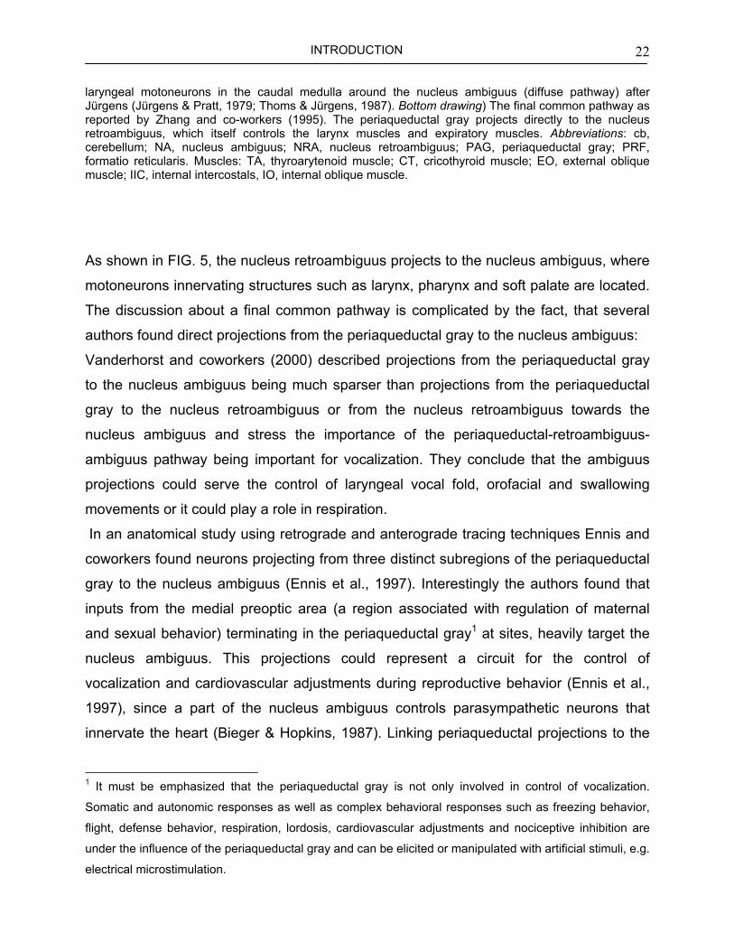
INTRODUCTION 22
laryngeal motoneurons in the caudal medulla around the nucleus ambiguus (diffuse pathway) after Jürgens (Jürgens & Pratt, 1979; Thoms & Jürgens, 1987). Bottom drawing) The final common pathway as reported by Zhang and co-workers (1995). The periaqueductal gray projects directly to the nucleus retroambiguus, which itself controls the larynx muscles and expiratory muscles. Abbreviations: cb, cerebellum; NA, nucleus ambiguus; NRA, nucleus retroambiguus; PAG, periaqueductal gray; PRF, formatio reticularis. Muscles: TA, thyroarytenoid muscle; CT, cricothyroid muscle; EO, external oblique muscle; IIC, internal intercostals, IO, internal oblique muscle.
As shown in FIG. 5, the nucleus retroambiguus projects to the nucleus ambiguus, where
motoneurons innervating structures such as larynx, pharynx and soft palate are located.
The discussion about a final common pathway is complicated by the fact, that several
authors found direct projections from the periaqueductal gray to the nucleus ambiguus:
Vanderhorst and coworkers (2000) described projections from the periaqueductal gray
to the nucleus ambiguus being much sparser than projections from the periaqueductal
gray to the nucleus retroambiguus or from the nucleus retroambiguus towards the
nucleus ambiguus and stress the importance of the periaqueductal-retroambiguus-
ambiguus pathway being important for vocalization. They conclude that the ambiguus
projections could serve the control of laryngeal vocal fold, orofacial and swallowing
movements or it could play a role in respiration.
In an anatomical study using retrograde and anterograde tracing techniques Ennis and
coworkers found neurons projecting from three distinct subregions of the periaqueductal
gray to the nucleus ambiguus (Ennis et al., 1997). Interestingly the authors found that
inputs from the medial preoptic area (a region associated with regulation of maternal
and sexual behavior) terminating in the periaqueductal gray1 at sites, heavily target the
nucleus ambiguus. This projections could represent a circuit for the control of
vocalization and cardiovascular adjustments during reproductive behavior (Ennis et al.,
1997), since a part of the nucleus ambiguus controls parasympathetic neurons that
innervate the heart (Bieger & Hopkins, 1987). Linking periaqueductal projections to the
1 It must be emphasized that the periaqueductal gray is not only involved in control of vocalization.
Somatic and autonomic responses as well as complex behavioral responses such as freezing behavior,
flight, defense behavior, respiration, lordosis, cardiovascular adjustments and nociceptive inhibition are
under the influence of the periaqueductal gray and can be elicited or manipulated with artificial stimuli, e.g.
electrical microstimulation.

INTRODUCTION 23
nucleus ambiguus with the control of vocalization only on the base of neuroanatomical
results, as done by Dennis and coworkers (see above) does not seem to be very
expressive. Neuroanatomical studies must be combined with electrical microstimulation
experiments and selective lesion experiments to establish a functional involvement of
particular areas in the control of vocalization.
3. Role of the periaqueductal gray in vocalization – a brief summary Although the contribution of the periaqueductal gray to the control of different behaviors
is not clear in detail, there is current agreement on its crucial role in control of
vocalization:
• Vocalizations can be elicited in the periaqueductal gray in numerous animals and
man: in the rat (Yajima et al., 1980), guinea pig (Martin, 1976), bat (Schuller &
Radtke-Schuller, 1990; Suga et al., 1973; Valentine et al., 2002), cat
(Hunsberger, 1956), squirrel monkey (Jürgens & Ploog, 1970), rhesus monkey
(Magoun et al., 1937), gibbon (Apfelbach, 1972), chimpanzee (Brown, 1915) and
man (Sem-Jacobsen & Torkildsen, 1960).
• vocalizations elicited in the periaqueductal gray generally resemble natural calls
(Jürgens, 1994; Vanderhorst et al., 2000).
• The obvious lack of correlation between vocalization and emotional reaction
indicates that periaqueductally evoked vocalizations do not represent secondary
reactions to stimulation-induced motivational changes but are triggered more
directly (Jürgens, 1994; Jürgens, 2002).

INTRODUCTION 24
• The periaqueductal gray displays very short latencies for vocalization when being
stimulated with electrical microstimulation. In the squirrel monkey latencies down
to 50 ms where found within the periaqueductal gray (Jürgens, 2002).
• While in most other vocalization-eliciting areas repetitive stimulation causes a
vanishing of the vocal reaction, periaqueductally evoked vocalization lasts for the
whole duration of the stimulation showing no habituation to the stimulus (Jürgens,
1994; Jürgens, 2002).
• Lesions invading the periaqueductal gray can cause mutism without general
akinesia. Lesions do not have to destroy the periaqueductal gray completely, as
partial lesion leads to a loss of some vocal reactions, leaving others intact
(Jürgens, 1994).
• Single-unit recording studies in the periaqueductal gray showed vocalization-
correlated activity changes in several animals: in the cat (Adams, 1968), bat
(Suga & Yajima, 1988), squirrel monkey (Düsterhoft et al., 2000) and macaque
spec. (Larson & Kistler, 1984). A systematic relationship between periaqueductal
activity changes and fundamental frequency changes of calls has not been
demonstrated so far (Düsterhoft et al., 2000). Most of the cells found in this study
did not even differentiate between individual call types.
• Afferent projections of relevance for vocal behavior reach the periaqueductal gray
from essentially two groups of structures (Jürgens, 1994): The first group, when
electrically stimulated, leads to the production of vocalizations. These structures
all belong to the limbic system (see FIG. 2 fore details) depending upon an intact
PAG for their production of vocalization (Fernandez de Molina & Hunsperger,
1962; Jürgens, 1982; Jürgens & Pratt, 1970; Jürgens & Pratt, 1979). The second
group consists of sensory relay structures. They provide the periaqueductal gray
with visual, auditory, gustatory and somatosensory information (Jürgens, 1994).
Massive efferent projections where found from the periaqueductal gray to the
reticular formation which itself is connected with all phonatory motor nuclei

INTRODUCTION 25
(Holstege, 1989; Jürgens & Pratt, 1970; Thoms & Jürgens, 1987). Other efferents
from the periaqueductal gray reach the nucleus retroambiguus, a group of
premotor neurons (Vanderhorst et al., 2000) and the nucleus ambiguus.

INTRODUCTION 26
4. Vocalization in bats
Bats use vocalizations for communication and for echolocation. Like in any other
mammal, social calls are used for intraspecific interactions. Echolocation in general is
used for orientation gathering information about the environment, despite the possible
situation that echolocation calls of one bat could serve another bat in a non-intentional
communicative manner to indicate potential food sources (Fenton, 1984). Echolocation
capability was developed independently from some taxa among the following groups:
shrews, birds, tenrecs, cetaceans, rodents, bats (Busnel & Fish, 1980) and seals
(Renouf & Davis, 1982). While most of these echolocators use brief broadband clicks,
which are unstructured and easy to produce, microchiroptera produce calls structured in
time (Fenton, 1984).
Communication calls and echolocation calls can clearly be separated from each other
acoustically by their specific spectral patterns. Although clearly distinguishable, under
physiological aspects the production of echolocation calls must access the same
laryngeal and expiratory components as the production of communication calls. The
current discussion on control of vocalization together with the functional brain areas
involved in it could take advantage from results found in bats.
4.1 Echolocation calls can be elicited in the anterior cingulate cortex Since the beginning of systematic research on vocalization related sites within the
vertebrata central nervous system in the late 1960s, a variety of different animals had
been under investigation (see chapter 2 for an overview). Interestingly, bats which have
a very specialized vocalization system were never in the center of attention. These
animals utter echolocation calls and species-specific communication calls produced by
the larynx while shaping by the vocal tract is very limited (in contrast to humans where
articulation is the main shaping mechanism (Ploog, 1988)). Production of these different
types of calls in bats presumably shares the same neural control centers as in higher
mammals (Schuller & Radtke-Schuller, 1990).

INTRODUCTION 27
After a few experiments using electrical stimulation to explore vocally active sites within
the brain of bats were published (Suga et al., 1973; Suthers & Fattu, 1982), it was not
until 1987 that a rather systematic work was done on the elicitity of vocalizations in the
anterior cingulate cortex of bats (Gooler & O´Neill, 1987). The authors mention that
echolocation calls, virtually indistinguishable from natural calls can be elicited by
microstimulation within this region. Posterior to this region audible calls resembling
social calls can also be elicited with microstimulation. From there findings they finally
concluded that vocalization, if uttered in the purpose of social communication [this
applies for bats as well as for higher primates like monkeys] or for foraging and
navigation in echolocating bats, must be a highly motivated behavior (Gooler & O´Neill,
1987).
4.2 The periaqueductal gray in bats
For the periaqueductal gray it was shown by stimulation experiments that echolocation
calls as well as calls of non-echolocation type (Suga et al., 1973; Valentine et al., 2002)
could be elicited in bats. Suga and coworkers elicited sounds similar to orientation
signals in the genus Myotis from the lateral part of the periaqueductal gray (Suga et al.,
1973). In Pteronotus parnellii and Pteronotus suapurensis, stimulation of the
periaqueductal gray yield vocalizations indistinguishable from natural orientation sounds
and for Eptesicus fuscus and Noctilio leporinus, electrical stimulation elicited
vocalizations similar to natural orientation sounds (Suga et al., 1973). The authors also
reported that stimulation of the ventro-medial area of the periaqueductal gray in Myotis
elicited vocalizations quite different from orientation sounds, but these vocalizations
were in association with various large body movements. For Eptesicus fuscus it was
shown by Valentine and coworkers (2002) that electrical stimulation of the
periaqueductal gray leads to vocalizations comparable to communication signals emitted
by this species.
But a systematic work on the elicitation of different types of communication calls on the
one hand and echolocation calls on the other hand applying the same criteria on

INTRODUCTION 28
triggered vocalizations as it was done for higher primates (short latencies of triggered
calls, no habituation to stimuli, low thresholds) is not available to date.
4.3 Other neuronal substrates in bats yielding echolocation calls On the search of brainstem areas where electrical microstimulation could trigger natural
echolocation calls, three areas were found where vocalizations could be elicited without
temporal or spectral distortions (Schuller & Radtke-Schuller, 1990): The deep layers of
the superior colliculus, the deep mesencephalic nucleus in the reticular formation and a
restricted area medial to the rostral parts of the dorsal nucleus of the lateral lemniscus.
To ensure that the brain areas were “specific” for the production of biosonar
vocalizations, the authors applied the following criteria:
a) Threshold currents for triggering vocalizations were below 10 µA.
b) Vocalizations corresponded to spontaneously produced echolocation calls with
respect to spectral and temporal patterns.
c) No body movements other than of ear, mouth or nose accompanied
vocalizations.
d) Latency between onset of vocalization and stimulus train was stable and below
100 ms.
e) Vocalizations were not uttered as a consequence of stimulus-induced arousal of
the animal.
The shortest latencies (mean latency: 30.9 ± 9.1 ms s.d.) for eliciting echolocation calls
at lowest thresholds (< 10 µA) and/or pinna movements not accompanied by any
arousal of the animal where found in the area medial to the dorsal nucleus of the lateral
lemniscus (Schuller et al., 1997). Strikingly, by fulfilling the above listed requirements,
this area shares most of the criteria which awarded the periaqueductal gray a crucial
role in the production of communication calls.

INTRODUCTION 29
4.4 The paralemniscal tegmental area The expression “paralemniscal tegmental area” only expresses the position of the
restricted area medial to the rostral parts of the dorsal nucleus of the lateral lemniscus
and does not describe any identified nucleus. A discussion is complicated by the fact
that different authors had different motivations to investigate this area, which led to
different functional or anatomical findings suggesting rather an aggregation of different
fields than a clearly delimited area:
On the basis of neural responses to acoustic stimuli (pure tones, frequency-modulated
sweeps and noise bursts) Covey (1993) differentiated between a dorsal paralemniscal
nuclei (DPL), located rostral to the dorsal nucleus of the lateral lemniscus and separated
it from the paralemniscal zone (PL) and the dorsal nucleus of the lateral lemniscus
(DNLL). At monaural presentation of the stimuli, most of the neurons in the DNLL and in
the PL were activated by contralateral sound, and also in the DPL all tested neurons
responded only to a contralateral sound. In a binaural testing situation (contralateral
stimulus at 20 dB above threshold, ipsilateral sound level varied) the most common
response pattern in all three areas was suppression of the contralateral response to
ipsilateral sound level increase. In the DPL, 29% of all neurons tested showed binaural
responses, in the DNLL 84% of all neurons showed binaural responses and in the PL
88% were binaural. Thus the author reserved an auditory function for the DPL.
Henkel and Edwards defined a tegmental area in the cat as the paralemniscal zone and
investigated this area under the aspect of pinna movements (Henkel, 1981; Henkel &
Edwards, 1978). In their anatomical studies using orthograde and retrograde tracers
they injected HRP (horseradish peroxidase) into pinna muscles to determine the facial
nucleus regions projecting into these muscles. Additionally brainstem regions projecting
to the facial nucleus were identified with HRP and superior colliculus projections to these
areas were identified autoradiographically. They concluded that superior colliculus
control of pinna movements is mediated by the paralemniscal zone via indirect
connections to the facial nucleus. HRP injections into the paralemniscal zone showed
also afferents from the external nucleus of the inferior colliculus and the periolivary cell
group (auditory afferents), the nucleus prepositus hypoglossus, the adjacent pontine

INTRODUCTION 30
reticular formation and the vestibular nuclei (premotor regions, among other things
involved in gaze control), the nucleus cuneiformis and the periaqueductal gray (Henkel,
1981). Since they found relatively sparse labeling in the auditory regions, Henkel
concluded that “sensorimotor” integration necessary to guide pinna movements does not
take place primarily in the lateral midbrain tegmentum and mentions that the inferior
colliculus is as likely to be the site for this kind of “sensorimotor” integration.
In research related to oculomotor questions the tegmental region was termed lateral
tegmental region (LTR), where neurons, probed in the cat responded to visual,
vestibular and auditory stimulation (Gerlach et al., 1991; Gerlach & Their, 1995). Their
anatomical studies using HRP and fluorescent tracers, injected into different sites of the
LTR, revealed afferents from the vestibular nuclei, the nucleus prepositus hypoglossus,
the superior colliculus, the periaqueductal gray and the contralateral LTR. According to
the authors, the afferents from the superior colliculus could be the source of visual
signals fed into the LTR while head movement-related information could come from the
nucleus prepositus hypoglossus and the vestibular nuclei (Gerlach & Their, 1995).
In a neuroanatomical study using Rhinolophus rouxi, Metzner (1996) found evidence
that the paralemniscal tegmentum (PL) could provide an interface between the
pathways for auditory sensory processing and for the motor control of vocalization.
WGA-HRP (wheat germ agglutinin conjugated to horseradish peroxidase), injected into
different sites of the PL, yield afferents from the dorsal nucleus of the lateral lemniscus,
the central nucleus and the rostral portion of the inferior colliculus, the lateral superior
olive, the superior colliculus and the nucleus of the central acoustic tract (all are part of
the auditory pathway). Efferents where found to the superior colliculus, the facial
nucleus and the reticular formation rostral to the nucleus ambiguus. In an
electrophysiological study the same author reported, that the activity of neurons in the
PL was correlated to sound emission and auditory stimuli differentially. Responses also
varied according to the time delay between an auditory stimulus and a preceding
vocalization, while this delay-sensitivity completely disappeared when vocalizations
were simulated acoustically. Since mainly vocalization and “echo-parameters”, which

INTRODUCTION 31
occur in Doppler-shift compensation were encoded, Metzner (1989) emphasizes the role
of the PL in this special behavior.
Schuller and coworkers (Schuller & Radtke-Schuller, 1990; Schuller et al., 1997)
delimited the paralemniscal area in Rhinolophus rouxi using the ability to electrically
elicit vocalizations (See chapter 4.3 for details). In a combined electrophysiological and
neuroanatomical study, Schuller and coworkers found similar results for Pteronotus p.
parnellii (Schuller et al., 1997). The paralemniscal tegmental area (PLA) was functionally
identified as location, where vocalizations could be elicited at thresholds below 10 µA.
The authors describe three different sizes for neurons within the PLA (medium
elongated cells, small elongated cells and small peripheral cells) and found that
stimulation in the PLA was effective for eliciting vocalizations in an area which contains
the medium elongated cells. This area was restricted to a zone ventral to the inferior
colliculus. Ventrally it was bordered by the superior cerebellar peduncle and laterally by
the nuclei of the lateral lemniscus. Its rostral limit reached the substantia nigra (Schuller
et al., 1997). Stimulations of this area elicited vocalizations indistinguishable from
spontaneously uttered echolocation calls at very short latencies (25 – 30 ms) without
any motor activity except pinna movements. By placing WGA-HRP into vocally active
sites of the PLA they found as main afferent connection projections from the superior
colliculus. Using this input, the tegmental area might have motor controlling functions for
pinna movements via efferent projections to the facial nucleus (Schuller et al., 1997).
Additionally, auditory and visual information could be fed into the paralemniscal
tegmental area after being processed across the superior colliculus (Schuller et al.,
1997) and vestibular informations could come from afferent projections originating from
the nucleus prepositus hypoglossus. Input also came from the substantia nigra
[functional part of the basal ganglia, motor intention], the putamen [part of the striatum, a
nucleus of the basal ganglia, modulation of motor impulses], the reticular formation
[numerous functions such as control of respiration, circulation, vomiting, extrapyramidal
motor pathway] and the contralateral paralemniscal tegmental area (Schuller et al.,
1997).
On the efferent side projections of the tegmental area could be traced towards the
putamen, the nucleus accumbens [part of the striatum, supposed to represent an

INTRODUCTION 32
interface for the converting of motivation into action] the substantia nigra, the pretectal
area [senso-motoric integration, (Nixdorf, 2003)] and the superior colliculus (Schuller et
al., 1997). Efferent projections from the tegmental area also reached the cuneiforme
nucleus, the parabrachial region and the periaqueductal gray (Schuller et al., 1997). The
authors concluded that the PLA has a potential sensory-motor function: On the sensory
side the PLA receives information coming from superior colliculus (visual and auditory)
and from the nucleus prepositus hypoglossus (vestibular). Motor involvement in
vocalization and pinna movements could be realized via the nucleus facialis and the
nucleus cuneiformis. The PLA does not interfere with low level motor control, but rather
might play a role in co-ordinating emission of echolocation calls or start of pinna
movements with other behaviors such as head orientation (Schuller et al., 1997).
4.5 The paralemniscal tegmental area – a brief summary The paralemniscal tegmental area has been considered from different perspectives.
Several different functional involvements have been characterized, but it seems hard to
compare the different results in terms of a uniform paralemniscal tegmental area:
The “paralemniscal zone” of the cat defined by Henkel and Edwards (see above)
corresponds partly to the connectional patterns found in bats, but the area defined with
electrical microstimulation in the bat is more restricted and only occupies the dorsal half
of the paralemniscal zone as defined by Henkel and Edwards (Schuller et al., 1997).
Discrepancies in connectional patterns between findings from Metzner (see above) and
Schuller (see above) and thus different interpretations of functional involvements of the
paralemniscal tegmental area can most probably be attributed to differences in the
location of tracer injection sites (Schuller et al., 1997).
In general, the definitions of the paralemniscal area and surrounding regions are
different in different species of bats or in different mammals so that direct comparisons
of the tegmental area have to be treated with caution.

INTRODUCTION 33
5. Central questions of this thesis
This thesis strictly follows the definition suggested by Schuller and coworkers on a
sensory-motor function of the paralemniscal tegmental area.
A direct vocal controlling pathway from the periaqueductal gray towards the nucleus
retroambiguus (as outlined by Zhang, see FIG. 5) or from the periaqueductal gray to the
reticular formation (as outlined by Jürgens, see FIG. 5) seems to be insufficient to
explain findings from Schuller and coworkers on the involvement of the paralemniscal
tegmental area in motor control of vocalizations.
This leads to the following questions:
• How does the paralemniscal area interfere with the final common pathway for
vocalization in bats?
• How does the paralemniscal area interfere with the production of echolocation
calls and communication calls in bats?
• How does the paralemniscal area and the periaqueductal gray interfere with each
other in terms of the production of echolocation calls and communication calls?
• How does the periaqueductal gray interfere with the final common pathway for
vocalization in bats?
• How does the periaqueductal gray interfere with the production of echolocation
calls and communication calls in bats?

AIMS AND ACHIEVEMETS 34
IV. Aims and achievements of the Thesis
The main intention of this Ph.D. project was to understand the involvement of the
periaqueductal gray and the paralemniscal area in the production of communication calls
and echolocation calls in the animal model Phyllostomus discolor. It is the first time that
the production mechanisms of these different types of vocalizations are investigated in
the same animal. To gain insight into mechanisms of neural control of vocal behavior at
the level of these two brain regions several approaches were used:
(1) With its rich repertoire of about 20 different social calls (Esser & Pistohl, 1998)
and at the same time its usage of stereotyped frequency modulated echolocation calls
(Rother & Schmidt, 1982), the bat Phyllostomus discolor represents an ideal animal
model for investigations on control of both types of vocalizations. Together with Andreas
Nixdorf (Ph.D. student) a preliminary stereotaxic brain atlas of this species (unpublished)
was developed in order to have stereotaxic access to brain regions.
The results are described and discussed in detail in paper 1, a brief discussion is given
in chapter V, part 1.
(2) Identification of the periaqueductal gray and the paralemniscal area in
Phyllostomus discolor was carried out electrophysiologically and neuroanatomically.
With electrical microstimulation several types of communication calls and echolocation
calls could be elicited within the periaqueductal gray and echolocation calls in the
paralemniscal area. Delimited areas of the two brain regions could be linked to individual
types of calls for the first time in this species. In histologically processed brain sections
the two brain areas could be identified and delimited towards surrounding areas.
Results are described and discussed in detail in paper 1, a brief discussion is given in
chapter V, part 1.
(3) In order to exclude activation of fibers of passage within vocally active areas of
the periaqueductal gray and the paralemniscal area as source of vocal responses, kainic
acid (GLU agonist) was locally applied using microdialysis. Communication calls could

AIMS AND ACHIEVEMETS 35
be elicited in the periaqueductal gray, and echolocation calls could be triggered in the
paralemniscal area upon pharmacological stimulation, which demonstrated that
stimulation of neurons and not fibers of passage are responsible for the vocal
responses. Respiration is closely linked to vocalization and influenced by the stimulation
of the two areas.
Results are described and discussed in detail in paper 1, a brief discussion is given in
chapter V, part 2.
These results indicated that descending control for communication calls and
echolocation calls seem to differ in its functional organization. The periaqueductal gray
and the paralemniscal area may interact differentially with the final common pathway
controlling vocalization.
(4) In order to test for the possibility of differentiated neural pathways for vocal
control, electrodes for chronic electrical microstimulation were developed and implanted
stereotaxically within the periaqueductal gray at sites, where echolocation calls or
communication calls could be elicited. Simultaneously, the paralemniscal area was
reversibly blocked by kynurenic acid (GLU antagonist). The results showed that
periaqueductally induced echolocation calls depend on an intact paralemniscal area
contralateral to the stimulation site. The emission of communication calls upon electrical
stimulation within the periaqueductal gray cannot be influenced by such blockades of the
paralemniscal area.
Results are described and discussed in detail in paper 2, a brief discussion is given in
chapter V, part 3.
(5) Tracer studies with WGA-HRP in vocally active sites of the periaqueductal gray
showed projections towards the region of the nucleus ambiguus/retroambiguus complex
without well defined terminal endings in this area. The nucleus ambiguus was identified
by its cholinergic properties using the AChE staining procedure.

AIMS AND ACHIEVEMETS 36
Results are described and discussed in detail in paper 2, a brief discussion is given in
chapter V, part 4.
The results can be summarized as follows: A vocal pathway leading from the
periaqueductal gray directly to the nucleus ambiguus cannot explain the involvement of
the paralemniscal area on the production of echolocation calls. Although direct
connections between the periaqueductal gray and the paralemniscal area could not be
demonstrated, the physiological experiments suggest a functional connection of these
two brain areas, at least for the production of echolocation calls.
It can be concluded that the paralemniscal area is essential for the production of
periaqueductally induced echolocation calls but not for particular types of communication
calls. The control of particular types of communication calls on the one hand and
echolocation calls on the other hand may be realized via different vocal pathways being
more complex than the direct periaqueductal-nucleus retroambiguus pathway suggested
in the literature.
The periaqueductal gray and the paralemniscal area do interact differentially with the
final common pathway for vocalization.

DISCUSSION 37
V. Discussion
(1) The relative location of the periaqueductal gray in Phyllostomus discolor is similar
to what is found in other mammals like rats (Van Bockstaele et al., 1991) or other bats
(Suga et al., 1973). The region of the paralemniscal area in Phyllostomus discolor is
similar to what is found in other bats such as Rhinolophus rouxi (Metzner, 1993; Radtke-
Schuller, Anat. Anst. LMU, Munich, Germany, brain atlas, unpublished) and Myotis
montivagus (Baron et al., 1996).
The periaqueductally induced vocalizations in Phyllostomus discolor can be subdivided
into echolocation calls and several types of communication calls. In behavioral
experiments it has been shown that this bat species has a repertoire of at least 20
different social calls (Esser & Pistohl, 1998). The stimulation experiments carried out in
this thesis did not yield all communication calls described by Esser and Pistohl. The
majority of periaqueductally induced social calls resemble a group of calls which was
described as aggressive calls by Esser and Pistohl. This dominance of aggressive calls
obtained in an experimental situation is confirmed by findings in cat and monkey, where
stimulation experiments also induce call types of agonistic kind more often than calls
related to other behaviors (Jürgens, 1994; Jürgens, 2002). The constraints of the
experiment could provoke an agonistic disposition of the animal encouraging
vocalizations of agonistic kind.
Periaqueductally induced echolocation calls of Phyllostomus discolor closely resemble
natural echolocation calls in their spectral composition, whereas the duration of the calls
shows a wider variability than described for spontaneous calls emitted in laboratory
(Rother & Schmidt, 1982). The natural variation of echolocation calls of free moving bats
in the field is generally larger than recorded in the laboratory (Surlykke & Moss, 2000),
reflected in longer durations, longer interpulse intervals and a greater variability in
bandwidth.
Electrical stimulation in the paralemniscal area elicits exclusively echolocation calls
showing the typical harmonic structure of Phyllostomid echolocation calls (Esser &
Daucher, 1996; Rother & Schmidt, 1982) with small variations. Since these authors

DISCUSSION 38
recorded calls from freely moving bats in the laboratory the differences in duration and
bandwidth may again not represent the real range of these parameters.
Vocalization-eliciting sites in the periaqueductal gray overlap to a large extend and are
not organized in distinct clusters for individual call types (Jürgens, 1994). Such overlap
can also be found in Phyllostomus discolor and stimulation at identical sites can result in
the emission of different call types in subsequent penetrations. Therefore different types
of calls are possibly elements of the same behavior and stimulus locations might rather
represent a behavioral context than a particular type of vocalization.
Short latencies and low thresholds together with the lack of habituation to repetitive
stimuli of periaqueductally and paralemniscally induced vocalizations support the
assumption that both areas have a relatively direct access to the descending vocal
pathway system.
(2) Kainic acid was effective in both the periaqueductal gray and the paralemniscal
area. This Glutamate agonist (selective at kainite receptors) activates specifically
neurons and not fibers of passage originating from remote brain areas.
Delayed onset of vocalization to microdialysed kainic acid may be explained with slow
diffusion dynamics of the agonist. The delayed fading and offset of vocalization after
completion of microdialysis can be explained with slow dilution of the substance within
brain tissue.
Application of kainic acid in the periaqueductal gray provokes an increase of respiration
rate starting always earlier than the onset of kainic acid-induced vocalizations. The
control of respiration rate and the triggering of vocalizations seem to depend on
separate mechanisms. An adaptation of the respiration rate could also be linked to
particular behaviors, e.g. different stages of defense (Fanselow, 1991) or hypotension
plus decreased vasoconstrictor tone to allow the animal to rest and recuperate efficiently
(Carrive, 1991) are modulated by periaqueductal gray stimulations. The periaqueductal
gray could therefore serve as an integrator between a particular behavior and the
adequate respiration rate.
The same pattern of delayed vocalization onset after respiration rate increase is found in
the paralemniscal area. Modulation of respiratory rate by the paralemniscal area may
either use a separate pathway or be a result of coactivation of periaqueductal circuits.

DISCUSSION 39
(3) Are different classes of vocalizations (communication vs. echolocation calls)
processed by separate vocal pathways in the bat? The paralemniscal area and the
periaqueductal gray are involved in vocal control, with the distinction that communication
calls cannot be elicited in the paralemniscal area, whereas echolocation calls can be
triggered in both regions.
The experiments demonstrated a functional linkage between both areas for the
production of vocalizations, at least what concerns the control of echolocation calls.
Echolocation call, but not social call emission triggered in the periaqueductal gray was
dependent on an intact contralateral paralemniscal area, which shows that there are
functional differentiations between control mechanisms for communication call and
echolocation call production.
Communication calls would be controlled by a pathway described by several authors
(Ennis et al., 1997; Zhang et al., 1994; Zhang et al., 1995). In this pathway a direct
connection from the periaqueductal gray towards the nucleus retroambiguus is
proposed. This nucleus includes premotor neurons which send projections to thoracic
and upper lumbar motoneurons (Vanderhorst et al., 2000; Vanderhorst et al., 2001)
involved in expiration, and to the nucleus ambiguus containing laryngeal and pharyngeal
motoneurons (Holstege, 1989; Vanderhorst et al., 2000; Vanderhorst et al., 2001). This
network is sufficient to explain the unimpaired production of communication calls but
cannot explain the findings that periaqueductally induced echolocation calls can be
blocked by paralemniscal inactivation. A possible “echolocation call pathway” originating
in the periaqueductal gray must therefore integrate paralemniscal activity at some stage,
which has not yet been determined.
(4) Several authors report a direct projection from the periaqueductal gray to the nucleus
retroambiguus (Gerrits & Holstege, 1996; Zhang et al., 1995), a nucleus displaying
functional connections with the rostrally adjacent nucleus ambiguus. Dense projections
from the periaqueductal gray towards the region of the nucleus ambiguus were found for
Phyllostomus discolor, although discrete terminal endings were not identified. It is
unclear whether a nucleus retroambiguus can be distinguished anatomically for this
species. Projections from the periaqueductal gray aim to this caudal region which could
correspond to the nucleus retroambiguus as defined in other mammals.
DISCUSSION 40
The preliminary data point to a direct periaqueductal / nucleus retroambiguus pathway in
Phyllostomus discolor as implementation of vocal control, for at least a subgroup of
communication calls. Other, more indirect pathways for neural control of vocalization, at
least for the control of echolocation calls, must exist as the experiment with
paralemniscal blockade suggest.
However, no direct projections from the periaqueductal gray to the paralemniscal area,
as described by other authors (Gerlach & Their, 1995; Schuller et al., 1997) could be
traced in Phyllostomus discolor so far. The reason may be that the projections of the
periaqueductal gray to the paralemniscal area are of different density in different species
and could therefore have escaped from detection in Phyllostomus discolor.
On the other hand, involvement of the paralemniscal area would not necessitate a serial
“periaqueductal – paralemniscal connection” but could influence vocal production in a
parallel way to a common vocal pathway.
VI. Summary and conclusion
Echolocation calls and communication calls can be elicited with electrical
microstimulation in the periaqueductal gray. The periaqueductal gray, involved in the
neural control of communication calls as well as echolocation calls, constitutes part of
the vocal pathway. The periaqueductal gray would contribute an integrator for particular
behaviors and its appropriate vocalizations under both conditions (echolocate and
communicate). The paralemniscal area, in terms of vocalization exclusively controls
echolocation calls. Reversible blockade of the vocally active paralemniscal area totally
blocks periaqueductally induced echolocation calls leaving communication calls
triggered in the same region unaffected. The paralemniscal area therefore seems to be
essential for periaqueductally induced echolocation calls but not for particular types of
communication calls. This suggests a differentiated organization of vocal pathways for
certain types of communication calls and echolocation calls, and suggests that the
descending vocalization system is more complex than the network connecting the
periaqueductal gray directly to the nucleus ambiguus/retroambiguus complex.

THINGS TO DO 41
VII. Things to do ... We are far from understanding neural control of vocalization in every detail. To help
putting together the pieces of the puzzle research on vocalization systems in bats could
contribute through several approaches:
Tracer experiments using anterograd and retrograde tracers injected into vocal
active sites of the periaqueductal gray and the paralemniscal area should bring more
insight into vocally relevant interconnections. Since different types of communication
calls and echolocation calls can be triggered at different sites within the periaqueductal
gray, precise applications of tracers into the individual vocally active sites could reflect
differentiated vocal pathways.
A reversible blockade of the paralemniscal area has no influence on the
production of a particular type of communication call. It would be of great interest to
know how such blockades would affect other types of communication calls. The
experiments with blockades of the paralemniscal area therefore should be continued
testing all types of communication calls potentially triggered in the periaqueductal gray
on its ability to survive such blockades. These findings could uncover whether
differentiated vocal pathways discriminate only between echolocation calls and
communication calls or whether more global aspects of vocalizations, like call structure
etc., constitute criteria for this distinction.
Single cell recordings in the paralemniscal area with simultaneous elicitation of
vocalizations in the periaqueductal gray could give insight into a possible interplay of
both areas. These recordings should also be executed in the periaqueductal gray during
electrical stimulation of the paralemniscal area. Additionally simultaneous electrical
stimulations of both structures could reveal access priorities of one or the other structure
on the control of vocalization. This set of experiments could be completed with
independent electrical stimulations in both areas together with single cell and multi unit
recordings in the nucleus ambiguus/retroambiguus complex.

THINGS TO DO 42
The findings that respiration can be influenced by stimulations in the periaqueductal gray
and paralemniscal area is another aspect of interest. Stimulation experiments in both
areas together with single cell or multi unit recordings in the pre-Bötzinger complex for
example, an area within the groups of respiratory neurons containing all classes of
neurons important for respiratory rhythm generation (Connelly et al., 1992;
Schwarzacher, 1995) should shed light on the functional interaction of respiratory and
vocal control.
On a long scale experiments with Phyllostomus discolor with its very stereotypic
echolocation calls and a variety of communication calls could help on the search of the
vocal pattern generator, one, in my opinion central questions of research on vocalization
systems.

REFERENCES 43
Reference List
Adametz,J., O´Leary,J.L. (1959). Experimental mutism resulting from periaqueductal lesions in cats. Neurology 9, 636-642
Adams,D.B. (1968). Cells related to fighting behavior recorded from the midbrain central gray neuropil of cat. Science 159, 894-896
Apfelbach,R. (1972). Electrically elicited vocalizations in the gibbon Hylobates lar (Hylobatidae), and their behavioral significance. Z.Tierpsychol. 30, 420-430
Bandler,R., Carrive,P., Dapaulis,A. (1991). Emerging principles of organization of the midbrain periaqueductal gray matter. In: Depaulis,A., Bandler,R. (Eds.), The Midbrain Periaqueductal Gray Matter; Functional, Anatomical and Neurochemical Organization. Plenum Press, New York, pp. 1-8
Bandler,R., Tork,I. (1987). Midbrain periaqueductal grey region in the cat has afferent and efferent connections with solitary tract nuclei. Neurosci.Lett. 74, 1-6
Barnes,R.D., Dorit,R.L., Walker,W.F., 1991. Receptors and Sense Organs. In: Barnes,R.D., Dorit,R.L., Walker,W.F. (Eds.), Zoology. Saunders College Publishing, Orlando.
Baron,G., Stephan,H., Frahm,H.D. (1996). Comparative Neurobiology in Chiroptera. Birkhaueser Verlag, Switzerland, Basel
Beier,E.G., Zautra,A.J. (1972). Identification of vocal communication of emotions across cultures. J.Consult.Clin.Psychol. 39, 166
Bieger,D., Hopkins,D.A. (1987). Viscerotopic representation of the upper alimentary tract in the medulla oblongata in tha cat: the nucleus ambiguus. J.Comp.Neurol. 262, 546-562
Brickner,R.M. (1940). A human cortical area producing repititve phenomena when stimulated. J.Neurophysiol. 3, 128-130
Brown,J.L. (1965). Vocalization evoked from the optic lobe of a songbird. Science 149, 1002-1003
Brown,T.G. (1915). Note on physiology of basal ganglia and midbrain of anthropoid ape especially in reference to act of laughter. J Physiol 49, 195-207
Busnel,R.G., Fish,J.F. (1980). Animal Sonar Systems. Plenum Press, New York.
Carrive,P. (1991). Functional organization of PAG neurons controlling regional vascular beds. In: Depaulis,A., Bandler,R. (Eds.), The Midbrain Periaqueductal Gray Matter; Functional, Anatomical and Neurochemical Organization. Plenum Press, New York, pp. 67-100
REFERENCES 44
Connelly,C.A., Dobbins,E.G., Feldman,J.L. (1992). Pre-Botzinger complex in cats: respiratory neuronal discharge patterns. Brain Res. 590, 337-340
Covey,E. (1993). Response properties of single units in the dorsal nucleus of the lateral lemniscus and paralemniscal zone of an echolocating bat. J.Neurophysiol. 69, 842-859
Delius,J.D. (1971). Neural substrates of vocalizations in gulls and pigeons. Exp.Brain Res. 12, 64-80
Demski,L.S., Gerald,J.W. (1974). Sound production and other behavioral effects of midbrain stimulation in a free-swimming toadfish, Opsanus beta. Brain Behav.Evol. 9, 41-59
Dusterhoft,F., Häusler,U., Jürgens,U. (2000). On the search for the vocal pattern generator. A single-unit recording study. Neuroreport 11, 2031-2034
Eibl-Eibesfeldt,I. (1973). The expressive behaviour of the deaf-and-blind-born. In: Von Cranach,M., Vine,J. (Eds.), Social communication and movement. Academic, London
Ennis,M., Xu,S.J., Rizvi,T.A. (1997). Discrete subregions of the rat midbrain periaqueductal gray project to nucleus ambiguus and the periambigual region. Neuroscie. 80, 829-845
Erickson,T.C., Woolsey,C.N. (1951). Observations on the supplementary motor area of man. Trans.Am.Neurol.Assoc. 76, 50-56
Esser,K.H., Daucher,A. (1996). Hearing in the FM-bat Phyllostomus discolor: a behavioral audiogram. J.Comp.Physiol A 178, 779-785
Esser, K.-H. and Pistohl, D. (1998). The vocal repertoire of the lesser spear-nosed bat (Phyllostomus discolor). Zoology 101(Suppl. I), 84
Fanselow,M.S. (1991). The midbrain periaqueductal gray as a coordinator of action in response to fear and anxiety. In: Depaulis,A., Bandler,R. (Eds.), The Midbrain Periaqueductal Gray Matter; Functional, Anatomical and Neurochemical Organization. Plenum Press, New York, pp. 151-173
Fenton,M. (1984). Echolocation: implications for ecology and evolution of bats. Q.Rev.Biol. 59, 33-52
Fernandez de Molina,A., Hundsperger,R.W. (1962). Organization of the subcortical system governing defence and flight reactions in the cat. J.Physiol. 160, 200-213
Gemba,H., Miki,N., Sasaki,K. (1995). Cortical field potentials preceding cocalization and influences of cerebellar hemispherectomy upon them in monkeys. Brain Res. 697, 143-151
Gerlach,I., Thier,P. (1995). Brainstem afferents to the lateral mesencephalic tegmental region of the cat. J.Comp.Neurol. 358, 219-232
REFERENCES 45
Gerlach,I., Thier,P., Koehler,W. (1991). Visual and vestibular signals in the lateral mesencephalic tegmental region of the cat. Exp.Brain Res. 85, 641-649
Gerrits,P.O., Holstege,G. (1996). Pontine and medullary projections to the nucleus retroambiguus: a wheat germ agglutinin-horseradish peroxidase and autoradiographic tracing study in the cat. J.Comp.Neurol. 373, 173-185
Gibbs,E.L., Gibbs,F.A. (1936). A purring centre in the cat´s brain. J.Comp.Neurol. 64, 209-211
Gooler,D.M., O'Neill,W.E. (1987). Topographic representation of vocal frequency demonstrated by microstimulation of anterior cingulate cortex in the echolocating bat, Pteronotus parnelli parnelli. J.Comp.Physiol A 161, 283-294
Groswasser,Z., Korn,Z., Groswasser-Reider,I., Solzi,P. (1988). Mutism associated with buccofacial apraxia and behemisheric lesions. Brain Lang. 34, 157-168
Harmann,P.A., Carlton,S.M., Willis,W.D. (1988). Collaterals of spinothalamic tract cells to the periaqueductal gray: a flourescent double-labeling study in the cat. Brain Res. 441, 87-97
Henkel,C.K. (1981). Afferent sources of a lateral midbrain tegmental zone associated with the pinnae in the cat as mapped by retrograde transport of horseradish peroxidase. J.Comp.Neurol. 203, 213-226
Henkel,C.K., Edwards,S.B. (1978). The superior colliculus control of pinna movements in the cat: possible anatomical connections. J.Comp.Neurol. 182, 763-776
Hewes,G.W. (1973a). An explicit formulation of the relationship between tool-using, tool-making and the emergence of language. Visible Language 7, 101-127
Hewes,G.W. (1973b). Primate communication and the gestural origin of language. Current Anthropology 14, 5-24
Hewes,G.W. (1977). Language Origin Theories. In: Rumbaugh,D.M. (Ed.), Language Learning by a Chimpanzee (Communication and Behavior Series). Academic Press, New York, San Francisco, London, pp. 3-50
Holstege,G. (1989). Anatomical study of the final common pathway for vocalization in the cat. J.Comp.Neurol. 284, 242-252
Hunsperger,R.W. (1956). Affektreaktionen auf elektrische Reizung im Hirnstamm der Katze. Helv.physiol.pharmacol.Acta 14, 70-92
Jürgens,U. (1976). Reinforcing concomitants of electrically elicited vocalizations. Exp. Brain Res. 26, 203-214
Jürgens,U. (1979). Role of the PAG in vocal expression of emotion. Brain Res. 167, 367-378

REFERENCES 46
Jürgens,U. (1982). Amygdalar vocalization pathways in the squirrel monkey. Brain Res. 241, 189-196.
Jürgens,U. (1988). Central control of monkey calls. In: Todt,D., Goedeking, Symmes,D. (Eds.), Primate vocal communication. Springer-Verlag, Berlin, Heidelberg, pp. 162-167
Jürgens,U. (1991). Neurochemical study of PAG control of vocal behavior. In: Depaulis,A., Bandler,R. (Eds.), The Midbrain Periaqueductal Gray Matter; Functional, Anatomical and Neurochemical Organization. Plenum Press, New York, pp. 11-21
Jürgens,U. (1994). The role of the periaqueductal grey in vocal behaviour. Behav. Brain Res. 62, 107-117
Jürgens,U. (1998). Neuronal control of mammalian vocalization, with special reference to the squirrel monkey. Naturwissenschaften 85, 376-388
Jürgens,U. (2002). Neural pathways underlying vocal control. Neurosci.Biobehav.Rev. 26, 235-258
Jürgens,U., Kirzinger,A., von Cramon,D. (1982). The effects of deep reaching leasions in the cortical face area on phonation. A combined case report and experimental monkey study. Cortex 18, 125-140
Jürgens,U., Maurus,M., Ploog,D., Winter,P. (1967). Vocalization in the squirrel monkey (Saimiri sciureus). Exp.Brain Res. 4, 114-117
Jürgens,U., Ploog,D. (1970). Cerebral representation of vocalization in the squirrel monkey. Exp.Brain Res. 10, 532-554
Jürgens,U., Ploog,D. (1976). Zur Evolution der Stimme. Arch.Psychiat.Nervenkr. 222, 117-137
Jürgens,U., Ploog,D. (1981). On the neural control of mammalian vocalization. TINS 6, 135-137
Jürgens,U., Pratt,R. (1970). Role of the periaqueductal gray in vocal expression of emotion. Brain Res. 167, 367-378
Jürgens,U., Pratt,R. (1979). The cingular vocalization pathway in the squirrel monkey. Exp.Brain Res. 34, 499-510
Jürgens,U., von Cramon,D. (1982). On the role of the anterior cingulate cortex in phonation: a case report. Brain Lang. 15, 234-248
Jürgens,U., Zwirner,P. (1996). The role of the periaqueductal grey in limbic and neocortical vocal fold control. Neuroreport 7, 2921-2923
Kennedy,M.C. (1975). Vocalization elicited in a lizard by electrical stimulation of the midbrain. Brain Res. 91, 321-325
REFERENCES 47
Kuypers,H.G.J.M. (1958). Corticobulbar connexions to the pons and lower brain-stem in man. Brain 81, 364-388
Larson,C.R. (1991). On the relation of PAG neurons to laryngeal and respiratory muscles during vocalization in the monkey. Brain Res. 552, 77-86
Larson,C.R., Kistler,M. (1984). Periaqueductal gray neuronal activity associated with laryngeal EMG and vocalization in the awake monkey. Neurosci.Lett. 46, 261-266
Lu,C.L., Jurgens,U. (1993). Effects of chemical stimulation in the periaqueductal gray on vocalization in the squirrel monkey. Brain Res.Bull. 32, 143-151
Magoun,H.W., Atlas,D., Ingersoll,E.H., Ranson,S.W. (1937). Associated facial, vocal and respiratory components of emotional expression. An experimental study. J.Neurol.Psychopath. 17, 241-255
Mantyh,P.W. (1982). The ascending input to the midbrain periaqueductal gray of the primate. J Comp.Neurol. 211, 50-64
Martin,J.R. (1976). Motivated behaviors elicited from hypothalamus, midbrain, and pons of the guinea pig (Cavia porcellus). J Comp.Physiol.Psychol. 90, 1011-1034
Meller,S.T., Dennis,B.J. (1986). Afferent projections to the periaqueductal gray in the rabbit. Neuroscience 19, 927-964
Metzner,W. (1989). A possible neuronal basis for Doppler shift compensation in echo-locating horseshoe bats. Nature 341, 529-532
Metzner,W. (1993). An audio-vocal interface in echolocating horseshoe bats. J.Neurosci. 13, 1899-1915
Metzner,W. (1996). Anatomical basis for audio-vocal integration in echolocating horseshoe bats. J.Comp.Neurol. 368, 252-269
Nixdorf, A. (2003). Die prätektale Region bei der Fledermaus Phyllostomus discolor - eine audio-motorische Schnittstelle? PhD.-Thesis, Dept. Biol. II., Ludwig-Maximilians-Universität München. Payne,K., Payne,R. (1985). Large scale changes over 19 years in songs of humpback whales in Bermuda. Z.Tierpsychol. 68, 89-114
Place,U.L. (2000). The role of the hand in the evolution of language. Psycoloquy 11, http://psycprints.ecs.soton.ac.uk/view/topics/language-gesture.html.
Ploog,D. (1988). Neurobiology and Pathology of Subhuman Vocal Communication and Human Speech. In: Todt,D., Goedeking,P., Symmes,D. (Eds.), Primate Vocal Communication. Springer-Verlag, Berlin, Heidelberg, New York, pp. 195-212

REFERENCES 48
Renouf,D., Davis,M.B. (1982). Evidence that seals may use echolocation. Nauture 300, 635-637
Robinson,B.W. (1967). Vocalization evoked from forebrain in Macaca mulatta. Physiol.Behav. 2, 345-354
Rother,G., Schmidt,U. (1982). Der Einfluss visueller Information auf die Echoortung bei Phyllostomus discolor. Z.Säugetierkunde 47, 324-334
Schmidt,R.S. (1966). Central mechanisms of frog calling. Bahaviour 26, 251-285
Schuller,G., Fischer,S., Schweizer,H. (1997). Significance of the paralemniscal tegmental area for audio-motor control in the moustached bat, Pteronotus p. parnellii: the afferent and efferent connections of the paralemniscal area. Eur.J.Neurosci. 9, 342-355
Schuller,G., Radtke-Schuller,S. (1990). Neural control of vocalization in bats: mapping of brainstem areas with electrical microstimulation eliciting species-specific echolocation calls in the rufous horseshoe bat. Exp.Brain Res. 79, 192-206
Schwarzacher,S.W., Smith,J.C., Richter,D.W. (1995). Pre-Botzinger complex in the cat. J.Neurophysiol. 73, 1452-1461
Sem-Jacobsen,C., Torkildsen,A. (1960). Depth recording and electrical stimulation in the human brain. In: Ramey,E.R., O´Doherty,D.S. (Eds.), Electrical studies on the unanesthetized brain. Paul B. Hoeber, New York, pp. 275-287
Skultety,F.M. (1965). Mutism in cats with rostral midbrain lesions. Arch.Neurol.(Chic.) 12, 211-225
Suga,N., Yajima,Y. (1988). Auditory-vocal integration in the midbrain of the mustached bat: Periaqueductal Gray and Reticular Formation. In: Newman,J.D. (Ed.), The physiological control of mammalian vocalization. Plenum Press, New York, pp. 87-107
Suga,N., Schlegel,P., Shimozawa,T., Simmons,J. (1973). Orientation sounds evoked from echolocating bats by electrical stimulation of the brain. J.Acoust.Soc.Am. 54, 793-797
Surlykke,A., Moss,C.F. (2000). Echolocation behavior of big brown bats, Eptesicus fuscus, in the field and he laboratory. J.Acoust.Soc.Am. 108, 2419-2429
Suthers,R.A., Fattu,J.M. (1982). Selective laryngeal neurotomy and the control of phonation by the echolocating bat, Eptesicus. J Comp.Physiol. 145, 529-537
Sutton,D., Larson,C., Lindeman,R.C. (1974). Neocortical and limbic lesion effects on primate phonation. Brain Res. 71, 61-75
Sutton,D., Larsson,C., Taylor,E.M., Lindeman,R.C. (1973). Vocalization in rhesus monkey: condition-ability. Brain Res 52, 225-231
REFERENCES 49
Thoms,G., Jürgens,U. (1987). Common input of the cranial motor nuclei involved in phonation in squirrel monkey. Exp.Neurol. 95, 85-99
Van Bockstaele,E.J., Aston,J.G., Pieribone,V.A., Ennis,M., Shipley,M.T. (1991). Subregions of the periaqueductal gray topographically innervate the rostral ventral medulla in the rat. J.Comp.Neurol. 309, 305-327
Valentine,D.E., Sinha,S.R., Moss,C.F. (2002). Orienting responses and vocalizations produced by microstimulation in the superior colliculus of the echolocating bat, Eptesicus fuscus. J Comp.Physiol. A 188, 89-108
Vanderhorst,V.G., Terasawa,E., Ralston,H.J., III, Holstege,G. (2000). Monosynaptic projections from the lateral periaqueductal gray to the nucleus retroambiguus in the rhesus monkey: implications for vocalization and reproductive behavior. J.Comp.Neurol. 424, 251-268
Vanderhorst,V.G., Terasawa,E., Ralston,H.J., III, Holstege,G. (2000). Monosynaptic projections from the nucleus retroambiguus to motoneurons supplying the abdominal wall, axial, hindlimb, and pelvic floor muscles in the female rhesus monkey. J.Comp. Neurol. 424, 233-250
Vanderhorst,V.G., Terasawa,E., Ralston,H.J. (2001). Monosynaptic projections from the nucleus retroambiguus region to laryngeal motoneurons in the rhesus monkey. Neuroscience 107, 117-125
von Holst,E., von Saint Paul,U. (1960). Vom Wirkungsgefüge der Triebe. Naturwissenschaften 47, 409-422
West,R.A., Larson,C.R. (1995). Neurons of the anterior mesial cortex related to faciovocal activity in the awake monkey. J.Neurophysiol. 74, 1856-1869
Winter,P., Handley,P., Ploog,D., Schott,D. (1973). Ontogeny of squirrel monkey calls under normal conditions and under acoustic isolation. Bahaviour 47, 230-239
Yajima,Y., Hayashi,Y., Yoshii,N. (1980). The midbrain central gray substance as a highly sensitive neural structure for the production of ultrasonic vocalization in the rat. Brain Res. 198, 446-452
Zhang,S.P., Bandler,R., Davis,P.J. (1995). Brain stem integration of vocalization: role of the nucleus retroambigualis. J.Neurophysiol. 74, 2500-2512
Zhang,S.P., Davis,P.J., Bandler,R., Carrive,P. (1994). Brain stem integration of vocalization: role of the midbrain periaqueductal gray. J.Neurophysiol. 72, 1337-1356

PAPER 1 50
Paper 1

Periaqueductal gray and the region of the paralemniscalarea have different functions in the control of vocalizationin the neotropical bat, Phyllostomus discolor
T. Fenzl and G. SchullerDepartment Biologie II, Ludwig-Maximilians-UniversitaÈ t, Luisenstrasse 14, D-80333, MuÈnchen, Germany
Keywords: communication, echolocation, kainic acid, PAG, PLA
Abstract
The periaqueductal gray matter and the region of the paralemniscal area were neuroanatomically delineated in the brain of the
neotropical bat Phyllostomus discolor [Wagner (1843) Arch. Naturgesch., 9, 365±368] and were probed with electricalmicrostimulation for eliciting vocalizations. In a well-delimited rostral portion of the periaqueductal gray exclusively,
communication calls could be triggered at low stimulation currents. Communication calls as well as echolocation calls could be
elicited at the dorsal and ventral edges of this area. Pharmacological stimulation with microdialysed kainic acid in this particular
periaqueductal gray area demonstrated that neurons and not ®bres of passage are activated for triggering vocalization. Solelyecholocation calls were emitted upon electrical microstimulation or with microdialysed kainic acid in the region of the
paralemniscal area. The periaqueductal gray appears to be involved in vocal pathways that control both communication calls and
echolocation calls, while the region of the paralemniscal area seems to be specialized for control of echolocation calls only.Respiration is similarly in¯uenced by stimulation in the periaqueductal gray and the region of the paralemniscal area.
Periaqueductal gray and paralemniscal area interact differently with the ®nal common pathway for vocalization, and may represent
different functional organization in the vocal controlling pathways for communication calls and echolocation calls.
Introduction
The control of echolocation calls in bats has been dealt with in
numerous publications (Suga et al., 1973; Metzner, 1996), whereas
communication call control has been neglected in these mammals so
far, although bats have large repertoires of communication calls
(Esser, 1990; Esser, 1994; Esser & Daucher, 1996). In general,
echolocation calls and social calls can clearly be distinguished
acoustically from each other by their spectral pattern and different
kinds of vocalizations refer to clearly distinct behavioural contexts
(i.e. communicating and orientating). In this context the question
arises whether the vocal control also shows differences in the neural
and functional organization of vocal pathways. Social calls can be
elicited by stimulation of the periaqueductal gray matter (PAG) as
shown in a variety of mammals, including squirrel monkey (JuÈrgens,
1979; Lu & JuÈrgens, 1993), rhesus monkey (Larson & Kistler, 1984),
rat (Yajima et al., 1980), guinea pig (Martin, 1976) and gibbon
(Apfelbach, 1972). Additionally stimulating the PAG in bats evokes
echolocation calls (Suga et al., 1973; Suga & Yajima, 1988) as well
as calls of nonecholocation type (Suga et al., 1973; Valentine et al.,
2002). This study outlines a more thorough examination of the types
of social calls elicited by stimulation of the PAG in Phyllostomus
discolor, a bat which has a rich repertoire of social calls.
The paralemniscal tegmental area (PLA), located in the dorsal
tegmentum rostral and medial to the dorsal nucleus of the lateral
lemniscus, is an integral part of audio-motor control of ultrasonic
vocalization and pinna movements in Pteronotus p. parnellii
(Schuller et al., 1997). In Rhinolophus rouxi, electrical microstimula-
tion within that area elicits ultrasonic vocalization at lower stimu-
lation currents and shorter latencies in comparison to all other vocal
stimulation sites in the brainstem (Schuller & Radtke-Schuller, 1990).
Both bat species, although belonging to different families, show
striking parallels in echolocation call patterns (constant frequency±
frequency-modulated, CF±FM) and in audio-vocal behaviour.
The paralemniscal tegmentum has been described in detail as an
audio-vocal interfacing structure in Rhinolophus rouxi, and contains
neurons responsive to auditory stimuli as well as neurons in its rostral
part which are active during spontaneous vocalization (Metzner,
1993; Metzner, 1996). However, the paralemniscal tegmentum as
described by Metzner extends more caudally and ventromedially than
the paralemniscal tegmental area in which vocalization can be
elicited as described by Schuller & Radtke-Schuller (1990).
In contrast to CF±FM bats, Phyllostomid bats typically emit brief
multiharmonic echolocation calls which are characterized by a steep
downward-directed frequency modulation (Esser & Daucher, 1996) at
a faint sound pressure level (86 dB max), at least in stationary
animals (Rother & Schmidt, 1982). This study examines the PLA of
Phyllostomus discolor as a part of the vocal pathway responsible for
the production of echolocation calls.
In the Phyllostomid bat the substrates for vocal control of
echolocation calls (a region of the PLA) and of communication
calls (the PAG) are investigated. The ®ndings that communication
calls and echolocation calls can be elicited by stimulation of the PAG
while stimulation of the PLA only triggers echolocation calls
demonstrates a possible functional and connectional difference in
respect to vocalization of these two mesencephalic regions.
Correspondence: T. Fenzl, as above.Email: [email protected]
Received 22 March 2002, revised 8 August 2002, accepted 4 September 2002
doi:10.1046/j.1460-9568.2002.02261.x
European Journal of Neuroscience, Vol. 16, pp. 1974±1986, 2002 ã Federation of European Neuroscience Societies
Differentiated results concerning the organization of vocal control
would foster a concept of partially separate pathways for echoloca-
tion calls and communication calls.
Materials and methods
Seven male neotropical bats, Phyllostomus discolor, were used for
this study. All animals came from a pure male colony, separated from
a mixed breeding colony. During the experiments, the particular
animal under investigation was kept separate from the male colony
but under the same seminatural conditions (12-h light: 12-h dark
cycle, 65±70% humidity, 25±30 °C).
Surgical preparation of the animals was performed under
Halothaneâ (Hoechst, Germany) anaesthesia with an initial dose of
3% Halothaneâ and additional 20±30-s doses of 1.5% Halothaneâ
when needed to maintain the anaesthetic state of the animal. Skin and
muscles covering the skull were in addition locally anaesthetized with
2% Xylocainâ (Astia, Germany).
The skin and muscles overlying the skull were cut rostro-caudally
along the midline, re¯ected to the sides and kept in place using dental
sponge (Gelastyptâ, Hoechst, Germany). Minor bleedings were
staunched with the coagulant Orbatâ (lege artis Pharma, Germany).
A small holding tube to allow later ®xation of the head in a
stereotaxic device during experiments was attached to the skull
surface with a light-curing dental compound (Microglassâ, Kulzer,
Germany).
Throughout the experiment the wound margins were treated with
Orbatâ if needed and additionally 2% H2O2 and/or Betaisodonaâ
(Mundipharma, Germany) were used to disinfect the wound margins.
Later, during experiments the animals were awake.
The experiments were conducted in an acoustic chamber lined with
structured foam to reduce acoustical interference from the environ-
ment and to minimize the re¯ections of call signals. The animals were
placed in a sandwich-like holder which prevented gross body
movements and the head was immobilized by attaching the surgically
af®xed tube to a head-holder that allowed accurate repositioning
(< 10 mm) of the animal in the stereotaxic device throughout
experimental procedures.
The orientation of the skull and consequently the brain within the
stereotaxic coordinates was determined by scanning the pro®le lines
of the exposed skull in parasagittal and transverse directions at the
®rst postoperative day. Details of the stereotaxic device, the
procedures to determine the skull position, and the reconstruction
of the stimulation and microdialysis sites are described elsewhere
(Schuller et al., 1986). This method typically yields accuracy better
than 100 mm in all three dimensions. As a control of the stereotaxic
procedure, the stimulation sites within the brain were marked by
placing electrical lesions for post-experimental anatomical veri®ca-
tion and comparison with the stereotaxically known coordinates. The
lesions were made by delivering ±2 mA DC for 5 min through the tip
of the stimulation electrodes. Starting from the stereotaxically placed
lesion with its known coordinates all stimulation sites could be
calculated and compared with the three-dimensional coordinates of
the brain area under investigation to verify the range of stimulation.
The stimulation experiments typically started on the third
postoperative day and did not last longer than 5 h per day. To insert
the small stimulation electrodes, a small hole was drilled into the
skull using a miniature drill (homemade) while wound surfaces were
treated with the surface active local anaesthetic, legecain (lege artis
Pharma, Germany). Holes had a typical diameter of 200 mm and
several probe penetrations with different rostro-caudal and medio-
lateral inclinations were made through the same hole, in order to
reach different locations. Kainic acid probes were inserted through
rectangular trepanations (800 3 500 mm) made with microrazor-
blades. Coordinates of all penetrations, including lesioning penetra-
tions, were referred to the reference point of the equipment, and thus
could be mathematically transformed to the coordinate system of a
standard series of frontal sections, especially prepared for this bat
species as a working brain atlas (T. Fenzl and A. Nixdorf,
unpublished data).
Electrical microstimulation
For electrical microstimulation, parylene-coated tungsten electrodes
(type TM33A20; WPI Inc., Sarasota, USA) with impedances between
1.9 and 2.2 MW were used. The electrodes were placed on the brain's
surface and were lowered in steps of 100 or 200 mm using a
piezoelectric micropositioner. The exposed electrode tips of the metal
electrodes had diameters of 1 mm (sharpened tip). A sharpened
tungsten wire mounted on an additional micromanipulator was
inserted between the muscle fold and skull as indifferent electrode.
A Grass stimulator (type S 48; Grass, Quincy, USA) provided the
electrical stimuli which were delivered as current stimuli by a
photoelectric stimulus isolation unit (type PSI U6; Grass) to the
electrodes at negative polarity. Typical stimuli consisted of bursts of
rectangular pulses of 0.1 ms width at a frequency of 1 kHz. The
bursts lasted 15 ms and were presented at repetition rates of 2, 3, 4 or
6 bursts/s. Stimuli currents ranged from 3 to 50 mA. Stimulus
currents of 80 mA were used only for wide-range exploring within a
particular area of interest.
Kainic acid stimulation
Kainic acid (a-Kainic acid, 98%; Diagnostic Chemicals Limited,
Canada) was applied into PAG using microdialysis probes. The
exposed tips of the homemade probes (R. Landgraf, Max Planck
Institute of Psychiatry, Munich, Germany) had dimensions of
350 3 150 mm and a length of 1600 mm at the membrane. Within
PLA, concentric microdialysis probes, purchased from Microbiotech
(type MAB4.15.1.CA; Stockholm, Sweden) were used. These probes
had tip diameters of 200 mm and 1000 mm of exposed membrane.
Kainic acid, dissolved in 0.9% NaCl at a concentration of 144 pM,
was pumped through the dialysis tubes at rates of 0.6, 1 or 1.2 mL/
min, using a syringe pump (TSE-Systems, Bad Homburg, Germany)
driving 2.5 mL Microlitreâ syringes (Hamilton, Switzerland).
Current spread and chemical spread
The intracranial current spread for a stimulation current of 20 mA
with a monopolar electrode is » 70 mm when cell bodies are
stimulated and » 110 mm when myelinated ®bres passing through
(Ranck, 1975). In our experiments we found similar data. In
particular stimulation sites of optimal vocal responses, a 100-mm
dorsal or ventral shift of the stimulation electrode lead to a total lack
of vocal answer when the stimulation current was held at the same
level. Only with increasing stimulation current could a similar vocal
answer be triggered at these remote areas.
McGeer et al. (1978) reported that diffusion of kainic acid to areas
remote from the injection site cannot be seen when using small
volumes and long injection times. For this study we used the
microdialysed application of kainic acid to obtain these criteria
because the only driving force to apply the substance is the
concentration difference between the inner lumen of the probe and
the tissue itself. Additionally, the stereotaxic device was used to place
the microdialysis probes with an accuracy of better than 100 mm into
the area probed for vocal response with electrical stimulation.
Vocal control in bats 1975
ã 2002 Federation of European Neuroscience Societies, European Journal of Neuroscience, 16, 1974±1986
Monitoring of respiration rate and animal reaction
Respiration activity was measured with a small thermistor unit as
part of a bridge circuit (homemade). The tiny thermistor probe
was placed in front of the bat's nostril, the signals were ampli®ed
and stored on a Hameg storage scope (HM 205±3; Frankfurt a.
M., Germany) for online monitoring of electrical and chemical
induced reactions. During experiments the animal's reaction was
continuously picked up by a TV camera (Teli, Tokyo, Japan)
under infrared light illumination with an LED array (12 V/28
LED; Conrad Electronics, Germany), observed on a monitor (TC-
800 E4D, Panasonic, Osaka, Japan) and recorded on a video
recorder (NV-8200, Panasonic, Osaka, Japan). Body temperature
of the animal was measured continuously with a temperature
probe (Thermo clock Nr. 120145, Conrad Electronics) integrated
into the sandwich holder.
Neuroanatomy
At the termination of experiments, the animals were lethally
anaesthetized with Barbitalâ (16 mg/mL solution, 0.1 mL/10 g
body weight) and transcardially perfused with 4% paraformaldehyd
in 0.05 M phosphate buffer solution. Cryoprotection for freeze cutting
was achieved by soaking the brains with 30% sucrose in 0.05 M
phosphate buffer solution. The brains were embedded in egg yolk in a
small Perspex chamber following a protocol (Schuller et al., 1986)
which allows the alignment of the brain within the embedding block
so that the ensuing section plane would optimally correspond to that
of the reference brain sections of the brain atlas. The brains were
freeze cut on a cryostat (Frigocut type 2700, Reichert-Jung,
Germany) into 42-mm slices and generally three adjacent series
were processed. Staining depended on the purpose of neuroanatomi-
cal processing and was performed following different protocols: Nissl
stain or ®bre stain (Gallyas, 1979)
Due to the standardized cutting procedure, data from individual
brains could be well correlated to the sections of the reference brain,
and thus made precise comparison of data between individual animals
possible.
Sound recording and data analysis
The vocalizations of the bats were picked up with a 1/4-inch
ultrasonic microphone (type 4135, Bruel & Kjaer, Darmstadt,
Germany), ampli®ed and sampled (250 kHz) with an A±D
converter board (type CIO-DAS16/M1, Computer boards Inc.,
Mans®eld, USA), and stored on a personal computer. Signals
were recorded with the software `Bat Sound' (Pettersson
Electronic AB, Sweden) and/or with a self-programmed recording
program based on Agilent-VEE (version 6.0 pro, Agilent, USA).
Vocalizations were spectrally analysed and compared with known
spontaneously uttered echolocation calls (echolocation calls were
recorded in an acoustic chamber from a freely moving bat;
Table 3) and communication calls of P. discolor, and correlations
to the stimulation sites within the PAG and PLA were
established. Types of calls were named after Kanwal (Kanwal
et al., 1994); refer to Table 4 for details about the speci®cations
of the calls we recorded. Only calls with a 20-dB peak amplitude
above noise were used for evaluation. For analysis of the
recorded vocalizations the software `Bat Sound' (Petterson
Electronic AB, Sweden) was used. Call length was evaluated
using the envelopes of the vocalizations, peak amplitude of
vocalizations was evaluated using the particular power spectrums
and frequency shifts of vocalizations where measured within the
spectograms.
Animal care
Principals of laboratory animal care were followed and experiments
were conducted under the regulations of the current version of
German Law and Animal Protection. Reference Government of
Bavaria (Az. Reg. vs. Obb. 211-2531-37/98).
Results
Electrical stimulations and pharmacological stimulations in seven bats
showed that the periaqueductal gray (PAG) and the paralemniscal area
(PLA) have differentiated roles in the control of communication calls
and echolocation calls. Stimulation of the PAG triggered diverse
communication calls plus echolocation calls thus the PAG is involved
in a vocal pathway that controls both types of calls. Stimulation of the
PLA only triggered echolocation calls. The PLA seems to be
exclusively involved in the control of this type of calls. These
differences support our assumption of differential functional organ-
izations in the vocal controlling pathways for communication calls and
echolocation calls.
(1) Electrical stimulation in the PAG
The PAG around the mesencephalic aqueduct in P. discolor has a
rostro-caudal extension of maximally 2500 mm, reaches laterally to a
maximum of » 1000 mm from the midline, and extends » 2000 mm
over the dorso-ventral dimension (brain atlas, T. Fenzl and A.
Nixdorf) (Fig. 1B). The relative location and extension of the PAG in
P. discolor is not different from that found in other mammals like rats
(Van Bockstaele et al., 1991) or other bats (Suga et al., 1973).
Electrical microstimulation in the PAG of P. discolor induces calls
only in very restricted lateral and ventro-lateral areas within
» 700 mm caudal to the rostral margin of the PAG. Electrical
microstimulations in the caudal half of the PAG did not trigger any
vocalizations at all. Depending on the positive stimulation sites,
different vocalization types can be distinguished in a single animal
(Table 1; for classi®cation see Table 4).
The recorded vocalizations can be classi®ed as echolocation
calls (Fig. 2A) or communication calls (Fig. 2B). Echolocation
calls of P. discolor are typically frequency-modulated calls (FM)
covering a frequency range between 45 and 100 kHz with the
3rd±5th harmonic (Rother & Schmidt, 1982), and constitute one
class including 340 downward frequency-modulated calls (DFM;
46.4%) listed in Table 1. The 340 DFM calls could be subdivided
(see Table 2) as follows: 88 of the calls (25.8%, type I) were
typical short echolocation calls of P. discolor with a mean
duration of 0.76 6 0.28 ms (SD), whereas 58 calls (17%, type
IV) were similar to echolocation calls with respect to their
spectral composition, but showed overlap of the individual
harmonics which cover wider frequency ranges and have a longer
duration (mean 2.14 6 0.91 ms). Thirty-four calls (10%, type III)
were long DFM calls (mean 5.31 6 2.25 ms) with the individual
harmonics covering a frequency range narrower than in the
echolocation calls. Twelve calls (3.52%, type II) showed curved
downward modulated harmonics (mean 1.96 6 0.68 ms SD). Of
the remaining calls, 38 (11.2%) were presyllables of social calls,
nine (2.6%) were fragments of calls and 101 (29.7%) could not
be classi®ed (see Table 1). Either peak amplitude was not 20 dB
above noise or spectral composition was disturbed in such a way
that correlation was not de®nite.
Besides the DFM calls, the communication calls were classi®ed as
follows (for all call abbreviations see Table 4): 102 vocalizations
were broadband noise burst (BNB) calls; 23 vocalizations were
1976 T. Fenzl and G. Schuller
ã 2002 Federation of European Neuroscience Societies, European Journal of Neuroscience, 16, 1974±1986

quasi-sinusoidal frequency-modulated (qSFM) calls; 13 vocalizations
were downward frequency-modulated±wrinkled frequency-modu-
lated (DFM±WFM) calls; and 8 vocalizations were wrinkled
frequency-modulated long (WFMl) calls. Two hundred and forty-
six calls could not be classi®ed at all. Classi®cation was again not
de®nite because peak amplitudes were < 20 dB above noise or
because spectral composition was disturbed.
Stimulus-synchronized respiration can be triggered together with
vocalizations or without eliciting vocalizations, e.g. at currents below
the threshold for eliciting vocalization.
(2) Pharmacological stimulation in the PAG
In order to exclude stimulation of ®bres passing through the
stimulated area, kainic acid as a glutamate agonist was used to
TABLE 1. Calls elicited by electrical microstimulation of the PAG
Vocalizationtype
Numberof calls
Percentageof total(732) calls
Latency (ms)(mean 6 SD)
Durationrange(ms)
Threshold(mA)
DFM 340 46.4 29.5 6 9.79 0.21±8.9 10BNB 102 13.9 42.9 6 15.95 39±196 10qSFM 23 3.1 31.2 6 6.98 40.1±216 20DFM-WFM 13 1.8 56.1 6 9.79 19±151.2 20WFMl 8 1.1 111.6 6 20.35 137±213 16Not speci®ed 246 33.6 ± ± ±
Duration, minimum and maximum duration of all vocalizations of one type. Threshold, minimum value at which vocalization could be triggered. Not speci®ed,unidenti®ed vocalizations, either fragments of vocalizations or vocalizations with peak amplitude < 20 dB above noise.
FIG. 1. (A) Sagittal view of the brain of Phyllostomus discolor. The arrows on the abscissa mark the two frontal sections at (B) 8820 mm and (C) 10836 mm.(B) Frontal section at 8820 mm with the rostral PAG region where vocalizations can be triggered. (C) Frontal section at 10836 mm showing the PLArepresented by relatively large neurons in comparison to its surrounding areas. The sections in B and C are Nissl stained.
Vocal control in bats 1977
ã 2002 Federation of European Neuroscience Societies, European Journal of Neuroscience, 16, 1974±1986

activate neurons and elicit vocalizations in the PAG. Those regions in
the PAG where electrical stimulation reproducibly triggered vocali-
zations other than echolocation calls at lowest thresholds were probed
with kainic acid using microdialysis. Experiments with kainic acid
were performed on one animal over a period of 10 consecutive days.
The microdialysis probe was inserted into the PAG, and dialysis of
kainic acid was turned on when the respiration rate was stable at the
resting level of 3±4 cycles/s.
Figure 3A and B show the timing of the experiment and the effect
of kainic acid dialysed at a ¯ow rate of 1.2 mL/min on respiration and
vocalization rate. The insertion of the dialysis probe took 6 min,
starting at minute 4 of the experiment. The removal of the probe took
2.5 min, starting at minute 41 of the experiment. The dialysis pump
was turned on at minute 10 and turned off at minute 41 of the
experiment. The gap in the respiration rate graph (Fig. 3B) at minute
4 is due to experimental procedure while inserting the probe. On
kainic acid application the respiration rate rose from » 4 cycles/s to a
maximum of » 13 cycles/s and dropped back to its resting rate again
when dialysis was turned off. Clearly the increase in respiration rate
preceded the increase in vocalization rate. Steady supply of kainic
acid provoked a clear increase in the parameters vocal length, number
of harmonics per call and call rate towards their respective maxima.
FIG. 2. (A) Two typical echolocation calls from Phyllostomus discolorelicited by stimulation of the PAG and PLA together with a naturalecholocation call. The vocalization on the left is of vocalization type DFM(Table 1) and was elicited by stimulation of the PAG with a 30 mA current,the vocalization in the middle represents vocal type I (Table 5) and showsthe answer of the PLA to a stimulation with 16 mA. The naturalecholocation call on the right was recorded from a freely moving bat in anacoustic chamber; 2nd, 3rd and 4th refer to the individual harmonic. (B)Four communication calls. All four calls were triggered within the PAG.The upper call is classi®ed as BNB because of its noisy character. Theduration, intensity and proportion of noisy to sinusoidally modulated partsof the calls strongly depend on the stimulation current. The majority of thecalls other than echolocation calls are of this type (see Table 1). This calltogether with the other three types of calls shown here were elicited in asingle animal at stereotaxically different sites within the PAG.
TABLE 2. Classi®cation of DFM calls
Vocalizationtype
Numberof calls
Percentage oftotal DFM calls
Duration(mean 6 SD)
Type I 88 25.8 0.76 6 0.28Type II 12 3.5 1.96 6 0.68Type III 34 10 5.31 6 2.25Type IV 58 17 2.14 6 0.91Pre-syllables 38 11.2 ±Fragments of calls 9 2.6 ±Not speci®ed 101 29.7 ±
DFM calls are further subdivided into types I, II, III and IV calls; see Results,(1) Electrical stimulation in the PAG, for details.
TABLE 3. Spontaneously emitted echolocation calls
Numberof naturalcalls
Duration(ms)(mean 6 SD)
Starting frequencyof third harmonic(kHz; mean 6 SD)
Ending frequencyof third harmonic(kHz; mean 6 SD)
46 1.4 6 0.46 78 6 2.96 50 6 1.05
Echolocation calls of a freely ¯ying male P. discolor, recorded for comparisonwith electrically induced DFM calls shown in Tables 1 and 2.
TABLE 4. Description of sounds according to Kanwal et al. (1994)
Vocal type Description
DFM Downward frequency-modulated sound, includingunidirectional downward sweeps with frequencies varying> 20% of the mean frequency of a single harmonic
BNB Broadband noise burst with predominant bandwidth > 20%of the value of its centre frequency
qSFM Quasi-sinusoidal frequency-modulated sounds where therate of frequency modulation may vary and the sinusoidallyfrequency modulated pattern may be ramped upward ordownward, or interrupted
DFM±WFM Sounds with a wrinkled (irregular frequency modulation)part, initiated by a short DFM beginning
WFMl Wrinkled (irregular frequency modulation) sounds of longduration
The classi®cation given by Kanwal was used to categorize the elicited calls as®ve different types. The call lengths from our recorded vocalizations wereevaluated using the envelopes of the vocalizations, peak amplitudes ofvocalizations were evaluated using the particular power spectra and frequencyshifts of vocalizations were measured within the spectograms.
1978 T. Fenzl and G. Schuller
ã 2002 Federation of European Neuroscience Societies, European Journal of Neuroscience, 16, 1974±1986

When vocalization rate and respiration rate showed a saturation at
maximum response, the pump was turned off again.
Following probe removal, the vocalization stopped after 22 min
and respiration rate dropped back to its resting frequency of
» 4 cycles/s, reaching control conditions. A recording of vocaliza-
tions at the time of maximum vocal length (Fig. 3, minute 35±40) is
shown in Fig. 4, characterizing all vocalizations from the experiment
shown in Fig. 3. Only one type of call was elicited with kainic acid.
The spectral structure of this call characterized it as a communication
call, especially when the call is compared with the single
echolocation call marked with `E' in Fig. 4. The control experiment
with 0.9% NaCl (¯ow rate 1.2 mL/min) through the microdialysis
probe at the same brain position did not show any effect on
respiration, nor did it elicit vocalization.
The effect of kainic acid was dose-dependent and increasing
dialysis rates of 0.6, 1 and 1.2 mL/min induced rises in vocal length
and number of harmonics per call. Clearly the impact on the
parameter `vocal length' was strongest with 1.2 mL/min (Fig. 5A),
decreasing with smaller doses.
Independent of the kainic acid dose applied, the leading gain in
respiration rate with a delayed onset of vocalization could be
determined in all experiments (data not shown).
Two days after completion of the experimental series, the animal
was perfused and the different lesions localized (Fig. 6). The
electrically induced lesion, produced by passing a negative current
of 2 mA for 5 min through the stimulating electrode, was placed for
veri®cation of the stereotaxic position of the electrode and thus the
dialysis probe (L1). Further lesion effects could be seen at the site
where kainic acid was applied (L2). The multiple application of
kainic acid over a period of 11 days could provoke lethal effects of
kainic acid on neurons responsible for vocalizations.
(3) Electrical stimulation in the PLA
The region of the paralemniscal area (PLA) of P. discolor was ®rst
outlined on the brain atlas of P. discolor by analogy with the
paralemniscal area as characterized neuroanatomically in
Rhinolophus rouxi (Metzner, 1993) (S. Radtke-Schuller, Anat.
Anst. LMU, Munich, Germany, brain atlas, unpublished data) and
Myotis montivagus (Baron et al., 1996). In P. discolor the PLA is
located medially, adjacent to the dorsal nucleus of the lateral
lemniscus, and contains neurons larger than those in surrounding
areas. Dorsally the PLA is bordered by the ventral margin of the
inferior colliculus. The overall rostro-caudal extension of the PLA is
» 1200 mm and the medio-lateral dimensions are » 750±800 mm at its
maximum (Fig. 1C). Vocalizations could be triggered in a restricted
area outlined in Fig. 7 without, or with only minor, pinna and/or body
movements accompanying the vocalizations at low stimulation
currents (minimum threshold was 3.4 mA). An increase in stimulation
current typically caused body movements and arousal of the animal
due to an enlarged radius of current spread. At stimulation sites with
higher stimulus current thresholds, between 20 and 50 mA, elicited
vocalizations were therefore in general accompanied by body
movements and by arousal of the animal. Body movements were
often accompanied by lasting nonspeci®c vocalizations due to the
commotion of the animal. Detection of echolocation calls is
impossible within these vocalizations of high intensity. Licking in
addition to vocalization could be triggered at the medial and lateral
margins of the caudal end of the PLA.
The elicited vocalizations at the optimum stimulation sites, with
stimulation currents < 15 mA, were always DFM calls and never
communication calls as elicited by stimulation of the PAG. All 984
recorded calls are listed in Table 5. Of these calls, 71.1% were DFM
calls; 283 calls, also indicated as DFM calls, are not included as their
peak amplitude was < 20 dB above noise.
Calls identi®ed as DFM calls may differ in duration and frequency
ranges when considering the starting frequency of the third harmonic.
Straight downward modulated harmonics are present in vocalization
types I, II, III and IV, whereas only vocalization type V displays a
slightly curved course of frequency modulation. The typical DFM
calls from PAG stimulation and from PLA stimulation in Fig. 2 show
no apparent differences in spectral structure. Both echolocation calls
have almost identical duration and their corresponding harmonics
cover the same frequency ranges. As in the PAG, the respiration rate
could also be in¯uenced by stimulating the PLA. Of the 484 recorded
PLA stimulation runs, 44% show correlation of the respiration rate
with the stimulating frequency. In 17% of stimulation runs the
amplitude of the respiration signal increased, which suggests a
stronger activity of the expiratory muscles due to stimulation of the
FIG. 3. (A) The effect of 1.2 mL/min microdialysed kainic acid on a vocallyactive PAG region. Both plots (A and B) originate from the sameexperimental run. The ordinate in plot A includes two dimensions: `ms'refers to the vocal length per call, `number' refers to the number ofharmonics per call. In plot B, `s±1' refers to exhalations per second and tocall rate. Probe inserted (Probe ins.), Probe removed (Probe rem.). `Pumpon' and `Pump off' are indicated with arrows in both plots. The wholekainic acid experiment took 20 days. The experiment plotted here wasperformed on day 15.
Vocal control in bats 1979
ã 2002 Federation of European Neuroscience Societies, European Journal of Neuroscience, 16, 1974±1986

PLA, independent of whether the respiration signal was correlated
with the electrical stimulus.
(4) Pharmacological stimulation in the PLA
The PLA was also probed with kainic acid to exclude ®bres of
passage as a possible source of vocal response when stimulated by
electrical microstimulation. Because a microdialysis rate of 1.2 mL/
min showed best results in the PAG, it was also used in the PLA
region. This experiment was performed with one animal on one day
and is shown in Fig. 8.
The probe was inserted at the PLA coordinates determined
previously with electrical microstimulation. The insertion of the
dialysis probe took 2 min starting with minute 2.5 of the experiment.
The temporary drop in respiration rate down to 1 cycles/s after
insertion of the probe may be a distortion rather than the resting
respiration rate of the bat. The dialysis pump was turned on at minute
7.5 and turned off at minute 20 of the experiment. Removal of the
probe took 1.5 min starting with minute 22.5 of the experiment.
Kainic acid induced a relatively quick onset of respiration rate
increase which preceded the onset of vocal activity in the PLA.
Because the effect of kainic acid on neurons can be seen within the
®rst third of the experiment and respiration rate as well as
vocalization were maintained throughout the rest of the experiment
until the bat stopped vocalizing, only 40 min of the 130-min
experiment are plotted.
All kainic acid-induced vocalizations from the PLA were DFM
calls. Figure 9 shows a record of pharmacologically triggered
vocalizations. The calls form clusters of three, separated by distinct
intervals between the groups of calls with a mean call interval of
31.66 6 3.57 ms and a mean group interval of 54.53 6 7.68 ms.
This grouping was encountered in all 46 recordings (Table 6) of
the 40 min experimental run shown in Fig. 8. The kainic acid-
induced vocalizations were of type II (refer to Table 5), as shown in
Fig. 10.
Discussion
This study has demonstrated that the control of different types of
vocalizations may rely on different neural substrates.
It is well established that the PAG plays a crucial role in the
production of communication calls (JuÈrgens, 2002). Our examin-
ations of the PAG in the bat Phyllostomus discolor using electrical
and pharmacological stimuli support these ®ndings. Four different
types of communication calls could be elicited by electrical
stimulation. Additionally, echolocation calls could be triggered in
the PAG. This supports the statement that the PAG is involved in
vocal pathways that control both echolocation and communication
calls. In contrast, electrical stimulation of the PLA could trigger only
echolocation calls, as has been shown in other bats (Schuller &
Radtke-Schuller, 1990; Schuller, Fischer et al., 1997). The PAG and
the PLA therefore may interact differentially with the pathway for
vocalization.
Periaqueductal gray in Phyllostomus discolor
The periaqueductal gray of the midbrain plays an important role in
vocal control of several species (JuÈrgens, 1998). A lesion in the PAG
can lead to complete mutism. Also, in humans, ischemic lesions in
this region cause irreversible mutism, although language compre-
hension functions and nonverbal expression capacity can be pre-
served (Esposito et al., 1999). Electrical stimulation in the PAG
FIG. 4. (A) Microdialysed kainic acid triggered vocalization in the PAG. Twenty-nine minutes after the dialysis pump was turned on (see Fig. 3 for details),1.2 mL/min kainic acid caused vocalizations as shown in A. Among the 17 calls with nearly identical spectral and harmonic structure, one echolocation call isemitted, marked with E. (B) Spectral structure of a single call (duration 15 ms), taken from plot A and marked with an arrow.
1980 T. Fenzl and G. Schuller
ã 2002 Federation of European Neuroscience Societies, European Journal of Neuroscience, 16, 1974±1986

triggers vocalizations in different mammalian (Kirzinger & JuÈrgens,
1991); (Zhang et al., 1994) and nonmammalian (Kennedy, 1975)
species.
The location and anatomical structure of the PAG in P. discolor is
similar to that of other mammals (Bandler et al., 1991; Van
Bockstaele et al., 1991). The vocally active sites in the PAG of P.
discolor are restricted to lateral and ventro-lateral areas within the
rostral half of the PAG whereas the entire caudal half of the PAG is
vocally not active.
Stimulation in the lateral margins of the PAG in the bat
Rhinolophus rouxi elicit distorted vocalizations accompanied by
various degrees of arousal of the animal (Schuller & Radtke-Schuller,
1990), but a complete scan of the entire PAG with electrical
stimulation has not been performed in this species.
Ennis et al. (1997) traced the connection of the PAG to the nucleus
ambiguus region in rats by retrograde labelling of PAG neurons.
Labeled neurons are found laterally in the rostral two thirds and
ventrolaterally also through the caudal third of the PAG. Larson &
Kistler (1984) report on cellular activity connected to vocalization
only in a small area in the dorsal part of the PAG of monkeys. JuÈrgens
(1994), however, mentions that vocalization-eliciting sites are not
restricted to a speci®c subarea of the PAG across different species.
According to his studies in the squirrel monkey, such an extensive
distribution is only found if the data from several animals are pooled.
In the single animal, the vocalization-eliciting area in the monkey
seems to be as restricted (JuÈrgens, 1994) as in our study with P.
discolor.
(1) Electrical stimulation in the PAG
The PAG-induced vocalizations of P. discolor can be subdivided
into echolocation calls on the one hand and natural-sounding
FIG. 5. The effect on the vocalization behaviour of kainic acid (Kainat)applied in the same area with different doses. (A) Vocal length ofindividual calls each minute. Clearly for 0.6 mL/min dialysed kainic acidthe response was weak, rising with 1 mL/min kainic acid. Maximum valueswere reached with a dose of 1.2 mL/min dialysed kainic acid (pump on forall three experiments at minute 0). In all experiments the gain in respirationrate preceded the onset of vocalization (not shown here). (B) Gain in thenumber of harmonics from the same three experiments shown in A. Notethat the application time for 1 mL kainic acid is longer than for 1.2 mLkainic acid, thus leading to a slightly extended vocalization period for 1 mLkainic acid (plot A). The whole kainic acid experiment took 20 days. The0.6- and 1.2-mL experiments were performed on day 7, the 1-mL experimenton day 5. At day 20 an additional control experiment with 0.9% NaCl wasperformed after the animal was tested positive with electricalmicrostimulation on the explored sites.
FIG. 6. A kainic acid-induced lesion (L2), where vocalizations can betriggered, is clearly visible within the lateral PAG (LPAG). Additionally, anelectrically induced lesion (L1) for exact measurement of stereotaxic data inthe ventrolateral PAG can be seen. DLPAG, dorso-lateral PAG; VPAG,ventral PAG. Kainic acid-induced vocalizations as shown in Fig. 4 weretriggered at the L2 area.
FIG. 7. The PLA in the brain of Phyllostomus discolor. The region whereelectrical stimulation triggers vocalizations is outlined in this Nissl-stainedsection. 4V, 4th ventricle; CER, cerebellum; IC, inferior colliculus; LL,lateral lemniscus; PLA, paralemniscal tegmental area.
Vocal control in bats 1981
ã 2002 Federation of European Neuroscience Societies, European Journal of Neuroscience, 16, 1974±1986

communication calls on the other. It has been shown that
echolocation calls can be elicited by stimulation of the lateral part
of the PAG in Myotis spec. and without speci®c localization in the
PAG of Pteronotus parnellii, P. suapurensis and Noctilio leporinus
(Suga et al., 1973).
In their spectral composition, the calls elicited in P. discolor of
type DFM (Table 1) closely resemble the natural echolocation calls,
whereas their duration shows a wider spread than described for
spontaneously emitted calls (Rother & Schmidt, 1982). The duration,
bandwidth and interpulse intervals of echolocation calls as published
for this species may not re¯ect the real potential of these parameters
because they were recorded in the laboratory (see Discussion,
electrical stimulation in the region of the PLA).
Subtypes I and IV of DFM calls (Table 2) correspond closest to
echolocation calls. Subtypes II and III (Table 2) show a curved
pattern and long calls up to » 9 ms occur. The calls of type II and III
could also correspond to what Esser & Pistohl (1998) describe as
social calls of a possibly nonagonistic behaviour. The BNB calls
(Table 1) roughly resemble in mean duration and frequency range a
threat call with a noisy character and an aggressive call with a
sinusoidal character that have been recorded from P. discolor in
behavioural experiments (Esser & Pistohl, 1998). It is noticeable,
however, that the stimulation experiments did not result in the
production of all communication calls described by Esser & Pistohl
(1998). The majority of PAG-induced call types in the cat and
squirrel monkey are of agonistic kind (JuÈrgens, 1994; JuÈrgens, 2002)
and Table 1 reveals equivalent ®ndings because the majority of
elicited calls other than DFM calls are BNB calls. JuÈrgens (2002)
concludes that the PAG has a basic gating function relating to
voluntary as well as unconditioned vocal reactions. Because the PAG
belongs to the limbic system, a network responsible for the control of
motivation, the constraints of the experiment could be directly
re¯ected in an agonistic disposition of the animal and its appertaining
types of vocalizations, controlled by the limbic system and the PAG
as part of it. However, all types of calls elicited by stimulation of the
PAG should be re¯ected in the vocalization repertoire of the animal.
Such an equation between elicited calls and natural calls is supported
by JuÈrgens. He mentions that PAG-induced vocalizations generally
resemble natural calls (JuÈrgens, 1994). Based on this, not only the
DFM but also all other types of calls elicited by stimulation of the
PAG should conform with natural calls, even though we cannot
establish a connection between all of the communication calls elicited
and a particular behaviour.
The locations of speci®c vocalization-eliciting sites for different
call types in the PAG overlap to a large degree and are not organized
in distinct contiguous clusters for individual call types in the PAG
(JuÈrgens, 1994). Therefore different types of calls, possibly embedded
in the same behaviour of the animal, may be evoked in a single area
representing the behavioural context.
Such overlap of sites with elicited vocalizations of different
characteristics can also be found in P. discolor. Stimulation at the
same coordinates of particular PAG areas in subsequent penetrations
may result in the emission of different call types. Call types can also
change when the stimulation current is increased.
However, most of the vocalizations are consistently elicited over
even long periods and at relatively short latencies in the PAG
(Table 1). Much longer latencies of the response would be expected if
vocalizations were induced as an integral part of a behaviour. This
argues against the concept that vocalizations were secondary
reactions to motivational changes due to stimulation (JuÈrgens,
1994), or the experiment itself. Our results support conclusions
from ®ndings in the squirrel monkey (JuÈrgens, 1994) that vocaliza-
tions triggered in the PAG are not directly controlled by behavioural
activation and that the PAG has a rather direct access to the
descending vocalization pathway.
(2) Pharmacological stimulation in the PAG
Kainic acid, one of the most powerful glutamate analogues that has
been studied for excitatory potential (Olney, 1978), induced calls
which differed from those electrically elicited at the same site in the
PAG and which were different from echolocation calls. This could be
due to the overlap of vocalization-eliciting sites within the PAG, or a
consequence of the much larger diameter of the dialysis probes, thus
delivering the kainic acid to a much larger area at the same time.
TABLE 5. Calls elicited by electrical microstimulation of the PLA
Vocalizationtype
Numberof callsrecorded
Percentageof total984 calls
Duration(ms, mean 6 SD)
Starting frequencyof third harmonic(kHz, mean 6 SD)
Ending frequencyof third harmonic(kHz, mean 6 SD)
Latency(ms, mean 6 SD)
I 343 34.8 1.53 6 0.61 72 6 4.45 53 6 3.33 33 6 11.87II 36 3.6 3.45 6 1.07 85 6 1.84 53 6 2.78 27 6 1.26III 290 29.5 2.38 6 0.83 78 6 4.06 53 6 3.59 32 6 12.16IV 28 2.8 2.12 6 0.68 65 6 3.74 48 6 4.00 35 6 2.09V 4 0.4 3.36 6 1.60 73 6 1.41 54 6 7.78 34 6 1.56Not speci®ed 283 28.9 ± ± ± ±
All types of vocalizations (types I±V and not speci®ed) represent DFM calls. `Not speci®ed' (unidenti®ed) comprises all calls with a peak amplitude < 20 dBabove noise.
FIG. 8. The effect of microdialysed 1.2 mL/min kainic acid on the vocallyactive PLA region. With respect to the temporal development of respirationrate and call rate the results are similar to those in kainic acid PAGexperiments. Probe inserted (Probe ins.), Probe removed (Probe rem.).`Pump on' and `pump off' are indicated with arrows.
1982 T. Fenzl and G. Schuller
ã 2002 Federation of European Neuroscience Societies, European Journal of Neuroscience, 16, 1974±1986

Irrespective of this it must be supposed that vocalizations can only
be triggered by exceeding a particular minimum concentration of
kainic acid within the brain tissue. At this point the individual calls
show a very constant duration (Fig. 4) while at the onset and end of
vocalization the duration of the calls is decreased. Concentration
increase and concentration decrease effect in the application area can
be seen at the beginning and the end of each kainic acid experiment.
The delayed onset and slow rise of vocalization rate can be explained
with an increase towards the concentration required for the stimu-
lation of vocalization areas. In contrast to pressure injection of kainic
acid, the use of microdialysis allows only a very slow gain of
concentration within the tissue because the only driving force to
apply the substance is the concentration difference between the inner
lumen of the probe and the tissue itself. The delayed and relatively
long fading of vocalization can be explained with slow dilution of
kainic acid within the brain tissue.
An inhibitory control of vocalization in the PAG is exerted by
GABA (JuÈrgens & Lu, 1993). GABAergic afferents seem to have a
tonic inhibitory control on the PAG vocalization mechanism (Lu &
JuÈrgens, 1993), and Sakamoto et al. (1993) discuss how GABAergic
inhibition regulates the excitability of the cells within the ventro-
lateral PAG, which exerts an excitatory effect on the lower brain
structures that produce vocalization. Additionally it has been shown
repeatedly that kainic acid does depress GABA-mediated inhibition
in CA1 pyramidal cells (Ben Ari & Cossart, 2000). Even though the
mechanisms and the physiological relevance of this effect is unclear,
kainic acid-induced depression of inhibition should also be discussed
as an additional explanation for the slow attenuation of vocalizations
after kainic acid application to PAG neurons.
The response of the respiration rate to kainic acid is somewhat
surprising, because all kainic acid experiments show that the onset of
increasing respiration rates always precedes the onset of vocalizations
itself (Fig. 3), whereas after ending of vocalizations the respiration
shows a delayed response towards decreasing kainic acid concentra-
tion. It can be assumed that the respiration rate increase is not a
consequence of triggering vocalization but rather a separately
modulated mechanism. Behaviours such as vocalizations, different
stages of defence (Fanselow, 1991), defence freezing and ¯ight
reactions (Morgan et al., 1998), reproductive behaviour (Ogawa et al.,
1991) and hypotension plus decreased vasoconstrictor tone to allow
FIG. 9. Microdialysed kainic acid triggered vocalization in the PLA. (A) A vocalization train 15.5 min after the dialysis pump was turned on. Calls are ofDFM type, and typically clustered in groups of three. (B) A single echolocation call (duration 3.5 ms) taken from the recording in A, indicated by the arrow.
TABLE 6. List of groupings within all 46 recordings from the experiment shown in Fig. 8
Grouping (number of individual calls per syllable)
No grouping1/2 2 2/3 3 3/4 2/3/4 8
Number of recordings 11 19 10 1 1 1 1 2
Grouping refers to the total number of individual calls per syllable: 1/2, single calls or groups of two calls within one syllable; 2, groups of two calls only withinone syllable; 2/3, groups of two or three calls only within one syllable and so on.
Vocal control in bats 1983
ã 2002 Federation of European Neuroscience Societies, European Journal of Neuroscience, 16, 1974±1986

the animal to rest and recuperate ef®ciently (Carrive, 1991), can be
induced or modulated by PAG stimulation. An adaptation of
respiration rate with respect to the behaviour being performed can
be assumed. The PAG therefore could serve as an integrator between
a particular behaviour and the adequate respiration rate.
The respiration rate can also be in¯uenced by electrical stimulation
in vocally active areas without eliciting vocalizations. It has been
shown in the cat that respiratory-related cells can be found in the
PAG (Ni et al., 1990). The authors argue that a subset of PAG cells
may play a role in state-related respiratory patterning. In addition, it
has been suggested that different constellations of activated PAG
cells during vocalizations may lead to varying patterns of muscle
activities and thereby assist in determining the quantitative and
qualitative aspects of vocalization (Larson, 1991). In the rat, an
electrophysiological link between the PAG and respiratory neurons of
the nucleus tractus solitarius was shown (Huang et al., 2000), and the
authors suggest an integral role of the PAG as a cardiorespiratory
modulator to a number of behavioural responses initiated in other
areas of the PAG. In combination with our ®ndings, an independent
PAG control of respiration commanded through different behaviours
embedded in the PAG could be conceivable.
The region of the paralemniscal area in Phyllostomus discolor
The vocally active paralemniscal tegmental area in the bats
Rhinolophus rouxi and Pteronotus p. parnellii is located in a
restricted tegmental region rostral and medial to the dorsal nucleus of
the lateral lemniscus (Schuller & Radtke-Schuller, 1990; Schuller
et al., 1997). The paralemniscal area in the brain of P. discolor can be
found at a similar position, but the vocally active sites extend » 400±
500 mm more ventrally.
Other than in bats, the paralemniscal area has rarely been
investigated for its integration in the production of vocalizations.
Only in the squirrel monkey the ventral end of the paralemniscal area
has been found to be involved in vocalizations (JuÈrgens, 2000).
(1) Electrical stimulation in the region of the PLA
The electrical microstimulation and pharmacological stimulation in
the part of the paralemniscal area marked in Fig. 7 yield results
similar to those found in Rhinolophus rouxi (Pillat & Schuller, 1998).
All calls elicited by stimulation of the PLA are DFM calls (Table 5)
showing the typical harmonic structure of a Phyllostomid-like
echolocation call (Rother & Schmidt, 1982; Esser & Daucher,
1996) with small variations. The frequency ranges of all ®ve types of
calls show similar values for their ending frequencies while the
starting frequencies show more variation, which re¯ects the natural
variation of echolocation calls. For a longer call the bat either can
sweep through a broader frequency range by raising the starting
frequency of the harmonics, or it can prolong the call itself without
changing the bandwidth. When approaching a target, typically the
bandwidth of the signal increases while the sound duration decreases
(Kalko & Schnitzler, 1998). This could explain the variations in
duration and frequency shifts shown in Table 5 for the DFM calls.
Furthermore, the echolocation calls of free moving bats in the ®eld
have longer durations, longer interpulse intervals and a greater
variability in bandwidth than signals recorded in the laboratory
(Surlykke & Moss, 2000).
The very short and stable response latencies (Table 5) and low
thresholds in P. discolor, which both argue for a direct access of the
PLA to the descending vocal pathway for echolocation calls, are also
found in Rhinolophus rouxi and Pteronotus p. parnellii (Schuller &
Radtke-Schuller, 1990; Schuller et al., 1997). In addition, pinna
movements are elicited with thresholds equal to or lower than
triggering vocalization in these species, as well as in P. discolor. In
the present work, experiments con®rm these statements.
(2) Pharmacological stimulation in the region of the PLA
Different from ®ndings in the PAG, kainic acid induced the same
echolocation call as electrical microstimulation at similar stimulation
sites in the PLA (Fig. 10). This was independent of the much larger
diameter of the dialysis probe and thus a much larger area of
stimulation.
However, kainic acid induces grouping of calls (Fig. 9 and
Table 6), which is not found in electrically induced calls with a
one-to-one stimulus±call relationship. Such grouping of echolocation
calls in clusters is also found in spontaneously vocalizing P. discolor,
when stationary or ¯ying in an arena (Rother & Schmidt, 1982). As
kainic acid stimulation of the PLA leads to emission of echolocation
calls in a naturally occurring constellation, the PLA might represent
an area in¯uencing this behaviour. The grouping of vocalizations in
P. discolor is paralleled by a corresponding change in respiration rate
and does not represent multiple vocalizations. Multiple vocalizations
within exhalation have been triggered with electrical microstimula-
tion in Rhinolophus rouxi (Schuller & Radtke-Schuller, 1990) in the
nucleus cuneiformis and the adjacent area ventrally.
The respiration rate onset and call rate onset after kainic acid
application is similar in PAG and PLA experiments (Figs 3 and 8),
and therefore may access control of respiration similarly via a ®nal
common pathway. Possible stages of convergence of the postulated
pathways towards a ®nal common pathway could be the bilaterally
organized dorsal and ventral groups of respiratory neurons (Bianchi
et al., 1995; Richter & Spyer, 2001) within the lower brainstem. Here
the pre-BoÈtzinger complex within the ventral groups of respiratory
neurons contains all classes of neurons important for respiratory
rhythm generation (Connelly et al., 1992; Schwarzacher et al., 1995).
It cannot be excluded that the two pathways also converge in this
respect above the level of the medulla, and neurons, responsible for
respiration muscle patterns inside the PAG (Davis et al., 1996), could
get input from the PLA, in¯uencing respiration patterns.
Latencies for echolocation calls
The latencies for echolocation calls elicited from the PAG and PLA
appear similar (Tables 1 and 5). These measured latencies represent
FIG. 10. Two DFM calls of Phyllostomus discolor elicited by stimulation ofthe PLA. The left call (taken from vocal type II in Table 5) in the plot isthe answer to a stimulation with 14 mA while the right call represents theanswer to 1.2 mL/min kainic acid. Apart from the small difference of thedurations the two calls are spectrally almost identical. The starting andending frequencies in the 2nd to 4th harmonic are the same and the multiharmonic downward frequency modulated pattern is similar. The recordingis limited to 125 kHz and therefore crops the upper frequencies in the callon the right side.
1984 T. Fenzl and G. Schuller
ã 2002 Federation of European Neuroscience Societies, European Journal of Neuroscience, 16, 1974±1986
the composition of several time components, namely the onset of the
stimulus, the latency of interneurons and premotoric neurons and the
latency of the motor neurons itself. RuÈbsamen showed that, at the
level of the nucleus retroambiguus, motor neurons answer between 10
and 15 ms prior to the onset of spontaneously emitted vocalizations
(RuÈbsamen & Betz, 1986), so the given latencies for elicited
vocalizations are consequently a summation of individual latencies
given for each step along the vocal pathways. An exact calculation of
these individual latencies is impossible because we do not know all
components of the pathways and thus we have not discussed different
pathways on the basis of latencies.
Summary and conclusion
Electrical microstimulation in the PAG triggers communication calls
and echolocation calls. The PAG, involved in control of social calls
as well as of echolocation calls, constitutes part of the vocal pathway.
Assuming that echolocation calls developed from communication
calls (Gould, 1971; Gould, 1977; Gould, 1983; Fenton, 1984), the
PAG as an integrator for particular behaviours and its required
vocalizations could possibly embed both types of vocalizations.
The PLA, however, seems to be exclusively involved in the control
of echolocation calls, and this echolocation pathway could be a
special adaptation required for the ®ne contour of echolocation calls.
Descending vocalization control for communication calls and
echolocation calls seem to differ in their functional organization.
PAG and PLA therefore may interact differently with the ®nal
common pathway for vocalization.
Acknowledgements
The authors like to thank Uwe Firzlaff for extensive discussions, ClaudiaSchulte, Christoph Romanowski and Horst KoÈnig for their technical support,Karl-Heinz Esser (University of Ulm, Germany) and Hans Erkert (Universityof TuÈbingen, Germany) for helping to establish a breeding colony ofPhyllostomus discolor by supplying bats and Doug Truskowsky for proofreading the manuscript.
Abbreviations
BNB, broadband noise burst; DFM, downward frequency-modulated; DFM±WFM, downward frequency-modulated±wrinkled frequency-modulated; PLA,paralemniscal tegmental area; PAG, periaqueductal gray; qSFM, quasi-sinusoidal frequency-modulated; WFMl, wrinkled frequency-modulated long.
References
Apfelbach, R. (1972) Electrically elicited vocalizations in the gibbonHylobates lar (Hylobatidae), and their behavioral signi®cance. Z.Tierpsychol., 30, 420±430.
Bandler, R., Carrive, P. & Dapaulis, A. (1991) Emerging principles oforganization of the midbrain periaqueductal gray matter. In Depaulis, A. &Bandler, R. (eds), The Midbrain Periaqueductal Gray Matter; Functional,Anatomical and Neurochemical Organization. Plenum Press, New York, pp.1±8.
Baron, G., Stephan, H. & Frahm, H.D. (1996) Comparative Neurobiology inChiroptera. Birkhaueser Verlag, Basel, Switzerland.
Ben Ari, Y. & Cossart, R. (2000) Kainate, a double agent that generatesseizures: Two decades of progress. Trends Neurosci., 23, 580±587.
Bianchi, A.L., Denavit-Saubie, M. & Champagnat, J. (1995) Central control ofbreathing in mammals: neuronal circuitry, membrane properties, andneurotransmitters. Physiol. Rev., 75, 1±45.
Carrive, P. (1991) Functional organization of PAG neurons controllingregional vascular beds. In Depaulis, A. & Bandler, R. (eds), The MidbrainPeriaqueductal Gray Matter; Functional, Anatomical and NeurochemicalOrganization. Plenum Press, New York, pp. 67±100.
Connelly, C.A., Dobbins, E.G. & Feldman, J.L. (1992) Pre-Botzinger complexin cats: respiratory neuronal discharge patterns. Brain Res., 590, 337±340.
Davis, P.J., Zhang, S.P., Winkworth, A. & Bandler, R. (1996) Neural controlof vocalization: respiratory and emotional in¯uences. J. Voice, 10, 23±38.
Ennis, M., Xu, S.J. & Rizvi, T.A. (1997) Discrete subregions of the ratmidbrain periaqueductal gray project to nucleus ambiguus and theperiambigual region. Neuroscience, 80, 829±845.
Esposito, A., Demeurisse, G., Alberti, B. & Fabbro, F. (1999) Completemutism after midbrain periaqueductal gray lesion. Neuroreport, 10, 681±685.
Esser, K.-H. (1990) Der Ein¯uss von Lernprozessen auf die ontogenetischeEntwicklung sozialer Laute bei der kleinen Lanzennase P. discolor. Thesis,Rheinische Friedrich-Wilhelms-Universitaet, Bonn.
Esser, K.H. (1994) Audio-vocal learning in a non-human mammal: the lesserspear-nosed bat Phyllostomus discolor. Neuroreport, 5, 1718±1720.
Esser, K.H. & Daucher, A. (1996) Hearing in the FM-bat Phyllostomusdiscolor: a behavioral audiogram. J. Comp. Physiol. A, 178, 779±785.
Esser, K.H. & Pistohl, D. (1998) The vocal repertoire of the lesser spear-nosedbat (Phyllostomus discolor). Zoology, 101 (Suppl. I), 84.
Fanselow, M.S. (1991) The midbrain periaqueductal gray as a coordinator ofaction in response to fear and anxiety. In Depaulis, A. & Bandler, R. (eds),The Midbrain Periaqueductal Gray Matter; Functional, Anatomical andNeurochemical Organization. Plenum Press, New York, pp. 151±173.
Fenton, M. (1984) Echolocation: implications for ecology and evolution ofbats. Q. Rev. Biol., 59, 33±52.
Gallyas, F. (1979) Silver staining of myelin by means of physicaldevelopment. Neurol. Res., 1, 203±209.
Gould, E. (1971) Studies of maternal-infant communication and developmentof vocalizations in the bats Myotis and Eptesicus. Commun. Behav. Biol., 5,263±313.
Gould, E. (1977) Echolocation and communication. In Baker, R.J., Jones, J.K.& Carter, D.C. (eds), Biology of Bats of the New World FamilyPhyllostomatidae, Part II. Tech University Press, Lubbock, Texas, pp.247±280.
Gould, E. (1983) Mechanisms of mammalian auditory communication. InEisenberg, J.F. & Kleiman, D.G. (eds), Advances in the Study of MammalianBehavior (Am. Soc. Of Mammalogists Spec. Publications, No. 7). AmericanSociety of Mammalogists, Laurence, KS, USA, pp. 265±342.
Huang, Z.G., Subramanian, S.H., Balnave, R.J., Turman, A.B. & Moi-Chow,C. (2000) Roles of periaqueductal gray and nucleus tractus solitarius incardiorespiratory function in the rat brainstem. Respir. Physiol., 120, 185±195.
JuÈrgens, U. (1979) Role of the PAG in vocal expression of emotion. BrainRes., 167, 367±378.
JuÈrgens, U. (1994) The role of the periaqueductal grey in vocal behaviour.Behav. Brain Res., 62, 107±117.
JuÈrgens, U. (1998) Neuronal control of mammalian vocalization, with specialreference to the squirrel monkey. Naturwissenschaften, 85, 376±388.
JuÈrgens, U. (2000) Localization of a pontine vocalization-controlling area. J.Acoust. Soc. Am., 108, 1393±1396.
JuÈrgens, U. (2002) Neural pathways underlying vocal control. Neurosci.Biobeh. Rev., 26, 235±258.
JuÈrgens, U. & Lu, C.L. (1993) The effects of periaqueductally injectedtransmitter antagonists on forebrain-elicited vocalization in the squirrelmonkey. Eur. J. Neurosci., 5, 735±741.
Kalko, E.K.V. & Schnitzler, H.J. (1998) How echolocating bats approach andaquire food. In Kunz, T.H. & Racey, P.A. (eds), Bat Biology andConservation. Smithsonian Institution Press, Washington, pp. 197±204.
Kanwal, J.S., Matsumura, S., Ohlemiller, K. & Suga, N. (1994) Analysis ofacoustic elements and syntax in communication sounds emitted bymustached bats. J. Acoust. Soc. Am., 96, 1229±1254.
Kennedy, M.C. (1975) Vocalization elicited in a lizard by electricalstimulation of the midbrain. Brain Res., 91, 321±325.
Kirzinger, A. & JuÈrgens, U. (1991) Vocalization-correlated single-unit activityin the brain stem of the squirrel monkey. Exp. Brain Res., 84, 545±560.
Larson, C.R. (1991) On the relation of PAG neurons to laryngeal andrespiratory muscles during vocalization in the monkey. Brain Res., 552, 77±86.
Larson, C. & Kistler, M. (1984) Periaqueductal gray neuronal activityassociated with laryngeal EMG and vocalization in the awake monkey.Neurosci. Lett., 46, 261±266.
Lu, C.L. & JuÈrgens, U. (1993) Effects of chemical stimulation in theperiaqueductal gray on vocalization in the squirrel monkey. Brain Res.Bull., 32, 143±151.
Martin, J.R. (1976) Motivated behaviors elicited from hypothalamus,
Vocal control in bats 1985
ã 2002 Federation of European Neuroscience Societies, European Journal of Neuroscience, 16, 1974±1986

midbrain, and pons of the guinea pig (Cavia porcellus). J. Comp. Physiol.Psychol., 90, 1011±1034.
McGeer, P.L. & McGeer, E.G. & Hattori, T. (1978) Kainic acid as a Tool inNeurobiology. Raven Press, New York, pp. 123±139.
Metzner, W. (1993) An audio±vocal interface in echolocating horseshoe bats.J. Neurosci., 13, 1899±1915.
Metzner, W. (1996) Anatomical basis for audio-vocal integration inecholocating horseshoe bats. J. Comp. Neurol., 368, 252±269.
Morgan, M.M., Whitney, P.K. & Gold, M.S. (1998) Immobility and ¯ightassociated with antinociception produced by activation of the ventral andlateral/dorsal regions of the rat periaqueductal gray. Brain Res., 804, 159±166.
Ni, H.F., Zhang, J.X. & Harper, R.M. (1990) Respiratory-related discharge ofperiaqueductal gray neurons during sleep-waking states. Brain Res., 511,319±325.
Ogawa, S., Kow, L.-M., McCarthy, M.M., Pfaff, D.W. & Giblin, S.S. (1991)Midbrain PAG control of female reproductive behavior: In vitroelectrophysiological characterization of actions of lordosis-relevantsubstances. In Depaulis, A. & Bandler, R. (eds), The MidbrainPeriaqueductal Gray Matter; Functional, Anatomical and NeurochemicalOrganization. Plenum Press, New York, pp. 211±235.
Olney, J.W. (1978) Neurotoxicity of excitatory amino acids. In McGeer, E.G.(ed. ), Kainic Acid as a Tool in Neurobiology. Raven Press, New York, pp.95±121.
Pillat, J. & Schuller, G. (1998) Audiovocal behavior of Doppler-shiftcompensation in the horseshoe bat survives bilateral lesion of theparalemniscal tegmental area. Exp. Brain Res., 119, 17±26.
Ranck, J.B. (1975) Which elements are excited in electrical stimulation ofmammalian central nervous system: a review. Brain Res., 98, 417±440.
Richter, D.W. & Spyer, K.M. (2001) Studying rhythmogenesis of breathing:comparison of in vivo and in vitro models. Trends Neurosci., 24, 464±472.
Rother, G. & Schmidt, U. (1982) Der Ein¯uss visueller Information auf dieEchoortung bei Phyllostomus discolor. Z. SaÈugetierkunde, 47, 324±334.
RuÈbsamen, R. & Betz, M. (1986) Control of echolocation pulses by neurons ofthe nucleus ambiguus in the rufous horseshoe bat, Rhinolophus rouxi. J.Comp. Physiol. A, 159, 675±687.
Sakamoto, T., Wada, K., Yamanaka, Y., Shiba, K. & Nakajima, Y. (1993)Effects of microinjection of a GABA antagonist into the periaqueductal
gray upon electrically-induced vocalization in decerebrate cats. Neurosci.Res., 18, 235±240.
Schuller, G., Fischer, S. & Schweizer, H. (1997) Signi®cance of theparalemniscal tegmental area for audio-motor control in the moustachedbat, Pteronotus, p. parnellii: the afferent off efferent connections of theparalemniscal area. Eur. J.Neurosci., 9, 342±355.
Schuller, G., Radtke-Schuller, S. & Betz, M. (1986) A stereotaxic method forsmall animals using experimentally determined reference pro®les. J.Neurosci. Meth., 18, 339±350.
Schuller, G. & Radtke-Schuller. S. (1990) Neural control of vocalization inbats: mapping of brainstem areas with electrical microstimulation elicitingspecies-speci®c echolocation calls in the rufous horseshoe bat. Exp. BrainRes., 79, 192±206.
Schwarzacher, S.W., Smith, J.C. & Richter, D.W. (1995) Pre-Botzingercomplex in the cat. J. Neurophysiol., 73, 1452±1461.
Suga, N., Schlegel, P., Shimozawa, T. & Simmons, J. (1973) Orientationsounds evoked from echolocating bats by electrical stimulation of the brain.J. Acoust. Soc. Am., 54, 793±797.
Suga, N. & Yajima, Y. (1988) Auditory-vocal integration in the midbrain ofthe mustached bat: periaqueductal gray and reticular formation. In Newman,J.D. (ed. ), The Physiological Control. of Mammalian Vocalization. PlenumPress, New York, pp. 87±107.
Surlykke, A. & Moss, C.F. (2000) Echolocation behavior of big brown bats,Eptesicus fuscus, in the ®eld and the laboratory. J. Acoust. Soc. Am., 108,2419±2429.
Valentine, D.E., Sinha, S.R. & Moss, C.F. (2002) Orientating responses andvocalizations produced by microstimulation in the superior colliculus of theecholocating bat, Eptesicus fuscus. J. Comp. Physiol. A, 188, 89±108.
Van Bockstaele, E.J., Aston, J.G., Pieribone, V.A., Ennis, M. & Shipley, M.T.(1991) Subregions of the periaqueductal gray topographically innervate therostral ventral medulla in the rat. J. Comp. Neurol., 309, 305±327.
Wagner (1843) Diagnosen neuer Arten Brasilischer Hand¯ugler. Arch.Naturgesch., 9, 365±368.
Yajima, Y., Hayashi, Y. & Yoshii, N. (1980) The midbrain central graysubstance as a highly sensitive neural structure for the production ofultrasonic vocalization in the rat. Brain Res., 198, 446±452.
Zhang, S.P., Davis, P.J., Bandler, R. & Carrive, P. (1994) Brain stemintegration of vocalization: role of the midbrain periaqueductal gray. J.Neurophysiol., 72, 1337±1356.
1986 T. Fenzl and G. Schuller
ã 2002 Federation of European Neuroscience Societies, European Journal of Neuroscience, 16, 1974±1986

PAPER 2 51
Paper 2

Echolocation calls and communication calls are processed
differentially in the brainstem of the bat Phyllostomus
discolor.
T. Fenzl and G. Schuller
Department Biologie II, Ludwig-Maximilians-Universität, Luisenstrasse 14, D-80333,
München, Germany
Correspondence to:
T. Fenzl Department Biologie II, Ludwig-Maximilians-Universität, Luisenstrasse 14, D-80333, München, Germany e-mail: [email protected] Tel: +49-89-5902-202 Fax: +49-89-5902-210
Running title: Neural processing of vocalizations
Keywords: kynurenic acid, periaqueductal gray, paralemniscal area,
reversible lesion, chronic electrodes
# Pages: 33 # Figures: 7 # Tables: 1 Abstract: 170 words Introduction: 511 words Total: 9643 words

Abstract
Vocalizations of bats can be separated in two different classes, namely echolocation
calls and communication calls. Independent from this differentiation the production of
all types of calls must have access to the same pool of motoneurons responsible for
vocalizations. In the periaqueductal gray matter (PAG) communication calls and
echolocation calls can be elicited, whereas in the paralemniscal area (PLA) only
echolocation calls can be elicited. Both structures show low current thresholds for
triggering natural vocalizations without habituation to the stimuli even for long
stimulation periods meaning that these structures have relative direct access to the
final common pathway for vocalization. Reversible blockades of the PLA prevent
periaqueductally induced echolocation calls while communication calls elicited in the
same area are not affected. Tracer studies were executed to obtain insight into
neuroanatomical connections between PLA and PAG in respect to their access to the
final common pathway for vocalization. It is concluded that neural control of
echolocation and communication calls is differentially organized, before it shares the
final common pathway for vocalization.
Introduction
Motoneurons controlling the larynx, the vocal tract and the respiratory muscles are
accessed via interneurons from the nucleus ambiguus/retroambiguus complex
(Zhang et al., 1995) through the final common pathway for vocalization. One major
subcortical structure for the control of vocalizations above the level of the medulla is
the periaqueductal gray matter (PAG). Here social calls can be elicited in several
mammals like Squirrel monkey (Lu & Jürgens, 1993), Rhesus monkey (Larson &
Kistler, 1984), rat (Yajima et al., 1980), guinea pig (Martin, 1976), gibbon (Apfelbach,
1972) and bat (Fenzl & Schuller, 2003; Valentine et al., 2002) While in most areas,
where vocalizations can be elicited, long-lasting stimulation causes a vanishing of the
vocal reaction, the PAG is one of the very few regions where natural communication
calls can be elicited without changes in the emotional state of the animal and without
habituation to the stimulus, even over extended stimulation periods (Jürgens, 1994).

In bats stimulating the PAG evokes natural echolocation calls (Suga et al., 1973,
Suga & Yajima, 1988) in addition to communication calls without motor concomitants
(except adequate ear and nose leaf movements) also without habituation to the
stimulus (Fenzl & Schuller, 2002). The PAG in bats is therefore involved in control of
both types of vocalization, social calls as well as echolocation calls.
Phyllostomus discolor uses a rich repertoire of about 20 different social calls (Esser &
Pistohl, 1998) and very stereotyped frequency modulated echolocation calls (Rother
& Schmidt, 1982). Echolocation calls cannot only be triggered in the PAG but in a
variety of brainstem areas (Schuller & Radtke-Schuller, 1990). Of all these areas, in
Rhinolophus rouxi the lowest current thresholds and short latencies for eliciting
echolocation calls are found in the paralemniscal area (PLA) (Schuller & Radtke-
Schuller, 1990). The PLA seems to posses this property in bats in general, as also
been demonstrated in Pteronotus p. parnellii (Schuller t al., 1997) as well as in the
bat used in this study, Phyllostomus discolor (Fenzl & Schuller, 2002; Nixdorf, 2003).
As the PLA displays the same characteristics as the PAG, in terms of
microstimulation parameters for eliciting vocalizations, it can be assumed that the
PLA also must have access to neural networks for the production of vocalizations
similar to the PAG.
A vocal pathway from the PAG directly to the nucleus ambiguous/retroambiguus
complex as described in the literature, is therefore not sufficient to explain findings on
the PLA and other lower brainstem structures involved in the control of echolocation
calls in bats. Rather a parallel organization of the descending control system for
vocalizations can be assumed at this level, and it is of interest whether this
organization is a general mammalian feature.
The present study investigates whether the PAG and the PLA interact during the
PAG-induced production of communication calls and/or echolocation calls.
Differences of the effect of reversible blockades of the PLA on triggering
communication calls and echolocation calls in the PAG, respectively, should provide
arguments for or against the hypothesis that vocal pathways for the production of
communication calls and echolocation calls are in part separately organized.

Material and Methods
Experimental design
The experiments were designed to explore a potentially parallel organization of the
neural pathways for vocal control of communication calls and/or orientation calls.
Electrical stimulation within the PAG triggers either echolocation calls or particular
types of communication calls, depending on the stimulation site. Two stereotaxically
implanted electrodes in the PAG yielded either echolocation calls or communication
calls upon electrical micro-stimulation. Concurrent blockades of one or two PLA sites
should either block the production of PAG induced communication calls, echolocation
calls or both, if a vocal pathway for vocalization from the PAG to the final common
pathway for vocalization involves PLA sites.
Two male neotropical bats, Phyllostomus discolor, were used for the blockade
studies and additionally ten male bats where used for tracer studies. All animals
originated from a pure male colony, separated from a mixed breeding colony. During
the experiments the particular animal under investigation was kept separate from the
male colony but under the same semi-natural conditions (light cycle 12 h/12 h, 65 % -
70 % humidity, 25 - 30 degree centigrade).
Surgical preparation of the animals was done under Isoflurane (CuraMED Pharma,
Karlsruhe, Germany) anesthesia with an initial dose of 4% Isoflurane and a sustained
dose of 3.4 – 4% to maintain the anesthetic state of the animal. Skin and muscles
overlying the skull were locally anaesthetized with 2% Xylocain (Astia, Germany).
The skin and muscles overlaying the skull were cut rostro-caudally along the midline,
reflected to the sides and kept in place using dental sponge (Gelastypt, Hoechst,
Germany). Minor bleedings were stanched with the coargulant Orbat (lege artis
Pharma, Germany).
A small holding tube to allow later fixation of the head in a stereotaxic device during
experiments was attached to the skull surface with a light curing dental compound
(Microglass ,Kulzer, Germany). The experiments were conducted in an anechoic
chamber lined with structured foam to reduce acoustical interferences from the
environment and to minimize the reflections of call signals. The animals were placed
in a sandwich-like holder which prevented gross body movements and the head was

immobilized by attaching the surgically affixed tube to a head-holder that allowed
accurate repositioning (≤ 10 µm) of the animal in the stereotaxic device throughout
experimental procedures.
The orientation of the skull and consequently the brain within the stereotaxic co-
ordinates was determined by scanning the profile lines of the exposed skull in
parasagittal and transverse directions at the first postoperative day. Details of the
stereotaxic device, the procedures to determine the skull position and the
reconstruction of the stimulation and iontophoresis sites are described elsewhere
(Schuller et al., 1986). This method typically yields accuracy better than 100 µm in all
three dimensions. As a control of the stereotaxic procedure, the stimulation and
iontophoresis sites within the brain were marked by placing electrical lesions for post
experimental anatomical verification and comparison with the stereotaxically known
coordinates.
Electrical lesions were made by delivering -4 µA DC for 5 minutes through the tip of
the stimulation electrodes. Starting from the stereotaxically placed lesion with its
known coordinates all stimulation and iontophoresis sites could be calculated and
compared with the three dimensional coordinates of the brain area under
investigation to verify the range of stimulation.
The stimulation experiments within the PAG and PLA typically started on the third
postoperative day and did not last longer than 5 hours per day. To insert the small
stimulation electrodes and iontophoresis probes, a small hole was drilled into the
skull using a miniature drill (custom made). Holes had a typical diameter of 200 µm
and several probe penetrations with different rostro-caudal and medio-lateral
inclinations were made through the same hole, in order to reach different locations.
Coordinates of all penetrations, including lesioning penetrations, were referred to the
reference point of the equipment, and thus could be mathematically transformed to
the coordinate system of a standard series of frontal sections, especially prepared for
this bat species as standard working brain atlas (T. Fenzl and A. Nixdorf,
unpublished).

Electrical micro-stimulation
To localize the stereotaxic coordinates for the chronically implanted electrodes within
the PAG (vocally active sites) and for the iontophoresis probes within the PLA
(vocally active sites), electrical micro-stimulation was used.
For electrical micro-stimulation parylene-coated tungsten electrodes (type TM33A20,
WPI Inc., Sarasota, USA) with impedances between 1.9 MΩ and 2.3 MΩ were used.
The electrodes were placed on the brain surface and were lowered in steps of
100 µm or 200 µm using a piezoelectric micro-positioner. The exposed electrode tips
of the metal electrodes had diameters of ∼1 µm (sharpened tip). A sharpened
tungsten wire mounted on an additional micromanipulator was inserted between the
muscle fold and skull as indifferent electrode.
A Grass stimulator (type S 48, Grass, Quincy, USA) provided the electrical stimuli,
which were delivered as current stimuli by a photoelectric stimulus isolation unit (type
PSIU6, Grass, Quincy, USA) to the electrodes at negative polarity. Typical stimuli
consisted of bursts of rectangular pulses of 0.1 ms width at a frequency of 1 kHz. The
bursts lasted 15 ms and were presented at repetition rates of 6 bursts per second.
Stimulus current ranged from 3 µA to 50 µA. 80 µA stimulus current was used only
for wide range exploring within a particular area of interest.
The identical setup and stimulation paradigm was used with the chronically implanted
electrodes except for the stimulus current, which was adjusted to the slightly
changing threshold each experimental day (FIG. 2).
Iontophoresis within PLA
For reversible blockade of the PLA the glutamate antagonist kynurenic acid (Sigma-
Aldrich, Steinheim, Germany) was used. Dissolved in 165 mM NaCl the
concentration of the active agent was 75 mM (~ pH 9, adjusted with NaOH). Aliquots
were kept frozen at –20 degrees centigrade. The iontophoresis probes were pulled
on a capillary puller (model P-87, Sutter Instruments, Novato, USA) using borosilicate
glass (type GB150F-10, Science Products, Hofheim, Germany). The tips of the
probes typically had a diameter between 3 µm and 5 µm. Retaining current was 30
nA, the ejection current ranged between 200 nA and 250 nA, delivered by
Neurophore BH-2 system (Medical Systems Corp., Greenvale, USA). The
iontophoresis probes were mounted either on a piezo drive or on a additional
hydraulically driven micromanipulator (custom made), mounted on the x-axis of the
stereotaxic device. Combined with electrical micro-stimulation in the PAG, a
iontophoresis session typically lasted between 4 to 8 hours.
Sedation of the animal
To ensure a stable position of the animal during the whole period of session,
Rompun® (Bayer, Leverkusen, Germany) with a concentration of 0.04% was applied
subcutaneously (3.5 µl to 5.5 µl/min) through a winged needle infusion set (type
Butterfly® -23, Abbott Laboratories, Dublin, Ireland) for sedation. The infusion set was
attached to a syringe pump (TSE-Systems, Bad Homburg, Germany) driving a 2.5 ml
micro-liter syringe (Hamilton, Switzerland). Forty minutes before the experiment
started a initial dose of 0.4 ml sedative (0.04% Rompun®/0.9% NaCl) was injected
subcutaneously.
Preparation and implantation of chronic stimulation electrodes into PAG
Single Teflon isolated silver wires (type AGT0510, WPI, Sarasota, USA) with a
diameter of 125 µm were stretched to straighten the wire. Fixed in the stretched
position the Teflon isolation was cut leaving a 2 cm bare silver ending on one side of
the wire. According to the stimulation depth of the PAG sites the other end of the wire
was cut leaving the isolation intact. This section ideally formed a sharpened bare
silver tip (Ø 125 µm, length ~ 100 - 200 µm) for stimulation followed by an isolated
segment long enough for the whole length of the penetration. Each single electrode
was stereotaxically placed by holding it with a drill chuck (custom made) which itself
was mounted to the piezo drive of the stereotaxic apparatus. After being placed in
the PAG where electrical stimulation via these electrodes elicited the same
vocalizations as electrical stimulation with the earlier described tungsten electrodes
(see section electrical micro-stimulation), the silver wire electrodes were secured in
place with cyanoacrylic glue to ensure safe positioning for further manipulations. After
placing the electrodes a solid base using light curing dental compound cement was
built up from the skull´s surface around the electrodes. The proper placement of the
electrodes was immediately tested by the ability of the electrodes to trigger

vocalizations. A pair of IC-sockets (FIG. 1) was used as a connector box. Through
each of the pins one of the bare silver wire endings was guided, cut to proper length
and fixed around the outside walls of the socket. The IC-sockets together with the
bare silver endings were mounted on the solid base with light curing dental
compound cement. As a connector again IC-sockets with sharpened tips for easy
fitting into the mounted sockets with its wires were used. Each single electrode could
easily be controlled be switching the electrical stimulus between the socket pins
using a remote controlled electric relay switch, interposed between the isolation unit
and the stimulation electrodes.
Sound recording, processing and data analysis
The vocalizations of the bats were picked up with a ¼” ultrasonic microphone (type
4135, Bruel & Kjaer, Darmstadt, Germany), amplified and sampled (250 kHz) with an
A/D converter board (type CIO-DAS16/M1, Computer boards Inc., Mansfield, USA),
and stored on a personal computer. Signals were recorded with a custom-
programmed recording program written in Agilent-VEE (version 6.0 pro, Agilent,
USA). Vocalizations were spectrally analyzed and compared with known
echolocation calls and communication calls of P. discolor (Fenzl & Schuller, 2002).
For analysis of the recorded vocalizations the software “Bat Sound” (Petterson
Electronic AB, Sweden ) was used. Call length was evaluated using the envelopes of
the vocalizations, peak amplitude of vocalizations was evaluated using the particular
power spectrum and frequency shifts of vocalizations where measured within the
spectograms.
A total blockade of PAG triggered vocalizations was reached when iontophoresis in
the PLA led to an arbitrarily defined 75% failure to elicit vocalizations across the
whole stimulation period for a time interval of at least 10 coherent minutes (refer to
FIG. 2 for stimulation currents). One stimulation test period lasted in general 2
seconds with a total of 12 stimuli (6 Hz). Recovery was reached when during a
stimulation period of at least 10 coherent minutes, 75% or more of the stimuli led to
the emission of a vocalization. Refer to FIG. 3, 4 and 5 for temporal intervals between
test stimulations.

Monitoring of animal reaction
During experiments the animals were continuously monitored by a TV camera (Teli,
Tokyo, Japan) under infrared light illumination (LED array, 12 V/28 LED; Conrad
Electronics, Germany) and observed on a monitor (TC-800 E4D, Osaka, Japan)
Tracer experiments
Wheat Germ Agglutinin marked with Horseradish Peroxidase (WGA-HRP, L 3892,
Sigma, Germany) was iontophoretically injected into vocally active PAG sites,
specified through electrical microstimulation in ten animals. Iontophoresis probes
were pulled on a capillary puller (model P-87, Sutter Instruments, Novato, USA) using
borosilicate glass (type GB150F-10, Science Products, Hofheim, Germany) and filled
with a solution of approximately 2% in 0.9% NaCl. The tips of the electrodes typically
had a diameter of 25 µm. The ejection current was 0.8 µA for 15 min, delivered by a
custom made constant current source. Before the electrodes were removed, they
were left on-site for additional 2 minutes after iontophoresis.
Neuroanatomical processing
After a survival period of 48 hours after WGA-HRP-injection, the animals were
lethally anaesthetized with Nembutal (16 mg/ml solution, 0.1 ml/10g BW) and trans-
cardially perfused with 4 % glutaraldehyd in 0.05 M phosphate buffer solution.
Cryoprotection for freeze cutting was done by soaking the brains with 30 % sucrose
in 0.05 M phosphate buffer solution. The brains were embedded in egg yolk in a
small Perspex chamber following a protocol (Schuller et al., 1986) that allows the
alignment of the brain within the embedding block so that the ensuing section plane
would optimally correspond to that of the reference brain sections of the brain atlas.
The brains were freeze cut on a cryostat (Frigocut type 2700, Reichert-Jung,
Germany) into 42 µm slices and generally three adjacent series were processed.
Staining depended on the purpose of neuroanatomical processing and was done
following different protocols: Nissl stain and fiber stain (Gallyas, 1979) to identify
anatomical structures and a AChE procedure (Hardy et al., 1976) to identify
cholinergic activity within the brainstem. Sections treated with TMB-tungstate

(Llewellyn-Smith et al., 1993) were counterstained with Nissl stain to visualize the
transport of the tracer directly within anatomical structures.
Due to the standardized cutting procedure, data from individual brains could be well
correlated to the sections of the reference brain, and thus made precise comparison
of data from individual animals possible.
Animal care
Principals of laboratory animal care were followed and experiments were conducted
under the regulations of the current version of German Law and Animal Protection.
Reference Government of Bavaria (Az. Reg. v. Obb. 211-2531-37/98).
Results
Electrical stimulations within the periaqueductal gray (PAG), carried out via
chronically implanted stimulation electrodes, triggers echolocation calls and
communication calls, depending on the stimulation site. Kynurenic acid, applied
iontophoretically into the paralemniscal area (PLA) seated contralaterally to the PAG
stimulation site temporary blocks the production of echolocation calls derived from
the PAG whereas ipsilateral blockade of the PLA only shows a weak influence on
PAG induced echolocation calls. PAG induced communication calls could neither be
blocked through ipsilateral nor contralateral blockade of the PLA. These differences
display partly differentiated functional organizations of vocal controlling pathways for
communication calls and echolocation calls below PAG level.
(1) Implants – Stability of chronical microstimulation
In each bat a pair of chronical electrodes for electrical microstimulation was
implanted into vocally active sites within the PAG (FIG. 1a). The thresholds for
eliciting identical vocalizations varied from probe to probe and from one experimental
day to the next experimental day within relatively narrow limits. In FIG. 2a thresholds
for four implants over a time period of 22 (animal 1) and 33 days (animal 2),
respectively, are plotted. Only in experiment A the threshold declined by about 30%

Connectors
Connector Box (A)
Implanted Probes (B1/B2)
Skull´s Surface
C2C1
PAGPLA left
PLA right
A
E
B1 B2
1a 1b
FIG. 1. Arrangement of chronically implanted stimulation electrodes and iontophoresis probes. (1a)
The two chronically implanted silver wire electrodes (B1/B2) were connected to a connector box (A).
They were positioned in order to stimulate different vocally active sites within the PAG. The
iontophoresis probes (C1 and C2) allowed to pharmacologically manipulate regions of PLA.
Stimulation sites in the PAG and iontophoresis sites in the PLA have been combined on an artificial
level to clarify the arrangement of the probes. In vivo the iontophoresis probes and stimulation probes
have a rostro-caudal offset of about 2000 µm. (1b) The connections to the stimulator output for the
electrical stimulation electrodes are shown in detail including the remote controlled relay switch (E)
and the photoelectric stimulus isolation unit (PSIU).
between the first measuring point at day 1 and day 33. The threshold in experiment
B increases by 33%, in experiment C by 56% and in experiment D by 95%. The
stimulation current in the PAG was individually chosen for each experiment at a
supra-threshold level providing a one-to-one relationship between electrical stimulus
and vocal answer. Figure 2b displays the mean percentage of stimulation currents
above thresholds used with the chronical implants shown in Fig. 2a. For implant A the
median value is 20% above threshold (P25=20, P75=25). The medians for implants
B, C and D are 14% (P25=11, P75=15), 13% (P25=8, P75=14) and 14% (P25=7,
P75=17) above threshold.
In animal 1 (FIG. 2a, graphs C and D) seven blockade experiments with comparable
results were carried out. In animal 2 (graphs A and B) 12 blockade experiments were
executed. The first nine experiments also showed comparable results until day 18 of
the experiment. Two experiments at day 23 and day 29 allowed no PLA blockade of

PAG induced vocalizations although the iontophoresis probes where stereotaxically
placed into identical PLA sites as in the experiments before. After a period of 22 days
without subjecting the animal to further experiments blockade of PAG induced
vocalizations was possible again at day 52 of the experiment (threshold for A: 60µA,
threshold for B: 94 µA, data not shown).
M
ean
Per
cent
age
abov
e Th
resh
old
20
40
A B C D
20
14 13 14
20
25
11
15
8
14
7
17
2b
A
BC
D
Threshold/µA
2a
Number ofExperiment
0 1 2 3 4 5 6 7 8 9 10 11 12 13 140
20
40
60
80
100
120
FIG. 2. Stability of stimulation threshold currents and time course at chronical microstimulation probes.
Implantation of electrodes at day 0 of the graph 2a. (2a) Thresholds of four chronically implanted
stimulation probes (A/B and C/D, in one animal each). Stimuli through implant A and B (animal 1) and
through implant C (animal 2) elicited echolocation calls. Stimuli through implant D (animal 2) triggered
communication calls. (2b) “Mean percentage above threshold”-values for all four implants (A to D) as
plotted in 2a. Median values are indicated as numbers, additionally P25 and P75 values are plotted in
italics. Note that the graphs in 2a display all experiments including experiments where blockade failed.

(2) Sedation of animals during experiments
To ensure that sedation had no influence on activation of electrically induced
vocalizations, one experiment with PLA blockade (FIG. 3) and without blocking the
PLA sites was carried out under identical depressant conditions. Neither the initial
dose of 0.5 ml sedative prior to the experiment nor the continuous application of 6
µl/min showed any influence on the triggering of electrically induced vocalizations.
Again in all blockade experiments communication calls as well as echolocation calls
could be elicited at the peak of sedation after injection of the initial dose without any
deficits such as raise of threshold or disturbances in vocal patterns (see FIG. 4 and
FIG. 5, initiation of individual experiments).
0
20
40
60
80
100
0 20 40 60 80 100 120 140 160 180 200 220
Probe 1
Probe 2
0
20
40
60
80
100
0 20 40 60 80 100 120 140 160 180 200 220
Probe 1
Probe 2
t[min]
Answer [%]
6 µl/min subcutan
FIG. 3. Influence of sedation on vocal performances. Prior to electrical stimulation an initial dose of 0.5
ml sedative (0.04% Rompun® in 0.9% NaCl) was injected subcutaneously. Starting 30 min. later at
minute 0 of the plot a continuous dose of 6 µl/min of 0.04% Rompun® was infused subcutaneously for
a total of 205 minutes. The elicitability of electrically triggered vocalizations is shown for a time period
of 205 minutes.

(3) Echolocation calls versus communication calls
Application of the broad spectrum and nonselective glutamate antagonist kynurenic
acid into the PLA totally blocks PAG-induced echolocation calls whereas the
production of communication calls is not affected at all. This effect is dependent on
whether the PLA blockade is applied ipsilaterally or contralaterally to PAG stimulation
sites. Figure 4 shows the time course of elicitability of vocalization for different
combinations of ipsi- and contralateral PLA/PAG blockages/stimulations for
echolocation calls and communication calls. In FIG. 4a kynurenic acid is applied to
the left PLA of the animal for 85 min. Vocalizations, in this case echolocation calls,
induced electrically within the ipsilateral PAG cannot be blocked although a
depression can be seen between about 50 minutes after onset. The production of
communication calls elicited within the contralateral PAG site is only slightly affected,
rarely falling below 75% elicitability. A completely different pattern of vocal responses
appears by PLA blockade contralaterally to the stimulated PAG inducing
echolocation calls (FIG. 4b). Here kynurenic acid is applied to the right PLA of the
animal for 85 min. The production of echolocation calls, elicited in the contralateral
PAG site is totally blocked. While in the first 10 min of kynurenic application each
electrical stimulus triggers a vocalization (answer = 100%), the response on PAG
stimulation clearly starts to decrease after minute 10 indicating the beginning impact
of the GLU antagonist. Shortly before minute 80 the production of contralaterally PAG
induced echolocation calls is totally blocked persisting until the termination of the
experiment after 240 minutes. In FIG. 4c kynurenic acid is applied bilaterally into the
both PLA sites for 15 min. Again the production of PAG induced echolocation calls
can be stopped totally developing shortly after the beginning of kynurenic acid
application. This effect can be maintained throughout the whole experiment whereas
the production of communication calls cannot be cut.

0
20
40
60
80
100
0 40 80 120 160 200 240
PAG ipsilateral (echolocation call)
PAG contralateral (communication call)
t[min]
Answer[%]
PLA Block left1 85
0
20
40
60
80
100
PAG ipsilateral (communication call)
PAG contralateral (echolocation call)
t[min]
Answer[%]
PLA Block right1 850 40 80 120 160 200 240 280
0
20
40
60
80
100
0 40 80 120 160 200 240
PAG left
PAG right
t[min]
Answer[%]
1 15PLA Block left/right
4a
4b
4c
(echolocation call)
(communication call)
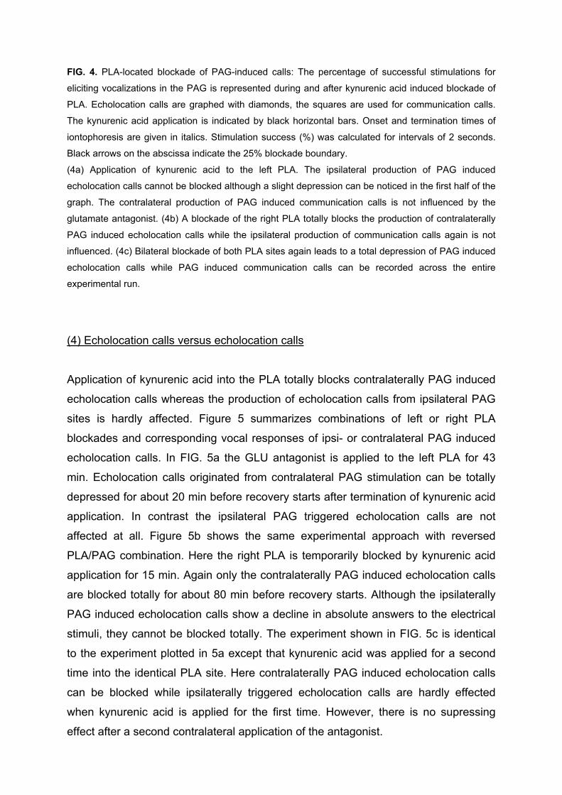
FIG. 4. PLA-located blockade of PAG-induced calls: The percentage of successful stimulations for
eliciting vocalizations in the PAG is represented during and after kynurenic acid induced blockade of
PLA. Echolocation calls are graphed with diamonds, the squares are used for communication calls.
The kynurenic acid application is indicated by black horizontal bars. Onset and termination times of
iontophoresis are given in italics. Stimulation success (%) was calculated for intervals of 2 seconds.
Black arrows on the abscissa indicate the 25% blockade boundary.
(4a) Application of kynurenic acid to the left PLA. The ipsilateral production of PAG induced
echolocation calls cannot be blocked although a slight depression can be noticed in the first half of the
graph. The contralateral production of PAG induced communication calls is not influenced by the
glutamate antagonist. (4b) A blockade of the right PLA totally blocks the production of contralaterally
PAG induced echolocation calls while the ipsilateral production of communication calls again is not
influenced. (4c) Bilateral blockade of both PLA sites again leads to a total depression of PAG induced
echolocation calls while PAG induced communication calls can be recorded across the entire
experimental run.
(4) Echolocation calls versus echolocation calls
Application of kynurenic acid into the PLA totally blocks contralaterally PAG induced
echolocation calls whereas the production of echolocation calls from ipsilateral PAG
sites is hardly affected. Figure 5 summarizes combinations of left or right PLA
blockades and corresponding vocal responses of ipsi- or contralateral PAG induced
echolocation calls. In FIG. 5a the GLU antagonist is applied to the left PLA for 43
min. Echolocation calls originated from contralateral PAG stimulation can be totally
depressed for about 20 min before recovery starts after termination of kynurenic acid
application. In contrast the ipsilateral PAG triggered echolocation calls are not
affected at all. Figure 5b shows the same experimental approach with reversed
PLA/PAG combination. Here the right PLA is temporarily blocked by kynurenic acid
application for 15 min. Again only the contralaterally PAG induced echolocation calls
are blocked totally for about 80 min before recovery starts. Although the ipsilaterally
PAG induced echolocation calls show a decline in absolute answers to the electrical
stimuli, they cannot be blocked totally. The experiment shown in FIG. 5c is identical
to the experiment plotted in 5a except that kynurenic acid was applied for a second
time into the identical PLA site. Here contralaterally PAG induced echolocation calls
can be blocked while ipsilaterally triggered echolocation calls are hardly effected
when kynurenic acid is applied for the first time. However, there is no supressing
effect after a second contralateral application of the antagonist.

Answer[%]5a
0
20
40
60
80
100
0 20 40 60 80 100 120 140
Answer[%]
t[min]
PAG ipsilateral
PAG contralateral
1 15 65 105PLA Block left PLA Block left
t[min]
0
20
40
60
80
100
0 20 40 60 80 100 120 140
Answer[%]
PAG contralateral
PAG ipsilateral
PLA Block right151
0
20
40
60
80
100
0 20 40 60 80 100 120 140t[min]
PAG ipsilateral
PAG contralateral
PLA Block left431
5c
5b

FIG. 5. PLA-located blockade of PAG-induced echolocation calls: ipsi- and contralateral differences.
Refer to FIG. 4 for explanations on graph. (5a) Application of kynurenic acid to the left PLA does not
influence the elicitability of ipsilaterally PAG-induced echolocation calls whereas it lowers dramatically
the efficiency of contralaterally induced echolocation calls. The elicitability does not recover to the 75%
mark within 120 min of experiment duration. (5b) A blockade of the right PLA totally blocks the
production of contralaterally PAG induced echolocation calls while ipsilateral induced echolocation
calls are little affected although some drops in elicitablity can be detected. (5c) Repeated application of
kynurenic acid principally shows comparable results although at the second application the
contralateral PAG induced production of echolocation calls cannot be cut off as effectively as shown
for the first application.
Although the application time was prolonged from 15 min in the first cycle of the
experiment to 40 min in the second cycle a total blockade cannot be accomplished
anymore. At the beginning of the second application at 65 minutes a short trend of
declining elicitability in PAG induced echolocation calls can be noticed, but the 25%
criteria for a blockade cannot be reached. Already within kynurenic acid application
time the contralaterally induced echolocation calls reach 100% elicitability again.
(5) Effect of PLA blockades on latencies of electrically induced PAG-vocalizations
Figure 6 shows that contralateral PLA blockades not only effect the elicitability of
vocalizations within the PAG but also influence the latencies of vocal responses to
electrical microstimulation. The blockade is marked with a gray bar at the bottom of
each plot. Section one (I) represents the first part of the experiment until a 25%
blockade was achieved, section two (II) represents the part of the experiment where
blockade was active and vocal responses did not reach the 75% recovery boundary
and section three (III) finally marks the part of the experiment where vocal responses
recovered and crossed the 75% boundary again. Under the influence of kynurenic
acid the mean latencies for vocal responses show significant differences when
particular sections of the graphs are compared. Table 1 summarizes the statistical
analysis for the latencies of ipsilaterally PAG induced vocalizations (echolocation
calls as well as communication calls). The PLA blockade not only stops contralateral
PAG-induced echolocation calls but also seem to have an influence on the latency of
electrically induced echolocation calls triggered on ipsilateral PAG sites.


FIG. 6. Mean latencies for vocalizations of experiments represented in FIG. 5. Figure 6a, b, c
correspond to FIG. 5a, b, c, respectively. The black bars below each plot mark the period of kynurenic
acid application. The gray bars on top of the abscissas indicate the duration of effective blockade
(25% and 75% limits taken from FIG. 5). Gaps within the contralateral graphs mark 100% blockade.
Standard deviations for mean latencies are plotted only positively. (6a) The blockade of contralaterally
blocked echolocation calls (black graph) does not reach the 75% recovery before the termination of
the experiment. The mean latencies of ipsilaterally (!) elicited calls show a significant difference (p ≤
0.020, table 1) when compared between the interval before or during contralateral blockade. (6b) The
mean ipsilateral latency is calculated for 3 sections (I, II and III) before, during and after blocking the
contralaterally elicited calls. The mean latencies differ for section I versus section II (p ≤ 0.025, table 1)
but not for section I versus section III (p ≤ 1.000, table 1). (6c) The mean latencies for section II versus
section III are significantly different (p ≤ 0.000, table 1), whereas the data available before blockade
are not sufficient.
(6) Histological verification of electrode positions
In Phyllostomus discolor the PAG is located around the mesencephalic aqueduct and
has a lateral extension of about 1000 µm from the midline whereas the PLA (around
800 µm in its maximal medio-lateral dimension) is located medially, adjacent to the
dorsal nucleus of the lateral lemniscus and contains neurons larger than those in
surrounding areas (Fenzl & Schuller, 2002). Electrical lesions to mark the PLA
stimulation and kynurenic acid-injection sites were placed stereotaxically 400 µm
(range of lesion in dorso-ventral spread: 400 µm) ventral to the PLA (FIG. 7b) to
ensure that the function of the PLA was not impaired during the ongoing
experiments. Lesions in the PAG (7a) resulted from the microstimulation via the
implants and could easily be detected in anatomical sections. In FIG. 7a the left
lesion (L1) marks the site in the PAG where echolocation calls could be triggered
while L2 shows the spot for the elicitation of a particular type of communication call.
(7) Tracer experiments
In ten animals WGA-HRP was iontophoretically ejected into sites within the PAG
which had been identified as vocally active with electrical microstimulation prior to
tracer application. In no case retrograde or anterograde connections between the

PAG and one or both PLA sites could be found. Strong anterograde and some
retrograde projections however led from the PAG into several regions within the
lower brainstem. Labelled fibers leave the ventro-lateral part of the PAG ipsilaterally
to the injection site reaching the caudal part of the Medulla oblongata. At this level
the labeled fibers separate into a ipsilateral and a contralateral stream. Although the
ipsilateral stream is more distinct, both projections can be traced more caudally.
Slightly dorso-lateral to the lateral nucleus of the formatio reticularis, the nucleus
ambiguus can be identified (FIG. 7c/7d). At this and more caudal levels the
anterograde projections from the PAG bilaterally terminate near the median half of
the nucleus ambiguus and the medially adjacent part of the formatio reticularis,
without forming clearly stained terminal endings.
AQPAG
B
L1L2
4V
LL
PLA
L
NA
FRLNFR
ML
ventral
dorsal
FIG. 7. Histological verification of electrode locations and location of the nucleus ambiguus. 7a, b and 7d show Nissl stained 42 µm frontal sections, 7c shows a AChE stain of a frontal 42 µm section. (7a) Two lesions (L1/L2) caused by repeated electrical stimulation through chronically implanted electrodes placed into vocally active sites within the PAG. AQ, aqueduct; B, boundary between PAG and surrounding tissue. (7b) Electrically induced lesion (L) 400 µm below the location of the iontophoresis probes in the PLA. Due to the 400 µm offset of the lesion below the PLA the function of the PLA during
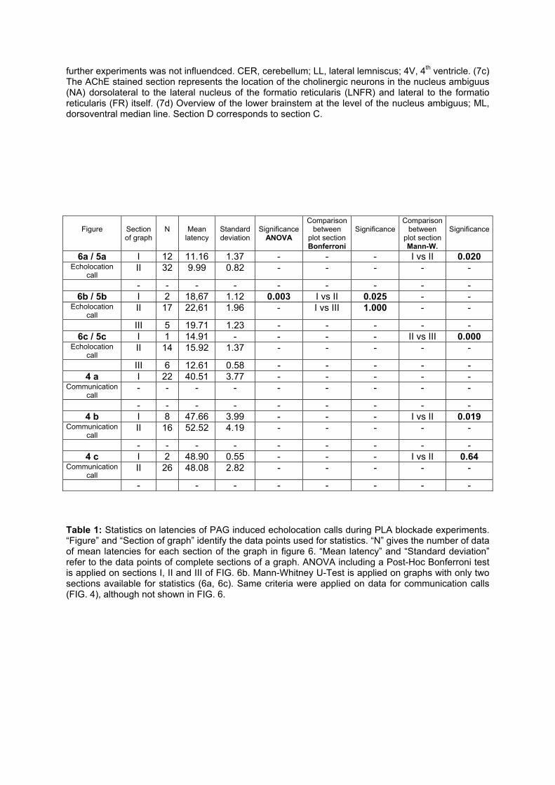
further experiments was not influendced. CER, cerebellum; LL, lateral lemniscus; 4V, 4th ventricle. (7c) The AChE stained section represents the location of the cholinergic neurons in the nucleus ambiguus (NA) dorsolateral to the lateral nucleus of the formatio reticularis (LNFR) and lateral to the formatio reticularis (FR) itself. (7d) Overview of the lower brainstem at the level of the nucleus ambiguus; ML, dorsoventral median line. Section D corresponds to section C.
Figure
Section of graph
N
Mean
latency
Standard deviation
Significance
ANOVA
Comparison between
plot section Bonferroni
Significance
Comparison between
plot section Mann-W.
Significance
6a / 5a I 12 11.16 1.37 - - - I vs II 0.020 Echolocation
call II 32 9.99 0.82 - - - - -
- - - - - - - - - 6b / 5b I 2 18,67 1.12 0.003 I vs II 0.025 - -
Echolocation call
II 17 22,61 1.96 - I vs III 1.000 - -
III 5 19.71 1.23 - - - - - 6c / 5c I 1 14.91 - - - - II vs III 0.000
Echolocation call
II 14 15.92 1.37 - - - - -
III 6 12.61 0.58 - - - - - 4 a I 22 40.51 3.77 - - - - -
Communication call
- - - - - - - - -
- - - - - - - - - 4 b I 8 47.66 3.99 - - - I vs II 0.019
Communication call
II 16 52.52 4.19 - - - - -
- - - - - - - - - 4 c I 2 48.90 0.55 - - - I vs II 0.64
Communication call
II 26 48.08 2.82 - - - - -
- - - - - - - - Table 1: Statistics on latencies of PAG induced echolocation calls during PLA blockade experiments. “Figure” and “Section of graph” identify the data points used for statistics. “N” gives the number of data of mean latencies for each section of the graph in figure 6. “Mean latency” and “Standard deviation” refer to the data points of complete sections of a graph. ANOVA including a Post-Hoc Bonferroni test is applied on sections I, II and III of FIG. 6b. Mann-Whitney U-Test is applied on graphs with only two sections available for statistics (6a, 6c). Same criteria were applied on data for communication calls (FIG. 4), although not shown in FIG. 6.

Discussion
This study has demonstrated that the neural control of different types of calls involves
different neural substrates for vocalization.
The periaqueductal gray is involved in the control of both echolocation calls and
communication calls (Fenzl & Schuller, 2002), whereas the paralemniscal area,
where only echolocation calls can be triggered (Fenzl & Schuller, 2002; Schuller et
al., 1997; Schuller & Radtke-Schuller, 1990) is only involved the control of
echolocation calls. The hypothesis that the production of particular communication
calls and the production of echolocation calls must revert to differently organized
neural substrates and pathways in the bat below the functional level of the PAG has
been supported by the results presented in this paper. Thus the production of
echolocation calls triggered in the PAG can totally be inhibited by a reversible
blockade of the PLA, whereas, the production of communication calls elicited within
the PAG is not influenced by such a blockade.
A straight forward vocal pathway from the PAG to the nucleus
ambiguous/retroambiguus as described in the literature is not sufficient to explain
these findings. A more complex organization of the descending vocal control system
below the PAG level seems to be mandatory.
(1) Sedation of animals and stability of chronic implants
To ensure a stable position of the animal during the iontophoresis experiments
Rompun was chronically applied subcutaneously during the sessions. The reaction
of the animal on external acoustic stimuli was used to evaluate the depth of sedation,
meaning the animal responded with ear movement but without arousal or ample body
movements. This condition could easily be maintained during the experiment by
applying the permanent infusion of the sedative. The experiments typically started
when the initial dose of Rompun had its peak effect. At this time all bats showed
perfect elicitability of vocalization (FIG. 3, FIG. 4 and FIG. 5) so that the effect of
sedation on the ongoing experiments can be considered as minor. This sedative was
adequate to both achieve optimal stability of the animal and minimal influence on
vocal responses.

The stability of the chronically implanted electrodes for electrical micro-stimulation
was very satisfactory, in that, both echolocation calls and communication calls
without any change in spectral composition of the calls were elicited consistently over
a long stimulation period of up to 33 days. The slight increase of stimulation
thresholds for the individual electrodes (FIG. 2) can be attributed to an accumulation
of glia cells and debris caused by the presence of implants. A decrease in elicitibility
and/or quality of triggered vocalizations was not observed throughout the
experiments. Also the intensity of motor reactions associated with vocalizations, like
pinna and nose leaf movement with echolocation calls or mouth movements with
communication calls, did not change during the experiments.
(2) Definition of PAG and PLA
Vocalizations are complex motor patterns imbedded into differentiated behaviors of
an animal. It is well established that the periaqueductal gray (PAG) plays an
important role in vocal control of communication calls (Jürgens, 1998; Jürgens,
2002), i.e., social calls can be triggered in several mammalian (Fenzl & Schuller,
2002; Kirzinger & Jürgens, 1991; Zhang et al., 1994) and non-mammalian species
(Kennedy, 1975). In addition, echolocation calls can also be elicited in bats within
restricted areas of the PAG (Valentine et al., 2002) separated from those areas
where communication calls can be triggered (Fenzl & Schuller, 2002). The PAG
seems to have control on both types of calls (Fenzl & Schuller, 2002).
Natural echolocation calls cannot be elicited in the PAG only. Very short latencies for
eliciting ultrasonic vocalization and/or pinna movements were found in a restricted
paralemnisal tegmental region rostral and medial to the dorsal nucleus of the lateral
lemniscus (Schuller et al., 1997; Schuller & Radtke-Schuller, 1990).
Other examinations on functional aspects of the paralemniscal tegmental area are
very rare. Covey (1993) differentiates between a dorsal paralemniscal zone and a
paralemniscal zone on the basis of neural responses to acoustic stimuli. The
paralemniscal area described by this author at the level of the intermediate nucleus
of the lateral lemniscus and dorso-lateral to the medial superior olive is located more
caudally than the paralemniscal area we define.
In contrast to a pure neuroanatomical definition, the paralemniscal area is functionally
identified with electrical microstimulation, which offers a highly reliable possibility to

identify the vocally active area of the paralemniscal region. Histological verifications
demonstrate the reliability of the functional identification. Only within a very restricted
paralemniscal area vocalizations can be elicited at very low thresholds (3.4 µA – 15
µA) without, or with only minor pinna movements (Fenzl & Schuller, 2002; Schuller &
Radtke-Schuller 1990).
Henkel and Edwards (Henkel, 1981; Henkel & Edwards, 1978) defined a tegmental
area in the cat as the “paralemniscal zone” (PL) that corresponds partly in its
connectional pattern to the situation found in the moustached bat (Schuller et al.,
1997). However the area defined by the authors in the cat cannot be aligned properly
with our anatomical data due to a lack of comparable anatomical cues.
In oculomotor function research a similar tegmental region is termed lateral tegmental
region (LTR) where neurons, probed in the cat do respond to visual, vestibular and
auditory stimulation (Gerlach & Their, 1995). According to these authors the PL
described by Henkel and Edwards constitutes only a subdivision of the larger LTR.
The comparable vocally active region in bats is even more restricted and only
occupies the dorsal half of the paralemniscal zone as defined by Henkel and
Edwards (Schuller et al., 1997).
Another characterization of the “lateral tegmentum underneath the inferior colliculus”
is given by Jürgens (1994). This region along the medial edge of the lateral
lemniscus is linked by the author with a vocal pathway from the PAG towards the
periambigual reticular formation although only lesion-experiments were carried out
leaving unanswered whether this region represents only fibers of passage or neurons
involved in vocal control.
Due to the functional differences demonstrated by different authors, together with the
definition of the tegmental region by various different methods, there is no clear
definition of this area given, neither anatomically nor physiologically. It cannot be
decided whether the different regions are part of a functionally coherent tegmental
area responsible for diverse tasks or whether the tegmental area is an ensemble of
independent functional regions.
The definition of the PLA used in this paper is strictly based on electrical
microstimulation and the delimitation towards adjacent regions is recognized in a
significant threshold increase for eliciting natural echolocation calls. Previous findings

about the tegmental region in bats (Fenzl & Schuller, 2002; Schuller & Radtke-
Schuller, 1988; Schuller et al., 1997; Schuller & Pillat, 1998; Schuller & Radtke-
Schuller, 1990) and this paper are all based on this functional definition and results
can be directly compared.
(3) Differentiated vocal pathways ?
Could different types of vocalizations be processed via at least partly separated vocal
pathways in the bat? The initial position of this question came from results that
communication calls (Fenzl & Schuller, 2002; Suga et al., 1973; Valentine et al.,
2002) and echolocation calls (Fenzl & Schuller, 2002, Suga et al., 1973; Suga &
Yajima, 1988) could be elicited within the PAG, while within the PLA only
echolocation calls could be triggered (Fenzl & Schuller, 2002; Schuller et al., 1997;
Schuller & Radtke-Schuller, 1990). The two mesencephalic brain areas, PAG and
PLA have never been investigated so far in bats in order to explore interactions
between both regions in terms of vocalization. Our experiments clearly show a
functional linkage between the PAG and both PLA sites for the control of
vocalizations, which however reveals functional differentiation between two classes
of vocalizations, i.e. communication calls and echolocation calls.
A vocal pathway for the production of communication calls as reported by several
authors (Ennis et al., 1997; Vanderhorst et al., 2000; Zhang et al., 1995; Zhang et al.,
1994) does not necessarily involve activity of the PLA. In this pathway a direct
connection from the PAG towards the nucleus retroambiguus (NRA) is described.
The NRA includes a group of premotor neurons which send direct projections to
thoracic and upper lumbar motoneurons (Vanderhorst et al., 2000; Vanderhorst et al.,
2000) involved in expiration, and to the nucleus ambiguus containing laryngeal and
pharyngeal motoneurons (Holstege, 1989; Vanderhorst et al., 2000; Vanderhorst et
al., 2000). This pathway alone cannot explain our findings that PAG induced
echolocation calls can be blocked by PLA blockades but would be sufficient to
explain that the production of communication calls could never be cut off totally by
PLA blockade.
Jürgens (2000) demonstrated that injections of kynurenic acid into the ventrolateral
pons around the ventral nucleus of the lateral lemniscus and superior olive of

monkeys, a region located ventral and caudal to the paralemniscal area and certainly
not identical with the PLA in bats, can block periaqueductally elicited vocalizations .
Only one type of communication call with characteristic frequency modulations over
several kHz was effected by this blockade. Jürgens (2000) suggests that vocal
patterns are generally controlled in different brainstem regions, and that vocalizations
with frequency modulations seem to depend on an intact periolivary region.
Echolocation calls of P. discolor are typically frequency-modulated calls (FM)
covering a frequency range between 45 and 100 kHz with the 3rd – 5th harmonic
(Rother & Schmidt, 1982). These FM calls can successfully be blocked in all
experiments we carried out so far which could be analogous to what Jürgens found in
his experiments with squirrel monkeys, however, in a different region.
The effect of PLA induced blockades on PAG triggered vocalizations strictly depends
on the side of the application since echolocation calls can only be blocked when
kynurenic acid is applied contralateral to the stimulation site in the PAG (FIG. 5). This
is in contrast to what Jürgens (2000) describes for squirrel monkeys. He mentions
that periaqueductally elicited vocalizations are only affected by ipsilateral, not by
contralateral injection of kynurenic acid. This elementary difference could be due to
different brain regions injected with kynurenic acid. The blockade sites in the monkey
were all located in the ventrolateral pons, as described earlier and is definitely not the
same area as the paralemniscal area, defined in this paper.
The results from the monkey cannot support the findings from Holstege (1989) and
Zhang (1995) that the vocal control pathway consists only of a direct connection from
the PAG to the nucleus retroambiguus in the medulla oblongata (Jürgens, 2000). Our
findings do not rule out the direct connection from the PAG to the nucleus
retroambiguus (see discussion on anatomical studies) but also demonstrate that a
differentiated control of certain types of vocalizations via parallel or at least partly
separated pathways for echolocation calls must exist. This evidence of a more
complicated network for vocal control at the level below the PAG in the monkey as
well as in the bat, underlines the broader significance of this concept on a
mammalian level. The bat vocal control system cannot be considered as being
specialized but rather being a general mammalian vocalization system with distinct
emphasized features.
That only contralateral PLA blockades affect PAG induced echolocation calls while
ipsilateral manipulations show no consequences is surprising. Besides strong
reciprocal connections between the PLAs of both sides there are no major midline
crossing projections that could account for the contralateral influence.
(2) Influence of PLA blockades on PAG induced vocalizations
It is noteworthy that the onset of a contralateral PLA blockade is extraordinary
variable between only three minutes and almost 80 minutes under comparable
experimental conditions. Slightly different positions of the iontophoresis probe at the
PLA site may be responsible as even a deviation as small as 100 µm corresponds to
almost 15% of the medio-lateral dimension (≈ 800 µm, Fenzl & Schuller, 2002) of the
PLA in P. discolor. At marginal application sites kynurenic acid should take longer to
influence a necessary number of neurons in the PLA, than when injected to the
geometrically optimal PLA location. The onset time differences indicate that the
suppressive effect of PLA inactivation depends on the size of population of PLA
neurons involved. PLA blockades also leads to an increase of latency of PAG-
triggered vocalizations indicating again influence of the PLA on cell elicitability.
The general role of the PLA is still not clear. Based on our findings the PLA should be
a necessary link in the pathway for the control of PAG-induced echolocation calls,
although the production of spontaneously emitted echolocation calls evidently
survives bilateral electrolytic lesioning of the PLA (Pillat & Schuller, 1998).
Neuroanatomy
For the cat (Gerlach & Their, 1995) retrograde labelling in the ipsilateral PAG was a
consistent result of tracer injections into the LTR (lateral tegmental region) but it
cannot be certified that LTR and PLA are homologous brain regions.
In this study we could not demonstrate any direct connections between vocally active
PAG sites and the PLA, using WGA-HRP injections into the PAG. Such connections
could have been expected from the blockade experiments in the PLA and from
findings that injections of WGA-HRP into vocally active sites within the PLA lead to
labelled neurons in the PAG (Schuller et al., 1997). The lack of labelling in
Phyllostomus discolor may be a species-specific methodological problem with WGA-
HRP. Quality of labelling in Pyllostomus disclor did generally not reach that in
Pteronotus p. parnellii or Rhinolophus rouxi under identical conditions of tracer
application.
Several authors report a direct projection from the PAG to the nucleus retroambiguus
(Davis et al., 1996; Gerrits & Holstege, 1996; Holstege et al., 1997; Jürgens, 2002;
Zhang et al., 1995; Zhang et al., 1994). The nucleus retroambiguus (NRA) itself is a
group of premotor neurons in the caudal medulla sending direct projections to
thoracic and upper lumbar motoneurons involved in expiration and to the nucleus
ambiguus (NA) containing motoneurons to the larynx, pharynx and soft palate
(Vanderhorst et al., 2000). Physiological studies revealed that the NRA is involved in
vocalization activating expiratory and laryngeal muscles (Larson et al., 1994; Zhang
et al., 1995). Ennis and coworkers (1997) demonstrated that periaqueductal gray
neurons terminate in close contiguity to cholinergic neurons in the “compact,
semicompact, loose and external formations of nucleus ambiguus”.
The NA in Phyllostomus discolor was identified using its cholonergic properties and
is situated slightly dorsolateral to the lateral nucleus to the formatio reticularis.
Massive ipsilateral projections from the PAG into the ventral brainstem target the
caudal part of this identified NA-region. The labelled fibers ended in this region but
terminals were not found. At medullary level projections from the PAG cross the
midline and reach the corresponding regions. The fibers from the PAG to NRA in
Phyllostomus discolor seem to serve also for vocal control, at least for a subgroup of
communication calls.
Other control pathways for the control of vocalization besides the PAG to NRA route
must exist as the blockade of PAG-elicited calls by inactivation of the PLA shows.
However, the projections of the PAG into the region of the PLA as described by other
authors either are very faint in Phyllostomus discolor or the PLA influences
vocalization not via a serial but rather parallel route from the PAG to the common
vocal pathway.
Summary and conclusion
Communication calls and echolocation calls can be elicited with electrical micro-
stimulation through chronically implanted electrodes in the PAG. Direct projections
from the PAG towards the region of the nucleus ambiguus/retroambiguus (NA/NRA)
complex could be traced implicating a vocal pathway from the PAG to
premotoneurons within this region. Reversible blockade of the vocally active PLA,
where only echolocation calls can be triggered, totally block PAG induced
echolocation calls but not communication calls. The PAG to NA/NRA pathway may
not be the only pathway processing vocal activity. The PLA seems to be essential for
the production of echolocation calls but not for particular types of communication
calls elicited in the PAG. This suggests differential pathway organization for particular
types of communication calls on the one hand and echolocation calls on the other
hand. Whether the differentiation of pathways applies to the two classes of
echolocation calls and communication calls in general, or whether it is more directly
dependent on specific call properties in the acoustic pattern domain remains open to
further experimentation.
Acknowledgements
The authors like to thank Uwe Firzlaff, Andreas Nixdorf, Markus Drexl and Rudolph
Marsch for fruitful discussions during the experiments. Herrmann Schweizer for his
kind help on anatomical verifications of brain sections, Claudia Schulte and Horst
König for their technical support. Karl-Heinz Esser (University of Hannover,
Germany) and Hans Erkert (University of Tübingen, Germany) for helping to establish
a breeding colony of Phyllostomus discolor by supplying bats.

Reference List
Apfelbach R. (1972) Electrically elicited vocalizations in the gibbon Hylobates lar (Hylobatidae), and their behavioral significance. Z.Tierpsychol 30: 420-430
Covey E. (1993) Response properties of single units in the dorsal nucleus of the lateral lemniscus and paralemniscal zone of an echolocating bat. J.Neurophysiol 69: 842-859
Davis P.J., Zhang S.P., Winkworth A., Bandler R. (1996) Neural control of vocalization: respiratory and emotional influences. J.Voice 10: 23-38
Ennis M., Xu S.J., Rizvi T.A. (1997) Discrete subregions of the rat midbrain periaqueductal gray project to nucleus ambiguus and the periambigual region. Neurosci. 80: 829-845
Esser, K.-H. and Pistohl, D. (1998) The vocal repertoire of the lesser spear-nosed bat (Phyllostomus discolor). Zoology 101[Suppl. I], 84. Fenzl T., Schuller G. (2002) Periaqueductal gray and the region of the paralemniscal area have different functions in the control of vocalization in the neotropical bat, Phyllostomus discolor. Eur.J.Neurosci. 16: 1974-1986
Gallyas F. (1979) Silver staining of myelin by means of physical development. Neurol.Res 1: 203-209
Gerlach I., Thier P. (1995) Brainstem afferents to the lateral mesencephalic tegmental region of the cat. J.Comp.Neurol. 358: 219-232
Gerrits P.O., Holstege G. (1996) Pontine and medullary projections to the nucleus retroambiguus: a wheat germ agglutinin-horseradish peroxidase and autoradiographic tracing study in the cat. J.Comp.Neurol. 373: 173-185
Hardy H., Heimer L., Switzer R., Watkins D. (1976) Simultaneous Demonstration of Horseradish Peroxidase and Acetylcholinesterase. Neurosci.Lett. 3: 1-5
Henkel C.K. (1981) Afferent sources of a lateral midbrain tegmental zone associated with the pinnae in the cat as mapped by retrograde transport of horseradish peroxidase. J.Comp.Neurol. 203: 213-226
Henkel C.K., Edwards SB (1978) The superior colliculus control of pinna movements in the cat: possible anatomical connections. J.Comp.Neurol. 182: 763-776
Holstege G. (1989) Anatomical study of the final common pathway for vocalization in the cat. J.Comp.Neurol. 284: 242-252
Holstege G., Kerstens L., Moes M.C., Vanderhorst V.G. (1997) Evidence for a periaqueductal gray-nucleus retroambiguus-spinal cord pathway in the rat. Neurosci. 80: 587-598
Jürgens U. (1994) The role of the periaqueductal grey in vocal behaviour. Behav. Brain Res. 62: 107-117

Jürgens U. (1998) Neuronal control of mammalian vocalization, with special reference to the squirrel monkey. Naturwissenschaften 85: 376-388
Jürgens U. (2000) Localization of a pontine vocalization-controlling area. J.Acoust. Soc.Am. 108: 1393-1396
Jürgens U. (2002) Neural pathways underlying vocal control. Neurosci.Biobehav.Rev. 26: 235-258
Kennedy M.C. (1975) Vocalization elicited in a lizard by electrical stimulation of the midbrain. Brain.Res. 91: 321-325
Kirzinger A., Jurgens U (1991) Vocalization-correlated single-unit activity in the brain stem of the squirrel monkey. Exp.Brain Res. 84: 545-560
Larson C.R., Kistler M (1984) Periaqueductal gray neuronal activity associated with laryngeal EMG and vocalization in the awake monkey. Neurosci.Lett. 46: 261-266
Larson C.R., Yajima Y, Ko P (1994) Modification in activity of medullary respiratory-related neurons for vocalization and swallowing. J.Neurophysiol. 71: 2294-2304
Llewellyn-Smith I.J., Pilowsky P., Minson J.B. (1993) The tungstate-stabilized tetramethylbenzidine reaction for light and electron microscopic immunocytochemistry and for revealing biocytin-filled neurons. J.Neurosci.Methods 46: 27-40
Lu C.L., Jürgens U. (1993) Effects of chemical stimulation in the periaqueductal gray on vocalization in the squirrel monkey. Brain Res.Bull. 32: 143-151
Martin J.R. (1976) Motivated behaviors elicited from hypothalamus, midbrain, and pons of the guinea pig (Cavia porcellus). J.Comp.Physiol.Psychol. 90: 1011-1034
Nixdorf, A. (2003) Die prätektale Region bei der Fledermaus Phyllostomus discolor - eine audio-motorische Schnittstelle? PhD-Thesis, Dept. Biol. II., Ludwig-Maximilians-Universität München Pillat J, Schuller G. (1998) Audiovocal behavior of Doppler-shift compensation in the horseshoe bat survives bilateral lesion of the paralemniscal tegmental area. Exp Brain Res. 119: 17-26
Rother G., Schmidt U. (1982) Der Einfluss visueller Information auf die Echoortung bei Phyllostomus discolor. Z.Säugetierkunde 47: 324-334
Schuller G., Radtke-Schuller S. (1988) Midbrain areas as candidates for audio-vocal interface in echolocating bats. In: Nachtigall P.E., Moore P.W.B. (Eds.), Animal Sonar, Processes and Performance. Plenum Press, New York, pp. 93-98
Schuller G., Fischer S., Schweizer H. (1997) Significance of the paralemniscal tegmental area for audio-motor control in the moustached bat, Pteronotus p. parnellii: the afferent and efferent connections of the paralemniscal area. Eur.J.Neurosci. 9: 342-355

Schuller G., Pillat J. (1998) Audiovocal behavior of doppler-shift compensation in the horseshoe bat survieves bilateral lesion of the paralemniscal tegmental area. Exp. Brain Res. 119(1): 17-26
Schuller G., Radtke-Schuller S. (1990) Neural control of vocalization in bats: mapping of brainstem areas with electrical microstimulation eliciting species-specific echolocation calls in the rufous horseshoe bat. Exp.Brain Res. 79: 192-206
Schuller G., Radtke-Schuller S., Betz M. (1986) A stereotaxic method for small animals using experimentally determined reference profiles. J.Neurosci.Methods 18: 339-350
Suga N., Yajima Y. (1988) Auditory-vocal integration in the midbrain of the mustached bat: Periaqueductal Gray and Reticular Formation. In: The physiological control of mammalian vocalization (Newman JD, ed), pp 87-107. New York: Plenum Press
Suga N., Schlegel P., Shimozawa T., Simmons J. (1973) Orientation sounds evoked from echolocating bats by electrical stimulation of the brain. J.Acoust.Soc. Am 54: 793-797
Valentine D.E., Sinha S.R., Moss C.F. (2002) Orienting responses and vocalizations produced by microstimulation in the superior colliculus of the echolocating bat, Eptesicus fuscus. J.Comp.Physiol. A 188: 89-108
Vanderhorst V.G., Terasawa E., Ralston H.J., III, Holstege G (2000) Monosynaptic projections from the lateral periaqueductal gray to the nucleus retroambiguus in the rhesus monkey: implications for vocalization and reproductive behavior. J.Comp. Neurol. 424: 251-268
Vanderhorst V.G., Terasawa E., Ralston H.J., III, Holstege G (2000) Monosynaptic projections from the nucleus retroambiguus to motoneurons supplying the abdominal wall, axial, hindlimb, and pelvic floor muscles in the female rhesus monkey. J.Comp. Neurol. 424: 233-250
Yajima Y., Hayashi Y., Yoshii N. (1980) The midbrain central gray substance as a highly sensitive neural structure for the production of ultrasonic vocalization in the rat. Brain Res. 198: 446-452
Zhang S.P., Bandler R., Davis P.J. (1995) Brain stem integration of vocalization: role of the nucleus retroambigualis. J.Neurophysiol. 74: 2500-2512
Zhang S.P., Davis P.J., Bandler R., Carrive P. (1994) Brain stem integration of vocalization: role of the midbrain periaqueductal gray. J.Neurophysiol. 72: 1337-1356

THANKS TO 52
Thanks to ... Als aller erstes möchte ich mich ganz besonders herzlich bei Prof. Gerd Schuller für die Aufnahme in seine Arbeitsgruppe bedanken, in der ich neben fachlichen Aspekten ganz nebenbei lernte, auf meinen eigenen „wissenschaftlichen Beinen“ zu stehen. Mein Dank gebührt auch Dr. Hermann Schweizer. Ohne ihn wäre ich in anatomischen Belangen so manches mal ein klein wenig überfordert gewesen. Auch ehrte mich sein Vertrauen, indem er mir bei der Betreuung zoologischer Kurse freie Hand ließ. Vielen Dank auch an Prof. Gerhard Neuweiler. Durch sein Interesse an meiner Arbeit verhalf er mir über so manche antriebsarme Phase hinweg. Petra Weissenbacher(in), Elisabeth Föller, Alexandra Stein, Andreas Nixdorf, Uwe Firzlaff, Rudi Marsch(inger), Markus Drexl, Jan Grunwald, Lutz Wiegrebe. Das nenne ich Kollegen! Nicht zuletzt, weil sie mir auch als Freunde zur Seite stehen. Claudia Schulte, Sabine Peisker und Horst König möchte ich ganz besonders für ihre technische Unterstützung danken. Kathrin Schmidt, Edda Eibinger und Dieter Leippert, vielen Dank für die ausgezeichnete Versorgung der Tiere. Christoph Romanowski, vielen Dank bei der Unterstützung während Routinearbeiten. Herrn Zaschka sowie seinen Mitarbeitern möchte ich für die Herstellung einiger Bestandteile meines Setups danken. Vielen Dank auch an Alexandra für die schöne Zeit (trotz Laboralltag) und ihren Beistand während einer für mich schwierigen Zeit. Anja und Lara. Mit euch beiden kann man selbst trister Schreibarbeit noch eine schöne Seite abgewinnen (Los, zerstreu mich!). Mein allergrößter Dank jedoch gilt meinen Eltern. Für mich war es nie einfach nur selbstverständlich, wie sehr sie mich dabei unterstützten, meinen Weg zu verfolgen. Von der mittleren Reife zur Dissertation ist ein langer Weg, doch ihr Vertrauen in mich half mir sehr, mein Ziel nie aus den Augen zu verlieren. Die an dieser Stelle meist übliche Floskel „Dank gebührt auch meinen Versuchstieren“ verkneife ich mir, um meiner Empörung über die teilweise doch sehr mangelhafte Kooperation der Tiere ein wenig Ausdruck zu verleihen.
Alf Poier

CURRICULUM VITAE 53
CURRICULUM VITAE
Thomas Fenzl Address Professional: Department Biology II Ludwig-Maximilians-University Munich Luisenstrasse 14 80333 Munich
Germany Tel +49-89-5902-202
Fax +49-89-5902-210 email: [email protected]
Personal: Rosa-Luxemburg-Platz 1 80637 Munich Germany Tel: +49-89-15987713 Born: 1965, September 30 Nationality: German EDUCATION AND UNIVERSITY TITLES 1983-1987 Vocational training as machinist for electrical machines 1987-1991 German Federal Armed Forces 1991-1994 Upper vocational school (qualification for university entrance) 1994-1999 University of Regensburg Faculty III Biology Institute of Zoology Universitaetsstrasse 31, 93059 Regensburg, Germany
1996 Pre-master degree (Vordiplom) in Zoology, Botany, Biochemistry, Genetics, Microbiology, Chemistry and
Physics. 1999 Masters (Diplom) in Biology
main subject: Zoology subsidiary subjects: Botany, Biophysics
Since 2000 Enrollment in doctoral program (PhD) Ludwig-Maximilians-University Munich
CURRICULUM VITAE 54
LABORATORY EXPERIENCE 1998 Internship at the Institute of Zoology, University of Regensburg
Techniques: basic techniques in neurophysiology. Behavioral tests on chemo- and water sense organs in flies. 1998/1999 “Electrophysiological examinations of antennal structures in the
larvae of Tenebrio molitor”. Masters (Diplom) at the Institute of Zoology, University of Regensburg. Techniques: REM-imaging, extracellular recording on different sense organs on the larval antennae.
Since Mar. 2000 “Differentiated roles of the periaqueductal gray and the
paralemniscal area on the control of echolocation calls and communication calls in the Neotropical bat Phyllostomus discolor”.
PhD work at the Ludwig Maximilians University of Munich, Germany. Techniques: Electrical microstimulation, microdialysis, iontophoresis, experimental development of chronically implanted microelectrodes, anterograde and retrograde labeling, slicing and histological processing of samples, behavioral tests, development of a stereotaxic atlas, stereotaxic handling of the experimental animal, experimental development of cooled stimulation probes for subcortical stimulation.
EXTERNAL LABORATORY EXPERIENCE 1997 Internship at the Field Museum of Natural History Roosevelt Road at Lake Shore Drive Chicago, IL 60605-2496, USA
Techniques: basic techniques in cladistic, invertebrate collection, preparation and dissection techniques.
1999/2000 Visiting Scientist at Hort+Research Centre 120 Mt Albert Road Auckland, New Zealand Techniques: basic molecular biology techniques, Gel-electrophoresis, 2D-electrophoresis, protein activity tests.
CURRICULUM VITAE 55
TEACHING Annual course “Morphology of Animals”
Theoretical and practical instructions for graduate students. 2000/2003 “Behavioral work with bats”
Theoretical and practical instructions for undergraduate and graduate students.
EXTERNAL COURSES Mar. 2001 “Macroscopy of the human brain”
Dr. Senkenbergische Anatomy, University of Frankfurt am Main, Germany. Techniques: Preparation of human brains, preparation of the limbic system, identification of brain structures.
Jun. 2001 “Rhetoric and presentation techniques in science” Graduiertenkolleg Munich. Apr. 2002 “Imaging and patch-clamp in cellular neuroscience”
Neurologische Klinik der Charite, Humbolt University Berlin, Germany. Techniques: Calcium-imaging, patch-clamp recordings in vivo and in vitro.
May 2002 “Matlab in Science”
Institut für Mathematik und Datenverarbeitung, University of German Federal Armed Forces, Munich, Germany. Techniques: Basics of signal processing in Matlab.
FINANCIAL SUPPORTS 1997 Financial Support from “Verein der Freunde und Foerderer der
Universitaet Regensburg” for internship at the Field Museum. Since 2000 University of Munich (BAT IIa/2). Since 2001 Member of the DFG Graduiertenkolleg “Sensory interaction in biological
und technical systems”, DFG-GRK 267 (Special PhD program of the Deutsche Forschungsgemeinschaft).

CURRICULUM VITAE 56
PUBLICATIONS P. Sierwald, T. Fenzl, (1999) Argyrodes in webs of the Floridian red widow spider (Araneae: Therediidae), Florida Entomologist 82(2) 359-361. T. Fenzl, G. Schuller, (2002) Periaqueductal gray and the region of the paralemniscal area have different functions in the control of vocalization in the Neotropical bat, Phyllostomus discolor, Eur. J. Neurosci. 16, 1974-1986. T. Fenzl, G. Schuller, Echolocation calls and communication calls are processed differentially in the brainstem of the bat Phyllostomus discolor. Submitted to Eur. J. Neurosci
ABSTRACTS AND POSTERS Aug. 2001 T. Fenzl and G. Schuller, Different roles of the Periaqueductal gray
(PAG) and the paralemniscal area (PLA) on vocal control in the Neotropical bat Phyllostomus discolor (Wagner, 1843).
6th International Congress of Neuroethology, Bonn, Germany. Jan 2002 T. Fenzl and G. Schuller, Periaqueductal Gray and the Paralemniscal
Region control echolocation and communication calls differentially in the bat Phyllostomus discolor.
25th ARO 2002 Mid Winter Meeting, St. Petersburg Beach, USA. Nov 2002 T. Fenzl and G. Schuller, Differential neural control of communication
calls and echolocation calls in the bat Phyllostomus discolor. 32nd SFN Annual Meeting, Orlando, USA. INVITED SEMINARS Jan 2002 T. Fenzl and G. Schuller, Differentiated roles of periaqueductal gray and
region of paralemniscal area on control of echolocation calls and communication calls in Phyllostomus discolor.
Meeting of national research groups, MPI - Seewiesen, Germany. Feb 2002 T. Fenzl. and G. Schuller, Physiological control of mammalian
vocalizations: Differentiated roles of the periaqueductal gray and the paralemniscal region on the control of echolocation calls and communication calls in Phyllostomus discolor.
Washington State University, Vancouver, USA. Jun 2002 T. Fenzl and G. Schuller, Echolocation calls and communication calls are
controlled differentially by the periaqueductal gray and the paralemniscal area in the bat Phyllostomus discolor.
University of Ulm, Germany.

CURRICULUM VITAE 57
Nov 2002 T. Fenzl and G. Schuller, Differential neural control of communication calls and echolocation calls in the bat Phyllostomus discolor.
University of Washington, Seattle, USA