differentiated paju cells have increased resistance to ... · pdf filedifferentiated paju...
TRANSCRIPT
Communication
Differentiated Paju cells have increased resistance to toxiceffects of potassium ionophores��
Vera Teplova1, Elina Jääskeläinen2, Mirja Salkinoja-Salonen2, Nils-Erik L. Saris2�,Martina Serlachius3, Feng-Yen Li2,3 and Leif C. Andersson3,4
1Institute of Theoretical and Experimental Biophysics, Russian Academy of Sciences,RU-142290 Pushchino, Moscow region, Russia; 2Department of Applied Chemistry andMicrobiology, Viikki Biocenter 1; 3Department of Pathology, Haartman Institute, University ofHelsinki, Helsinki, Finland 4HusLab HUCH, Helsinki, Finland
Received: 19 April, 2004; accepted: 23 April, 2004
Key words: cell differentiation, cereulide, mitochondria, membrane potential, valinomycin, Paju cells
In this study we have investigated the impact of differentiation of neuronal cells ontheir sensitivity to microbial toxins. We used the human neural crest-derived tumorcell line Paju, which can be induced to differentiation in vitro by treatment withphorbol 12-myristate 13-acetate. Addition of the highly toxic potassium ionophorescereulide (4.5 and 9.0 ng/ml) or valinomycin (20 ng/ml), to cultures of undifferenti-ated Paju cells caused collapse of the mitochondrial membrane potential — measuredwith the fluorescent probe 5,5�,6,6�-tetrachloro-1,1�,3,3�-tetrabenzimidazole carbo-cyanine iodide (JC-1) followed by detachment of the cells and their apoptotic death.After induced differentiation of the Paju cells, their mitochondria retained the mem-brane potential upon exposure to the toxins and the cells displayed increased resis-tance to apoptosis as compared with undifferentiated cells. This effect may be
Vol. 51 No. 2/2004
539–544
QUARTERLY
�This work was presented in poster form at the 29th Congress of the Federation of European Biochemi-cal Societies, Warsaw, Poland, 26 June–1 July 2004.�This study was supported by grants from the Sigrid Juselius and the Magnus Ehrnrooth Foundations,the Academy of Finland to Microbial Resources Unit (53305), the King Gustaf V and Queen VictoriaFoundation and the NIH/FIC MIRT T37 TW00032 grant to Kim H. Tan.
�Corresponding author: Nils-Erik Saris, Department of Applied Chemistry and Microbilogy, ViikkiBiocenter 1, POB 56, FIN-00014 University of Helsinki, Helsinki, Finland; tel: (35 89) 1915 9347; fax:-01, e-mail: [email protected]
Abbreviations: ��m, mitochondrial transmembrane potential; JC-1, 5,5�,6,6�-tetrachloro-1,1�,3,3�-te-trabenzimidazole carbocyanine iodide; PI, propidium iodide; PMA, phorbol 12-myristate 13-acetate.

caused by an elevated expression of the anti-apoptotic protein Bcl-2 and of theneuroprotective factor, stanniocalcin, in differentiated cells.
When neural Paju cells are induced to differ-entiation in culture, the expression of Bcl-2rises (Zhang et al., 1996). Bcl-2 is known toprotect against the mitochondrial permeabil-ity transition induced by Ca2+ and/orprooxidants (Shimizu et al., 1998; Murphy etal., 2001). Neuronal differentiation, both invivo and in vitro, also leads to a stronglyupregulated expression of stanniocalcin(Zhang et al., 1998). Stanniocalcin confers aprotective effect on neurons during cerebralischemia (Zhang et al., 2000). Mitochondriahave a central role in the signal transductionof apoptosis and necrosis (Gottlieb, 2000;Lemasters et al., 2002). Valinomycin, a potas-sium ionophore, facilitates the selective trans-port of K+ ions across the inner membrane ofmitochondria (Duax et al., 1996) and inducesapoptosis in ascites hepatoma cells (Inai etal., 1997) by disrupting the mitochondrialmembrane potential. Valinomycin has beenreported to cause apoptosis in other mamma-lian cell lines as well (Penning et al., 2000; Li& El-Mallahk, 2000; Krick et al., 2001). An-dersson et al. (1998a) isolated from an indoorenvironment Streptomyces griseus strains thatproduce valinomycin. It inhibited human NKcell activity and induced apoptosis as did com-mercially available valinomycin (Paananen etal., 2000). Cereulide is a toxin, which maycause serious food poisoning in man. Wehave characterised cereulide isolated fromBacillus cereus and found that it acts as a po-tassium ionophore with valinomycin-like ef-fects on mitochondrial functions (Mikkola etal., 1999). It induced loss of mitochondrialmembrane potential (��m) in human cells(HeLa, Caco-2 and Calu-3) (Jääskeläinen etal., 2003) and initiated apoptosis of humanNK cells as a consequence of mitochondrialdamage (Paananen et al., 2002).Here we report that the mitochondria in
Paju cells that have been induced to neuraldifferentiation are more resistant to the
toxic effects of cereulide and valinomycinthan the mitochondria in undifferentiatedPaju cells. It is suggested that this could bedue to higher expression of Bcl-2 andstanniocalcin in differentiated Paju cells.
MATERIALS AND METHODS
RPMI 1640 medium, EDTA, phorbol12-myristate 13-acetate (PMA), propidium io-dide (PI), L-glutamine, penicillin G, strepto-mycin sulfate, valinomycin were obtainedfrom Sigma Chemical Co. (St. Louis, MO,U.S.A). 5,5�,6,6�-tetrachloro-1,1�,3,3�-tetraben-zimidazole carbocyanine iodide (JC-1) wasfrom Molecular Probes Inc. (Eugene, OR,U.S.A). Other chemicals were of the highestpurity available commercially.The Paju cell line was established in the De-
partment of Pathology, Haartman Institute(Zhang et al., 1996) from the pleural fluid of agirl who had a widespread metastatic neuralcrest-derived tumour. The cells grow sur-face-adherent in RPMI 1640 medium, supple-mented with 10% FCS (fetal calf serum), peni-cillin G (6 mg/ml), streptomycin sulfate (10mg/ml), and 1 mM glutamine. Forsubculturing, the cells were detached by treat-ment with 0.5 M EDTA. After detachment thecells were seeded (40000 cells/chamber) onto8-well chambered cover glasses (Naperville,U.S.A.), grown to 20% confluence whereafter10 nM PMA was added for 4 days to induceterminal neural differentiation.Bacterial toxin from S. griseus was purified
by HPLC and shown to be valinomycin as pre-viously described (Andersson et al., 1998a).The toxin is hydrophobic and therefore it wasdiluted in methanol and applied to cells in8-well chambers at a concentration of 5 ng/ml(isolated from S. griseus strain 10/A1) or 20ng/ml (from the same strain or commercialfrom Sigma). In controls, methanol alone was
540 V. Teplova and others 2004
used as a solvent. Cereulide, the emetic toxin,was isolated from B. cereus strain F4810/72as described (Andersson et al., 1998b) and ap-plied to the cells at a concentration of 4.5 and9 ng/ml.Changes in ��m in intact cells were exam-
ined with fluorescence microscope after stain-ing with the fluorescent probe JC-1 (Reers etal., 1995). The plasma membrane integritywas assessed by PI, which enters into deadcells only (Yeh et al., 1981). These dyes wereused in combination as a cell viability test.Staining of the slide cultures was done at 3�M JC-1 and 5 �M PI for 20 min at 37°C inthe dark in culture medium under humidifiedatmosphere containing 5% CO2. Free dye wasremoved by washing with RPMI-1640 me-dium and the slide was analysed immediatelywith a Leitz photomicroscope with epifluo-rescence optics and a 100-W mercury lamp.To visualize green and red fluorescence si-multaneously, a long-pass filter system LP520 nm, dichromic mirror RKP 510, and ex-citer (450–490 nm) were used.
RESULTS
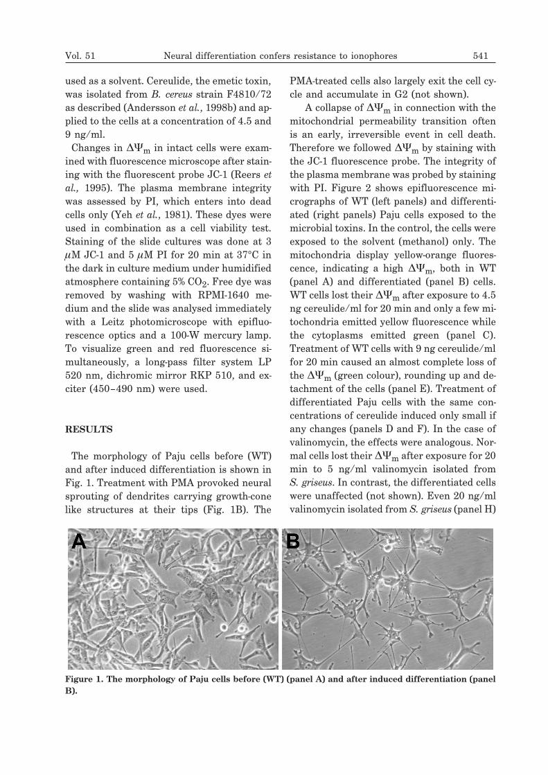
The morphology of Paju cells before (WT)and after induced differentiation is shown inFig. 1. Treatment with PMA provoked neuralsprouting of dendrites carrying growth-conelike structures at their tips (Fig. 1B). The
PMA-treated cells also largely exit the cell cy-cle and accumulate in G2 (not shown).
A collapse of ��m in connection with themitochondrial permeability transition oftenis an early, irreversible event in cell death.Therefore we followed ��m by staining withthe JC-1 fluorescence probe. The integrity ofthe plasma membrane was probed by stainingwith PI. Figure 2 shows epifluorescence mi-crographs of WT (left panels) and differenti-ated (right panels) Paju cells exposed to themicrobial toxins. In the control, the cells wereexposed to the solvent (methanol) only. Themitochondria display yellow-orange fluores-cence, indicating a high ��m, both in WT(panel A) and differentiated (panel B) cells.WT cells lost their ��m after exposure to 4.5ng cereulide/ml for 20 min and only a few mi-tochondria emitted yellow fluorescence whilethe cytoplasms emitted green (panel C).Treatment of WT cells with 9 ng cereulide/mlfor 20 min caused an almost complete loss ofthe ��m (green colour), rounding up and de-tachment of the cells (panel E). Treatment ofdifferentiated Paju cells with the same con-centrations of cereulide induced only small ifany changes (panels D and F). In the case ofvalinomycin, the effects were analogous. Nor-mal cells lost their ��m after exposure for 20min to 5 ng/ml valinomycin isolated fromS. griseus. In contrast, the differentiated cellswere unaffected (not shown). Even 20 ng/mlvalinomycin isolated from S. griseus (panel H)
Vol. 51 Neural differentiation confers resistance to ionophores 541
Figure 1. The morphology of Paju cells before (WT) (panel A) and after induced differentiation (panelB).
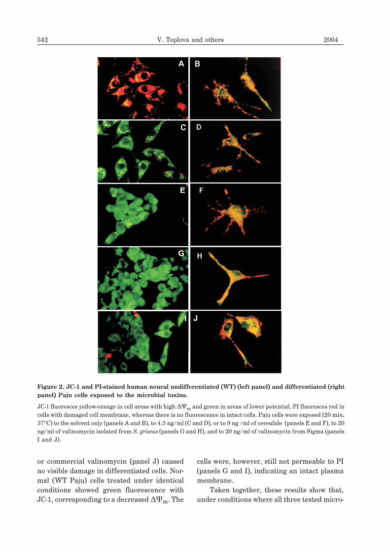
or commercial valinomycin (panel J) causedno visible damage in differentiated cells. Nor-mal (WT Paju) cells treated under identicalconditions showed green fluorescence withJC-1, corresponding to a decreased ��m. The
cells were, however, still not permeable to PI(panels G and I), indicating an intact plasmamembrane.
Taken together, these results show that,under conditions where all three tested micro-
542 V. Teplova and others 2004
Figure 2. JC-1 and PI-stained human neural undifferentiated (WT) (left panel) and differentiated (rightpanel) Paju cells exposed to the microbial toxins.
JC-1 fluoresces yellow-orange in cell areas with high ��m and green in areas of lower potential. PI fluoresces red incells with damaged cell membrane, whereas there is no fluorescence in intact cells. Paju cells were exposed (20 min,37°C) to the solvent only (panels A and B), to 4.5 ng/ml (C and D), or to 9 ng /ml of cereulide (panels E and F), to 20ng/ml of valinomycin isolated from S. griseus (panels G and H), and to 20 ng/ml of valinomycin from Sigma (panelsI and J).

bial toxins caused loss of ��m in WT Pajucells, the mitochondria in differentiated Pajucells displayed resistance to the toxins.
DISCUSSION
In this study Paju cells were used to monitorfor neurotoxicity of cereulide from B. cereus(Mikkola et al., 1999) and of valinomycin,both commercial and isolated from S. griseus(Andersson et al., 1998a). We examined theeffect of neuronal cell differentiation on thesensitivity to these microbial toxins. Pro-found effects of the toxins (all potassiumionophores) on undifferentiated Paju cellswere observed (Fig. 2). JC-1 staining showeda loss of ��m in normal cells within 20 minof exposure to the toxins, the cells roundedand detached from the monolayer. The cellslost viability without obvious damage to theplasma membrane. In contrast, in differenti-ated cells, mitochondria were largely intactafter similar exposure to the same toxins, in-dicating an increased resistance.Mitochondria may loose their ��m by three
main mechanisms: 1) ��m-driven uptake ofK+ as a positively charged ionophore com-plex; 2) Opening of the permeability transi-tion pore; 3) Increased transmembrane trans-port of protons, i.e. protonophorous uncou-pling. Since we deal here with K+ ionophores,mechanism 1) is mainly responsible for thedepolarisation. Given that K+ is the main cat-ion in the cytosol, the uptake of K+ could beconsiderable, resulting in extensive swellingof mitochondria. We might in addition havestimulation of the mitochondrial permeabilitytransition. This and swelling will cause somesecondary increase in H+ flux, i.e. uncouplingwith decreased synthesis of ATP, and hydro-lysis of ATP by the FoF1-ATPase. Mitochon-drial volume is regulated by the combined ac-tivities of the ATP-sensitive K+ channel andthe K+/H+ antiporter (Garlid & Paucek,2003). In the presence of potassium iono-phores it is to be expected, that the stimulated
influx of K+ and swelling results in activationof the antiporter, which in turn causes K+
efflux and influx of H+. This cycling mecha-nism may contribute to an uncoupling.The toxicity of the studied potassium
ionophores is mainly due to their effects onmitochondria, i.e. loss of ��m and swelling.This generally occurs in the mitochondrialpermeability transition, however, valino-mycin was found also to cause release ofcytochrome c without inducing permeabilitytransition (Shinohara et al., 2002). Swellingitself would rupture the outer membrane andrelease proapoptotic factors like cytochrome c(Cai et al., 1998). The associated decrease inATP would be expected to result in cell necro-sis (Lemasters et al., 2002), but the cell deathseen in this study rather corresponds toapoptosis.The molecular mechanism(s), conferring
higher resistance to the toxins in differen-tiated Paju cells, is not clear. The expres-sion of several proteins is increased duringinduced neural differentiation, amongwhich Bcl-2 is known to suppress the mito-chondrial permeability transition (Shimizuet al., 1998; Murphy et al., 2001). This prop-erty of Bcl-2 is not of interest since valino-mycin was found to release cytochrome cand induce apoptosis without permeabilitytransition (Shinohara et al., 2002). Stan-niocalcin has a protective effect on neu-rons during cerebral ischemia (Zhang etal., 2000). Moreover, binding of stannio-calcin to the inner membrane of mitochon-dria was recently reported and stan-niocalcin was found to stimulate electrontransport in submitochondrial particles invitro (McCudden et al., 2002). These obser-vations suggest that stanniocalcin maycontribute to the maintenance of the en-ergy state even under stressful conditions.Studies are in progress to elucidatewhether stanniocalcin or/and some otherfactors are instrumental for the increasedresistance to bacterial toxins in differenti-ated neuronal cells.
Vol. 51 Neural differentiation confers resistance to ionophores 543
R E F E R E N C E S
Andersson MA, Mikkola R, Kroppenstedt F,Rainey J, Reijula E, Hintikka E-L,Salkinoja-Salonen M. (1998a) Appl EnvironmMicrobiol.; 64: 4767–73.
Andersson MA, Mikkola R, Helin J, AnderssonMC, Salkinoja-Salonen M. (1998b) ApplEnvironm Microbiol.; 64: 1338–43.
Cai J, Yang J, Jones DP. (1998) Biochim BiophysActa.; 1366: 139–49.
Duax WL, Griffin JF, Langs DA, Smith GD,Groshulski P, Pletnev V, Ivanov V. (1996)Biopolymers.; 40: 141–55.
Garlid KD, Paucek P. (2003) Biochim. Biophys.Acta.; 1606: 23–41.
Gottlieb RA. (2000) FEBS Lett.; 482: 6–12.
Inai Y, Yabuki M, Akiyama J, Yasuda K. (1997)Cell Struct Funct.; 22: 555–63.
Krick S, Platoshin O, Sweeney M, Kim H, YuanJX. (2001) Am J Physiol Cell Physiol.; 280:C970–9.
Jääskeläinen E, Teplova V, Andersson MA,Andersson LC, Tammela P, Andersson MC,Pirhonen TI, Saris N-E, Vuorela P,Salkinoja-Salonen M. (2003) Toxicol In Vitro.;17: 737–44.
Lemasters JJ, Qian T, He L, Kim J-S, ElmoreSP, Cascio WE, Brenner DA. (2002) AntioxidRedox Signal.; 4: 769–81.
Li R, Mallahk RS. (2000) Neurosci Lett.; 294:147–50.
McCudden CR, James KA, Hasilo C, WagnerGF. (2002) J Biol Chem.; 277: 45249–58.
Mikkola R, Saris N-E, Grigoriev PA, AnderssonMA, Salkinoja-Salonen M. (1999) Eur JBiochem.; 263: 112–7.
Murphy RC, Schneider E, Kinnally KW. (2001)FEBS Lett.; 497: 73–6.
Paananen A, Mikkola R, Sareneva T,Matikainen S, Andersson M, Julkunen I,Salkinoja-Salonen M, Timonen T. (2000) ProcNatl Acad Sci USA.; 93: 4504–8.
Paananen A, Mikkola R, Sareneva T,Matikainen S, Hess M, Andersson M,Julkunen I, Salkinoja-Salonen M, Timonen T.(2002) Clin Exp Immunol.; 129: 420–8.
Penning LC, Denecker G, Vercammen D,Declercq W, Snipper RG, Vandenabeele P.(2000) Cytokine.; 12: 747–50.
Reers M, Smiley ST, Mottola-Hartshorn C, ChenA, Lin M, Chen LB. (1995) MethodsEnzymol.; 260: 406–17.
Shimizu S, Eguchi Y, Kamiike W, Funahashi Y,Mignov A, Lacronique V, Matsuda H,Tsujimoto Y. (1998) Proc Natl Acad SciUSA.; 95: 1455–9.
Shinohara Y, Almofti MR, Yamamoto T, IshidaT, Kita F, Kanzaki H, Ohnishi M, YamashitaK, Shimizu S, Terada H. (2002) Eur JBiochem.; 269: 5224–30.
Yeh CJ, His BL, Faulk WP. (1981) J ImmunolMethods.; 43: 269–75.
Zhang K-Z, Westberg JA, Hölttä E, AnderssonLC. (1996) Proc Natl Acad Sci USA.; 93:4504–8.
Zhang K-Z, Westberg JA, Paetau A,Bogyslawsky K, Lindsberg P, Erlander M,Guo H, Su J, Olsen HS, Andersson LC.(1998) Am J Pathol.; 153: 439–45.
Zhang K-Z, Lindsberg PJ, Tatlisumak T, KasteM, Olsen HS, Andersson LC. (2000) ProcNatl Acad Sci USA.; 97: 3637–42.
544 V. Teplova and others 2004