differentiallyexpressedmicrorna-483confersdistinct ... ·...
TRANSCRIPT

Differentially Expressed MicroRNA-483 Confers DistinctFunctions in Pancreatic �- and �-Cells*
Received for publication, March 8, 2015, and in revised form, June 15, 2015 Published, JBC Papers in Press, June 24, 2015, DOI 10.1074/jbc.M115.650705
Ramkumar Mohan‡1, Yiping Mao‡1, Shungang Zhang‡, Yu-Wei Zhang§, Cheng-Ran Xu§, Gérard Gradwohl¶,and Xiaoqing Tang‡2
From the ‡Department of Biological Sciences, Michigan Technological University, Houghton, Michigan 49931, the §College of LifeSciences, Peking-Tsinghua Center for Life Sciences, Peking University, Beijing 100871, China, and the ¶Institute of Genetics andMolecular and Cellular Biology, Department of Development and Stem cells, University of Strasbourg, 67404 Illkirch, France
Background: MicroRNAs are noncoding RNAs and have emerged as important regulators in �-cell function.Results: miR-483 is highly expressed in �-cells but expressed at much lower levels in �-cells, and increased miR-483 inducesinsulin production in �-cells while repressing glucagon production in �-cells.Conclusion: miR-483 has opposite effects in �- and �-cells by targeting SOCS3.Significance: miR-483 is a potential therapeutic target for diabetes.
Insulin secreted from pancreatic �-cells and glucagonsecreted from pancreatic �-cells are the two major hormonesworking in the pancreas in an opposing manner to regulate andmaintain a normal glucose homeostasis. How microRNAs(miRNAs), a population of non-coding RNAs so far demon-strated to be differentially expressed in various types of cells,regulate gene expression in pancreatic �-cells and its closelyassociated �-cells is not completely clear. In this study, miRNAprofiling was performed and compared between pancreatic�-cells and their partner �-cells. One novel miRNA, miR-483,was identified for its highly differential expression in pancreatic�-cells when compared to its expression in �-cells. Overexpres-sion of miR-483 in �-cells increased insulin transcription andsecretion by targeting SOCS3, a member of suppressor of cyto-kine signaling family. In contrast, overexpression of miR-483decreased glucagon transcription and secretion in �-cells.Moreover, overexpressed miR-483 protected against proinflam-matory cytokine-induced apoptosis in �-cells. This correlateswith a higher expression level of miR-483 and the expanded�-cell mass observed in the islets of prediabetic db/db mice.Together, our data suggest that miR-483 has opposite effects in�- and �-cells by targeting SOCS3, and the imbalance of miR-483 and its targets may play a crucial role in diabetespathogenesis.
Pancreatic islets consist of two major types of cells, the insu-lin-producing �-cells (50 –70%) and the glucagon-producing�-cells (20 –30%), which are physically organized asymmetri-cally together to maintain a normal glucose homeostasis (1).Insulin and glucagon are two recognized hormones and aresecreted in a coordinated manner in pancreatic cells (2). An
increase in insulin, triggered by high glucose, suppresses gluca-gon secretion and a decrease in insulin, in concert with lowglucose, stimulates glucagon secretion in healthy individuals(3). In type 2 diabetic patients, �-cell loss and insufficient insu-lin secretion contribute to hyperglycemia (4, 5). Furthermore, anumber of studies have reported that �-cell expansion and ele-vated glucagon secretion worsen the hyperglycemia in diabeticpatients (2, 6 – 8), suggesting that an imbalanced ratio between�-cell and �-cell mass leads to an imbalanced ratio betweeninsulin and glucagon, which contributes to the development ofdiabetes. Such imbalance has been well documented in diabetespatients, but the underlying mechanisms that promote thisimbalance are not fully understood.
Cell mass remodeling is the result of several processesincluding proliferation, neogenesis, cell size, and apoptosis (9).In diabetes patients, �-cell loss mainly reflects a combination ofimpaired �-cell proliferation and increased �-cell apoptosis(10). �-Cell proliferation is stimulated by nutrition and growthfactors including glucose, insulin, glucagon-like peptide-1(GLP-1,) and insulin-like growth factor (IGF-1/2)3 through sev-eral intracellular signaling pathways connecting cell surfacereceptors to proliferation machinery (11, 12). These signalingpathways include the insulin receptor substrate/PI3K/Akt(IRS-PI3K-Akt) pathways, JAK-STAT, MAPK, and calcineu-rin/nuclear factor of activated T-cells (NFAT) (13, 14). How-ever, high concentrations of glucose, leptin, free fatty acids,reactive oxygen species, or proinflammatory cytokines con-verge toward a common cell death signaling pathway (JNK/p38kinase) and cause induction of �-cell apoptosis in the pathogen-esis of type 2 diabetes (15).
In contrast, the mechanisms regulating �-cell proliferationand glucagon secretion are poorly understood. It is believedthat activated insulin signaling pathway stimulated by high glu-cose directly or indirectly via �-cell-secreted insulin repressesglucagon secretion in �-cells (16, 17). On the other hand, low* This work was supported by National Institutes of Health Grant DK084166
(to X. T.). The authors declare that they have no conflicts of interest with thecontents of this article.
1 Both authors contributed equally to this work.2 To whom correspondence should be addressed: Dept. of Biological Sci-
ences, Michigan Technological University, 1400 Townsend Dr., Houghton,MI 49931. Tel.: 906-487-1872; Fax: 906-487-3167; E-mail: [email protected].
3 The abbreviations used are: IGF-2, insulin-like growth factor 2; IRS2, insulinreceptor substrate 2; miRNA, microRNA; miR, microRNA; SOCS3, the sup-pressor of cytokine signaling 3; DIG, digoxigenin; KRB, Krebs-Ringer buffer;Ad, adenovirus; HPRT, hypoxanthine guanine phosphoribosyl transferase.
THE JOURNAL OF BIOLOGICAL CHEMISTRY VOL. 290, NO. 32, pp. 19955–19966, August 7, 2015© 2015 by The American Society for Biochemistry and Molecular Biology, Inc. Published in the U.S.A.
AUGUST 7, 2015 • VOLUME 290 • NUMBER 32 JOURNAL OF BIOLOGICAL CHEMISTRY 19955
by guest on October 8, 2018
http://ww
w.jbc.org/
Dow
nloaded from

glucose leads to inactivation of insulin signaling proteins that inturn stimulates glucagon secretion (18, 19). However, activatedinsulin signaling is considered to induce �-cell proliferationbased on the findings from �-cell-specific insulin receptorknock-out (�IRKO) mice and pancreas-specific IRS2 knock-out mice (20, 21). A recent study also demonstrated that insulinstimulates �-cell proliferation through the IR/IRS2/AKT/mTOR (mammalian target of rapamycin) signaling pathway(22). Much remains to be learned about the cross-talk betweenor among these pathways.
As a key player in gene expression regulation, microRNAs(miRNAs) are endogenous, noncoding RNAs of 21–24 nucleo-tides that bind to the 3�-UTR of target mRNAs thereby repress-ing their translation and/or promoting their decay (23, 24).With no exceptions to most eukaryotic cells, recent studies sug-gested that miRNAs have also emerged as important regulatorsin �-cell function and proliferation (25, 26). Several miRNAshave been identified to function in controlling and maintaining�-cell proliferation and mass expansion through targeting var-ious cellular signaling pathways. miR-375, the most abundantmiRNA detected in pancreatic islets, plays a key role in main-taining normal �-cell and �-cell mass in mice (27). Mice lackingmiR-375 have chronic hyperglycemia due to a decreased �-cellmass and increased �-cell numbers (27). Unlike miR-375,miR-7a was reported to inhibit mature �-cell proliferation bytargeting the mTOR signaling pathway, and transgenic miceoverexpressing miR-7a in �-cells developed diabetes due toimpaired insulin secretion and �-cell dedifferentiation (28, 29).Taken together, dysfunctional miRNAs contribute to inappro-priate �-cell responses during diabetes pathogenesis.
However, little is known about the functions of specificmiRNAs in �-cells and the mechanism for their differentialexpressions/functions detected in �- and �-cells. In this study,we have performed an miRNA screening to comprehensivelyassess miRNA expressions in �TC3 in contrast with �TC1-6,which are excellent surrogate systems for equivalent functionalstudies of primary �- and �-cells (30). We identified a numberof miRNAs including miR-483 that were differentiallyexpressed between �- and �-cells. In particular, miR-483 wasinvestigated for its unique higher expression in �-cells than in�-cells. Moreover, the glucose-stimulated miR-483 promotedinsulin secretion, insulin transcription, and cell proliferation bytargeting the suppressor of cytokine signaling 3 (SOCS3) in�-cells, whereas overexpression of miR-483 inhibits glucagontranscription and secretion in �-cells. Thus, miR-483 has oppo-site effects in �- and �-cells, and the imbalance of miR-483expression may contribute to diabetes pathophysiology.
Experimental Procedures
Cell Culture—Pancreatic �-cell line (�TC1-6) and �-cell line(�TC3) were purchased from the American Type Culture Col-lection (ATCC). Another �-cell line (MIN6) was kindly pro-vided by Dr. Miyazaki (University of Tokyo, Japan) (31). Cellswere routinely maintained in DMEM containing 25 mM glucosesupplemented with 15% FBS and 1% penicillin-streptomycin(Invitrogen) as described (32).
Mouse Islet Isolation and Flow Cytometric Cell Sorting—Wildtype C57BL/6 mice (number 000664), diabetic mice (db/db,
BKS.Cg-m�/�leprdb/J, number 000642), and their heterozy-gous lean controls (db/�) were 8 weeks old and obtained fromThe Jackson Laboratory (Bar Harbor, ME). All mice werehoused in the animal facility of Michigan Technological Uni-versity on a 12-h light/dark cycle with ad libitum access towater and normal chow. Pancreatic islets were isolated andpurified by intra-ductal perfusion of collagenase V (0.5 mg/ml)(Sigma) following the protocol described (33). The purifiedislets were cultured in RPMI 1640 medium supplemented with10% FBS and 1% penicillin-streptomycin for 24 –72 h accordingto the experiments. All experiments were carried out in accord-ance with the approval by the Animal Care Committee at theMichigan Technological University.
We performed FACS to obtain the purified �- and �-cellsfrom Ins1-mRFP (34) and glucagon-Cre/Rosa26R-YFP (35)mice, respectively. In preparation for sorting, isolated isletswere hand-picked and dissociated at 37 °C by adding 0.05%trypsin-EDTA as described previously (36). Digestion was inac-tivated by the addition of FCS, and dissociated cells were cen-trifuged and resuspended in PBS containing 10% FBS for sort-ing. Flow cytometric sorting was performed on a FACSAria (BDBiosciences) using 561 and 488 excitation lines for RFP andYFP, respectively. Sorted �- and �-cells were then collected inlysis buffer for subsequent RNA extraction. On average, thesorted populations were �98% pure with the final yield rangingbetween 60 and 80%.
MicroRNA Array and Data Analysis—Total RNA was iso-lated from both �TC3 and �TC1-6 cells using TRIzol (Invitro-gen), and the harvested small RNAs were radiolabeled andhybridized to the mouse miRNA array platform developed inour laboratory as described previously (37). The obtained datawere clustered using Cluster 3.0 (38) and visualized using JavaTreeView (39).
Quantitative Real-time PCR for miRNA and mRNA—TotalRNA from islets or cell lines was extracted using the miRNeasykit (Qiagen) according to the manufacturer’s instructions andtreated with rDNase I (Sigma). The TaqMan miRNA quantita-tive real-time PCR detection system (Applied Biosystems) wasused for quantification of miR-483, and its expression was nor-malized to the relative expression of RNU19. For mRNA quan-tification, cDNAs were generated using the High CapacitycDNA reverse transcription kit (Applied Biosystems), andquantitative real-time PCR was performed using the PowerSYBR Green PCR master mix (Applied Biosystems). Real-timePCR was performed on a StepOnePlusTM system (Applied Bio-systems) using the following procedure: 10 min at 95 °C, 40cycles of 95 °C for 15 s, and 60 °C for 1 min. All samples wererun in duplicate, and the RNA expression was determined usingrelative comparison method (��Ct), with hypoxanthine gua-nine phosphoribosyl transferase (Hprt) mRNA as an internalstandard. The following are the primers used in the study: pre-insulin, GGGGAGCGTGGCTTCTTCTA (forward) andGGGGACAGAATTCAGTGGCA (reverse); glucagon, AGAA-GAAGTCGCCATTGCTG (forward) and CCGCAGAGAT-GTTGTGAAGA (reverse); Hprt, TCAGTCAACGGGGGA-CATAAA (forward) and GGGGCTGTACTGCTTAACCAG(reverse).
Functions of MicroRNA-483 in Pancreatic �- and �-Cells
19956 JOURNAL OF BIOLOGICAL CHEMISTRY VOLUME 290 • NUMBER 32 • AUGUST 7, 2015
by guest on October 8, 2018
http://ww
w.jbc.org/
Dow
nloaded from

In Situ Hybridization and Immunohistochemistry Staining—Dissected mouse pancreas were fixed in 4% formaldehyde (pH7.4) for 24 h at 4 °C and then processed routinely for paraffinembedding. Tissues were cut into 5-�m sections and adheredto glass slides (Superfrost, Fisher Scientific). For in situ hybrid-ization, sections were first deparaffinized and rehydrated andthen treated with proteinase K (Roche Applied Science, 40�g/ml) as described (40). Briefly, a total of 3 pmol of DIG-labeled Locked Nucleic Acid (LNA) probes (Exiqon) weremixed with 200 �l of hybridization buffer and applied onto theslides to hybridize at 37 °C for overnight. Slides were thenwashed using 2� SSC solution and incubated with alkalinephosphatase-conjugated sheep anti-DIG antibody (RocheApplied Science) at 4 °C overnight. Alkaline phosphatase reac-tion was carried out with 50 mg/ml NBT/BCIP (4-nitro-bluetetrazolium/5-brom-4-chloro-3�-indolylphosphate) stainingsolution at room temperature for 1–3 days.
For combined in situ and immunofluorescence staining, fluo-rescent in situ hybridization was processed as above, except thatdetection of the DIG-labeled LNA probes was done with per-oxidase-conjugated sheep anti-DIG (Roche Applied Science)followed by Tyramide Signal Amplification (TSA)-FITC sub-strate (PerkinElmer) according to the manufacturer’s recom-mendations. The same slides were blocked in 0.5% BSA for 30min and incubated with mouse anti-insulin (Sigma) and rabbitanti-glucagon (Sigma) at 4 °C overnight. The next day, theimmunodetection was processed with Alexa Fluor 405- orAlexa Fluor 594-conjugated secondary antibodies (Invitrogen)at room temperature for 2 h. Slides were coverslipped usinganti-fading mounting medium (Vector Laboratories). Theimages were captured on an Olympus FluoView FV1000 con-focal microscope.
miRNA Transfection and Adenovirus Transduction—MIN6cells were electroporated with 5 �g of oligonucleotides or 10 �gof plasmids using the Amaxa Nucleofector system (Amaxa Inc.)according to the manufacturer’s instructions. 2 days after trans-fection, cells were treated with low (1 mM) or high (25 mM)glucose without serum for 16 h, and then cell lysates or totalRNA was prepared and subjected to analysis by Western blot-ting or real-time RT-PCR, respectively. For cytokine treatment,cells were incubated with 10 ng/ml cytokine mixture (TNF-�,IL-1�, and IFN�) in 25 mM glucose medium with 1% fetalbovine serum for the selected time. The following oligonucleo-tides were used in this study: miR-483 mimic, miRNA mimicnegative control (mimic control), anti-miR483, anti-miRNAnegative control (anti-control), and siRNA for Socs3. All theoligonucleotides were purchased from Life Technologies.
For overexpression of miR-483 in isolated islets, adenovirusvector containing miR-483 stem-loop precursor sequence (AD-miR483) was constructed using the RAPAd� miRNA adenovi-ral expression system (Cell Biolabs). Adenovirus containing theGFP was prepared as a control. The isolated islets were partiallydispersed, infected with purified adenovirus in RPMI 1640 with2% FBS for 2 h, and further incubated in the complete RPMI1640 medium overnight. Infected islets were cultured in thevirus-free complete RPMI 1640 medium for an additional 48 hprior to experimentation. Overexpression of miR-483 in isletswas confirmed by real-time PCR analysis of miR-483.
Immunoblot Analysis—Cells were lysed in lysis buffer sup-plemented with protease inhibitors and phosphatase inhibitorcocktails (Sigma). Lysates were resolved on a 10% SDS-PAGE,transferred to Immobilon transfer membrane (Millipore), andincubated with the following antibodies: SOCS3, IRS2 (Cell Sig-naling), Pdx-1 (EMD Millipore), MafA (Bethyl Laboratories),and �-actin (Sigma). Anti-rabbit and anti-mouse secondaryantibodies were purchased from GE Healthcare. Immunoblotswere developed using SuperSignal West Pico chemilumines-cent substrates (Thermo Scientific) and visualized on a Fujiimager. Protein levels were quantified using the ImageJsoftware.
Luciferase Assays for miRNA Target Validation—For evalua-tion of the predicted miR-483 complementary sites at the3�-UTR of Socs3 gene, the mouse Socs3 3�-UTR containingthree miR-483 binding sites (50 – 450 and 1000 –1320 bp) wassynthesized and subcloned into the pRLTK vector (Promega).For the luciferase reporter assay, pRLTK reporter constructs (5�g) were electroporated into MIN6 cells (1 � 106) with miR-483 mimic or control using Amaxa (Lonza). The plasmid PGL-3containing firefly luciferase (5 �g) was co-transfected togetherto normalize for transfection efficiency. Luciferase activity wasmeasured with a Dual-Luciferase reporter assay kit (Promega) 2days after transfection. All the related luciferase assay vectorswere provided by Dr. Peter Nelson (University of Kentucky,Lexington, KY) (40).
Insulin and Glucagon Secretion Assay—For quantification ofinsulin secretion, MIN6 cells were preincubated at 37 °C for 2 hin Krebs-Ringer buffer (KRB) (128.8 mM NaCl, 4.8 mM KCl, 1.2mM KH2PO4, 1.2 mM MgSO4, 2.5 mM CaCl, 5 mM NaHCO3, 10mM HEPES, 0.1% BSA) containing 1 mM glucose and then stim-ulated in the same buffer containing 25 mM glucose for 1 h. Thesupernatant from each treatment was then collected for insulinassay using a mouse insulin ELISA assay kit (Mercodia). Cellswere lysed in acid ethanol solution for total DNA. For glucagonsecretion assay, �TC1-6 cells were preincubated in KRB con-taining 25 mM glucose at 37 °C for 2 h. Subsequently, the cellswere stimulated with KRB containing 1 mM glucose for 1 h, andthe secreted glucagon in the collected supernatants was mea-sured by a mouse glucagon ELISA assay kit (Wako Chemicals).Both insulin and glucagon values were normalized to total cel-lular DNA content from the respective lysates.
For quantification of insulin and glucagon secretion in islets,virus-infected islets were transferred to 1.5-ml Eppendorf tubesand preincubated at 37 °C for 30 min in KRB supplementedwith 2.7 mM glucose (for insulin secretion) or 16.7 mM glucose(for glucagon secretion). The islets were then stimulated withKRB containing either 2.7 mM or 16.7 mM glucose for 1 h, andthe supernatant from each treatment was collected for insulinand glucagon assay. Islets were also lysed in acid ethanol solu-tion for total DNA isolation, and the total cellular DNA contentwas used to normalize the insulin and glucagon values.
Cell Apoptosis and Proliferation Assay—Two days aftertransfection, cells were seeded in 96-well plates (1 � 104 cells/well) and cultured for 16 h with or without cytokines (10 ng/ml)to induce apoptosis. Cytoplasmic histone-associated DNAfragments were quantified by the cell death detection ELISA kit(Roche Applied Science), which detects DNA laddering derived
Functions of MicroRNA-483 in Pancreatic �- and �-Cells
AUGUST 7, 2015 • VOLUME 290 • NUMBER 32 JOURNAL OF BIOLOGICAL CHEMISTRY 19957
by guest on October 8, 2018
http://ww
w.jbc.org/
Dow
nloaded from

from apoptotic cell death with anti-histone-biotin antibody andanti-DNA-peroxidase antibody. For proliferation assay, thethymidine analog BrdU (10 �M) was added to the culturemedium for the last 16 h of incubation prior to fixation, andDNA synthesis was measured using the cell proliferation ELISAkit (Roche Applied Science) according to the protocol.
Statistical Analysis—Data are expressed as means � S.D. ofthree independent experiments. Statistical significance wasdetermined by unpaired Student’s t test (two-tailed) or one-wayanalysis of variance with Tukey’s post hoc test with differencesconsidered significant at p � 0.05 (marked as *) and p � 0.01(marked as **).
Results
miR-483 Is Differentially Expressed between Pancreatic �-and �-Cells—To understand what are the major miRNAs thatare differentially expressed between pancreatic �- and �-cellsand their potential cellular functions, we profiled 553 selectedhuman and mouse miRNAs in �TC1-6 and �TC3 cells, respec-tively, using our optimized homemade miRNA array platform(37). About 108 miRNAs in total were detectable in the assay
analysis. Among these detectable miRNAs, most miRNAsshowed approximately equally expression in the two types ofpancreatic cells with a -fold change from 0.5 to 1.5 (Fig. 1, A andB). Among the differentially expressed miRNAs, nine, includ-ing miR-483, miR-375, miR-99b, and miR-24, were expressedsignificantly higher in �TC3, whereas 14 miRNAs, includingmiR-124 and miR-103/107, were expressed much higher in�TC1-6 (Fig. 1B).
We carefully examined the short list of candidate miRNAsthat potentially have a role in distinguishing the two types ofpancreatic cells and found that miR-483 expression was signif-icantly higher in �TC3, but almost undetectable in �TC1-6.Real-time PCR confirmed that the miR-483 level was 100-foldhigher in �TC3 than �TC1-6 (Fig. 1C). It is well known thathigh glucose induces opposite effects in �- and �-cells. We fur-ther examined the effect of glucose concentration on miR-483expression level. High glucose (25 mM) dramatically inducedmiR-483 level by 2.5-fold in �TC3 when compared with lowglucose (1 mM) (Fig. 1C). However, miR-483 level was not sig-nificantly induced by high glucose in �TC1-6. Similar to �TC3cells, miR-483 was also detected in another pancreatic � cell
FIGURE 1. miR-483 is highly expressed in pancreatic �-cells but detected at much lower levels in �-cells. A, miRNA array analysis in �TC3 and �TC1-6 cells.Total RNA was isolated, and small RNA was harvested by gel cleaning for radiolabeling. The radiolabeled small RNAs were then hybridized to an array platformcontaining a collection of mouse/human miRNAs. The identified miR-483 is marked with an oval. B, hierarchical clustering of the identified miRNAs based ontheir expression profiles using Gene Cluster 3.0 (average linkage and Euclidean distance as similarity measure). The data are the average of three independentexperiments � S.D. The expression of each miRNA was normalized to external and internal controls and median-centered prior to clustering. The expressionlevels ranged from �0.12 log10 to 0.12 log10. C, validation of miR-483 expression by real-time PCR. miR-483 was highly expressed and glucose-stimulated in�TC3 cells when compared with �TC1-6 cells. The expression of miR-483 was normalized to U6 RNA level, and the data are the average of three independentexperiments � S.D. **, p � 0.01
Functions of MicroRNA-483 in Pancreatic �- and �-Cells
19958 JOURNAL OF BIOLOGICAL CHEMISTRY VOLUME 290 • NUMBER 32 • AUGUST 7, 2015
by guest on October 8, 2018
http://ww
w.jbc.org/
Dow
nloaded from
line, MIN6 cells, a currently popular cell line for studying insu-lin production and signaling, so this cell line was therefore usedfor further functional studies of miR-483.
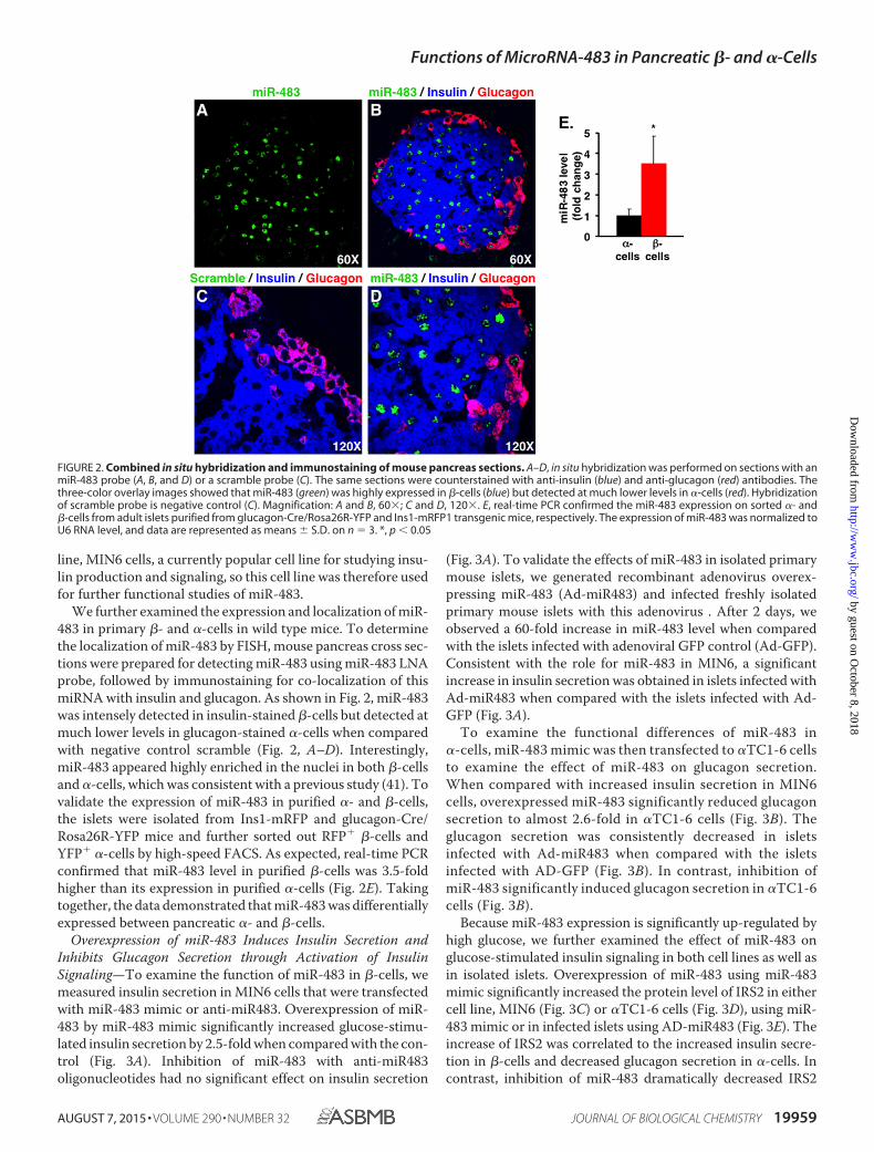
We further examined the expression and localization of miR-483 in primary �- and �-cells in wild type mice. To determinethe localization of miR-483 by FISH, mouse pancreas cross sec-tions were prepared for detecting miR-483 using miR-483 LNAprobe, followed by immunostaining for co-localization of thismiRNA with insulin and glucagon. As shown in Fig. 2, miR-483was intensely detected in insulin-stained �-cells but detected atmuch lower levels in glucagon-stained �-cells when comparedwith negative control scramble (Fig. 2, A–D). Interestingly,miR-483 appeared highly enriched in the nuclei in both �-cellsand �-cells, which was consistent with a previous study (41). Tovalidate the expression of miR-483 in purified �- and �-cells,the islets were isolated from Ins1-mRFP and glucagon-Cre/Rosa26R-YFP mice and further sorted out RFP� �-cells andYFP� �-cells by high-speed FACS. As expected, real-time PCRconfirmed that miR-483 level in purified �-cells was 3.5-foldhigher than its expression in purified �-cells (Fig. 2E). Takingtogether, the data demonstrated that miR-483 was differentiallyexpressed between pancreatic �- and �-cells.
Overexpression of miR-483 Induces Insulin Secretion andInhibits Glucagon Secretion through Activation of InsulinSignaling—To examine the function of miR-483 in �-cells, wemeasured insulin secretion in MIN6 cells that were transfectedwith miR-483 mimic or anti-miR483. Overexpression of miR-483 by miR-483 mimic significantly increased glucose-stimu-lated insulin secretion by 2.5-fold when compared with the con-trol (Fig. 3A). Inhibition of miR-483 with anti-miR483oligonucleotides had no significant effect on insulin secretion
(Fig. 3A). To validate the effects of miR-483 in isolated primarymouse islets, we generated recombinant adenovirus overex-pressing miR-483 (Ad-miR483) and infected freshly isolatedprimary mouse islets with this adenovirus . After 2 days, weobserved a 60-fold increase in miR-483 level when comparedwith the islets infected with adenoviral GFP control (Ad-GFP).Consistent with the role for miR-483 in MIN6, a significantincrease in insulin secretion was obtained in islets infected withAd-miR483 when compared with the islets infected with Ad-GFP (Fig. 3A).
To examine the functional differences of miR-483 in�-cells, miR-483 mimic was then transfected to �TC1-6 cellsto examine the effect of miR-483 on glucagon secretion.When compared with increased insulin secretion in MIN6cells, overexpressed miR-483 significantly reduced glucagonsecretion to almost 2.6-fold in �TC1-6 cells (Fig. 3B). Theglucagon secretion was consistently decreased in isletsinfected with Ad-miR483 when compared with the isletsinfected with AD-GFP (Fig. 3B). In contrast, inhibition ofmiR-483 significantly induced glucagon secretion in �TC1-6cells (Fig. 3B).
Because miR-483 expression is significantly up-regulated byhigh glucose, we further examined the effect of miR-483 onglucose-stimulated insulin signaling in both cell lines as well asin isolated islets. Overexpression of miR-483 using miR-483mimic significantly increased the protein level of IRS2 in eithercell line, MIN6 (Fig. 3C) or �TC1-6 cells (Fig. 3D), using miR-483 mimic or in infected islets using AD-miR483 (Fig. 3E). Theincrease of IRS2 was correlated to the increased insulin secre-tion in �-cells and decreased glucagon secretion in �-cells. Incontrast, inhibition of miR-483 dramatically decreased IRS2
FIGURE 2. Combined in situ hybridization and immunostaining of mouse pancreas sections. A–D, in situ hybridization was performed on sections with anmiR-483 probe (A, B, and D) or a scramble probe (C). The same sections were counterstained with anti-insulin (blue) and anti-glucagon (red) antibodies. Thethree-color overlay images showed that miR-483 (green) was highly expressed in �-cells (blue) but detected at much lower levels in �-cells (red). Hybridizationof scramble probe is negative control (C). Magnification: A and B, 60�; C and D, 120�. E, real-time PCR confirmed the miR-483 expression on sorted �- and�-cells from adult islets purified from glucagon-Cre/Rosa26R-YFP and Ins1-mRFP1 transgenic mice, respectively. The expression of miR-483 was normalized toU6 RNA level, and data are represented as means � S.D. on n 3. *, p � 0.05
Functions of MicroRNA-483 in Pancreatic �- and �-Cells
AUGUST 7, 2015 • VOLUME 290 • NUMBER 32 JOURNAL OF BIOLOGICAL CHEMISTRY 19959
by guest on October 8, 2018
http://ww
w.jbc.org/
Dow
nloaded from

expression in both MIN6 and �TC1-6 cells (Fig. 3C). Takentogether, the data suggested that miR-483 played a critical rolein regulating insulin/glucagon secretion via activating insulinsignaling.
Glucose-stimulated miR-483 Induces Insulin Transcriptionwhile Inhibiting Glucagon Transcription—To understandwhich step of insulin and glucagon expression is critical for therole of miR-483, we further examined the effect of miR-483 oninsulin and glucagon transcription. The miR-483 mimic oranti-miR483 was transfected into MIN6 and �TC1-6, followedby real-time PCR for the expression of pre-insulin and glucagonat their transcript levels. As shown in Fig. 4A, high glucose-stimulated pre-insulin mRNA was significantly increased inMIN6 cells transfected with miR-483 mimic when comparedwith the control. In contrast, transfection with anti-miR483resulted in an �2-fold reduction in pre-insulin mRNA, sug-gesting that miR-483 promotes insulin gene expression atthe transcriptional level under high glucose condition. How-ever, low glucose-induced glucagon mRNA was significantlyrepressed by overexpression of miR-483 or induced by anti-miR483 in �TC1-6 cells (Fig. 4B). These opposite effects inregulating insulin and glucagon transcription were furtherconfirmed in islets infected with Ad-miR483 (Fig. 4, A andB). We hypothesized that the induction of insulin gene tran-scription by miR-483 may be involved through the regula-tion of insulin transcription factors, such as Pdx-1 and MafA.However, expression of both Pdx-1 and MafA has no effectupon miR-483 overexpression or inhibition in MIN6 andislets Fig. 4C).
SOCS3 Is One of the Targets of miR-483 and Plays OppositeFunctions in �- and �-Cells—To understand which target genesare negatively regulated by miR-483 in activating insulin pro-duction and/or inhibiting glucagon production, we predictedcandidate target genes using miRNA target prediction pro-grams. Among many candidates, SOCS3 was shown to havetwo additional miR-483 complementary sites on its 3�-UTR atthe 1043- and 1230-bp regions (Fig. 5A), in addition to the onereported at the 295-bp region of the 3�-UTR in a study with theliver Hepa1-6 cells (42). These two complementary sites arehighly conserved among its homologs in various mammalianspecies including humans and mice, suggesting a critical role ofmiR-483 in regulating SOCS3, which may function in pancre-atic cells (Fig. 5A).
To examine a potential physical interaction between miR-483 and the three predicted binding sites at the 3�-UTR ofSOCS3 transcript, we synthesized two segments of mouseSOCS3 3�-UTR (SOCS3-3�-UTRI between 50 and 450 bp andSOCS3-3�-UTRII between 1000 and 1332 bp, respectively) andsubcloned them into the pRLTK vector (Promega). Thereporter constructs were co-electroporated into MIN6 cellswith anti-miR483 inhibitor or control using Amaxa (Lonza).When compared with the control, introduction of anti-miR483increased reporter activities of both SOCS3-3�-UTRI andSOCS3-3�-UTRII by 60%, suggesting that inhibition of miR-483abolished the repression of miR-483 on the luciferase reporteractivities (Fig. 5B).
To further determine whether SOCS3 is a potential target ofmiR-483, we examined the effect of miR-483 on SOCS3 protein
FIGURE 3. Overexpression of miR-483 induces insulin secretion but inhibits glucagon secretion via activated IRS2 signaling. A, insulin secretion wasmeasured in MIN6 transfected with miR-483 mimic and control mimic, or anti-miR483 and anti-control at 25 mM glucose, or in isolated islets infected withrecombinant adenovirus overexpressed miR-483 (AD-miR483) or control (Ad-GFP) at 16.7 mM glucose. B, glucagon secretion was analyzed in �TC1-6 trans-fected with miR-483 mimic or anti-miR483 at 1 mM glucose or in isolated islets infected with AD-miR483 at 2.7 mM glucose. Secreted insulin and glucagon levelsin the medium were quantified using mouse insulin and glucagon ELISA, respectively, and normalized to total cellular DNA content and then presented asrelative -fold. The presented data are the average of three independent experiments � S.D. *, p � 0.05, **, p � 0.01 versus control. C–E, transfected cells wereincubated with 1 or 25 mM glucose in DMEM for 16 h, and the expression of IRS2 and actin was analyzed by Western blot in MIN6 (C) and �TC1-6 (D) and isolatedislets (E). All the experiments were repeated 3– 6 times.
Functions of MicroRNA-483 in Pancreatic �- and �-Cells
19960 JOURNAL OF BIOLOGICAL CHEMISTRY VOLUME 290 • NUMBER 32 • AUGUST 7, 2015
by guest on October 8, 2018
http://ww
w.jbc.org/
Dow
nloaded from

level in MIN6, �TC1-6, and isolated islets. Overexpression ofmiR-483 using miR-483 mimic decreased SOCS3 protein levelin both MIN6 and �TC1-6 when compared with the control(Fig. 5, C and D). AD-miR483-infected islets also showed areduction in SOCS3 expression when compared with the AD-GFP control (Fig. 5E). In contrast, inhibition of miR-483 usinganti-miR483 significantly increased SOCS3 protein level (Fig.5C). Taken together, these data demonstrated that SOCS3 is adirect target of miR-483 in both �-cells and �-cells.
In the insulin-sensitive peripheral tissues, including whiteadipose tissue, liver, and muscle, SOCS3 inhibits insulin actionthrough binding IRS2 and causing IRS2 ubiquitination and deg-radation (43– 46). To validate the function of SOCS3 in MIN6and �TC1-6 cells, we silenced SOCS3 by transfecting the cellswith SOCS3 siRNA (Applied Biosystems). As shown in Fig. 5F,significant reduction of SOCS3 expression was observed in cellsexpressing either SOCS3 siRNA-1 or siRNA-2. As expected,silencing of SOCS3 significantly increased IRS2 expression byalmost 3-fold in MIN6 (Fig. 5F). Moreover, SOCS3 silencing ledto an increase in both insulin secretion and insulin transcrip-tion (Fig. 5G), which is consistent with a previous study inwhich SOCS3 negatively regulated insulin secretion and insulintranscription in �-cell lines and SOCS3 transgenic mice (47–49). In contrast, silencing of SOCS3 significantly inhibited bothglucagon secretion and transcription in �TC1-6 cells (Fig. 5H).
miR-483 Protects against Cytokine-mediated Apoptosis in�-Cells—Studies have shown that SOCS3 is induced by inflam-matory cytokines and can inhibit excessive cell growth andinduce apoptosis as part of maintaining cell stability (50 –52).We hypothesized that miR-483 may be involved in protecting�-cell growth by targeting SOCS3. A cytokine mixture with 10ng/ml each of TNF�, IL-1�, and IFN� was incubated withMIN6 cells to induce apoptosis. Although overexpression of
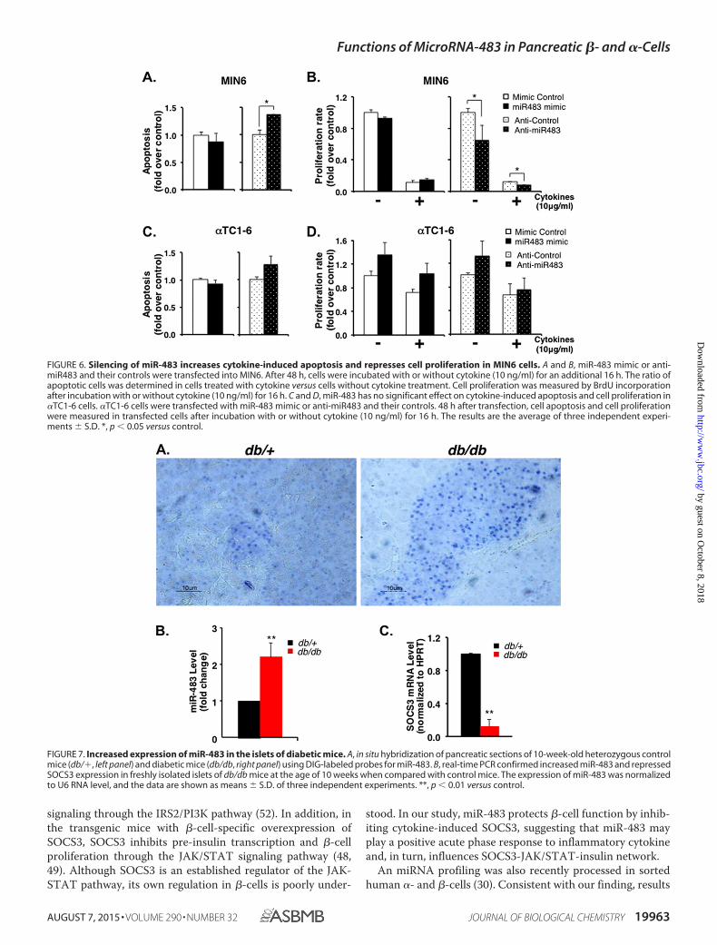
miR-483 had no significant effect on apoptosis, silencing ofmiR-483 by anti-miR483 increased cytokine-induced apoptosisin MIN6 cells (Fig. 6A). Consistently, the cell proliferation ratewas significantly decreased by anti-miR483 when cells wereexposed to cytokine (Fig. 6B), suggesting that miR-483 is nec-essary to inhibit target protein SOCS3 to protect �-cells againstcytokine-induced apoptosis. However, such profound protec-tion was not observed in �TC1-6 cells. Overexpression or inhi-bition of miR-483 had no significant effect on apoptosis (Fig.6C) and cell proliferation under both normal growth conditionsand cytokine treatment in �TC1-6 cells (Fig. 6D). Takentogether, pancreatic �- and �-cells have distinct characteristicsin the expressions of miR-483 and its target gene SOSC3 as wellas their responses to cytokines in terms of cell apoptosis andproliferation.
Increased Expression of miR-483 in the Islets of DiabeticMice—To examine the change of miR-483 expression duringthe development of diabetes, we harvested the pancreases from10-week-old db/db or control lean mice (db/�) and preparedthem for in situ hybridization. When compared with the con-trol mice, miR-483 expression increased in the islets of predia-betic mice in correspondence with the increase of the �-cellmass (53) (Fig. 7A). Real-time PCR further confirmed that thisincrease of miR-483 was almost 2-fold in isolated islets fromprediabetic mice when compared with control mice (Fig. 7B).
The isolated islets were further analyzed for the expression ofSOCS3 by real-time PCR. When compared with control leanmice, SOCS3 mRNA level declined 8-fold in prediabetic islets,correlating with the increase of miR-483 in prediabetic islets(Fig. 7C). Thus, our in vitro data for functions of miR-483 werealso supported by the in vivo changes of miR-483 and SOCS3 inprediabetic mice.
FIGURE 4. miR-483 promotes insulin transcription while repressing glucagon transcription. A, MIN6 were transfected with miR-483 mimic or anti-miR483,or isolated islets were infected with AD-miR483. After 48 h, transfected cells were incubated with 25 mM glucose in DMEM (for MIN6) or 16.7 mM glucose (forislets) for 16 h, and total RNA was extracted to analyze the expression of pre-insulin by real-time PCR. B, 48 h after miR-483 mimic transfection in �TC1-6 orAD-miR483 in isolated islets, transfected cells were incubated with 1 mM glucose in DMEM (for �TC1-6) or 2.7 mM glucose (for islets) for 16 h, and total RNA wasextracted to analyze the expression of glucagon by real-time PCR. The data were normalized to internal control HPRT, and the results are the average of threeindependent experiments � S.D.; **, p � 0.01 versus mimic control. C, the expression of MafA, Pdx-1, and actin was analyzed by Western blot in transfectedMIN6 and islets. Results are representative of three independent experiments.
Functions of MicroRNA-483 in Pancreatic �- and �-Cells
AUGUST 7, 2015 • VOLUME 290 • NUMBER 32 JOURNAL OF BIOLOGICAL CHEMISTRY 19961
by guest on October 8, 2018
http://ww
w.jbc.org/
Dow
nloaded from

Discussion
In this study, we first observed that the expression of miR-483 was much higher in pancreatic �-cells than in �-cells. Glu-cose-stimulated miR-483 stimulated IRS2-mediated insulinsignaling, leading to increased insulin secretion in �-cells.Moreover, miR-483 promoted insulin transcription by directlytargeting SOCS3. In contrast, overexpression of miR-483repressed low glucose-stimulated glucagon secretion and tran-scription in �-cells. In addition, miR-483 serves as a �-cell sur-vival factor to suppress cytokine-induced apoptosis in �-cellswhile serving no significant effect in �-cells. Increased miR-483is shown in the islets of prediabetic mice, suggesting that miR-483 may facilitate compensatory �-cell mass expansion duringthe early stage of diabetes.
miRNAs typically recognize their target mRNAs throughcomplementary sites of their seed region to the binding siteslocated on the 3�-UTR of the target mRNA. However, we
revealed that the 3�-UTR of SOCS3 contains three atypicalmiR-483 recognition sites that are not perfectly base-paringwith the seed region of miR-483 (Fig. 5A). We demonstratedthat these atypical miR-483 recognition sites were valid andsufficient to mediate the suppression of SOCS3 by miR-483.These atypical recognitions between miRNAs and their targetsare also supported by previous studies with numerous bio-chemical and structural findings on miRNA-target interactions(54, 55).
miR-483 has been reported to be expressed in mouse liverand adipocyte tissues and regulates fat metabolism by targetingSOCS3 (42). SOCS3 belongs to the SOCS family of inhibitoryproteins, which can interfere with insulin signaling in muscle,liver, and adipose tissue by inhibition of tyrosine phosphoryla-tion of IRS2 (56). In pancreatic �-cells, SOCS3 is induced byvarious cytokines and complexes with insulin receptor, leadingto reduced insulin receptor autophosphorylation and impaired
FIGURE 5. SOCS3 is one of the targets of miR-483 in pancreatic �-cells. A, bioinformatic prediction of the interaction between miR-483 and the 3�-UTRs ofSOCS3 of various species. Has, Ptr, Rno, and Mmu represent human, chimpanzee, rat, and mouse, respectively. The predicted three binding sites are indicatedin blue. B, luciferase assay confirmed that miR-483 inhibited the luciferase reporter activity, in which the SOCS3 3�-UTR fragments (50 – 450 and 1000 –1320 bp)were fused with a Renilla luciferase reporter gene in pRLTK, respectively. The reporter constructs were co-transfected into MIN6 cells with either anti-miR483or anti-control. The pGL3 firefly luciferase plasmid was co-transfected for detection of transfection efficiency. 48 h after transfection, luciferase activity wasassayed using the Dual-Luciferase reporter assay kit. Data represent means � S.E. of three independent experiments. **, p � 0.01. C–E, overexpression ofmiR-483 down-regulates SOCS3 protein expression in MIN6 (C), �TC1-6 (D), and isolated islets (E) validated by Western blots. Data are representative of threeindependent experiments. F, silencing of SOCS3 activated the expression of IRS2. 48 h after transfection with siRNA against SOCS3 (#1 and #2) or Scramble (Scr)in MIN6 cells, cell lysates were prepared for Western blots to detect the expression of IRS2, SOCS3, and actin. Results are representative of three independentexperiments. G, silencing of SOCS3 increased insulin secretion and insulin transcription. H, silencing of SOCS3 reduced glucagon secretion and transcription.Secreted insulin and glucagon levels in the medium were quantified using mouse insulin or glucagon ELISA and normalized to total cellular DNA contents.Total RNA was extracted to analyze the expression of insulin or glucagon by real-time PCR. The data were normalized to internal control HPRT. The presenteddata are the average of three independent experiments � S.D. *, p � 0.05, **, p � 0.01.
Functions of MicroRNA-483 in Pancreatic �- and �-Cells
19962 JOURNAL OF BIOLOGICAL CHEMISTRY VOLUME 290 • NUMBER 32 • AUGUST 7, 2015
by guest on October 8, 2018
http://ww
w.jbc.org/
Dow
nloaded from
signaling through the IRS2/PI3K pathway (52). In addition, inthe transgenic mice with �-cell-specific overexpression ofSOCS3, SOCS3 inhibits pre-insulin transcription and �-cellproliferation through the JAK/STAT signaling pathway (48,49). Although SOCS3 is an established regulator of the JAK-STAT pathway, its own regulation in �-cells is poorly under-
stood. In our study, miR-483 protects �-cell function by inhib-iting cytokine-induced SOCS3, suggesting that miR-483 mayplay a positive acute phase response to inflammatory cytokineand, in turn, influences SOCS3-JAK/STAT-insulin network.
An miRNA profiling was also recently processed in sortedhuman �- and �-cells (30). Consistent with our finding, results
FIGURE 6. Silencing of miR-483 increases cytokine-induced apoptosis and represses cell proliferation in MIN6 cells. A and B, miR-483 mimic or anti-miR483 and their controls were transfected into MIN6. After 48 h, cells were incubated with or without cytokine (10 ng/ml) for an additional 16 h. The ratio ofapoptotic cells was determined in cells treated with cytokine versus cells without cytokine treatment. Cell proliferation was measured by BrdU incorporationafter incubation with or without cytokine (10 ng/ml) for 16 h. C and D, miR-483 has no significant effect on cytokine-induced apoptosis and cell proliferation in�TC1-6 cells. �TC1-6 cells were transfected with miR-483 mimic or anti-miR483 and their controls. 48 h after transfection, cell apoptosis and cell proliferationwere measured in transfected cells after incubation with or without cytokine (10 ng/ml) for 16 h. The results are the average of three independent experi-ments � S.D. *, p � 0.05 versus control.
FIGURE 7. Increased expression of miR-483 in the islets of diabetic mice. A, in situ hybridization of pancreatic sections of 10-week-old heterozygous controlmice (db/�, left panel) and diabetic mice (db/db, right panel) using DIG-labeled probes for miR-483. B, real-time PCR confirmed increased miR-483 and repressedSOCS3 expression in freshly isolated islets of db/db mice at the age of 10 weeks when compared with control mice. The expression of miR-483 was normalizedto U6 RNA level, and the data are shown as means � S.D. of three independent experiments. **, p � 0.01 versus control.
Functions of MicroRNA-483 in Pancreatic �- and �-Cells
AUGUST 7, 2015 • VOLUME 290 • NUMBER 32 JOURNAL OF BIOLOGICAL CHEMISTRY 19963
by guest on October 8, 2018
http://ww
w.jbc.org/
Dow
nloaded from

form their study confirmed that human �-cells contain highermiR-483 than �-cells (30). A pivotal observation in our work isthat miR-483 is stimulated by high glucose and exerts an oppo-site effect on insulin and glucagon release. First, glucose-stim-ulated miR-483 induces insulin production and release from�-cells, which in turn suppresses glucagon release from �-cells.Insulin has been considered as a negative regulator of glucagonrelease (3, 16). Second, miR-483-activated IRS2 expression andinsulin signaling inhibit glucagon secretion. In addition, ourstudy showed that highly expressed miR-483 protects �-cellgrowth against cytokine-induced apoptosis, suggesting thatthese cells, although derived from common precursor cells,evolved into closely related types of cells with distinct func-tions. Taken together, the differentially expressed miR-483plays a crucial role in the maintenance of �- and �-cell mass andfunction.
Over one-third of mammalian miRNAs are located withinthe introns of protein-coding genes, referred to as intronicmiRNAs (57). miR-483 is an intronic miRNA and is locatedwithin the intron of the Igf2 gene (42). IGF-2 was detected at arelatively high level in islets, and reduced IGF-2 expression wasobserved in the impaired islets of the adult Goto-Kakizaki ratmodel of type 2 diabetes (58). The transgenic mice with �-cell-specific overexpression of IGF-2 showed an increase in insulinmRNA level and �-cell mass (59). Moreover, the persistentpresence of circulating IGF-II largely prevented �-cell apopto-sis in the early postnatal period (60, 61). It is possible that miR-483 is not only transcriptionally linked to IGF-2 expression, butalso coordinately regulated under different physiological con-ditions. Interestingly, miR-483 was found to be highly enrichedin the nucleus of both �-cells and �-cells. In support, a numberof miRNAs have been identified to directly bind to the genepromoter, potentially inducing overexpression or down-regu-lation of target genes (62, 63). More recently, overexpressedmiR-483 in the pediatric kidney cancer cells localized in thenuclei and enhanced the transcription of IGF-2 transcripts bybinding to its 5�-UTR (41). However, the interaction betweennuclear miR-483 and IGF-2 in �- and �-cells still needs to befurther examined under physiological conditions. Further-more, insulin gene is clustered closely together with IGF-2gene, suggesting that miR-483 and IGF-2, together with insu-lin, might be controlled by a common regulatory mecha-nism. Therefore, identification of the coordinated interac-tion between miR-483 and IGF-2 will be important forunderstanding the differential expression of miR-483 between�- and �-cells and establishing a functional relationshipbetween them.
Two studies have recently reported miRNA profiling in thesorted human �- and �-cells or �- and �-cell lines by themiRNA PCR array method (30, 64). Among the identifiedmiRNAs in these studies, most of them, including miR-483,miR-375, and miR-99b, expressed at a relatively higher level in�-cells, as confirmed in our array. Other miRNAs, such as miR-124 and miR-103/107, expressed relatively higher in �-cells inour array. miR-124 has been shown to regulate pancreas devel-opment and insulin exocytosis and induce neural differentia-tion and proliferation (65, 66). miR-103/107 has been demon-strated to induce insulin resistance and glucose intolerance in
both liver and adipocytes (67). Therefore, the data suggest thatpro- or anti-proliferative miRNAs co-exist in both �-cells and�-cells and function interactively in maintaining the identityand functions of �- and �-cells. It will be of interest to dissectthe functional interactions of these miRNAs and their homeo-static roles in the development of diabetes.
In summary, we characterized the distinct expression andfunctions of miR-483 between �- and �-cells. miR-483 can bestimulated by glucose and insulin, and activated miR-483 isrequired for glucose-stimulated IRS2 signaling and protects�-cells against apoptosis. The balance of miR-483 and its tar-gets would not only protect �-cell function, but also help topreserve �-cell mass. Future dissection of the interaction ofmiR-483 with the SOCS3-mediated functional pathway mayextend our understanding on the potential therapeutic role ofmiR-483.
Author Contributions—X. T. performed the experiments shown inFig. 1, coordinated the study, and wrote the paper. R. M. designed,performed, and analyzed the experiments shown in Figs. 3–7. Y. M.designed and performed the experiments shown in Figs. 2 and 7. S. Z.constructed vectors and performed the luciferase reporter assayshown in Fig. 5. Y. Z., and C. X. performed the fluores-cence-activated cell sorting analysis shown in Fig. 2. G. G. providedIns1-mRFP mice. All authors analyzed the results and approved thefinal version of the manuscript.
Acknowledgments—We thank Dr. Peter Nelson for providing lucifer-ase reporter plasmids, Dr. Guiliang Tang for critical reading andediting of this manuscript, and the undergraduate students AustinBurke and Pedro Rodrigues for technical assistance.
References1. Cabrera, O., Berman, D. M., Kenyon, N. S., Ricordi, C., Berggren, P. O., and
Caicedo, A. (2006) The unique cytoarchitecture of human pancreatic is-lets has implications for islet cell function. Proc. Natl. Acad. Sci. U.S.A.103, 2334 –2339
2. Breckenridge, S. M., Cooperberg, B. A., Arbelaez, A. M., Patterson, B. W.,and Cryer, P. E. (2007) Glucagon, in concert with insulin, supports thepostabsorptive plasma glucose concentration in humans. Diabetes 56,2442–2448
3. Cooperberg, B. A., and Cryer, P. E. (2010) Insulin reciprocally regulatesglucagon secretion in humans. Diabetes 59, 2936 –2940
4. Porte, D., Jr., and Kahn, S. E. (2001) �-Cell dysfunction and failure in type2 diabetes: potential mechanisms. Diabetes 50, Suppl. 1, S160 –S163,10.2337/diabetes.50.2007.S160
5. Butler, A. E., Janson, J., Bonner-Weir, S., Ritzel, R., Rizza, R. A., and Butler,P. C. (2003) �-Cell deficit and increased �-cell apoptosis in humans withtype 2 diabetes. Diabetes 52, 102–110
6. D’Alessio, D. (2011) The role of dysregulated glucagon secretion intype 2 diabetes. Diabetes Obes. Metab. 13, Suppl. 1, 126 –132,10.1111/j.1463-1326.2011.01449.x
7. Yoon, K. H., Ko, S. H., Cho, J. H., Lee, J. M., Ahn, Y. B., Song, K. H., Yoo,S. J., Kang, M. I., Cha, B. Y., Lee, K. W., Son, H. Y., Kang, S. K., Kim, H. S.,Lee, I. K., and Bonner-Weir, S. (2003) Selective �-cell loss and �-cell ex-pansion in patients with type 2 diabetes mellitus in Korea. J. Clin. Endo-crinol. Metab. 88, 2300 –2308
8. Deng, S., Vatamaniuk, M., Huang, X., Doliba, N., Lian, M. M., Frank, A.,Velidedeoglu, E., Desai, N. M., Koeberlein, B., Wolf, B., Barker, C. F., Naji,A., Matschinsky, F. M., and Markmann, J. F. (2004) Structural and func-tional abnormalities in the islets isolated from type 2 diabetic subjects.Diabetes 53, 624 – 632
Functions of MicroRNA-483 in Pancreatic �- and �-Cells
19964 JOURNAL OF BIOLOGICAL CHEMISTRY VOLUME 290 • NUMBER 32 • AUGUST 7, 2015
by guest on October 8, 2018
http://ww
w.jbc.org/
Dow
nloaded from

9. Montanya, E., and Téllez, N. (2009) Pancreatic remodeling: �-cell apopto-sis, proliferation and neogenesis, and the measurement of �-cell mass andof individual �-cell size. Methods Mol. Biol. 560, 137–158
10. Kassem, S. A., Ariel, I., Thornton, P. S., Scheimberg, I., and Glaser, B.(2000) �-Cell proliferation and apoptosis in the developing normal humanpancreas and in hyperinsulinism of infancy. Diabetes 49, 1325–1333
11. Heit, J. J., Karnik, S. K., and Kim, S. K. (2006) Intrinsic regulators of pan-creatic �-cell proliferation. Annu. Rev. Cell Dev. Biol. 22, 311–338
12. Kulkarni, R. N. (2005) New insights into the roles of insulin/IGF-I in thedevelopment and maintenance of �-cell mass. Rev. Endocr. Metab. Disord.6, 199 –210
13. Kulkarni, R. N., Mizrachi, E. B., Ocana, A. G., and Stewart, A. F. (2012)Human �-cell proliferation and intracellular signaling: driving in the darkwithout a road map. Diabetes 61, 2205–2213
14. Bernal-Mizrachi, E., Kulkarni, R. N., Scott, D. K., Mauvais-Jarvis, F., Stew-art, A. F., and Garcia-Ocaña, A. (2014) Human �-cell proliferation andintracellular signaling part 2: still driving in the dark without a road map.Diabetes 63, 819 – 831
15. Mandrup-Poulsen, T. (2001) �-Cell apoptosis: stimuli and signaling. Dia-betes 50, Suppl. 1, S58 –S63, 10.2337/diabetes.50.2007.S58
16. Ravier, M. A., and Rutter, G. A. (2005) Glucose or insulin, but not zincions, inhibit glucagon secretion from mouse pancreatic �-cells. Diabetes54, 1789 –1797
17. Diao, J., Asghar, Z., Chan, C. B., and Wheeler, M. B. (2005) Glucose-regulated glucagon secretion requires insulin receptor expression in pan-creatic �-cells. J. Biol. Chem. 280, 33487–33496
18. Zhou, H., Tran, P. O., Yang, S., Zhang, T., LeRoy, E., Oseid, E., and Rob-ertson, R. P. (2004) Regulation of �-cell function by the �-cell duringhypoglycemia in Wistar rats: the “switch-off” hypothesis. Diabetes 53,1482–1487
19. Hope, K. M., Tran, P. O., Zhou, H., Oseid, E., Leroy, E., and Robertson,R. P. (2004) Regulation of �-cell function by the �-cell in isolated humanand rat islets deprived of glucose: the “switch-off” hypothesis. Diabetes 53,1488 –1495
20. Cantley, J., Choudhury, A. I., Asare-Anane, H., Selman, C., Lingard, S.,Heffron, H., Herrera, P., Persaud, S. J., and Withers, D. J. (2007) Pancreaticdeletion of insulin receptor substrate 2 reduces � and � cell mass andimpairs glucose homeostasis in mice. Diabetologia 50, 1248 –1256
21. Kawamori, D., Kurpad, A. J., Hu, J., Liew, C. W., Shih, J. L., Ford, E. L.,Herrera, P. L., Polonsky, K. S., McGuinness, O. P., and Kulkarni, R. N.(2009) Insulin signaling in � cells modulates glucagon secretion in vivo.Cell Metab. 9, 350 –361
22. Liu, Z., Kim, W., Chen, Z., Shin, Y. K., Carlson, O. D., Fiori, J. L., Xin, L.,Napora, J. K., Short, R., Odetunde, J. O., Lao, Q., and Egan, J. M. (2011)Insulin and glucagon regulate pancreatic �-cell proliferation. PLoS One 6,e16096
23. Bartel, D. P. (2009) MicroRNAs: target recognition and regulatory func-tions. Cell 136, 215–233
24. Broderick, J. A., and Zamore, P. D. (2011) MicroRNA therapeutics. Gene.Ther. 18, 1104 –1110
25. Guay, C., Roggli, E., Nesca, V., Jacovetti, C., and Regazzi, R. (2011) Diabe-tes mellitus, a microRNA-related disease? Transl. Res. 157, 253–264
26. Mao, Y., Mohan, R., Zhang, S., and Tang, X. (2013) MicroRNAs as phar-macological targets in diabetes. Pharmacol. Res. 75, 37– 47
27. Poy, M. N., Hausser, J., Trajkovski, M., Braun, M., Collins, S., Rorsman, P.,Zavolan, M., and Stoffel, M. (2009) miR-375 maintains normal pancreatic�- and �-cell mass. Proc. Natl. Acad. Sci. U.S.A. 106, 5813–5818
28. Wang, Y., Liu, J., Liu, C., Naji, A., and Stoffers, D. A. (2013) MicroRNA-7regulates the mTOR pathway and proliferation in adult pancreatic �-cells.Diabetes 62, 887– 895
29. Latreille, M., Hausser, J., Stützer, I., Zhang, Q., Hastoy, B., Gargani, S.,Kerr-Conte, J., Pattou, F., Zavolan, M., Esguerra, J. L., Eliasson, L., Rülicke,T., Rorsman, P., and Stoffel, M. (2014) MicroRNA-7a regulates pancreatic� cell function. J. Clin. Invest. 124, 2722–2735
30. Klein, D., Misawa, R., Bravo-Egana, V., Vargas, N., Rosero, S., Piroso, J.,Ichii, H., Umland, O., Zhijie, J., Tsinoremas, N., Ricordi, C., Inverardi, L.,Domínguez-Bendala, J., and Pastori, R. L. (2013) MicroRNA expression in� and � cells of human pancreatic islets. PLoS One 8, e55064
31. Miyazaki, J., Araki, K., Yamato, E., Ikegami, H., Asano, T., Shibasaki, Y.,Oka, Y., and Yamamura, K. (1990) Establishment of a pancreatic � cell linethat retains glucose-inducible insulin secretion: special reference to ex-pression of glucose transporter isoforms. Endocrinology 127, 126 –132
32. Tang, X., Muniappan, L., Tang, G., and Ozcan, S. (2009) Identification ofglucose-regulated miRNAs from pancreatic � cells reveals a role for miR-30d in insulin transcription. RNA 15, 287–293
33. Brissova, M., Fowler, M., Wiebe, P., Shostak, A., Shiota, M., Radhika, A.,Lin, P. C., Gannon, M., and Powers, A. C. (2004) Intraislet endothelial cellscontribute to revascularization of transplanted pancreatic islets. Diabetes53, 1318 –1325
34. Piccand, J., Meunier, A., Merle, C., Jia, Z., Barnier, J. V., and Gradwohl, G.(2014) Pak3 promotes cell cycle exit and differentiation of �-cells in theembryonic pancreas and is necessary to maintain glucose homeostasis inadult mice. Diabetes 63, 203–215
35. Herrera, P. L. (2000) Adult insulin- and glucagon-producing cells differ-entiate from two independent cell lineages. Development 127, 2317–2322
36. Xu, C. R., Li, L. C., Donahue, G., Ying, L., Zhang, Y. W., Gadue, P., andZaret, K. S. (2014) Dynamics of genomic H3K27me3 domains and role ofEZH2 during pancreatic endocrine specification. EMBO J. 33, 2157–2170
37. Tang, X., Gal, J., Zhuang, X., Wang, W., Zhu, H., and Tang, G. (2007) Asimple array platform for microRNA analysis and its application in mousetissues. RNA 13, 1803–1822
38. de Hoon, M. J., Imoto, S., Nolan, J., and Miyano, S. (2004) Open sourceclustering software. Bioinformatics 20, 1453–1454
39. Saldanha, A. J. (2004) Java Treeview: extensible visualization of microarraydata. Bioinformatics 20, 3246 –3248
40. Wang, W. X., Rajeev, B. W., Stromberg, A. J., Ren, N., Tang, G., Huang, Q.,Rigoutsos, I., and Nelson, P. T. (2008) The expression of microRNA miR-107 decreases early in Alzheimer’s disease and may accelerate diseaseprogression through regulation of �-site amyloid precursor protein-cleav-ing enzyme 1. J. Neurosci. 28, 1213–1223
41. Liu, M., Roth, A., Yu, M., Morris, R., Bersani, F., Rivera, M. N., Lu, J.,Shioda, T., Vasudevan, S., Ramaswamy, S., Maheswaran, S., Diederichs, S.,and Haber, D. A. (2013) The IGF2 intronic miR-483 selectively enhancestranscription from IGF2 fetal promoters and enhances tumorigenesis.Genes Dev. 27, 2543–2548
42. Ma, N., Wang, X., Qiao, Y., Li, F., Hui, Y., Zou, C., Jin, J., Lv, G., Peng, Y.,Wang, L., Huang, H., Zhou, L., Zheng, X., and Gao, X. (2011) Coexpres-sion of an intronic microRNA and its host gene reveals a potential role formiR-483–5p as an IGF2 partner. Mol. Cell Endocrinol. 333, 96 –101
43. Rui, L., Yuan, M., Frantz, D., Shoelson, S., and White, M. F. (2002) SOCS-1and SOCS-3 block insulin signaling by ubiquitin-mediated degradation ofIRS1 and IRS2. J. Biol. Chem. 277, 42394 – 42398
44. Shi, H., Tzameli, I., Bjørbaek, C., and Flier, J. S. (2004) Suppressor of cyto-kine signaling 3 is a physiological regulator of adipocyte insulin signaling.J. Biol. Chem. 279, 34733–34740
45. Ueki, K., Kondo, T., and Kahn, C. R. (2004) Suppressor of cytokine signal-ing 1 (SOCS-1) and SOCS-3 cause insulin resistance through inhibition oftyrosine phosphorylation of insulin receptor substrate proteins by discretemechanisms. Mol. Cell. Biol. 24, 5434 –5446
46. Ueki, K., Kondo, T., Tseng, Y. H., and Kahn, C. R. (2004) Central role ofsuppressors of cytokine signaling proteins in hepatic steatosis, insulin resis-tance, and the metabolic syndrome in the mouse. Proc. Natl. Acad. Sci.U.S.A. 101, 10422–10427
47. Rønn, S. G., Hansen, J. A., Lindberg, K., Karlsen, A. E., and Billestrup, N.(2002) The effect of suppressor of cytokine signaling 3 on GH signaling in�-cells. Mol. Endocrinol. 16, 2124 –2134
48. Laubner, K., Kieffer, T. J., Lam, N. T., Niu, X., Jakob, F., and Seufert, J.(2005) Inhibition of preproinsulin gene expression by leptin induction ofsuppressor of cytokine signaling 3 in pancreatic �-cells. Diabetes 54,3410 –3417
49. Lindberg, K., Rønn, S. G., Tornehave, D., Richter, H., Hansen, J. A., Rømer,J., Jackerott, M., and Billestrup, N. (2005) Regulation of pancreatic �-cellmass and proliferation by SOCS-3. J. Mol. Endocrinol. 35, 231–243
50. Emanuelli, B., Peraldi, P., Filloux, C., Chavey, C., Freidinger, K., Hilton,D. J., Hotamisligil, G. S., and Van Obberghen, E. (2001) SOCS-3 inhibitsinsulin signaling and is up-regulated in response to tumor necrosis fac-
Functions of MicroRNA-483 in Pancreatic �- and �-Cells
AUGUST 7, 2015 • VOLUME 290 • NUMBER 32 JOURNAL OF BIOLOGICAL CHEMISTRY 19965
by guest on October 8, 2018
http://ww
w.jbc.org/
Dow
nloaded from

tor-� in the adipose tissue of obese mice. J. Biol. Chem. 276, 47944 – 4794951. Karlsen, A. E., Rønn, S. G., Lindberg, K., Johannesen, J., Galsgaard, E. D.,
Pociot, F., Nielsen, J. H., Mandrup-Poulsen, T., Nerup, J., and Billestrup, N.(2001) Suppressor of cytokine signaling 3 (SOCS-3) protects �-cellsagainst interleukin-1�- and interferon-�-mediated toxicity. Proc. Natl.Acad. Sci. U.S.A. 98, 12191–12196
52. Emanuelli, B., Glondu, M., Filloux, C., Peraldi, P., and Van Obberghen, E.(2004) The potential role of SOCS-3 in the interleukin-1�-induced desen-sitization of insulin signaling in pancreatic �-cells. Diabetes 53, Suppl. 3,S97–S103, 10.2337/diabetes.53.suppl_3.S97
53. Wang, Q., and Brubaker, P. L. (2002) Glucagon-like peptide-1 treatmentdelays the onset of diabetes in 8 week-old db/db mice. Diabetologia 45,1263–1273
54. Grimson, A., Farh, K. K., Johnston, W. K., Garrett-Engele, P., Lim, L. P.,and Bartel, D. P. (2007) MicroRNA targeting specificity in mammals: de-terminants beyond seed pairing. Mol. Cell 27, 91–105
55. Shin, C., Nam, J. W., Farh, K. K., Chiang, H. R., Shkumatava, A., and Bartel,D. P. (2010) Expanding the microRNA targeting code: functional siteswith centered pairing. Mol. Cell 38, 789 – 802
56. Jorgensen, S. B., O’Neill, H. M., Sylow, L., Honeyman, J., Hewitt, K. A.,Palanivel, R., Fullerton, M. D., Öberg, L., Balendran, A., Galic, S., van derPoel, C., Trounce, I. A., Lynch, G. S., Schertzer, J. D., and Steinberg, G. R.(2013) Deletion of skeletal muscle SOCS3 prevents insulin resistance inobesity. Diabetes 62, 56 – 64
57. Monteys, A. M., Spengler, R. M., Wan, J., Tecedor, L., Lennox, K. A., Xing,Y., and Davidson, B. L. (2010) Structure and activity of putative intronicmiRNA promoters. RNA 16, 495–505
58. Cornu, M., Yang, J. Y., Jaccard, E., Poussin, C., Widmann, C., and Thorens,B. (2009) Glucagon-like peptide-1 protects �-cells against apoptosis byincreasing the activity of an IGF-2/IGF-1 receptor autocrine loop. Diabe-tes 58, 1816 –1825
59. Devedjian, J. C., George, M., Casellas, A., Pujol, A., Visa, J., Pelegrín, M.,Gros, L., and Bosch, F. (2000) Transgenic mice overexpressing insulin-like
growth factor-II in � cells develop type 2 diabetes. J. Clin. Invest. 105,731–740
60. Petrik, J., Arany, E., McDonald, T. J., and Hill, D. J. (1998) Apoptosis in thepancreatic islet cells of the neonatal rat is associated with a reduced ex-pression of insulin-like growth factor II that may act as a survival factor.Endocrinology 139, 2994 –3004
61. Hill, D. J., Strutt, B., Arany, E., Zaina, S., Coukell, S., and Graham, C. F.(2000) Increased and persistent circulating insulin-like growth factor II inneonatal transgenic mice suppresses developmental apoptosis in the pan-creatic islets. Endocrinology 141, 1151–1157
62. Place, R. F., Li, L. C., Pookot, D., Noonan, E. J., and Dahiya, R. (2008)MicroRNA-373 induces expression of genes with complementary pro-moter sequences. Proc. Natl. Acad. Sci. U.S.A. 105, 1608 –1613
63. Kim, D. H., Saetrom, P., Snøve, O., Jr., and Rossi, J. J. (2008) MicroRNA-directed transcriptional gene silencing in mammalian cells. Proc. Natl.Acad. Sci. U.S.A. 105, 16230 –16235
64. Barbagallo, D., Piro, S., Condorelli, A. G., Mascali, L. G., Urbano, F., Par-rinello, N., Monello, A., Statello, L., Ragusa, M., Rabuazzo, A. M., Di Pietro,C., Purrello, F., and Purrello, M. (2013) miR-296 –3p, miR-298 –5p andtheir downstream networks are causally involved in the higher resistanceof mammalian pancreatic � cells to cytokine-induced apoptosis as com-pared to � cells. BMC Genomics 14, 62
65. Baroukh, N., Ravier, M. A., Loder, M. K., Hill, E. V., Bounacer, A., Schar-fmann, R., Rutter, G. A., and Van Obberghen, E. (2007) MicroRNA-124aregulates Foxa2 expression and intracellular signaling in pancreatic �-celllines. J. Biol. Chem. 282, 19575–19588
66. Lovis, P., Gattesco, S., and Regazzi, R. (2008) Regulation of the expressionof components of the exocytotic machinery of insulin-secreting cells bymicroRNAs. Biol. Chem. 389, 305–312
67. Trajkovski, M., Hausser, J., Soutschek, J., Bhat, B., Akin, A., Zavolan, M.,Heim, M. H., and Stoffel, M. (2011) MicroRNAs 103 and 107 regulateinsulin sensitivity. Nature 474, 649 – 653
Functions of MicroRNA-483 in Pancreatic �- and �-Cells
19966 JOURNAL OF BIOLOGICAL CHEMISTRY VOLUME 290 • NUMBER 32 • AUGUST 7, 2015
by guest on October 8, 2018
http://ww
w.jbc.org/
Dow
nloaded from

Gérard Gradwohl and Xiaoqing TangRamkumar Mohan, Yiping Mao, Shungang Zhang, Yu-Wei Zhang, Cheng-Ran Xu,
-Cellsα- and βDifferentially Expressed MicroRNA-483 Confers Distinct Functions in Pancreatic
doi: 10.1074/jbc.M115.650705 originally published online June 24, 20152015, 290:19955-19966.J. Biol. Chem.
10.1074/jbc.M115.650705Access the most updated version of this article at doi:
Alerts:
When a correction for this article is posted•
When this article is cited•
to choose from all of JBC's e-mail alertsClick here
http://www.jbc.org/content/290/32/19955.full.html#ref-list-1
This article cites 67 references, 39 of which can be accessed free at
by guest on October 8, 2018
http://ww
w.jbc.org/
Dow
nloaded from