different pathways leading to integrase inhibitors resistance · mutation resistance pathways at...
TRANSCRIPT

fmicb-07-02165 January 9, 2017 Time: 11:30 # 1
REVIEWpublished: 11 January 2017
doi: 10.3389/fmicb.2016.02165
Edited by:Hirofumi Akari,
Kyoto University, Japan
Reviewed by:Takao Masuda,
Tokyo Medical and Dental University,Japan
Hiroyuki Yamamoto,National Institute of Infectious
Diseases, Japan
*Correspondence:Olivier Delelis
Specialty section:This article was submitted to
Virology,a section of the journal
Frontiers in Microbiology
Received: 10 November 2016Accepted: 23 December 2016
Published: 11 January 2017
Citation:Thierry E, Deprez E and Delelis O(2017) Different Pathways Leadingto Integrase Inhibitors Resistance.
Front. Microbiol. 7:2165.doi: 10.3389/fmicb.2016.02165
Different Pathways Leading toIntegrase Inhibitors ResistanceEloïse Thierry, Eric Deprez and Olivier Delelis*
Laboratoire de Biologie et Pharmacologie Appliquée, CNRS UMR8113, Ecole Normale Supérieure de Cachan, UniversitéParis-Saclay, Cachan, France
Integrase strand-transfer inhibitors (INSTIs), such as raltegravir (RAL), elvitegravir, ordolutegravir (DTG), are efficient antiretroviral agents used in HIV treatment in order toinhibit retroviral integration. By contrast to RAL treatments leading to well-identifiedmutation resistance pathways at the integrase level, recent clinical studies report severalcases of patients failing DTG treatment without clearly identified resistance mutation inthe integrase gene raising questions for the mechanism behind the resistance. Thesecompounds, by impairing the integration of HIV-1 viral DNA into the host DNA, lead toan accumulation of unintegrated circular viral DNA forms. This viral DNA could be at theorigin of the INSTI resistance by two different ways. The first one, sustained by a recentreport, involves 2-long terminal repeat circles integration and the second one involvesexpression of accumulated unintegrated viral DNA leading to a basal production of viralparticles maintaining the viral information.
Keywords: HIV-1, integrase, strand-transfer inhibitors, unintegrated viral DNA, viral resistance
INTRODUCTION
Although both the incidence and the number of AIDS-related deaths decreased since 1997 and2006, respectively, AIDS remains a global health issue. Since the beginning of the pandemic, morethan 35 million of people died. In 2014, the World Health Organization had estimated that 36.9million people living with HIV, including 2.6 million of children below 15 years old. Moreover,more than 5000 new infections occur each day all over the world. Due to the high morbidity andmortality, many efforts have been made to discover efficient inhibitors of HIV replication.
After the entry of HIV into the target cell, reverse transcription occurs, coupled to bothuncoating and nuclear import, leading to the conversion of viral RNA into linear double strandedviral DNA (for a review, see Campbell and Hope, 2015). During this step, high mutation frequencydue to the lack of a 3′ to 5′ exonuclease proofreading allows an extensive genomic heterogeneity(Hu and Hughes, 2012).
Integrase (IN), released from the viral particle, catalyses the insertion of the resulting viral linearDNA into the host cell genome during the integration step. This process involves two consecutivereactions catalyzed by IN: the 3′-processing (3′-P) and the strand-transfer (ST) reactions (for areview, see Delelis et al., 2008b). The 3′-P consists in an endonucleolytic cleavage at each viralDNA end ensuring the positioning of viral DNA ends in the active site necessary for the STstep, consisting in their insertion in the cellular genome. Once integrated, the viral DNA, namedprovirus, is the starting point of the post-integrative steps from transcription to release of infectiousviral particles. The integration step is crucial in the overall HIV-1 replication cycle since (i) itensures the stability of the viral information and (ii) the provirus is described to be the sole templatefor an efficient viral transcription responsible in turn for the synthesis of new infectious viral
Frontiers in Microbiology | www.frontiersin.org 1 January 2017 | Volume 7 | Article 2165
fmicb-07-02165 January 9, 2017 Time: 11:30 # 2
Thierry et al. Unintegrated Viral DNA-INSTI Resistance
particles (for a review, see Vandegraaff and Engelman, 2007). Dueto its central role in HIV-1 replication, many inhibitors targetingthe integration step have been developed since the end of the1990s (for a review, see Park et al., 2015).
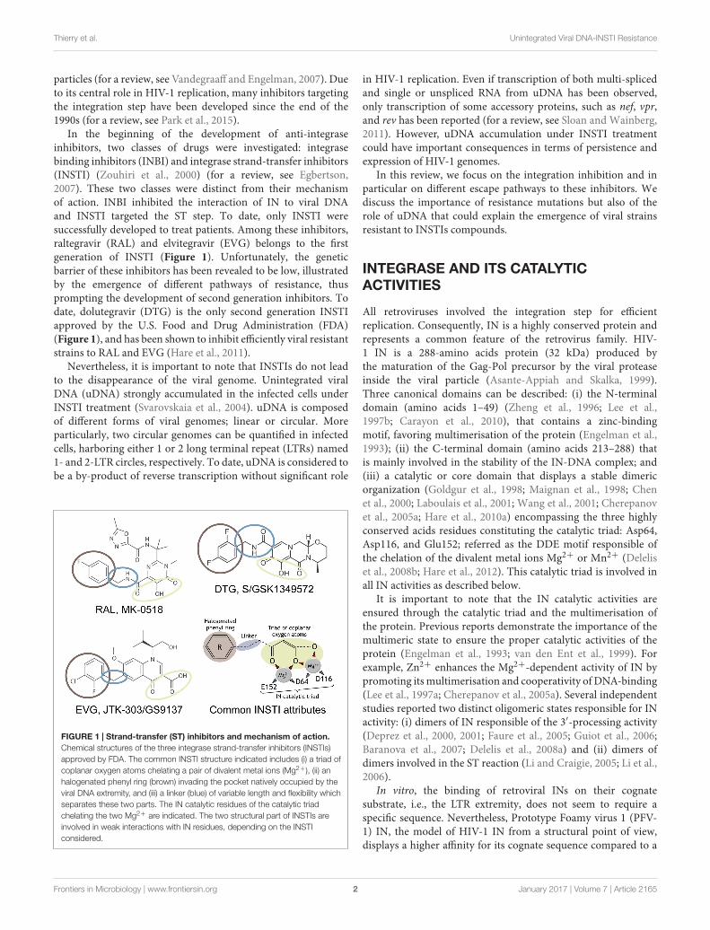
In the beginning of the development of anti-integraseinhibitors, two classes of drugs were investigated: integrasebinding inhibitors (INBI) and integrase strand-transfer inhibitors(INSTI) (Zouhiri et al., 2000) (for a review, see Egbertson,2007). These two classes were distinct from their mechanismof action. INBI inhibited the interaction of IN to viral DNAand INSTI targeted the ST step. To date, only INSTI weresuccessfully developed to treat patients. Among these inhibitors,raltegravir (RAL) and elvitegravir (EVG) belongs to the firstgeneration of INSTI (Figure 1). Unfortunately, the geneticbarrier of these inhibitors has been revealed to be low, illustratedby the emergence of different pathways of resistance, thusprompting the development of second generation inhibitors. Todate, dolutegravir (DTG) is the only second generation INSTIapproved by the U.S. Food and Drug Administration (FDA)(Figure 1), and has been shown to inhibit efficiently viral resistantstrains to RAL and EVG (Hare et al., 2011).
Nevertheless, it is important to note that INSTIs do not leadto the disappearance of the viral genome. Unintegrated viralDNA (uDNA) strongly accumulated in the infected cells underINSTI treatment (Svarovskaia et al., 2004). uDNA is composedof different forms of viral genomes; linear or circular. Moreparticularly, two circular genomes can be quantified in infectedcells, harboring either 1 or 2 long terminal repeat (LTRs) named1- and 2-LTR circles, respectively. To date, uDNA is considered tobe a by-product of reverse transcription without significant role
FIGURE 1 | Strand-transfer (ST) inhibitors and mechanism of action.Chemical structures of the three integrase strand-transfer inhibitors (INSTIs)approved by FDA. The common INSTI structure indicated includes (i) a triad ofcoplanar oxygen atoms chelating a pair of divalent metal ions (Mg2+), (ii) anhalogenated phenyl ring (brown) invading the pocket natively occupied by theviral DNA extremity, and (iii) a linker (blue) of variable length and flexibility whichseparates these two parts. The IN catalytic residues of the catalytic triadchelating the two Mg2+ are indicated. The two structural part of INSTIs areinvolved in weak interactions with IN residues, depending on the INSTIconsidered.
in HIV-1 replication. Even if transcription of both multi-splicedand single or unspliced RNA from uDNA has been observed,only transcription of some accessory proteins, such as nef, vpr,and rev has been reported (for a review, see Sloan and Wainberg,2011). However, uDNA accumulation under INSTI treatmentcould have important consequences in terms of persistence andexpression of HIV-1 genomes.
In this review, we focus on the integration inhibition and inparticular on different escape pathways to these inhibitors. Wediscuss the importance of resistance mutations but also of therole of uDNA that could explain the emergence of viral strainsresistant to INSTIs compounds.
INTEGRASE AND ITS CATALYTICACTIVITIES
All retroviruses involved the integration step for efficientreplication. Consequently, IN is a highly conserved protein andrepresents a common feature of the retrovirus family. HIV-1 IN is a 288-amino acids protein (32 kDa) produced bythe maturation of the Gag-Pol precursor by the viral proteaseinside the viral particle (Asante-Appiah and Skalka, 1999).Three canonical domains can be described: (i) the N-terminaldomain (amino acids 1–49) (Zheng et al., 1996; Lee et al.,1997b; Carayon et al., 2010), that contains a zinc-bindingmotif, favoring multimerisation of the protein (Engelman et al.,1993); (ii) the C-terminal domain (amino acids 213–288) thatis mainly involved in the stability of the IN-DNA complex; and(iii) a catalytic or core domain that displays a stable dimericorganization (Goldgur et al., 1998; Maignan et al., 1998; Chenet al., 2000; Laboulais et al., 2001; Wang et al., 2001; Cherepanovet al., 2005a; Hare et al., 2010a) encompassing the three highlyconserved acids residues constituting the catalytic triad: Asp64,Asp116, and Glu152; referred as the DDE motif responsible ofthe chelation of the divalent metal ions Mg2+ or Mn2+ (Deleliset al., 2008b; Hare et al., 2012). This catalytic triad is involved inall IN activities as described below.
It is important to note that the IN catalytic activities areensured through the catalytic triad and the multimerisation ofthe protein. Previous reports demonstrate the importance of themultimeric state to ensure the proper catalytic activities of theprotein (Engelman et al., 1993; van den Ent et al., 1999). Forexample, Zn2+ enhances the Mg2+-dependent activity of IN bypromoting its multimerisation and cooperativity of DNA-binding(Lee et al., 1997a; Cherepanov et al., 2005a). Several independentstudies reported two distinct oligomeric states responsible for INactivity: (i) dimers of IN responsible of the 3′-processing activity(Deprez et al., 2000, 2001; Faure et al., 2005; Guiot et al., 2006;Baranova et al., 2007; Delelis et al., 2008a) and (ii) dimers ofdimers involved in the ST reaction (Li and Craigie, 2005; Li et al.,2006).
In vitro, the binding of retroviral INs on their cognatesubstrate, i.e., the LTR extremity, does not seem to require aspecific sequence. Nevertheless, Prototype Foamy virus 1 (PFV-1) IN, the model of HIV-1 IN from a structural point of view,displays a higher affinity for its cognate sequence compared to a
Frontiers in Microbiology | www.frontiersin.org 2 January 2017 | Volume 7 | Article 2165

fmicb-07-02165 January 9, 2017 Time: 11:30 # 3
Thierry et al. Unintegrated Viral DNA-INSTI Resistance
random sequence (Delelis et al., 2008a). This observation couldbe explained by a higher solubility of PFV-1 IN compared toHIV-1 IN. Indeed, even if the oligomeric state depends on INconcentration, less aggregates was found in PFV-1 IN purificationcompared to HIV-1 IN. This could favor a better fixation ofIN monomers and/or dimers to the detriment of aggregates inthe case of PFV-1 IN and then increase the number of specificIN/DNA complexes requiring the correct positioning of IN onits sequence (Delelis et al., 2008a). In vitro, the monitoring ofthe IN binding onto oligonucleotide (ODN) mimicking the viralDNA have shown cooperativity mediated by the Zn2+ motif inthe N-terminal domain only in the presence of the specific ODNand Mg2+ (Carayon et al., 2010). The highly conserved terminal5′-CA dinucleotide is critical for the 3′-P activity (Leavitt et al.,1992; Brown et al., 1999), but the length of the viral attachmentsequence (att) involved in the formation of the IN competentcomplex is not precisely determined. First, previous studies haveshown that the terminal 12 base pairs were involved in HIV-1 IN/DNA specific contacts (Masuda et al., 1998). A recentstudy based on the PFV-1 IN structure complexed to its cognatesequence, revealed specific contacts between IN and DNA onthe last 10 nucleotides from the LTR extremity, and non-specificinteractions on the 17 last nucleotides (Hare et al., 2010a).
Four activities are ensured by IN. 3′-P and ST activities arethe two main activities described both in vitro and in vivo. Bothreactions require the full-length protein, the integrity of thecatalytic triad and a metallic cation (Mn2+ or Mg2+) (Deleliset al., 2008b). 3′-P reaction, corresponding to a nucleophilicattack by a water molecule on the viral DNA, is highlyspecific and strictly requires the CA-dinucleotide sequence, justbefore the terminal GT dinucleotide that is removed duringthe reaction (Esposito and Craigie, 1998). Thus, this reactionensures the maturation of both viral DNA ends necessary forthe subsequent reaction, ST. In an infected cell, the 3′-P reactionis efficient since the linear DNA from the reverse transcriptionis immediately cleaved by IN after its formation (Munir et al.,2013). Interestingly, in the case of PFV-1, the 3′-P occurs onlyon the 3′-LTR whereas the 5′-LTR is not involved in thisprocess (Juretzek et al., 2004) (for a review, see Delelis et al.,2004). The consequence of this asymmetrical maturation onthe overall integration process catalyzed by PFV-1 IN remainselusive.
The resulting 3′-processed DNA is then used as a substratefor the integration process. During this reaction, the nucleophilicagent is constituted by the 3′-OH of the 3′-processed DNA end(Li and Craigie, 2005). ST reaction, performed preferentiallyby a dimer of IN, corresponds to the integration of one DNAextremity. This half-site ST reaction can be easily recordedin vitro using ODN or long substrate DNA (Sinha et al., 2002;Sinha and Grandgenett, 2005; Li et al., 2006; Benleulmi et al.,2015). Concerted integration involves the integration of twoviral DNA extremities in the same location leading to the 5-bpduplication (in the case of HIV-1) of the sequence flanking theintegration site, and is catalyzed by a tetramer of IN (dimer ofdimer) (Lesbats et al., 2008; Benleulmi et al., 2015). This overallprocess actually corresponds to the full-site integration processthat occurs in vivo and can be performed in vitro by recombinant
IN and purified PIC (Faure et al., 2005; Sinha and Grandgenett,2005).
A third activity of IN, requiring the full length protein, hasbeen identified by several independent groups and consists ina specific endonucleolytic activity of IN onto a short ODNmimicking the palindromic sequence found at the LTR-LTRjunction of 2-LTRc (Delelis et al., 2005, 2007; Shadrina et al., 2014;Zhang et al., 2014). This reaction occurs symmetrically on the twoDNA strands, at the CA position involved in the 3′-P reaction.This reaction is also highly specific using a plasmid harboring theLTR-LTR junction (Delelis et al., 2007). Importantly, this activityhas been recently reported to occur during HIV-1 replication(Thierry et al., 2015). Finally, disintegration can be consideredto be the reverse of the ST reaction. However, this reaction wasonly observed in vitro and can be performed by IN lacking theN-terminal or the C-terminal domain, in contrast to the threeabove-mentioned activities (Gerton and Brown, 1997; Leh et al.,2000; Zhang et al., 2013).
Integrase interacts with numerous host cell proteins, such asHSP60, BAF (Barrier-to-autointegration factor), HMG I(Y), INI-1 (Integrase interactor 1), and Gemin2 (Kalpana et al., 1994;Li et al., 2000; Parissi et al., 2001; Lin and Engelman, 2003;Hamamoto et al., 2006; Mathew et al., 2013). These partnersmodulate HIV-1 replication by direct or indirect interactionswith IN, not exclusively at the integration step but also at post-integrative steps in the case of INI-1 (Mathew et al., 2013). Themain cellular partner of HIV-1 IN is LEDGF/p75 (for a recentreview Debyser et al., 2015). LEDGF/p75 interacts directly withIN and has a major role in the integration efficiency. LEDGF/p75greatly enhances both 3′-P, ST and concerted viral integration(Cherepanov et al., 2003; Llano et al., 2006; Botbol et al., 2008;Engelman and Cherepanov, 2008; Maillot et al., 2013; Fadelet al., 2014). Moreover, LEDGF/p75 has been reported to havemajor role in post-integration step by silencing expression ofthe provirus by maintaining histone occupancy at the HIV-1promoter thanks to its interaction with Spt6 and Iws1 (Gerardet al., 2015). Due to the central role of LEDGF in the overallreplication process, many efforts are under investigations toimpair the LEDGF/p75 interaction.
TARGETING HIV-1 IN
Due to the crucial role of IN in HIV-1 replication and consideringthe absence of cellular counterpart, IN represents an importanttarget to treat HIV infection. Two main strategies are investigatedto develop inhibitors: (i) catalytic inhibitors targeting 3′-P or STreaction and (ii) non catalytic inhibitors targeting IN/LEDGFinteractions.
Inhibitors targeting the catalytic site or other regions involvedin the binding of DNA substrate were the first to be developed.This family includes nucleic acids or nucleotide-based inhibitors(Mazumder et al., 1997; Pinskaya et al., 2004), peptides (Sourgenet al., 1996), small organic polycyclic compounds (Robinson et al.,1996; Deprez et al., 2004) and impair the binding of IN tothe viral DNA end. However, only inhibitors that preferentiallyor specifically target the ST reaction have reached clinical use,
Frontiers in Microbiology | www.frontiersin.org 3 January 2017 | Volume 7 | Article 2165

fmicb-07-02165 January 9, 2017 Time: 11:30 # 4
Thierry et al. Unintegrated Viral DNA-INSTI Resistance
belonging to the INSTI family. To date, from a chemical pointof view, nine classes can be determined among INSTIs, based ontheir scaffolds (for a review, see Li et al., 2015). RAL, the first anti-integrase inhibitor approved by FDA in 2007 (Grinsztejn et al.,2007; Summa et al., 2008), was followed by EVG in 2012 andDTG in 2013, the latter belonging to the second generation ofINSTI (for a review, see Serrao et al., 2009) (Hare et al., 2011). Allthese compounds target the IN/DNA complex and not IN alone(Espeseth et al., 2000).
Raltegravir, EVG, and DTG share a structure in two moietiesjoined by a linker (Figure 1). The first one contains three oxygenatoms chelating the metallic cations indispensable for the INcatalytic activities. The second one, the halogenated benzyl group,interacts with the G:C base pair of the viral DNA end, precedingthe terminal adenine, and with residues 145 and 146 of IN. Thisinteraction leads to the displacement of the terminal adenine ofthe 3′-processed DNA from the active site. Furthermore, INSTIbinding competes with the binding of the target DNA in the activesite (Espeseth et al., 2000; Maertens et al., 2010) (for a review, seeEngelman and Cherepanov, 2012).
Raltegravir and EVG, the latter being given with apharmacokinetic booster, such as cobicistat, demonstratedtheir strong efficacy to counteract HIV-1 replication even inhighly therapy antiretroviral-experienced patients (Steigbigelet al., 2008; Marchand, 2012). This efficacy is similar for HIV-2in the case of RAL (Charpentier et al., 2011; Ni et al., 2011).
However, RAL and EVG treatments lead to resistance causedby mutations in IN gene, involving for instance the Q148/G140and N155/E92 pathways (Cooper et al., 2008; Malet et al., 2008;Shimura et al., 2008; Delelis et al., 2009). A third pathwayinvolving the Y143 residue has been specifically described forRAL (da Silva et al., 2010; Delelis et al., 2010). While INpolymorphism has low impact on INSTI susceptibility when noassociated to a resistance mutation (Van Baelen et al., 2008;Low et al., 2009), some mutations such as S119R have beenshown to increase the resistance to INSTIs when combined tothe primary mutations Y143C, Q148H, and N155H (Hachiyaet al., 2015). Otherwise, secondary non-polymorphic mutationsare selected according to the observed resistance pathway(Rhee et al., 2008). Primary resistance mutations confer aselective advantage explaining their emergence (Quercia et al.,2009) but can be associated with different IN activity defects(Marinello et al., 2008; Delelis et al., 2009), depending onthe nature of the residue substituted. For example, Y143R/Cmutations lead to a similar decrease in 3′-P activity while theST activity of the Y143C mutant is more reduced compared tothe Y143R mutant (Delelis et al., 2010). Secondary mutationshave been described to increase the INSTI resistance, suchas the E92Q mutation associated with the Y143 or N155Hpathways, or to restore the defect of activity due to theprimary mutation, exemplified by the G140S that leads to arecovery of the activity of the Q148H mutant (Fransen et al.,2009; Huang et al., 2013). Interestingly, some mutations in thereverse transcriptase or protease can compensate the decreaseof IN activity due to primary mutations (Buzon et al., 2010a),highlighting the functional cooperation between IN and otherviral proteins.
Similar susceptibilities between PFV-1 and HIV-1 INs,coupled with the PFV-1 structure, allowed to obtain informationfor a better comprehension of mechanisms involved in resistance(Valkov et al., 2009; Hare et al., 2010a).
Structures of PFV-1 intasome, complexed with INSTIs,confirmed the importance of the halogenated benzyl group, aswell as the three oxygen atoms allowing complete octahedralcoordination of both Mg2+ in the active site (Hare et al., 2010a,b,2011).
Raltegravir interaction was observed with the Y212 residueof PFV-1 (equivalent to the Y143 in HIV-1), its oxadiazole ringlaying π interactions with the aromatic ring of tyrosine. Lossof this interaction in the mutants of the Y143 pathway canexplain emergence of resistant mutants belonging to the Y143pathway (Hare et al., 2010b). Structural studies highlight that,in the context of R224H mutant of PFV-1 (equivalent to theN155H mutant in HIV-1), an interaction occurs between thehistidine and the phosphate group of the terminal adenine (interminal 3′ position of the processed viral DNA). RAL wasshown to be inefficient to abolish this distinctive interactionthus explaining the resistance of the N155H mutant (Hare et al.,2010b).
Finally, the resistance of the Q148H mutant was explained bythe need for large and energetically unfavorable conformationalchanges to allow RAL binding (Hare et al., 2010b). The rapidemergence of pathways involved in resistance of RAL and EVGdemonstrated that both RAL and EVG have a low genetic barrier.
SECOND GENERATION INSTIs
To impair these resistance pathways described previously, INSTIsbelonging to the second generation, such as DTG, have beendeveloped. DTG has proven its efficacy in naive patientswhen combined with nucleotide reverse transcriptase inhibitors(NRTIs) with non-inferiority efficiency compared to RAL (Raffiet al., 2013; Walmsley et al., 2013). Furthermore, the VIKINGtrial (Eron et al., 2013; Castagna et al., 2014; Akil et al., 2015)reported the efficacy of DTG when administrated to patients withvirological failure due to the emergence of primary mutationsconferring resistance to RAL and EVG. However, the same studyreported that DTG was less efficient concerning mutants of theQ148 pathway (Eron et al., 2013; Castagna et al., 2014; Akilet al., 2015). To date, no pathway leading to DTG resistancehas been highlighted by in vitro selection. Only some mutationsin the C-terminal domain of IN have been reported to confera moderate resistance to DTG (Anstett et al., 2015; Cutillaset al., 2015). The study of the susceptibility to DTG of themutants resistant to the first generation of INSTI confirmedits highest genetic barrier (Underwood et al., 2012; Canducciet al., 2013). The intrinsic stability of DTG onto the IN/DNAcomplex mainly explains the higher efficacy of DTG comparedto RAL and EVG. Indeed, the dissociation half-time for RALand EVG are 8.8 and 2.7 h, respectively, compared to 71 h forDTG. Structural studies using PFV-1 reported that DTG bindingis similar to other INSTIs belonging to the first generation (Hareet al., 2011), i.e., that the three coplanar oxygen atoms allow the
Frontiers in Microbiology | www.frontiersin.org 4 January 2017 | Volume 7 | Article 2165

fmicb-07-02165 January 9, 2017 Time: 11:30 # 5
Thierry et al. Unintegrated Viral DNA-INSTI Resistance
chelation of Mg2+ cations, while the halogenated group competeswith the binding of the 3′-processed end (Hare et al., 2010a).However, some features characterized the DTG binding to theIN/DNA complex. DTG is characterized by a straighter structurecompared to RAL and EVG which enables the deeper penetrationof this compound into the space released by the movement ofthe terminal adenine of the 3′-processed DNA leading to morestable interactions with the adjacent cytidine (Hare et al., 2011).This explains why mutations at position G118 confer resistanceto DTG (Malet et al., 2014; Munir et al., 2015). Moreover, vander Waals interactions between the two-fluoro atom of DTG andthe Cγ and Cδ atoms of the E221 residue (equivalent to theE152 in HIV-1) (Hare et al., 2011) and between the four-fluoroatom of DTG and the C6 of the guanine are closer to thoseinvolved with RAL and EVG. In a general manner, structuralstudy indicates a greater adaptability of DTG, compared to RALand EVG, to the structural modifications induced by the mutantsfrom the first INSTI generation (Hare et al., 2011; DeAnda et al.,2013).
Other second generation INSTIs are under development.Cabotegravir (GSK1265744), showing a similar structure to DTG,is under phase II clinical testing and was shown to be efficientin the reduction of the viral load (for a review, see Spreenet al., 2013). In vitro assays demonstrated its efficiency to impairreplication of resistant mutants from the first generation withthe notable exception of the mutants belonging to the Q148pathway (Yoshinaga et al., 2015). The intrinsic properties ofcabotegravir allow formulation of injectable nanosuspension inorder to develop a long-acting antiretroviral treatment (Spreenet al., 2013).
NON-CATALYTIC IN INHIBITORS
Anti-IN inhibitors were first focused on the inhibition ofcatalytical activities. However, due to the emergence of viralstrains resistant to INSTIs, compounds with another mode ofaction were developed. Interestingly, the IN binding domain(IBD) over-expression leads to a decrease in HIV-1 integrationefficiency by a competition with the endogenous LEDGF (DeRijck et al., 2006). The essential role of LEDGF in HIV-1 integration mentioned above and the determination of thestructural determinants involved in IN/LEDGF interaction allowto define a therapeutic target (Cherepanov et al., 2005a,b;Emiliani et al., 2005; Christ and Debyser, 2013). Severalpeptides, derived from the IBD, were efficient to impairIN oligomerization and thus prevented its catalytic activities(Hayouka et al., 2007; Al-Mawsawi et al., 2008). Moreover,inhibition efficiency increased when cyclic peptides were used(Hayouka et al., 2010a,b). Other peptides were developed tospecifically impair IN oligomerization belonging to the “shiftides”family (Kessl et al., 2009; Maes et al., 2012). A similarapproach, based on the design of peptides impairing IN/LEDGFinteraction but targeting LEDGF, was employed. Expression ofthese peptides using lentiviral vectors was efficient to inhibitviral replication without cellular toxicity (Desimmie et al.,2012).
Screenings of existing or virtual chemical libraries, as wellas the development of compounds based on the IN/LEDGFinterface, have been performed (Du et al., 2008; Hou et al.,2008; De Luca et al., 2009, 2010; Christ et al., 2010, 2012; Fanet al., 2011; Peat et al., 2012). Several molecules have been shownto be efficient under the micromolar range (EC50 < 100 nM)(Christ et al., 2012; Fader et al., 2014) and are now referred to asLEDGINs, NCINIs (non-catalytic site IN inhibitors) or ALLINIs(Allosteric IN inhibitors). A common feature of these compoundsis the presence of an acetic acid mimicking the D366 residue ofLEDGF, the latter involved in the IN interaction using the D170,H171, and T174 residues (for a review, see Demeulemeesteret al., 2014). These inhibitors have been described to displaythree modes of action. First, they inhibit HIV-1 integrationby impairing the IN/LEDGF interaction (Christ et al., 2010).Second, they can favor the formation of inactive IN multimers(Desimmie et al., 2013; Le Rouzic et al., 2013). By enhancing INmultimerization, LEDGINs interfere with IN catalytic activitiesin an allosteric manner, leading to 2-LTR circles accumulationsimilarly to RAL treatment (Hayouka et al., 2007). Finally, theyare also able to target the post-integrative steps leading to inactiveviral particles formation with aberrant capsids (Balakrishnanet al., 2013; Sharma et al., 2014). Interestingly, only inhibitorstargeting integrase catalytic activities lead to 2-LTR circlesaccumulation, which is not observed with NCINIs such as GS-B(Al-Mawsawi et al., 2008). These results highlight that 2-LTRcircles accumulation is not systematically observed when HIV-1integration is inhibited, but depends on inhibition of IN catalyticactivities. The lack of 2-LTR circles accumulation after NCINIStreatment could be explained by their impact on viral DNAsynthesis.
Among the NCINIs, the compound BI-224436 is under phaseI clinical testing after showing its efficacy in in vitro assays, ininfected cells and in experiments on animals (Fenwick et al.,2014). This compound has shown no cross-resistance withRAL and EVG (Fenwick et al., 2014). The resistant mutantsG140S/Q148H are efficiently inhibited by BI-224436 (Fenwicket al., 2014). Conversely, the resistant mutants selected in vitroby BI-224436 (for example A128N) were sensitive to RAL andEVG. These promising results obtained by these inhibitors andthe fact that there is no cross-resistance with INSTIs provides anopportunity to use them in combination for future treatments.However, due to differences in the residues of HIV-2 IN involvedin the interaction with LEDGF and targeted by these compounds,their development has to be specifically investigated for HIV-2 toovercome this natural resistance (Christ et al., 2010; Desimmieet al., 2012).
Emergence of IN mutations, leading to INSTI resistance,constitutes the classical way for the virus to escape when INSTIsare used. However, recent clinical trials involving DTG treatmentin naïve patients did not report any resistance mutation in thecases of virological failure (Raffi et al., 2013; Walmsley et al.,2013; Molina et al., 2014). This observation suggests anotherpathway of escape used by the virus to replicate under INSTItreatment. One hypothesis is based on recent studies underlyingthe roles of unintegrated HIV genome, accumulated under INSTItreatment.
Frontiers in Microbiology | www.frontiersin.org 5 January 2017 | Volume 7 | Article 2165

fmicb-07-02165 January 9, 2017 Time: 11:30 # 6
Thierry et al. Unintegrated Viral DNA-INSTI Resistance
UNINTEGRATED VIRAL DNA
As mentioned previously, integration of the viral genome is acentral step in the HIV-1 replication life cycle since it ensuresthe stability of the viral information and efficient transcription.The provirus is considered to be the sole template for HIV-1 expression. It is important to note that, impairing HIV-1integration does not lead to the disappearance of HIV-1 genomesin infected cells but to the formation of uDNA instead ofintegrated DNA (Chun et al., 1997; Sharkey et al., 2000). Multipleforms of uDNA could be detected as linear (issued from thereverse transcription step and precursor of the provirus) orcircular forms. Two circular forms exist, harboring 1 or 2-LTR,and are called 1 and 2-LTR circles, respectively. 1-LTR circles(1-LTRc) and 2-LTR circles (2-LTRc) are mainly detected in thenucleus of the infected cell. The origin of such circular forms isdiverse but the common point is that these circular forms derivedfrom linear viral DNA and are found in the nucleus of infectedcells (Munir et al., 2013). The circular nature confers them agreater stability compared to the linear viral DNA, the latter beingquickly degraded. They are only diluted by cell division (Sharkeyet al., 2000; Butler et al., 2002; Pierson et al., 2002; Munir et al.,2013). Due to their apparent stability, they were used as a markerof recent infections even if they can persist for a long time in cellswith a weak division rate such as macrophages (Sharkey et al.,2000; Gillim-Ross et al., 2005).
1-LTRc are mainly due to recombination between each LTRby homologous recombination (HR) despite several conflictingreports. Viral extremities are recognized by the MRN complex(MRE11/RAD50/NBS1), activated by the ATM pathway as soonas the reverse transcription step occurs, and then supported bythe proteins of the HR pathway (Kilzer et al., 2003). However,1-LTRc quantification in cells deficient in MRE11 protein didnot result in a decrease of 1-LTRc amount (Sakurai et al., 2009).Indeed, a significant proportion of 1-LTRc has been shown to begenerated in the cytoplasm during reverse transcription (Muniret al., 2013).
2-LTRc are formed by circularization of linear DNA by thenon-homologous end-joining (NHEJ) pathway (Kilzer et al.,2003). A peculiar feature shared by many retroviruses is thepresence of a palindromic sequence at the LTR-LTR junction(Delelis et al., 2005, 2007). The amount of 1-LTRc can reach20–30% of the viral genome whereas 2-LTRc amount is quitelow in wt infection (2–5% of total viral DNA) (Munir et al.,2013). uDNA has been considered for a long time as a by-product of reverse transcription with no significance in theoverall process of HIV-1 replication (Sloan and Wainberg, 2011).However, it is important to note that inhibition of HIV-1 INcatalytic activities lead to an accumulation of uDNA and moreparticularly circular viral DNA forms (Munir et al., 2013). Whilethe 1-LTRc representativeness can reach more than 50% oftotal vDNA, the greater increase is observed with the amountof 2-LTRc, that can be increased by a 10-fold factor (Muniret al., 2013). Consequences of such accumulation are yet poorlydescribed.
Despite their efficiency to inhibit integration, it is worthy tonote that viral replication still occurs under INSTI treatment,
raising the question of the viral genome originating this residualreplication.
DIFFERENT WAYS TO BYPASS INSTIsEFFECTS
uDNA ExpressionThe role of uDNA expression in the HIV-1 cycle is still a matter ofdebate. Indeed, uDNA displays the same genomic organization asthe provirus. However, all reports agreed that uDNA expressionis weaker compared to the provirus. Interestingly, a recent reportclearly demonstrates that viral production could be detectedfrom uDNA after reactivation of resting CD4+ T cells (Chanet al., 2016). Depending on the experimental settings, expressionof uDNA is comprised between 10 and 70% of the proviralexpression (Stevenson et al., 1990; Iyer et al., 2009). Transcriptionfrom uDNA leads to the synthesis of unspliced and spliced viralRNAs but spliced RNAs are found in a greater amount (Wuand Marsh, 2001, 2003; Kelly et al., 2008). The low amount ofRev protein, that is essential for the late replication stages, hasbeen shown to contribute to the mechanisms leading to a weakerreplication from uDNA compared to proviral DNA. Indeed,infections of cells expressing Rev lead to an efficient replicationfrom uDNA (Sloan et al., 2011). However, uDNA expressionoccurs during infection of different cell lines using INSTIs oran IN catalytic mutant. In this case, viral gene expression ofearly genes such as Tat can be highlighted by the transcriptionalactivity of Tat on LTRs (Wu and Marsh, 2001; Gelderblom et al.,2008; Kelly et al., 2008). Among proteins translated from fullyspliced mRNA, only Nef was directly observed (Sloan et al.,2011). Furthermore, it has been clearly demonstrated that, underspecific conditions, HIV-1 replication could be evidenced withoutintegration (Gelderblom et al., 2008; Trinite et al., 2013). Moreparticularly, Chan et al. (2016) demonstrate that, in restingCD4 T cells, uDNA leads to the production of infectious viralparticles. Several parameters influence uDNA expression. Forexample, in conditions where integration is impaired (catalyticmutant IN or INSTI treatment), Vpr protein enclosed withinviral particles promotes uDNA transcription leading to Tatexpression (Trinite et al., 2013). Furthermore, HDAC inhibitorslead to an increase of uDNA expression, suggesting a chromatinorganization of uDNA (Kantor et al., 2009). However, thedetection of transcripts does not ensure the presence of the viralproteins since a post-transcriptional control could be involved,as suggested by a controversial report studying SLFN11 (Li et al.,2012).
Expression of uDNA is mainly due to circular genomes.Indeed, linear DNA can be excluded due to its weak stability inthe cell. Both 1-LTRc and 2-LTRc have been shown to lead toinfectious viral particles when transfected into HeLa cells evenwith a low efficiency compared to the proviral DNA (Cara et al.,1996). Although a specific type of mRNA transcribed from 2-LTRc has been detected (Brussel and Sonigo, 2004), it has beenreported that uDNA expression from 1-LTRc is stronger thanfrom 2-LTRc (Cara et al., 1996). A recent report sustains thisobservation since uDNA expression is similar after infection
Frontiers in Microbiology | www.frontiersin.org 6 January 2017 | Volume 7 | Article 2165

fmicb-07-02165 January 9, 2017 Time: 11:30 # 7
Thierry et al. Unintegrated Viral DNA-INSTI Resistance
of cells deficient in 2-LTRc formation (Thierry et al., 2016).The precise contribution of these two circular forms to uDNAexpression needs to be further investigated.
Due to the strong accumulation of circular forms underINSTIs treatment, uDNA could play an important role in viralexpression and could lead to a weak viral particle synthesisbypassing the INSTIs treatment (Figure 2). Despite their greatefficiency to inhibit the integration step, INSTIs are unable toimpair the late stages of the viral replication cycle, and thus theycan’t prevent new infections. Clinical studies have not only shownthat low-level viremia can persist in patients with undetectableplasma HIV RNA (Maldarelli et al., 2007) including patientstreated with RAL-based regimens (Baroncelli et al., 2015), butalso that RAL intensification is unable to suppress this persistentresidual viremia and is associated with an increase in circularuDNA (Dinoso et al., 2009; Buzon et al., 2010b; Gandhi et al.,2010; McMahon et al., 2010; Hatano et al., 2011; Vallejo et al.,2012). An evolution of HIV-1 envelope sequences despite potentantiviral therapy has previously been shown (Gunthard et al.,1999; Martinez et al., 1999) with the emergence of a NRTIresistance mutation (Martinez et al., 1999). Such ongoing basalreplication could occur from uDNA and, taking advantages of theinability of INSTIs to impair new infections, mutations leading
to INSTI resistance could occur during reverse transcription innewly infected cells.
INSTI ReversibilityAs mentioned previously, inhibition of integration by INSTIs isdependent from the residence time of the compounds on thecomplex formed by the drug, IN and viral DNA. Stability ofthe INSTI on the complex depends on the compounds studied,since different discordant half-life have been described for RAL,EVG, and DTG (8.8, 2.7, and 71 h, respectively) (Hightower et al.,2011).
It has been recently reported that removal of RAL fromcell medium until 72 h post-infection leads to viral resumptionmediated by de novo integrated events (Thierry et al., 2015).This viral resumption was due to the cleavage of the LTR-LTRjunction of 2-LTRc followed by their integration in the hostcell genome indicating that 2-LTRc, accumulated under INSTIstreatment, can be used as a substrate for integration process.Moreover, the observation of the 5 bp duplication associatedwith these integration events, considered as HIV-1 integrasemediated integration hallmark, underlined the specificity of theseevents. These results also highlight the biological relevance ofthe endonucleolytic in vitro activity of IN onto the LTR-LTR
FIGURE 2 | Fate of unintegrated viral DNA (uDNA). Linear viral DNA from the reverse transcription step can have several behaviors. Linear DNA is integrated inthe host cell genome or circularized leading to 1 or 2-long terminal repeat (LTR) circles. Basal transcription from uDNA could lead to the production of infectious viralparticles bypassing the effects of ST inhibitors.
Frontiers in Microbiology | www.frontiersin.org 7 January 2017 | Volume 7 | Article 2165

fmicb-07-02165 January 9, 2017 Time: 11:30 # 8
Thierry et al. Unintegrated Viral DNA-INSTI Resistance
junction, in the specific context of 2-LTRc accumulation causedby INSTI treatment. These data highly suggest that 2-LTRc canbe considered as a back-up molecule.
Based on this result, one can speculate that, infection of non-dividing cells such as quiescent CD4-T cells can lead to 2-LTRcaccumulation under INSTI treatment. Indeed, the presence ofuDNA in macrophages infected with a non-integrative virus hasbeen detected up to 30 days post-infection (Kelly et al., 2008).After cellular activation, 2-LTRc could be used as a substrate forintegration. To explain how this activity could be used in thevirological context several hypothesis can be advanced. The firstone is that the LTR-LTR junction maintained IN in an active formdue to the rather stability, in non-dividing cells, of uDNA. Thesecond one, supported by several reports, involves a faint viralproduction from uDNA probably due to its peculiar regulationcompared to the provirus (Gelderblom et al., 2008; Chan et al.,2016; Thierry et al., 2016). In this case, the faint viral production,under INSTI condition, could lead to a weak infection of newlycells providing newly complex formed by 2-LTR circles and INin these cells. If these hypotheses are confirmed, 2-LTRc couldbe considered as a reservoir for HIV-1 integration and thus amolecule involved in pre-integration latency.
CONCLUSION
Development of the INSTIs compounds is a great advancein treatment-naïve and experienced HIV-infected patients.
Inclusion of INSTI in the regimen is considered as a first-linetherapy for treatment-naïve infected patients. However, despitetheir efficacy to decrease the viral load, one must not onlymonitor the emergence of resistance mutations, but also takecare of the presence of uDNA that could be a source of viralescape, either by integration of 2-LTRc or by expression of uDNAat the origin of basal replication. The quantitative importanceof these uDNA forms under treatment with catalytic integraseinhibitors highlights the issue of reservoirs cells. In particular, thekey DNA forms in latent reservoirs such as quiescent memoryCD4-T cells, the only reservoir where long-term persistence ofHIV-1 in patients receiving optimal antiretroviral therapy hasbeen repeatedly described, could not be only integrated DNA butcould involve uDNA. Treatment with a non-catalytical integraseinhibitor in the context of antiretroviral therapy could thus havedifferent implications in terms of reservoirs.
These alternative pathways making use of uDNA to escapeINSTI treatment emphasizes the need to understand the natureof the viral DNA forms in the various reservoirs cells. Thisknowledge would fuel the research developing curative strategiesthat cannot bypass the question of reservoirs.
AUTHOR CONTRIBUTIONS
OD initiated the project. ET, ED, and OD wrote the article. Allthe authors reviewed the final version of the manuscript prior tosubmission for publication.
REFERENCESAkil, B., Blick, G., Hagins, D. P., Ramgopal, M. N., Richmond, G. J., Samuel,
R. M., et al. (2015). Dolutegravir versus placebo in subjects harbouring HIV-1 with integrase inhibitor resistance associated substitutions: 48-week resultsfrom VIKING-4, a randomized study. Antivir. Ther. 20, 343–348. doi: 10.3851/IMP2878
Al-Mawsawi, L. Q., Christ, F., Dayam, R., Debyser, Z., and Neamati, N. (2008).Inhibitory profile of a LEDGF/p75 peptide against HIV-1 integrase: insight intointegrase-DNA complex formation and catalysis. FEBS Lett. 582, 1425–1430.doi: 10.1016/J.Febslet.2008.02.076
Anstett, K., Mesplede, T., Oliveira, M., Cutillas, V., and Wainberg, M. A. (2015).Dolutegravir resistance mutation R263K cannot coexist in combination withmany classical integrase inhibitor resistance substitutions. J. Virol. 89, 4681–4684. doi: 10.1128/JVI.03485-14
Asante-Appiah, E., and Skalka, A. M. (1999). HIV-1 integrase: structuralorganization, conformational changes, and catalysis. Adv. Virus Res. 52,351–369.
Balakrishnan, M., Yant, S. R., Tsai, L., O’Sullivan, C., Bam, R. A., Tsai, A., et al.(2013). Non-catalytic site HIV-1 integrase inhibitors disrupt core maturationand induce a reverse transcription block in target cells. PLoS ONE 8:e74163.doi: 10.1371/journal.pone.0074163
Baranova, S., Tuzikov, F. V., Zakharova, O. D., Tuzikova, N. A., Calmels, C.,Litvak, S., et al. (2007). Small-angle X-ray characterization of the nucleoproteincomplexes resulting from DNA-induced oligomerization of HIV-1 integrase.Nucleic Acids Res. 35, 975–987. doi: 10.1093/Nar/Gkl1111
Baroncelli, S., Pirillo, M. F., Galluzzo, C. M., Antoni, A. D., Ladisa, N., Francisci, D.,et al. (2015). Rate and determinants of residual viremia in multidrug-experienced patients successfully treated with raltegravir-based regimens. AIDSRes. Hum. Retroviruses 31, 71–77. doi: 10.1089/AID.2014.0060
Benleulmi, M. S., Matysiak, J., Henriquez, D. R., Vaillant, C., Lesbats, P.,Calmels, C., et al. (2015). Intasome architecture and chromatin density
modulate retroviral integration into nucleosome. Retrovirology 12:13. doi: 10.1186/s12977-015-0145-9
Botbol, Y. R., Raghavendra, N. K., Rahman, S., Engelman, A., and Lavigne, M.(2008). Chromatinized templates reveal the requirement for the LEDGF/p75PWWP domain during HIV-1 integration in vitro. Nucleic Acids Res. 36,1237–1246. doi: 10.1093/Nar/Gkm1127
Brown, H. E. V., Chen, H. M., and Engelman, A. (1999). Structure-basedmutagenesis of the human immunodeficiency virus type 1 DNA attachmentsite: effects on integration and cDNA synthesis. J. Virol. 73, 9011–9020.
Brussel, A., and Sonigo, P. (2004). Evidence for gene expression by unintegratedhuman immunodeficiency virus type 1 DNA species. J. Virol. 78, 11263–11271.
Butler, S. L., Johnson, E. P., and Bushman, F. D. (2002). Human immunodeficiencyvirus cDNA metabolism: notable stability of two-long terminal repeat circles.J. Virol. 76, 3739–3747.
Buzon, M. J., Dalmau, J., Puertas, M. C., Puig, J., Clotet, B., and Martinez-Picado, J. (2010a). The HIV-1 integrase genotype strongly predicts raltegravirsusceptibility but not viral fitness of primary virus isolates. AIDS 24, 17–25.doi: 10.1097/Qad.0b013e328331c81e
Buzon, M. J., Massanella, M., Llibre, J. M., Esteve, A., Dahl, V., Puertas, M. C., et al.(2010b). HIV-1 replication and immune dynamics are affected by raltegravirintensification of HAART-suppressed subjects. Nat. Med. 16, 460–465. doi:10.1038/Nm.2111
Campbell, E. M., and Hope, T. J. (2015). HIV-1 capsid: the multifaceted key playerin HIV-1 infection. Nat. Rev. Microbiol. 13, 471–483. doi: 10.1038/nrmicro3503
Canducci, F., Ceresola, E. R., Saita, D., Castagna, A., Gianotti, N., Underwood, M.,et al. (2013). In vitro phenotypes to elvitegravir and dolutegravir in primarymacrophages and lymphocytes of clonal recombinant viral variants selectedin patients failing raltegravir. J. Antimicrob. Chemother. 68, 2525–2532. doi:10.1093/jac/dkt220
Cara, A., Cereseto, A., Lori, F., and Reitz, M. S. Jr. (1996). HIV-1 protein expressionfrom synthetic circles of DNA mimicking the extrachromosomal forms of viralDNA. J. Biol. Chem. 271, 5393–5397.
Frontiers in Microbiology | www.frontiersin.org 8 January 2017 | Volume 7 | Article 2165

fmicb-07-02165 January 9, 2017 Time: 11:30 # 9
Thierry et al. Unintegrated Viral DNA-INSTI Resistance
Carayon, K., Leh, H., Henry, E., Simon, F., Mouscadet, J. F., and Deprez, E. (2010).A cooperative and specific DNA-binding mode of HIV-1 integrase depends onthe nature of the metallic cofactor and involves the zinc-containing N-terminaldomain. Nucleic Acids Res. 38, 3692–3708. doi: 10.1093/nar/gkq087
Castagna, A., Maggiolo, F., Penco, G., Wright, D., Mills, A., Grossberg, R., et al.(2014). Dolutegravir in antiretroviral-experienced patients with raltegravir-and/or elvitegravir-resistant HIV-1: 24-week results of the phase III VIKING-3study. J. Infect. Dis. 210, 354–362. doi: 10.1093/infdis/jiu051
Chan, C. N., Trinite, B., Lee, C. S., Mahajan, S., Anand, A., Wodarz, D., et al. (2016).HIV-1 latency and virus production from unintegrated genomes followingdirect infection of resting CD4 T cells. Retrovirology 13:1. doi: 10.1186/s12977-015-0234-9
Charpentier, C., Roquebert, B., Delelis, O., Larrouy, L., Matheron, S., Tubiana, R.,et al. (2011). Hot spots of integrase genotypic changes leading to HIV-2resistance to raltegravir. Antimicrob. Agents Chemother. 55, 1293–1295. doi:10.1128/AAC.00942-10
Chen, J. C. H., Krucinski, J., Miercke, L. J. W., Finer-Moore, J. S., Tang, A. H.,Leavitt, A. D., et al. (2000). Crystal structure of the HIV-1 integrase catalyticcore and C-terminal domains: a model for viral DNA binding. Proc. Natl. Acad.Sci. U.S.A. 97, 8233–8238.
Cherepanov, P., Ambrosio, A. L., Rahman, S., Ellenberger, T., and Engelman, A.(2005a). Structural basis for the recognition between HIV-1 integrase andtranscriptional coactivator p75. Proc. Natl. Acad. Sci. U.S.A. 102, 17308–17313.doi: 10.1073/pnas.0506924102
Cherepanov, P., Maertens, G., Proost, P., Devreese, B., Van Beeumen, J.,Engelborghs, Y., et al. (2003). HIV-1 integrase forms stable tetramers andassociates with LEDGF/p75 protein in human cells. J. Biol. Chem. 278, 372–381.doi: 10.1074/Jbc.M209278200
Cherepanov, P., Sun, Z. Y., Rahman, S., Maertens, G., Wagner, G., andEngelman, A. (2005b). Solution structure of the HIV-1 integrase-bindingdomain in LEDGF/p75. Nat. Struct. Mol. Biol. 12, 526–532. doi: 10.1038/nsmb937
Christ, F., and Debyser, Z. (2013). The LEDGF/p75 integrase interaction, a noveltarget for anti-HIV therapy. Virology 435, 102–109. doi: 10.1016/j.virol.2012.09.033
Christ, F., Shaw, S., Demeulemeester, J., Desimmie, B. A., Marchand, A., Butler, S.,et al. (2012). Small-molecule inhibitors of the LEDGF/p75 binding site ofintegrase block HIV replication and modulate integrase multimerization.Antimicrob. Agents Chemother. 56, 4365–4374. doi: 10.1128/AAC.00717-12
Christ, F., Voet, A., Marchand, A., Nicolet, S., Desimmie, B. A., Marchand, D.,et al. (2010). Rational design of small-molecule inhibitors of the LEDGF/p75-integrase interaction and HIV replication. Nat. Chem. Biol. 6, 442–448. doi:10.1038/nchembio.370
Chun, T. W., Carruth, L., Finzi, D., Shen, X., DiGiuseppe, J. A., Taylor, H., et al.(1997). Quantification of latent tissue reservoirs and total body viral load inHIV-1 infection. Nature 387, 183–188. doi: 10.1038/387183a0
Cooper, D. A., Steigbigel, R. T., Gatell, J. M., Rockstroh, J. K., Katlama, C., Yeni, P.,et al. (2008). Subgroup and resistance analyses of raltegravir for resistant HIV-1infection. N. Engl. J. Med. 359, 355–365.
Cutillas, V., Mesplede, T., Anstett, K., Hassounah, S., and Wainberg, M. A.(2015). The R262K substitution combined with H51Y in HIV-1 subtype Bintegrase confers low-level resistance against dolutegravir. Antimicrob. AgentsChemother. 59, 310–316. doi: 10.1128/AAC.04274-14
da Silva, D., Van Wesenbeeck, L., Breilh, D., Reigadas, S., Anies, G., VanBaelen, K., et al. (2010). HIV-1 resistance patterns to integrase inhibitorsin antiretroviral-experienced patients with virological failure on raltegravir-containing regimens. J. Antimicrob. Chemother. 65, 1262–1269. doi: 10.1093/Jac/Dkq099
De Luca, L., Barreca, M. L., Ferro, S., Christ, F., Iraci, N., Gitto, R., et al. (2009).Pharmacophore-based discovery of small-molecule inhibitors of protein-protein interactions between HIV-1 integrase and cellular cofactor LEDGF/p75.ChemMedChem 4, 1311–1316. doi: 10.1002/Cmdc.200900070
De Luca, L., Ferro, S., Gitto, R., Barreca, M. L., Agnello, S., Christ, F., et al.(2010). Small molecules targeting the interaction between HIV-1 integrase andLEDGF/p75 cofactor. Bioorg. Med. Chem. 18, 7515–7521. doi: 10.1016/j.bmc.2010.08.051
De Rijck, J., Vandekerckhove, L., Gijsbers, R., Hombrouck, A., Hendrix, J.,Vercammen, J., et al. (2006). Overexpression of the lens epithelium-derived
growth factor/p75 integrase binding domain inhibits human immunodeficiencyvirus replication. J. Virol. 80, 11498–11509. doi: 10.1128/Jvi.00801-06
DeAnda, F., Hightower, K. E., Nolte, R. T., Hattori, K., Yoshinaga, T., Kawasuji, T.,et al. (2013). Dolutegravir interactions with HIV-1 integrase-DNA: structuralrationale for drug resistance and dissociation kinetics. PLoS ONE 8:e77448.doi: 10.1371/journal.pone.0077448
Debyser, Z., Christ, F., De Rijck, J., and Gijsbers, R. (2015). Host factors forretroviral integration site selection. Trends Biochem. Sci. 40, 108–116. doi: 10.1016/j.tibs.2014.12.001
Delelis, O., Carayon, K., Guiot, E., Leh, H., Tauc, P., Brochon, J. C., et al. (2008a).Insight into the integrase-DNA recognition mechanism – A specific DNA-binding mode revealed by an enzymatically labeled integrase. J. Biol. Chem. 283,27838–27849. doi: 10.1074/Jbc.M803257200
Delelis, O., Carayon, K., Saib, A., Deprez, E., and Mouscadet, J. F. (2008b). Integraseand integration: biochemical activities of HIV-1 integrase. Retrovirology 5:114.doi: 10.1186/1742-4690-5-114
Delelis, O., Lehmann-Che, J., and Saib, A. (2004). Foamy viruses–a world apart.Curr. Opin. Microbiol. 7, 400–406. doi: 10.1016/j.mib.2004.06.009
Delelis, O., Malet, I., Na, L., Tchertanov, L., Calvez, V., Marcelin, A. G., et al. (2009).The G140S mutation in HIV integrases from raltegravir-resistant patientsrescues catalytic defect due to the resistance Q148H mutation. Nucleic AcidsRes. 37, 1193–1201. doi: 10.1093/nar/gkn1050
Delelis, O., Parissi, V., Leh, H., Mbemba, G., Petit, C., Sonigo, P., et al. (2007).Efficient and specific internal cleavage of a retroviral palindromic DNAsequence by tetrameric HIV-1 integrase. PLoS ONE 2:e608. doi: 10.1371/journal.pone.0000608
Delelis, O., Petit, C., Leh, H., Mbemba, G., Mouscadet, J. F., and Sonigo, P.(2005). A novel function for spumaretrovirus integrase: an early requirementfor integrase-mediated cleavage of 2 LTR circles. Retrovirology 2:31. doi: 10.1186/1742-4690-2-31
Delelis, O., Thierry, S., Subra, F., Simon, F., Malet, I., Alloui, C., et al. (2010). Impactof Y143 HIV-1 integrase mutations on resistance to Raltegravir in vitro andin vivo. Antimicrob. Agents Chemother. 54, 491–501. doi: 10.1128/AAC.01075-09
Demeulemeester, J., Chaltin, P., Marchand, A., De Maeyer, M., Debyser, Z., andChrist, F. (2014). LEDGINs, non-catalytic site inhibitors of HIV-1 integrase: apatent review (2006 – 2014). Expert Opin. Ther. Pat. 24, 609–632. doi: 10.1517/13543776.2014.898753
Deprez, E., Barbe, S., Kolaski, M., Leh, H., Zouhiri, F., Auclair, C., et al.(2004). Mechanism of HIV-1 integrase inhibition by styrylquinoline derivativesin vitro. Mol. Pharmacol. 65, 85–98.
Deprez, E., Tauc, P., Leh, H., Mouscadet, J. F., Auclair, C., and Brochon, J. C.(2000). Oligomeric states of the HIV-1 integrase as measured by time-resolvedfluorescence anisotropy. Biochemistry 39, 9275–9284. doi: 10.1021/Bi000397j
Deprez, E., Tauc, P., Leh, H., Mouscadet, J. F., Auclair, C., Hawkins, M. E., et al.(2001). DNA binding induces dissociation of the multimeric form of HIV-1integrase: a time-resolved fluorescence anisotropy study. Proc. Natl. Acad. Sci.U.S.A. 98, 10090–10095.
Desimmie, B. A., Humbert, M., Lescrinier, E., Hendrix, J., Vets, S., Gijsbers, R., et al.(2012). Phage display-directed discovery of LEDGF/p75 binding cyclic peptideinhibitors of HIV replication. Mol. Ther. 20, 2064–2075. doi: 10.1038/mt.2012.132
Desimmie, B. A., Schrijvers, R., Demeulemeester, J., Borrenberghs, D., Weydert, C.,Thys, W., et al. (2013). LEDGINs inhibit late stage HIV-1 replication bymodulating integrase multimerization in the virions. Retrovirology 10:57. doi:10.1186/1742-4690-10-57
Dinoso, J. B., Kim, S. Y., Wiegand, A. M., Palmer, S. E., Gange, S. J., Cranmer, L.,et al. (2009). Treatment intensification does not reduce residual HIV-1 viremiain patients on highly active antiretroviral therapy. Proc. Natl. Acad. Sci. U.S.A.106, 9403–9408. doi: 10.1073/pnas.0903107106
Du, L., Zhao, Y. X., Yang, L. M., Zheng, Y. T., Tang, Y., Shen, X., et al. (2008).Symmetrical 1-pyrrolidineacetamide showing anti-HIV activity through a newbinding site on HIV-1 integrase. Acta Pharmacol. Sin. 29, 1261–1267. doi:10.1111/J.1745-7254.2008.00863.X
Egbertson, M. S. (2007). HIV integrase inhibitors: from diketoacids to Heterocyclictemplates: a history of HIV integrase medicinal chemistry at Merck west pointand Merck Rome (IRBM). Curr. Top. Med. Chem. 7, 1251–1272.
Frontiers in Microbiology | www.frontiersin.org 9 January 2017 | Volume 7 | Article 2165

fmicb-07-02165 January 9, 2017 Time: 11:30 # 10
Thierry et al. Unintegrated Viral DNA-INSTI Resistance
Emiliani, S., Mousnier, A., Busschots, K., Maroun, M., Van Maele, B., Tempe, D.,et al. (2005). Integrase mutants defective for interaction with LEDGF/p75 areimpaired in chromosome tethering and HIV-1 replication. J. Biol. Chem. 280,25517–25523.
Engelman, A., Bushman, F. D., and Craigie, R. (1993). Identification of discretefunctional domains of Hiv-1 integrase and their organization within an activemultimeric complex. EMBO J. 12, 3269–3275.
Engelman, A., and Cherepanov, P. (2008). The lentiviral integrase binding proteinLEDGF/p75 and HIV-1 replication. PLoS Pathog. 4:E1000046. doi: 10.1371/Journal.Ppat.1000046
Engelman, A., and Cherepanov, P. (2012). The structural biology of HIV-1:mechanistic and therapeutic insights. Nat. Rev. Microbiol. 10, 279–290. doi:10.1038/nrmicro2747
Eron, J. J., Clotet, B., Durant, J., Katlama, C., Kumar, P., Lazzarin, A., et al.(2013). Safety and efficacy of dolutegravir in treatment-experienced subjectswith raltegravir-resistant HIV type 1 infection: 24-week results of the VIKINGStudy. J. Infect. Dis. 207, 740–748. doi: 10.1093/infdis/jis750
Espeseth, A. S., Felock, P., Wolfe, A., Witmer, M., Grobler, J., Anthony, N., et al.(2000). HIV-1 integrase inhibitors that compete with the target DNA substratedefine a unique strand transfer conformation for integrase. Proc. Natl. Acad. Sci.U.S.A. 97, 11244–11249.
Esposito, D., and Craigie, R. (1998). Sequence specificity of viral end DNA bindingby HIV-1 integrase reveals critical regions for protein-DNA interaction. EMBOJ. 17, 5832–5843.
Fadel, H. J., Morrison, J. H., Saenz, D. T., Fuchs, J. R., Kvaratskhelia, M., Ekker,S. C., et al. (2014). TALEN knockout of the PSIP1 gene in human cells: analysesof HIV-1 replication and allosteric integrase inhibitor mechanism. J. Virol. 88,9704–9717. doi: 10.1128/JVI.01397-14
Fader, L. D., Malenfant, E., Parisien, M., Carson, R., Bilodeau, F., Landry, S., et al.(2014). Discovery of BI 224436, a noncatalytic site integrase inhibitor (NCINI)of HIV-1. ACS Med. Chem. Lett. 5, 422–427. doi: 10.1021/ml500002n
Fan, X., Zhang, F. H., Al-Safi, R. I., Zeng, L. F., Shabaik, Y., Debnath, B., et al. (2011).Design of HIV-1 integrase inhibitors targeting the catalytic domain as well as itsinteraction with LEDGF/p75: a scaffold hopping approach using salicylate andcatechol groups. Bioorg. Med. Chem. 19, 4935–4952. doi: 10.1016/j.bmc.2011.06.058
Faure, A., Calmels, C., Desjobert, C., Castroviejo, M., Caumont-Sarcos, A.,Tarrago-Litvak, L., et al. (2005). HIV-1 integrase crosslinked oligomers areactive in vitro. Nucleic Acids Res. 33, 977–986. doi: 10.1093/Nar/Gki241
Fenwick, C., Amad, M., Bailey, M. D., Bethell, R., Bos, M., Bonneau, P., et al. (2014).Preclinical profile of BI 224436, a novel HIV-1 non-catalytic-site integraseinhibitor. Antimicrob. Agents Chemother. 58, 3233–3244. doi: 10.1128/AAC.02719-13
Fransen, S., Gupta, S., Danovich, R., Hazuda, D., Miller, M., Witmer, M., et al.(2009). Loss of raltegravir susceptibility by human immunodeficiency virustype 1 is conferred via multiple nonoverlapping genetic pathways. J. Virol. 83,11440–11446. doi: 10.1128/Jvi.01168-09
Gandhi, R. T., Zheng, L., Bosch, R. J., Chan, E. S., Margolis, D. M., Read, S., et al.(2010). The effect of raltegravir intensification on low-level residual viremia inHIV-infected patients on antiretroviral therapy: a randomized controlled trial.PLoS Med. 7:321. doi: 10.1371/journal.pmed.1000321
Gelderblom, H. C., Vatakis, D. N., Burke, S. A., Lawrie, S. D., Bristol, G. C., andLevy, D. N. (2008). Viral complementation allows HIV-1 replication withoutintegration. Retrovirology 5:60. doi: 10.1186/1742-4690-5-60
Gerard, A., Segeral, E., Naughtin, M., Abdouni, A., Charmeteau, B., Cheynier, R.,et al. (2015). The integrase cofactor LEDGF/p75 associates with Iws1 and Spt6for postintegration silencing of HIV-1 gene expression in latently infected cells.Cell Host Microbe 17, 107–117. doi: 10.1016/j.chom.2014.12.002
Gerton, J. L., and Brown, P. O. (1997). The core domain of HIV-1 integraserecognizes key features of its DNA substrates. J. Biol. Chem. 272, 25809–25815.
Gillim-Ross, L., Cara, A., and Klotman, M. E. (2005). HIV-1 extrachromosomal2-LTR circular DNA is long-lived in human macrophages. Viral Immunol. 18,190–196.
Goldgur, Y., Dyda, F., Hickman, A. B., Jenkins, T. M., Craigie, R., and Davies, D. R.(1998). Three new structures of the core domain of HIV-1 integrase: an activesite that binds magnesium. Proc. Natl. Acad. Sci. U.S.A. 95, 9150–9154.
Grinsztejn, B., Nguyen, B. Y., Katlama, C., Gatell, J. M., Lazzarin, A., Vittecoq, D.,et al. (2007). Safety and efficacy of the HIV-1 integrase inhibitor raltegravir
(MK-0518) in treatment-experienced patients with multidrug-resistant virus:a phase II randomised controlled trial. Lancet 369, 1261–1269.
Guiot, E., Carayon, K., Delelis, O., Simon, F., Tauc, P., Zubin, E., et al. (2006).Relationship between the oligomeric status of HIV-1 integrase on DNAand enzymatic activity. J. Biol. Chem. 281, 22707–22719. doi: 10.1074/jbc.M602198200
Gunthard, H. F., Frost, S. D., Leigh-Brown, A. J., Ignacio, C. C., Kee, K.,Perelson, A. S., et al. (1999). Evolution of envelope sequences of humanimmunodeficiency virus type 1 in cellular reservoirs in the setting of potentantiviral therapy. J. Virol. 73, 9404–9412.
Hachiya, A., Ode, H., Matsuda, M., Kito, Y., Shigemi, U., Matsuoka, K., et al.(2015). Natural polymorphism S119R of HIV-1 integrase enhances primaryINSTI resistance. Antiviral Res. 119, 84–88. doi: 10.1016/j.antiviral.2015.04.014
Hamamoto, S., Nishitsuji, H., Amagasa, T., Kannagi, M., and Masuda, T. (2006).Identification of a novel human immunodeficiency virus type 1 integraseinteractor, Gemin2, that facilitates efficient viral cDNA synthesis in vivo. J. Virol.80, 5670–5677. doi: 10.1128/Jvi.02471-05
Hare, S., Gupta, S. S., Valkov, E., Engelman, A., and Cherepanov, P. (2010a).Retroviral intasome assembly and inhibition of DNA strand transfer. Nature464, 232–236. doi: 10.1038/Nature08784
Hare, S., Maertens, G. N., and Cherepanov, P. (2012). 3′-processing and strandtransfer catalysed by retroviral integrase in crystallo. EMBO J. 31, 3020–3028.doi: 10.1038/emboj.2012.118
Hare, S., Smith, S. J., Metifiot, M., Jaxa-Chamiec, A., Pommier, Y., Hughes,S. H., et al. (2011). Structural and functional analyses of the second-generation integrase strand transfer inhibitor dolutegravir (S/GSK1349572).Mol. Pharmacol. 80, 565–572. doi: 10.1124/mol.111.073189
Hare, S., Vos, A. M., Clayton, R. F., Thuring, J. W., Cummings, M. D.,and Cherepanov, P. (2010b). Molecular mechanisms of retroviral integraseinhibition and the evolution of viral resistance. Proc. Natl. Acad. Sci. U.S.A. 107,20057–20062. doi: 10.1073/pnas.1010246107
Hatano, H., Hayes, T. L., Dahl, V., Sinclair, E., Lee, T. H., Hoh, R., et al. (2011).A randomized, controlled trial of raltegravir intensification in antiretroviral-treated. HIV-infected patients with a suboptimal CD4+ T cell response.J. Infect. Dis. 203, 960–968. doi: 10.1093/infdis/jiq138
Hayouka, Z., Hurevich, M., Levin, A., Benyamini, H., Iosub, A., Maes, M.,et al. (2010a). Cyclic peptide inhibitors of HIV-1 integrase derived from theLEDGF/p75 protein. Bioorg. Med. Chem. 18, 8388–8395. doi: 10.1016/j.bmc.2010.09.046
Hayouka, Z., Levin, A., Maes, M., Hadas, E., Shalev, D. E., Volsky, D. J., et al.(2010b). Mechanism of action of the HIV-1 integrase inhibitory peptide LEDGF361-370. Biochem. Biophys. Res. Commun. 394, 260–265. doi: 10.1016/J.Bbrc.2010.02.100
Hayouka, Z., Rosenbluh, J., Levin, A., Kotler, M., Loyter, A., and Friedler, A. (2007).Inhibiting proteins by shifting their oligomerization equilibrium: applicationfor HIV-1 integrase. Biopolymers 88, 519–519.
Hightower, K. E., Wang, R., Deanda, F., Johns, B. A., Weaver, K., Shen, Y., et al.(2011). Dolutegravir (S/GSK1349572) exhibits significantly slower dissociationthan raltegravir and elvitegravir from wild-type and integrase inhibitor-resistant HIV-1 integrase-DNA complexes. Antimicrob. Agents Chemother. 55,4552–4559. doi: 10.1128/AAC.00157-11
Hou, Y., McGuinness, D. E., Prongay, A. J., Feld, B., Ingravallo, P., Ogert,R. A., et al. (2008). Screening for antiviral inhibitors of the HIV integrase -LEDGF/p75 interaction using the AlphaScreen (TM) luminescent proximityassay. J. Biomol. Screen. 13, 406–414. doi: 10.1177/1087057108317060
Hu, W. S., and Hughes, S. H. (2012). HIV-1 reverse transcription. Cold Spring Harb.Perspect. Med. 2:a006882. doi: 10.1101/cshperspect.a006882
Huang, W., Frantzell, A., Fransen, S., and Petropoulos, C. J. (2013). Multiplegenetic pathways involving amino acid position 143 of HIV-1 integrase arepreferentially associated with specific secondary amino acid substitutions andconfer resistance to raltegravir and cross-resistance to elvitegravir. Antimicrob.Agents Chemother. 57, 4105–4113. doi: 10.1128/AAC.00204-13
Iyer, S. R., Yu, D., Biancotto, A., Margolis, L. B., and Wu, Y. (2009). Measurementof human immunodeficiency virus type 1 preintegration transcription by usingRev-dependent Rev-CEM cells reveals a sizable transcribing DNA populationcomparable to that from proviral templates. J. Virol. 83, 8662–8673. doi: 10.1128/JVI.00874-09
Frontiers in Microbiology | www.frontiersin.org 10 January 2017 | Volume 7 | Article 2165

fmicb-07-02165 January 9, 2017 Time: 11:30 # 11
Thierry et al. Unintegrated Viral DNA-INSTI Resistance
Juretzek, T., Holm, T., Gartner, K., Kanzler, S., Lindemann, D., Herchenroder, O.,et al. (2004). Foamy virus integration. J. Virol. 78, 2472–2477.
Kalpana, G. V., Marmon, S., Wang, W., Crabtree, G. R., and Goff, S. P. (1994).Binding and stimulation of HIV-1 integrase by a human homolog of yeasttranscription factor SNF5. Science 266, 2002–2006.
Kantor, B., Ma, H., Webster-Cyriaque, J., Monahan, P. E., and Kafri, T. (2009).Epigenetic activation of unintegrated HIV-1 genomes by gut-associated shortchain fatty acids and its implications for HIV infection. Proc. Natl. Acad. Sci.U.S.A. 106, 18786–18791. doi: 10.1073/Pnas.0905859106
Kelly, J., Beddall, M. H., Yu, D. Y., Iyer, S. R., Marsh, J. W., and Wu, Y. T. (2008).Human macrophages support persistent transcription from unintegrated HIV-1 DNA. Virology 372, 300–312. doi: 10.1016/J.Virol.2007.11.007
Kessl, J. J., Eidahl, J. O., Shkriabai, N., Zhao, Z. J., Mckee, C. J., Hess, S., et al. (2009).An allosteric mechanism for inhibiting HIV-1 integrase with a small molecule.Mol. Pharmacol. 76, 824–832. doi: 10.1124/Mol.109.058883
Kilzer, J. M., Stracker, T., Beitzel, B., Meek, K., Weitzman, M., and Bushman, F. D.(2003). Roles of host cell factors in circularization of retroviral DNA. Virology314, 460–467. doi: 10.1016/S0042-6822(03)00455-0
Laboulais, C., Deprez, E., Leh, H., Mouscadet, J. F., Brochon, J. C., and Le Bret, M.(2001). HIV-1 integrase catalytic core: molecular dynamics and simulatedfluorescence decays. Biophys. J. 81, 473–489.
Le Rouzic, E., Bonnard, D., Chasset, S., Bruneau, J. M., Chevreuil, F., Le Strat, F.,et al. (2013). Dual inhibition of HIV-1 replication by integrase-LEDGF allostericinhibitors is predominant at the post-integration stage. Retrovirology 10:144.doi: 10.1186/1742-4690-10-144
Leavitt, A. D., Rose, R. B., and Varmus, H. E. (1992). Both substrate and targetoligonucleotide sequences affect in vitro integration mediated by human-immunodeficiency-virus type-1 integrase protein produced in Saccharomycescerevisiae. J. Virol. 66, 2359–2368.
Lee, S. P., Xiao, J., Knutson, J. R., Lewis, M. S., and Han, M. K. (1997a). Zn2+
promotes the self-association of HIV-1 integrase in vitro. Biophys. J. 72, M297–M297.
Lee, S. P., Xiao, J. M., Knutson, J. R., Lewis, M. S., and Han, M. K. (1997b).Zn2+ promotes the self-association of human immunodeficiency virus type-1integrase in vitro. Biochemistry 36, 173–180.
Leh, H., Brodin, P., Bischerour, J., Deprez, E., Tauc, P., Brochon, J. C., et al.(2000). Determinants of Mg2+-dependent activities of recombinant humanimmunodeficiency virus type 1 integrase. Biochemistry 39, 9285–9294. doi:10.1021/Bi000398b
Lesbats, P., Metifiot, M., Calmels, C., Baranova, S., Nevinsky, G., Andreola, M. L.,et al. (2008). In vitro initial attachment of HIV-1 integrase to viral ends: controlof the DNA specific interaction by the oligomerization state. Nucleic Acids Res.36, 7043–7058. doi: 10.1093/Nar/Gkn796
Li, L., Yoder, K., Hansen, M. S. T., Olvera, J., Miller, M. D., and Bushman, F. D.(2000). Retroviral cDNA integration: stimulation by HMG I family proteins.J. Virol. 74, 10965–10974.
Li, M., and Craigie, R. (2005). Processing of viral DNA ends channels the HIV-1integration reaction to concerted integration. J. Biol. Chem. 280, 29334–29339.doi: 10.1074/Jbc.M505367200
Li, M., Kao, E., Gao, X., Sandig, H., Limmer, K., Pavon-Eternod, M., et al. (2012).Codon-usage-based inhibition of HIV protein synthesis by human schlafen 11.Nature 491, 125–128. doi: 10.1038/nature11433
Li, M., Mizuuchi, M., Burke, T. R., and Craigie, R. (2006). Retroviral DNAintegration: reaction pathway and critical intermediates. EMBO J. 25, 1295–1304. doi: 10.1038/Sj.Emboj.7601005
Li, Y., Xuan, S., Feng, Y., and Yan, A. (2015). Targeting HIV-1 integrase with strandtransfer inhibitors. Drug Discov. Today 20, 435–449. doi: 10.1016/j.drudis.2014.12.001
Lin, C. W., and Engelman, A. (2003). The barrier-to-autointegration factor is acomponent of functional human immunodeficiency virus type 1 preintegrationcomplexes. J. Virol. 77, 5030–5036. doi: 10.1128/Jvi.77.8.5030-5036.2003
Llano, M., Saenz, D. T., Meehan, A., Wongthida, P., Peretz, M., Walker, W. H.,et al. (2006). An essential role for LEDGF/p75 in HIV integration. Science 314,461–464. doi: 10.1126/science.1132319
Low, A., Prada, N., Topper, M., Vaida, F., Castor, D., Mohri, H., et al. (2009).Natural polymorphisms of human immunodeficiency virus type 1 integrase andinherent susceptibilities to a panel of integrase inhibitors. Antimicrob. AgentsChemother. 53, 4275–4282. doi: 10.1128/Aac.00397-09
Maertens, G. N., Hare, S., and Cherepanov, P. (2010). The mechanism of retroviralintegration from X-ray structures of its key intermediates. Nature 468, 326–329.doi: 10.1038/nature09517
Maes, M., Loyter, A., and Friedler, A. (2012). Peptides that inhibit HIV-1 integraseby blocking its protein-protein interactions. FEBS J. 279, 2795–2809. doi: 10.1111/j.1742-4658.2012.08680.x
Maignan, S., Guilloteau, J. P., Zhou-Liu, Q., Clement-Mella, C., and Mikol, V.(1998). Crystal structures of the catalytic domain of HIV-1 integrase free andcomplexed with its metal cofactor: high level of similarity of the active site withother viral integrases. J. Mol. Biol. 282, 359–368.
Maillot, B., Levy, N., Eiler, S., Crucifix, C., Granger, F., Richert, L., et al. (2013).Structural and functional role of INI1 and LEDGF in the HIV-1 preintegrationcomplex. PLoS ONE 8:e60734. doi: 10.1371/journal.pone.0060734
Maldarelli, F., Palmer, S., King, M. S., Wiegand, A., Polis, M. A., Mican, J., et al.(2007). ART suppresses plasma HIV-1 RNA to a stable set point predictedby pretherapy viremia. PLOS Pathog. 3:e46. doi: 10.1371/journal.ppat.0030046
Malet, I., Delelis, O., Valantin, M. A., Montes, B., Soulie, C., Wirden, M.,et al. (2008). Mutations associated with failure of raltegravir treatment affectintegrase sensitivity to the inhibitor in vitro. Antimicrob. Agents Chemother. 52,1351–1358. doi: 10.1128/AAC.01228-07
Malet, I., Gimferrer Arriaga, L., Artese, A., Costa, G., Parrotta, L., Alcaro, S., et al.(2014). New raltegravir resistance pathways induce broad cross-resistance to allcurrently used integrase inhibitors. J. Antimicrob. Chemother. 69, 2118–2122.doi: 10.1093/jac/dku095
Marchand, C. (2012). The elvitegravir Quad pill: the first once-daily dual-targetanti-HIV tablet. Expert Opin. Investig. Drugs 21, 901–904. doi: 10.1517/13543784.2012.685653
Marinello, J., Marchand, C., Mott, B. T., Bain, A., Thomas, C. J., and Pommier, Y.(2008). Comparison of raltegravir and elvitegravir on HIV-1 integrase catalyticreactions and on a series of drug-resistant integrase mutants. Biochemistry 47,9345–9354. doi: 10.1021/bi800791q
Martinez, M. A., Cabana, M., Ibanez, A., Clotet, B., Arno, A., and Ruiz, L. (1999).Human immunodeficiency virus type 1 genetic evolution in patients withprolonged suppression of plasma viremia. Virology 256, 180–187. doi: 10.1006/viro.1999.9601
Masuda, T., Kuroda, M. J., and Harada, S. (1998). Specific and independentrecognition of U3 and U5 att sites by human immunodeficiency virus type 1integrase in vivo. J. Virol. 72, 8396–8402.
Mathew, S., Nguyen, M., Wu, X., Pal, A., Shah, V. B., Prasad, V. R., et al. (2013).INI1/hSNF5-interaction defective HIV-1 IN mutants exhibit impaired particlemorphology, reverse transcription and integration in vivo. Retrovirology 10:66.doi: 10.1186/1742-4690-10-66
Mazumder, A., Uchida, H., Neamati, N., Sunder, S., JaworskaMaslanka, M.,Wickstrom, E., et al. (1997). Probing interactions between viral DNA andhuman immunodeficiency virus type 1 integrase using dinucleotides. Mol.Pharmacol. 51, 567–575.
McMahon, D., Jones, J., Wiegand, A., Gange, S. J., Kearney, M., Palmer, S., et al.(2010). Short-course raltegravir intensification does not reduce persistent low-level viremia in patients with HIV-1 suppression during receipt of combinationantiretroviral therapy. Clin. Infect. Dis. 50, 912–919. doi: 10.1086/650749
Molina, J. M., Clotet, B., van Lunzen, J., Lazzarin, A., Cavassini, M., Henry, K., et al.(2014). Once-daily dolutegravir is superior to once-daily darunavir/ritonavir intreatment-naive HIV-1-positive individuals: 96 week results from FLAMINGO.J. Int. AIDS Soc. 17(4 Suppl. 3), 19490. doi: 10.7448/IAS.17.4.19490
Munir, S., Thierry, E., Malet, I., Subra, F., Calvez, V., Marcelin, A. G., et al. (2015).G118R and F121Y mutations identified in patients failing raltegravir treatmentconfer dolutegravir resistance. J. Antimicrob. Chemother. 70, 739–749. doi: 10.1093/jac/dku474
Munir, S., Thierry, S., Subra, F., Deprez, E., and Delelis, O. (2013). Quantitativeanalysis of the time-course of viral DNA forms during the HIV-1 life cycle.Retrovirology 10:87. doi: 10.1186/1742-4690-10-87
Ni, X. J., Delelis, O., Charpentier, C., Storto, A., Collin, G., Damond, F., et al.(2011). G140S/Q148R and N155H mutations render HIV-2 Integrase resistantto raltegravir whereas Y143C does not. Retrovirology 8:68. doi: 10.1186/1742-4690-8-68
Parissi, V., Calmels, C., De Soultrait, V. R., Caumont, A., Fournier, M.,Chaignepain, S., et al. (2001). Functional interactions of human
Frontiers in Microbiology | www.frontiersin.org 11 January 2017 | Volume 7 | Article 2165

fmicb-07-02165 January 9, 2017 Time: 11:30 # 12
Thierry et al. Unintegrated Viral DNA-INSTI Resistance
immunodeficiency virus type 1 integrase with human and yeast HSP60.J. Virol. 75, 11344–11353.
Park, T. E., Mohamed, A., Kalabalik, J., and Sharma, R. (2015). Review of integrasestrand transfer inhibitors for the treatment of human immunodeficiency virusinfection. Expert Rev. Anti Infect. Ther. 13, 1195–1212. doi: 10.1586/14787210.2015.1075393
Peat, T. S., Rhodes, D. I., Vandegraaff, N., Le, G., Smith, J. A., Clark, L. J.,et al. (2012). Small molecule inhibitors of the LEDGF site of humanimmunodeficiency virus integrase identified by fragment screening andstructure based design. PLoS ONE 7:e40147. doi: 10.1371/journal.pone.0040147
Pierson, T. C., Kieffer, T. L., Ruff, C. T., Buck, C., Gange, S. J., and Siliciano,R. F. (2002). Intrinsic stability of episomal circles formed during humanimmunodeficiency virus type 1 replication. J. Virol. 76, 4138–4144.
Pinskaya, M., Romanova, E., Volkov, E., Deprez, E., Leh, H., Brochon, J. C.,et al. (2004). HIV-1 integrase complexes with DNA dissociate in the presenceof short oligonucleotides conjugated to acridine. Biochemistry 43, 8735–8743.doi: 10.1021/Bi049706m
Quercia, R., Dam, E., Perez-Bercoff, D., and Clavel, F. (2009). Selective-advantageprofile of human immunodeficiency virus type 1 integrase mutants explainsin vivo evolution of raltegravir resistance genotypes. J. Virol. 83, 10245–10249.doi: 10.1128/Jvi.00894-09
Raffi, F., Rachlis, A., Stellbrink, H. J., Hardy, W. D., Torti, C., Orkin, C., et al.(2013). Once-daily dolutegravir versus raltegravir in antiretroviral-naive adultswith HIV-1 infection: 48 week results from the randomised, double-blind, non-inferiority SPRING-2 study. Lancet 381, 735–743. doi: 10.1016/S0140-6736(12)61853-4
Rhee, S. Y., Liu, T. F., Kiuchi, M., Zioni, R., Gifford, R. J., Holmes, S. P., et al. (2008).Natural variation of HIV-1 group M integrase: implications for a new class ofantiretroviral inhibitors. Retrovirology 5:74. doi: 10.1186/1742-4690-5-74
Robinson, W. E., Cordeiro, M., AbdelMalek, S., Jia, Q., Chow, S. A.,Reinecke, M. G., et al. (1996). Dicaffeoylquinic acid inhibitors of humanimmunodeficiency virus integrase: inhibition of the core catalytic domain ofhuman immunodeficiency virus integrase. Mol. Pharmacol. 50, 846–855.
Sakurai, Y., Komatsu, K., Agematsu, K., and Matsuoka, M. (2009). DNA doublestrand break repair enzymes function at multiple steps in retroviral infection.Retrovirology 6:114. doi: 10.1186/1742-4690-6-114
Serrao, E., Odde, S., Ramkumar, K., and Neamati, N. (2009). Raltegravir,elvitegravir, and metoogravir: the birth of “me-too” HIV-1 integrase inhibitors.Retrovirology 6:33. doi: 10.1186/1742-4690-6-33
Shadrina, O., Krotova, O., Agapkina, J., Knyazhanskaya, E., Korolev, S.,Starodubova, E., et al. (2014). Consensus HIV-1 subtype A integrase andits raltegravir-resistant variants: design and characterization of the enzymaticproperties. Biochimie 102, 92–101. doi: 10.1016/j.biochi.2014.02.013
Sharkey, M. E., Teo, I., Greenough, T., Sharova, N., Luzuriaga, K., Sullivan, J. L.,et al. (2000). Persistence of episomal HIV-1 infection intermediates in patientson highly active anti-retroviral therapy. Nat. Med. 6, 76–81. doi: 10.1038/71569
Sharma, A., Slaughter, A., Jena, N., Feng, L., Kessl, J. J., Fadel, H. J., et al. (2014).A new class of multimerization selective inhibitors of HIV-1 integrase. PLoSPathog. 10:e1004171. doi: 10.1371/journal.ppat.1004171
Shimura, K., Kodama, E., Sakagami, Y., Matsuzaki, Y., Watanabe, W.,Yamataka, K., et al. (2008). Broad Antiretroviral activity and resistance profileof the novel human immunodeficiency virus integrase inhibitor elvitegravir(JTK-303/GS-9137). J. Virol. 82, 764–774. doi: 10.1128/Jvi.01534-07
Sinha, S., and Grandgenett, D. P. (2005). Recombinant human immunodeficiencyvirus type 1 integrase exhibits a capacity for full-site integration in vitro thatis comparable to that of purified preintegration complexes from virus-infectecells. J. Virol. 79, 8208–8216. doi: 10.1128/Jvi.79.13.8208-8216.2005
Sinha, S., Pursley, M. H., and Grandgenett, D. P. (2002). Efficient concertedintegration by recombinant human immunodeficiency virus type 1 integrasewithout cellular or viral cofactors. J. Virol. 76, 3105–3113. doi: 10.1128/Jvi.76.7.3105-3113.2002
Sloan, R. D., Kuhl, B. D., Donahue, D. A., Roland, A., Bar-Magen, T.,and Wainberg, M. A. (2011). Transcription of preintegrated HIV-1 cDNAmodulates cell surface expression of major histocompatibility complex class Ivia Nef. J. Virol. 85, 2828–2836. doi: 10.1128/JVI.01854-10
Sloan, R. D., and Wainberg, M. A. (2011). The role of unintegrated DNA in HIVinfection. Retrovirology 8:52. doi: 10.1186/1742-4690-8-52
Sourgen, F., Maroun, R. G., Frere, V., Bouziane, M., Auclair, C., Troalen, F.,et al. (1996). A synthetic peptide from the human immunodeficiency virustype-1 integrase exhibits coiled-coil properties and interferes with the in vitrointegration activity of the enzyme – Correlated biochemical and spectroscopicresults. Eur. J. Biochem. 240, 765–773.
Spreen, W. R., Margolis, D. A., and Pottage, J. C. Jr. (2013). Long-acting injectableantiretrovirals for HIV treatment and prevention. Curr. Opin. HIV AIDS 8,565–571. doi: 10.1097/COH.0000000000000002
Steigbigel, R. T., Cooper, D. A., Kumar, P. N., Eron, J. E., Schechter, M.,Markowitz, M., et al. (2008). Raltegravir with optimized background therapyfor resistant HIV-1 infection. N. Engl. J. Med. 359, 339–354.
Stevenson, M., Haggerty, S., Lamonica, C. A., Meier, C. M., Welch, S. K., andWasiak, A. J. (1990). Integration is not necessary for expression of humanimmunodeficiency virus type 1 protein products. J. Virol. 64, 2421–2425.
Summa, V., Petrocchi, A., Bonelli, F., Crescenzi, B., Donghi, M., Ferrara, M., et al.(2008). Discovery of Raltegravir, a potent, selective orally bioavailable HIV-integrase inhibitor for the treatment of HIV-AIDS infection. J. Med. Chem. 51,5843–5855. doi: 10.1021/Jm800245z
Svarovskaia, E. S., Barr, R., Zhang, X., Pais, G. C., Marchand, C., Pommier, Y.,et al. (2004). Azido-containing diketo acid derivatives inhibit humanimmunodeficiency virus type 1 integrase in vivo and influence the frequency ofdeletions at two-long-terminal-repeat-circle junctions. J. Virol. 78, 3210–3222.
Thierry, S., Munir, S., Thierry, E., Subra, F., Leh, H., Zamborlini, A., et al. (2015).Integrase inhibitor reversal dynamics indicate unintegrated HIV-1 dna initiatede novo integration. Retrovirology 12:24. doi: 10.1186/s12977-015-0153-9
Thierry, S., Thierry, E., Subra, F., Deprez, E., Leh, H., Bury-Mone, S., et al. (2016).Opposite transcriptional regulation of integrated vs unintegrated HIV genomesby the NF-kappaB pathway. Sci. Rep. 6:25678. doi: 10.1038/srep25678
Trinite, B., Ohlson, E. C., Voznesensky, I., Rana, S. P., Chan, C. N., Mahajan, S.,et al. (2013). An HIV-1 replication pathway utilizing reverse transcriptionproducts that fail to integrate. J. Virol. 87, 12701–12720. doi: 10.1128/JVI.01939-13
Underwood, M. R., Johns, B. A., Sato, A., Martin, J. N., Deeks, S. G., andFujiwara, T. (2012). The activity of the integrase inhibitor dolutegravir againstHIV-1 variants isolated from raltegravir-treated adults. J. Acquir. Immune Defic.Syndr. 61, 297–301. doi: 10.1097/QAI.0b013e31826bfd02
Valkov, E., Gupta, S. S., Hare, S., Helander, A., Roversi, P., McClure, M., et al.(2009). Functional and structural characterization of the integrase from theprototype foamy virus. Nucleic Acids Res. 37, 243–255. doi: 10.1093/Nar/Gkn938
Vallejo, A., Gutierrez, C., Hernandez-Novoa, B., Diaz, L., Madrid, N., Abad-Fernandez, M., et al. (2012). The effect of intensification with raltegravir on theHIV-1 reservoir of latently infected memory CD4 T cells in suppressed patients.Aids 26, 1885–1894. doi: 10.1097/QAD.0b013e3283584521
Van Baelen, K., Van Eygen, V., Rondelez, E., and Stuyver, L. J. (2008).Clade-specific HIV-1 integrase polymorphisms do not reduce raltegravir andelvitegravir phenotypic susceptibility. AIDS 22, 1877–1880. doi: 10.1097/Qad.0b013e32830f9703
van den Ent, F. M. I., Vos, A., and Plasterk, R. H. A. (1999). Dissecting therole of the N-terminal domain of human immunodeficiency virus integrase bytrans-complementation analysis. J. Virol. 73, 3176–3183.
Vandegraaff, N., and Engelman, A. (2007). Molecular mechanisms of HIVintegration and therapeutic intervention. Expert Rev. Mol. Med. 9, 1–19. doi:10.1017/S1462399407000257
Walmsley, S. L., Antela, A., Clumeck, N., Duiculescu, D., Eberhard, A.,Gutierrez, F., et al. (2013). Dolutegravir plus abacavir-lamivudine for thetreatment of HIV-1 infection. N. Engl. J. Med. 369, 1807–1818. doi: 10.1056/NEJMoa1215541
Wang, J. Y., Ling, H., Yang, W., and Craigie, R. (2001). Structure of a two-domainfragment of HIV-1 integrase: implications for domain organization in the intactprotein. EMBO J. 20, 7333–7343.
Wu, Y., and Marsh, J. W. (2001). Selective transcription and modulation of restingT cell activity by preintegrated HIV DNA. Science 293, 1503–1506. doi: 10.1126/science.1061548
Wu, Y., and Marsh, J. W. (2003). Gene transcription in HIV infection. MicrobesInfect. 5, 1023–1027.
Yoshinaga, T., Kobayashi, M., Seki, T., Miki, S., Wakasa-Morimoto, C., Suyama-Kagitani, A., et al. (2015). Antiviral characteristics of GSK1265744, an HIV
Frontiers in Microbiology | www.frontiersin.org 12 January 2017 | Volume 7 | Article 2165

fmicb-07-02165 January 9, 2017 Time: 11:30 # 13
Thierry et al. Unintegrated Viral DNA-INSTI Resistance
integrase inhibitor dosed orally or by long-acting injection. Antimicrob. AgentsChemother. 59, 397–406. doi: 10.1128/AAC.03909-14
Zhang, D. W., He, H. Q., and Guo, S. X. (2014). Hairpin DNA probe-based fluorescence assay for detecting palindrome cleavage activity of HIV-1integrase. Anal. Biochem. 460C, 36–38. doi: 10.1016/j.ab.2014.05.010
Zhang, D. W., Zhao, M. M., He, H. Q., and Guo, S. X. (2013). Real-time monitoringof disintegration activity of catalytic core domain of HIV-1 integrase usingmolecular beacon. Anal. Biochem. 440, 120–122. doi: 10.1016/j.ab.2013.05.032
Zheng, R. L., Jenkins, T. M., and Craigie, R. (1996). Zinc folds the N-terminaldomain of HIV-1 integrase, promotes multimerization, and enhances catalyticactivity. Proc. Natl. Acad. Sci. U.S.A. 93, 13659–13664.
Zouhiri, F., Mouscadet, J. F., Mekouar, K., Desmaele, D., Savoure, D., Leh, H., et al.(2000). Structure-activity relationships and binding mode of styrylquinolines
as potent inhibitors of HIV-1 integrase and replication of HIV-1 in cell culture.J. Med. Chem. 43, 1533–1540.
Conflict of Interest Statement: The authors declare that the research wasconducted in the absence of any commercial or financial relationships that couldbe construed as a potential conflict of interest.
Copyright © 2017 Thierry, Deprez and Delelis. This is an open-access articledistributed under the terms of the Creative Commons Attribution License (CC BY).The use, distribution or reproduction in other forums is permitted, provided theoriginal author(s) or licensor are credited and that the original publication in thisjournal is cited, in accordance with accepted academic practice. No use, distributionor reproduction is permitted which does not comply with these terms.
Frontiers in Microbiology | www.frontiersin.org 13 January 2017 | Volume 7 | Article 2165