differences in human cell lines to support stable replication of epstein-barr virus-based vectors
TRANSCRIPT

Biochimica et Biophysica Acta, 1048 (1990) 171-177 171 Elsevier
BBAEXP 92033
Differences in human cell lines to support stable replication of Epstein-Barr virus-based vectors
M i g u e l V i d a l 1, C h r i s t o p h e r W r i g h t o n 1, S a r a h E c c l e s 1, J u l i a n B u r k e :
a n d F r a n k G r o s v e l d 1
1 Laboratory of Gene Structure and Expression, National Institute for Medical Research, The Ridgeway, Mill Hill, London and 2 Biochemistry Laboratory, University of Sussex, Falmer, Brighton, Sussex (U.K.)
(Received 17 July 1989)
Key words: Eukaryotic episomal vector; Epstein-Barr virus; (Human cell line)
Vectors carrying the origin of replication, ori-P, of the Epstein-Barr virus (EBV) are maintained extrachromosomally in human cells expressing the EBV nuclear antigen 1 (EBNA-1). We have studied the EBV vectors p201 and p292 in which both ori-P and EBNA-1 functions are present using the human cell lines A431 and HeLa. The two lines showed differences in their transfectability by the EBV vectors. Thousands of HeLa transfectants were obtained with either vector and these remained intact as episomes. A431 could only be efficiently transfected with p292 and a high ratio of chromosomal integrations and rearrangements were observed. The vector p292 expressed the EBNA-I gene more efficiently than p201 and this was found to be associated with a harmful effect on the grown of both HeLa and A431 lines. These results indicate that EBV vectors behave differently, depending on the cell line and that over-expression of EBNA-1 from these vectors may be detrimental to the cells.
Introduction
Extrachromosomal vectors provide an efficient way to express cloned genes in tissue culture cells [1-3]. Moreover, they overcome the variability of expression seen when transfected genes integrate into the host genome, probably due to the effect of adjacent se- quences. These vectors are maintained extrachromo- somally as a consequence of their origin of replication (usually from viral replicons) and the presence of t rans-
acting factor(s) which act upon the replication origin. In SV40 and Polyoma-based vectors the large T-antigens bind to the origin of replication and initiate multiple round of replication leading to many thousands of copies per cell, providing efficient transient expression systems [2,3]. In contrast, vectors with origins of repli- cation from viruses found as stable episomes in the nuclei of infected cells, i.e., bovine papilloma virus (BPV) or Epstein-Barr virus (EBV), maintain a constant number of copies per cell and are more appropriate for
Abbreviations: EBV, Epstein-Barr virus; EBNA-1, EBV nuclear anti- gen 1; BPV, bovine papilloma virus;
Correspondence: F. Grosveld, National Institut for Medical Research, The Ridgeway, Mill Hill, London NW7 1AA, U.K.
generating stable transfectants. BPV-based vectors are maintained as multi-copy plasmids in murine cells [4,5], while EBV-based vectors can be maintained in EBV-in- fected human cells. EBV-based vectors carry the origin of replication of EBV (termed ori-P) and the nuclear antigen encoded by the EBV gene EBNA-1 is the only virally encoded t r a n s - a c t i n g factor needed for repli- cation [6]. However, EBV vectors can also be main- tained extrachromosomally in normal human and other mammalian cell lines if the EBNA-1 function is pro- vided in t rans [6]. In this paper we describe the study of EBV-based shuttle vectors in two human cell lines, A431 and HeLa. We find that the efficiency of transfec- tion and stability of the plasmids is cell line-dependent, and that vectors designated to increase EBNA-1 expres- sion are detrimental to the cells.
Materials and Methods
P l a s m i d s a n d cells
The EBV plasmids p201 and p291 (p291 which was modified to give p292), were obtained from B. Sugden (Madison, WI, Ref. 6) through D. Kioussis. p292-H2K was obtained by subcloning the H2-K b gene [8] under the control of the Rous Sarcoma virus LTR [9] into the Sail site of p292. p228 was obtained from pHeBo [7] by D. Kioussis. A431 cells (obtained from Oncogene Sci-
0167-4781/90/$03.50 © 1990 Elsevier Science Publishers B.V. (Biomedical Division)

172
ence Inc., New York) and HeLa cells were cultured in Dulbecco modified Eagle's medium (Gibco Laborato- ries) supplemented with 100 U of penicillin per ml, 100 mg of streptomycin per ml, and 10% foetal calf serum (Gibco Laboratories). BW14 is an EBV-infected lymphoblastoid cell line obtained from M. Gallem (U.C., London).
DNA transfections Plasmids were introduced into cells by the calcium
phosphate co-precipitation method [10]. Cells ( (5-8) . 105 plated the previous day) were treated with 1 ml of a calcium phosphate D N A coprecipitate containing 10 #g of DNA for 20 min at 37 ° C, 9 ml of complete medium was added, and the cells were incubated for 4 -6 h at 37°C and then exposed to 20% glycerol in phosphate- buffered saline for 2 min. 48 h after transfection, cells were trypsinized and plated at 1:10, 1 :20 and 1 :40 dilutions. The next day, the cells were fed with complete medium containing hygromycin B (Calbiochem) at 200 /~g per ml. Hygromycin resistant colonies were picked at about 3 -4 weeks after transfection. The resulting cell lines were maintained in medium containing 200/~g of hygromycin per ml.
DNA analysis Low molecular weight D N A was prepared from
( 1 - 2 ) - 1 0 7 cells as described [11]. To check for losses during isolation a known amount of a purified recombi- nant plasmid D N A (M13 phage, replicative form, con- taining a human 7-globin gene) was added prior to the precipitation of proteins and high molecular weight DNA. For plasmid rescue experiments DNA from ( 2 - 4 ) . 10 6 cells was used to transform Escherichia coli HB101 as described [12].
For Southern blot analysis digested D N A was elec- trophoresed on 0.8% agarose gels and blotted onto nitrocellulose filters. Probes were radiolabelled by the random oligonucleotide priming method [13]. Hybridi- zations were done at 65% in 3 × SSC, 10 × Denhardt solution, 0.1% SDS, 100/~g/ml denatured salmon sperm DNA and 10% dextran sulphate (1 x SSC is 0.1 M NaC1, 0.015 M sodium citrate; 1 x Denhardt solution is 0.02% Ficoll 400, 0.02% polyvinylpirrolydone, 0.02% bovine serum albumin). Filters were washed with 0.1 × SSC, 0.1% SDS at 65°C and autoradiographed.
S1 protection analysis Total RNA was prepared by the lithium chloride
method [14]. Levels of EBNA-1 RNA were measured by S1 analysis [15]. To prepare a probe the HindlII frag- ment of p292 containing the EBNA-1 gene was sub- cloned into pGEM4 (Promega). An NaeI-NcoI frag- ment of that plasmid was labelled with [7-32p]ATP and T4 polynucleotide kinase and hybridized to 20 #g of total RNA at 54°C for 18 h. After S1 digestion, prod-
ucts were analysed by electrophoresis on 8% acryla- rnide-urea gels and autoradiography.
R e s u l t s
Transfection of A431 and HeLa cells We first transfected A431 with the EBV vector p201
(Fig. 1) which carries both ori-P and EBNA-1 functions, as well as the hph gene (encoding a hygromycin B phosphotransferase under an HSV thymidine kinase promoter) as a selectable marker [16]. Very few colonies grew in selective media (2 -4 colonies/10 ~tg D N A per 8.105 cells), which indicates a transfection efficiency several hundred-fold lower than previously that re- ported for this vector [6]. When the same D N A prepara- tion of p201 was used to transfect HeLa ceils, many resistant colonies were obtained ( (2-3) . 103/10/~g D N A per 8 • 105 cells). This is probably not due to differences in DNA uptake because A431 cells are readily transfec- table as assayed by transient expression assays (data not shown), indicating that differences in the numbers of stable transfected colonies are cell line-dependent. As the ability of an EBV-based vector to replicate is depen- dent on EBNA-1 in trans, a plasmid containing EBNA-1 under the control of the SV40 early promoter (p292) was used to increase EBNA-1 expression. (In p201 EBNA-1 is apparently transcribed from a fortuitous promoter in pBR322). This plasmid also contains the mouse H2-K b gene (p292-H2K, Fig. 1) which allows the study of heterologous gene expression. After transfec- tion into A431 ceils colonies were observed at high frequency ( (1-3) . 103/10 ~g D N A per 8.105 cells), which was 500-1000-times greater than that observed with p201, and is similar to the frequency observed with p201 or p292 vectors in HeLa cells. This suggests that an insufficient level of expression of EBNA-1 was re-
I ,L I'~ [ / / /A ~ p201
EBNA-1 Ori P hph Amp r
R R,H H B,S R R R
SM40 EBNA-1 / ~ O r i P hph Amp t
S R / ~ Bg B ~ B S
I RSV H2K b
1.0kb I t
Fig. 1. Maps of EBV plasmids p201 and p292 [6]. The hph gene encoding hygromycin resistance uses the herpes virus thymidine kinase (TK) promoter and poly(A) addition sequences (hatched boxes). Ori-P lies on a SphI-Sacll fragment from EBV (nucleotides 7358-9517 in the EBV genome [24]). EBNA-1 is encoded on a BamHI-PouI1 fragment from EBV (nucleotides 107566-110177) in p201. In p292 it is a Sau3AI-HindllI fragment (nucleotides 107930-110493) and is transcribed from the SV40 early region. The H2-K b gene subcloned in p292 is transcribed from the LTR of RSV. Other sequences, including the fl-lactamase gene, are from pBR322. Relevant restriction sites are shown: EcoRI (RI), BamHI (B), SalI (S), HindlII (H), Bglll (Bg).
a b c d e f g h i j k I m n o pc l r s 1 2 3
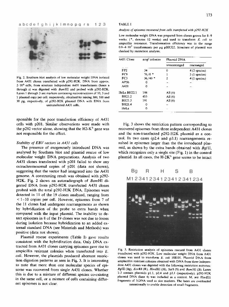
Fig. 2. Southern blot analysis of low molecular weight DNA isolated from A431 clones transfected with p292-H2K. DNA from approx. 2.106 cells, from nineteen independent A431 transfectants (lanes a through s) was digested with BamHl and probed with p292-H2K. Lanes 1 through 3 are markers containing reconstructions of 10, 3 and 1 plasmid copy per cell, respectively, obtained by mixing 300, 100 and 30 pg, respectively, of p292-H2K plasmid DNA with DNA from
untransfected A431 cells.
173
TABLE I
Analysis of episomes recovered from cells transfected with p292-H2K
Low molecular weight DNA was prepared from clones grown for 8-9 weeks. (*, denotes 12 weeks) and used to transform E. coli to ampicillin resistance. Transformation efficiency was in the range 0.9-4.107 transformants per #g pBR322. Structure of plasmid was checked by restriction analysis.
A431 Clone amp r colonies Plasmid DNA
unrearranged rearranged
FP2 34 0 FC9 76/0 * 1 FC3 36/40 * 2 AP5b 0 - A431 0 -
HeLa BH22.1 198 All (6) BH21.2 453 All (6) BH21.3 191 All (6) BH21.4 0 - HeLa 0 -
4 (2 species) 5 (1 species) 4 (1 species)
sponsible for the poor transfection efficiency of A431 cells with p201. Similar observations were made with the p292 vector alone, showing that the H2-K b gene was not responsible for the effect.
Stabi l i ty o f E B V vectors in A 4 3 1 cells
The presence of exogenously in t roduced D N A was
analyzed by Southern blot and plasmid rescue of low molecular weight D N A preparat ions. Analysis of two
A431 clones transfected with p201 failed to show any
extrachromosomal copies of p201 (data not shown),
suggesting that the vector had integrated into the A431
genome. A constras t ing result was obta ined with p292-
H2K. Fig. 2 shows an autoradiograph of B a m H I di-
gested D N A from p292-H2K transfected A431 clones probed with the total p292-H2K DNA. Episomes were
detected in 11 of the 19 clones analyzed, ranging from
< 1 -10 copies per cell. However, episomes from 7 of the 11 clones had undergone rearrangements as shown
by hybr idizat ion of the probe to extra bands when compared with the inpu t plasmid. The inabi l i ty to de- tect episomes in 8 of the 19 clones was not due to losses
dur ing isolation because hybr idizat ion to an added ex-
ternal s tandard D N A (see Materials and Methods) was positive (data not shown).
Plasmid rescue experiments (Table I) gave results consis tent with the hybr idizat ion data. Only D N A ex- tracted from A431 clones carrying episomes gave rise to
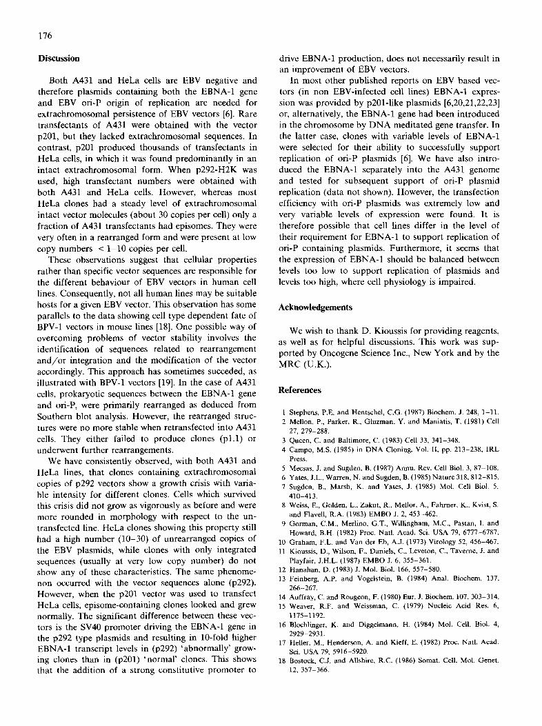
ampici l l in resistant colonies when transfected into E. co i l However, the plasmids produced aberrant restric- t ion digestion pat terns as seen in Fig. 3. It is interest ing to note that more than one molecular species of epi- some was recovered from single A431 clones. Whether this is due to a mixture of different species co-existing in the same cell, or a mixture of cells conta in ing differ- ent episomes is not clear.
Fig. 3 shows the restriction pattern corresponding to recovered episomes from three independent A431 clones and the non-transfected p292-H2K plasmid as a con- trol. In two cases (p2.4 and p3.1) rearrangements re- suited in episomes larger than the the introduced plas- mid, as shown by the extra bands obtained with BglII, which recognizes only a single site (Fig. 1) in the control plasmid. In all cases, the H-2K b gene seems to be intact
Bg R H S B
M1 2341 2341 2341 2341 234
Fig. 3. Restriction analysis of episomes rescued from A431 clones transfected with p292-H2K. Low molecular weight DNA from A431 clones was used to transform E. coli HB101. Plasmid DNA from amphicillin resistant colonies obtained with DNA from three indepen- dent A431 clones was digested with the following restriction enzymes: BglII (Bg), EcoRI (R), HindIII (H), SalI (S) and BamHI (B). Lanes 1-3 contain plasmids p1.1, p2.4 and p3.1 (respectively). p292-H2K plasmid DNA (lane 4) was included as a control. M, are HindIII fragments of ~, DNA used as size markers. The lanes are overloaded
intentionally to enable detection of small fragments.

174
A B 1 2 3 4 5 6 7 1 2 3 4 5 6 7 8 9 1 0
Fig. 4. Southern blot analysis of low molecular weight DNA isolated from A431 clones harboring rearrangement forms of p292-H2K. DNA from approx. 2-106 cells was digested with Bam HI and probed with p292-H2K. (A) Clones obtained by transfection of A431 cells with rescued p2.4; five independent clones (lanes 3 through 7) as well as a population of p2.4 transfectants (lane 2) are shown, p2.4 plasmid DNA was included as a control (lane 1). (B) Subclones of the A431 cell line which gave rise to p3.1 (lane 2 through 10). p3.1 plasmid
DNA was included as a control (lane 1).
as judged by the Barn HI digestion pattern. The EBNA-1 gene, which is contained on a HindlII fragment, is intact in two cases (p2.4 and p3.1). All the episomes have the correct 2.2 kb EcoRI fragment encompassing most of the pBR322 sequences. The latter is perhaps not surprising as it contains the fl-lactamase gene and the origin of replication which were selected for during the rescue procedure. The other bias in the rescue analysis would be the hygromycin resistance gene and the ori-P regions, since these are required for replication in hu- man cells and it is therefore likely that most changes have occurred in pBR322 sequences flanking the insert.
It is possible that the rearranged EBV plasmids have a selective advantage when grown in A431 cells and do not undergo further changes after the initial rearrange- ments. To test this possibility, we introduced pl.1, p2.4 and p3.1 into A431 cells. Transfection with pl.1. was 500-fold less efficient than p2.4 and p3.1, the latter gave colonies ((0.5-1.0)- 103 colonies/10/~g DNA per 8- 105 cells) at a frequency similar to that of the original p292-H2K plasmid. However, only the p2.4 transfected clones survived in culture prior to analysis. Fig. 4A shows the Southern analysis of low molecular weight DNA from A431 clones transfected with p2.4. One clone has an additional band and another clone has no episomes; the other three, as well as a population of p2.4 transfected cells (about 100 clones), gave an un- changed pattern. The A431 clone from which p2.4 was obtained, however, lost its episomes after 12 weeks in
culture. An additional experiment on the stability of these
rearranged EBV plasmids was carried out with the parental line which produced p3.1. The line was re- cloned after 8 weeks in culture by limiting dilution and
low molecular weight DNA from nine subclones was analyzed. Fig. 4B shows that in two cases episomes are missing, in another two cases further rearrangements occurred, while five sub-clones yielded the same pattern as the original 3.1 parent cells. This suggests that the initial rearrangements which gave rise to p3.1 do not provide an obvious selective advantage in A431 cells.
Stability of EBV-derived plasmids in HeLa cells The unexpected instability of the EBV vectors could
be due to a property of the A431 cell line. We tested this possibility by analyzing HeLa clones transfected with p292-H2K. Fig. 5 shows an autoradiograph of BamHI digests of low molecular weight DNA from ten independent clones hybridized with p292-H2K. All but one had a relatively high copy number (30 copies per cell) of episomes and only one showed an extra band compared to p292-H2K plasmid DNA control.
Similar observations were made with plasmid ob- tained from bacterial colonies resulting from transfor- mation with low molecular weight DNA extracted from HeLa cells. In all cases, a restriction pattern identical to p292-H2K was observed (Table I). Furthermore, the cloned H-2K b gene was efficiently expressed in HeLa and H-2K b specific mRNA levels were found to be related to the copy number of episomes (data not shown). The same conclusion about stable extrach- romosomal replication of EBV vectors in HeLa cells was obtained from the analysis of HeLa clones trans- fected with p201. Fig. 6 shows a Southern blot of total DNA probe with p228 (a plasmid derived from pHeBo (7) which contains ori-P, the hph gene and pBR322 sequences). This was used to avoid possible cross-hy- bridization with endogenous sequences homologous to H-2 and EBNA-1 genes [17]. Enzymes which do not cut
a b c d e f g h j k 1 2 3
Fig. 5. Southern blot analysis of low molecular weight episomal DNA isolation from HeLa clones transfectexl with p292-H2K. DNA from approx. 2.106 cells from ten independent HeLa transfectants (lanes a through k) was digested with Bam HI and probed with p292-H2K. Lanes 1 to 3 are markers containing reconstructions of 1, 3 and 10 plasmid per cell, respectively, obtained by mixing 30, 100 and 300 pg of p292-H2K plasmid DNA with DNA from untransfected HeLa
cells.

1 2 a b c d e f g h i j k I 3 4
1"1
I r I
Fig. 6. Southern blot analysis of total DNA isolated from HeLa clones transfected with p201. Six clones, representative of a total of ten independent clones, are shown. Lanes a through f are DNA samples digested with BstEII (which linearizes p201). Lane g through 1 are DNA samples p201. p201 plasmid DNA was mixed with toal DNA from untransfected cells to provide markers for 1 and 10 copies of plasmid per cell. Migration of form III is indicated by BstEII digested p201 (lanes 1 and 2, corresponding to 10 and 1 plasmid copy per cell, respectively). Migration of forms I and II is indicated by BglII-treated p201 (lanes 3 and 4, corresponding to 1 and 10 plasmid
copy per cell, respectively).
(BgllI) or cut p201 once (BstEII) were chosen. This analysis showed tha t these clones have in tac t extrach- r o m o s o m a l copies of p201 (in nine out of ten clones analyzed) , f rom 3 - 3 0 copies pe r cell, and that no re- a r r angemen t had occurred.
Growth of A431 and HeLa cells transfected with p201 and p292-H2K vectors
A l t h o u g h the p la smid p 2 9 2 - H 2 K is more s table in H e L a cells than in A431, we observed in bo th A431 and in H e L a cells that af ter 8 - 9 weeks in cul ture the cells underwent a crisis dur ing which the growth rate was decreased and many dead cells a ccompan ied every pas- sage. Interes t ingly , H e L a cells t ransfec ted with p201 and the rare A431 t ransfec tants ob ta ined with p201 d id not show any crisis and grew faster than their counter - pa r t s ob ta ined with p292, with or wi thout the c loned H - 2 K b gene.
A poss ib le exp lana t ion for the occurrence of this crisis is that the loss of the d o m i n a n t selectable marke r (due to inact iva t ion , or lack of rep l ica t ion of the vector) cou ld give rise to cells unable to survive selection. However , the change occurred rap id ly and, in the case of a l r eady charac te r ized clones, it was known that they con ta ined 1 0 - 3 0 in tac t ep isomes per cell, which makes the above exp lana t ion unlikely. Af t e r con t inued growth, some clones survived the crisis, bu t their g rowth rate was slower than that of the pa ren ta l l ine and they showed differences in morphology . Nevertheless , in tac t ep isomes were ma in t a ined in H e L a cells, as shown by Southern hybr id i za t ion of three of the H e L a clones in Fig. 5 ( lanes a, b and d).
175
Expression of the EBNA-1 gene in p201 and p292 based vectors
Because the p201 and p292 p l a smids have a d i f ferent p r o m o t e r on the EBNA-1 gene, the di f ferent g rowth proper t ies of the cells t ransfec ted with p201 and p292 based vectors could be caused by di f ferent ia l express ion of the EBNA-1 gene in the two vectors. W e therefore analyzed R N A from H e L a cells t ransfec ted with p201 and p 2 9 2 - H 2 K vectors for the s teady-s ta te level of EBNA-1 m R N A . Fig. 7 shows S1 analysis of to ta l R N A from four i ndependen t H e L a clones as well as a popu l a -
t ion of t ransfec tants ob t a ined with p201 or p292. Clones car ry ing p292 have 5 to 10-fold higher levels
of s teady-s ta te EBNA-1 m R N A than clones wi th p201. These levels were even higher than in an EBV infec ted
l y m p h o b l a s t o m a line used as a control . I t therefore seems l ikely that the level of EBNA-1 m R N A m a y de te rmine the phys io logica l changes lead ing to the dif-
ferences observed be tween cells t ransfec ted with EBV vectors.
5 1 7 -
220 -
154-
1 2 3 4 5 6 7 8 9 10 11 12 13
7 5 -
Hind III Sau 3A I N c o l
p292 SV 4 0 EBNA I
S a u 3 A I N c o l
/ = [ p201 ! EBNA I
Nae I H ind I ~ N c o I
~ probe pGEM E B N A I
p292 protected 150 nt
p201 protected 138 nt
Fig. 7. S1 analysis of EBNA-1 transcripts in HeLa clones transfected with p292 and p201. Lanes 3 through 7 are individual clones and lane 2 is a population of cells transfected with p292. Lanes 9 through 12 are individual clones and lane 8 is a population of p201 transfectants. Lane 1 (untransfected cells) and lane 13 (EBV-infected lympho- blastoid BW14 cell line) are the negative and positive controls respec- tively. Size markers shown at the left are those of Hinfl digested
pBR322.
176
Discussion
Both A431 and HeLa cells are EBV negative and therefore plasrnids containing both the EBNA-1 gene and EBV ori-P origin of replication are needed for extrachromosomal persistence of EBV vectors [6]. Rare transfectants of A431 were obtained with the vector p201, but they lacked extrachromosomal sequences. In contrast, p201 produced thousands of transfectants in HeLa cells, in which it was found predominantly in an intact extrachromosomal form. When p292-H2K was used, high transfectant numbers were obtained with both A431 and HeLa cells. However, whereas most HeLa clones had a steady level of extrachromosomal intact vector molecules (about 30 copies per cell) only a fraction of A431 transfectants had episomes. They were very often in a rearranged form and were present at low copy numbers < 1-10 copies per cell.
These observations suggest that cellular properties rather than specific vector sequences are responsible for the different behaviour of EBV vectors in human cell lines. Consequently, not all human lines may be suitable hosts for a given EBV vector. This observation has some parallels to the data showing cell type dependent fate of BPV-1 vectors in mouse lines [18]. One possible way of overcoming problems of vector stability involves the identification of sequences related to rearrangement a n d / o r integration and the modification of the vector accordingly. This approach has sometimes succeded, as illustrated with BPV-1 vectors [19]. In the case of A431 cells, prokaryotic sequences between the EBNA-1 gene and ori-P, were primarily rearranged as deduced from Southern blot analysis. However, the rearranged struc- tures were no more stable when retransfected into A431 cells. They either failed to produce clones (pl.1) or underwent further rearrangements.
We have consistently observed, with both A431 and HeLa lines, that clones containing extrachromosomal copies of p292 vectors show a growth crisis with varia- ble intensity for different clones. Cells which survived this crisis did not grow as vigorously as before and were more rounded in morphology with respect to the un- transfected line. HeLa clones showing this property still had a high number (10-30) of unrearranged copies of the EBV plasmids, while clones with only integrated sequences (usually at very low copy number) do not show any of these characteristics. The same phenome- non occurred with the vector sequences alone (p292). However, when the p201 vector was used to transfect HeLa cells, episome-containing clones looked and grew normally. The significant difference between these vec- tors is the SV40 promoter driving the EBNA-1 gene in the p292 type plasmids and resulting in 10-fold higher EBNA-1 transcript levels in (p292) 'abnormally' grow- ing clones than in (p201) 'normal ' clones. This shows that the addition of a strong constitutive promoter to
drive EBNA-1 production, does not necessarily result in an improvement of EBV vectors.
In most other published reports on EBV based vec- tors (in non EBV-infected cell lines) EBNA-1 expres- sion was provided by p201-1ike plasmids [6,20,21,22,23] or, alternatively, the EBNA-1 gene had been introduced in the chromosome by DNA meditated gene transfer. In the latter case, clones with variable levels of EBNA-1 were selected for their ability to successfully support replication of ori-P plasmids [6]. We have also intro- duced the EBNA-1 separately into the A431 genome and tested for subsequent support of ori-P plasmid replication (data not shown). However, the transfection efficiency with ori-P plasmids was extremely low and very variable levels of expression were found. It is therefore possible that cell lines differ in the level of their requirement for EBNA-1 to support replication of ori-P containing plasmids. Furthermore, it seems that the expression of EBNA-1 should be balanced between levels too low to support replication of plasrnids and levels too high, where cell physiology is impaired.
Acknowledgements
We wish to thank D. Kioussis for providing reagents, as well as for helpful discussions. This work was sup- ported by Oncogene Science Inc., New York and by the MRC (U.K.).
References
1 Stephens, P.E. and Hentschel, C.G. (1987) Biochem. J. 248, 1-11. 2 Mellon, P., Parker, R., Gluzman, Y. and Maniatis, T. (1981) Cell
27, 279-288. 3 Queen, C. and Baltimore, C. (1983) Cell 33, 341-348. 4 Campo, M.S. (1985) in DNA Cloning, Vol. If, pp. 213-238, IRL
Press. 5 Mecsas, J. and Sugden, B. (1987) Annu. Rev. Cell Biol. 3, 87-108. 6 Yates, J.L., Warren, N. and Sugden, B. (1985) Nature 318, 812-815. 7 Sugden, B., Marsh, K. and Yates, J. (1985) Mol. Cell Biol. 5,
410-413. 8 Weiss, E., Golden, L., Zakut, R., Mellor, A., Fahrner, K., Kvist, S.
and Flavell, R.A. (1983) EMBO J. 2, 453-462. 9 Gorman, C.M., Merlino, G.T., Willingham, M.C., Pastan, I. and
Howard, B.H. (1982) Proc. Natl. Acad. Sci. USA 79, 6777-6787. 10 Graham, F.L. and Van der Eb, A.J. (1973) Virology 52, 456-467. 11 Kioussis, D., Wilson, F., Daniels, C., Leveton, C., Taverne, J. and
Playfair, J.H.L. (1987) EMBO J. 6, 355-361. 12 Hanahan, D. (1983) J. Mol. Biol. 166, 557-580. 13 Feinberg, A.P. and Vogelstein, B. (1984) Anal. Biochem. 137,
266-267. 14 Auffray, C. and Rougeon, F. (1980) Eur. J. Biochem. 107, 303-314. 15 Weaver, R.F. and Weissman, C. (1979) Nucleic Acid Res. 6,
1175-1192. 16 Blochlinger, K. and Diggelmann, H. (1984) Mol. Cell. Biol. 4,
2929-2931. 17 Heller, M., Henderson, A. and Kieff, E. (1982) Proc. Natl. Acad.
Sci. USA 79, 5916-5920. 18 Bostock, C.J. and Allshire, R,C. (1986) Somat. Cell. Mol. Genet.
12, 357-366.

19 Green, L., Schlaffer, I., Wright, K., Moreno, M.L., Berand, D., Hager, G., Stein, J. and Stein, G. (1986) Proc. Natl. Acad. Sci. USA 83, 2315-2319.
20 Haver, C.A., Getty, R.R. and Tykocinski, M.L. (1989) Nucleic Acids Res. 13, 1989-2003.
21 DuBridge, R.B., Tang, P., Hsia, H.C., Leong, P., Miller, J.H. and Calos, M.P. (1987) Mol. Cell. Biol. 7, 379-387.
22 Hambor, J.E., Haver, C.A., Shu, H.-K., Groger, R.K., Kaplan,
177
D.R. and Tykocinski, M.L. (1988) Proc. Natl. Acad. Sci. USA 85, 4010-4014.
23 Reisman, D., Greenberg, M. and Rotter, V. (1988) Proc. Natl. Acad. Sci. USA 85, 5146-5250.
24 Baer, R., Bambier, A.T., Biggin, M.D., Deininger, P.L., Farrell, P.J., Gibson, T.J., Hatfull, G., Hudson, G.S., Satchwell, S.C., Seguin, C., Tuffnell, P.S. and Barrell, B.G. (1984) Nature 310, 207-211.