differences in fatty acid compositionbetween vegetative cells and
TRANSCRIPT

Proc. Nati. Acad. Sci. USAVol. 86, pp. 3399-3403, May 1989Microbiology
Differences in fatty acid composition between vegetative cells andN2-fixing vesicles of Frankia sp. strain Cpu1ANDERS TUNLID*t, NANCY A. SCHULTZt, DAVID R. BENSON*, D. BERNIE STEELE*, AND DAVID C. WHITE**Institute for Applied Microbiology, University of Tennessee, Knoxville, TN 37932; and tDepartment of Molecular and Cell Biology, University ofConnecticut, Storrs, CT 06268
Communicated by John G. Torrey, December 9, 1988 (received for review September 28, 1988)
ABSTRACT When growing on N2, actinomycetes from thegenus Frankia form multicellular structures that contain ni-trogenase. The structures are referred to as vesicles and areindistinguishable from vesicles formed when Frankia sp. are inroot-nodule symbioses. Vesicles isolated from N2-grown cells ofFrankia sp. strain CpI1 had a significantly higher amount anddifferent composition of fatty acids than did vegetative cellsrecovered fromNWt-containing medium. Lipids from vesicles,whole cells grown on N2, and whole cells grown on NH:+ werefractionated by silicic acid chromatography into neutral lipids,glycolipids, and polar lipids. The fatty acids were transester-ified by methanolysis and analyzed by gas chromatography andmass spectrometry. Vesicles had considerably higher amountsof fatty acids in the neutral and glycolipid fractions but loweramounts of polar lipid fatty acids than did vegetative cells.Polar lipids from vesicles had a higher proportion of mono-unsaturated and cyclopropane fatty acids and a lower propor-tion of isobranched fatty acids than did polar lipids fromNH:-grown or N2-grown cells. The neutral lipid and glycolipidfractions contained several long-chain compounds with molec-ular ions at m/z 408 and 410. The proportions of thesecompounds were significantly higher in the lipids from vesiclesthan from vegetative cells. These results suggest that lipids invesicles might be involved in the protection of nitrogenase from02 and suggest a parallel with the glycolipids involved inprotecting nitrogenase from 02 in the cyanobacterial hetero-cysts.
Frankia sp. are N2-fixing actinomycetes that induce rootnodules on several nonleguminous plants including species ofAlnus, Myrica, and Casuarina (1). When fixing N2 in sym-biosis and in culture, most Frankia sp. form specializedcellular structures called vesicles. Vesicles contain nitrogen-ase as shown by direct assay after isolation (2, 3) and byimmunogold-labeling using antibodies against nitrogenase(4).
In culture, Frankia sp. fix N2 at atmospheric partialpressures of 02 (P02) (5-7). Since Frankia sp. grow andrespire slowly and since their nitrogenase is as sensitive to 02inactivation as other nitrogenases (8), some mechanismsmust exist that protect the enzyme from 02- Part of thisprotection has been proposed to reside in the cell envelope ofvesicles, which may function as a diffusion barrier (6, 9, 10).Evidence for this function includes the observation thatnitrogenase activity occurs without vesicles in Frankia cul-tures grown under microaerobic conditions; such activity issensitive to inactivation by 02 (11). A parallel occurs incyanobacteria, where cultures lacking heterocysts can fix N2only under microaerobic conditions, while aerobic fixationoccurs only when heterocysts are present (12).
Frankial vesicles have a cell envelope or capsule thatcompletely surrounds the vesicle stem and head regions (4, 9,
10, 13). In freeze-fracture and in freeze-substituted prepara-tions, the envelope material appears as repeated lipid mono-layers with as many as 50 or more layers observed (10, 13).Recent studies have shown that the number of monolayersincreases in proportion to the level of02 under which frankialcultures are induced for nitrogenase activity (10). The mul-tilayered structure of the vesicle envelope is similar to theheterocyst envelope structure (12). The inner laminated layerof the heterocyst envelope is composed of glycolipids withlong-chain polyhydroxyl alcohols and hydroxy acids notfound in vegetative cells (14-16). Mutant studies on Ana-baena vulgaris have suggested that these lipids are essentialfor protecting cyanobacterial nitrogenase from 02 (17), andgas-diffusion studies using gas vesicle collapse have shownthe diffusion resistance of intact heterocysts to gases to bequite high (18), primarily because of the glycolipid laminae inthe heterocyst envelope.
In the present paper we have taken advantage of a newlydescribed technique for isolating vesicles from N2-fixingcultures of Frankia (2). We have examined the fatty acidprofiles of NH'-grown cells lacking vesicles, cells fromN2-fixing cultures, and vesicles isolated from N2-fixing cul-tures to identify differences in fatty acid composition of polarlipids, glycolipids, and neutral lipids.
MATERIALS AND METHODSOrganism and Culture Media. Frankia sp. strain CpIl, a
Comptonia peregrina isolate (19), was grown in definedsuccinate/NH' or succinate/combined nitrogen-free liquidmedium as described (2).
Vesicle Isolation. Nitrogen-fixing cells grown in 3-literbatch cultures were harvested by vacuum filtration and werehomogenized in 50% (wt/wt) sucrose (2). The homogenatewas transferred to 50-ml polycarbonate centrifuge tubes. Thesucrose was overlaid with 10% (vol/vol) glycerol in distilledH20, and the tubes were centrifuged for 1 hr at 16,000 X g.The vesicle band was removed with a Pasteur pipet. Both theisolated vesicles and gradient pellet were washed with 20mMimidazole/HCI buffer (pH 6.9) containing 2 mM MgCl2 and1% glycerol.
Cells grown on NH' or on N2 were collected by vacuumfiltration and stored at -80°C. The frozen cell pastes fromNH+-grown and N2-grown cultures and the gradient pelletand vesicle fractions from the sucrose density gradientcentrifugation were lyophilized with a Virtis lyophilizer(Gardiner, NY). The freeze-dried samples were held underN2 atmosphere until analysis.
Lipid Extraction and Fractionation. Approximately 10 mg(dry weight) of the lyophilized samples was extracted withthe single-phase mixture chloroform/methanol/water (1:2:0.8, vol/vol) as described by Bligh and Dyer (20). Theextracts were split into two phases by the addition of 1
tPresent address: Laboratory of Ecological Chemistry, Lund Uni-versity, Lund, Sweden.
3399
The publication costs of this article were defrayed in part by page chargepayment. This article must therefore be hereby marked "advertisement"in accordance with 18 U.S.C. §1734 solely to indicate this fact.

Proc. Natl. Acad. Sci. USA 86 (1989)
volume each of chloroform and water. The organic phase wasrecovered, and the extracted lipids were fractionated oncolumns of silicic acid (Unisil, 100-200 mesh; Clarkson,Williamsport, PA) containing 1 g (dry weight) of adsorbent.The lipids were applied to the columns in a minimal volumeof chloroform and were eluted with solvents of increasingpolarity: 10 ml of chloroform, 40 ml of acetone, and 10 ml ofmethanol. For lipids ofmost bacteria, the chloroform fractioncontains neutral lipids; the acetone fraction, glycolipids; andthe methanol fraction, polar lipids (21, 22). The lipid fractionswere dried under a stream of N2.Mild Alkaline Methanolysis. The lipid fractions were sub-
jected to a mild alkaline methanolysis, which yields themethyl esters of the ester-linked fatty acids (23).Gas Chromatography (GC). The methyl esters were ana-
lyzed by capillary gas GC with a Hewlett-Packard 5880 gaschromatograph equipped with a flame-ionization detector.The esters were separated on a 50m x 0.2 mm i.d. fused silicacolumn coated with a cross-linked methylsilicone stationaryphase (Hewlett-Packard). The initial oven temperature washeld at 800C for 1 min and then was increased at a rate of100C/min to 240'C and finally at 50C/min to 300'C. Theinjector temperature was 250'C, and the detector tempera-ture was 270'C. Hydrogen at a linear gas flow rate of 80cm/sec was used as the carrier gas. Injections were per-formed in the splitless mode.
Tentative peak identification, prior to mass spectrometric(MS) analysis, was based on comparison of retention timeswith data from standards obtained from Supelco, Sigma, NuChek Prep (Elysian, MN), and from compounds previouslyidentified in our laboratory. Peak areas were quantified byadding an internal standard, methyl nonadecanoate, prior tothe GC injections. The GC data were acquired and manipu-lated by a Nelson chromatography data system (Nelson,Paramus, NJ).GC/MS. GC/MS analyses were performed on a VG
TRIO-3 GC/MS/MS instrument equipped with aHP 5890 gaschromatograph. The GC conditions were those described asabove, but helium was used as the carrier gas. The electronenergy in electron-impact ionization was 70 eV, and thesource temperature was 2000C. Isobutane (purity, 99.99%)was used as the reagent gas in chemical ionization. Thesource temperature in chemical ionization was 1500C.MS identifications of the methyl esters were based on
comparison with spectra from standards or with spectrareported in the literature (24-26). The positions of doublebonds were determined by preparing the dimethyldisulfideadducts (27). Positions of cyclopropane rings were deter-mined after hydrogenation of the methylesters in the pres-ence of Adam's catalyst (PtO2) (28).
Fatty Acid Nomenclature. Fatty acids are designated by thetotal number ofcarbon bonds: number of double bonds, withthe position closest to the aliphatic end of the moleculeindicated (w) along with the geometry (c for cis and t fortrans). The prefixes "i" and "a" refer to iso- and anti-isobranching, respectively. Cyclopropane fatty acids aredesignated "cy."
RESULTSFatty Acid Content. The data in Figs. 1-4 are presented as
the amount, by weight, of the most abundant fatty acids ineach cell type. The data in Table 1 present all fatty acidsidentified in the polar lipids in the methanol fraction from thesilicic acid column and are expressed in mol %. Majordifferences were observed in the amount and composition offatty acids between the various Frankia cell types. Vesicleshad the highest proportion of their dry weight as fatty acids;N2-grown cells had less, and NH'-grown cells had only halfthe amount found in vesicles (Fig. 1).
There were also significant differences in the relativeproportions of fatty acids recovered in the methanol, ace-tone, and chloroform fractions between the three cell types(Fig. 1). Sixty percent by weight of the total fatty acids invegetative cells from NH'-containing medium were frompolar lipids extracted in the methanol fraction (Fig. 1). Thisfraction was considerably smaller in N2-grown cells (26%)and much smaller in the vesicles (10%); the latter samples hadhigher proportions of their fatty acids in the glycolipid andneutral lipid fractions. Approximately 70% by weight of thefatty acids in vesicles and 55% in N2-grown cells were foundin the neutral lipid fraction compared with 30% in vegetativecells.
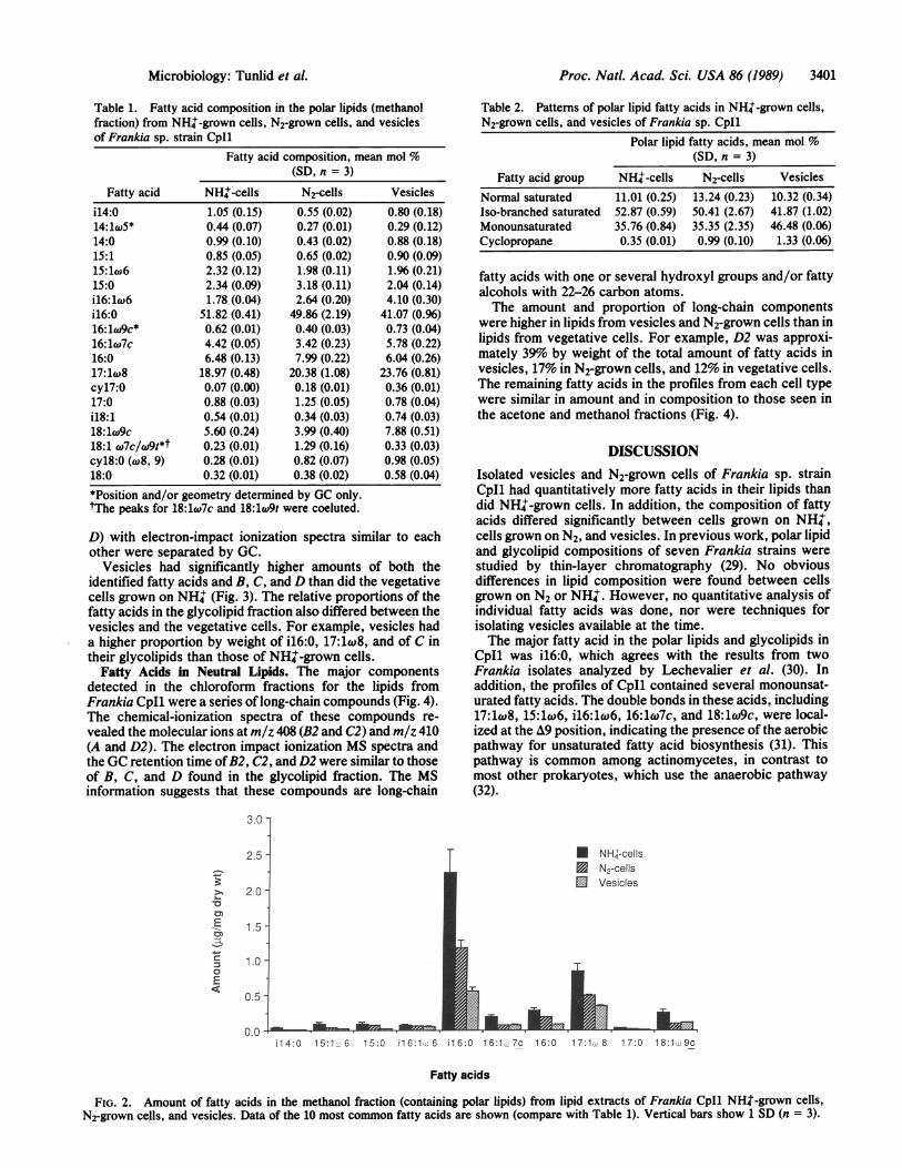
Polar Lipid Fatty Acids. Ester-linked fatty acids from themethanol fraction had 14.18 carbon atoms (Table 1, Fig. 2).The predominant fatty acid in NHW-grown cells, N2-growncells, and vesicles was i16:0, which comprised between 41%and 51% by weight of the total polar fatty acids. Othercommon fatty acids in these profiles were monounsaturatedfatty acids with double bonds localized at the A9 position,including 17:1w8, 15:1w6, il6:lc6, 16:17c, and 18:1co9c,and normal saturated fatty acids with 15, 16, and 17 carbons.Among the minor compounds were two cyclopropane fattyacids. The most prominent of these had 18 carbon atoms andwas identified as cyl8:O(*8, 9) (Table 1).The composition of fatty acids in the polar lipids of
NH+-grown cells and N2-grown cells were quite similar.However, by comparing the relative proportions expressed inmol %, it was shown that polar lipids from the vesicles hada significantly lower mol % of isobranched fatty acids but ahigher proportion of monounsaturated and cyclopropanefatty acids than did polar lipids from vegetative cells (Table2).
Fatty Acids in Glycolipids. The acetone eluent from thesilicic acid column contained the isobranched, monounsat-urated, and normal saturated fatty acids as identified in thepolar lipids. The two major fatty acids were i16:0 (17-25% byweight of the glycolipid fatty acids) and 17:1w8 (18-27%)(Fig. 3). Furthermore, the acetone fraction contained severallonger chain components. The electron impact ionizationspectra of these showed extensive fragmentation of themolecules with no molecular ions present. The molecularweight of the most common long-chain compound wasdetermined by chemical-ionization MS to be m/z 408. Thiscompound was designated C. Two other compounds (B and
20 -
.0)
B.,0)
0
E
CD
15 -
10 -
5 -
0-
* NH+-cellsN2-cells
E VesiclesT
Total MeOH Acetone CHC13
Lipid fractions
FIG. 1. Total amount of fatty acids in NH'-grown cells, N2-grown cells, and vesicles of Frankia sp. strain Cpu1. The lipidextracts were fractionated on a silicic acid column. The methanolfraction contains polar lipids, the acetone fraction contains glycolip-ids, and the chloroform fraction contains neutral lipids. Vertical barsrepresent 1 SD (n = 3).
3400 Microbiology: Tunfid et al.
Proc. Nati. Acad. Sci. USA 86 (1989) 3401
Table 1. Fatty acid composition in the polar lipids (methanolfraction) from NH'-grown cells, N2-grown cells, and vesiclesof Frankia sp. strain CpI1
Fatty acid composition, mean mol %(SD, n = 3)
Fatty acid NH+-cells N2-cells Vesicles
i14:0 1.05 (0.15) 0.55 (0.02) 0.80 (0.18)14:1w5* 0.44 (0.07) 0.27 (0.01) 0.29 (0.12)14:0 0.99 (0.10) 0.43 (0.02) 0.88 (0.18)15:1 0.85 (0.05) 0.65 (0.02) 0.90 (0.09)15:1w6 2.32 (0.12) 1.98 (0.11) 1.% (0.21)15:0 2.34 (0.09) 3.18 (0.11) 2.04 (0.14)il6:1w6 1.78 (0.04) 2.64 (0.20) 4.10 (0.30)i16:0 51.82 (0.41) 49.86 (2.19) 41.07 (0.96)16:lw9c* 0.62 (0.01) 0.40 (0.03) 0.73 (0.04)16:1w7c 4.42 (0.05) 3.42 (0.23) 5.78 (0.22)16:0 6.48 (0.13) 7.99 (0.22) 6.04 (0.26)17:1w8 18.97 (0.48) 20.38 (1.08) 23.76 (0.81)cyl7:0 0.07 (0.00) 0.18 (0.01) 0.36 (0.01)17:0 0.88 (0.03) 1.25 (0.05) 0.78 (0.04)i18:1 0.54 (0.01) 0.34 (0.03) 0.74 (0.03)18:1w9c 5.60 (0.24) 3.99 (0.40) 7.88 (0.51)18:1 w7c/w9t*t 0.23 (0.01) 1.29 (0.16) 0.33 (0.03)cyl8:0 (8, 9) 0.28 (0.01) 0.82 (0.07) 0.98 (0.05)18:0 0.32 (0.01) 0.38 (0.02) 0.58 (0.04)
*Position and/or geometry determined by GC only.tThe peaks for 18:17c and 18:1w9t were coeluted.
D) with electron-impact ionization spectra similar to eachother were separated by GC.
Vesicles had significantly higher amounts of both theidentified fatty acids and B, C, and D than did the vegetativecells grown on NH' (Fig. 3). The relative proportions of thefatty acids in the glycolipid fraction also differed between thevesicles and the vegetative cells. For example, vesicles hada higher proportion by weight of i16:0, 17:1cA8, and of C intheir glycolipids than those of NH'-grown cells.
Fatty Acids in Neutral Lipids. The major componentsdetected in the chloroform fractions for the lipids fromFrankia CpI1 were a series of long-chain compounds (Fig. 4).The chemical-ionization spectra of these compounds re-vealed the molecular ions at m/z 408 (B2 and C2) and m/z 410(A and D2). The electron impact ionization MS spectra andthe GC retention time ofB2, C2, and D2 were similar to thoseof B, C, and D found in the glycolipid fraction. The MSinformation suggests that these compounds are long-chain
3.0 -
2.5 -
20-
C
-E
=3
0E
1.5
1.0 -
0.5 -
0.0 -
T
Table 2. Patterns of polar lipid fatty acids in NH'-grown cells,N2-grown cells, and vesicles of Frankia sp. CpI1
Polar lipid fatty acids, mean mol %(SD, n = 3)
Fatty acid group NH'-cells N2-cells VesiclesNormal saturated 11.01 (0.25) 13.24 (0.23) 10.32 (0.34)Iso-branched saturated 52.87 (0.59) 50.41 (2.67) 41.87 (1.02)Monounsaturated 35.76 (0.84) 35.35 (2.35) 46.48 (0.06)Cyclopropane 0.35 (0.01) 0.99 (0.10) 1.33 (0.06)
fatty acids with one or several hydroxyl groups and/or fattyalcohols with 22-26 carbon atoms.The amount and proportion of long-chain components
were higher in lipids from vesicles and N2-grown cells than inlipids from vegetative cells. For example, D2 was approxi-mately 39% by weight of the total amount of fatty acids invesicles, 17% in N2-grown cells, and 12% in vegetative cells.The remaining fatty acids in the profiles from each cell typewere similar in amount and in composition to those seen inthe acetone and methanol fractions (Fig. 4).
DISCUSSIONIsolated vesicles and N2-grown cells of Frankia sp. strainCpI1 had quantitatively more fatty acids in their lipids thandid NH+-grown cells. In addition, the composition of fattyacids differed significantly between cells grown on NH+,cells grown on N2, and vesicles. In previous work, polar lipidand glycolipid compositions of seven Frankia strains werestudied by thin-layer chromatography (29). No obviousdifferences in lipid composition were found between cellsgrown on N2 or NH'. However, no quantitative analysis ofindividual fatty acids was done, nor were techniques forisolating vesicles available at the time.The major fatty acid in the polar lipids and glycolipids in
CpI1 was i16:0, which agrees with the results from twoFrankia isolates analyzed by Lechevalier et al. (30). Inaddition, the profiles of CpI1 contained several monounsat-urated fatty acids. The double bonds in these acids, including17:1w8, 15:1cw6, i16:1co6, 16:1l7c, and 18:1w9c, were local-ized at the A9 position, indicating the presence of the aerobicpathway for unsaturated fatty acid biosynthesis (31). Thispathway is common among actinomycetes, in contrast tomost other prokaryotes, which use the anaerobic pathway(32).
* NH,-cells* N2-cellsl Vesicles
& hii14:0 15:1. 6 15:0 i16:1 6 1 6:0 16:1 7c 16:0 17:1. 8 17:0 1 8:1. 9c
Fatty acids
FIG. 2. Amount of fatty acids in the methanol fraction (containing polar lipids) from lipid extracts of Frankia CpI1 NHJ-grown cells,N2-grown cells, and vesicles. Data of the 10 most common fatty acids are shown (compare with Table 1). Vertical bars show 1 SD (n = 3).
Microbiology: Tunfid et aL

Proc. Natl. Acad. Sci. USA 86 (1989)
0.8 -
* NHR-ceIls
0.6~~~~ ~ ~ ~ ~ ~ ~~~~~~~~ ~~~N2-cellsEli vesicles
0.4
o 0.2-.....
0.015:0 i16:1 6 i16:0 16:1 7c 16:0 17:1. 8 18:1 .Sc B C D
Fatty acids
FIG. 3. Amount offatty acids in the acetone fraction (containing glycolipids) from lipid extracts ofFrankia CpH NHZ-grown cells, Nrgrowncells, and vesicles. Data of the 10 most common fatty acids are shown. B, C, andD are unidentified long-chain components. Vertical bars show1 SD (n = 3).
Polar lipids are important components of the plasmamembrane in actinomycetes, and a functional membranerequires a suitable selection of various types of such lipids,particularly phospholipids (33). Phosphatidylinositol anddiphosphatidylglycerol are the major polar lipids identified inFrankia (29, 30). The higher proportion of monounsaturatedand cyclopropane fatty acids in the polar lipids of vesiclesthan in hyphae from CpI1 would be expected to influence thephysical properties and thereby the physiological function (inas yet unidentified ways) of the membranes (34).The chemical structures of the long-chain compounds
detected in the neutral lipid and glycolipid fractions have notbeen identified. Preliminary characterization, based on MS,suggests that they are polyhydroxy fatty acids or alcoholswith 22-26 carbon atoms. Therefore, they have some simi-larity with the C26 to C28 hydroxy alcohols and fatty acids thathave been identified in the glycolipids from heterocysts ofcyanobacteria (16). Further work is needed to finalize thestructure of the long-chain compounds in Frankia CpI1.
6-
5-
4-
L-
cm
0)
0E
4-
3-
2-
0
The cellular location of the long-chain compounds withinthe vesicles is not known, but light and electron microscopicstudies suggest that they could be constituents of the lami-nated layers seen in the cell envelope. In freeze-fractureelectron microscopy, up to 50 lipid monolayers have beenobserved in envelopes, with each layer having a thicknessfrom 3.5 to 4.0 nm (9, 10), which is between the thickness ofa monolayer and a bilayer of palmitic acid. In addition,studies with Nile red fluorescence staining indicate that theenvelope of the Frankia vesicle is composed largely of lipid(35). These observations, together with the abundance of thelong-chain compounds, strongly suggest that they are local-ized in the laminae. Ultrastructurally similar laminae areoccasionally seen in vegetative hyphae of CpI1, but neverwith the abundance noted in vesicles (36). Thus, lipidsassociated with vesicle laminae may be normal cell compo-nents that accumulate during vesicle differentiation.The apparent similarity in structure with the long-chain
compounds of the heterocysts as well as the significantly
T* NHW-cells3 N2-cellsE3 Vesicles
v---_-....15:0 il6:0 16:1 t7c 17:1 8 17:0 18:1. 9c A
Fatty acidsB2 C2 02
FIG. 4. Amount of fatty acids in the chloroform fraction (containing neutral lipids) from lipid extracts of Frankia Cpu1 NH't-grown cells,N2-grown cells, and vesicles. Data of the 10 most common fatty acids are shown. A, B2, C2, and D2 are long-chain compounds with unknownstructure. Vertical bars show 1 SD (n = 3).
.........Jay .Nmlk.[F'Jr .........&. .M%:--..:.:.jFF79PIM.
3402 Microbiology: Tunlid et al.

Proc. Natl. Acad. Sci. USA 86 (1989) 3403
higher amounts of these compounds in vesicles than inhyphae suggest that long-chain fatty acids or alcohols can bean important part of the cell envelope and can function as adiffusion barrier to 02 in Frankia vesicles.
This research has been supported by U.S. Department of Agri-culture Grant 85-CRCR-1-1657 from the Competitive ResearchGrants Office (to D.R.B.) and by Swedish Natural Research CouncilGrant B-BU 8564-300 (to A.T.).
1. Torrey, J. G. (1978) Bioscience 28, 586-592.2. Noridge, N. A. & Benson, D. R. (1986) J. Bacteriol. 166, 301-
305.3. Tisa, L. S. & Ensign, J. C. (1987) J. Bacteriol. 169, 5054-5059.4. Meesters, T. M. (1987) Arch. Microbiol. 146, 327-331.5. Gauthier, D. L., Diem, H. G. & Dommergues, Y. (1981) Appl.
Environ. Microbiol. 41, 306-308.6. Murry, M. A., Fontaine, M. S. & Tjepkema, J. D. (1984) Arch.
Microbiol. 139, 162-166.7. Tjepkema, J. D., Ormerod, W. & Torrey, J. G. (1980) Nature
(London) 287, 633-635.8. Benson, D. R., Arp, D. J. & Burris, R. H. (1978) Science 205,
688-689.9. Torrey, J. G. & Callaham, D. (1982) Can. J. Microbiol. 28,749-
757.10. Parsons, R., Silvester, W. B., Harris, S., Gruijters, W. T. M.
& Bullivant, S. (1987) Plant Physiol. 83, 728-731.11. Murry, M. A., Zhongze, Z. & Torrey, J. G. (1985) Can. J.
Microbiol. 31, 804-809.12. Stewart, W. D. P. (1980) Annu. Rev. Microbiol. 34, 497-536.13. Lancelle, S. A., Torrey, J. G., Hepler, P. K. & Callaham,
D. A. (1985) Protoplasma 127, 64-72.14. Nichols, B. W. & Wood, B. J. B. (1968) Nature (London) 217,
767-768.15. Winkenbach, F., Wolk, C. P. & Jost, M. (1972) Planta 107, 69-
80.16. Lambein, F. & Wolk, C. P. (1973) Biochemistry 12, 791-798.
17. Haury, J. F. & Wolk, C. P. (1978) J. Bacteriol. 136, 688-692.18. Walsby, A. E. (1985) Proc. R. Soc. London Ser. B 226, 345-
366.19. Callaham, D., DelTredici, P. & Torrey, J. G. (1978) Science
199, 899-902.20. Bligh, E. G. & Dyer, W. J. (1959) Can. J. Biochem. Physiol. 31,
911-917.21. Kates, M. (1986) Techniques ofLipidology, Isolation, Analysis,
and Identification ofLipids (Elsevier, Amsterdam), 2nd Ed., p.194.
22. King, J. D., White, D. C. & Taylor, C. W. (1977) Appl.Environ. Microbiol. 33, 1177-1183.
23. White, D. C., Davis, W. M., Nickels, J. S., King, J. D. &Bobbie, R. J. (1979) Qecologia (Berlin) 40, 51-62.
24. Ryhage, R. & Stenhagen, E. (1960) J. Lipid Res. 1, 361-390.25. Campbell, I. M. & Naworal, J. (1%9) J. LipidRes. 10, 589-592.26. Boon, J. J., van de Graaf, B., Schuyl, P. J. W., de Lange, F.
& de Leeuw, J. W. (1977) Lipids 12, 717-721.27. Nichols, P. D., Guckert, J. B. & White, D. C. (1986) J.
Microbiol. Methods 5, 49-55.28. McCloskey, J. A. & Law, J. H. (1967) Lipids 2, 225-230.29. Lopez, M. F., Whaling, C. S. & Torrey, J. G. (1983) Can. J.
Bot. 61, 2834-2842.30. Lechevalier, M. P., Baker, D. & Horriere, F. (1983) Can. J.
Bot. 61, 2826-2833.31. Scheuerbrandt, G. & Bloch, K. (1962)J. Biol. Chem. 237, 2064-
2068.32. Fulco, A. J. (1983) Prog. Lipid Res. 22, 133-160.33. Minnikin, D. E. & O'Donnell, A. G. D. (1983) in The Biology
of Actinomycetes, eds. Goodfellow, M., Mordarski, M. &Williams, S. T. (Academic, London), pp. 337-388.
34. Cronan, J. E. & Gelmann, E. P. (1975) Bacteriol. Rev. 39, 232-256.
35. Lamont, H. C., Silvester, W. B. & Torrey, J. G. (1988) Can. J.Microbiol. 34, 656-660.
36. Newcomb, W. & Wood, S. M. (1987) Int. Rev. Cytol. 109, 1-88.
Microbiology: Tunlid et al.