dietary walnut oil modulates liver steatosis in the obese
TRANSCRIPT
ORIGINAL CONTRIBUTION
Dietary walnut oil modulates liver steatosisin the obese Zucker rat
Anja Fink • Corinna E. Rufer • Julie Le Grandois •
Alexander Roth • Dalal Aoude-Werner •
Eric Marchioni • Achim Bub • Stephan W. Barth
Received: 26 June 2013 / Accepted: 31 July 2013 / Published online: 13 August 2013
� The Author(s) 2013. This article is published with open access at Springerlink.com
Abstract
Purpose Non-alcoholic fatty liver disease (NAFLD) is
the hepatic manifestation of the metabolic syndrome. We
aimed to clarify the impact of dietary walnut oil versus
animal fat on hepatic steatosis, representing the initial step
of multistage pathogenesis of NAFLD, in Zucker obese
rats.
Methods Zucker lean ad libitum (a.l.), Zucker obese a.l. or
Zucker obese pair fed (p.f.) to the lean received isocaloric
diets containing 8 % walnut oil (W8), W14 or 14 % lard
(L14) (n = 10/group). Body weight, clinical serology, liver
weight, lipid content and fatty acid composition and hepatic
lipid metabolism-related transcripts were evaluated.
Results Compared to lean, Zucker obese a.l. and p.f.
showed hepatic triacylglyceride (TAG) accumulation. In
Zucker obese p.f., W14 compared to W8 and L14 reduced
liver lipids, TAG as well as hepatic omega-6 (n-6)/n-3 ratio
and SCD activity index [(C18:0 ? C18:1)/C18:0 ratio]
paralleled by decreased lipoprotein lipase mRNA in obese
p.f. and elevated microsomal triglyceride transfer protein
mRNA in lean and obese. Further, W14 elevated the fast-
ing blood TAG and reduced cholesterol levels in obese.
Conclusions In our model, consumption of W14 inhibited
hepatic lipid accumulation along with modulated hepatic
gene expression implicated in hepatic fatty acid influx or
lipoprotein assembly. These results provide first indication
that dietary lipids from walnut oil are modulators of hepatic
steatosis as the initial step of progressive NAFLD
pathogenesis.
Keywords NAFLD � Lard � SFA � MUFA � PUFA
Introduction
Non-alcoholic fatty liver disease (NAFLD) is a common
public health problem worldwide with an increased prev-
alence during the last three decades [1–3]. The prevalence
of this disorder is strongly associated with several risk
factors of the metabolic syndrome (MetS) such as obesity,
dyslipidemia, hypertension and type 2 diabetes [4–7].
Therefore, NAFLD is regarded as the hepatic manifestation
of the MetS [1, 8]. NAFLD, a multistage and progressive
disease, ranges from lipid accumulation in hepatocytes
(hepatic steatosis) to hepatic steatosis with a necroinflam-
matory component (non-alcoholic steatohepatitis; NASH)
and might finally progresses to liver cirrhosis [5, 6, 9].
Liver triacylglyceride (TAG) levels are the result of the
balance between influx of fatty acids, de novo lipogenesis,
TAG synthesis and delivery by very low density lipopro-
tein (VLDL) assembly [10–13]. Thus, the accumulation
of TAG in the steatotic liver reflects a difference between
the rate at which fatty acids reach hepatocytes, or are
A. Fink � A. Roth � A. Bub � S. W. Barth (&)
Department of Physiology and Biochemistry of Nutrition,
Max Rubner-Institut, Haid-und-Neu-Str. 9, 76131 Karlsruhe,
Germany
e-mail: [email protected]
C. E. Rufer
Department of Safety and Quality of Fruit and Vegetables,
Max Rubner-Institut, Haid-und-Neu-Str. 9, 76131 Karlsruhe,
Germany
J. Le Grandois � D. Aoude-Werner
Aerial, Parc d’Innovation, rue Laurent Fries,
B.P. 40443, 67412 Illkirch-Cedex, France
E. Marchioni
Equipe de Chimie Analytique des Molecules BioActives,
Faculte de Pharmacie, IPHC, Universite de Strasbourg,
74 route du Rhin, 67400 Illkirch, France
123
Eur J Nutr (2014) 53:645–660
DOI 10.1007/s00394-013-0573-z

synthesized therein, and the rate at which they are metab-
olized, stored or assembled [10, 12, 13]. Further, this initial
expansion of ectopic lipid levels during steatosis develop-
ment tends to be associated with progressive metabolic
dysregulation and, in particular, altered hepatic insulin
resistance and the development of inflammation.
Until now, there has no specific therapy been developed
to directly treat the pathogenesis and progression of NA-
FLD. Instead, indirect approaches are pursued by treating
major risk factors of NAFLD. However, this treatment
strategy is complex, because not a single but various risk
factors influence and associate the induction and progres-
sion of NAFLD. Among others, mainly the developing
insulin resistance and lipid dysmetabolism promote the
pathological hepatic TAG accumulation [5, 6]. Hence, the
cornerstones of the therapy as well as prevention of NA-
FLD are to ameliorate the metabolic risk factors by
reducing body weight and improving insulin sensitivity and
hyperlipidemia [6, 9].
The enhanced prevalence of NAFLD is also driven by
changes of dietary habits and primarily increased con-
sumption of dietary fat. Recent studies have further indi-
cated that not solely the quantity of dietary fat, but also fatty
acid composition is a risk-determining factor for NAFLD:
Patients with NAFLD not only showed a higher intake of
saturated fatty acids (SFA), but also increased omega-6
(n-6) polyunsaturated fatty acids (PUFA) consumption
while n-3 PUFA uptake is reduced, leading to a significant
shift toward increased dietary n-6/n-3 ratio [3, 8, 14].
Recent literature provides indications that these dietary
changes might also lead to endogenous alterations in the
hepatic fatty acid composition toward an increase in the
n-6/n-3 PUFA ratio [15, 16], which associates with the
development of an adverse metabolic profile and contribute
to the pathogenesis of NAFLD [16]. Further, animal studies
have demonstrated that an excess of n-6 PUFA in the liver
is associated with a proinflammatory [3, 17] as well as
steatogenic state with pronounced hepatic fat accumulation
[18–20]. Given these observations, a dietary approach
changing the n-6/n-3 PUFA ratio in the diet might be
effective enough to change hepatic n-6/n-3 PUFA ratio in a
way that positively influence NAFLD-related hepatic
pathogenesis [21–23].
Nuts and seeds share a fatty acid profile which is char-
acterized by low amounts of SFA and high content of
monounsaturated fatty acids (MUFA) and PUFA [24].
Among the numerous nut varieties, recent research has
focused on walnuts (Juglans regia). One reason for this
might be the ‘‘optimal’’ 4:1 ratio of n-6/n-3 PUFA and
additionally the high content of tocopherols (mainly
c-tocopherol), phytosterols, polyphenolic antioxidants
(ellagitannins) and fiber [25, 26]. With regard to a signif-
icant bioactivity, recent investigations have shown that a
walnut-rich diet improved hyperlipidemia [26–30], type 2
diabetes [31, 32], cardiovascular disease [33–35], amelio-
rated the antioxidative status [36, 37] and provided general
parenchyma-protecting effects in the liver [30, 38]. How-
ever, to our best knowledge, no data are published
regarding bioactivity of walnut on NAFLD mainly focus-
ing on the initial step of liver steatosis.
Thus, the aim of the present study was to investigate the
effect of different amounts of walnut oil versus lard on
hepatic lipid metabolism in a rodent steatosis model and
identify hepatic pathways which might be implicated in
any observed bioactivity. Therefore, we have targeted
hepatic lipid content, serum lipids and sICAM, as an
artherogenic and proinflammatory marker. Additionally,
we focused on the expression of genes involved in hepatic
lipid metabolism to explore underlying mechanisms.
Materials and methods
Phospholipid analysis of walnut oil
Standard solutions of soy lecithin-mix standard (certified:
14.41 % phosphatidylcholine (PC), 12.06 % phosphati-
dylethanolamine (PE) and 9.64 % phosphatidylinosi-
tol (PI)) from Spectral Service GmbH (Cologne, Germany)
were prepared at six concentrations between 0.1 and
1.1 mg/mL. Sand (Fontainebleau) was provided by VWR
(Strasbourg, France).
The walnut oil (Ohlmuhle Walz, Oberkirch, Germany) has
been produced by a traditional two-step process performed in
a hydraulic press driven by water power. The first step con-
sists on a cold press of walnut seeds at 100 bar, followed by a
400 bar step at 40 �C. In subsequent to the pressing proce-
dure, the oil is filtered through a rough filter mesh and bottled.
For phospholipid (PL) analysis, 75 mg of oil samples were
dissolved in 1 mL of CHCl3, transferred onto a preparative
silica gel column (35 cm 9 2 cm i.d., 15 g of Si 60 silica gel,
particle size 40–63 lm, Geduran, Merck) which was pre-
activated with 10 mL of CHCl3. Elution was performed at a
flow rate of 4 mL/min. First, neutral lipids such as triacyl-
glycerols and carotenoids were removed by 250 mL of
CHCl3. Then, PL were eluted with 200 mL of CH3OH/1M
aqueous formic acid (adjusted to pH = 3 with triethylamine)
(98:2, v/v) mixture. The solvent was removed by rotary
evaporation, and the residue containing pure PL was stored at
-20 �C for the chromatographic separation.
A chromatographic system, made of a 616 controller, a
2424 ELS detector and a 717 Plus autosampler (Waters,
Saint-Quentin-Fallavier, France) controlled with Empower
2 software (Waters), was used to analyze PL classes. High-
purity nitrogen from a nitrogen generator (Domnik Hunter,
Villefranche-sur-Saone, France) was used as a nebulizing
646 Eur J Nutr (2014) 53:645–660
123

gas at a pressure of 310 kPa. The drift tube temperature
was set at 45 �C. PL were separated into their respective
classes (PE, PI, PC) using a 150 9 3 mm, 3 lm Luna
normal phase column (Phenomenex, Le Pecq, France). The
flow rate of mobile phase was 0.5 mL/min, and separations
were performed at room temperature using a 20-min linear
gradient ranging from CHCl3/CH3OH (88/12, v/v) to
CHCl3/CH3OH/1M aqueous formic acid (adjusted to
pH = 3 with triethylamine) (28/60/12, v/v/v). Each extract
was dissolved in a mixture chloroform/methanol (2/1, v/v),
filtered through a 0.45-lm filter (Macherey–Nagel, Hoerdt,
France) to eliminate particles and injected (20 lL) in the
chromatographic system. PL classes were identified by
comparing their retention times with those obtained under
the same analytical conditions with standards. Quantifica-
tion of each PL class was performed based on a quadratic
model of external calibration obtained by using standard
solutions.
Phytosterol analysis of walnut oil
Solvents used for extraction and purification were of analytical
grade. Ethanol (95 %), diethyl ether, n-hexane and cyclo-
hexane were purchased from LPCR (Strasbourg, France).
Analytical-grade potassium hydroxide (LPCR, Strasbourg
France) was used for saponification. 1-methyl imidazole
(Sigma-Aldrich, Saint-Quentin-Fallavier, France) and N-
methyl-N-trimethylsilyl-heptafluorobutyramide (MSHFBA)
(Sigma-Aldrich, Saint-Quentin-Fallavier, France) were used
for sterols silylation. Sterols analysis was adapted from EN
ISO 12228 [39].
One mL of internal standard (betulin solution at 1 mg/
mL) is added to 250 mg of walnut oil in a round-bottom
flask. This sample is dissolved in 5 mL of potassium
hydroxide, 0.5 M in ethanol (95 %) and heated under
reflux for 15 min. After cooling, the whole saponified
sample is subjected to column chromatography on an alu-
mina column (10 g) preconditioned with 20 mL of ethanol
95 %. Elution of unsaponifiable matter was first performed
with 5 mL ethanol, then with 30 mL diethyl ether. Eluents
were combined and evaporated under vacuum. Residue
was dissolved in 1 mL cyclohexane/diethyl ether (9:1, v/v)
before being subjected to solid-phase extraction on
Chromabond SiOH cartridge (3 mL, 500 mg, Macherey–
Nagel, Hoerdt, France) preconditioned with 5 mL cyclo-
hexane. Washing step was performed with 5 mL of
cyclohexane/diethyl ether (9:1, v/v). Finally, sterols elution
was achieved using 8 mL of cyclohexane/diethyl ether
(1:1, v/v). The eluent was evaporated under vacuum.
Sterols were silylated using 50 lL of 1-methyl imidazole
and 1 mL MSHFBA, before injection (1 lL) in GC-FID.
Separation was made by a CP Sil 8-CB column (50 m,
0.25 mm, 0.25 lm, Agilent Technologies). Helium of high
purity (99.9995 %) was used as carrier gas at a flow rate of
1.4 mL/min. The injector was set to 250 �C and the flame
ionization detector to 320 �C. The column was set at
80 �C, held for 1 min, and raised to 280 �C (rate of 10 �C/
min). The temperature was then raised to 308 �C (rate of
0.5 �C/min) and then to 320 �C (rate of 2 �C/min). The
final temperature was maintained for 3 min. Peaks were
identified by a comparison with standards and to relative
retention times (RRT). Quantification was performed using
betuline as internal standard, considering response factors
between sterols and betuline as equal to 1.
Tocopherol analysis of walnut oil
The method used for analysis was adapted from Skrivanova
et al. [40]. About two grams of walnut oil were diluted in
25 mL of HPLC grade n-hexane (LPCR, Strasbourg, France).
The solution is filtered through a 0.45 lm PTFE membrane
(Macherey–Nagel, Hoerdt, France) before injection. Separa-
tion of tocopherols (a-, b-, v- and d-tocopherol) is performed
on a Lichrospher Si 60 column (250 9 4.6 mm, 5 lm,
Merck, Darmstadt, Germany) with an isocratic mobile phase
based on n-hexane/2-propanol (98:2, v/v). Detection was
done by fluorimetry, with excitation and emission wave-
lengths set to 284 and 330 nm, respectively. Quantification of
each tocopherol was performed using a calibration curve
prepared with tocopherol standards (Sigma-Aldrich, Saint-
Quentin-Fallavier, France).
Animals and diets
Obese (fa/fa; n = 60) and lean (Fa/?; n = 30) female
Zucker rats were purchased from Charles River Laborato-
ries (Lyon, France) at the age of 6 weeks. All animals were
housed in a temperature- and humidity-controlled animal
facility under ambient temperature of 21 ± 2 �C, 55–65 %
of relative humidity and a 12–12 h light–dark cycle. Dur-
ing the first week of adaption, all rats were provided
ad libitum (a.l.) tap water and a standard experimental diet
AIN93G with 7 % corn oil (Ssniff, Soest, Germany). In
subsequent to the adaptation, rats were randomly allocated
to one of the following groups: lean a.l., obese pair fed
(p.f.) or obese a.l. The group of the obese p.f. rats were fed
the same amount of food consumed at the previous day by
the age-matched lean counterparts. This pair feeding con-
cept has been conducted, because the total energy intake is
a major determinant for obesity (Table 5), dyslipidemia
(Fig. 2a) as well as hepatic steatosis (Fig. 1a, b). As the
pair feeding group receives the same amount of isocaloric
diets and thus the same amount of energy compared to the
lean group, this confounding factor has been controlled by
this concept. Thus, any observed differences between
lean and obese p.f. are determined by genotype, while
Eur J Nutr (2014) 53:645–660 647
123
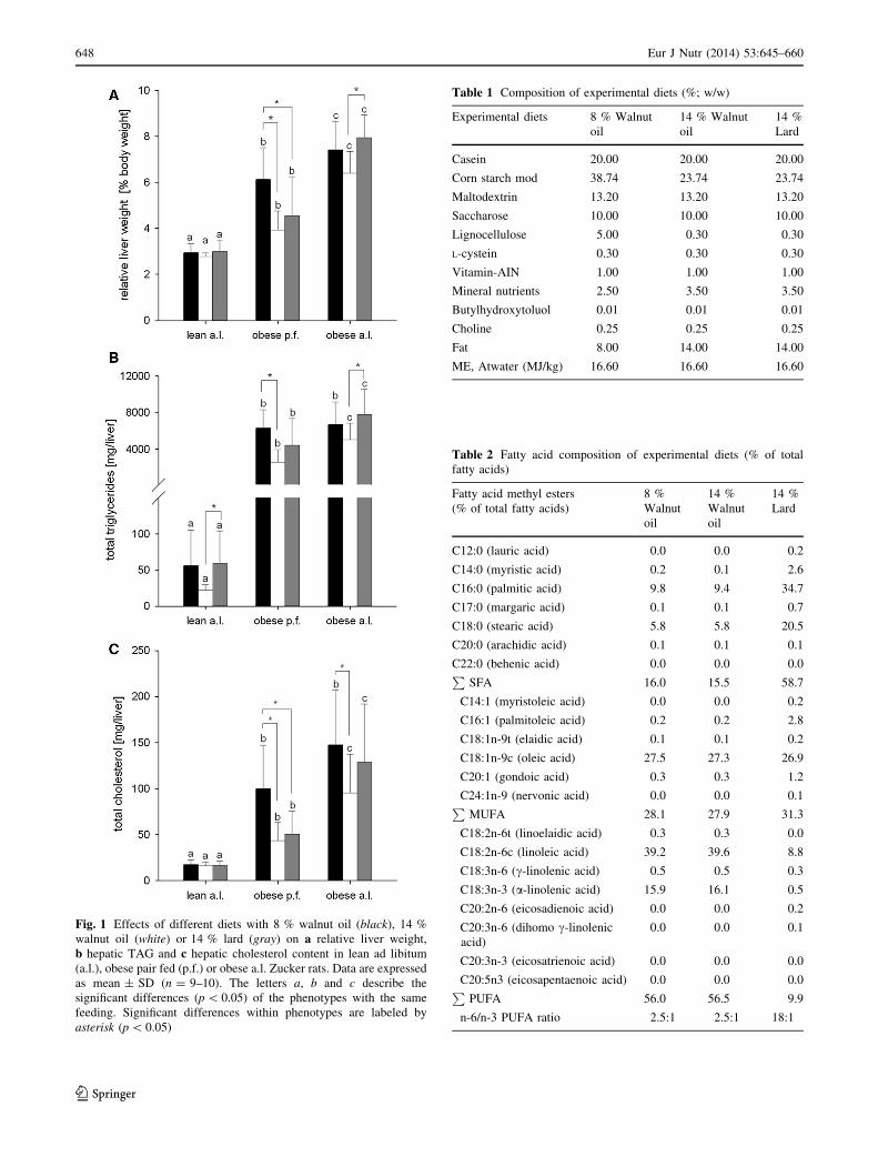
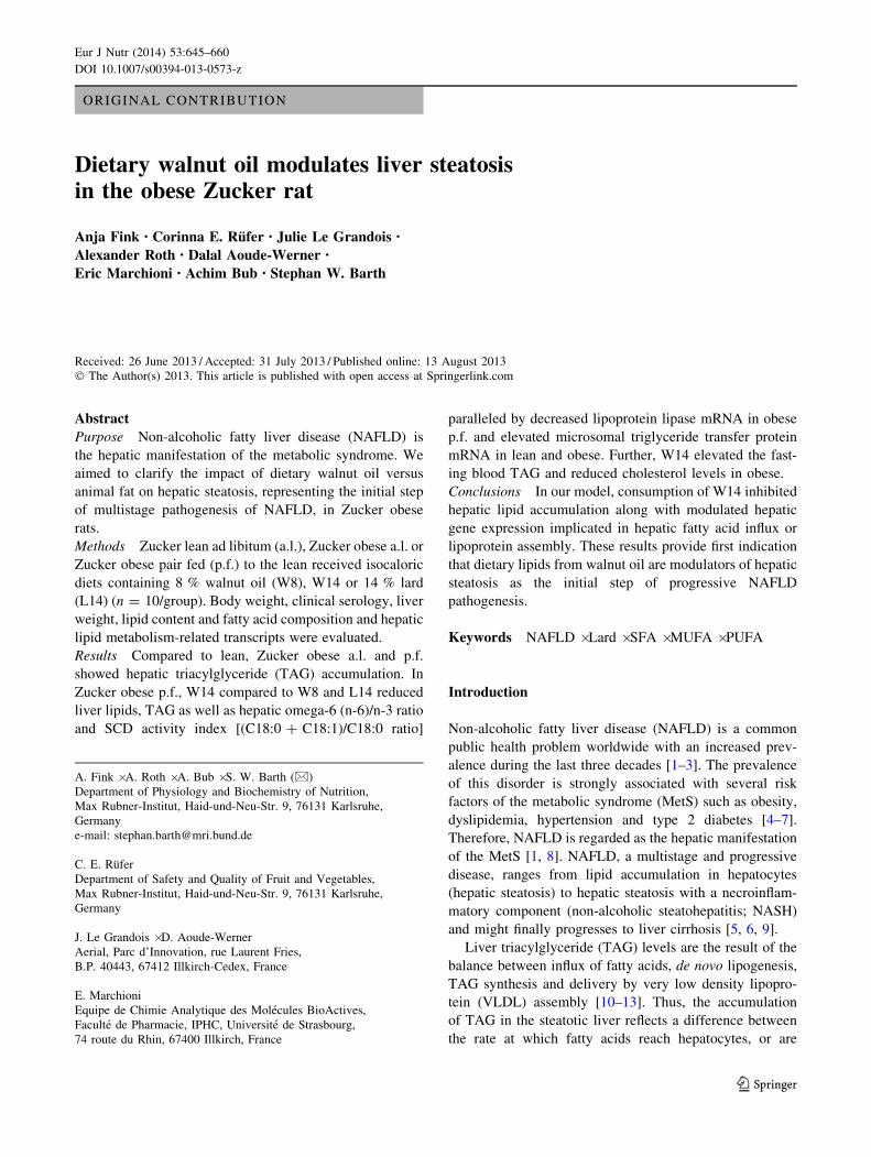
Fig. 1 Effects of different diets with 8 % walnut oil (black), 14 %
walnut oil (white) or 14 % lard (gray) on a relative liver weight,
b hepatic TAG and c hepatic cholesterol content in lean ad libitum
(a.l.), obese pair fed (p.f.) or obese a.l. Zucker rats. Data are expressed
as mean ± SD (n = 9–10). The letters a, b and c describe the
significant differences (p \ 0.05) of the phenotypes with the same
feeding. Significant differences within phenotypes are labeled by
asterisk (p \ 0.05)
Table 1 Composition of experimental diets (%; w/w)
Experimental diets 8 % Walnut
oil
14 % Walnut
oil
14 %
Lard
Casein 20.00 20.00 20.00
Corn starch mod 38.74 23.74 23.74
Maltodextrin 13.20 13.20 13.20
Saccharose 10.00 10.00 10.00
Lignocellulose 5.00 0.30 0.30
L-cystein 0.30 0.30 0.30
Vitamin-AIN 1.00 1.00 1.00
Mineral nutrients 2.50 3.50 3.50
Butylhydroxytoluol 0.01 0.01 0.01
Choline 0.25 0.25 0.25
Fat 8.00 14.00 14.00
ME, Atwater (MJ/kg) 16.60 16.60 16.60
Table 2 Fatty acid composition of experimental diets (% of total
fatty acids)
Fatty acid methyl esters
(% of total fatty acids)
8 %
Walnut
oil
14 %
Walnut
oil
14 %
Lard
C12:0 (lauric acid) 0.0 0.0 0.2
C14:0 (myristic acid) 0.2 0.1 2.6
C16:0 (palmitic acid) 9.8 9.4 34.7
C17:0 (margaric acid) 0.1 0.1 0.7
C18:0 (stearic acid) 5.8 5.8 20.5
C20:0 (arachidic acid) 0.1 0.1 0.1
C22:0 (behenic acid) 0.0 0.0 0.0P
SFA 16.0 15.5 58.7
C14:1 (myristoleic acid) 0.0 0.0 0.2
C16:1 (palmitoleic acid) 0.2 0.2 2.8
C18:1n-9t (elaidic acid) 0.1 0.1 0.2
C18:1n-9c (oleic acid) 27.5 27.3 26.9
C20:1 (gondoic acid) 0.3 0.3 1.2
C24:1n-9 (nervonic acid) 0.0 0.0 0.1P
MUFA 28.1 27.9 31.3
C18:2n-6t (linoelaidic acid) 0.3 0.3 0.0
C18:2n-6c (linoleic acid) 39.2 39.6 8.8
C18:3n-6 (c-linolenic acid) 0.5 0.5 0.3
C18:3n-3 (a-linolenic acid) 15.9 16.1 0.5
C20:2n-6 (eicosadienoic acid) 0.0 0.0 0.2
C20:3n-6 (dihomo c-linolenic
acid)
0.0 0.0 0.1
C20:3n-3 (eicosatrienoic acid) 0.0 0.0 0.0
C20:5n3 (eicosapentaenoic acid) 0.0 0.0 0.0P
PUFA 56.0 56.5 9.9
n-6/n-3 PUFA ratio 2.5:1 2.5:1 18:1
648 Eur J Nutr (2014) 53:645–660
123

differences between obese p.f. and obese a.l. are due to the
different intake of amount of food. All groups were further
randomly subdivided into one of the three intervention
groups (n = 10) receiving either an isocaloric diet based
on AIN93G containing 8 % (w/w) walnut oil (W8; Ohl-
muhle Walz, Oberkirch), 14 % walnut oil (W14) or 14 %
(w/w) lard (L14) (ssniff, Soest, Germany) for 10 weeks
(Table 1). The fatty acid composition of diets and the
analytical data of walnut oil phytosterols, tocopherols and
phospholipids are summarized in Tables 2 and 3, respec-
tively. Food intake was measured daily and body weight
four times a week. At the end of the feeding period, the
animals were fasted overnight, deeply anesthetized by
carbon dioxide and killed by decapitation. Liver tissues
were collected and stored at -80 �C. All animal experi-
ments were approved by the Animal Care Committee of
the Regional Administrative Authority, Karlsruhe (35-
9185.81/G-89/09), and all animal care and handling were
conducted in accordance with the guidelines of the German
law on animal care.
Blood parameters
Blood was collected into serum monovettes (Sarstedt,
Numbrecht, Germany), and serum was prepared from clot-
ted blood by centrifugation (2,5009g, 10 min) and stored at
-30 �C until further analysis. Serum cholesterol (CHOD-
PAP, Roche) and TAG (GPO-PAP, Roche) were measured
using enzymatic assay kits. Serum level of soluble inter-
cellular adhesion molecule (sICAM)-1 (R&D systems,
Wiesbaden, Germany) was measured using a rat ELISA kit.
Liver lipid and fatty acid analyses
Lipid extraction of 2 g liver tissues was carried out
according to Hara and Radin [41] with slight modifications.
The liver tissue was homogenized in 18 mL of hexane/
isopropanol (3/2; v/v) (Carl Roth, Karlsruhe, Germany)
containing 0.01 % butylhydroxytoluol (BHT; Merck,
Darmstadt, Germany). After sonification and addition of
12 mL aqueous sodium sulfate (Merck), samples were
placed on a horizontal shaker for overnight extraction. The
samples were centrifuged and hexane overlayer was
removed to a fresh pre-weighted vial. The remainder bottom
layer was again extracted with 18 mL of hexane/isopropanol
(7/2; v/v) containing 0.01 % BHT, and after centrifugation,
the hexane overlayer was given to the vial already contain-
ing the hexane layer of the initial extraction. After evapo-
ration under a stream of nitrogen gas, total lipid content
within sample vials was weighted for gravimetric estimation
of total liver fat. Fatty acids were analyzed by gas chroma-
tography (GC). Fatty acid methyl esters (FAME) were pre-
pared by transesterification of total lipids with TMSH.
50 mg of the lipid extract dissolved in 1 mL dichlorometh-
ane and aliquots of 10 lL were used for FAME analysis.
After addition of 100 lL of 90 mg/L methyl-nonadecanoate
as internal standard, 30 lL of TMSH, as well as 70 lL
methanol containing 1 % BHT, the organic extract was
evaporated to complete dryness under a stream of nitrogen.
The residue was dissolved in 100 lL TMSH and 500 lL
methanol, stirred in the dark overnight and subjected to GC
analysis. GC analysis was carried out on a gas chromato-
graph with mass spectrometric detection (GC-MS-QP-2010
Ultra, Shimadzu, Kyoto, Japan) using split/splitless injec-
tion. Chromatographic separation of FAME were achieved
on a fused silica capillary column with a non-bonded cy-
anopropyl-type phase (SP-2560, 75 m 9 0.18 mm ID,
0.14 lm film thickness, Supelco, Taufkirchen, Germany)
using a helium carrier gas flow of 0.9 mL/min and a linear
temperature gradient (100 �C for 5 min, then 4 �C/min to
230 �C and hold at 230 �C for 25 min). The injector port
temperature was set to 230 �C. The temperatures of transfer
line and ion source were set to 250 and 200 �C, respectively.
The injection volume was 1 lL with a split of 1:5. Individual
methyl esters were identified and quantified using a standard
mixture of 37 FAME (37 Component FAME Mix, Supelco,
Taufkirchen, Germany). As based on the fatty acid quanti-
fication, hepatic stearoyl CoA desaturase (SCD-1) activity
index was calculated by the (C18:0 ? C18:1)/C18:0 ratio.
Hepatic cholesterol and TAG were measured as previously
described for serum samples.
Table 3 Analysis of phytosterols, tocopherols and phospholipids in
walnut oil (mg/100 g)
Walnut oil (mg/100 g)
Brassicasterol 0.00
Campesterol 8.89
Cholesterol 0.39
d5-Avenasterol 1.09
d7-Avenasterol 0.00
d7-Campesterol 0.00
d7-Stigmasterol 49.46
Sitostanol 17.91
Sitosterol 163.48
Stigmasterol n.d.
Phytosterols total 241.21
a-Tocopherol 2.394
c-Tocopherol 45.450
d-Tocopherol 5.044
Tocopherols total 52.890
Phosphatidylethanolamine 22.5
Phosphatidylinositol n.d.
Phosphatidylcholine n.d.
Phospholipids total 22.5
n.d. not detectable
Eur J Nutr (2014) 53:645–660 649
123

Real-time quantitative PCR
Total RNA was isolated from the liver tissue using a
commercial kit according to the manufacturer’s instruction
(Total RNA and protein isolation kit, Macherey–Nagel,
Duren, Germany). cDNA was prepared by reverse tran-
scription of 2 lg total RNA using the Transcriptor First
Strand cDNA Synthesis Kit and oligo(dT) primers (Roche).
Samples were stored at -20 �C until further use. Semi-
quantitative real-time PCR was carried out using the Light
Cycler480� Instrument (Roche). Primer and probe
sequences were designed by Universal Probe Library
(Roche) as listed in Table 4. The reaction mixture con-
tained 5 lL cDNA, corresponding to 50 ng total RNA,
0.5 lM of each primer, 1 lM probe and Light Cycler480�
Probe Master Mix (29 conc.) (Roche). The PCR conditions
were as follows: 10 min of initial denaturation at 95 �C
followed by 45 amplification cycles each at 95 �C for 10 s,
60 �C for 30 s and 72 �C for 1 s with a terminal cooling
period of 10 s at 40 �C. The analysis was carried out with
the Light Cycler480� Software (Roche) using the relative
quantification DDCT-method and normalized by beta-actin
as reference gene.
Statistical analysis
Due to high heterogeneity of variances between lean and
obese, a statistical model, namely, generalized least
squares ANOVA (GLS-ANOVA) was chosen that is
capable of handling unequal variances for the different
groups. The independent variables for the two-factorial
ANOVA were ‘‘phenotype’’ and ‘‘food’’, resulting in a
3 9 3 factorial model. As a first step, we calculated a
simple model which assumes equal variances within fac-
tors ‘‘phenotype’’ and ‘‘food’’. Subsequently, this simple
model was then modified by adjusting the variance struc-
ture for ‘‘phenotype’’ and ‘‘food’’. A likelihood ratio test
was used to determine if the variance-adapted models fitted
better than the simple model. When main effects or inter-
actions were found significant, a post hoc test (Tukey–
Kramer) for pairwise comparisons was applied. The p
values that were given in the text and figures result from
these post hoc tests. To test for the assumptions that have to
be met for the GLS-ANOVA, we made plots to visually
inspect residuals. Studentized residuals were plotted
against fitted values to assess homoscedasticity. QQ-plots
were used to test normal distribution of the residuals.
Further, both plots were used to identify outliers. All cal-
culations were carried out by R 2.15.2 [42]. GLS-ANOVAs
were calculated by R package nlme [43].
Results
Analysis of dietary lipids and experimental diets
Fatty acid spectra of walnut oil and lard-containing diets
were characteristic for the respective lipid. While the
PUFA linoleic and a-linolenic are the major fatty acids in
walnut oil, lard mainly contained the SFA palmitic and
stearic acids. The resulting ratio of n-6/n-3 PUFA were
2.5:1 and 18:1 in walnut oil and lard-containing experi-
mental diets, respectively (Table 2).
The analytical results of walnut oil phospholipid content
(Table 3) showed significant concentrations only for phos-
phatidylethanolamine (22.5 mg/100 g), while no trace of
phosphatidylinositol was recovered, and phosphatidylcho-
line, the major phospholipid in common foods, was present
only in trace amounts and therefore was not quantified here.
Further, sitosterol (163 mg/100 g) and d7-stigmasterol
(49.5 mg/100 g) represent the major phytosterol constitu-
ents in walnut oil, while content of c-tocopherol (45.5 mg/
100 g) was highest compared to a-(2.4 mg/100 g) and
d-tocopherols (5.0 mg/100 g) (Table 3).
Table 4 Primer sequences and probes used for quantitative real-time PCR
Genes Forward primer Reverse primer UPL no. Genbank accession no.
ACTB cccgcgagtacaaccttct cgtcatccatggcgaact #17 NM_031144.2
ACC1 acagagatggtggctgatgtc gatccccatggcaatctg #4 NM_022193.1
ChREBP aatcccagcccctacacc ctgggaggagccaatgtg #10 AB074517.1
DGAT2 gctggtgccctactccaag agcttagggacggtgatgg #9 NM_001012345.1
FASN ggccacctcagtcctgttat agggtccagctagagggtaca #6 NM_017332.1
GPAm tccagacaccacatcaagga ctcctccatgactcaacgtg #127 NM_017274.1
LPL atgatgtggccaggttcatc gggctccaagactgtacccta #20 L03294.1
PPAR-a tgcggactaccagtacttaggg gctggagagagggtgtctgt #116 NM_013196.1
PPAR-c ggtgaaactctgggagatcct aatggcatctctgtgtcaacc #115 NM_013124.2
SREBP1 gtacagcatggctgggaac ggctgagcgatacagttcaa #1 AF286470.2
650 Eur J Nutr (2014) 53:645–660
123
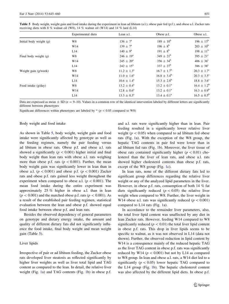
Body weight and food intake
As shown in Table 5, body weight, weight gain and food
intake were significantly affected by genotype as well as
the feeding regimen, namely the pair feeding versus
ad libitum in obese rats. Obese p.f. and obese a.l. rats
showed a significantly (p \ 0.001) higher initial and final
body weight than lean rats with obese a.l. rats weighing
more than obese p.f. rats (p \ 0.001). Further, the mean
body weight gain was significantly lower in lean than in
obese a.l. (p \ 0.001) and obese p.f. (p \ 0.001) Zucker
rats and obese p.f. rats gained less weight throughout the
experiment when compared to obese a.l. (p \ 0.001). The
mean food intake during the entire experiment was
approximately 25 % higher in obese a.l. than in lean
(p \ 0.001) and the matched obese p.f. rats (p \ 0.001). As
a result of the established pair feeding regimen, statistical
evaluation between the lean and obese p.f. showed equal
food intake between obese p.f. and lean rats.
Besides the observed dependency of general parameters
on genotype and dietary energy intake, the amount and
quality of different dietary fats did not significantly influ-
ence the food intake, final body weight and mean weight
gain (Table 5).
Liver lipids
Irrespective of pair or ad libitum feeding, the Zucker obese
rats developed liver steatosis as reflected significantly by
higher liver weights as well as liver total lipid and TAG
content as compared to the lean. In detail, the relative liver
weight (Fig. 1a) and TAG contents (Fig. 1b) in obese p.f.
and a.l. rats were significantly higher than in lean. Pair
feeding resulted in a significantly lower relative liver
weight (p \ 0.05) when compared to ad libitum fed obese
rats (Fig. 1a). With the exception of the W8 group, the
hepatic TAG contents in pair fed were lower than in
ad libitum fed rats (Fig. 1b). Moreover, the liver tissue of
obese rats contained significantly higher (p \ 0.01) cho-
lesterol than the liver of lean rats, and obese a.l. rats
showed higher cholesterol contents than obese p.f. rats,
except of the W8 group (Fig. 1c).
In lean rats, none of the different dietary fats led to
significant group differences regarding the relative liver
weight or any of the analyzed lipid parameters in the liver.
However, in obese p.f. rats, consumption of both 14 % fat
diets significantly reduced (p \ 0.05) the relative liver
weight when compared to W8. Further, the liver weight in
W14 obese a.l. rats was significantly reduced (p \ 0.001)
compared to L14 rats (Fig. 1a).
In accordance to the remainder liver parameters, also,
the total liver lipid content was unaffected by any diet in
lean Zucker rats. However, feeding W14 compared to W8
significantly reduced (p \ 0.01) the total liver lipid content
in obese p.f. rats. This drop in liver lipids seems to be
specific to walnut, as it was not observed in L14 (data not
shown). Further, the observed reduction in lipid content by
W14 is a consequence mainly of the reduced hepatic TAG
as the liver TAG content in obese p.f. rats was significantly
reduced by W14 (p \ 0.001) but not by L14 as compared
to W8 group. In lean and obese a.l. rats, a W14 diet led to a
significantly (p \ 0.05) lower hepatic TAG compared to
the L14 group (Fig. 1b). The hepatic cholesterol content
was also affected by the different lipid diets. In obese p.f.
Table 5 Body weight, weight gain and food intake during the experiment in lean ad libitum (a.l.), obese pair fed (p.f.), and obese a.l. Zucker rats
receiving diets with 8 % walnut oil (W8), 14 % walnut oil (W14) and 14 % lard (L14)
Experimental diets Lean a.l. Obese p.f. Obese a.l.
Initial body weight (g) W8 136 ± 7a 189 ± 10b 196 ± 13b
W14 139 ± 7a 196 ± 8b 203 ± 10b
L14 140 ± 9a 191 ± 8b 198 ± 11b
Final body weight (g) W8 246 ± 19a 349 ± 21b 395 ± 21c
W14 245 ± 20a 356 ± 34b 406 ± 36c
L14 242 ± 15a 337 ± 27b 386 ± 38c
Weight gain (g/week) W8 11.2 ± 1.3a 16.7 ± 1.7b 20,3 ± 1.7c
W14 11.0 ± 1.6a 16.8 ± 3.4b 20.3 ± 3.5c
L14 10.6 ± 1.4a 15.3 ± 2.8b 18.8 ± 3.6c
Food intake (g/day) W8 13.2 ± 0.4a 13.2 ± 0.1a 16.4 ± 1.2b
W14 12.8 ± 0.6a 13.2 ± 0.1a 16.3 ± 0.9b
L14 13.3 ± 0.3a 13.4 ± 0.1a 16.5 ± 0.5b
Data are expressed as mean ± SD (n = 9–10). Values in a common row of the identical intervention labeled by different letters are significantly
different between phenotypes
Significant differences within phenotypes are labeled by * (p \ 0.05 compared to W8)
Eur J Nutr (2014) 53:645–660 651
123
rats, W14 and L14 diets reduced the hepatic cholesterol
content significantly (p \ 0.01) compared to the W8 group,
and obese a.l. W14 rats had also a lower cholesterol
(p \ 0.05) content compared to the W8 group (Fig. 1c).
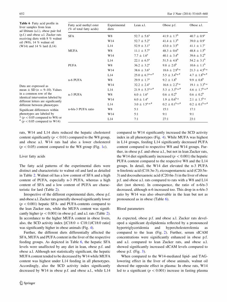
Liver fatty acids
The fatty acid patterns of the experimental diets were
distinct and characteristic to walnut oil and lard as detailed
in Table 2. Walnut oil has a low content of SFA and a high
content of PUFA, especially n-3 PUFA, whereas a high
content of SFA and a low content of PUFA are charac-
teristic for lard (Table 2).
Irrespective of the different experimental diets, obese p.f.
and obese a.l. Zucker rats generally showed significantly lower
(p \ 0.001) hepatic SFA- and PUFA-contents compared to
the lean Zucker rats, while the MUFA content was signifi-
cantly higher (p \ 0.001) in obese p.f. and a.l. rats (Table 2).
In accordance to the higher MUFA content in obese livers,
also, the SCD activity index [(C18:0 ? C18:1)/C18:0 ratio]
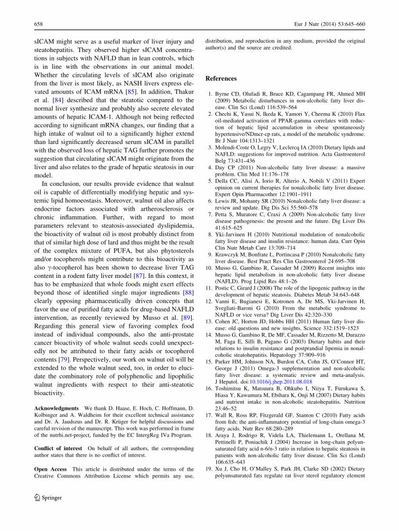
was significantly higher in obese animals (Fig. 4).
Further, the different diets differentially affected the
SFA, MUFA and PUFA content in the liver of the respective
feeding groups. As depicted in Table 6, the hepatic SFA
levels were unaffected by any diet in lean, obese p.f. and
obese a.l. Although not statistically significant, the hepatic
MUFA content tended to be decreased by W14 while MUFA
content was highest under L14 feeding in all phenotypes.
Accordingly, also the SCD activity index significantly
decreased by W14 in obese p.f. and obese a.l., while L14
compared to W14 significantly increased the SCD activity
index in all phenotypes (Fig. 4). While MUFA was highest
in L14 groups, feeding L14 significantly decreased PUFA
content compared to respective W8 and W14 groups. Fur-
ther, in obese p.f. and obese a.l., but not in lean Zucker rats,
the W14 diet significantly increased (p \ 0.001) the hepatic
PUFA content compared to the respective W8 and the L14
groups. In detail, the W14 diet elevated the n-3 PUFA
a-linolenic acid (C18:3n-3), eicosapentaenoic acid (C20:5n-
3) and docosahexaenoic acid (C20:6n-3) in the liver of obese
p.f. and obese a.l. rats compared to rats with a W8 and L14
diet (not shown). In consequence, the ratio of n-6/n-3
decreased, although n-6 increased too. This drop in n-6/n-3
ratio by W14 was also observable in the lean but not as
pronounced as in obese (Table 6).
Blood parameters
As expected, obese p.f. and obese a.l. Zucker rats devel-
oped a significant dyslipidemia reflected by a pronounced
hypertriglyceridemia and hypercholesterolemia as
compared to the lean (Fig. 2). Further, serum sICAM
concentrations were significantly enhanced in obese p.f.
and a.l. compared to lean Zucker rats, and obese a.l.
showed significantly increased sICAM levels compared to
obese p.f. (Fig. 3).
When compared to the W14-mediated lipid- and TAG-
lowering effect in the liver of obese animals, walnut oil
showed the opposite effect in plasma: In obese rats, W14
led to a significant (p \ 0.001) increase in fasting plasma
Table 6 Fatty acid profile in
liver samples from lean
ad libitum (a.l.), obese pair fed
(p.f.) and obese a.l. Zucker rats
receiving diets with 8 % walnut
oil (W8), 14 % walnut oil
(W14) and 14 % lard (L14)
Data are expressed as
mean ± SD (n = 9–10). Values
in a common row of the
identical intervention labeled by
different letters are significantly
different between phenotypes
Significant differences within
phenotypes are labeled by
* (p \ 0.05 compared to W8) or# (p \ 0.05 compared to W14)
Fatty acid methyl ester
(% of total fatty acids)
Experimental
diets
Lean a.l. Obese p.f. Obese a.l.
SFA W8 52.7 ± 5.6a 41.9 ± 1.7b 40.7 ± 0.9c
W14 52.7 ± 5.2a 41.4 ± 1.3b 39.0 ± 0.9c
L14 52.9 ± 3.1a 43.0 ± 3.5b 41.1 ± 1.7c
MUFA W8 11.1 ± 5.7a 48.3 ± 0.6b 48.8 ± 1.5b
W14 7.7 ± 1.6a 40.1 ± 3.4b 39.6 ± 5.2b
L14 22.1 ± 6.5a 51.5 ± 4.8b 54.2 ± 3.1b
PUFA W8 36.2 ± 3.2a 9.8 ± 2.0b 10.6 ± 1.1b
W14 38.6 ± 3.6a 18.6 ± 2.9b,* 21.3 ± 4.5b,*
L14 25.0 ± 6.7a,*,# 5.5 ± 3.4b*,# 4.7 ± 1.8b*,#
n-6 PUFA W8 29.9 ± 1.7a 9.2 ± 1.8b 9.9 ± 0.8b
W14 32.2 ± 2.4a 16.6 ± 2.2b,* 19.1 ± 3.3b,*
L14 21.9 ± 5.3a,*,# 5.3 ± 3.3b,*,# 4.6 ± 1.7b,*,#
n-3 PUFA W8 6.0 ± 1.6a 0.6 ± 0.2b 0.6 ± 0.2b
W14 6.0 ± 1.4a 1.9 ± 0.6b,* 2.1 ± 1.3b,*
L14 3.0 ± 1.5a,*,# 0.2 ± 0.1b,*,# 0.2 ± 0.1b,*,#
n-6/n-3 PUFA ratio W8 5:1 15:1 17:1
W14 5:1 9:1 9:1
L14 7:1 27:1 23:1
652 Eur J Nutr (2014) 53:645–660
123
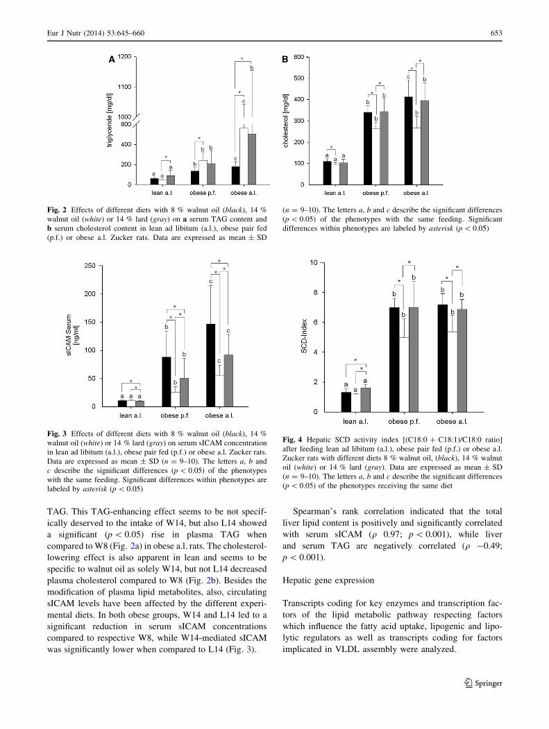
TAG. This TAG-enhancing effect seems to be not specif-
ically deserved to the intake of W14, but also L14 showed
a significant (p \ 0.05) rise in plasma TAG when
compared to W8 (Fig. 2a) in obese a.l. rats. The cholesterol-
lowering effect is also apparent in lean and seems to be
specific to walnut oil as solely W14, but not L14 decreased
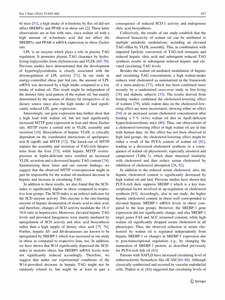
plasma cholesterol compared to W8 (Fig. 2b). Besides the
modification of plasma lipid metabolites, also, circulating
sICAM levels have been affected by the different experi-
mental diets. In both obese groups, W14 and L14 led to a
significant reduction in serum sICAM concentrations
compared to respective W8, while W14-mediated sICAM
was significantly lower when compared to L14 (Fig. 3).
Spearman’s rank correlation indicated that the total
liver lipid content is positively and significantly correlated
with serum sICAM (q 0.97; p \ 0.001), while liver
and serum TAG are negatively correlated (q -0.49;
p \ 0.001).
Hepatic gene expression
Transcripts coding for key enzymes and transcription fac-
tors of the lipid metabolic pathway respecting factors
which influence the fatty acid uptake, lipogenic and lipo-
lytic regulators as well as transcripts coding for factors
implicated in VLDL assembly were analyzed.
Fig. 2 Effects of different diets with 8 % walnut oil (black), 14 %
walnut oil (white) or 14 % lard (gray) on a serum TAG content and
b serum cholesterol content in lean ad libitum (a.l.), obese pair fed
(p.f.) or obese a.l. Zucker rats. Data are expressed as mean ± SD
(n = 9–10). The letters a, b and c describe the significant differences
(p \ 0.05) of the phenotypes with the same feeding. Significant
differences within phenotypes are labeled by asterisk (p \ 0.05)
Fig. 3 Effects of different diets with 8 % walnut oil (black), 14 %
walnut oil (white) or 14 % lard (gray) on serum sICAM concentration
in lean ad libitum (a.l.), obese pair fed (p.f.) or obese a.l. Zucker rats.
Data are expressed as mean ± SD (n = 9–10). The letters a, b and
c describe the significant differences (p \ 0.05) of the phenotypes
with the same feeding. Significant differences within phenotypes are
labeled by asterisk (p \ 0.05)
Fig. 4 Hepatic SCD activity index [(C18:0 ? C18:1)/C18:0 ratio]
after feeding lean ad libitum (a.l.), obese pair fed (p.f.) or obese a.l.
Zucker rats with different diets 8 % walnut oil, (black), 14 % walnut
oil (white) or 14 % lard (gray). Data are expressed as mean ± SD
(n = 9–10). The letters a, b and c describe the significant differences
(p \ 0.05) of the phenotypes receiving the same diet
Eur J Nutr (2014) 53:645–660 653
123

The gene expression of hepatic enzymes involved in
hydrolysis of TAG to free fatty acids, namely lipoprotein
lipase (LPL) showed significant phenotype differences
between obese and lean Zucker rats. The liver LPL gene
expression was up-regulated in obese p.f. and a.l. Zucker
rats, except for obese p.f. W14, compared to respective
lean groups (Fig. 5). The expression of peroxisome
proliferator-activated receptor (PPAR)-a, a transcription
factor involved in fatty acid oxidation was significantly
higher in obese a.l. rats than in obese p.f. rats (Table 7).
Amount of transcript coding for lipogenic enzymes acetyl-
CoA carboxylase (ACC) and fatty acid synthase (FAS)
showed significantly enhanced mRNA levels in obese p.f.
Zucker rats compared to lean and obese a.l Zucker rats
Fig. 5 Hepatic (a) LPL and
(b) MTTP gene expression after
feeding lean ad libitum (a.l.),
obese pair fed (p.f.) or obese a.l.
Zucker rats with different diets
8 % walnut oil, (black), 14 %
walnut oil (white) or 14 % lard
(gray). Data are expressed as
mean ± SD (n = 9–10). The
letters a, b and c describe the
significant differences
(p \ 0.05) of the phenotypes
receiving the same diet
Table 7 Expression of key
genes involved in hepatic lipid
metabolism was determined by
real-time PCR in liver samples
lean ad libitum (a.l.), obese pair
fed (p.f.) and obese a.l. Zucker
rats receiving diets with 8 %
walnut oil (W8), 14 % walnut
oil (W14) and 14 % lard (L14)
Data are expressed as
mean ± SD (n = 8–10). Values
in a common row of the
identical intervention containing
labeled by different letters are
significantly different between
phenotypes
Significant differences within
phenotypes are labeled by
* (p \ 0.05 compared to W8) or# (p \ 0.05 compared to W14)
Experimental diet Lean a.l. Obese p.f. Obese a.l.
ACC/b-Actin W8 2.9 ± 2.8a 3.3 ± 1.7b 2.0 ± 0.6a
W14 2.7 ± 2.6a 5.4 ± 1.8b 2.8 ± 1.1a
L14 3.4 ± 3.2a 5.2 ± 2.4b 2.3 ± 1.1a
ChREBP/b-Actin W8 1.3 ± 0.5a 0.7 ± 0.2b 0.6 ± 0.1c
W14 1.2 ± 0.3a 0.8 ± 0.2b 0.7 ± 0.1c
L14 1.1 ± 0.4a 0.7 ± 0.2b 0.6 ± 0.2c
DGAT/b-Actin W8 1.2 ± 0.3a 0.8 ± 0.3a,b 0.9 ± 0.4b
W14 1.3 ± 0.4a 1.4 ± 0.5a,b 1.0 ± 0.4b
L14 1.4 ± 0.4a 1.3 ± 0.6a,b 0.9 ± 0.5b
FAS/b-Actin W8 1.7 ± 2.1a 2.7 ± 1.8b 1.6 ± 0.7a
W14 1.7 ± 1.7a 4.9 ± 1.8b 2.1 ± 0.8a
L14 2.0 ± 1.8a 4.3 ± 1.8b 1.9 ± 0.7a
GPAm/b-Actin W8 0.9 ± 0.2a 2.2 ± 1.9b 1.3 ± 0.5c
W14 1.2 ± 0.8a 5.0 ± 2.3b 1.7 ± 1.2c
L14 1.2 ± 0.6a 3.2 ± 2.8b 1.2 ± 0.7c
ICAM/b-Actin W8 0.6 ± 0.1a 1.6 ± 0.4b 1.6 ± 0.4b
W14 0.8 ± 0.3a 1.2 ± 0.2a 1.9 ± 0.7b
L14 0.6 ± 0.2a 1.3 ± 0.3b 1.6 ± 0.3b
PPAR-a/b-Actin W8 2.0 ± 0.6a,b 1.8 ± 0.6a 2.3 ± 0.6b
W14 2.0 ± 0.7a,b 1.8 ± 0.8a 2.6 ± 1.0b
L14 2.1 ± 0.7a,b 1.5 ± 0.4a 2.1 ± 0.5b
PPAR-y/b-Actin W8 1.2 ± 0.6a 6.4 ± 4.1b 5.2 ± 1.6b
W14 1.1 ± 0.4a 5.3 ± 2.1b 6.5 ± 4.3b
L14 1.1 ± 0.5a 5.8 ± 2.2b 5.9 ± 2.6b
SREBP1c/b-Actin W8 0.8 ± 0.4a 1.7 ± 1.1b 1.9 ± 0.6b
W14 1.1 ± 0.4a 2.3 ± 0.8b 2.0 ± 0.7b
L14 1.1 ± 0.7a 2.0 ± 0.8b 2.0 ± 0.8b
654 Eur J Nutr (2014) 53:645–660
123

(Table 7). Furthermore, lipogenic transcription factors
PPAR-c and sterol regulatory element-binding protein
(SREBP)-1c were up-regulated, whereas carbohydrate
responsive element-binding protein (ChREBP) was down-
regulated in obese Zucker rats compared to the lean
(Table 7). Amount of mitochondrial glycerol-3-phosphate
acyltransferase (GPAm) mRNA, which codes for a key
enzyme implicated in TAG esterification, was significantly
higher in obese p.f. rats compared to obese a.l and lean rats.
Diglyceride acyltransferase (DGAT), a key enzyme for the
esterification of TAG, was significantly induced in lean rats
compared to obese a.l. rats. Furthermore, the expression of
microsomal TAG transfer protein (MTTP), a key factor
regulating hepatic TAG export by VLDL synthesis and
assembly, was significantly down-regulated in obese p.f.
and a.l. compared to lean Zucker rats. With the exception
of the obese p.f. W14 group, the hepatic ICAM gene
expression was significantly up-regulated in all obese
compared to lean Zucker rats (Table 7).
The intervention with the different experimental diets
differentially modulated the expression of LPL in liver
tissue. In obese p.f., both diets containing 14 % fat content
led to a significant reduction of LPL mRNA in the liver
compared to obese p.f. W8 Zucker rats. In contrast, no
effects were observed in lean Zucker rats (Fig. 5). Spear-
man’s rank correlation showed that the total liver lipid
content is positively and significantly correlated with the
hepatic LPL mRNA content (q 0.785; p \ 0.001).
Irrespective of the phenotype, the MTTP gene expres-
sion has been significantly increased in all W14 groups as
compared to groups receiving W8 or L14 diets (Fig. 5). All
other transcript levels of ACC, FAS, SREBP-1c, ChREBP,
PPAR-c, PPAR-a, GPAm, DGAT, MTTP, ICAM were
unaffected by any diet (Table 7).
Discussion
The key finding of this study stated that the quality and
the quantity of dietary lipids differentially influence
hepatic and circulating lipid metabolites in vivo. In detail,
a high intake of walnut oil decreased hepatic TAG while
fasting serum TAG levels were increased in obese Zucker
rats. The decreased hepatic TAG concomitantly appeared
with significant changes in fatty acid patterns in the liver
and a reduced SCD activity index as well as reduced/
normalized n-6/n-3 ratio in the liver. These qualitative
and quantitative changes in lipid contents were associated
with decreased hepatic LPL mRNA in obese p.f. rats and
increased MTTP mRNA irrespective of the phenotype.
Finally, a diet high in walnut oil significantly reduced the
artherogenic and inflammatory marker sICAM in obese
Zucker rats.
The development of efficient (diet-based) prevention
and therapeutic options for NAFLD is based on a clear
understanding of the etiology and mechanisms of this
condition, which in turn is limited by the quality of the
study model. NAFLD in lab rodents can be induced and
promoted by a wide variety of factors, initially leading to
changes in hepatic lipid deposition. These include diets
which are high in fat or carbohydrates (e.g., fructose) or
methionine and choline deficient (MCD) diets. The diet-
induced NAFLD models are completed by various genetic
models; the most commonly used are represented by the
leptin and/or leptin receptor variants. All these currently
available animal models of NAFLD are associated with
various drawbacks in that they do not or only partially
reflect the real picture of human NAFLD in terms of eti-
ology, pathogenesis and disease mechanisms. For example,
most of the diet-induced NAFLD models also implicate
potential confounding factors such as high energy intake
(by high-fat and/or high-carbohydrate diets) or the high-
(fructose, lipid) or deficient-(MCD) intake of particular
macronutrients. On the other side, genetic models carrying
a single mutation do not share the same etiology as mul-
tifactorially/multigenetically generated human NAFLD,
although clinical parameters related to NAFLD show
identical aberrations [44–48]. Based on this knowledge
about respective model-associated advantages and disad-
vantages, we chose the obese Zucker rat, integrated this
model into an energy-controlled pair feeding design and
further used isocaloric diets irrespective of the different fat
contents. According to this experimental design, the pair
feeding groups received the amount of diet adjusted to the
respective lean a.l. group which received approximately
75 % of dietary calories as compared to the obese a.l. fed
groups. This concept combined with isocaloric diets irre-
spective of fat content enabled us to analyze a ‘‘pure’’
intervention effect of dietary lipid quality and/or quantity
on NAFLD-associated metabolic aberrations largely
excluding the mentioned potential confounders such as
variable energy intake.
The present study shows that an isocaloric diet with a
high content of walnut oil (14 % w/w) not solely mediated
anti-steatotic effects in obese rats, but it also influenced the
hepatic fatty acid composition.
It has been described that various dietary fats with dif-
ferent fatty acid compositions differentially affect the
hepatic TAG accumulation [49, 50]. In vivo studies have
already shown that dietary fats from fish oil rich in long-
chain n-3 PUFA resulted in a reduction in hepatic TAG
concentration [49–52], which has recently been shown to
be associated with a preferred incorporation of long-chain
n-3 PUFA into hepatic phospholipids [53]. Further, Chechi
et al. [2] have demonstrated that a flax oil diet, rich in
a-linolenic acid, also reduced the hepatic TAG in obese
Eur J Nutr (2014) 53:645–660 655
123

SHR/NDmcr-cp rats. However, as recently published in
ApoE-deficient mice, a diet containing 20 % (w/w) walnut
oil did not change liver lipid content [54], which might be
due to the complex functional impairments of this rodent
model. Besides its artherogenic effect, the ApoE deficiency
also results in impaired VLDL assembly [55, 56], which
might be the mechanism by which walnut oil in our study
mobilized and reduced the hepatic lipid content as dis-
cussed below.
While low levels of hepatic n-3 PUFA are associated
with the development of liver steatosis in NAFLD patients
[18, 57], a supplementation with n-3 PUFA decreased
hepatic TAG content in vivo [23, 57]. Based on these data,
we suggest that the observed bioactivity of high walnut oil
intake in obese rats might be caused by rather than ran-
domly associated with the observed increased levels of
hepatic PUFA, especially n-3 PUFA. The predominating
n-3 PUFA of walnut oil, a-linolenic acid (Table 2), is
endogenously converted into eicosapentaenoic acid (EPA,
C20:5n-3) and docosahexaenoic acid (DHA, C22:6n-3) by
the D-6- and D-5-desaturase and elongase-5 and -2 enzyme
systems, respectively. Although the efficacy of this con-
version of a-linolenic acid into EPA and DHA is not
quantitative, but has been estimated below 21 % [58, 59],
significant two to fourfold enrichment of hepatic EPA or
DHA by W14 compared to W8 was obvious (data not
shown) and paralleled by a general improvement of n-6/n-3
ratio under anti-steatotic feeding with walnut oil.
Epidemiological studies have reported that besides its
impact on hepatic lipid homoeostasis, a low n-6/n-3 PUFA
ratio is also capable of improving the blood lipid profile
[22, 51]. Riediger et al. [22] reported decreased plasma
TAG levels by flax oil diet with a low n-6/n-3 PUFA ratio.
A high-fat diet containing PUFA compared to high SFA
diet led to a decreased concentration of TAG in the serum
of C57BL/6J mice [51]. However, recently published data
show that a high intake of walnut oil did not change plasma
TAG which might also be the result of the ApoE deficiency
in this model [54].
As opposed to these latter-published data in our study,
the drop in hepatic TAG content by high intake of walnut
oil was paralleled by a significant elevation of fasting
serum TAG in energy-controlled obese rats. In obese
ad libitum fed rats, also, the high intake of lard shows this
effect. These results might be an indication that besides
being substrate for hepatic TAG synthesis, fatty acids are
also capable of being signaling molecules directly affecting
hepatic receptors (e.g., PPARs), enzymes and transport
molecules and also catalyzing hepatocyte VLDL assembly.
It has been established in several studies in human and
animal experiments that dietary n-3 PUFA decrease plasma
TAG by suppressing hepatic VLDL assembly [60–63].
However, the results presented here let us suggest that a
high intake of walnut oil rather increased than decreased
VLDL secretion as reflected by elevated plasma concen-
tration of fasting TAG in steatotic animals.
Increasing fasting plasma lipids might be an indication
for an elevating insulin resistance characterized by diabetic
dyslipidemia [64]. However, in the present study, changes
of insulin resistance causing the observed hypertriglyceri-
demia may be excluded, as plasma insulin and glucose as
well as adiponectin were not significantly affected by
walnut oil in obese pair fed (data not shown), which
showed the strongest modulation of plasma and hepatic
lipids by walnut oil. Further, also, the fasting plasma
concentration of non-esterified fatty acids (NEFA), liber-
ated from peripheral fat tissue by promoted lipolysis under
insulin-resistant circumstances, was not affected by walnut
oil intervention (data not shown).
Besides changes in insulin sensitivity, changes in the
hepatocyte redox status might be relevant for the liver
response to dietary lipid intervention. The pathogenesis of
steatosis is mainly characterized by excess accumulation of
lipids which also impairs the oxidative capacity of the
mitochondria, increasing the reduced state of the electron
transport chain complexes and stimulating peroxisomal and
microsomal pathways of lipid oxidation [65]. Interestingly,
Botham et al. [66] have demonstrated that delivery of n-3
PUFA to hepatocytes objected to minor changes in cellular
redox level rather increased than decreased VLDL secre-
tion as observed by n-3 PUFA under normal conditions.
Therefore, also, interactions between dietary walnut
ingredients and the cellular redox status might have been
responsible for the observed unexpected results of an
increased hepatic lipid assembly and resulting hyperlipid-
emia. Similar to our results, attenuation of hepatic steatosis
in Zucker obese rats by different dietary constituents have
been paralleled by increased fasting plasma TAG [67, 68].
These data are in line with ours and share common features
with the basic principle to lower ectopic lipid stores in the
liver by increasing hepatic lipid disposal.
As based on this hypothesis of an increased VLDL
assembly, we have conducted further attempts to elucidate
possible signaling pathways which might be responsible for
the observed reduction in hepatic and increase in serum
TAG by a high intake of walnut oil in steatotic rodents.
It has been generally accepted that n-3 and n-6 PUFA
are key regulators of hepatic gene transcription. However,
recent studies have shown that the ability of PUFA regu-
lating the expression of target genes, such as PPAR-a or
SREBP1c and ChREBP, together with downstream lipo-
genic enzymes, e.g., FAS and ACC, might be determined
by the kind of fatty acid supplied by a distinct oil [37, 51,
57]. While Yang et al. [51] demonstrated that high intake
of DHA and EPA by fish oil reduced the hepatic expression
of SREBP1c and the target genes FAS and ACC in C57BL/
656 Eur J Nutr (2014) 53:645–660
123
6J mice [51], a high intake of a-linolenic by flax oil did not
affect SREBP1c and PPAR-a in obese rats [2]. These latter
observations are in line with ours, since walnut oil with a
high amount of a-linolenic acid did not affect the
SREBP1c and PPAR-a mRNA expression in obese Zucker
rats.
LPL is an enzyme which plays a role in plasma TAG
regulation. It promotes plasma TAG clearance by hydro-
lyzing triglycerides from chylomicrons and VLDL [69, 70].
Previous studies have demonstrated that the development
of hypertriglyceridemia is closely associated with the
downregulation of LPL activity [71]. In our study in
energy-controlled obese pair fed rats, the amount of LPL
mRNA was decreased by a high intake compared to a low
intake of walnut oil. This result might be independent of
the distinct fatty acid pattern of the walnut oil, but mainly
determined by the amount of dietary fat irrespective of its
dietary source since also the high intake of lard signifi-
cantly reduced LPL gene expression.
Interestingly, our gene expression data further show that
a high load with walnut oil, but not lard significantly
increased MTTP gene expression in lean and obese Zucker
rats. MTTP exerts a central role in VLDL assembly and
secretion [10]. Biosynthesis of hepatic VLDL is critically
dependent on the coordinated interactions of apolipopro-
tein B (apoB) and MTTP [72]. The knock-out of MTTP
impairs the assembly and secretion of TAG-rich lipopro-
teins from the liver [73], while hepatic MTTP overex-
pression in leptin-deficient mice resulted an increased
VLDL secretion and a decreased hepatic TAG content [74].
Summarizing these latter and our current findings, we
suggest that the observed MTTP overexpression might in
part be responsible for the walnut oil-mediated decrease in
hepatic and increase in circulating TAG.
In addition to these results, we also found that the SCD-
index is significantly higher in obese compared to respec-
tive lean groups. The SCD-index is an indirect indicator for
the SCD enzyme activity. This enzyme is the rate-limiting
enzyme of hepatic desaturation of stearic acid to oleic acid,
and therefore, changes of SCD activity modulate the 18:1/
18:0 ratio in hepatocytes. Moreover, elevated hepatic TAG
levels and provoked lipogenesis were mainly mediated by
upregulation of SCD activity and oleic acid biosynthesis
rather than a high supply of dietary oleic acid [75, 76].
Further, hepatic D5- and D6-desaturases are known to be
upregulated by SREBP-1 which was increased in our study
in obese as compared to respective lean, too. In addition,
we have shown that W14 significantly depressed the SCD-
index in steatotic obese, but SREBP1 mRNA levels were
not significantly reduced accordingly. Therefore, we
suggest that under our experimental conditions of the
W14-provoked decrease in hepatic TAG might not be
randomly related to, but might be at least in part a
consequence of reduced SCD-1 activity and endogenous
oleic acid biosynthesis.
Collectively, the results of our study establish that the
observed bioactivity of walnut oil can be attributed to
multiple metabolic modulations, including an elevated
TAG efflux by VLDL assembly. This, in combination with
impaired lipolytic conversion of TAG-rich remnants and
reduced hepatic oleic acid and subsequent reduced TAG
synthesis results in subsequent reduced hepatic and ele-
vated circulating TAG levels.
Besides the walnut oil-mediated modulation of hepatic
and circulating TAG concentration, a high walnut-intake
reduces total cholesterol as summarized in the framework
of a meta-analysis [77], which has been confirmed more
recently by a randomized cross-over study in free-living
[78] and diabetic subjects [33]. The results derived from
feeding studies confirmed the cholesterol-lowering effect
of walnuts [79], while rodent data on the cholesterol-low-
ering effect are more inconsistent, showing either no effect
[54] or an increased serum cholesterol concentration after
feeding a 5 % (w/w) walnut oil diet in ApoE-deficient
hypercholesterolemic mice [80]. Thus, our observations of
a cholesterol-lowering effect of high walnut oil are in line
with human data. As this effect has not been observed in
high lard groups, the cholesterol-lowering effect might be
either a result of the PUFA content of walnut oil [81],
leading to a decreased cholesterol synthesis or a conse-
quence of walnut oil phytosterols, especially sitosterol and
campesterol (Table 3), which share structural similarity
with cholesterol and thus reduce serum cholesterol by
inhibition of cholesterol absorption [26, 82].
In addition to the reduced serum cholesterol, also, the
hepatic cholesterol content is significantly decreased by
high walnut oil and lard. Previous studies have shown that
PUFA-rich diets suppress SREBP-1 which is a key tran-
scriptional factor involved in up-regulation of cholesterol
synthesis [83]. Accordingly, also in our study, the higher
hepatic cholesterol content in obese well corresponded to
elevated hepatic SREBP-1 mRNA levels in obese com-
pared to the lean groups. However, the SREBP-1 gene
expression did not significantly change, and also SREBP-1
target genes FAS and ACC remained constant, while high
walnut oil significantly dropped serum cholesterol in all
phenotypes. Thus, the observed reduction in serum cho-
lesterol by walnut oil is regulated independently from
hepatic SREBP-1 or changes in SREBP-1 expression due
to post-transcriptional regulation, e.g., by changing the
maturation of SREBP-1 protein, as described previously
for PUFA-rich fish oil [83].
Patients with NAFLD have increased circulating level of
artherosclerotic biomarkers like sICAM [84–86]. Although
classically synthesized and secreted by vascular endothelial
cells, Thakur et al. [84] suggested that circulating levels of
Eur J Nutr (2014) 53:645–660 657
123
sICAM might serve as a useful marker of liver injury and
steatohepatitis. They observed higher sICAM concentra-
tions in subjects with NAFLD than in lean controls, which
is in line with the observations in our animal model.
Whether the circulating levels of sICAM also originate
from the liver is most likely, as NASH livers express ele-
vated amounts of ICAM mRNA [85]. In addition, Thakur
et al. [84] described that the steatotic compared to the
normal liver synthesize and probably also secrete elevated
amounts of hepatic ICAM-1. Although not being reflected
according to significant mRNA changes, our finding that a
high intake of walnut oil to a significantly higher extend
than lard significantly decreased serum sICAM in parallel
with the observed loss of hepatic TAG further promotes the
suggestion that circulating sICAM might originate from the
liver and also relates to the grade of hepatic steatosis in our
model.
In conclusion, our results provide evidence that walnut
oil is capable of differentially modifying hepatic and sys-
temic lipid homoeostasis. Moreover, walnut oil also affects
endocrine factors associated with artherosclerosis or
chronic inflammation. Further, with regard to most
parameters relevant to steatosis-associated dyslipidemia,
the bioactivity of walnut oil is most probably distinct from
that of similar high dose of lard and thus might be the result
of the complex mixture of PUFA, but also phytosterols
and/or tocopherols might contribute to this bioactivity as
also c-tocopherol has been shown to decrease liver TAG
content in a rodent fatty liver model [87]. In this context, it
has to be emphasized that whole foods might exert effects
beyond those of identified single major ingredients [88]
clearly opposing pharmaceutically driven concepts that
favor the use of purified fatty acids for drug-based NAFLD
intervention, as recently reviewed by Musso et al. [89].
Regarding this general view of favoring complex food
instead of individual compounds, also the anti-prostate
cancer bioactivity of whole walnut seeds could unexpect-
edly not be attributed to their fatty acids or tocopherol
contents [79]. Perspectively, our work on walnut oil will be
extended to the whole walnut seed, too, in order to eluci-
date the combinatory role of polyphenolic and lipophilic
walnut ingredients with respect to their anti-steatotic
bioactivity.
Acknowledgments We thank D. Haase, E. Hoch, C. Hoffmann, D.
Kolbinger and A. Waldheim for their excellent technical assistance
and Dr. A. Jaudszus and Dr. R. Kruger for helpful discussions and
careful revision of the manuscript. This work was performed in frame
of the nutrhi.net-project, funded by the EC IntergReg IVa Program.
Conflict of interest On behalf of all authors, the corresponding
author states that there is no conflict of interest.
Open Access This article is distributed under the terms of the
Creative Commons Attribution License which permits any use,
distribution, and reproduction in any medium, provided the original
author(s) and the source are credited.
References
1. Byrne CD, Olufadi R, Bruce KD, Cagampang FR, Ahmed MH
(2009) Metabolic disturbances in non-alcoholic fatty liver dis-
ease. Clin Sci (Lond) 116:539–564
2. Chechi K, Yasui N, Ikeda K, Yamori Y, Cheema K (2010) Flax
oil-mediated activation of PPAR-gamma correlates with reduc-
tion of hepatic lipid accumulation in obese spontaneously
hypertensive/NDmcr-cp rats, a model of the metabolic syndrome.
Br J Nutr 104:1313–1321
3. Molendi-Coste O, Legry V, Leclercq IA (2010) Dietary lipids and
NAFLD: suggestions for improved nutrition. Acta Gastroenterol
Belg 73:431–436
4. Day CP (2011) Non-alcoholic fatty liver disease: a massive
problem. Clin Med 11:176–178
5. Della CC, Alisi A, Iorio R, Alterio A, Nobili V (2011) Expert
opinion on current therapies for nonalcoholic fatty liver disease.
Expert Opin Pharmacother 12:1901–1911
6. Lewis JR, Mohanty SR (2010) Nonalcoholic fatty liver disease: a
review and update. Dig Dis Sci 55:560–578
7. Petta S, Muratore C, Craxi A (2009) Non-alcoholic fatty liver
disease pathogenesis: the present and the future. Dig Liver Dis
41:615–625
8. Yki-Jarvinen H (2010) Nutritional modulation of nonalcoholic
fatty liver disease and insulin resistance: human data. Curr Opin
Clin Nutr Metab Care 13:709–714
9. Krawczyk M, Bonfrate L, Portincasa P (2010) Nonalcoholic fatty
liver disease. Best Pract Res Clin Gastroenterol 24:695–708
10. Musso G, Gambino R, Cassader M (2009) Recent insights into
hepatic lipid metabolism in non-alcoholic fatty liver disease
(NAFLD). Prog Lipid Res 48:1–26
11. Postic C, Girard J (2008) The role of the lipogenic pathway in the
development of hepatic steatosis. Diabetes Metab 34:643–648
12. Vanni E, Bugianesi E, Kotronen A, De MS, Yki-Jarvinen H,
Svegliati-Baroni G (2010) From the metabolic syndrome to
NAFLD or vice versa? Dig Liver Dis 42:320–330
13. Cohen JC, Horton JD, Hobbs HH (2011) Human fatty liver dis-
ease: old questions and new insights. Science 332:1519–1523
14. Musso G, Gambino R, De MF, Cassader M, Rizzetto M, Durazzo
M, Faga E, Silli B, Pagano G (2003) Dietary habits and their
relations to insulin resistance and postprandial lipemia in nonal-
coholic steatohepatitis. Hepatology 37:909–916
15. Parker HM, Johnson NA, Burdon CA, Cohn JS, O’Connor HT,
George J (2011) Omega-3 supplementation and non-alcoholic
fatty liver disease: a systematic review and meta-analysis.
J Hepatol. doi:10.1016/j.jhep.2011.08.018
16. Toshimitsu K, Matsuura B, Ohkubo I, Niiya T, Furukawa S,
Hiasa Y, Kawamura M, Ebihara K, Onji M (2007) Dietary habits
and nutrient intake in non-alcoholic steatohepatitis. Nutrition
23:46–52
17. Wall R, Ross RP, Fitzgerald GF, Stanton C (2010) Fatty acids
from fish: the anti-inflammatory potential of long-chain omega-3
fatty acids. Nutr Rev 68:280–289
18. Araya J, Rodrigo R, Videla LA, Thielemann L, Orellana M,
Pettinelli P, Poniachik J (2004) Increase in long-chain polyun-
saturated fatty acid n-6/n-3 ratio in relation to hepatic steatosis in
patients with non-alcoholic fatty liver disease. Clin Sci (Lond)
106:635–643
19. Xu J, Cho H, O’Malley S, Park JH, Clarke SD (2002) Dietary
polyunsaturated fats regulate rat liver sterol regulatory element
658 Eur J Nutr (2014) 53:645–660
123

binding proteins-1 and -2 in three distinct stages and by different
mechanisms. J Nutr 132:3333–3339
20. Zivkovic AM, German JB, Sanyal AJ (2007) Comparative review
of diets for the metabolic syndrome: implications for nonalco-
holic fatty liver disease. Am J Clin Nutr 86:285–300
21. El-Badry AM, Graf R, Clavien PA (2007) Omega 3–Omega 6:
what is right for the liver? J Hepatol 47:718–725
22. Riediger ND, Othman R, Fitz E, Pierce GN, Suh M, Moghadasian
MH (2008) Low n-6:n-3 fatty acid ratio, with fish- or flaxseed oil,
in a high fat diet improves plasma lipids and beneficially alters
tissue fatty acid composition in mice. Eur J Nutr 47:153–160
23. Sekiya M, Yahagi N, Matsuzaka T, Najima Y, Nakakuki M,
Nagai R, Ishibashi S, Osuga J, Yamada N, Shimano H (2003)
Polyunsaturated fatty acids ameliorate hepatic steatosis in obese
mice by SREBP-1 suppression. Hepatology 38:1529–1539
24. Maguire LS, O’Sullivan SM, Galvin K, O’Connor TP, O’Brien
NM (2004) Fatty acid profile, tocopherol, squalene and phytos-
terol content of walnuts, almonds, peanuts, hazelnuts and the
macadamia nut. Int J Food Sci Nutr 55:171–178
25. Bolling BW, McKay DL, Blumberg JB (2010) The phytochem-
ical composition and antioxidant actions of tree nuts. Asia Pac J
Clin Nutr 19:117–123
26. Amaral JS, Casal S, Pereira JA, Seabra RM, Oliveira BP (2003)
Determination of sterol and fatty acid compositions, oxidative
stability, and nutritional value of six walnut (Juglans regia L.)
cultivars grown in Portugal. J Agric Food Chem 51:7698–7702
27. Tapsell LC, Gillen LJ, Patch CS, Batterham M, Owen A, Bare M,
Kennedy M (2004) Including walnuts in a low-fat/modified-fat
diet improves HDL cholesterol-to-total cholesterol ratios in
patients with type 2 diabetes. Diabetes Care 27:2777–2783
28. Iwamoto M, Sato M, Kono M, Hirooka Y, Sakai K, Takeshita A,
Imaizumi K (2000) Walnuts lower serum cholesterol in Japanese
men and women. J Nutr 130:171–176
29. Wu H, Pan A, Yu Z, Qi Q, Lu L, Zhang G, Yu D, Zong G, Zhou
Y, Chen X, Tang L, Feng Y, Zhou H, Chen X, Li H, Demark-
Wahnefried W, Hu FB, Lin X (2010) Lifestyle counseling and
supplementation with flaxseed or walnuts influence the manage-
ment of metabolic syndrome. J Nutr 140:1937–1942
30. Shimoda H, Tanaka J, Kikuchi M, Fukuda T, Ito H, Hatano T,
Yoshida T (2009) Effect of polyphenol-rich extract from walnut
on diet-induced hypertriglyceridemia in mice via enhancement of
fatty acid oxidation in the liver. J Agric Food Chem
57:1786–1792
31. Tapsell LC, Batterham MJ, Teuss G, Tan SY, Dalton S, Quick
CJ, Gillen LJ, Charlton KE (2009) Long-term effects of increased
dietary polyunsaturated fat from walnuts on metabolic parameters
in type II diabetes. Eur J Clin Nutr 63:1008–1015
32. Gillen LJ, Tapsell LC, Patch CS, Owen A, Batterham M (2005)
Structured dietary advice incorporating walnuts achieves optimal
fat and energy balance in patients with type 2 diabetes mellitus.
J Am Diet Assoc 105:1087–1096
33. Ma Y, Njike VY, Millet J, Dutta S, Doughty K, Treu JA, Katz DL
(2010) Effects of walnut consumption on endothelial function in
type 2 diabetic subjects: a randomized controlled crossover trial.
Diabetes Care 33:227–232
34. Ros E, Nunez I, Perez-Heras A, Serra M, Gilabert R, Casals E,
Deulofeu R (2004) A walnut diet improves endothelial function
in hypercholesterolemic subjects: a randomized crossover trial.
Circulation 109:1609–1614
35. Feldman EB (2002) The scientific evidence for a beneficial health
relationship between walnuts and coronary heart disease. J Nutr
132:1062–1101
36. Canales A, Sanchez-Muniz FJ, Bastida S, Librelotto J, Nus M,
Corella D, Guillen M, Benedi J (2011) Effect of walnut-enriched
meat on the relationship between VCAM, ICAM, and LTB4
levels and PON-1 activity in ApoA4 360 and PON-1 allele car-
riers at increased cardiovascular risk. Eur J Clin Nutr 65:703–710
37. Anderson KJ, Teuber SS, Gobeille A, Cremin P, Waterhouse AL,
Steinberg FM (2001) Walnut polyphenolics inhibit in vitro
human plasma and LDL oxidation. J Nutr 131:2837–2842
38. Shimoda H, Tanaka J, Kikuchi M, Fukuda T, Ito H, Hatano T,
Yoshida T (2008) Walnut polyphenols prevent liver damage
induced by carbon tetrachloride and d-galactosamine: hepato-
protective hydrolyzable tannins in the kernel pellicles of walnut.
J Agric Food Chem 56:4444–4449
39. EN ISO 12228 (1999) Animal and vegetable fats and oils—
determination of individual and total sterols contents—gas
chromatographic method
40. Skrivanova E, Marounek M, De SS, Raes K (2007) Influence of
dietary selenium and vitamin E on quality of veal. Meat Sci
76:495–500
41. Hara A, Radin NS (1978) Lipid extraction of tissues with a low-
toxicity solvent. Anal Biochem 90:420–426
42. R Core Team (2013) R: A language and environment for statis-
tical computing. Vienna, Austria
43. Pinheiro J, Bates D, DebRoy S, Deepayan S, R Core Team (2012)
nlme: linear and nonlinear mixed effects models. R package
version 3.1-106
44. Fan JG, Qiao L (2009) Commonly used animal models of non-
alcoholic steatohepatitis. Hepatobiliary Pancreat Dis Int 8:233–
240
45. Hebbard L, George J (2011) Animal models of nonalcoholic fatty
liver disease. Nat Rev Gastroenterol Hepatol 8:35–44
46. Anstee QM, Goldin RD (2006) Mouse models in non-alcoholic
fatty liver disease and steatohepatitis research. Int J Exp Pathol
87:1–16
47. Takahashi Y, Soejima Y, Fukusato T (2012) Animal models of
nonalcoholic fatty liver disease/nonalcoholic steatohepatitis.
World J Gastroenterol 18:2300–2308
48. Liu Y, Meyer C, Xu C, Weng H, Hellerbrand C, Ten DP, Dooley
S (2013) Animal models of chronic liver diseases. Am J Physiol
Gastrointest Liver Physiol 304:G449–G468
49. Levy JR, Clore JN, Stevens W (2004) Dietary n-3 polyunsatu-
rated fatty acids decrease hepatic triglycerides in Fischer 344 rats.
Hepatology 39:608–616
50. Oosterveer MH, van Dijk TH, Tietge UJ, Boer T, Havinga R,
Stellaard F, Groen AK, Kuipers F, Reijngoud DJ (2009) High fat
feeding induces hepatic fatty acid elongation in mice. PLoS One
4:e6066–e6075
51. Yang ZH, Miyahara H, Takeo J, Hatanaka A, Katayama M
(2011) Pollock oil supplementation modulates hyperlipidemia
and ameliorates hepatic steatosis in mice fed a high-fat diet.
Lipids Health Dis 10:189–198
52. Abete I, Goyenechea E, Zulet MA, Martinez JA (2011) Obesity
and metabolic syndrome: potential benefit from specific nutri-
tional components. Nutr Metab Cardiovasc Dis 21(Suppl 2):B1–
B15
53. Lamaziere A, Wolf C, Barbe U, Bausero P, Visioli F (2013)Lipidomics of hepatic lipogenesis inhibition by omega 3 fatty
acids. Prostaglandins Leukot Essent Fatty Acids 88:149–154
54. Nergiz-Unal R, Kuijpers MJ, de Witt SM, Heeneman S, Feijge
MA, Garcia Caraballo SC, Biessen EA, Haenen GR, Cosemans
JM, Heemskerk JW (2013) Atheroprotective effect of dietary
walnut intake in ApoE-deficient mice: involvement of lipids and
coagulation factors. Thromb Res 131:411–417
55. Mensenkamp AR, Jong MC, van Goor H, van Luyn MJ, Bloks V,
Havinga R, Voshol PJ, Hofker MH, van Dijk KW, Havekes LM,
Kuipers F (1999) Apolipoprotein E participates in the regulation
of very low density lipoprotein-triglyceride secretion by the liver.
J Biol Chem 274:35711–35718
Eur J Nutr (2014) 53:645–660 659
123

56. Kuipers F, Jong MC, Lin Y, Eck M, Havinga R, Bloks V,
Verkade HJ, Hofker MH, Moshage H, Berkel TJ, Vonk RJ,
Havekes LM (1997) Impaired secretion of very low density
lipoprotein-triglycerides by apolipoprotein E-deficient mouse
hepatocytes. J Clin Invest 100:2915–2922
57. Masterton GS, Plevris JN, Hayes PC (2010) Review article:
omega-3 fatty acids—a promising novel therapy for non-alco-
holic fatty liver disease. Aliment Pharmacol Ther 31:679–692
58. Hussein N, Ah-Sing E, Wilkinson P, Leach C, Griffin BA,
Millward DJ (2005) Long-chain conversion of [13C]linoleic acid
and alpha-linolenic acid in response to marked changes in their
dietary intake in men. J Lipid Res 46:269–280
59. Burdge GC, Wootton SA (2002) Conversion of alpha-linolenic
acid to eicosapentaenoic, docosapentaenoic and docosahexaenoic
acids in young women. Br J Nutr 88:411–420
60. Harris WS (1989) Fish oils and plasma lipid and lipoprotein
metabolism in humans: a critical review. J Lipid Res 30:785–807
61. Wong SH, Nestel PJ, Trimble RP, Storer GB, Illman RJ, Topping
DL (1984) The adaptive effects of dietary fish and safflower oil
on lipid and lipoprotein metabolism in perfused rat liver. Biochim
Biophys Acta 792:103–109
62. Harris WS, Bulchandani D (2006) Why do omega-3 fatty acids
lower serum triglycerides? Curr Opin Lipidol 17:387–393
63. Brown AM, Wiggins D, Gibbons GF (1999) Glucose phosphor-
ylation is essential for the turnover of neutral lipid and the second
stage assembly of triacylglycerol-rich ApoB-containing lipopro-
teins in primary hepatocyte cultures. Arterioscler Thromb Vasc
Biol 19:321–329
64. Arca M, Pigna G, Favoccia C (2012) Mechanisms of diabetic
dyslipidemia: relevance for atherogenesis. Curr Vasc Pharmacol
10:684–686
65. Podrini C, Borghesan M, Greco A, Pazienza V, Mazzoccoli G,
Vinciguerra M (2012) Redox homeostasis and epigenetics in
non-alcoholic fatty liver disease (NALFD). Curr Pharm
Des 19:2737–2746
66. Botham KM, Zheng X, Napolitano M, Avella M, Cavallari C,
Rivabene R, Bravo E (2003) The effects of dietary n-3 polyun-
saturated fatty acids delivered in chylomicron remnants on the
transcription of genes regulating synthesis and secretion of very-
low-density lipoprotein by the liver: modulation by cellular oxi-
dative state. Exp Biol Med (Maywood) 228:143–151
67. Moran-Ramos S, Avila-Nava A, Tovar AR, Pedraza-Chaverri J,
Lopez-Romero P, Torres N (2012) Opuntia ficus indica (nopal)
attenuates hepatic steatosis and oxidative stress in obese Zucker
(fa/fa) rats. J Nutr 142:1956–1963
68. Nagao K, Inoue N, Wang YM, Shirouchi B, Yanagita T (2005)
Dietary conjugated linoleic acid alleviates nonalcoholic fatty
liver disease in Zucker (fa/fa) rats. J Nutr 135:9–13
69. Ahn J, Lee H, Chung CH, Ha T (2011) High fat diet induced
downregulation of microRNA-467b increased lipoprotein lipase
in hepatic steatosis. Biochem Biophys Res Commun 414:664–
669
70. Pardina E, Baena-Fustegueras JA, Llamas R, Catalan R, Galard
R, Lecube A, Fort JM, Llobera M, Allende H, Vargas V, Peinado-
Onsurbe J (2009) Lipoprotein lipase expression in livers of
morbidly obese patients could be responsible for liver steatosis.
Obes Surg 19:608–616
71. Benhizia F, Hainault I, Serougne C, Lagrange D, Hajduch E,
Guichard C, Malewiak MI, Quignard-Boulange A, Lavau M,
Griglio S (1994) Effects of a fish oil-lard diet on rat plasma
lipoproteins, liver FAS, and lipolytic enzymes. Am J Physiol
267:E975–E982
72. Blasiole DA, Davis RA, Attie AD (2007) The physiological and
molecular regulation of lipoprotein assembly and secretion. Mol
BioSyst 3:608–619
73. Raabe M, Veniant MM, Sullivan MA, Zlot CH, Bjorkegren J,
Nielsen LB, Wong JS, Hamilton RL, Young SG (1999) Analysis
of the role of microsomal triglyceride transfer protein in the liver
of tissue-specific knockout mice. J Clin Invest 103:1287–1298
74. Chen Z, Newberry EP, Norris JY, Xie Y, Luo J, Kennedy SM,
Davidson NO (2008) ApoB100 is required for increased VLDL-
triglyceride secretion by microsomal triglyceride transfer protein
in ob/ob mice. J Lipid Res 49:2013–2022
75. Sampath H, Miyazaki M, Dobrzyn A, Ntambi JM (2007) Stea-
royl-CoA desaturase-1 mediates the pro-lipogenic effects of
dietary saturated fat. J Biol Chem 282:2483–2493
76. Dobrzyn A, Ntambi JM (2005) The role of stearoyl-CoA desat-
urase in the control of metabolism. Prostaglandins Leukot Essent
Fatty Acids 73:35–41
77. Banel DK, Hu FB (2009) Effects of walnut consumption on blood
lipids and other cardiovascular risk factors: a meta-analysis and
systematic review. Am J Clin Nutr 90:56–63
78. Torabian S, Haddad E, Cordero-MacIntyre Z, Tanzman J, Fer-
nandez ML, Sabate J (2010) Long-term walnut supplementation
without dietary advice induces favorable serum lipid changes in
free-living individuals. Eur J Clin Nutr 64:274–279
79. Davis PA, Vasu VT, Gohil K, Kim H, Khan IH, Cross CE, Yo-
koyama W (2012) A high-fat diet containing whole walnuts
(Juglans regia) reduces tumour size and growth along with
plasma insulin-like growth factor 1 in the transgenic adenocar-
cinoma of the mouse prostate model. Br J Nutr 108:1764–1772
80. Iwamoto M, Kono M, Kawamoto D, Tomoyori H, Sato M,
Imaizumi K (2002) Differential effect of walnut oil and safflower
oil on the serum cholesterol level and lesion area in the aortic root
of apolipoprotein E-deficient mice. Biosci Biotechnol Biochem
66:141–146
81. Sampath H, Ntambi JM (2005) Polyunsaturated fatty acid regu-
lation of genes of lipid metabolism. Annu Rev Nutr 25:317–340
82. Wu T, Fu J, Yang Y, Zhang L, Han J (2009) The effects of
phytosterols/stanols on blood lipid profiles: a systematic review
with meta-analysis. Asia Pac J Clin Nutr 18:179–186
83. Kim HJ, Kim JH, Lee JW (1998) Steroid receptor coactivator-1
interacts with serum response factor and coactivates serum
response element-mediated transactivations. J Biol Chem 273:
28564–28567
84. Thakur ML, Sharma S, Kumar A, Bhatt SP, Luthra K, Guleria R,
Pandey RM, Vikram NK (2012) Nonalcoholic fatty liver disease
is associated with subclinical atherosclerosis independent of
obesity and metabolic syndrome in Asian Indians. Atheroscle-
rosis 223:507–511
85. Sookoian S, Castano GO, Burgueno AL, Rosselli MS, Gianotti
TF, Mallardi P, Martino JS, Pirola CJ (2010) Circulating levels
and hepatic expression of molecular mediators of atherosclerosis
in nonalcoholic fatty liver disease. Atherosclerosis 209:585–591
86. Ito S, Yukawa T, Uetake S, Yamauchi M (2007) Serum inter-
cellular adhesion molecule-1 in patients with nonalcoholic ste-
atohepatitis: comparison with alcoholic hepatitis. Alcohol Clin
Exp Res 31:S83–S87
87. Yachi R, Igarashi O, Kiyose C (2010) Protective effects of
vitamin E analogs against carbon tetrachloride-induced fatty liver
in rats. J Clin Biochem Nutr 47:148–154
88. Jacobs DR, Tapsell LC (2013) Food synergy: the key to a healthy
diet. Proc Nutr Soc 72:200–206
89. Musso G, Anty R, Petta S (2013) Antioxidant therapy and drugs
interfering with lipid metabolism: could they be effective in
NAFLD patients? Curr Pharm Des 19:5297–5313
660 Eur J Nutr (2014) 53:645–660
123