diastolic heart failure - american college of osteopathic ... › sites › default › files ›...
TRANSCRIPT
Diastolic Heart Failure (HFpEF)Felix J. Rogers, DO, FACOI
April 29, 2018

Case presentation
• MSO, an 81 year old woman was admitted to HFWH because of progressive dyspnea and difficult to control hypertension
• Active medical problems:• ASPVD Hypertension• Diabetes Acute on chronic CKD• Anemia Lung nodule• Recurrent pleural effusion• Possible renal artery stenosis
• Examination• 5’ tall, weight 87.5 lbs• BP last 24 hours: systolic 146 – 198/ diastolic 68 – 94• JVD to angle of jaw at 90 degrees• 2+ PTE

ECG, April 1, 2014

CXR, April 1, 2014

Case presentation
EKG: NSR at 76,
Non-specific right precordial T inversion
CXR: Pulmonary edema
Right pleural effusion
Lab: Lytes – Normal
BUN 52, Creatinine 2.13
Hb 10.6, Hct 31.8
BNP 2742
Troponin 0.04

Echocardiogram, April 2, 2014
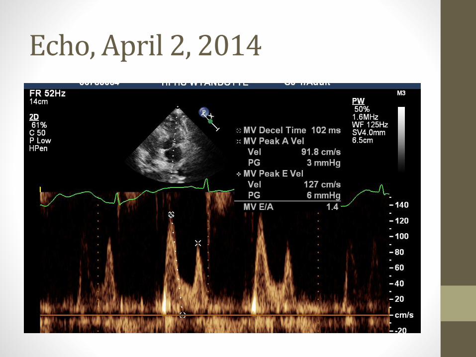
Echo, April 2, 2014
Let’s brush up on the evaluation of diastolic function
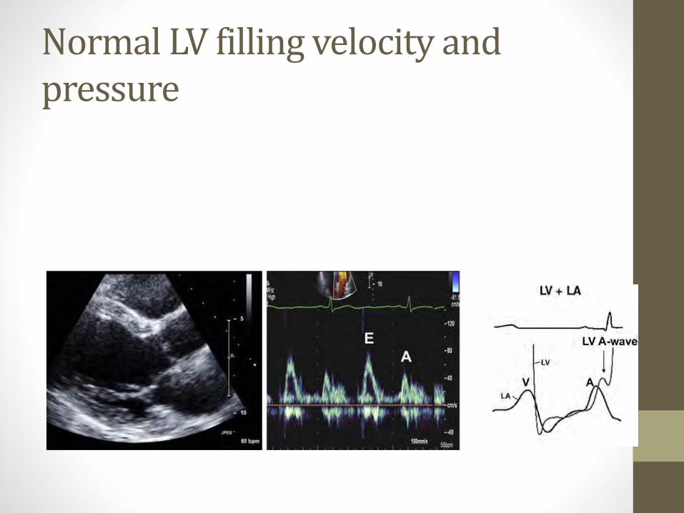
Normal LV filling velocity and pressure

Mitral inflow patterns in diastole
Normal Grade I Dysfunction

LV relaxation: tissue Doppler

Diastolic parameters
• Tissue Doppler records the actual movement of the LV in early diastole, and reflects LV relaxation
• The mitral flow characteristics reflect not only the flow velocity, but the left ventricular filling pressure when that flow occurs.

Normal LV filling
ASE Guidelines, April, 2016. (J Am Soc Echocardiogr 2016;29:277-314.)

Diastolic parameters
• Tissue Doppler records the actual movement of the LV in early diastole, and reflects LV relaxation
• The mitral flow characteristics reflect not only the flow velocity, but the left ventricular filling pressure when that flow occurs.
• The best estimate of the diastolic function takes into account the mitral flow velocity (E wave) and the LV relaxation (tissue Doppler), the tissue Doppler index (E/e’)

Grades of Diastolic Dysfunction
• Grade 1 Delayed early relaxation with normal filling pressure
• Grade 2 Delayed relaxation and increased LV end diastolic pressure
• Grade 3 Progressive reduction in LV compliance and elevation of LV filling pressures

Back to our patient.Tissue Doppler Index, MSO• Velocity of the lateral mitral annulus (Lateral e’) 3.12 cm/s
• Velocity of the medial mitral annulus (Medial e’) 3.02 cm/s
(A measure of LV relaxation, normal > 9 cm/s)
• Peak E velocity 127 cm/s
(A measure of LV filling pressure)
• Tissue Doppler Index, E/e’ 40.7
• Normal < 8
• Gray zone 8 – 15
• Abnormal > 15

Hospital course
• BP difficult to treat
• Fluids hard to control
• Worsening renal function: Creatinine increased to 3.8 with proteinuria. Is this actually renal disease, some sort of glomerulopathy?
• Arrange 24 hour urine, schedule renal biopsy
• 24 hour urine: 1.4 grams of protein. Biopsy cancelled.
• Pt elected to enter hospice.

So, what is this disease entity?
• Active medical problems:
• ASPVD Hypertension
• Diabetes Acute on chronic CKD
• Anemia Lung nodule
• Recurrent pleural effusion
• Possible renal artery stenosis
• Add to that:
• Sarcopenia
• Constipation
• Mitral regurgitation
• Hip, spine and back surgery

So, what is this disease entity?
• Is this diastolic heart failure?
• Are all the other problems just coincidental?

HFpEF
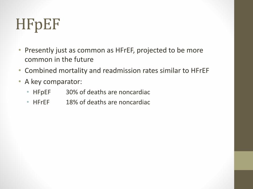
• Presently just as common as HFrEF, projected to be more common in the future
• Combined mortality and readmission rates similar to HFrEF
HFpEF
• Presently just as common as HFrEF, projected to be more common in the future
• Combined mortality and readmission rates similar to HFrEF
• A key comparator:
• HFpEF 30% of deaths are noncardiac
• HFrEF 18% of deaths are noncardiac

HFpEF
• Presently just as common as HFrEF, projected to be more common in the future
• Combined mortality and readmission rates similar to HFrEF
• A key comparator:
• HFpEF 30% of deaths are noncardiac
• HFrEF 18% of deaths are noncardiac
• Heart Failure with preserved LV EF is a disorder characterized by comorbidities: diabetes renal disease
obesity hypertension
systemic and pulmonary vascular abn.

HFpEF Overview

Phenogroups of HFpEFShah SJ, et al. Circulation 2015; 131: 269-79
1. Younger patients with moderate diastolic dysfunction with relatively normal BNP
2. Obese, diabetic patients with high prevalence of obstructive sleep apnea who had the worst LV relaxation
3. Older patients with significant chronic kidney disease, electrical and myocardial remodeling, pulmonary hypertension and RV dysfunction

HFpEF : Diagnosis
• European Society of Cardiology
• 3 basic aspects
• Signs or symptoms of heart failure
• Normal or nearly normal LV EF (~50%)
• Evidence of diastolic dysfunction

HFpEF : Diagnosis
• European Society of Cardiology
• 3 basic aspects
1. Signs or symptoms of heart failure
2. Normal or nearly normal LV EF (~50%)
3. Evidence of diastolic dysfunction
Evidence of abnormal LV relaxation, abnormal filling, diastolic stiffness
• Cardiac cath – elevated LVEDP > 16 mm Hg, mean PCWP > 12 mm Hg
• BNP > 200
• Tissue Doppler Index E/e’ > 15

Impaired relaxation transmitral flow pattern, from Penicka M, Heart 2014

Impaired relaxation pattern (E < A) with corresponding tissue Doppler of the lateral corner of the mitral annulus, from Penicka M, Heart 2014

Impaired flow pattern and low e’ in male with dyspnea, from Penicka M, Heart, 2014


Stepwise approach to the diagnosis of heart failure with preserved EF in elderly ambulatory patients with equivocal symptoms. Penicka M, Heart 2014;100: 68-76

HF preserved EF, Am Soc Echo, 2016

Pathophysiology of HFpEF
• Breathlessness is the predominant symptom due to elevated left ventricular diastolic pressure.
• Focus on abnormalities in active relaxation and passive stiffness
• Extracellular matrix
• Interstitial fibrosis
• Cardiomyocyte itself
• Incomplete relaxation of myocardial strips
• Increased myocardial stiffness
Pathophysiology of HFpEF
• A new paradigm – Paulus & Tschope – comorbidities such as obesity, diabetes and COPD lead to a systemic pro-inflammatory state that induces coronary microvascularendothelial inflammation.
• This inflammation and resultant oxidative stress cause stiff myocytes and interstitial fibrosis.
• Although hypertension is commonly felt to cause HFpEF by afterload excess, this model changes the emphasis to inflammation

Date of download:
5/14/2014
Copyright © The American College of Cardiology.
All rights reserved.
From: A Novel Paradigm for Heart Failure With Preserved Ejection Fraction: Comorbidities Drive Myocardial
Dysfunction and Remodeling Through Coronary Microvascular Endothelial Inflammation
J Am Coll Cardiol. 2013;62(4):263-271. doi:10.1016/j.jacc.2013.02.092
Myocardial Dysfunction and Remodeling in HFPEF and HFREF
In HFPEF, myocardial dysfunction and remodeling are driven by endothelial inflammation and oxidative stress. In HFREF, oxidative
stress originates in the cardiomyocytes because of ischemia, infection, or toxic agents. ROS trigger cardiomyocyte autophagy,
apoptosis, or necrosis. The latter attracts leukocytes. Dead cardiomyocytes are replaced by fibrous tissue. cGMP = cyclic guanosine
monophosphate; HFREF = heart failure with reduced ejection fraction; other abbreviations as in Figure 1.
Figure Legend:

Pathophysiology of HFpEF
• Vascular abnormalities
• Arterial stiffness increases with aging and is amplified by hypertension, diabetes and renal disease
• This leads to impaired LV reserve function, labile systemic hypertension, diminished coronary flow reserve and increased diastolic filling pressures, leading to breathlessness.

Pathophysiology of HFpEF
• The end systolic stiffness of the LV and the arteries increases with aging, especially in women, who are disproportionately represented in HFpEF
• Women also develop more concentric LVH in the setting of pressure overload compared to men.
• With exercise, the patient with HFpEF has a limited vasodilator response to activity.
• These patients often have marked systemic hypertension with exercise stress.

Treatment of HFpEF
• Pharmacologic management of HFpEF
• Agents in investigational trials
• Sildenafil (RELAX Trial)
• Aldosterone antagonists (TOPCAT Trial)
• ARB/neprilipsin inhibitor- ARNI (PARAMOUNT Trial)
• In each case, the information for each trial shows no benefit of treatment.

More on TOPCAT
Patients Sites Pt/site/mo Mortality
Overall 3,445 233 0.22 4.2-4.6
N & S Am 1,767 188 0.14 6.5-7.7
East. Eur. 1,676 45 0.56 2.0-2.3
One more case: BS
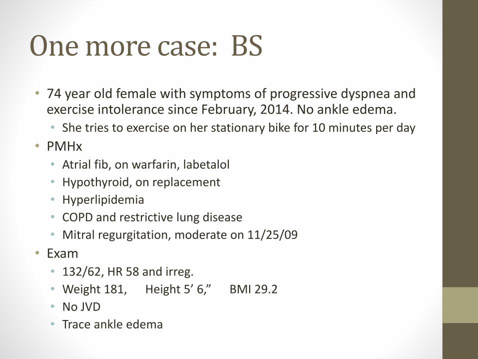
• 74 year old female with symptoms of progressive dyspnea and exercise intolerance since February, 2014. No ankle edema.• She tries to exercise on her stationary bike for 10 minutes per day
• PMHx• Atrial fib, on warfarin, labetalol
• Hypothyroid, on replacement
• Hyperlipidemia
• COPD and restrictive lung disease
• Mitral regurgitation, moderate on 11/25/09
• Exam• 132/62, HR 58 and irreg.
• Weight 181, Height 5’ 6,” BMI 29.2
• No JVD
• Trace ankle edema

One more case: BS

One more case: BS, Prior study.

One more case: BS

One more case: BS

One more case: BS
• You would order
A) Add furosemide, increase until dyspnea resolved
B) Start sildenafil
C) Start spironolactone
D) Cardiac rehabilitation exercise, paid by medicare
E) Cardiac rehab, not covered by insurance
One more case: BS
• You would order
A) Add furosemide, increase until dyspnea resolved
B) Start sildenafil
C) Start spironolactone
D) Cardiac rehabilitation exercise, paid by medicare
E) Cardiac rehab, not covered by insurance

Exercise and HFpEF (From Borlaug, JACC)

Exercise in HFpEF (From Rogers, Serajian. JAOA, 2015)

Exercise and aging

Exercise and aging (Aging Cell 10.1111/2018)
• 100 cyclists aged 55-79
• All cycle more than 100 miles/week
• Quadriceps muscle biopsy
• Muscle fiber type
• Muscle fiber size
• ATP activity
• Capillary density
• Mitochondrial proteins

Exercise and aging (Aging Cell 10.1111/2018)
• 100 cyclists aged 55-79
• All cycle more than 100 miles/week
• Quadriceps muscle biopsy
• Muscle fiber type
• Muscle fiber size
• ATP activity
• Capillary density
• Mitochondrial proteins
• Only capillary density decreased with age

HFpEF Conclusions
*HFpEF is a clinical syndrome of dyspnea and fatigue where there is normal LV EF, a stiff ventricle and stiff arteries and veins.
*The stiff LV and vasculature is worsened by inflammation, and the clinical syndrome of acute decompensated heart failure may be triggered by inflammation, especially lung disease, obesity, hypertension
*The signs and symptoms of HFpEF are dramatically more pronounced with exertion than they are at rest.
*Concerning treatment
With blood pressure and fluid excess, “go low and go slow”
There may be benefit to spironolactone
Exercise is demonstrated to improved functional status

Bottom Line (Personal opinion)
Failure with preserved ejection fraction is a disorder of increased stiffness of the heart, arteries and veins.
It is primarily a consequence of the natural aging process, which is worsened by deconditioning and accelerated by inflammation.