diagnostic methods in primary ciliary dyskinesia · pdf filekey words: primary ciliary...
TRANSCRIPT

Accepted Manuscript
Title: Diagnostic Methods in Primary Ciliary Dyskinesia
Author: Jane S Lucas Tamara Paff Patricia Goggin EricHaarman
PII: S1526-0542(15)00073-1DOI: http://dx.doi.org/doi:10.1016/j.prrv.2015.07.017Reference: YPRRV 1069
To appear in: YPRRV
Received date: 30-4-2015Accepted date: 30-7-2015
Please cite this article as: Lucas JS, Paff T, Goggin P, Haarman E, DiagnosticMethods in Primary Ciliary Dyskinesia, Paediatric Respiratory Reviews (2015),http://dx.doi.org/10.1016/j.prrv.2015.07.017
This is a PDF file of an unedited manuscript that has been accepted for publication.As a service to our customers we are providing this early version of the manuscript.The manuscript will undergo copyediting, typesetting, and review of the resulting proofbefore it is published in its final form. Please note that during the production processerrors may be discovered which could affect the content, and all legal disclaimers thatapply to the journal pertain.

Page 1 of 36
Accep
ted
Man
uscr
ipt
Diagnostic Methods in Primary Ciliary Dyskinesia
Authors
Jane S Lucas1, 2 Tamara Paff3, 4, Patricia Goggin1, 2 Eric Haarman3
Affiliations;
1. Primary Ciliary Dyskinesia Centre, University Hospital Southampton NHS Foundation
Trust, Southampton, UK
2. Academic Unit of Clinical and Experimental Sciences, Faculty of Medicine, University of
Southampton, Southampton, UK
3. Department of Pediatric Pulmonology, VU University Medical Center, Amsterdam, the
Netherlands
4. Department of Pulmonary Diseases, VU University Medical Center, Amsterdam, the Netherlands
Corresponding author: Dr Jane Lucas, Clinical and Experimental Sciences Academic Unit (Mail
Point 803), University of Southampton Faculty of Medicine, University Hospital Southampton
NHS Foundation Trust, Tremona Road, Southampton, SO16 6YD. [email protected]
Key Words: primary ciliary dyskinesia, diagnosis, high speed video analysis, electron microscopy,
genetics, nitric oxide
Educational Aims; The reader will be able to;
Describe the basis of the various diagnostic tests for PCD
Have an understanding of the strengths and limitations of each test
Understand that there is no ‘gold standard’ test
Understand why highly specialised centres should analyse samples and interpret results.

Page 2 of 36
Accep
ted
Man
uscr
ipt
Summary
Diagnosing primary ciliary dyskinesia is difficult. With no reference standard, a combination of
tests is needed; most tests require expensive equipment and specialist scientists. We review the
advances in diagnostic testing over the past hundred years, with emphasis on recent advances.
We particularly focus on use of high-speed video analysis, transmission electron microscopy,
nasal nitric oxide and genetic testing. We discuss the international efforts that are in place to
advance the evidence base for diagnostic tests.

Page 3 of 36
Accep
ted
Man
uscr
ipt
Introduction
Advances in diagnostic tests have accompanied technological developments and improved
understanding of the pathophysiology of primary ciliary dyskinesia (PCD). Following the first
case report of the syndrome, it took 70 years until transmission electron microscopy (TEM) was
proposed as a diagnostic tool. Since then, a number of protocols and methods have been shown
to aid diagnosis, but there remains no gold standard. In this review we discuss the historical
developments that have led to the current state-of-the-art; we describe the most commonly
used tests and consider the strengths and limitations for each (Table 1).
History of PCD Diagnostic Testing
In 1904, Siewart (or Zivat, Zivert, Sivert [1]) described a 21 year old with bronchiectasis
associated with situs inversus. Kartagener later reported the triad of bronchiectasis, situs
inversus and sinusitis in 1933, but it was not until the mid-1970s that Afzelius [2, 3] and
Pederson [4] recognized infertility as a feature and proposed the unifying role of cilia to explain
the syndrome. Having noted absent dynein arms in the cilia of patients with the syndrome,
Afzelius later demonstrated that the cilia were immotile, prompting the change of name from
“Kartagener’s Syndrome” to “Immotile Cilia Syndrome” [3]. These reports provided the
evidence for TEM and assessment of motility as the basis of diagnostic testing. Recognition that
outer dynein arm anomalies were not the only ultrastructural defect associated with the
syndrome gave early insights to the underlying heterogeneity of the disorder [5, 6]. Following
recognition that a number of patients had motile but dysfunctional cilia [7, 8] the name was
further changed in the mid-1980s to “primary ciliary dyskinesia” [9, 10]. Analysis of ciliary
motility is recommended as a diagnostic test in European guidelines [11].
Since the first description of a gene causing PCD in 1999 [12], there has been an exponential
increase in reports of PCD-associated genes. There are now over thirty known genes responsible
for >60% of cases. Recent publication of mutations in genes causing reduced numbers of cilia
with normal motility and ultrastructure [13, 14] in patients with a PCD phenotype, brings to
question whether yet another change of name is required. Gene panels are now used as part of
the diagnostic pathway in a number of countries.

Page 4 of 36
Accep
ted
Man
uscr
ipt
Diagnosis of PCD: state of the art
There is no single reference standard diagnostic test for PCD [15] and diagnosis usually requires
a number of technically demanding, sophisticated investigations. The availability and
combination of tests vary between countries [16], and there is no globally accepted consensus
as to which diagnostic results constitute a definite positive diagnosis, possible diagnosis or
excludes diagnosis. Moreover there is no global agreement regarding the standardisation of
conduct or reporting of any of the methods used to diagnose PCD.
Diagnosis in Europe is generally based on the following criteria [11, 15, 17, 18]: in a person with
a clinical history consistent with PCD confirmation of the diagnosis by at least two of the
following methods: (1) “hallmark” high speed videomicroscopy analysis (HVMA), (2) “hallmark”
TEM, (3) biallellic disease-causing mutations identified by gene testing, (4) abnormally low nasal
nitric oxide (nNO). If HVMA and nNO are the only abnormal tests, these tests should be
repeated before making a diagnosis.
Diagnostic criteria differ in North America where diagnosis is currently based on a combination
of nNO, TEM and genetic test results [19]. As the number of genes increases, the North
American Genetic Disorders of Mucociliary Clearance Consortium propose using nNO testing in
patients with a clinical phenotype to identify patients likely to have PCD, followed by genetic
testing to confirm the diagnosis, reserving HVMA and TEM for cases that are not identified by
PCD multigene panel testing [15]. The diagnostic pathway remains controversial, for example
some groups believe that HVMA should not be used in clinical practice but reserved as a
research tool [20, 21].
Who to refer for diagnostic testing: PCD diagnostic techniques are not widely available and
require extensive expertise and experience. Therefore, it is recommended that diagnosis should
only be made in specialized centers [11, 19]. Though PCD is a rare condition, with a prevalence
of approximately 1:10.000-20.000 people [16], its main presenting symptoms (recurrent
respiratory tract infections) are common in the general and, especially, the pediatric population.
The disease characteristics of PCD overlap with many more frequently occurring diseases like
asthma, immune deficiencies and cystic fibrosis. In many children, respiratory tract symptoms
are merely the result of frequent viral exposure without any underlying condition being present.

Page 5 of 36
Accep
ted
Man
uscr
ipt
It can be a challenge for a physician to decide which patients should be referred for further
diagnostic testing.
The clinical suspicion of PCD should be raised in case of a combination of a clinical history of
unexplained neonatal respiratory distress, early onset of nasal congestion, chronic wet sounding
cough, recurrent serous otitis, and situs abnormalities (figure 1). However, some of these
features can be mild or even absent in PCD patients [19]. In particular, 50% of PCD patients have
normal cardiac and visceral organ laterality. Parents of PCD patients often report nasal discharge
starting at the day of birth and it commonly persists throughout life. In addition, the majority of
neonates with PCD show neonatal distress [22], suggesting that cilia have a role in the effective
clearance of fetal lung fluid [23]. Serous otitis occurs frequently and, unlike in healthy children,
persists throughout adulthood [24]. Early onset of wet coughing and lower respiratory tract
infections occur in nearly all PCD patients. Although limited data on disease progression are
available, bronchiectasis is present in about half the pediatric PCD patients and all adult patients
[25]. In table 2 clinical features are summarized that should raise the suspicion of PCD and
prompt referral to a specialized diagnostic center [11]. In ethnic and consanguineous
populations where PCD is known to occur more frequently, the threshold for diagnostic testing
should be low.
Screening tests: Diagnosis of PCD requires access to a number of investigations which are
technically demanding and only available in specialist centres. Screening of high risk individuals
(i.e. individuals with an affected sibling or with symptoms suggestive of PCD) helps identify
patients for definitive testing.
Historically, estimation of nasal mucociliary clearance using the saccharine test was utilised as a
simple screen. A small particle of saccharine was placed on the inferior turbinate and the patient
asked to sit quietly for 1 hour; the test was considered positive if no taste of saccharine was
noted after 1 hour. However, results were difficult to interpret, even in compliant adults, since
patients with PCD find it difficult not to cough or sniff for the hour duration of the test. It is no
longer recommended.
In 1994 it was reported that nNO concentrations were significantly lower in patients with PCD
compared to healthy controls [26]. Whilst the reason for reduced nNO remains elusive [27],
measurement became widely established as a screening test [11, 17, 19, 28-31]. A recent meta-

Page 6 of 36
Accep
ted
Man
uscr
ipt
analysis of 11 studies comparing nNO during a velum closure breath hold reported a mean nNO
output of 19 nl/min (SD 18.6) in PCD (n=478) and 265 nl/min (SD 118.9) in healthy controls
(n=338) [32]. Although nNO is lower than normal in patients with CF [32] differentiation from
PCD remains good. nNO readings in healthy infants and young children are lower than in older
children, making false positive results more likely. This provides a major limitation of the test in
the age range that should be targeted for diagnosis.
A multi-centre study from North America derived a cut-off of 77 nL/min to differentiate PCD
patients from healthy controls [29]. They then validated the cut-off in consecutive patients
referred for diagnostic testing. 77 nL/min differentiated PCD (based on genotype and/ or TEM)
from patients with negative PCD test results with sensitivity of 0.98 and specificity of 0.75. Given
that genetic testing and TEM fail to detect 20-30% of patients, the true specificity is likely to be
higher and as new genes are added to panels, the reported specificity of nNO is likely to improve
but is unlikely to approach 100%. Patients with nNO<77nL/min but normal TEM and genetics in
the North American study had respiratory symptoms similar to patients with PCD, but only 9.5%
had laterality defects suggesting that perhaps 19% had ‘missed’ PCD; if these 4 patients were
reclassified as PCD positive, the specificity would moderately increase to 79%. In the authors’
centre, 77nl/min distinguished PCD positive and negative outcomes based on HSVMA and TEM
in consecutive referrals with sensitivity of 0.94 and specificity of 0.83 (unpublished). Therefore,
although nNO is highly specific when differentiating PCD from healthy controls [29], it is less
specific when differentiating PCD from patients with upper and lower respiratory tract
symptoms [32]. It should be noted that the youngest child in the North American study was 5.1
years and the mean age much higher. Also reference data for young healthy children is sparse.
Therefore strong evidence for use in the pre-school patient group, and reference norms is
awaited. PCD associated with normal nNO has been reported in a number of studies [29, 31, 33],
therefore nNO should not be used as a diagnostic test in isolation.
American Thoracic Society/ European Respiratory Society guidelines for the measurement of
nNO [33] recommend aspiration of gas from one nostril with gas entrained via the other naris
during a velum closure manoeuvre. The chemiluminescence analyser displays the nNO
concentration in real-time allowing measurements to be taken from the peak-plateau. This
method reliably differentiates patients with PCD from those with other respiratory diseases [29,

Page 7 of 36
Accep
ted
Man
uscr
ipt
31, 32, 34-36] as well as healthy controls. The limitations of this ‘gold standard’ method for
measuring nNO are that velum closure breath hold is difficult particularly for younger children
and that the chemiluminescence analysers are expensive and not easily transportable. To widen
accessibility of nNO as a screening test a small number of studies have investigated the use of
portable analysers [34, 37, 38]. These electromechanical analysers are more cost effective and
have excellent portability. The studies comparing portable and stationary analysers suggest that
portable analysers are a reliable means of measuring nNO, [32] but the machines have some
drawbacks. In particular, the lack of real-time visual display of NO does not allow the technician
to assess the acceptability of the measurement. In the author’s experience, a number of
patients referred to our centre with extremely low nNO measures using a portable device at the
referring centre, had normal levels when tested at our centre using a stationary
chemiluminescence analyser. Additionally the time required for the breath hold manoeuver
using portable devices is too long for many patients [37].
Although measurement is recommended during a velum closure breath-hold [33], tidal
breathing produces a reasonable alternative if breath-hold cannot be achieved e.g. young
children [32]. Tidal breathing measurements are lower in both PCD and controls [31, 32, 34, 35,
39, 40] and are less discriminatory than measures during a velum-closure breath-hold.
In summary, nNO provides an excellent screening test for PCD, and we now need
standardisation of methods of analysis and reporting.
Obtaining ciliated samples for analysis: In order to evaluate ciliary motility and ultrastructure, a
good quality epithelial sample is obtained from the upper or lower airways by a trained health
care professional. Nasal samples are most easily obtained, but if the patient is having a
bronchoscopy for other reasons, lower airway samples can be taken [41]. Epithelial cells can be
collected by brush, curette or forceps (figure 2). The advantages of brush biopsies are that they
are less painful, bleeding is unlikely and samples can be taken without sedation [41]. The
advantages of using a curette or forceps, are the larger amount of material obtained and the
possibility to evaluate ciliary motility in the context of other epithelial cells [41]. Depending on
local policies and age, sedation can be given during a nasal biopsy, although this is rarely
required for brush biopsies. Good explanations to both child and parents about the procedure
result in less stress and empower parents to provide support [42]. When sedation is required, it

Page 8 of 36
Accep
ted
Man
uscr
ipt
should be delivered by trained professionals, using monitoring and with rescue facilities
available [43]. It is useful to assess the quality of the sample using a light microscope while the
patient is still in clinic so that a repeat sample can be taken immediately if necessary.
Samples for HVMA should be placed in buffered culture medium and analyzed as soon as
possible [44] to provide optimal conditions for analysis. Biopsies taken for TEM are fixed in e.g.
3% buffered glutaraldehyde and can be stored.
High-Speed Video Microscopy: Assessment of ciliary beat frequency (CBF) and ciliary beat
pattern (CBP) using HVMA provide measures of ciliary function [45]. Ciliary frequency or pattern
are abnormal in all patients with PCD, and HVMA has a pivotal role in the 30% of PCD patients in
whom TEM is normal [46] and the 20-35% [19] of patients in whom the gene has not yet been
identified.
Abnormalities of pattern associated with PCD include static, slow, rotating, stiff, hyperfrequent
and vibrating cilia. These patterns correlate with abnormalities reported by TEM [47] (e.g.
rotating patterns and central pair defects) and with some genetic findings (e.g. DNAH5-mutant
cilia have a bent position with minimal movement) [48]. However, numbers of patients with
individual genetic defects were extremely low in the only study [48], and larger multi-centre
studies are called for to investigate genotype-ciliary phenotype relationships. Subtle functional
defects are increasingly recognised to cause phenotypic disease [49, 50] and analyses by
scientists with substantial experience of normal and abnormal ciliary beating is essential. In
expert hands, using HVMA to assess CBP, dyskinesia on >90% ciliated edges has 97% sensitivity
and 95% specificity to predict a TEM diagnosis of PCD [45].
Optical equipment should provide high magnification with excellent resolution to accurately
analyse the pattern. The quality of the objective is critical as the main determinant of resolution
and image quality. In the author’s laboratory we observe cilia at x1000 magnification using an
inverted microscope under bright field light conditions. Although lower magnification (e.g. x500)
is acceptable for calculation of CBF, the CBP would be difficult to decipher. The camera should
capture images at a fast rate so that they can subsequently be slowed down, allowing the
microscopists to qualitatively analyse the beat pattern. We record at 500 frames per second; so
for cilia beating at 15Hz, each beat is represented on 33 frames which can be played back at 30
frames per second for pattern analysis. Microscopic imaging is conducted under strictly

Page 9 of 36
Accep
ted
Man
uscr
ipt
controlled environmental conditions (e.g. temperature and pH) since minor variations effect
ciliary function [51, 52]. We analyse cilia within 4 hours of sampling, using buffered cell culture
medium to control pH and the sample is maintained at 37°C in an environmental chamber
during analysis. Under these conditions our reference range for CBF is 11-20Hz, but laboratories
using different conditions will have different reference ranges.
Abnormalities of CBF and CBP can occur secondary to infection, damage during sampling or
inflammation of the epithelia complicating the diagnostic picture. Following abnormal analysis it
is therefore necessary to reanalyse CBF and CBP following culture of the epithelial cells or
following a repeat brushing to confirm consistency of findings if genotype and TEM are not
diagnostic.
Whilst direct measurement of CBP and CBF using HVMA is generally considered the most
accurate and reproducible technique, it is time consuming and incurs risk of operator error due
to selection bias. Several groups have attempted to overcome these problems by developing
software to automate analysis from the digital images [53-55].
Although assessment of ciliary function by light microscopy is recognised as a critical diagnostic
test used as ‘first-line’ in many centres [11, 17, 48, 53, 54, 56] it should be noted that some
clinicians question its value and recommend that HVMA should be reserved for research
purposes [20]. A limitation of HVMA is the need for specialist equipment, rigorous quality
control and experienced technicians with a high through-put of normal and abnormal samples.
This restricts testing to a limited number of specialist centres.
Transmission Electron Microscopy: (TEM) allows the ultrastructure (figure 3) of ciliary
axonemes to be visualized. Ultrathin sections are prepared for TEM from fixed cells or tissue
biopsies using standard methods [47]. Sections should be examined at magnifications sufficient
to visualize the ultrastructural features (>x 60,000). Normal cilia have a structure of nine
peripheral microtubular doublets and a central pair (9+2 arrangement) (Figure 3). The accessory
axonemal components are the outer dynein arms (ODA), inner dynein arms (IDA), radial spokes
and the nexin-dynein regulatory complex (N-DRC). Dynein arms contain adenosine
triphosphatases and act as motors to achieve ciliary motion by sliding of adjacent microtubular
doublets. Defects in any ciliary components can cause immotility or dyskinetic beating.
Relationships between the ultrastructural defect and beating pattern have been described [47].
The most common ultrastructural defects are: ODA-defects (25-50%) and combined IDA- and

Page 10 of 36
Accep
ted
Man
uscr
ipt
ODA-defects (25-50%) [47, 57-59] (figure 4). IDA defects associated with microtubular
disorganisation occur in 15% of PCD, but isolated IDA defects as a cause of PCD are controversial
particularly as no mutations have been identified in IDA proteins. IDA are difficult to identify due
to the decreased repeats along the ciliary axoneme compared to the ODA [60] therefore false
positive IDA defects are likely. Reporting of isolated IDA defects requires repeated testing or
immunofluorescence staining of the IDA visualizing the entire axoneme [60]. Central pair defects
occur less frequently (5-15%) and are associated with a mix of both normal and abnormal cilia
[61, 62], therefore adequate numbers of cilia need to be viewed in longitudinal and transverse
section; in the author’s laboratory at least 100 and up to 300 cilia selected from healthy cells are
analysed in transverse section. It is also informative to examine the longitudinal sections as rare
defects e.g. RSPH4A, can be identified by distinctive patterning [50].
Previously TEM was considered the “gold standard” but it is now recognized that 20-30% of
patients have normal ultrastructure when analyzed by TEM [46, 61]. For example, defects of
nexin link components [63], central pair components [64], ciliary biogenesis defects [14] and
defects caused by DNAH11 [65, 66], usually cannot be visualized by classic TEM. Thus, normal
ultrastructure cannot rule out PCD.
An additional limitation of TEM is that inflammation and infection, can alter the normal 9+2
arrangement. Therefore, it can be difficult to differentiate acquired defects from PCD [19].
Similar problems can occur if cells are poorly fixed. Even in samples obtained from non-inflamed
tissue that have been properly fixed, correct identification of ultrastructural defects requires
highly specialized personnel with substantial experience of the range of normality and
abnormality.
Recently, electron tomography has evolved as a research tool providing 3D visualization of the
ultrastructure of cilia. This technique has demonstrated ultrastructural defects in PCD patients
with DNAH11 and HYDIN mutations, who did not appear to have defects on classic TEM [67]
[64]. However, there are some limitations to this technique mainly concerning microscopic
resolution, limited accessibility of the sample (penetration depth) and the speed of data
processing. However, with the current progress in both hardware and software, this technique
will be a valuable diagnostic tool in difficult PCD cases in the future.
Genetics: PCD is generally an autosomal recessive disease, though in rare cases other modes of
inheritance have been described (X-linked or autosomal dominance). Currently 31 genes have

Page 11 of 36
Accep
ted
Man
uscr
ipt
been identified, explaining approximately 60% of cases (Table 3). There seems to be a clear
association between genetic defects, ciliary ultrastructure and motion defects (table 3).
However, the association between genetic defects and the clinical phenotype is largely
unknown; international collaborations are developing large meta-cohorts to ensure sufficient
numbers of patients with mutations in each PCD-associated gene. Some genotype-phenotype
relations have been described, but caution needs to be kept as some descriptions are based on
low numbers of patients: mutations causing reduced generation of multiple motile cilia
(MCIDAS, CCNO) and those causing IDA with microtubular disorganization (CCDC39, CCDC40)
have been reported to result in relatively severe lung disease [13, 14, 68]. In contrast, mutations
in RSPH1 (a mutation in a gene coding for one of the radial spoke subunits) reportedly cause a
mild phenotype [69]. Male patients with mutations in CCDC114 (coding for a docking protein for
the ODA) are generally fertile [70]. Situs abnormalities are not observed in patients with
mutations affecting the central pair [64] or radial spokes [49, 50, 69], nor in patients with
reduced generation of multiple motile cilia [13, 14]. However, it is likely that variable clinical
pictures occur between patients with different mutations within a particular gene and even
between patients with identical mutations.
To date mutations have been identified in 6 genes encoding for proteins that are part of the
ODA (DNAH5, DNAI1, DNAI2, DNAL1, NME8 (TXNDC3) and DNAH11) [12, 71-75]. Mutations in
DNAH5 and DNAI1 are thought to account for the largest proportion of PCD patients: 30% and
9% respectively [19, 76-79]. Mutations in these genes cause structural and functional ODA
defects and consequently ciliary immotility [47, 48]. However, in DNAH11, TEM is normal and
thus not informative [65, 66, 75].
In addition, mutations in 3 genes have been described that, though not coding for proteins that
are structurally part of the ODA, cause selective absence of ODA on TEM: CCDC114 [70],
CCDC151[80] and ARMC4[81]. These genes code for ODA-associated docking complex proteins
that enable attachment of the ODA to the axoneme.
Mutations in CCDC39 [82, 83], CCDC40 [82, 84], CCDC65 [85] and CCDC164 [63] cause variable
inner dynein arm defects with microtubular disorganization. These genes encode for proteins of
the N-DRC proteins that connect the peripheral microtubular doublets with each other. The cilia
beat in a fast and stiff matter.
Patients with mutations in genes encoding for proteins that are part of the central apparatus
and radial spokes (RSPH9, RSPH4A, RSPH1 and HYDIN) can pose diagnostic difficulties [49, 50,
Page 12 of 36
Accep
ted
Man
uscr
ipt
64]. Patients have normal situs because healthy nodal cilia responsible for lateralization during
embryogenesis differ from healthy respiratory cilia, lacking the central pair. Approximately half
the cilia from patients with these mutations have normal ultrastructure on TEM and movement
on HVMA evaluation. Additionally, RSPH1 mutations generally cause a relatively mild clinical
picture and borderline to normal nasal NO concentrations [69].
Numerous proteins are involved in cytoplasmic biogenesis of cilia. The identification of genes
involved in cytoplasmic assembly of cilia (HEATR2, DNAAF1, DNAAF2, DNAAF3, DYX1C1,
CCDC103, LRRC6, ZMYND10, SPAG1, ARMC4 and C21orf59) has provided clear insights into this
process [81, 85-95]. Defects in the assembly line can result in ODA or ODA/IDA defects.
Recently, two genetic defects have been identified in patients previously classified as ciliary
aplasia: mutations in CCNO and MCIDAS [13, 14]. Mutations in CCNO and MCIDAS result in
defective centriole generation and placement on the outer surface of the epithelial membrane.
These basal bodies normally nucleate the motile ciliary axonemes. Consequently, there is a
severe reduction in the number of motile cilia leading to the clinical picture of PCD. The few
residual cilia in CCNO-mutants have normal axonemal ultrastructure on TEM and do not show
any obvious beating defects. Previously, these patients were often misclassified as secondary
dyskinesia. In MCIDAS-mutants, the few residual cilia lack many of the axonemal motor
components, and are immotile.
Thanks to high-throughput genetic testing, many disease-causing mutations have been
identified, and genotyping can currently identify >60% of patients. However, for a significant
proportion of patients the genetic defect has not yet been elucidated. Therefore genetic testing
cannot be used to rule out the diagnosis of PCD yet.
Additional tests: Tests that are currently used by only a handful of centers, but with increased
evidence may come into more common use include immunofluorescence (IF) labelling of ciliary
proteins and radioaerosol mucociliary clearance (MCC). IF was developed as a research tool to
improve understanding of the impact of disease-causing genes on ciliary proteins [96]. Specific
antibodies are used for subcellular localization of proteins in human respiratory epithelial cells
using high-resolution IF imaging. A number of antibodies against ciliary proteins are now
commercially available including DNAH5 (an outer dynein arm protein), DNALI1 (an inner dynein
arm protein), RSPH4A (a radial spoke head protein- central pairs) and GAS8 (nexin links).
Although the literature is confined to the research arena, several centers now use IF to aid

Page 13 of 36
Accep
ted
Man
uscr
ipt
diagnosis, and this is likely to increase as more antibodies become readily available. Antibodies
against gene-related proteins associated with normal TEM would be a particularly useful (e.g.
HYDIN and DNAH11). To promote IF as a diagnostic test, standardized protocols are required
along with studies to establish the sensitivity and specificity.
Pulmonary radioaerosol MCC has been reported to differentiate PCD from healthy individuals
[97, 98]. The method is based on clearance patterns after the inhalation of a radioaerosol
tracer, providing a whole-lung functional test for pulmonary radioaerosol MCC. Further data is
now required to confirm whether the technique will be useful for differentiating PCD patients
from people with secondary ciliary damage, or for diagnosing patients with inconclusive tests
using other methods.
Cell culture: Culturing respiratory epithelial cells provides an opportunity to confirm that
abnormalities seen on the fresh nasal brushing are due to PCD rather than a secondary
dyskinesia. This removes the need to repeat nasal brushing to obtain a sample for reanalysis
which is required if diagnosis depends on HVMA. The ciliary phenotype changes following cell
culture helping to differentiate primary from secondary dyskinesia and this change is therefore
advantageous [99]. Jorissen et al [57] first reported the use of cell culture to aid PCD diagnosis
using a submerged method (monolayer-suspension cell culture). Culture followed by ciliation at
air-liquid interface (ALI) [99-101] has the advantage of yielding more cells and cilia than the
submerged method. Both submerged and ALI-culture techniques allow reanalysis by HVMA [57,
99, 101] and TEM [99, 101, 102] aiding the diagnosis of PCD. ALI-culture of respiratory cells
from PCD patients additionally provides an excellent ex vivo model for research [103, 104].
Equivocal outcome from diagnostic testing
There are a number of patients with a clinical phenotype suggestive of PCD, whose diagnostic
tests are equivocal, or abnormalities very subtle. In particular, in the experience of the authors,
there are a small number of patients with subtle abnormality of ciliary pattern on PCD, with
normal TEM and equivocal/normal nNO; we label this sub-group of patients ‘possible PCD’.
Having investigated them for any alternative diagnosis, we clinically manage them in the PCD
service. We anticipate that further advances in PCD diagnostic testing, and in our understanding
of the underlying pathophysiology of the condition will lead to a significant proportion of this
group becoming positive over time.

Page 14 of 36
Accep
ted
Man
uscr
ipt
International perspectives and future needs
Recent international networks and collaborations have led to advances in the understanding
and conduct of diagnosing PCD, but there remains no ‘gold standard’. A European Respiratory
Society Taskforce (ERS TF 2007-9) provided a focus for paediatric pulmonologists to undertake
epidemiological studies [16, 105] and develop a consensus statement for diagnosis and
management [11]. The taskforce highlighted disparity in diagnosis across Europe [16], with
missed and delayed diagnosis particularly likely in countries with lower health expenditure. One
of several projects being conducted by European Union’s Seventh Framework Programme
BESTCILIA (2012-15), aims to improve equity of care by establishing diagnostic centres in three
areas of Europe currently unable to fulfil ERS consensus guidelines [11]: Cyprus, Poland and
Greece. Recent advances in diagnostics have underpinned the development of a new ERS
taskforce (ERS TF 2014-16), to develop evidence-based guidelines for the diagnosis of PCD. The
taskforce is using rigorous methodology to develop practice guidelines for diagnostic testing
including measurement of nNO, HVMA of ciliary function, TEM, genetics testing and IF labelling
of ciliary proteins. In North America eight centres are members of the Genetic Disorders of
Mucociliary Clearance Consortium [19]. The Consortium has developed standardized protocols
for diagnostic testing, including nNO testing [29].
National collaborations are contributing to improved diagnostic management in a number of
individual countries including France [58, 106], and the United Kingdom [17, 107].
Concluding discussion
Advances in the diagnosis of PCD have occurred over the decades since cilia were first
implicated in the syndrome. National consortia and research from individual groups have moved
us forward, but we still have no gold standard reference nor international standards for conduct
of tests. A number of experts in the field are collaborating to develop evidence based guidelines
for diagnostic testing through the European Respiratory Society PCD Task Force and COST Action
BEAT-PCD. Initiatives are also underway to widen accessibility to diagnostic services (BEST-
CILIA).
Future Directions:
Establish the evidence base to develop international standards for conduct and
reporting of tests.

Page 15 of 36
Accep
ted
Man
uscr
ipt
Establish the evidence base for international development of a diagnostic
algorithm for (i) definite PCD, (ii) probable PCD (iii) PCD excluded.
Establish the accuracy (sensitivity, specificity, and predictive values of diagnostic
tests in well designed ‘blinded’ studies.
Page 16 of 36
Accep
ted
Man
uscr
ipt
Conflict of Interest Statement: JSL is a member of Aerocrine PCD Advisory Board and has
received expenses and honoraria. TP, PG and EH have not declared any potential conflicts of
interest.
Acknowledgements: JSL and PG: The National PCD Diagnostic Service at UHS is commissioned
and funded by NHS England. Research is supported by AAIR Charity, NIHR Southampton
Respiratory Biomedical Research Unit and NIHR Wellcome Trust Clinical Research Facility,
Southampton, UK. JSL chairs the European Respiratory Society Task Force development of a
practice guideline for diagnosis of PCD (ERS TF-2014-04); EH and TP: research is funded by the
PCD Belangengroep. JSL and EH: receive research funding from the European Union’s Seventh
Framework Programme under EC-GA No. 305404 BESTCILIA: Better Experimental Screening and
Treatment for Primary Ciliary Dyskinesia. All authors: are participants of COST Action BEAT-PCD:
Better Evidence to Advance Therapeutic options for PCD (BM 1407).

Page 17 of 36
Accep
ted
Man
uscr
ipt
Table 1: The advantages and disadvantages of diagnostic tests in current use.
Diagnostic Test
Advantages Disadvantages Diagnostic accuracy
Nasal nitric oxide
1. Guidelines exist for conduct of test [33]
2. Meta-analysis demonstrates good sensitivity and specificity[32]
3. Protocols can be standardized for multi-center use[29]
4. Alternatives to ‘gold standard’ method (velum closure using chemiluminescence analyzer) have acceptable accuracy [35, 37]
1. ‘Gold standard’ method impossible for young children and equipment expensive and non-portable.
2. Standardised approach to use in PCD diagnostics and reporting of results needed
3. Small % of patients have normal NO
4. Normal reference values for younger age groups are lacking
In consecutive patients for PCD diagnostic testing:1. Cut-off 53
nl/min: sensitivity 0.92, specificity 0.96 [31]
2. Cut-off 77 nl/min: sensitivity 0.98, specificity >0.75 [29]
HVMA 1. Provides assessment of functional defect
2. HSVMA is abnormal in all described cases of PCD.
3. Correlates with TEM [47] and genetic findings [48]
1. Absence of standardized methods of reporting
2. Abnormalities of CBP can be subtle
3. Requires specialist equipment
4. Requires rigorous adherence to quality control
5. Secondary defects are common and experienced scientists are needed with expert knowledge of normal and abnormal findings
Dyskinesia on >90% ciliated edges:
o sensitivity 0.97
o specificity 0.95
to predict a TEM diagnosis [45]
TEM 1. Provides assessment of the ultrastructural defects
2. Correlates to genetics and HVMA
3. Widely used
1. ≈30% of patients have no defect on TEM
2. Potentially altered by secondary dyskinesia
3. Requires specialist equipment and evaluation
Sensitivity: 70-80% Specificity: 100%[46, 61] (false positives occur, but can be avoided by evaluating sufficient cilia (>100) and adequate training of staff).
Genetic testing
1. Indisputable and fast diagnosis of PCD in
1. Cannot rule out PCD (yet) as 20-35% is
Sensitivity: 65-80% (estimated)

Page 18 of 36
Accep
ted
Man
uscr
ipt
case of biallellicpathogenic mutations in known genes
2. Has relevance to clinical phenotype
3. Provides possibility for carrier testing in isolated populations with high frequency of PCD
unknown2. Commercial testing
does not offer complete gene/exon panel
3. Can be difficult to prove pathogenicity/relation to PCD in cases of mutations in novel (candidate) genes or novel mutations in known PCD genes
Specificity:100%[19]
IF 1.Much interest for IF to become more widely available as a diagnostic tool2.Useful research tool3.A number of antibodies are commercially available4. Relatively low cost
1. No evidence for use as a clinical tool yet published
2. The antibodies currently available commercially will not detect all cases
3. Absence of standardized methods or reporting
No published data
Page 19 of 36
Accep
ted
Man
uscr
ipt
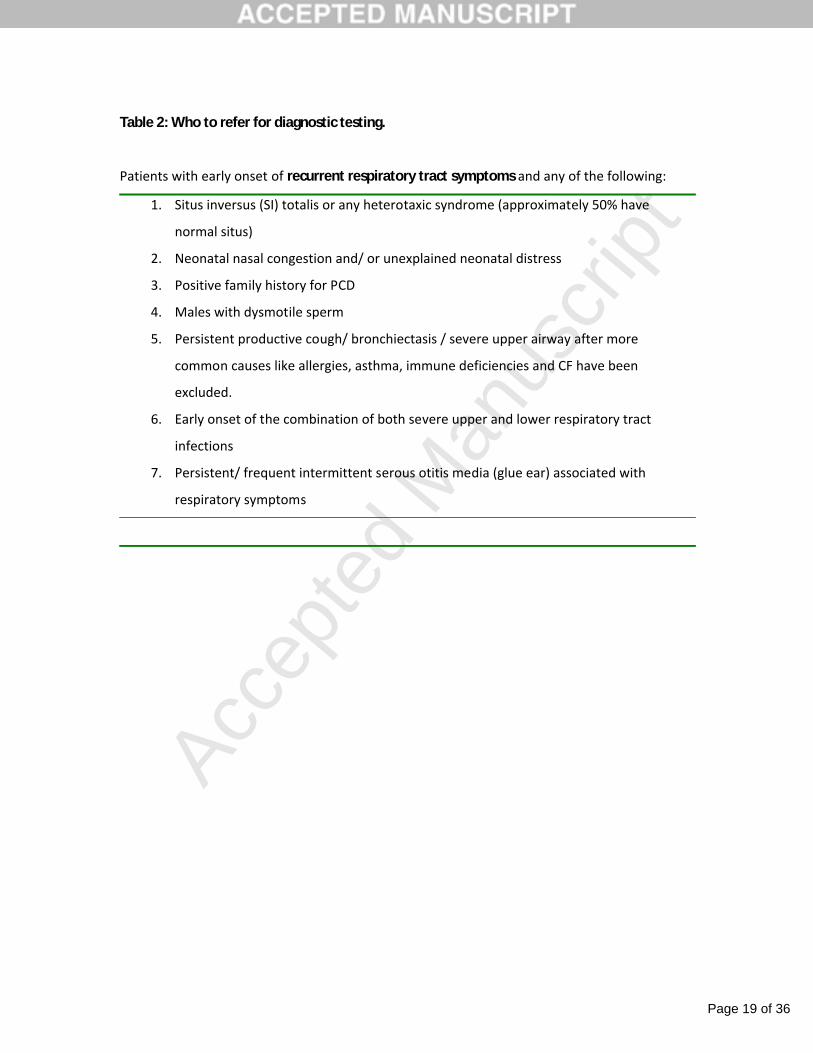
Table 2: Who to refer for diagnostic testing.
Patients with early onset of recurrent respiratory tract symptoms and any of the following:
1. Situs inversus (SI) totalis or any heterotaxic syndrome (approximately 50% have
normal situs)
2. Neonatal nasal congestion and/ or unexplained neonatal distress
3. Positive family history for PCD
4. Males with dysmotile sperm
5. Persistent productive cough/ bronchiectasis / severe upper airway after more
common causes like allergies, asthma, immune deficiencies and CF have been
excluded.
6. Early onset of the combination of both severe upper and lower respiratory tract
infections
7. Persistent/ frequent intermittent serous otitis media (glue ear) associated with
respiratory symptoms

Page 20 of 36
Accep
ted
Man
uscr
ipt
Table 3: Overview of PCD genes and the encompassing ciliary ultrastructural and movement
defects when mutated. Adapted from Paff (117) et al. 2014
Ultrastructural defect (by TEM)
Ciliary motion defect(by HVMA)
Clinical phenotype
Reference
ODA
DNAH5 Immotile with occasional stiff moving cilia Classic Olbrich [71] et al. 2002
DNAI1 Unknown Classic Pennarun [12]et al. 1999
DNAI2 Unknown Classic Loges [73] et al. 2008
DNAL1 Decreased CBF Classic Mazor [74] et al. 2011
NME8 (TXNDC3)
Mixed populations: normal to immotile Classic Duriez [72] et al. 2007
CCDC103 Complete immotility or lack of coordination with reduced amplitude
Classic Panizzi [108] et al. 2012
CCDC114 Largely immotile with some twitching cilia Normal male fertility
Onoufriadis[70] et al. 2013
ARMC4 Complete immotility or reduced CBF and amplitude
Classic Hjeij [81] et al. 2013
CCDC151 Complete immotility Classic Hjeij [80] et al. 2014
ODA/IDA
DNAAF1 (LRRC50) Complete immotility Classic Loges [109] et al. 2009
DNAAF2 (KTU) Complete immotility Classic Omran [92] et al. 2008
DNAAF3 Complete immotility Classic Mitchison [90]et al. 2012
HEATR2 Near complete immotility Classic Horani [87] etal. 2012
LRRC6 Complete immotility Classic Kott [89] et al. 2012
ZMYND10 Complete immotility or reduced CBF and amplitude
Classic Moore [91] et al. 2013**
SPAG1 Near complete immotility Classic Knowles [88] et al. 2013
C21orf59 Complete immotility Classic Austin-Tse [85]et al. 2013
DYX1C1 Largely complete immotility. Some cilia show Classic Tarkar [110] et
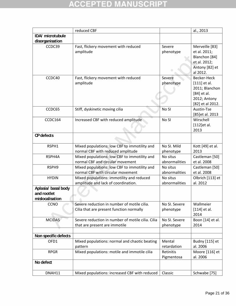
Page 21 of 36
Accep
ted
Man
uscr
ipt
reduced CBF al., 2013IDA/ microtubule disorganisation
CCDC39 Fast, flickery movement with reduced amplitude
Severe phenotype
Merveille [83]et al. 2011; Blanchon [84]et al. 2012; Antony [82] et al 2012.
CCDC40 Fast, flickery movement with reduced amplitude
Severe phenotype
Becker-Heck [111] et al. 2011; Blanchon [84] et al. 2012; Antony [82] et al 2012.
CCDC65 Stiff, dyskinetic moving cilia No SI Austin-Tse [85]et al. 2013
CCDC164 Increased CBF with reduced amplitude No SI Wirschell [112]et al. 2013
CP defects
RSPH1 Mixed populations: low CBF to immotility and normal CBF with reduced amplitude
No SI. Mild phenotype
Kott [49] et al. 2013
RSPH4A Mixed populations: low CBF to immotility and normal CBF and circular movement
No situs abnormalities
Castleman [50]et al. 2008
RSPH9 Mixed populations: low CBF to immotility and normal CBF with circular movement
No situs abnormalities
Castleman [50]et al. 2008
HYDIN Mixed populations: immotility and reduced amplitude and lack of coordination.
No situs abnormalities
Olbrich [113] et al. 2012
Aplasia/ basal body and rootlet mislocalisation
CCNO Severe reduction in number of motile cilia.Cilia that are present function normally
No SI. Severe phenotype
Wallmeier[114] et al. 2014
MCIDAS Severe reduction in number of motile cilia. Cilia that are present are immotile
No SI. Severe phenotype
Boon [14] et al. 2014
Non specific defectsOFD1 Mixed populations: normal and chaotic beating
patternMental retardation
Budny [115] et al. 2006
RPGR Mixed populations: motile and immotile cilia Retinitis Pigmentosa
Moore [116] et al. 2006
No defect
DNAH11 Mixed populations: increased CBF with reduced Classic Schwabe [75]

Page 22 of 36
Accep
ted
Man
uscr
ipt
amplitude and low CBF to immotility et al. 2008
Page 23 of 36
Accep
ted
Man
uscr
ipt
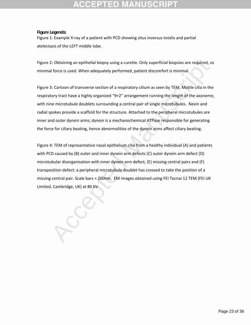
Figure Legends: Figure 1: Example X-ray of a patient with PCD showing situs inversus totalis and partial
atelectasis of the LEFT middle lobe.
Figure 2: Obtaining an epithelial biopsy using a curette. Only superficial biopsies are required, so
minimal force is used. When adequately performed, patient discomfort is minimal.
Figure 3: Cartoon of transverse section of a respiratory cilium as seen by TEM. Motile cilia in the
respiratory tract have a highly organized “9+2” arrangement running the length of the axoneme,
with nine microtubule doublets surrounding a central pair of single microtubules. Nexin and
radial spokes provide a scaffold for the structure. Attached to the peripheral microtubules are
inner and outer dynein arms; dynein is a mechanochemical ATPase responsible for generating
the force for ciliary beating, hence abnormalities of the dynein arms affect ciliary beating.
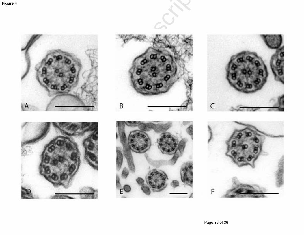
Figure 4: TEM of representative nasal epithelium cilia from a healthy individual (A) and patients
with PCD caused by (B) outer and inner dynein arm defects (C) outer dynein arm defect (D)
microtubular disorganisation with inner dynein arm defect, (E) missing central pairs and (F)
transposition defect: a peripheral microtubule doublet has crossed to take the position of a
missing central pair. Scale bars = 200nm. EM images obtained using FEI Tecnai 12 TEM (FEI UK
Limited, Cambridge, UK) at 80 kV.

Page 24 of 36
Accep
ted
Man
uscr
ipt
1. Berdon WE, McManus C, Afzelius B. More on Kartagener's syndrome and the contributions of Afzelius and A.K. Siewert. Pediatric radiology 2004, 34(7):585-586.
2. Afzelius BA. A human syndrome caused by immotile cilia. Science 1976, 193(4250):317-319.
3. Eliasson R, Mossberg B, Camner P, Afzelius BA. The immotile-cilia syndrome. A congenital ciliary abnormality as an etiologic factor in chronic airway infections and male sterility. NEnglJMed 1977, 297(1):1-6.
4. Pedersen H, Mygind N. Absence of axonemal arms in nasal mucosa cilia in Kartagener's syndrome. Nature 1976, 262(5568):494-495.
5. Sturgess JM, Chao J, Wong J, Aspin N, Turner JA. Cilia with defective radial spokes: a cause of human respiratory disease. The New England journal of medicine 1979, 300(2):53-56.
6. Afzelius BA, Eliasson R. Flagellar mutants in man: on the heterogeneity of the immotile-cilia syndrome. Journal of ultrastructure research 1979, 69(1):43-52.
7. Pedersen M, Mygind N. Ciliary motility in the 'immotile cilia syndrome'. First results of microphoto-oscillographic studies. BrJDisChest 1980, 74(3):239-244.
8. Rossman C, Forrest J, Newhouse M. Motile cilia in "immotile cilia" syndrome. Lancet 1980, 1(8182):1360.
9. Veerman AJ, van der Baan A, Weltevreden EF, Leene W, Feenstra L. Cilia: immotile, dyskinetic, dysfunctional. Lancet 1980, 2(8188):266.
10. Sleigh MA. Primary ciliary dyskinesia. Lancet 1981, 2(8244):476.aa11. Barbato A, Frischer T, Kuehni CE et al. Primary ciliary dyskinesia: a consensus
statement on diagnostic and treatment approaches in children. EurRespirJ 2009, 34(6):1264-1276.
12. Pennarun G, Escudier E, Chapelin C et al. Loss-of-function mutations in a human gene related to Chlamydomonas reinhardtii dynein IC78 result in primary ciliary dyskinesia. AmJHumGenet 1999, 65(6):1508-1519.
13. Wallmeier J, Al-Mutairi DA, Chen CT et al. Mutations in CCNO result in congenital mucociliary clearance disorder with reduced generation of multiple motile cilia. NatGenet 2014, 46(6):646-651.
14. Boon M, Wallmeier J, Ma L et al. MCIDAS mutations result in a mucociliary clearance disorder with reduced generation of multiple motile cilia. Nature communications 2014, 5:4418.
15. Lucas JS, Leigh MW. Diagnosis of primary ciliary dyskinesia: searching for a gold standard. EurRespirJ 2014, 44(6):1418-1422.
16. Kuehni CE, Frischer T, Strippoli MP et al. Factors influencing age at diagnosis of primary ciliary dyskinesia in European children. EurRespirJ 2010, 36(6):1248-1258.
17. Lucas JS, Burgess A, Mitchison HM, Moya E, Williamson M, Hogg C. Diagnosis and management of primary ciliary dyskinesia. ArchDisChild 2014, 99(9):850-856.

Page 25 of 36
Accep
ted
Man
uscr
ipt
18. Werner C, Onnebrink JG, Omran H. Diagnosis and management of primary ciliary dyskinesia. Cilia 2015, 4(1):2.
19. Knowles MR, Daniels LA, Davis SD, Zariwala MA, Leigh MW. Primary ciliary dyskinesia. Recent advances in diagnostics, genetics, and characterization of clinical disease. AmJRespirCrit Care Med 2013, 188(8):913-922.
20. Horani A, Brody SL, Ferkol TW. Picking up speed: advances in the genetics of primary ciliary dyskinesia. Pediatric Research 2014, 75(1-2):158-164.
21. Snijders D, Bertozzi I, Barbato A. Nasal NO, high-speed video microscopy, electron microscopy, and genetics: a primary ciliary dyskinesia puzzle to complete. Pediatric Research 2014, 76(3):321.
22. Mullowney T, Manson D, Kim R, Stephens D, Shah V, Dell S. Primary ciliary dyskinesia and neonatal respiratory distress. Pediatrics 2014, 134(6):1160-1166.
23. Ferkol T, Leigh M. Primary ciliary dyskinesia and newborn respiratory distress. Seminars in perinatology 2006, 30(6):335-340.
24. Wolter NE, Dell SD, James AL, Campisi P. Middle ear ventilation in children with primary ciliary dyskinesia. Int Jour Ped Otorhinolaryngology 2012, 76(11):1565-1568.
25. Kennedy MP, Noone PG, Leigh MW et al. High-resolution CT of patients with primary ciliary dyskinesia. AmJRoentgenol 2007, 188(5):1232-1238.
26. Lundberg JO, Weitzberg E, Nordvall SL et al. Primarily nasal origin of exhaled nitric oxide and absence in Kartagener's syndrome. The European respiratory journal 1994, 7(8):1501-1504.
27. Walker WT, Jackson CL, Lackie PM, Hogg C, Lucas JS. Nitric oxide in primary ciliary dyskinesia. EurRespirJ 2012, 40(4):1024-1032.
28. Lucas JS, Walker WT. Nasal nitric oxide is an important test in the diagnostic pathway for primary ciliary dyskinesia. AnnAmThoracSoc 2013, 10(6):645-647.
29. Leigh MW, Hazucha MJ, Chawla KK et al. Standardizing nasal nitric oxide measurement as a test for primary ciliary dyskinesia. AnnAmThoracSoc 2013, 10(6):574-581.
30. Marthin JK. Nasal nitric oxide and pulmonary radioaerosol mucociliary clearance as supplementary tools in diagnosis of primary ciliary dyskinesia. DanMedBull 2010, 57(8):B4174.
31. Marthin JK, Nielsen KG. Choice of nasal nitric oxide technique as first-line test for primary ciliary dyskinesia. EurRespirJ 2011, 37(3):559-565.
32. Collins SA, Gove K, Walker W, Lucas JS. Nasal nitric oxide screening for primary ciliary dyskinesia: systematic review and meta-analysis. EurRespirJ 2014, 44(6):1589-1599.
33. ATS/ERS recommendations for standardized procedures for the online and offline measurement of exhaled lower respiratory nitric oxide and nasal nitric oxide, 2005. AmJRespirCrit Care Med 2005, 171(8):912-930.
34. Marthin JK, Nielsen KG. Hand-held tidal breathing nasal nitric oxide measurement--a promising targeted case-finding tool for the diagnosis of primary ciliary dyskinesia. PLoSOne 2013, 8(2):e57262.
Page 26 of 36
Accep
ted
Man
uscr
ipt
35. Mateos-Corral D, Coombs R, Grasemann H, Ratjen F, Dell SD. Diagnostic value of nasal nitric oxide measured with non-velum closure techniques for children with primary ciliary dyskinesia. JPediatr 2011, 159(3):420-424.
36. Shoemark A, Wilson R. Bronchial and peripheral airway nitric oxide in primary ciliary dyskinesia and bronchiectasis. RespirMed 2009, 103(5):700-706.
37. Harris A, Bhullar E, Gove K et al. Validation of a portable nitric oxide analyzer for screening in primary ciliary dyskinesias. BMCPulmMed 2014, 14(1):18.
38. Montella S, Alving K, Maniscalco M et al. Measurement of nasal nitric oxide by hand-held and stationary devices. EurJClinInvest 2011, 41(10):1063-1070.
39. Boon M, Meyts I, Proesmans M et al. Diagnostic accuracy of nitric oxide measurements to detect Primary Ciliary Dyskinesia. EurJClinInvest 2014, 44(5):477-485.
40. Beydon N, Chambellan A, Alberti C et al. Technical and practical issues for tidal breathing measurements of nasal nitric oxide in children. Ped Pulm 2015.
41. Friedman NR, Pachigolla R, Deskin RW, Hawkins HK. Optimal technique to diagnose primary ciliary dyskinesia. The Laryngoscope 2000, 110(9):1548-1551.
42. Bishop PR, Nowicki MJ, May WL, Elkin D, Parker PH. Unsedated upper endoscopy in children. Gastrointestinal Endoscopy 2002, 55(6):624-630.
43. Leroy PL, Schipper DM, Knape HJ. Professional skills and competence for safe and effective procedural sedation in children: recommendations based on a systematic review of the literature. Int Jour Ped 2010, 2010:934298.
44. Veale D, Rodgers AD, Griffiths CJ, Ashcroft T, Gibson GJ. Variability in ciliary beat frequency in normal subjects and in patients with bronchiectasis. Thorax 1993, 48(10):1018-1020.
45. Stannard WA, Chilvers MA, Rutman AR, Williams CD, O'Callaghan C. Diagnostic testing of patients suspected of primary ciliary dyskinesia. American journal of respiratory and critical care medicine 2010, 181(4):307-314.
46. Boon M, Smits A, Cuppens H et al: Primary ciliary dyskinesia: critical evaluation of clinical symptoms and diagnosis in patients with normal and abnormalultrastructure. Orphanet journal of rare diseases 2014, 9:11.
47. Chilvers MA, Rutman A, O'Callaghan C. Ciliary beat pattern is associated with specific ultrastructural defects in primary ciliary dyskinesia. JAllergy ClinImmunol 2003, 112(3):518-524.
48. Raidt J, Wallmeier J, Hjeij R et al. Ciliary beat pattern and frequency in genetic variants of primary ciliary dyskinesia. EurRespirJ 2014, 44(6):1579-1588.
49. Kott E, Legendre M, Copin B et al. Loss-of-Function Mutations in RSPH1 Cause Primary Ciliary Dyskinesia with Central-Complex and Radial-Spoke Defects. AmJHumGenet 2013, 93(3):561-570.
50. Castleman VH, Romio L, Chodhari R et al. Mutations in radial spoke head protein genes RSPH9 and RSPH4A cause primary ciliary dyskinesia with central-microtubular-pair abnormalities. AmJHumGenet 2009, 84(2):197-209.
51. Smith CM, Hirst RA, Bankart MJ, Jones DW, Easton AJ, Andrew PW, O'Callaghan C: Cooling of cilia allows functional analysis of the beat pattern for diagnostic testing. Chest 2011, 140(1):186-190.

Page 27 of 36
Accep
ted
Man
uscr
ipt
52. Jackson CL, Goggin PM, Lucas JS. Ciliary beat pattern analysis below 37 degrees C may increase risk of primary ciliary dyskinesia misdiagnosis. Chest 2012, 142(2):543-544.
53. Papon JF, Bassinet L, Cariou-Patron G et al. Quantitative analysis of ciliary beating in primary ciliary dyskinesia: a pilot study. Orphanet journal of rare diseases 2012, 7:78.
54. Smith CM, Djakow J, Free RC et al. ciliaFA: a research tool for automated, high-throughput measurement of ciliary beat frequency using freely available software. Cilia 2012, 1(1):14.
55. Sears PR, Thompson K, Knowles MR, Davis CW. Human airway ciliary dynamics. American journal of physiology Lung cellular and molecular physiology 2013, 304(3):L170-183.
56. Hosie PH, Fitzgerald DA, Jaffe A, Birman CS, Rutland J, Morgan LC. Presentation of primary ciliary dyskinesia in children: 30 years' experience. Journal of paediatrics and child health 2014.
57. Jorissen M, Willems T, Van der Schueren B. Ciliary function analysis for the diagnosis of primary ciliary dyskinesia: advantages of ciliogenesis in culture. Acta Otolaryngol 2000, 120(2):291-295.
58. Papon JF, Coste A, Roudot-Thoraval F et al. A 20-year experience of electron microscopy in the diagnosis of primary ciliary dyskinesia. EurRespirJ 2010, 35(5):1057-1063.
59. Shoemark A, Dixon M, Corrin B, Dewar A. Twenty-year review of quantitative transmission electron microscopy for the diagnosis of primary ciliary dyskinesia. JClinPathol 2012, 65(3):267-271.
60. O'Callaghan C, Rutman A, Williams GM, Hirst RA. Inner dynein arm defects causing primary ciliary dyskinesia: repeat testing required. EurRespirJ 2011, 38(3):603-607.
61. Kim RH, D AH, Cutz E, Knowles MR et al. The role of molecular genetic analysis in the diagnosis of primary ciliary dyskinesia. Annals of the American Thoracic Society 2014, 11(3):351-359.
62. Stannard W, Rutman A, Wallis C, O'Callaghan C. Central microtubular agenesis causing primary ciliary dyskinesia. AmJRespirCrit Care Med 2004, 169(5):634-637.
63. Wirschell M, Olbrich H, Werner C et al. The nexin-dynein regulatory complex subunit DRC1 is essential for motile cilia function in algae and humans. NatGenet 2013, 45(3):262-268.
64. Olbrich H, Schmidts M, Werner C et al. Recessive HYDIN mutations cause primary ciliary dyskinesia without randomization of left-right body asymmetry. AmJHumGenet 2012, 91(4):672-684.
65. Knowles MR, Leigh MW, Carson JL et al. Mutations of DNAH11 in patients with primary ciliary dyskinesia with normal ciliary ultrastructure. Thorax 2012, 67(5):433-441.

Page 28 of 36
Accep
ted
Man
uscr
ipt
66. Lucas JS, Adam EC, Goggin PM et al. Static respiratory cilia associated with mutations in Dnahc11/DNAH11: a mouse model of PCD. HumMutat 2012, 33(3):495-503.
67. Burgoyne T, Lewis A, Dewar A et al. Characterizing the ultrastructure of primary ciliary dyskinesia transposition defect using electron tomography. Cytoskeleton (Hoboken, NJ) 2014, 71(5):294-301.
68. Davis SD, Ferkol TW, Rosenfeld M et al. Clinical features of childhood primary ciliary dyskinesia by genotype and ultrastructural phenotype. American journal of respiratory and critical care medicine 2015, 191(3):316-324.
69. Knowles MR, Ostrowski LE, Leigh MW et al. Mutations in RSPH1 cause primary ciliary dyskinesia with a unique clinical and ciliary phenotype. American journal of respiratory and critical care medicine 2014, 189(6):707-717.
70. Onoufriadis A, Paff T, Antony D et al. Splice-Site Mutations in the Axonemal Outer Dynein Arm Docking Complex Gene CCDC114 Cause Primary Ciliary Dyskinesia. AmJHumGenet 2013, 92(1):88-98.
71. Olbrich H, Haffner K, Kispert A et al. Mutations in DNAH5 cause primary ciliary dyskinesia and randomization of left-right asymmetry. NatGenet 2002, 30(2):143-144.
72. Duriez B, Duquesnoy P, Escudier E et al. A common variant in combination with a nonsense mutation in a member of the thioredoxin family causes primary ciliary dyskinesia. ProcNatlAcadSciUSA 2007, 104(9):3336-3341.
73. Loges NT, Olbrich H, Fenske L et al. DNAI2 mutations cause primary ciliary dyskinesia with defects in the outer dynein arm. AmJHumGenet 2008, 83(5):547-558.
74. Mazor M, Alkrinawi S, Chalifa-Caspi V et al. Primary ciliary dyskinesia caused by homozygous mutation in DNAL1, encoding dynein light chain 1. AmJHumGenet 2011, 88(5):599-607.
75. Schwabe GC, Hoffmann K, Loges NT et al. Primary ciliary dyskinesia associated with normal axoneme ultrastructure is caused by DNAH11 mutations. HumMutat 2008, 29(2):289-298.
76. Failly M, Saitta A, Munoz A et al. DNAI1 mutations explain only 2% of primary ciliary dykinesia. Respiration 2008, 76(2):198-204.
77. Guichard C, Harricane MC, Lafitte JJ et al. Axonemal dynein intermediate-chain gene (DNAI1) mutations result in situs inversus and primary ciliary dyskinesia (Kartagener syndrome). American journal of human genetics 2001, 68(4):1030-1035.
78. Hornef N, Olbrich H, Horvath J et al. DNAH5 mutations are a common cause of primary ciliary dyskinesia with outer dynein arm defects. AmJRespirCrit Care Med 2006, 174(2):120-126.
79. Zariwala M, Noone PG, Sannuti et al. Germline mutations in an intermediate chain dynein cause primary ciliary dyskinesia. American journal of respiratory cell and molecular biology 2001, 25(5):577-583.

Page 29 of 36
Accep
ted
Man
uscr
ipt
80. Hjeij R, Onoufriadis A, Watson CM et al. CCDC151 mutations cause primary ciliary dyskinesia by disruption of the outer dynein arm docking complex formation. American journal of human genetics 2014, 95(3):257-274.
81. Hjeij R, Lindstrand A, Francis R et al. ARMC4 mutations cause primary ciliary dyskinesia with randomization of left/right body asymmetry. American journal of human genetics 2013, 93(2):357-367.
82. Antony D, Becker-Heck A, Zariwala MA et al. Mutations in CCDC39 and CCDC40 are the major cause of primary ciliary dyskinesia with axonemal disorganization and absent inner dynein arms. HumMutat 2013, 34(3):462-472.
83. Merveille AC, Davis EE, Becker-Heck A et al. CCDC39 is required for assembly of inner dynein arms and the dynein regulatory complex and for normal ciliary motility in humans and dogs. NatGenet 2011, 43(1):72-78.
84. Blanchon S, Legendre M, Copin B et al. Delineation of CCDC39/CCDC40 mutation spectrum and associated phenotypes in primary ciliary dyskinesia. JMedGenet 2012, 49(6):410-416.
85. Austin-Tse C, Halbritter J, Zariwala MA et al. Zebrafish Ciliopathy Screen Plus Human Mutational Analysis Identifies C21orf59 and CCDC65 Defects as Causing Primary Ciliary Dyskinesia. AmJHumGenet 2013, 93(4):672-686.
86. Duquesnoy P, Escudier E, Vincensini L et al. Loss-of-function mutations in the human ortholog of Chlamydomonas reinhardtii ODA7 disrupt dynein arm assembly and cause primary ciliary dyskinesia. American journal of human genetics 2009, 85(6):890-896.
87. Horani A, Druley TE, Zariwala MA et al. Whole-exome capture and sequencing identifies HEATR2 mutation as a cause of primary ciliary dyskinesia. AmJHumGenet 2012, 91(4):685-693.
88. Knowles MR, Ostrowski LE, Loges NT et al. Mutations in SPAG1 Cause Primary Ciliary Dyskinesia Associated with Defective Outer and Inner Dynein Arms. AmJHumGenet 2013, 93(4):711-720.
89. Kott E, Duquesnoy P, Copin B et al. Loss-of-function mutations in LRRC6, a gene essential for proper axonemal assembly of inner and outer dynein arms, cause primary ciliary dyskinesia. AmJHumGenet 2012, 91(5):958-964.
90. Mitchison HM, Schmidts M, Loges NT et al. Mutations in axonemal dynein assembly factor DNAAF3 cause primary ciliary dyskinesia. NatGenet 2012, 44(4):381-382.
91. Moore DJ, Onoufriadis A, Shoemark A et al. Mutations in ZMYND10, a gene essential for proper axonemal assembly of inner and outer dynein arms in humans and flies, cause primary ciliary dyskinesia. AmJHumGenet 2013, 93(2):346-356.
92. Omran H, Kobayashi D, Olbrich H et al. Ktu/PF13 is required for cytoplasmic pre-assembly of axonemal dyneins. Nature 2008, 456(7222):611-616.
93. Panizzi JR, Becker-Heck A, Castleman VH et al. CCDC103 mutations cause primary ciliary dyskinesia by disrupting assembly of ciliary dynein arms. NatGenet 2012, 44(6):714-719.

Page 30 of 36
Accep
ted
Man
uscr
ipt
94. Tarkar A, Loges NT, Slagle CE et al. DYX1C1 is required for axonemal dynein assembly and ciliary motility. NatGenet 2013, 45(9):995-1003.
95. Zariwala MA, Gee HY, Kurkowiak M et al. ZMYND10 is mutated in primary ciliary dyskinesia and interacts with LRRC6. American journal of human genetics 2013, 93(2):336-345.
96. Fliegauf M, Olbrich H, Horvath J et al. Mislocalization of DNAH5 and DNAH9 in respiratory cells from patients with primary ciliary dyskinesia. AmJRespirCrit Care Med 2005, 171(12):1343-1349.
97. Walker WT, Young A, Bennett M et al. Pulmonary radioaerosol mucociliary clearance in primary ciliary dyskinesia. The European respiratory journal 2014, 44(2):533-535.
98. Marthin JK, Mortensen J, Pressler T, Nielsen KG. Pulmonary radioaerosol mucociliary clearance in diagnosis of primary ciliary dyskinesia. Chest 2007, 132(3):966-976.
99. Hirst RA, Jackson CL, Coles JL et al. Culture of primary ciliary dyskinesia epithelial cells at air-liquid interface can alter ciliary phenotype but remains a robust and informative diagnostic aid. PloS one 2014, 9(2):e89675.
100. de Jong PM, van Sterkenburg MA, Hesseling SC et al. Ciliogenesis in human bronchial epithelial cells cultured at the air-liquid interface. AmJRespirCell MolBiol 1994, 10(3):271-277.
101. Hirst RA, Rutman A, Williams G, O'Callaghan C. Ciliated air-liquid cultures as an aid to diagnostic testing of primary ciliary dyskinesia. Chest 2010, 138(6):1441-1447.
102. Jorissen M, Willems T, Van der Schueren B, Verbeken E, De BK. Ultrastructural expression of primary ciliary dyskinesia after ciliogenesis in culture. Acta OtorhinolaryngolBelg 2000, 54(3):343-356.
103. Walker WT, Jackson CL, Coles J et al. Ciliated cultures from patients with primary ciliary dyskinesia produce nitric oxide in response to Haemophilus influenzae infection and proinflammatory cytokines. Chest 2014, 145(3):668-669.
104. Ostrowski LE, Yin W, Patel M et al. Restoring ciliary function to differentiated primary ciliary dyskinesia cells with a lentiviral vector. Gene Ther 2014, 21(3):253-261.
105. Strippoli MP, Frischer T, Barbato A et al. Management of primary ciliary dyskinesia in European children: recommendations and clinical practice.EurRespirJ 2012, 39(6):1482-1491.
106. Vallet C, Escudier E, Roudot-Thoraval F et al. Primary ciliary dyskinesia presentation in 60 children according to ciliary ultrastructure. European journal of pediatrics 2013, 172(8):1053-1060.
107. Lucas JS, Chetcuti P, Copeland F et al. Overcoming challenges in the management of primary ciliary dyskinesia: the UK model. Paediatric respiratory reviews 2014, 15(2):142-145.
108. Panizzi JR, Becker-Heck A, Castleman VH et al. CCDC103 mutations cause primary ciliary dyskinesia by disrupting assembly of ciliary dynein arms. Nature genetics 2012, 44(6):714-719.

Page 31 of 36
Accep
ted
Man
uscr
ipt
109. Loges NT, Olbrich H, Becker-Heck A et al. Deletions and point mutations of LRRC50 cause primary ciliary dyskinesia due to dynein arm defects. AmJHumGenet 2009, 85(6):883-889.
110. Tarkar A, Loges NT, Slagle CE et al. DYX1C1 is required for axonemal dynein assembly and ciliary motility. Nature genetics 2013, 45(9):995-1003.
111. Becker-Heck A, Zohn IE, Okabe N et al: The coiled-coil domain containing protein CCDC40 is essential for motile cilia function and left-right axis formation. Nature genetics 2011, 43(1):79-84.
112. Wirschell M, Olbrich H, Werner C et al: The nexin-dynein regulatory complex subunit DRC1 is essential for motile cilia function in algae and humans. Nature genetics 2013, 45(3):262-268.
113. Olbrich H, Schmidts M, Werner C et al: Recessive HYDIN mutations cause primary ciliary dyskinesia without randomization of left-right body asymmetry. American journal of human genetics 2012, 91(4):672-684.
114. Wallmeier J, Al-Mutairi DA, Chen CT et al: Mutations in CCNO result in congenital mucociliary clearance disorder with reduced generation of multiple motile cilia. Nature genetics 2014, 46(6):646-651.
115. Budny B, Chen W, Omran H et al. A novel X-linked recessive mental retardation syndrome comprising macrocephaly and ciliary dysfunction is allelic to oral-facial-digital type I syndrome. HumGenet 2006, 120(2):171-178.
116. Moore A, Escudier E, Roger G et al. RPGR is mutated in patients with a complex X linked phenotype combining primary ciliary dyskinesia and retinitis pigmentosa. JMedGenet 2006, 43(4):326-333.
117. Paff T, Daniels JM, Pals G Haarman EG. Primary Ciliary Dyskinesia: from diagnosis to molecular mechanisms. Journal of Pediatric Genetics 2014: 115-127.
Page 32 of 36
Accep
ted
Man
uscr
ipt
Figure 1

Page 33 of 36
Accep
ted
Man
uscr
ipt
Figure 2a

Page 34 of 36
Accep
ted
Man
uscr
ipt
Figure 2b

Page 35 of 36
Accep
ted
Man
uscr
ipt
Figure 3
Page 36 of 36
Accep
ted
Man
uscr
ipt
Figure 4