diagnosis of hypersensitivity pneumonitis in adults. an
TRANSCRIPT

AMERICAN THORACIC SOCIETYDOCUMENTS
Diagnosis of Hypersensitivity Pneumonitis in AdultsAn Official ATS/JRS/ALAT Clinical Practice GuidelineGanesh Raghu, Martine Remy-Jardin, Christopher J. Ryerson, Jeffrey L. Myers, Michael Kreuter, Martina Vasakova,Elena Bargagli, Jonathan H. Chung, Bridget F. Collins, Elisabeth Bendstrup, Hassan A. Chami, Abigail T. Chua,Tamera J. Corte, Jean-Charles Dalphin†, Sonye K. Danoff, Javier Diaz-Mendoza, Abhijit Duggal, Ryoko Egashira,Thomas Ewing, Mridu Gulati, Yoshikazu Inoue, Alex R. Jenkins, Kerri A. Johannson, Takeshi Johkoh,Maximiliano Tamae-Kakazu, Masanori Kitaichi, Shandra L. Knight, Dirk Koschel, David J. Lederer, Yolanda Mageto,Lisa A. Maier, Carlos Matiz, Ferran Morell, Andrew G. Nicholson, Setu Patolia, Carlos A. Pereira, Elisabetta A. Renzoni,Margaret L. Salisbury, Moises Selman, Simon L. F. Walsh, Wim A. Wuyts, and Kevin C. Wilson; on behalf of theAmerican Thoracic Society, Japanese Respiratory Society, and Asociacion Latinoamericana de Torax
This guideline is dedicated to the memory of Prof. Jean-Charles Dalphin† (June 2, 1956–October 17, 2019)
THIS OFFICIAL CLINICAL PRACTICE GUIDELINE WAS APPROVED BY THE AMERICAN THORACIC SOCIETY, JAPANESE RESPIRATORY SOCIETY, AND ASOCIACION LATINOAMERICANA
DE TORAX MAY 2020
Background: This guideline addresses the diagnosis ofhypersensitivity pneumonitis (HP). It represents a collaborativeeffort among the American Thoracic Society, Japanese RespiratorySociety, and Asociacion Latinoamericana del Torax.
Methods: Systematic reviews were performed for six questions.Theevidencewasdiscussed, andthenrecommendationswere formulatedby amultidisciplinary committee of experts in the field of interstitial lungdisease and HP using the GRADE (Grading of Recommendations,Assessment, Development, and Evaluation) approach.
Results: The guideline committee defined HP, and clinical,radiographic, andpathological featuresweredescribed.HPwasclassifiedinto nonfibrotic and fibrotic phenotypes. There was limited evidencethat was directly applicable to all questions. The need for a thoroughhistory and a validated questionnaire to identify potential exposureswasagreed on. Serum IgG testing against potential antigens associated with
HP was suggested to identify potential exposures. For patients withnonfibrotic HP, a recommendation was made in favor of obtainingbronchoalveolar lavage (BAL)fluid for lymphocyte cellular analysis, andsuggestions for transbronchial lungbiopsy and surgical lungbiopsywerealsomade. For patientswithfibroticHP, suggestionsweremade in favorof obtaining BAL for lymphocyte cellular analysis, transbronchial lungcryobiopsy, and surgical lung biopsy. Diagnostic criteria wereestablished, and a diagnostic algorithmwas created by expert consensus.Knowledge gaps were identified as future research directions.
Conclusions: The guideline committee developed a systematicapproach to the diagnosis ofHP. The approach should be reevaluatedas new evidence accumulates.
Keywords: hypersensitivity pneumonitis; fibrotic hypersensitivitypneumonitis; nonfibrotic hypersensitivity pneumonitis; interstitiallung disease; pulmonary fibrosis
†Deceased.
ORCID IDs: 0000-0001-7506-6643 (G.R.); 0000-0003-1049-393X (C.J.R.); 0000-0001-8247-3028 (J.L.M.); 0000-0002-2831-6264 (M. Kreuter);0000-0002-0424-9941 (M.V.); 0000-0002-8351-3703 (E. Bargagli); 0000-0002-4238-6963 (E. Bendstrup); 0000-0002-9239-2528 (H.A.C.);0000-0002-5076-8929 (T.J.C.); 0000-0001-9172-8977 (S.K.D.); 0000-0003-4220-2359 (A.D.); 0000-0001-7566-546X (R.E.); 0000-0002-4082-1351 (M.G.);0000-0003-3994-874X (Y.I.); 0000-0002-4384-2342 (A.R.J.); 0000-0003-1205-5511 (K.A.J.); 0000-0003-1237-2349 (T.J.); 0000-0003-4943-1656 (M.T.-K.);0000-0002-4404-3833 (S.L.K.); 0000-0001-5258-0228 (D.J.L.); 0000-0003-4401-8758 (Y.M.); 0000-0001-6872-1769 (L.A.M.);0000-0002-7206-4543 (F.M.); 0000-0002-7819-068X (S.P.); 0000-0002-1118-797X (E.A.R.); 0000-0001-8217-6955 (M.L.S.); 0000-0002-1022-4783 (M.S.);0000-0003-0497-5297 (S.L.F.W.); 0000-0001-9648-3497 (W.A.W.); 0000-0003-4429-2263 (K.C.W.).
Supported by the American Thoracic Society, Japanese Respiratory Society, and Asociacion Latinoamericana de Torax.
An Executive Summary of this document is available at http://www.atsjournals.org/doi/suppl/10.1164/rccm.202005-2032ST.
You may print one copy of this document at no charge. However, if you require more than one copy, you must place a reprint order. Domestic reprint orders:[email protected]; international reprint orders: [email protected].
Correspondence and requests for reprints should be addressed to Ganesh Raghu, M.D., Center for Interstitial Lung Diseases, Department of Medicine andDepartment of Laboratory Medicine and Pathology (Adjunct), University of Washington, 1959 Northeast Pacific Street, Seattle, WA 98195. E-mail: [email protected].
This document has an online supplement, which is accessible from this issue’s table of contents at www.atsjournals.org.
Am J Respir Crit Care Med Vol 202, Iss 3, pp e36–e69, Aug 1, 2020
Copyright © 2020 by the American Thoracic Society
Originally Published in Press as DOI: 10.1164/rccm.202005-2032ST on July 24, 2020
Internet address: www.atsjournals.org
e36 American Journal of Respiratory and Critical Care Medicine Volume 202 Number 3 | August 1 2020
ContentsSummary of RecommendationsIntroductionHow to Use These GuidelinesMethodsDefinitionClinical Manifestations
Subtypes of HPSymptoms and SignsNatural History and PrognosisEpidemiology
PathogenesisInciting AgentsImmunological DysregulationGenetic/Host Susceptibility
Radiological FeaturesChest HRCT Scanning ProtocolRadiological Features of HP
Histopathological FeaturesHistopathological Features ofNonfibrotic, or Cellular, HP
Histopathological Features ofFibrotic HP
Diagnostic CriteriaDiagnostic Interventions
Question 1: Should patients withnewly detected ILD on chestradiographs or a CT scan of the
chest, with or without an overthistory of exposures capable ofcausing ILD in the patient’senvironment at home, work, orelsewhere, be subjected toformal questioning using aquestionnaire to raise thepossibility that a) potentialinciting agents of HP are theetiology of the ILD and b) thediagnosis of the ILD is HP?
Question 2: Should patients withnewly detected ILD on chestradiographs or a CT scan of thechest, with or without an overthistory of exposures capable ofcausing ILD in the patient’senvironment at home, work, orelsewhere, undergo serumtesting for IgG antibodiesagainst specific antigens toraise the possibility that a)potential inciting agents of HPare the etiology of the ILD and b)the diagnosis of the ILD is HP?
Question 3: Should patients withnewly detected ILD on chestradiographs or a CT scan of the
chest, with or without a history ofexposure capable of causing HP,undergo BAL fluid lymphocytecellular analysis to diagnose HP?
Question 4: Should patients withnewly detected ILD on chestradiographs or a CT scan of thechest, with or without a historyof exposure capable of causingHP, undergo transbronchialforceps lung biopsy to diagnoseHP?
Question 5: Should patients withnewly detected ILD on chestradiographs or a CT scan of thechest, with or without a historyof exposure capable of causingHP, undergo transbronchial lungcryobiopsy to diagnose HP?
Question 6: Should patients withnewly detected ILD on chestradiographs or a CT scan of thechest, with or without a historyof exposure capable of causingHP, undergo SLB to diagnoseHP?
Future DirectionsConclusions
Summary ofRecommendations
1. Hypersensitivity pneumonitis (HP) mustbe considered in the differential diagnosisfor patients with newly identifiedinterstitial lung disease (ILD).
2. The guideline committee categorizedHP into two clinical phenotypes—nonfibrotic and fibrotic HP—and madeseparate recommendations for each:a. For patients with clinical and
radiographic manifestationssuggestive of nonfibrotic HP(i.e., patients without radiologicaland/or histopathological evidence offibrosis), the guideline committee:
i. makes no recommendation orsuggestion for or against the use ofa questionnaire to identifypotential HP inciting agents andsources; instead, the guidelinecommittee recommendsdevelopment and validation of aquestionnaire. Remark: Pendingthe availability of a validatedquestionnaire, the guidelinecommittee advocates that clinicians
take a thorough history to identifypotential exposures and sources inthe patient’s environment thatare known to be associated withHP.
ii. suggests performing serum IgGtesting that targets potentialantigens associated with HP(suggestion, very low confidence inthe estimated effects).
iii. recommends obtaining bronchoalveolarlavage (BAL) fluid for lymphocytecellular analysis (recommendation,very low confidence in theestimated effects).
iv. suggests transbronchial forcepslung biopsy (suggestion, very lowconfidence in the estimated effects).
v. makes no recommendation orsuggestion for or againsttransbronchial lung cryobiopsy.
vi. suggests surgical lung biopsy onlywhen all other diagnostic testinghas not yielded a diagnosis(suggestion, very low confidence inthe estimated effects).
b. For patients with clinical andradiographic manifestations
suggestive of fibrotic HP(i.e., patients with radiological and/orhistopathological evidence offibrosis), the guideline committee:i. makes no recommendation orsuggestion for or against theuse of a questionnaire toidentify potential HP incitingagents and sources; instead, theguideline committee recommendsdevelopment and validation of aquestionnaire. Remark: Pendingthe availability of a validatedquestionnaire, the guidelinecommittee advocates thatclinicians take a thorough historyto identify potential exposuresand sources in the patient’senvironment that are known to beassociated with HP.
ii. suggests performing serumIgG testing that targets potentialantigens associated with HP(suggestion, very low confidence inthe estimated effects).
iii. suggests obtaining BAL fluid forlymphocyte cellular analysis
AMERICAN THORACIC SOCIETY DOCUMENTS
American Thoracic Society Documents e37

(suggestion, very low confidencein the estimated effects).
iv. makes no recommendation orsuggestion for or againsttransbronchial forceps lungbiopsy.
v. suggests transbronchial lungcryobiopsy (suggestion, very lowconfidence in the estimatedeffects).
vi. suggests surgical lung biopsy;this recommendation isintended to apply when all otherdiagnostic testing has notyielded a diagnosis (suggestion,very low confidence in theestimated effects).
Introduction
HP is typically an immune-mediateddisease that manifests as ILD in susceptibleindividuals after exposure to an identifiedor unidentified factor (1). Variousalternative definitions of HP have beenproposed, but agreement among expertsregarding disease definition, diagnosticcriteria, and diagnostic approach islacking, despite efforts by internationalgroups (2–8). Without a consensusdefinition, it is challenging to diagnose andresearch HP (7–11). Recent articles havehighlighted substantial gaps in ourknowledge about the epidemiology,pathogenesis, optimal diagnosticapproach, classification, treatment, andfollow-up of HP (9–11).
HP shares features of other acute andchronic pulmonary diseases; as a result,fibrotic/chronic HP can be misdiagnosed asidiopathic pulmonary fibrosis (IPF) oranother idiopathic interstitial pneumonia(IIP) (12). Many inciting agents have beenassociated with HP since its recognition in1700 (13), but the antigen and exposure arenot identified in up to 60% of patients withHP, despite a thorough history (14–18).This highlights the difficulty in identifying aculprit exposure and raises the possibilitythat HP can occur in the absence of aninhalational exposure. It also emphasizesthe difficulty in making a definitivediagnosis of HP (particularlyfibrotic/chronic HP), which is the reasonthat a diagnosis of HP requires amultidisciplinary approach that includesradiologists and pathologists. There aremany questions about the identification,
duration, quantity, frequency, intensityof exposure to the inciting agent and itssource that is required to induce HP, andfactors that may predispose people todevelop HP.
Clinical practice guidelines (CPGs) forthe diagnosis and management of HP arelacking. As a result, clinical practice variessubstantially from region to region andamong countries, agreement on HPdiagnosis is poor (19), and some clinicianscontinue to use a consensus statement fromnearly 30 years ago for guidance (6).This CPG was developed by an ad hoccommittee of experts appointed by theAmerican Thoracic Society (ATS), theJapanese Respiratory Society (JRS), and theAsociacion Latinoamericana del Torax(ALAT), as well as European andAustralian experts in HP. The targetaudience of this CPG is clinicians(i.e., pulmonologists, radiologists, andpathologists) who care for adults with ILD.The main objective is to help clinicianswho are evaluating patients with newlyidentified ILD to accurately recognizenonfibrotic HP and fibrotic HP in a timelymanner that will lead to avoidance ofculprit environmental factors andpotentially change the disease course. It isalso hoped that the CPG will stimulateresearch into environmental factors andmeasures to avoid exposure to factorsknown to induce HP in geneticallysusceptible persons, decreasing theincidence of HP and more severe forms ofthe disease.
How to Use These Guidelines
There are many similarities in the initialpresentation of patients with fibrotic ILD.This similarity lends itself to the question,“When should clinicians use theseguidelines and when should they use the2018 ATS/European Respiratory Society(ERS)/JRS/ALAT guidelines on thediagnosis of IPF (20)?” because bothguidelines address patients with newlyidentified fibrotic ILD.
Most patients with fibrotic ILD presentwith an insidious onset of cough, exertionaldyspnea, and bibasilar crackles withradiological evidence of fibrosis in lowerlobes. Both CPGs are applicable to suchpatients. Additional history is the first stepin evaluating such patients and is essential todeciding which guideline to follow. If the
patient has a potential culprit exposure, thisCPG should be followed, which means thatthe initial steps include a high-resolutioncomputed tomography (HRCT) scan andBAL fluid lymphocyte cellular analysis,followed by a multidisciplinary discussion(MDD). If the patient has no culpritexposures and is a male former smoker.60years old, the 2018 ATS/ERS/JRS/ALATguidelines on the diagnosis of IPF (20)should be followed, which means that theinitial steps include an HRCT scan followedby an MDD. For all other patients withnewly identified fibrotic ILD, the decisionof which CPG to initially follow shouldbe made on a case-by-case basis. Regardlessof which CPG is followed, the initial stepsare similar, and ongoing diagnosticevaluation may be redirected on the basis ofthe MDD.
It should be emphasized that cliniciansshould apply the recommendations withinthis CPG in the clinical context of eachindividual patient, considering the patient’svalues and preferences, and should notconsider any recommendations asmandates. No CPG or recommendationcan consider all potential clinicalcircumstances.
Methods
A multidisciplinary (pulmonologists,radiologists, methodologists, pathologists,and patient) panel of experts from the ATS,JRS, and ALAT was composed to identifyclinically important questions aboutdiagnostic testing for HP among patientswith newly identified ILD. The CPGwas created in two parts. The firstportion describes clinical, radiological,and pathological features of HP whileproposing a definition, diagnostic criteria,and a diagnostic algorithm. It wasapproached in a consensus fashion andinformed by a nonsystematic review of theliterature. The second portion makes gradedrecommendations that answer questionsabout whether to perform a diagnosticintervention. It was informed by NationalAcademy of Medicine–adherent guidelinemethodology, including a full systematicreview for each question and theformulation, writing, and grading ofrecommendations using the GRADE(Grading of Recommendations,Assessment, Development, andEvaluation) approach. For a detailed
AMERICAN THORACIC SOCIETY DOCUMENTS
e38 American Journal of Respiratory and Critical Care Medicine Volume 202 Number 3 | August 1 2020

description of the methods, see the onlinesupplement.
Implications of the different degrees ofrecommendation are described in Table 1.Using the GRADE approach, eachrecommendation was rated as either a“recommendation” or a “suggestion.” Themeaning of a recommendation is the sameas a strong recommendation in typicalGRADE nomenclature, and the meaning ofa suggestion is the same as a weak orconditional recommendation in typicalGRADE nomenclature. Typical GRADEnomenclature was altered for this guidelineto address prior criticism that the term“conditional” created uncertainty in thecontext of translation into non-Englishlanguages.
Definition
HP is an inflammatory and/or fibroticdisease affecting the lung parenchyma andsmall airways. It typically results from animmune-mediated reaction provoked by anovert or occult inhaled antigen in susceptibleindividuals.
HP was historically termed “extrinsicallergic alveolitis” and categorized as acute,subacute, or chronic. However, thesecategories are not easily demarcated, andtheir delineation has been variable andarbitrary in many studies. Becausethe presence of radiographic orhistopathological fibrosis is the primarydeterminant of prognosis (3, 21–29), theguideline committee decided unanimouslyto categorize HP as either fibrotic(i.e., mixed inflammatory plus fibrotic orpurely fibrotic) or nonfibrotic (i.e., purelyinflammatory), given the greater clinicalutility of this stratification. Some patientsmay have mixed features; in suchcircumstances, the categorization isdetermined by the predominance offeatures.
Although HP is characteristicallyassociated with an inhaled antigen,exposures may not be identified, despitea thorough evaluation in patients withotherwise typical features of HP (someexperts have used the term “cryptogenicHP” or “HP of undetermined cause”) (9, 14,15, 21, 30, 31). It is unknown whether thesesituations represent unidentified exposureor whether these patients instead havefeatures of HP that are primarily due to anindependent, intrinsic/primary process.
Although virtually all diseases occur in“susceptible individuals,” this phrase wasincluded in the definition of HP toemphasize the critical importance ofsensitization in the pathogenesis of HP.
Clinical Manifestations
Subtypes of HPHP is a disease with heterogeneous clinicalpresentations and outcomes, with subtypeshistorically categorized by disease durationat the time of presentation (i.e., acute,subacute, or chronic) (4). These categorieswere vaguely defined in the existingliterature and were not consistentlyassociated with outcomes; some patientshave a benign course with completerecovery once the relevant exposure hasbeen eliminated, whereas others do notrecover and progress to respiratory failure,irrespective of their classification as havingacute, subacute, or chronic HP (1, 14). Onthe basis of evolving knowledge and clinicalexperience, the guideline committeeconcluded that patients should be classifiedas having fibrotic HP or nonfibrotic HP, asdetermined by the predominant presenceor absence of radiological and/orhistopathological fibrosis. This newapproach reflects the consensus thatclassification as fibrotic or nonfibrotic HP ismore objective, may reflect diseasepresentation, and is likely to be moreconsistently associated with the clinicalcourse and other outcomes (9, 10).
Symptoms and SignsCommon symptoms and signs of bothnonfibrotic and fibrotic HP include dyspnea,cough, and midinspiratory squeaks (orchirping rales or squawks) (32). Lessfrequently, there may be constitutionalsymptoms such as weight loss, flu-likesymptoms (chills, low-grade fever, andmalaise), chest tightness, and wheezing, aswell as physical examination findings ofrales and cyanosis (1). Onset may beacute (developing over days to weeks,occasionally with pleural effusion) ormay also be insidious (developing andworsening over months to years); episodesmay be recurrent. Although an acutepresentation with or without constitutionalsymptoms seems more consistent withnonfibrotic HP and the insidiouspresentation seems more consistent with
fibrotic HP, duration of symptoms has notbeen rigorously characterized with respectto fibrosis status (1, 33).
Prevalence of HP is highest amongolder individuals (i.e., 65 yr and older, withthe average patient receiving a diagnosis intheir fifth or sixth decade) (34). It can alsobe diagnosed among younger adults andchildren (14, 35). Patients with fibroticHP are more likely to be older, have anunidentified inciting agent, and have alower vital capacity (VC), diffusioncapacity, and percentage of lymphocytesin their BAL fluid than patients withnonfibrotic HP (36).
Natural History and PrognosisThe natural history of HP ranges fromimprovement to progressive decline anddeath due to respiratory failure (15).Patients with nonfibrotic HP who avoidongoing exposure to the inciting agentmay have a favorable prognosis with thepossibility of stabilization or full recovery(15, 37, 38). Patients with fibrotic HP,particularly those with a usual interstitialpneumonia (UIP)-like pattern, havereduced survival (15, 22, 23, 25, 29, 30,38–42). Other features associated with poorprognosis include cigarette smoking, lowerbaseline VC, lack of BAL lymphocytosis(29, 42, 43), persistent exposure to theinciting agent, and/or inability to identifyan inciting agent (15). Notably, it has beenreported that an inciting agent is notidentified in 30–50% of cases evaluated atILD referral centers (15, 36, 44).
EpidemiologyThe prevalence of HP varies with regionaldisparities in climate, occupationalexposures, and environmental exposures(see Table E1 in the online supplement)(34, 45–57). Available studies estimate anincidence between 0.3 and 0.9 per 100,000individuals (34, 45–57), although theincidence may be even higher according toone study that reported bird breeder’sdisease in 4.9 per 100,000 individuals over a10-year period or 54.6 per 100,000 birdbreeders (58). Insurance claims–basedanalyses conducted between 2004 and 2013estimated 1-year prevalence to be 1.67–2.71per 100,000 in the U.S. population (34).The proportion of HP among all ILD casesvaries tremendously, ranging from 2% to47% in studies and registries (35, 59–67).Childhood HP is uncommon but mayrepresent 50% of all childhood ILDs
AMERICAN THORACIC SOCIETY DOCUMENTS
American Thoracic Society Documents e39

(68–70). Sporadic outbreaks of HP havebeen reported in a variety of exposedgroups, including lifeguards at swimmingpools (71), automobile workers exposedto polyurethane (72), and office workersexposed to a contaminated humidifier (73)or forced-air climate control (74).
Pathogenesis
Inciting AgentsHP develops in susceptible individuals afterrepeated exposure to one or more incitingagents. Several potential inciting agents andhundreds of sources of such agents havebeen reported (11) (Table 2). These incitingagents are diverse, vary by geographicregion, and are usually protein antigensderived from microorganisms, fungi, or
animals (e.g., avian antigens). They mayalso be polysaccharides or low-molecular-weight nonprotein chemicals(e.g., isocyanates) (9, 11, 12, 15, 75). Thelocation of exposure can be occupational,household related, or recreational. In manycases, an exposure is not identified (12, 15).Relationships between exposure-specificfactors (e.g., concentration, duration,frequency of exposure, particle size, andparticle solubility) and clinical course arefrequently observed but are not welldelineated (4, 11, 76–78). It has beenhypothesized that the inciting agent can bepart of a mixture of microbes, proteins,or other matter (e.g., dust). Commonantigenic motifs (epitopes) have also beenhypothesized; under this theory, sensitizationto one antigen may result in hypersensitivity
to multiple inciting agents (79–85). It isunknown why some exposed individuals alsodevelop other types of lung pathology(e.g., the higher-than-expected prevalence ofemphysema among patients with HP,independent of smoking status) (86, 87).
Immunological DysregulationIn sensitized individuals, the immunereaction after exposure to an antigenappears to consist of both humoral(i.e., antigen-specific IgG antibodies) andT-helper cell type 1 (Th1) cellular immuneresponses (83, 88). These responses lead toa predominantly lymphocytic inflammatorypattern and granulomatous inflammation(11, 75, 89). Neutrophilic inflammationmay play a role early in the disease course
Table 1. Strengths of Recommendations
From the GRADE working group Recommendation (“We recommend . . .”) Suggestion (“We suggest . . .”)
For patients The overwhelming majority of individuals inthis situation would want therecommended course of action and only asmall minority would not.
The majority of individuals in this situationwould want the suggested course of action,but a sizable minority would not.
For clinicians The overwhelming majority of individualsshould receive the recommendedcourse of action. Adherence to thisrecommendation according to theguideline could be used as a qualitycriterion or performance indicator. Formaldecision aids are not likely to be neededto help individuals make decisionsconsistent with their values andpreferences.
Different choices will be appropriate fordifferent patients, and you must help eachpatient arrive at a management decisionconsistent with her or his values andpreferences. Decision aids may be useful tohelp individuals make decisions consistentwith their values and preferences. Cliniciansshould expect to spend more time withpatients when working toward a decision.
For policy-makers The recommendation can be adapted aspolicy in most situations, including for useas performance indicators.
Policy-making will require substantial debatesand involvement of many stakeholders.Policies are also more likely to vary betweenregions. Performance indicators wouldhave to focus on the fact that adequatedeliberation about the management optionshas taken place.
From the ATS/JRS/ALAT Diagnosis ofHypersensitivity Pneumonitis Guidelinespanel discussion
It is the right course of action for .95% ofpatients.
It is the right course of action for .50% ofpatients.
“Just do it.” “Slow down, think about it, discuss it with thepatient.”
You would be willing to tell a colleague whodid not follow the recommendation thathe/she did the wrong thing.
You would not be willing to tell a colleaguewho did not follow the recommendationthat he/she did the wrong thing; it is “style”or “equipoise.”
The recommended course of action may bean appropriate performance measure.
The recommended course of action is notappropriate for a performance measure.
Definition of abbreviations: ALAT=Asociacion Latinoamericana del Torax; ATS=American Thoracic Society; GRADE=Grading of Recommendations,Assessment, Development, and Evaluation; JRS=Japanese Respiratory Society.The meaning of a suggestion is the same as a weak or conditional recommendation in typical GRADE nomenclature.
AMERICAN THORACIC SOCIETY DOCUMENTS
e40 American Journal of Respiratory and Critical Care Medicine Volume 202 Number 3 | August 1 2020
Table 2. Sources of Antigens Known to Cause HP
Matter Typical Sources HP “Disease”
Organic particulate matterI. Microbes
Fungi/moldsAspergillus spp. Contaminated plant material Farmer’s lungAlternaria alternata, Aureobasidiumspp.
Contaminated water Humidifier lung
Botrytis cinereaContaminated houses (flooded) Malt worker’s lung
Cephalosporium spp.Upholstered furniture Woodworker’s lung
Cladosporium spp.Contaminated stucco Indoor-air alveolitis (domestic HP)
Cryptococcus spp.Contaminated raw materials infood-processing industry
Compost lung
Fusarium spp. Organic wastesMushroom grower’s lung
Graphium spp. Contaminated sawdustMalt worker’s lung
Mucor spp. Moldy woodStucco worker’s lung
Penicillium spp. Aspergillus enzyme in baking agentsSuberosis
Rhizopus spp. Contaminated domestic ventilation andcooling systems
Baker’s lung
Trichoderma spp.Potted flowers, greenhouses
Waste sorter’s lung
Phytase (enzyme from Aspergillus orTrichoderma) Mold on grapes
Sauna taker’s lung
Contaminated wind instruments
Wine grower’s lung
Contaminated soil
Wind-instrument alveolitis
Peat
SequoiosisPeat worker’s lungCheese washer’s lungSalami producer’s lungPhytase alveolitis
YeastsCandida spp. Contaminated misting fountains and
humidifiersHumidifier lung
Geotrichum candidumMoldy hay, compost, mushrooms
Farmer’s lungSaccharomyces cerevisiae
Contaminated swimming poolsFootcare alveolitis
Saccharomonospora viridisContaminated wind instruments
Candida alveolitisSaccharopolyspora rectivirgula
Human intestine, fingernails, and skinIndoor-air alveolitis
Torulopsis glabrataMilk mold
Yeast-powder alveolitisTrichosporon cutaneum
Baker’s yeast, brewer’s yeast, wine yeastsThatched-roof lung
Contaminated housesMushroom worker’s lung
Dried grasses, leavesSummer-type HP
CompostWind-instrument lung
MushroomsEdible mushroomsMushrooms (shiitake, bunashimeji,Pleurotus, Pholiota, Lyophyllum,Agaricus)
Mushrooms growing in indoor environments Mushroom grower’s lung
BacteriaAcinetobacter spp. Contaminated water, whirlpools Machine operator’s lungBacillus spp. Contaminated machine fluid Humidifier lungKlebsiella spp. Sewage treatment plants Woodworker’s lungNontuberculous mycobacteria Sawdust Detergent worker’s alveolitisPhoma spp. Moist wood Summer-type HPPseudomonas spp. Detergents Farmer’s lungStenotrophomonas spp. Biological cleaning agents Hot-tub lungStaphylococcus spp. Washing powders Whirlpool alveolitisStreptomyces spp. Contaminated houses Wind-instrument alveolitisThermoactinomyces spp. Moldy plants Indoor-air alveolitisEndotoxin from pool-water sprays andfountains
Contaminated wind instruments Steam-iron alveolitis
Bacillus subtilis enzymes (subtilisin)Moldy shower curtains Mushroom grower’s lungCompost Thatched-roof diseaseEdible mushroom manure BagassosisContaminated soil Compost lungMoldy thatched roofs
ProtozoaAmoebae Contaminated humidifiers and
air-conditioning systemsHumidifier lung
(Continued )
AMERICAN THORACIC SOCIETY DOCUMENTS
American Thoracic Society Documents e41
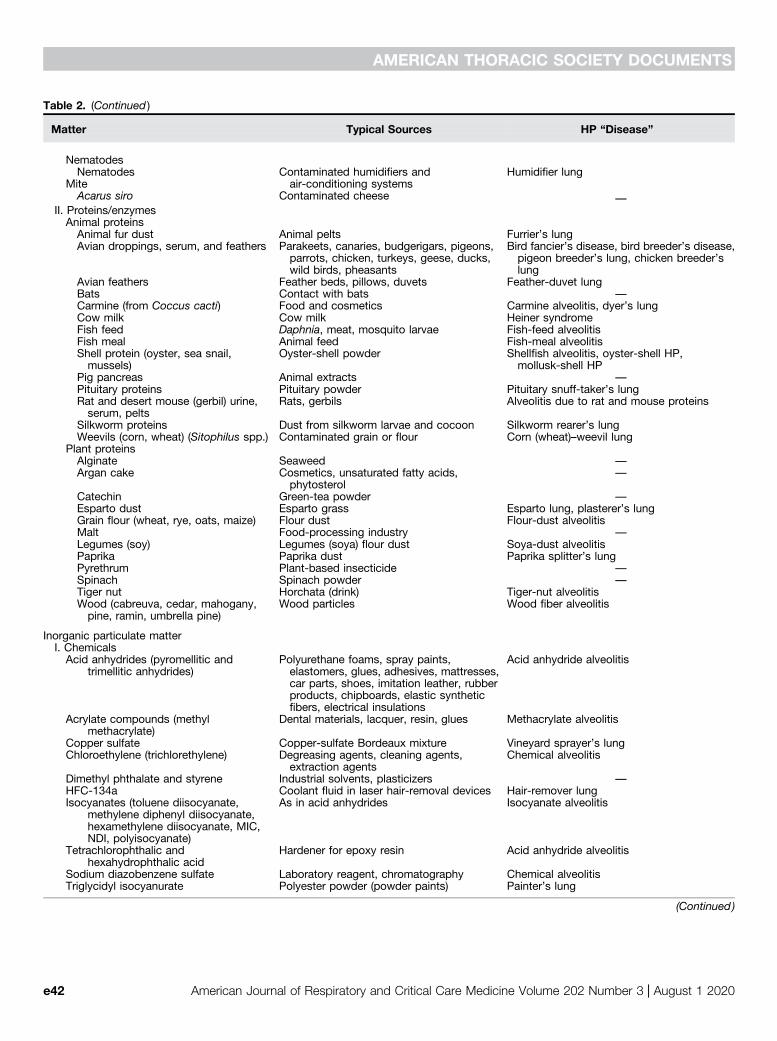
Table 2. (Continued )
Matter Typical Sources HP “Disease”
NematodesNematodes Contaminated humidifiers and
air-conditioning systemsHumidifier lung
MiteAcarus siro Contaminated cheese —
II. Proteins/enzymesAnimal proteinsAnimal fur dust Animal pelts Furrier’s lungAvian droppings, serum, and feathers Parakeets, canaries, budgerigars, pigeons,
parrots, chicken, turkeys, geese, ducks,wild birds, pheasants
Bird fancier’s disease, bird breeder’s disease,pigeon breeder’s lung, chicken breeder’slung
Avian feathers Feather beds, pillows, duvets Feather-duvet lungBats Contact with bats —Carmine (from Coccus cacti) Food and cosmetics Carmine alveolitis, dyer’s lungCow milk Cow milk Heiner syndromeFish feed Daphnia, meat, mosquito larvae Fish-feed alveolitisFish meal Animal feed Fish-meal alveolitisShell protein (oyster, sea snail,mussels)
Oyster-shell powder Shellfish alveolitis, oyster-shell HP,mollusk-shell HP
Pig pancreas Animal extracts —Pituitary proteins Pituitary powder Pituitary snuff-taker’s lungRat and desert mouse (gerbil) urine,serum, pelts
Rats, gerbils Alveolitis due to rat and mouse proteins
Silkworm proteins Dust from silkworm larvae and cocoon Silkworm rearer’s lungWeevils (corn, wheat) (Sitophilus spp.) Contaminated grain or flour Corn (wheat)–weevil lung
Plant proteinsAlginate Seaweed —Argan cake Cosmetics, unsaturated fatty acids,
phytosterol—
Catechin Green-tea powder —Esparto dust Esparto grass Esparto lung, plasterer’s lungGrain flour (wheat, rye, oats, maize) Flour dust Flour-dust alveolitisMalt Food-processing industry —Legumes (soy) Legumes (soya) flour dust Soya-dust alveolitisPaprika Paprika dust Paprika splitter’s lungPyrethrum Plant-based insecticide —Spinach Spinach powder —Tiger nut Horchata (drink) Tiger-nut alveolitisWood (cabreuva, cedar, mahogany,pine, ramin, umbrella pine)
Wood particles Wood fiber alveolitis
Inorganic particulate matterI. Chemicals
Acid anhydrides (pyromellitic andtrimellitic anhydrides)
Polyurethane foams, spray paints,elastomers, glues, adhesives, mattresses,car parts, shoes, imitation leather, rubberproducts, chipboards, elastic syntheticfibers, electrical insulations
Acid anhydride alveolitis
Acrylate compounds (methylmethacrylate)
Dental materials, lacquer, resin, glues Methacrylate alveolitis
Copper sulfate Copper-sulfate Bordeaux mixture Vineyard sprayer’s lungChloroethylene (trichlorethylene) Degreasing agents, cleaning agents,
extraction agentsChemical alveolitis
Dimethyl phthalate and styrene Industrial solvents, plasticizers —HFC-134a Coolant fluid in laser hair-removal devices Hair-remover lungIsocyanates (toluene diisocyanate,
methylene diphenyl diisocyanate,hexamethylene diisocyanate, MIC,NDI, polyisocyanate)
As in acid anhydrides Isocyanate alveolitis
Tetrachlorophthalic andhexahydrophthalic acid
Hardener for epoxy resin Acid anhydride alveolitis
Sodium diazobenzene sulfate Laboratory reagent, chromatography Chemical alveolitisTriglycidyl isocyanurate Polyester powder (powder paints) Painter’s lung
(Continued )
AMERICAN THORACIC SOCIETY DOCUMENTS
e42 American Journal of Respiratory and Critical Care Medicine Volume 202 Number 3 | August 1 2020
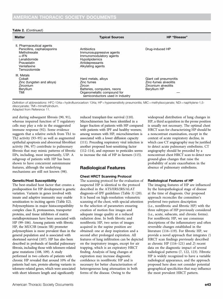
and during subsequent fibrosis (90, 91),whereas impaired function of T regulatorycells may play a role in the exaggeratedimmune response (92). Some evidencesuggests that a relative switch from Th1 toTh2 activity (93–95) as well as augmentedepithelial apoptosis and abnormal fibroblastactivity (96, 97) contribute to pulmonaryfibrosis that may mimic patterns of fibroticIIPs, including, most importantly, UIP. Asubgroup of patients with HP has beenshown to have concurrent autoimmunefeatures, although the underlyingmechanisms are still not known (98).
Genetic/Host SusceptibilityThe best-studied host factor that creates apredisposition for HP development is geneticvariation. Variants in genes involved withinnate and adaptive immunity may enablesensitization to inciting agents (Table E2).Polymorphisms in major histocompatibilitycomplex class II, proteasomes, transporterproteins, and tissue inhibitors of matrixmetalloproteinases have been associated withHP (99–106). Among patients with fibroticHP, the MUC5B (mucin 5B) promoterpolymorphism is more prevalent than in thegeneral population and is associated withshortened survival (107). HP has also beendescribed in probands of familial pulmonaryfibrosis, including those with telomere-relatedgene mutations (108, 109). A studyperformed in two cohorts of patients withchronic HP revealed that around 10% of thepatients had rare, protein-altering variants intelomere-related genes, which were associatedwith short telomere length and significantly
reduced transplant-free survival (110).Microchimerism has been identified in alarger fraction of patients with HP comparedwith patients with IPF and healthy women;among women with HP, microchimerism isassociated with a lower diffusion capacity(111). Preceding respiratory viral infection isanother proposed host-sensitizing factor(112–114), and exposure to pesticides seemsto increase the risk of HP in farmers (115).
Radiological Features
Chest HRCT Scanning ProtocolThe scanning protocol for the evaluation ofsuspected HP is identical to the protocoldescribed in the ATS/ERS/JRS/ALATdiagnosis-of-IPF guidelines (Table 3) (20).It is based on high-resolution volumetricscanning of the chest, with special attentionto the selection of parameters ensuringcreation of motion-free images andadequate image quality at a reducedradiation dose. In both fibrotic andnonfibrotic HP, two series of imagesacquired in the supine position areobtained: one at deep inspiration and asecond after prolonged expiration. Allfeatures of lung infiltration can be depictedon the inspiratory images, except for airtrapping, which is an expiratory HRCTfinding. Analysis of lung changes atexpiration may increase diagnosticconfidence in nonfibrotic HP and isnecessary for better characterization ofheterogeneous lung attenuation in bothforms of the disease. Owing to the
widespread distribution of lung changes inHP, a third acquisition in the prone positionis usually not necessary. The optimal chestHRCT scan for characterizing HP should bea noncontrast examination, except in thecontext of acute respiratory decline, inwhich case CT angiography may be justifiedto detect acute pulmonary embolisms. CTangiography should be preceded by anoncontrast chest HRCT scan to detect newground-glass changes that raise theprobability of acute exacerbation in theabsence of pulmonary embolisms.
Radiological Features of HPThe imaging features of HP are influencedby the histopathological stage of diseaseat the time of diagnosis. Our proposedapproach reconciles the committee’spreferred two-pattern description(i.e., nonfibrotic and fibrotic HP) with thethree subtypes of HP previously considered(i.e., acute, subacute, and chronic forms).For nonfibrotic HP, we use consensusdescriptions of the inflammatory and oftenreversible changes established in theliterature (116–119). For fibrotic HP, weprovide a novel approach that integrates 1)HRCT scan findings previously describedas chronic HP (116–121) and 2) recentdata on the diagnostic impact of severalradiological patterns (7, 122, 123). FibroticHP is widely recognized to have a variableradiological appearance, and the approachproposed here does not consider potentialgeographical specificities that may influencethe most prevalent HRCT pattern.
Table 2. (Continued )
Matter Typical Sources HP “Disease”
II. Pharmaceutical agentsPenicillins, cephalosporins Antibiotics Drug-induced HPMethotrexate Immunosuppressive agentsa-IFN Immunomodulatory agentsLenalidomide HypolipidemicsPravastatin AntidepressantsVenlafaxine Alkylating agentsTemozolomide
III. MetalsCobalt Hard metals, alloys Giant cell pneumonitisZinc (tungsten and alloys) Zinc fumes Zinc-fumes alveolitisZirconium Zircon Zirconium alveolitisBeryllium Batteries, computers, neons Beryllium HPTMI Organometallic compound for
semiconductors used in industry—
Definition of abbreviations: HFC-134a=hydrofluorocarbon 134a; HP=hypersensitivity pneumonitis; MIC=methylisocyanate; NDI = naphtylene-1,5-diisocyanate; TMI = trimethylindium.Adapted from Reference 11.
AMERICAN THORACIC SOCIETY DOCUMENTS
American Thoracic Society Documents e43
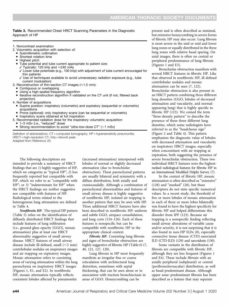
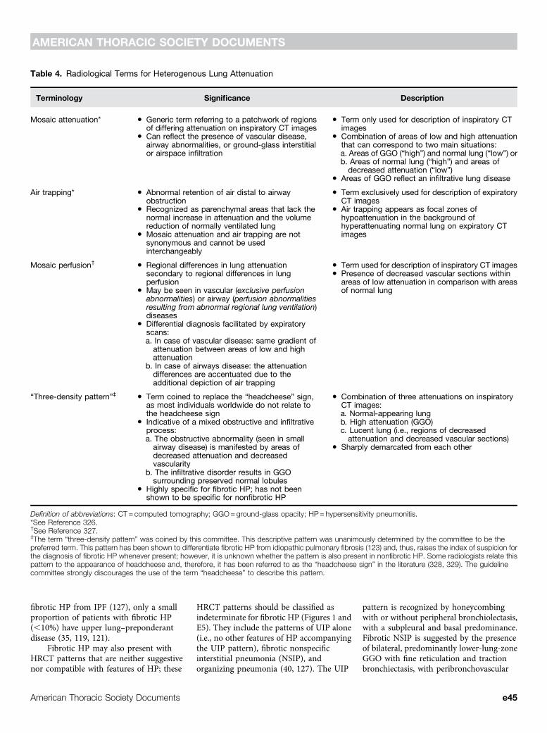
The following descriptions areintended to provide a summary of HRCTfindings that are 1) highly suggestive of HP,which we categorize as “typical HP”; 2) lessfrequently reported but compatible withHP, which we refer to as “compatible withHP”; or 3) “indeterminate for HP” whenthe HRCT findings are neither suggestivenor compatible with features of HP.Radiological terms related to theheterogenous lung attenuation are definedin Table 4.
Nonfibrotic HP. The typical HP pattern(Table 5) relies on the identification ofdiffusely distributed HRCT findings thatinclude features of lung infiltration(i.e., ground-glass opacity [GGO], mosaicattenuation) plus at least one HRCTabnormality suggestive of small airwaydisease. HRCT features of small airwaydisease include ill-defined, small (,5 mm)centrilobular nodules on inspiratory imagesand air trapping on expiratory images.Mosaic attenuation refers to coexistingareas of varying attenuation within the lungparenchyma on inspiratory HRCT images(Figures 1, E1, and E2). In nonfibroticHP, mosaic attenuation typically reflectscoexistent lobules affected by pneumonitis
(increased attenuation) interspersed withlobules of normal or slightly decreasedattenuation (due to bronchiolarobstruction). These parenchymal patternsare usually bilateral and symmetric with adiffuse distribution, both axially andcraniocaudally. Although a combination ofparenchymal abnormalities and features ofsmall airway disease is highly suggestiveof nonfibrotic HP, isolated air trapping isanother pattern that may be seen with HP.Three additional HRCT features have alsobeen described in nonfibrotic HP: uniformand subtle GGO, airspace consolidation,and lung cysts (124–126). Each of thesefeatures is nonspecific but can becompatible with nonfibrotic HP in theappropriate clinical context.
Fibrotic HP. Coexisting lung fibrosisand signs of bronchiolar obstruction arehighly suggestive of fibrotic HP (Table 6) (7,40, 121, 127, 128).
Lung fibrosis in HP most frequentlymanifests as irregular fine or coarsereticulation with architectural lungdistortion, sometimes with septalthickening, that can be seen alone or inassociation with traction bronchiectasis inareas of GGO. Honeycombing can be
present and is often described as minimal,but extensive honeycombing in severe formsof fibrotic HP may also occur. Lung fibrosisis most severe in the mid or mid and lowerlung zones or equally distributed in the threelung zones with relative basal sparing. Onaxial images, there is often no central orperipheral predominance of lung fibrosis(Figures 1 and E3).
Bronchiolar obstruction manifests withseveral HRCT features in fibrotic HP. Likethat observed in nonfibrotic HP, ill-definedcentrilobular nodules and mosaicattenuation can be seen (7, 122).Bronchiolar obstruction is also present inan HRCT pattern combining three differentlung densities (GGO, lobules of decreasedattenuation and vascularity, and normal-appearing lung) that is highly specific tofibrotic HP (123). We coined the term“three-density pattern” to describe thepresence of these three different lungdensities, which some radiologists havereferred to as the “headcheese sign”(Figure 2 and Table 4). This patternemphasizes the diagnostic value of lobuleswith decreased attenuation and vascularityon inspiratory HRCT images, especiallywhen concomitant with air trapping atexpiration, both suggesting the presence ofsevere bronchiolar obstruction. These twoindividual HRCT features were the highest-ranked radiological features for fibrotic HP inan International Modified Delphi Survey (7).
In the context of fibrotic HP, mosaicattenuation is often described as “extensive”(128) and “marked” (20), but thesedescriptors do not state specific numericalvalues. In a recent study, the threshold offive or more lobules of mosaic attenuationin each of three or more lobes bilaterallywas found to have the highest specificity forfibrotic HP and helped differentiate thisdisorder from IPF (123). Because airtrapping is a nonspecific finding reflectingsmall airway alterations of variable causeand/or severity, it is not surprising that it isalso found in non-HP ILDs (8), especiallyconnective tissue disease (CTD)-associatedILD (CTD-ILD) (129) and sarcoidosis (130).
Some variants in the distribution offibrosis are compatible with fibrotic HP,although they are less frequent (Figures 1and E4). These include fibrosis with anaxially peripheral (subpleural) or central(peribronchovascular) distribution, as wellas basal-predominant disease. Althoughupper zone–predominant fibrosis has beendescribed as a feature that may separate
Table 3. Recommended Chest HRCT Scanning Parameters in the DiagnosticApproach of HP
1. Noncontrast examination2. Volumetric acquisition with selection of:
d Submillimetric collimationd Shortest rotation timed Highest pitchd Tube potential and tube current appropriate to patient size:
✓ Typically: 120 kVp and <240 mAs✓ Lower tube potentials (e.g., 100 kVp) with adjustment of tube current encouraged forthin patients
✓ Use of techniques available to avoid unnecessary radiation exposure (e.g., tubecurrent modulation)
3. Reconstruction of thin-section CT images (<1.5 mm):d Contiguous or overlappingd Using a high-spatial-frequency algorithmd Iterative reconstruction algorithm if validated on the CT unit (if not, filtered back
projection)4. Number of acquisitions
d Supine position: inspiratory (volumetric) and expiratory (sequential or volumetric)acquisitions
d Prone (optional): only inspiratory scans (can be sequential or volumetric)d Inspiratory scans obtained at full inspiration
5. Recommended radiation dose for the inspiratory volumetric acquisition:d 1–3 mSv (i.e., “reduced” dose)d Strong recommendation to avoid “ultra–low-dose CT” (,1 mSv)
Definition of abbreviations: CT= computed tomography; HP=hypersensitivity pneumonitis;HRCT=high-resolution CT; kVp= kilovolt peak.Adapted from Reference 20.
AMERICAN THORACIC SOCIETY DOCUMENTS
e44 American Journal of Respiratory and Critical Care Medicine Volume 202 Number 3 | August 1 2020
fibrotic HP from IPF (127), only a smallproportion of patients with fibrotic HP(,10%) have upper lung–preponderantdisease (35, 119, 121).
Fibrotic HP may also present withHRCT patterns that are neither suggestivenor compatible with features of HP; these
HRCT patterns should be classified asindeterminate for fibrotic HP (Figures 1 andE5). They include the patterns of UIP alone(i.e., no other features of HP accompanyingthe UIP pattern), fibrotic nonspecificinterstitial pneumonia (NSIP), andorganizing pneumonia (40, 127). The UIP
pattern is recognized by honeycombingwith or without peripheral bronchiolectasis,with a subpleural and basal predominance.Fibrotic NSIP is suggested by the presenceof bilateral, predominantly lower-lung-zoneGGO with fine reticulation and tractionbronchiectasis, with peribronchovascular
Table 4. Radiological Terms for Heterogenous Lung Attenuation
Terminology Significance Description
Mosaic attenuation* d Generic term referring to a patchwork of regionsof differing attenuation on inspiratory CT images
d Term only used for description of inspiratory CTimages
d Can reflect the presence of vascular disease,airway abnormalities, or ground-glass interstitialor airspace infiltration
d Combination of areas of low and high attenuationthat can correspond to two main situations:a. Areas of GGO (“high”) and normal lung (“low”) orb. Areas of normal lung (“high”) and areas ofdecreased attenuation (“low”)
d Areas of GGO reflect an infiltrative lung disease
Air trapping* d Abnormal retention of air distal to airwayobstruction
d Term exclusively used for description of expiratoryCT images
d Recognized as parenchymal areas that lack thenormal increase in attenuation and the volumereduction of normally ventilated lung
d Air trapping appears as focal zones ofhypoattenuation in the background ofhyperattenuating normal lung on expiratory CTimagesd Mosaic attenuation and air trapping are not
synonymous and cannot be usedinterchangeably
Mosaic perfusion† d Regional differences in lung attenuationsecondary to regional differences in lungperfusion
d Term used for description of inspiratory CT images
d May be seen in vascular (exclusive perfusionabnormalities) or airway (perfusion abnormalitiesresulting from abnormal regional lung ventilation)diseases
d Presence of decreased vascular sections withinareas of low attenuation in comparison with areasof normal lung
d Differential diagnosis facilitated by expiratoryscans:a. In case of vascular disease: same gradient ofattenuation between areas of low and highattenuation
b. In case of airways disease: the attenuationdifferences are accentuated due to theadditional depiction of air trapping
“Three-density pattern”‡ d Term coined to replace the “headcheese” sign,as most individuals worldwide do not relate tothe headcheese sign
d Combination of three attenuations on inspiratoryCT images:a. Normal-appearing lung
d Indicative of a mixed obstructive and infiltrativeprocess:
b. High attenuation (GGO)
a. The obstructive abnormality (seen in smallairway disease) is manifested by areas ofdecreased attenuation and decreasedvascularity
c. Lucent lung (i.e., regions of decreasedattenuation and decreased vascular sections)
b. The infiltrative disorder results in GGOsurrounding preserved normal lobules
d Sharply demarcated from each other
d Highly specific for fibrotic HP; has not beenshown to be specific for nonfibrotic HP
Definition of abbreviations: CT= computed tomography; GGO=ground-glass opacity; HP=hypersensitivity pneumonitis.*See Reference 326.†See Reference 327.‡The term “three-density pattern” was coined by this committee. This descriptive pattern was unanimously determined by the committee to be thepreferred term. This pattern has been shown to differentiate fibrotic HP from idiopathic pulmonary fibrosis (123) and, thus, raises the index of suspicion forthe diagnosis of fibrotic HP whenever present; however, it is unknown whether the pattern is also present in nonfibrotic HP. Some radiologists relate thispattern to the appearance of headcheese and, therefore, it has been referred to as the “headcheese sign” in the literature (328, 329). The guidelinecommittee strongly discourages the use of the term “headcheese” to describe this pattern.
AMERICAN THORACIC SOCIETY DOCUMENTS
American Thoracic Society Documents e45
predominance in the axial distribution. Thepattern of organizing pneumonia relies onthe presence of consolidation in aperibronchovascular and/or peripheraldistribution, often seen with GGO andsometimes associated with a reverse halopattern. The presence of a reticularpattern superimposed on parenchymalconsolidation suggests an “organizingpneumonia–like” pattern of fibrotic HP. Asin other ILDs, HP may also present with atruly indeterminate HRCT pattern.
Combined pulmonary fibrosis andemphysema (82) and pleuroparenchymalfibroelastosis with emphysema (87) can alsooccur in HP (Figure E6), although they areinfrequent. Purely emphysematous forms ofHP can be seen independently of smokinghistory (Figure E7) (85, 116, 131, 132), andfibrotic HP may also be diagnosed at thetime of an acute exacerbation (Figure E8)(133).
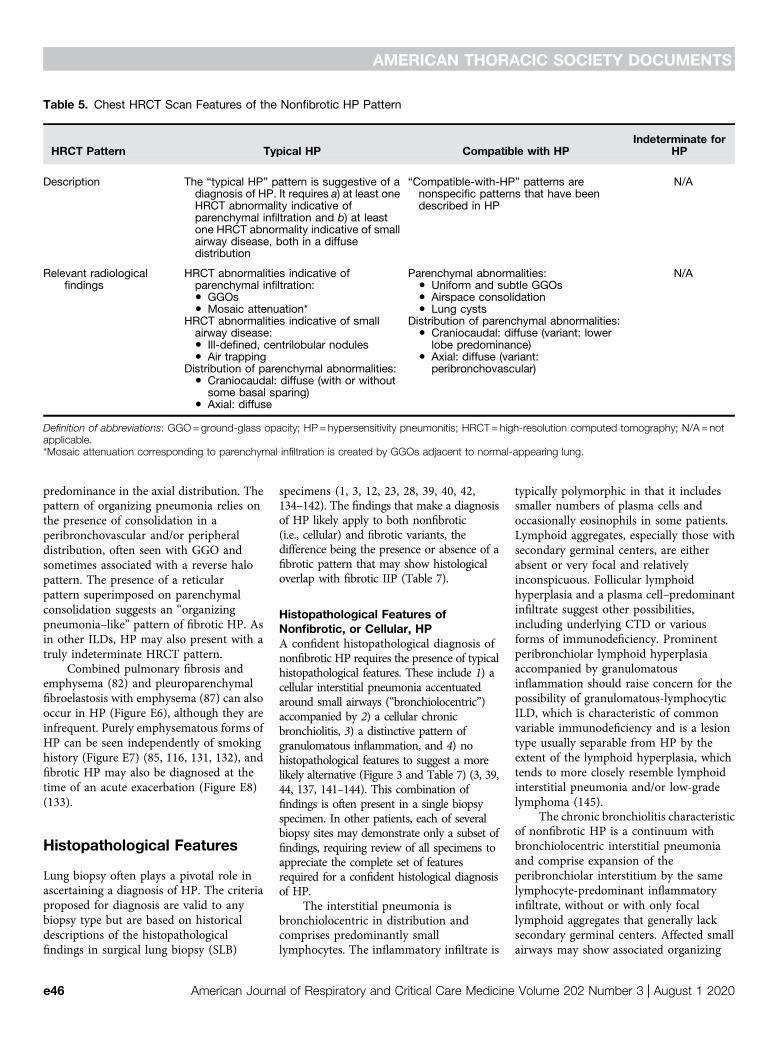
Histopathological Features
Lung biopsy often plays a pivotal role inascertaining a diagnosis of HP. The criteriaproposed for diagnosis are valid to anybiopsy type but are based on historicaldescriptions of the histopathologicalfindings in surgical lung biopsy (SLB)
specimens (1, 3, 12, 23, 28, 39, 40, 42,134–142). The findings that make a diagnosisof HP likely apply to both nonfibrotic(i.e., cellular) and fibrotic variants, thedifference being the presence or absence of afibrotic pattern that may show histologicaloverlap with fibrotic IIP (Table 7).
Histopathological Features ofNonfibrotic, or Cellular, HPA confident histopathological diagnosis ofnonfibrotic HP requires the presence of typicalhistopathological features. These include 1) acellular interstitial pneumonia accentuatedaround small airways (“bronchiolocentric”)accompanied by 2) a cellular chronicbronchiolitis, 3) a distinctive pattern ofgranulomatous inflammation, and 4) nohistopathological features to suggest a morelikely alternative (Figure 3 and Table 7) (3, 39,44, 137, 141–144). This combination offindings is often present in a single biopsyspecimen. In other patients, each of severalbiopsy sites may demonstrate only a subset offindings, requiring review of all specimens toappreciate the complete set of featuresrequired for a confident histological diagnosisof HP.
The interstitial pneumonia isbronchiolocentric in distribution andcomprises predominantly smalllymphocytes. The inflammatory infiltrate is
typically polymorphic in that it includessmaller numbers of plasma cells andoccasionally eosinophils in some patients.Lymphoid aggregates, especially those withsecondary germinal centers, are eitherabsent or very focal and relativelyinconspicuous. Follicular lymphoidhyperplasia and a plasma cell–predominantinfiltrate suggest other possibilities,including underlying CTD or variousforms of immunodeficiency. Prominentperibronchiolar lymphoid hyperplasiaaccompanied by granulomatousinflammation should raise concern for thepossibility of granulomatous-lymphocyticILD, which is characteristic of commonvariable immunodeficiency and is a lesiontype usually separable from HP by theextent of the lymphoid hyperplasia, whichtends to more closely resemble lymphoidinterstitial pneumonia and/or low-gradelymphoma (145).
The chronic bronchiolitis characteristicof nonfibrotic HP is a continuum withbronchiolocentric interstitial pneumoniaand comprise expansion of theperibronchiolar interstitium by the samelymphocyte-predominant inflammatoryinfiltrate, without or with only focallymphoid aggregates that generally lacksecondary germinal centers. Affected smallairways may show associated organizing
Table 5. Chest HRCT Scan Features of the Nonfibrotic HP Pattern
HRCT Pattern Typical HP Compatible with HPIndeterminate for
HP
Description The “typical HP” pattern is suggestive of adiagnosis of HP. It requires a) at least oneHRCT abnormality indicative ofparenchymal infiltration and b) at leastone HRCT abnormality indicative of smallairway disease, both in a diffusedistribution
“Compatible-with-HP” patterns arenonspecific patterns that have beendescribed in HP
N/A
Relevant radiologicalfindings
HRCT abnormalities indicative ofparenchymal infiltration:
Parenchymal abnormalities: N/A
d GGOsd Uniform and subtle GGOs
d Mosaic attenuation*d Airspace consolidationd Lung cysts
HRCT abnormalities indicative of smallairway disease:d Ill-defined, centrilobular nodules
Distribution of parenchymal abnormalities:
d Air trapping
d Craniocaudal: diffuse (variant: lowerlobe predominance)
d Axial: diffuse (variant:peribronchovascular)Distribution of parenchymal abnormalities:
d Craniocaudal: diffuse (with or withoutsome basal sparing)
d Axial: diffuse
Definition of abbreviations: GGO=ground-glass opacity; HP=hypersensitivity pneumonitis; HRCT=high-resolution computed tomography; N/A=notapplicable.*Mosaic attenuation corresponding to parenchymal infiltration is created by GGOs adjacent to normal-appearing lung.
AMERICAN THORACIC SOCIETY DOCUMENTS
e46 American Journal of Respiratory and Critical Care Medicine Volume 202 Number 3 | August 1 2020
pneumonia that is exquisitelybronchiolocentric. Foamy alveolarmacrophages may be conspicuous inperibronchiolar air spaces and are a form ofmicroscopic obstructive pneumonia thatreflects small airway dysfunction.
Granulomatous inflammation completesthe triad that allows a confident diagnosis ofHP on the basis of histology alone, but thediagnostic value is heavily dependent on thequalitative features of the granulomas(Figure 4) (44, 142, 144). The granulomas ofHP are typically small and poorly formed,comprising loose, poorly circumscribedclusters of epithelioid and multinucleated
cells (macrophages) that tend to be mostprevalent in the peribronchiolar interstitium.Isolated multinucleated giant cells arecommon and often show nonspecificcytoplasmic inclusions such as Schaumannbodies, asteroid bodies, or cholesterol-likeclefts. The poorly formed granulomas andmultinucleated giant cells spill intoperibronchiolar air spaces, where they maybe intimately associated with organizingpneumonia but should also involve theperibronchiolar interstitium (144). Well-formed granulomas resembling those seen insarcoidosis and granulomatous infections areuncommon and should raise the likelihood
of other conditions if they predominate (134,135). Aspiration is another importantconsideration that is characterized by well-formed intraluminal granulomas, often withsmall foci of central necrosis and associatedneutrophils. The granulomas are oftenaffiliated with aspirated foreign material,including a combination of organic and/ornonorganic particulates such as excipientsused in oral medications (146). This is true of“hot-tub lung,” a diffuse lung disease (DLD)associated with Mycobacterium aviumcomplex with clinical and radiologicalfindings that overlap with classical types ofHP, in which well-formed granulomas withor without central necrosis tend to be limitedto the lumens of distal bronchioles (FigureE9) (147).
Probable HP refers to cases in whichonly some of the features described aboveare present. It requires the presence ofboth a lymphocyte-rich, bronchiolocentricinterstitial pneumonia and an associatedbronchiolitis, but without thegranulomatous inflammation characteristicof classical HP. Indeterminate HPrefers to cases in which either a cellularbronchiolocentric interstitial pneumonia oran otherwise unexplained cellular chronicbronchiolitis is present, but without thecharacteristic granulomatous inflammation.The chronic bronchiolitis may includeperibronchiolar metaplasia (PBM),characterized by expansion of theperibronchiolar interstitium by mild,nondistorting fibrosis that extends intocontiguous alveolar septa in which liningpneumocytes have been replaced by acolumnar bronchiolar epithelium, withoutany of the other features to suggest fibroticHP, as discussed below. Foci of organizingpneumonia may also be present in thesecategories.
Histopathological Features of FibroticHPFibrotic HP differs from nonfibrotic HPin that the underlying chronic interstitialpneumonia and/or bronchiolitis iscomplicated by fibrosis. Typicalhistopathological features of fibrotic HPinclude subpleural and centriacinar fibrosis,with or without bridging fibrosis that spansboth subpleural and centriacinar regions, orwith neighboring centriacinar fibroticlesions (Figure 5) (23, 137, 140). Thepattern of fibrotic interstitial pneumoniamay include features that overlap with aUIP pattern, including patchy collagen
A B C
D E F
G H I
Figure 1. “Typical hypersensitivity pneumonitis (HP)” and “compatible-with-HP” high-resolutioncomputed tomography patterns. The nonfibrotic typical HP pattern is characterized by (A)centrilobular nodules, (B) mosaic attenuation on an inspiratory scan, and (C) air trapping on anexpiratory scan. (D) The nonfibrotic compatible-with-HP pattern is exemplified by uniform and subtleground-glass opacity and cysts. The fibrotic typical HP pattern consists of (E) coarse reticulation andminimal honeycombing in a random axial distribution with no zonal predominance in association with(F) small airway disease. The fibrotic compatible-with-HP pattern varies in the patterns and/ordistribution of lung fibrosis (e.g., basal and subpleural predominance, [G] upper-lung-zonepredominance, [H] central [or peribronchovascular] predominance [arrows], or [I] fibrotic ground-glassattenuation seen alone or in association with small airway disease). The fibrotic indeterminate-for-HPpattern includes the usual interstitial pneumonia pattern, nonspecific interstitial pneumonia pattern,organizing pneumonia–like pattern, or truly indeterminate findings.
AMERICAN THORACIC SOCIETY DOCUMENTS
American Thoracic Society Documents e47
fibrosis, fibroblast foci, and associatedsubpleural-dominant honeycombing (3, 12,39, 40, 42, 142, 143, 148, 149). Some haveapplied the term “UIP-like” to drawattention to the histological overlap with aUIP pattern, which frequently posesproblems in the differential diagnosis (3, 23,28, 39). Given the potentially confusingnature of the term “UIP-like” we havechosen not to apply it in this manuscript,although we acknowledge the histologicaloverlaps and highlight those histologicalfeatures helpful in distinguishing fibroticHP from other diffuse fibrotic lungdiseases. In others, the interstitialpneumonia may have a more uniform anddiffuse distribution without honeycombchange and may more closely resemble afibrotic NSIP pattern (“NSIP-like”).Bronchiolar fibrosis typically takes the formof PBM with fibrosis, a finding that showssignificant histological overlap with
descriptions of interstitial airway-centeredfibrosis (150, 151). Neither PBM norairway-centered fibrosis is unique to HP,and they therefore do not by themselvesestablish the diagnosis (140, 152), but theyare characteristic and tend to be moreprofuse in patients with fibrotic HPcompared with patients with fibrotic IIPs(140).
Distinguishing fibrotic HP fromfibrotic IIPs requires identification ofcentriacinar fibrotic lesions and thefeatures described in nonfibrotic HP. Thelatter features are usually observed in lessfibrotic lung tissue. This often requiressampling of more than one site. One sitemay show findings indistinguishable froma fibrotic interstitial pneumonia, whereasanother may show features typical ofnonfibrotic HP, including those that mightbe more appropriately characterized as“probable” or “indeterminate” (143). This
sort of diagnostic discordance betweensites is analogous to the histopathologicalvariability documented in patients withIPF, in whom NSIP-like changes arecommon and may be the sole finding insome samples (153). In other patients,much of a single-site biopsy specimenmay mimic a fibrotic IIP, whereas theevidence in support of HP is patchy andoften limited to less fibrotic lung tissue.Centriacinar fibrotic lesions, in additionto the subpleural-dominant fibroticlesions with or without honeycombing,prominent PBM, and/or isolatedperibronchiolar giant cells, often withconspicuous Schaumann bodies, may bethe clues to search more diligently for thefeatures that would make a diagnosis ofHP more likely. It is important todocument the fibrotic component whendiagnosing HP, as this is an adverseprognostic factor.
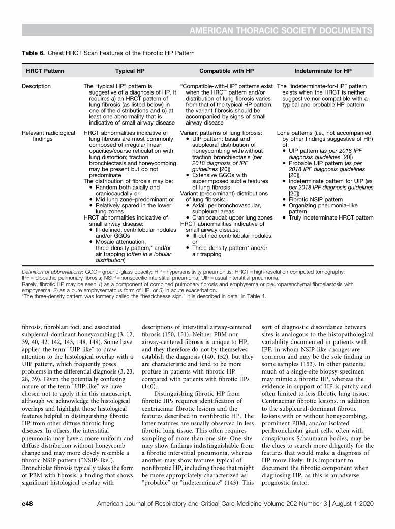
Table 6. Chest HRCT Scan Features of the Fibrotic HP Pattern
HRCT Pattern Typical HP Compatible with HP Indeterminate for HP
Description The “typical HP” pattern issuggestive of a diagnosis of HP. Itrequires a) an HRCT pattern oflung fibrosis (as listed below) inone of the distributions and b) atleast one abnormality that isindicative of small airway disease
“Compatible-with-HP” patterns existwhen the HRCT pattern and/ordistribution of lung fibrosis variesfrom that of the typical HP pattern;the variant fibrosis should beaccompanied by signs of smallairway disease
The “indeterminate-for-HP” patternexists when the HRCT is neithersuggestive nor compatible with atypical and probable HP pattern
Relevant radiologicalfindings
HRCT abnormalities indicative oflung fibrosis are most commonlycomposed of irregular linearopacities/coarse reticulation withlung distortion; tractionbronchiectasis and honeycombingmay be present but do notpredominate
Variant patterns of lung fibrosis: Lone patterns (i.e., not accompaniedby other findings suggestive of HP)of:
d UIP pattern: basal andsubpleural distribution ofhoneycombing with/withouttraction bronchiectasis (per2018 diagnosis of IPFguidelines [20])
d UIP pattern (as per 2018 IPFdiagnosis guidelines [20])
The distribution of fibrosis may be:d Random both axially and
craniocaudally or
d Extensive GGOs withsuperimposed subtle featuresof lung fibrosis
d Probable UIP pattern (as per2018 IPF diagnosis guidelines[20])
d Mid lung zone–predominant or
d Indeterminate pattern for UIP (asper 2018 IPF diagnosis guidelines[20])
d Relatively spared in the lowerlung zones
Variant (predominant) distributionsof lung fibrosis: d Fibrotic NSIP patternd Axial: peribronchovascular,
subpleural areasd Organizing pneumonia–like
patternHRCT abnormalities indicative ofsmall airway disease:
d Craniocaudal: upper lung zones d Truly indeterminate HRCT pattern
d Ill-defined, centrilobular nodulesand/or GGOs
d Mosaic attenuation,three-density pattern,* and/orair trapping (often in a lobulardistribution)
HRCT abnormalities indicative ofsmall airway disease:d Ill-defined centrilobular nodules,
ord Three-density pattern* and/or
air trapping
Definition of abbreviations: GGO=ground-glass opacity; HP=hypersensitivity pneumonitis; HRCT=high-resolution computed tomography;IPF= idiopathic pulmonary fibrosis; NSIP=nonspecific interstitial pneumonia; UIP= usual interstitial pneumonia.Rarely, fibrotic HP may be seen 1) as a component of combined pulmonary fibrosis and emphysema or pleuroparenchymal fibroelastosis withemphysema, 2) as a pure emphysematous form of HP, or 3) in acute exacerbation.*The three-density pattern was formerly called the “headcheese sign.” It is described in detail in Table 4.
AMERICAN THORACIC SOCIETY DOCUMENTS
e48 American Journal of Respiratory and Critical Care Medicine Volume 202 Number 3 | August 1 2020

Diagnostic Criteria
The diagnosis of HP requires integration ofmultiple domains that are ideally consideredin the context of an MDD. Given themultitude of presenting features, fibrotic HPshould be considered in the differentialdiagnosis for all patients with a fibrotic ILD.This is particularly challenging, given theabsence of an identifiable exposure in up to50% of patients with fibrotic HP (87, 122,131–133). Nonfibrotic HP is usuallyassociated with a clear exposure and lessfrequently poses a diagnostic dilemma, butit similarly lacks a single diagnosticpathway. For these reasons, acomprehensive multidisciplinary approachis important in diagnosing HP, particularlyfibrotic HP; however, there remainssubstantial diagnostic disagreement acrossexperienced MDD teams that likely reflectsthe absence of standardized diagnosticcriteria (17).
Previous studies have identifiedfeatures that increase the likelihood of HP,with diagnostic algorithms or criteriaproposed by multiple groups (1, 5–10). Thestudies on which these proposals are basedall have methodological limitations, mostnotably incorporation bias (e.g., serum IgGand BAL studies), incompleteconsideration of all potentially informativefeatures, absence of appropriate controlgroups, and inadequate validation(e.g., questionnaires). Despite theselimitations, some key features areconsistently identified as increasing thelikelihood of an HP diagnosis, including
exposure to a known offending agent (1, 7,8), typical imaging findings (7, 8, 122,154), and typical biopsy findings (7). BALlymphocytosis is an important feature (1,7); serum-specific immunoglobulins mightalso be helpful (1, 155, 156). Female sex,midinspiratory squeaks (or chirping ralesor squawks) (157, 158), absence of asmoking history, and obstructive or mixedrestrictive/obstructive physiology have alsobeen identified as potential predictors ofan HP diagnosis, but with more limiteddiagnostic utility. Other features are lessfrequently identified (e.g., episodes ofsymptoms and symptoms 4–8 h afterexposure) (12), likely reflecting variableproportions of fibrotic and nonfibrotic HPin previous studies.
Although the diagnosis of HP ispredominantly based on exposureidentification, chest HRCT scan pattern, andbronchoscopic/histopathological findings, amajor challenge is that no individual featureis sufficient in isolation, nor are anymandatory. This results in the potential formultiple combinations of abnormalitiesthat can result in a diagnosis of HP.Although a single diagnostic algorithmmay be applied to both fibrotic andnonfibrotic HP, these populations havefrequent differences in their underlyingfeatures. For example, patients withnonfibrotic HP more often have an acuteand identifiable exposure, rapid onset ofboth pulmonary and systemic symptoms,presence of centrilobular nodularity onchest CT scans, and lymphocytosis on BALcellular analysis (3, 25, 105–107).
Conversely, patients with fibrotic HP areless likely to have an identified exposureand more frequently have an insidious andchronic onset of isolated pulmonarysymptoms, fibrotic changes with orwithout more specific features of HP onchest imaging, and a nonspecificdifferential cell profile on BAL analysis (4,105–107). Additional features may beuseful in the context of an MDD toincrease or decrease the diagnosticconfidence of HP on a case-by-case basis,but these are not sufficiently sensitive orspecific to justify inclusion in formaldiagnostic criteria.
There is often substantial uncertaintyin the diagnosis of HP. This occurs mostfrequently in the distinction betweenfibrotic HP and IPF (12), reflecting theoverlapping features and lack of a single,definitive gold-standard test for bothdiagnoses. The diagnostic criteria for HPprovided in this guideline emphasize theimportance of three primary domains: 1)exposure identification (e.g., clinicalhistory with or without a questionnaire,serum IgG testing against potentialantigens associated with HP, and/orspecific inhalational challenge), 2) imagingpattern, and 3) BALlymphocytosis/histopathological findings,with each described in detail in thecorresponding sections of this document.Although the specific features that satisfyeach domain are different for fibrotic andnonfibrotic HP, a single approach is usedfor all patients who have a clinicalpresentation consistent with HP.
The diagnostic criteria are presentedin a way that explicitly conveys thediagnostic confidence associated withcommon combinations of specific features.We used an approach similar to theapproach proposed by an internationalworking group, which categorized ILDdiagnoses on the basis of confidence (159).We categorized diagnoses as definite(>90% confidence), high-confidence(80–89%), moderate-confidence (70–79%),and low-confidence (51–69%) diagnoses.This approach is supported by recentstudies suggesting the potential therapeuticand prognostic utility of assigningdiagnostic confidence in this manner(160, 161).
Criteria and an algorithm for establishinga diagnosis of HP are provided in Figures 6and 7, which may be applied to patientswith a clinical presentation consistent with
A B
Figure 2. Three-density pattern. High-resolution computed tomography (A) inspiratory and (B)expiratory images from a patient with hypersensitivity pneumonitis demonstrating the three differentdensities: high attenuation (ground-glass opacity) (red stars), lucent lung (regions of decreasedattenuation and decreased vascular sections) (red arrows), and normal lung (black arrows), which aresharply demarcated from each other.
AMERICAN THORACIC SOCIETY DOCUMENTS
American Thoracic Society Documents e49
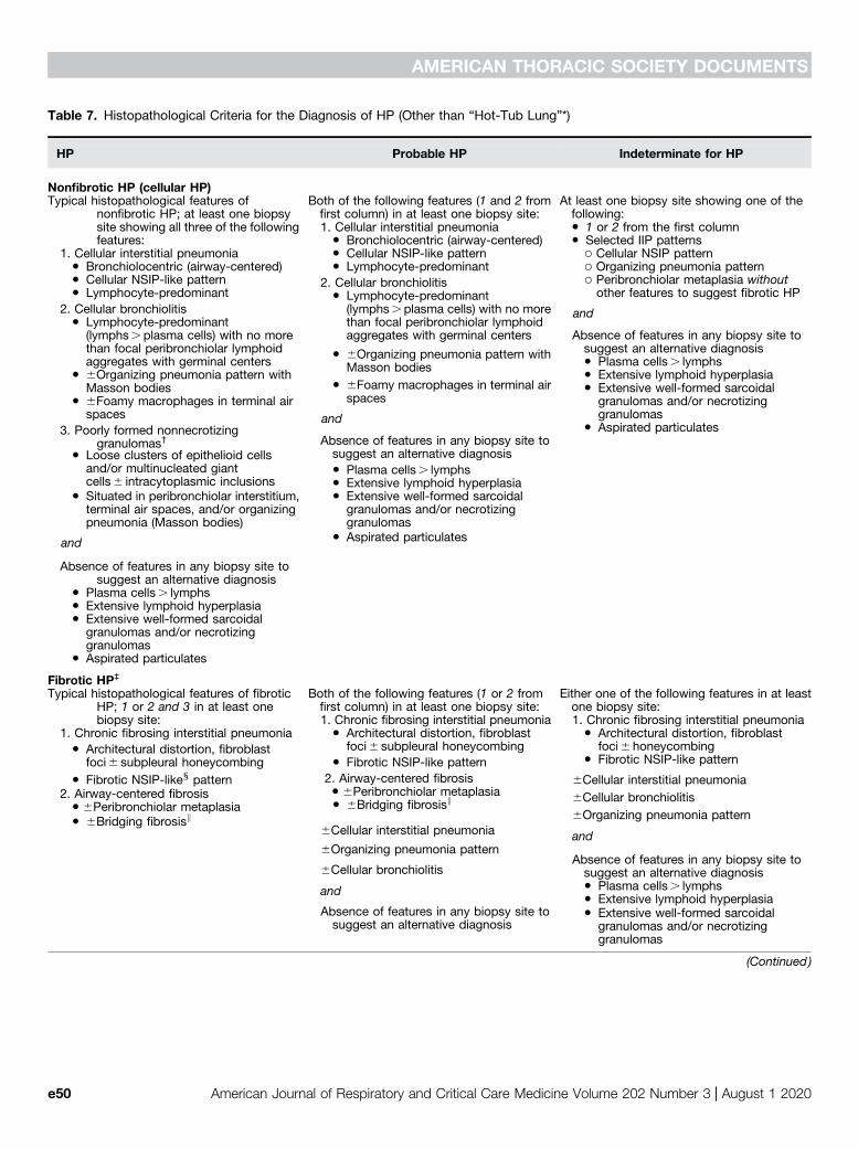
Table 7. Histopathological Criteria for the Diagnosis of HP (Other than “Hot-Tub Lung”*)
HP Probable HP Indeterminate for HP
Nonfibrotic HP (cellular HP)Typical histopathological features of
nonfibrotic HP; at least one biopsysite showing all three of the followingfeatures:
Both of the following features (1 and 2 fromfirst column) in at least one biopsy site:
At least one biopsy site showing one of thefollowing:
1. Cellular interstitial pneumonia
1. Cellular interstitial pneumonia d 1 or 2 from the first column
d Bronchiolocentric (airway-centered)
d Bronchiolocentric (airway-centered) d Selected IIP patterns
d Cellular NSIP-like pattern
d Cellular NSIP-like pattern ○ Cellular NSIP pattern
d Lymphocyte-predominant
d Lymphocyte-predominant ○ Organizing pneumonia pattern○ Peribronchiolar metaplasia withoutother features to suggest fibrotic HP
and2. Cellular bronchiolitis
2. Cellular bronchiolitis
Absence of features in any biopsy site tosuggest an alternative diagnosis
d Lymphocyte-predominant(lymphs.plasma cells) with no morethan focal peribronchiolar lymphoidaggregates with germinal centers
d Lymphocyte-predominant(lymphs.plasma cells) with no morethan focal peribronchiolar lymphoidaggregates with germinal centers
d Plasma cells. lymphsd 6Organizing pneumonia pattern with
Masson bodies
d 6Organizing pneumonia pattern withMasson bodies
d Extensive lymphoid hyperplasia
d 6Foamy macrophages in terminal airspaces
d 6Foamy macrophages in terminal airspaces
and
d Extensive well-formed sarcoidalgranulomas and/or necrotizinggranulomas
Absence of features in any biopsy site tosuggest an alternative diagnosis
d Aspirated particulates3. Poorly formed nonnecrotizinggranulomas†
d Plasma cells. lymphsd Loose clusters of epithelioid cells
and/or multinucleated giantcells6 intracytoplasmic inclusions d Extensive lymphoid hyperplasia
d Situated in peribronchiolar interstitium,terminal air spaces, and/or organizingpneumonia (Masson bodies)
and
d Extensive well-formed sarcoidalgranulomas and/or necrotizinggranulomas
Absence of features in any biopsy site tosuggest an alternative diagnosis
d Aspirated particulates
d Plasma cells. lymphsd Extensive lymphoid hyperplasiad Extensive well-formed sarcoidal
granulomas and/or necrotizinggranulomas
d Aspirated particulates
Fibrotic HP‡
Typical histopathological features of fibroticHP; 1 or 2 and 3 in at least onebiopsy site:
Both of the following features (1 or 2 fromfirst column) in at least one biopsy site:
Either one of the following features in at leastone biopsy site:
1. Chronic fibrosing interstitial pneumonia 1. Chronic fibrosing interstitial pneumonia1. Chronic fibrosing interstitial pneumonia d Architectural distortion, fibroblast
foci6 subpleural honeycombingd Architectural distortion, fibroblast
foci6honeycombingd Fibrotic NSIP-like pattern
d Architectural distortion, fibroblastfoci6 subpleural honeycombing d Fibrotic NSIP-like pattern
6Cellular interstitial pneumoniad Fibrotic NSIP-likex pattern 2. Airway-centered fibrosisd 6Peribronchiolar metaplasia 6Cellular bronchiolitisd 6Bridging fibrosisk
6Organizing pneumonia pattern
and6Cellular interstitial pneumonia
Absence of features in any biopsy site tosuggest an alternative diagnosis
2. Airway-centered fibrosisd 6Peribronchiolar metaplasia
6Cellular bronchiolitisd Plasma cells. lymphs
d 6Bridging fibrosisk
6Organizing pneumonia pattern
andd Extensive lymphoid hyperplasia
Absence of features in any biopsy site tosuggest an alternative diagnosis
d Extensive well-formed sarcoidalgranulomas and/or necrotizinggranulomas
(Continued )
AMERICAN THORACIC SOCIETY DOCUMENTS
e50 American Journal of Respiratory and Critical Care Medicine Volume 202 Number 3 | August 1 2020
either fibrotic or nonfibrotic HP. Bothwere developed through iterative discussionand consensus by the full guidelinecommittee on the basis of the evidencesyntheses and recommendations presentedbelow, supplemented by the guidelinecommittee’s collective clinical experience.
The primary goal in the diagnosis ofILD is to make a confident diagnosisusing the least invasive approach. HP can bediagnosed with high confidence in patientsin whom an exposure has been identifiedand who have a typical HP pattern at HRCTand have BAL lymphocytosis; such patientsdo not require additional testing. Patientswith any other combination of exposurehistory, HRCT pattern, and BAL resultsshould undergo an MDD that includes anexperienced expert in ILD (pulmonologist),a chest radiologist, and, if transbronchiallung biopsies were performed at the time ofBAL, a pathologist familiar withhistopathological features of interstitialpneumonias and HP. Additionalhistopathological sampling should beconsidered after the MDD in some patientswith a high-confidence diagnosis, moderate-confidence diagnosis, or low-confidencediagnosis or in patients for whom analternative diagnosis has not been established(161). A low-confidence diagnosis may be
adequate in patients for whom thedifferential diagnosis has been sufficientlynarrowed such that further investigations areunlikely to alter management, when invasivetesting has unacceptable risks, or when suchtests are declined by the patient. Thediagnosis should be reconsideredat subsequent visits, particularly forpatients without a definite diagnosis.
Diagnostic Interventions
Question 1: Should patients withnewly detected ILD on chestradiographs or a CT scan of the chest,with or without an overt history ofexposures capable of causing ILD inthe patient’s environment at home,work, or elsewhere, be subjected toformal questioning using aquestionnaire to raise the possibilitythat a) potential inciting agents of HPare the etiology of the ILD and b) thediagnosis of the ILD is HP?
Summary of evidence. A systematic search ofthe literature identified 1,141 potentiallyrelevant articles. The full text of 32 articleswas reviewed, and 2 observational studieswere selected to inform the guidelinecommittee (12, 162). One study enrolled
19 patients with definite or probable HP andused clinical history, a 22-itemquestionnaire, and serum IgG testing againstHP-associated antigens to identify potentialinciting agents of HP. The environments ofpatients with positive findings were sampled,and potential inciting agents were confirmedor excluded (162). Another study enrolled 46patients with IPF and used a nine-itemquestionnaire, serum IgG testing againstHP-associated antigens, and bronchial-challenge testing to identify potentialinciting agents of HP. BAL was performed,and histopathological specimens wererevisited to confirm IPF or reclassify thecondition as chronic HP (12). Neitherquestionnaire had been evaluated inprevious studies.
In the former study, the questionnaireidentified a potential inciting agent in 19 outof 19 (100%) patients; only 7 patientsunderwent subsequent environmentaltesting, with the potential inciting agentconfirmed in 5 out of the 7 (71%) (162).In the latter study, the questionnaireidentified a potential inciting agent in27 out of 46 (59%) patients; the finaldiagnosis was reclassifed from IPF tochronic HP in 18 out of 27 (67%) (12). Aquestionnaire was more likely to identify apotential inciting agent when compared
Table 7. (Continued )
HP Probable HP Indeterminate for HP
3. Poorly formed nonnecrotizinggranulomas†
d Plasma cells. lymphs d Aspirated particulates
6Cellular interstitial pneumonia
d Extensive lymphoid hyperplasia
6Cellular bronchiolitis
d Extensive well-formed sarcoidalgranulomas and/or necrotizinggranulomas
6Organizing pneumonia pattern
and
d Aspirated particulates
Absence of features in any biopsy site tosuggest an alternative diagnosis
d Plasma cells. lymphsd Extensive lymphoid hyperplasiad Extensive well-formed sarcoidal
granulomas and/or necrotizinggranulomas
d Aspirated particulates
Definition of abbreviations: HP=hypersensitivity pneumonitis; IIP = idiopathic interstitial pneumonias; lymphs= lymphocytes; NSIP=nonspecific interstitialpneumonia; UIP= usual interstitial pneumonia.*Histological findings in hot-tub lung are distinctly different from nonfibrotic and fibrotic forms of classic HP.†Granulomas in HP are smaller, less tightly clustered, and lack the perigranulomatous hyaline fibrosis commonly seen in sarcoidosis.‡Fibrotic HP may show classic features of nonfibrotic HP (cellular HP) in less fibrotic or nonfibrotic areas; if present, this combination of findings is ahistological clue to the diagnosis of HP.xUpdates to the classification of IIPs by Travis and colleagues (330) and diagnostic guidelines for idiopathic pulmonary fibrosis (20, 128) tightly link a UIPpattern with idiopathic pulmonary fibrosis and an NSIP pattern with idiopathic NSIP.kBridging fibrosis spans subpleural and centriacinar or neighboring centriacinar fibrotic foci.
AMERICAN THORACIC SOCIETY DOCUMENTS
American Thoracic Society Documents e51

with clinical history (relative risk [RR],3.80; 95% confidence interval [CI],1.79–8.06) or serum IgG testing (RR, 1.58;95% CI, 1.12–2.23), but there was nodifference when a questionnaire wascompared with the combination of serumIgG testing against potential antigensassociated with HP plus bronchial-challenge testing (RR, 0.90; 95% CI,0.65–1.24).
Committee discussion. The guidelinecommittee appreciated the potential of aquestionnaire to facilitate the systematic
identification of inciting agents of HP in apatient’s environment, compared withclinical history-taking alone, which may beless systematic. However, there is noquestionnaire that has been validated forthis purpose, and it seems likely thatdifferent questionnaires are appropriatefor different locations and populations(163). Thus, the guideline committeeconcluded that it is premature torecommend using a specific questionnairebut that use of a questionnaire may be animportant adjunct to the care of patients
with newly detected ILD in the future. Inthe interim, the committee encouragesclinicians to take a thorough history toidentify potential exposures and todevelop and use their own questionnairesto ensure routine inquiry about potentialexposures whenever they evaluate apatient with newly detected ILD. Suchquestionnaires should include ambientand occupational causes of HP relevant tothe patient’s geographical location andcultural habits.
Recommendation. For patients withnewly identified ILD whose differentialdiagnosis includes nonfibrotic HP orfibrotic HP, the guideline committeemakes no recommendation or suggestionfor or against the use of a specificquestionnaire to identify potentialinciting agents of HP; instead, theguideline committee recommends thedevelopment and validation of aquestionnaire. Voting results: unanimous,no recommendation or suggestion.Remark: Pending the availability of avalidated questionnaire, the guidelinecommittee advocates that clinicians take athorough history to identify potentialexposures and sources in the patient’senvironment that are known to beassociated with HP.
Question 2: Should patients withnewly detected ILD on chestradiographs or a CT scan of the chest,with or without an overt history ofexposures capable of causing ILD inthe patient’s environment at home,work, or elsewhere, undergo serumtesting for IgG antibodies againstspecific antigens to raise thepossibility that a) potential incitingagents of HP are the etiology of theILD and b) the diagnosis of the ILD isHP?
Summary of evidence. A systematic searchof the literature identified 926 potentiallyrelevant articles. The full text of 49 articleswas reviewed, and 15 observational studieswere selected to inform the guidelinecommittee (1, 12, 155, 164–175). Moststudies enrolled patients with known HP,usually farmer’s lung disease or birdfancier’s disease, and determined thesensitivity and specificity of serum IgGtesting against potential antigens associated
A B
C D
E
Figure 3. Surgical lung biopsy specimen from a patient with nonfibrotic hypersensitivity pneumonitis(HP). (A) Low-magnification photomicrograph showing preservation of lung architecture and acellular chronic interstitial pneumonia that is accentuated around bronchioles (asterisks).Magnification, 203. (B) Higher-magnification photomicrograph showing expansion of distal acinarand peribronchiolar interstitium by a cellular infiltrate of mononuclear inflammatory cells.Magnification, 883. (C) Photomicrograph showing a cellular bronchiolitis in which theperibronchiolar interstitium is expanded by cellular infiltrate, predominantly comprising lymphocyteswithout lymphoid aggregates or follicles. Magnification, 1083. (D) Higher-magnification view ofairway illustrated in C, demonstrating a poorly formed nonnecrotizing granuloma (arrow)characteristic of HP comprising loose clusters of epithelioid cells (macrophages). Magnification,4003. (E) High-magnification photomicrograph illustrating another poorly formed nonnecrotizinggranuloma (arrows) in the same biopsy specimen from a patient with nonfibrotic HP. Magnification,2643. Hematoxylin and eosin staining was used.
AMERICAN THORACIC SOCIETY DOCUMENTS
e52 American Journal of Respiratory and Critical Care Medicine Volume 202 Number 3 | August 1 2020

with HP using ELISA or precipitins,the latter including double diffusion,immunoelectrophoresis, or electrosyneresis.The antigens to which serum IgG were
measured varied from study to study. Manystudies included exposed and unexposedcontrol groups, the latter most oftencomprising blood donors.
Serum IgG testing against potentialantigens associated with HP distinguishedHP from other ILDs with a sensitivity andspecificity of 83% and 68%, respectively,derived from bivariate analysis of thesummary receiver operator curve created bypooling four studies. Serum IgG testingagainst potential antigens associated withHP performed best when ELISA was themethod used.
Serum IgG testing against potentialantigens associated with HP distinguishedpatients with HP from exposed controlsubjects with a sensitivity and specificity of90% and 91%, respectively, derived bypooling eight studies. Serum IgG testingagainst potential antigens associated withHP performed similarly for ELISA andprecipitin testing but performed best forpatients with metal worker’s lung, followedby farmer’s lung, bird fancier’s lung, andbagassosis.
Serum IgG testing against potentialantigens associated with HP distinguishedpatients with HP from unexposed controlsubjects with a sensitivity and specificity of93% and 100%, respectively, derived bypooling seven studies. Serum IgG testingagainst potential antigens associated withHP performed similarly for ELISA andprecipitin testing, and among all types ofHP.
A sensitivity analysis was performed toassess potential incorporation bias(overestimation of sensitivity and specificitydue to the inclusion of studies that useda positive serum IgG test result as adiagnostic criterion for HP). Five suchstudies were removed from the analyses,and the results remained similar.Therefore, the studies were kept in thefinal analysis.
Committee discussion. The evidencesynthesis estimated that serum IgGtesting against HP-associated antigensdistinguishes HP from other ILDs with asensitivity and specificity of 83% and 68%,respectively. The committee was unanimousin the opinion that both test characteristicsare suboptimal. Most committee membersconsidered testing convenient and adequatefor generating supportive data; however,they acknowledged that testing isinsufficient for confirming or excludinga diagnosis of HP because the testcharacteristics are inferior to most screeningtests currently in use.
It was emphasized that a positive serumIgG result does not mean that the exposure
A B
C D
E
Figure 4. (A–C) Poorly formed granulomas characteristic of hypersensitivity pneumonitis (HP)contrasted with (D and E) well-formed granulomas more typical of sarcoidosis. (A) High-magnification photomicrograph illustrating isolated multinucleated giant cells in a surgical lungbiopsy specimen from a patient with nonfibrotic HP. Magnification, 4003. (B) Anotherphotomicrograph illustrating giant cells in a patient with HP. These giant cells are distinguished bycytoplasmic cholesterol-like clefts, a nonspecific but common finding. Magnification, 4003. (C) Inthis high-magnification photomicrograph of a surgical lung biopsy specimen, the giant cells arelargely obscured by cytoplasmic Schaumann bodies (arrow), another nonspecific but characteristicfeature of the granulomatous response in HP. Magnification, 4003. (D) Low-magnificationphotomicrograph of surgical lung biopsy specimen from a patient with sarcoidosis showingcharacteristic “lymphangitic” distribution, in which the granulomas are limited to the interstitium andinvolve visceral pleura (asterisk), interlobular septa (arrow), and bronchovascular bundles.Magnification, 203. (E) High-magnification photomicrograph showing a well-formed nonnecrotizinggranuloma in a surgical lung biopsy specimen from a patient with sarcoidosis. The well-circumscribed, tight cluster of epithelioid cells (macrophages) is affiliated with a characteristic patternof circumferential lamellar fibrosis. Magnification, 4003. Hematoxylin and eosin staining was used.B=bronchovascular bundle.
AMERICAN THORACIC SOCIETY DOCUMENTS
American Thoracic Society Documents e53
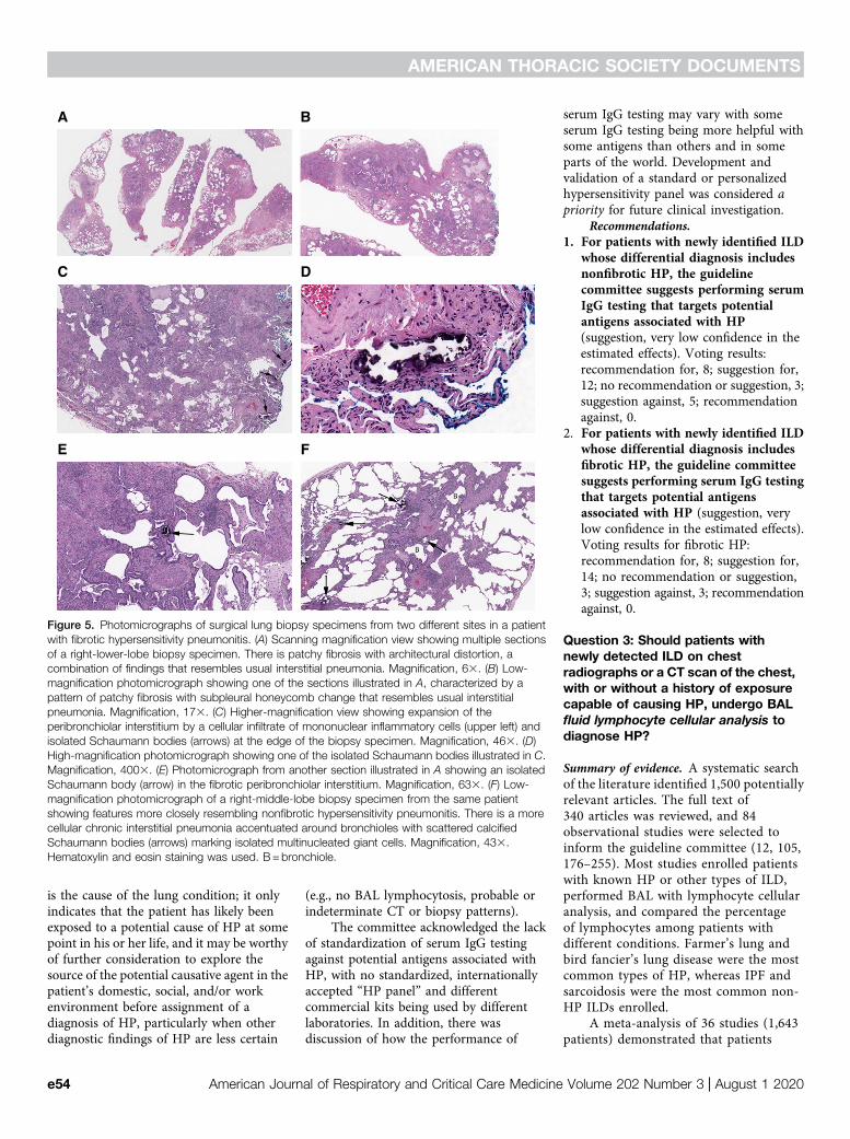
is the cause of the lung condition; it onlyindicates that the patient has likely beenexposed to a potential cause of HP at somepoint in his or her life, and it may be worthyof further consideration to explore thesource of the potential causative agent in thepatient’s domestic, social, and/or workenvironment before assignment of adiagnosis of HP, particularly when otherdiagnostic findings of HP are less certain
(e.g., no BAL lymphocytosis, probable orindeterminate CT or biopsy patterns).
The committee acknowledged the lackof standardization of serum IgG testingagainst potential antigens associated withHP, with no standardized, internationallyaccepted “HP panel” and differentcommercial kits being used by differentlaboratories. In addition, there wasdiscussion of how the performance of
serum IgG testing may vary with someserum IgG testing being more helpful withsome antigens than others and in someparts of the world. Development andvalidation of a standard or personalizedhypersensitivity panel was considered apriority for future clinical investigation.
Recommendations.1. For patients with newly identified ILD
whose differential diagnosis includesnonfibrotic HP, the guidelinecommittee suggests performing serumIgG testing that targets potentialantigens associated with HP(suggestion, very low confidence in theestimated effects). Voting results:recommendation for, 8; suggestion for,12; no recommendation or suggestion, 3;suggestion against, 5; recommendationagainst, 0.
2. For patients with newly identified ILDwhose differential diagnosis includesfibrotic HP, the guideline committeesuggests performing serum IgG testingthat targets potential antigensassociated with HP (suggestion, verylow confidence in the estimated effects).Voting results for fibrotic HP:recommendation for, 8; suggestion for,14; no recommendation or suggestion,3; suggestion against, 3; recommendationagainst, 0.
Question 3: Should patients withnewly detected ILD on chestradiographs or a CT scan of the chest,with or without a history of exposurecapable of causing HP, undergo BALfluid lymphocyte cellular analysis todiagnose HP?
Summary of evidence. A systematic searchof the literature identified 1,500 potentiallyrelevant articles. The full text of340 articles was reviewed, and 84observational studies were selected toinform the guideline committee (12, 105,176–255). Most studies enrolled patientswith known HP or other types of ILD,performed BAL with lymphocyte cellularanalysis, and compared the percentageof lymphocytes among patients withdifferent conditions. Farmer’s lung andbird fancier’s lung disease were the mostcommon types of HP, whereas IPF andsarcoidosis were the most common non-HP ILDs enrolled.
A meta-analysis of 36 studies (1,643patients) demonstrated that patients
A B
C D
E F
Figure 5. Photomicrographs of surgical lung biopsy specimens from two different sites in a patientwith fibrotic hypersensitivity pneumonitis. (A) Scanning magnification view showing multiple sectionsof a right-lower-lobe biopsy specimen. There is patchy fibrosis with architectural distortion, acombination of findings that resembles usual interstitial pneumonia. Magnification, 63. (B) Low-magnification photomicrograph showing one of the sections illustrated in A, characterized by apattern of patchy fibrosis with subpleural honeycomb change that resembles usual interstitialpneumonia. Magnification, 173. (C) Higher-magnification view showing expansion of theperibronchiolar interstitium by a cellular infiltrate of mononuclear inflammatory cells (upper left) andisolated Schaumann bodies (arrows) at the edge of the biopsy specimen. Magnification, 463. (D)High-magnification photomicrograph showing one of the isolated Schaumann bodies illustrated in C.Magnification, 4003. (E) Photomicrograph from another section illustrated in A showing an isolatedSchaumann body (arrow) in the fibrotic peribronchiolar interstitium. Magnification, 633. (F) Low-magnification photomicrograph of a right-middle-lobe biopsy specimen from the same patientshowing features more closely resembling nonfibrotic hypersensitivity pneumonitis. There is a morecellular chronic interstitial pneumonia accentuated around bronchioles with scattered calcifiedSchaumann bodies (arrows) marking isolated multinucleated giant cells. Magnification, 433.Hematoxylin and eosin staining was used. B=bronchiole.
AMERICAN THORACIC SOCIETY DOCUMENTS
e54 American Journal of Respiratory and Critical Care Medicine Volume 202 Number 3 | August 1 2020
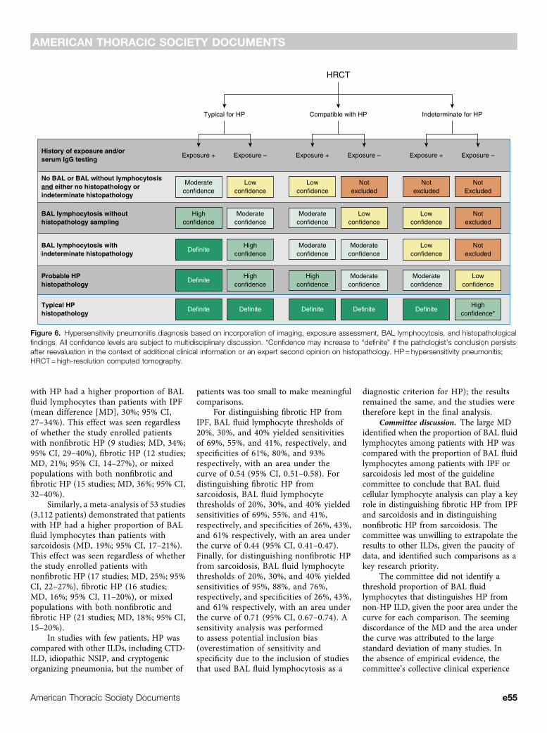
with HP had a higher proportion of BALfluid lymphocytes than patients with IPF(mean difference [MD], 30%; 95% CI,27–34%). This effect was seen regardlessof whether the study enrolled patientswith nonfibrotic HP (9 studies; MD, 34%;95% CI, 29–40%), fibrotic HP (12 studies;MD, 21%; 95% CI, 14–27%), or mixedpopulations with both nonfibrotic andfibrotic HP (15 studies; MD, 36%; 95% CI,32–40%).
Similarly, a meta-analysis of 53 studies(3,112 patients) demonstrated that patientswith HP had a higher proportion of BALfluid lymphocytes than patients withsarcoidosis (MD, 19%; 95% CI, 17–21%).This effect was seen regardless of whetherthe study enrolled patients withnonfibrotic HP (17 studies; MD, 25%; 95%CI, 22–27%), fibrotic HP (16 studies;MD, 16%; 95% CI, 11–20%), or mixedpopulations with both nonfibrotic andfibrotic HP (21 studies; MD, 18%; 95% CI,15–20%).
In studies with few patients, HP wascompared with other ILDs, including CTD-ILD, idiopathic NSIP, and cryptogenicorganizing pneumonia, but the number of
patients was too small to make meaningfulcomparisons.
For distinguishing fibrotic HP fromIPF, BAL fluid lymphocyte thresholds of20%, 30%, and 40% yielded sensitivitiesof 69%, 55%, and 41%, respectively, andspecificities of 61%, 80%, and 93%respectively, with an area under thecurve of 0.54 (95% CI, 0.51–0.58). Fordistinguishing fibrotic HP fromsarcoidosis, BAL fluid lymphocytethresholds of 20%, 30%, and 40% yieldedsensitivities of 69%, 55%, and 41%,respectively, and specificities of 26%, 43%,and 61% respectively, with an area underthe curve of 0.44 (95% CI, 0.41–0.47).Finally, for distinguishing nonfibrotic HPfrom sarcoidosis, BAL fluid lymphocytethresholds of 20%, 30%, and 40% yieldedsensitivities of 95%, 88%, and 76%,respectively, and specificities of 26%, 43%,and 61% respectively, with an area underthe curve of 0.71 (95% CI, 0.67–0.74). Asensitivity analysis was performedto assess potential inclusion bias(overestimation of sensitivity andspecificity due to the inclusion of studiesthat used BAL fluid lymphocytosis as a
diagnostic criterion for HP); the resultsremained the same, and the studies weretherefore kept in the final analysis.
Committee discussion. The large MDidentified when the proportion of BAL fluidlymphocytes among patients with HP wascompared with the proportion of BAL fluidlymphocytes among patients with IPF orsarcoidosis led most of the guidelinecommittee to conclude that BAL fluidcellular lymphocyte analysis can play a keyrole in distinguishing fibrotic HP from IPFand sarcoidosis and in distinguishingnonfibrotic HP from sarcoidosis. Thecommittee was unwilling to extrapolate theresults to other ILDs, given the paucity ofdata, and identified such comparisons as akey research priority.
The committee did not identify athreshold proportion of BAL fluidlymphocytes that distinguishes HP fromnon-HP ILD, given the poor area under thecurve for each comparison. The seemingdiscordance of the MD and the area underthe curve was attributed to the largestandard deviation of many studies. Inthe absence of empirical evidence, thecommittee’s collective clinical experience
History of exposure and/orserum IgG testing
BAL lymphocytosis withindeterminate histopathology
BAL lymphocytosis withouthistopathology sampling
No BAL or BAL without lymphocytosisand either no histopathology orindeterminate histopathology
Probable HPhistopathology
Typical HPhistopathology
Highconfidence
Moderateconfidence
Moderateconfidence
Lowconfidence
Highconfidence
Definite
Highconfidence
Definite
Definite Definite
Lowconfidence
Moderateconfidence
Lowconfidence
Notexcluded
Moderateconfidence
Moderateconfidence
Highconfidence
Moderateconfidence
Definite Definite
Lowconfidence
Notexcluded
NotExcluded
Notexcluded
Lowconfidence
Notexcluded
Lowconfidence
Moderateconfidence
Highconfidence*
Definite
Typical for HP
Exposure + Exposure +Exposure – Exposure – Exposure + Exposure –
Compatible with HP Indeterminate for HP
HRCT
Figure 6. Hypersensitivity pneumonitis diagnosis based on incorporation of imaging, exposure assessment, BAL lymphocytosis, and histopathologicalfindings. All confidence levels are subject to multidisciplinary discussion. *Confidence may increase to “definite” if the pathologist’s conclusion persistsafter reevaluation in the context of additional clinical information or an expert second opinion on histopathology. HP=hypersensitivity pneumonitis;HRCT=high-resolution computed tomography.
AMERICAN THORACIC SOCIETY DOCUMENTS
American Thoracic Society Documents e55

indicated that healthy nonsmokers have aproportion of BAL fluid lymphocytes of10–15% and, therefore, the committeeconsidered a 30% threshold to bereasonable (256).
Although the guideline committeeconcluded that BAL fluid cellularlymphocyte analysis is indicated toincrease the diagnostic likelihood of
HP, the committee made a strongerrecommendation for BAL in patients withsuspected nonfibrotic HP than for patientswith suspected fibrotic HP because there isan additional reason for BAL in patientswith suspected nonfibrotic HP—to identifyor exclude pulmonary infection, especiallyM. tuberculosis in patients from endemicareas with a high prevalence of M.
tuberculosis, which can progress to deathif untreated.
Recommendations.1. For patients with newly identified ILD
whose differential diagnosis includesnonfibrotic HP, the guideline committeerecommends BAL with lymphocytecellular analysis (recommendation, verylow confidence in the estimated effects).
Diagnosis of HP per Figure 6
Multidisciplinary discussion
Multidisciplinary discussion
Multidisciplinary discussion
Consider TBLC# or SLB##
High-confidence diagnosis of HP
Unclear diagnosisReconsiderexposures
All other combinations of exposure, HRCT,BAL, and TBLB findings
Exposure identified AND typical HPpattern on HRCT AND BAL lymphocytosis
Patient with newly detected interstitial lungabnormalities on chest imaging
Exposure assessment* and chest HRCT scan**
BAL with lymphocyte cellular analysis,with or without TBLB***
Figure 7. Algorithm for the diagnostic evaluation of possible hypersensitivity pneumonitis (HP). Specific features are described for all steps of the algorithmin the corresponding sections of the manuscript. A provisional diagnosis may be adequate in patients for whom the differential diagnosis has beensufficiently narrowed such that further investigations are unlikely to alter management, when invasive testing has unacceptable risks, or when such testsare declined by the patient. *Exposure assessment includes a thorough clinical history and/or serum IgG testing against potential antigens associated withHP and/or, in centers with the appropriate expertise and experience, specific inhalational challenge testing as described in References 9, 323, 324, and325. **High-resolution computed tomography should be performed using the technique described in Table 3 and then reviewed with a thoracicradiologist. ***Transbronchial lung biopsy is suggested for patients with potential nonfibrotic HP (see question 4, recommendation 1). #TBLC is suggestedfor patients with potential nonfibrotic HP, depending on local expertise (see question 5, recommendation 2). ##SLB is infrequently considered in patientswith nonfibrotic HP. HRCT=high-resolution computed tomography; SLB= surgical lung biopsy; TBLB= transbronchial lung biopsy; TBLC= transbronchiallung cryobiopsy.
AMERICAN THORACIC SOCIETY DOCUMENTS
e56 American Journal of Respiratory and Critical Care Medicine Volume 202 Number 3 | August 1 2020

Voting results: recommendation for, 19;suggestion for, 11; no recommendation orsuggestion, 0; suggestion against, 1;recommendation against, 0.
2. For patients with newly identified ILDwhose differential diagnosis includesfibrotic HP, the guideline committeesuggests BAL with lymphocyte cellularanalysis (suggestion, very lowconfidence in the estimated effects).Voting results: recommendation for, 10;suggestion for, 18; no recommendationor suggestion, 0; suggestion against, 3;recommendation against, 0.
Question 4: Should patients withnewly detected ILD on chestradiographs or a CT scan of the chest,with or without a history of exposurecapable of causing HP, undergotransbronchial forceps lung biopsy todiagnose HP?
Summary of evidence. A systematic searchof the literature identified 2,465 potentiallyrelevant articles. The full text of 24 articleswas reviewed, and 13 observational studieswere selected to inform the guidelinecommittee (14, 35, 257–267). Four studiesenrolled patients with known HP, sixenrolled patients with ILD, and threeenrolled patients with DLD. We initiallyconsidered ILD and DLD to besynonymous, but, on noting differentdiagnostic results in the two groups, weopted to analyze each separately. All studiesperformed transbronchial forceps lungbiopsy (TBBx), and most reported thehistopathological diagnostic yield of theprocedure; some also reported thefrequency of adverse effects.
Adequate specimens were obtainedin 87% (95% CI, 79–96%) of samplingprocedures. The diagnostic yield (defined asthe number of procedures that yielded ahistopathological diagnosis among the totalnumber of procedures performed) amongpatients with ILD was 37% (95% CI,32–42%). Inclusion of TBBx in themultimodality diagnostic approach ofpatients with ILD increased the likelihood ofarriving at a diagnosis compared with anapproach without TBBx (RR, 1.67; 95% CI,1.21–2.30). The diagnostic yield amongpatients with DLD was 68% (95% CI,50–86%). Patients with DLD were more likelythan patients with ILD to receive a non-ILDdiagnosis like malignancy, infection, etc. Thediagnostic yield could not be calculated for
patients with known HP, but 41% (95% CI,25–56%) of such patients who underwentTBBx were confirmed to have HP.
Among the studies that enrolled patientswith ILD or DLD, two studies reported thepneumothorax rate (7%; 95% CI, 1–13%), twostudies reported periprocedural mortality (nocases; one study monitored for 24 h and theother monitored for 6 mo), two studiesreported respiratory exacerbation or failure(no cases), four studies reported moderate-to-severe bleeding (4%; 95% CI, 0–8%), and sixstudies reported severe bleeding (no cases).One study reported the number ofpneumothoraces that required a chest tube(6%; 95% CI, 0–13%), with none lastinglonger than 72 hours. The studies thatenrolled patients with known HP did notreport adverse outcomes.
Committee discussion. There wasgeneral agreement that the diagnostic yieldof TBBx was suboptimal, with only half ofprocedures resulting in a diagnosis and withthe exact frequency depending on thepopulation. However, the guidelinecommittee was divided regarding theinterpretation of the evidence. Somemembers argued that roughly half ofpatients who undergo TBBx will be sparedan SLB, and other members contended thatroughly half of patients will require twosampling procedures instead of one, withthe former favoring TBBx and the latterarguing against TBBx. An additionalconcern is the potential for diagnosticmisclassification based on potentiallysuboptimal TBBx specimens.
Notably, the guideline committeeobserved that the diagnostic yield wassubstantially higher among patients withDLD than among patients with ILD (68% vs.37%). This was extrapolated to the notionthat diagnostic yield may be higher amongpatients with suspected nonfibrotic HP thanamong patients with suspected fibrotic HP.This potential difference was supported bynonsystematic clinical observations thatgranulomas are more likely to be detectedand to be diagnostic among patients withnonfibrotic HP than among patients withfibrotic HP. Thus, TBBx was judged to beworthwhile in patients with suspectednonfibrotic HP, but not in patients withsuspected fibrotic HP.
The committee acknowledged thatTBBx is safe, available in most institutions,minimally burdensome, and inexpensiveand that most pulmonary clinicians haveexperience performing the procedure. The
patient representative indicated that hewould choose a TBBx in the hope ofavoiding SLB, even with the knowledge thata second procedure would be necessary ifthe TBBx were nondiagnostic.
Recommendations.1. For patients with newly identified ILD
whose differential diagnosis includesnonfibrotic HP, the guideline committeesuggests TBBx (suggestion, very lowconfidence in the estimated effects).Voting results: recommendation for, 1;suggestion for, 20; no recommendation orsuggestion, 4; suggestion against, 3;recommendation against, 0.
2. For patients with newly identified ILDwhose differential diagnosis includesfibrotic HP, the guideline committeemakes no recommendation orsuggestion for or against TBBx. Votingresults: recommendation for, 1;suggestion for, 13; no recommendationor suggestion, 6; suggestion against, 7;recommendation against, 1.
Question 5: Should patients withnewly detected ILD on chestradiographs or a CT scan of the chest,with or without a history of exposurecapable of causing HP, undergotransbronchial lung cryobiopsy todiagnose HP?
Summary of evidence. A systematic searchof the literature identified 695 potentiallyrelevant articles. The full text of 34 articleswas reviewed, and 24 observational studieswere selected to inform the guidelinecommittee (257, 261, 262, 264, 268–287).One study enrolled patients with known orsuspected HP, 19 enrolled patients withILD, and 4 enrolled patients with DLD. Allstudies performed transbronchial lungcryobiopsy (TBLC) and reported thediagnostic yield of the procedure; some alsoreported the frequency of adverse effects.
The diagnostic yields were 91% (95%CI, 83–99%), 82% (95% CI, 78–86%), and82% (95% CI, 73–90%) among patientswith known or suspected HP, ILD, andDLD, respectively. Among patients withknown or suspected HP in whom adiagnosis was made by TBLC, 100% (95%CI, 91.4–100%) were confirmed to have HPand none had an alternative ILD or a non-ILD diagnosis. Among patients with ILD inwhom a diagnosis was made by TBLC, 7.2%(95% CI, 5.6–9.2%) were determined to
AMERICAN THORACIC SOCIETY DOCUMENTS
American Thoracic Society Documents e57
have HP, 77.5% (95% CI, 74.5–80.3%) werefound to have an alternative type of ILD,and 15.3% (95% CI, 13–18%) weredetermined to have a non-ILD diagnosis,such as an infection. Among patients withDLD in whom a diagnosis was made byTBLC, 13.4% (95% CI, 10.9–16.2%) weredetermined to have HP, 71.5% (95% CI,67.9–74.9%) were found to have analternative type of ILD, and 15.1% (95% CI,12.5–18.1%) were determined to have anon-ILD diagnosis.
Several studies reported adverse eventsof TBLC. In the only study that enrolledpatients with known or suspected HP,bleeding of any severity occurred in 4% (95%CI, 0–10%) and pneumothoraces occurred in27% (95% CI, 14–40%). Among studies thatenrolled patients with ILD or DLD, 16studies reported procedural mortality (rarecases; 95% CI, 0–1%); 11 studies did notspecify the follow-up duration, three studiesemployed 30 days of follow-up, one studyused 90 days of follow-up, and one studyhad both 30 days and 90 days of follow-up.Nine studies reported post-proceduralexacerbation or respiratory failure (rarecases; 95% CI, 0–1%), 17 studies reportedbleeding of any severity (11%; 95% CI,7–15%), 18 studies reported severebleeding (0%; 95% CI, 0–1%), and 23studies reported pneumothoraces (10%;95% CI, 8–13%).
Notably, several studies directlyevaluated TBLC and TBBx within the samepopulations (257, 261, 262). The studiesreported higher diagnostic yield with TBLC,although the incidence of bleeding was alsohigher with TBLC.
Committee discussion. There wasgeneral agreement among members of theguideline committee that the diagnosticyield of TBLC is favorable, with 82–91%of patients potentially avoiding SLB.Histopathological patterns of UIP identifiedby TBLC have been shown to havesubstantial agreement with findings fromSLB, including both histopathologicalagreement (kappa, 0.70; 95% CI, 0.55–0.86)and diagnostic agreement at MDD (kappa,0.62; 95% CI, 0.47–0.78) (288). The safetyprofile of TBLC is also favorable comparedwith SLB. However, the committeeacknowledged that most medical centers,particularly community medical centers,currently cannot provide TBLC due to lackof equipment and expertise, nor do theyhave a large enough volume to justifyestablishing a TBLC program.
The guideline committee related theevidence from studies that enrolled patientswith ILD to its recommendations forpatients with suspected fibrotic HP andrelated the evidence from studies thatenrolled patients with known or suspectedHP to its recommendations for patients withsuspected nonfibrotic HP. They concludedthat, in TBLC-capable medical centers,TBLC should be offered to those withsuspected fibrotic HP because it may lead toavoidance of a more burdensome,expensive, uncomfortable, and potentiallyharmful SLB. However, there was lack ofagreement about whether TBLC should besimilarly offered to patients with suspectednonfibrotic HP because most centers thatoffer TBLC can also perform TBBx; TBBxprobably has fewer complications and wasrecommended for this patient group asdescribed above.
Recommendations.1. For patients with newly identified ILD
whose differential diagnosis includesnonfibrotic HP, the guidelinecommittee makes no recommendationor suggestion for or against TBLC.Voting results: recommendation for, 4;suggestion for, 10; no recommendationor suggestion, 7; suggestion against, 6;recommendation against, 0.
2. For patients with newly identified ILDwhose differential diagnosis includesfibrotic HP, the guideline committeesuggests TBLC (suggestion, very lowconfidence in estimated effects). Votingresults: recommendation for, 8;suggestion for, 11; no recommendationor suggestion, 5; suggestion against, 3;recommendation against, 0.
Question 6: Should patients withnewly detected ILD on chestradiographs or a CT scan of the chest,with or without a history of exposurecapable of causing HP, undergo SLBto diagnose HP?
Summary of evidence. A systematic searchof the literature identified 501 potentiallyrelevant articles. The full text of 56 articleswas reviewed, and 34 nonrandomizedstudies were selected to inform the guidelinecommittee (14, 289–322). One studyenrolled patients with known or suspectedHP, 27 enrolled patients with ILD, and 6enrolled patients with DLD. All studiesperformed SLB and reported the diagnostic
yield of the procedure; some also reportedthe frequency of adverse effects.
The diagnostic yields were 96% (95%CI, 90–100%), 98% (95% CI, 98–99%), and96% (95% CI, 93–99%) among patientswith known or suspected HP, ILD, andDLD, respectively. Among patients withknown or suspected HP in whom adiagnosis was made by SLB, 91% (95% CI,79.3–96.5%) were confirmed to have HPand 9% (95% CI, 3.5–20.7%) had analternative ILD. Among patients with ILD(not specifically suspected or known tohave HP) in whom a diagnosis was made bySLB, 9% (95% CI, 5.9–13.1%) weredetermined to have HP, 61% (95% CI,54.3–66.4%) were found to have analternative type of ILD, and 31% (95% CI,25.1–36.6%) were determined to have anon-ILD diagnosis, such as an infection.Among patients with DLD in whom adiagnosis was made by SLB, 9% (95% CI,8.1–10.3%) were determined to have HP,61% (95% CI, 59.2–62.9%) were found tohave an alternative type of ILD, and 30%(95% CI, 28.1–31.6%) were determined tohave a non-ILD diagnosis.
Many studies reported adverse events ofSLB. Procedural mortality was 2% (95% CI,1–3%; after 30 days of follow-up). Post-procedural exacerbations or respiratoryfailure occurred in 2% (95% CI, 1–3%).Bleeding was detected in 1% (95% CI,0–1%). Pneumothorax persistence afterchest tube removal was observed in 4% (95%CI, 2–5%), with a prolonged air leakoccurring in 3% (95% CI, 2–4%).Respiratory infection and delayed woundhealing were complications in 3% (95% CI,2–3%) and 3% (95% CI, 1–5%), respectively.
Committee discussion. The differentialdiagnosis for ILD is wide, and managementvaries substantially among the causes ofILD. Thus, making a definitive or highlyconfident diagnosis is usually beneficial topatients. The guideline committeeconcluded that the diagnostic yield wassufficiently large and the adverse eventssufficiently few to warrant SLB, despite itsburdens and costs, once other diagnostictests had failed to result in a definitivediagnosis (including other forms of biopsy).The committee emphasized that thedecision to perform a SLB should be madeafter a comprehensive assessment of allavailable data, ideally in the context of anMDD, as well as after a thorough discussionwith the patient about the potential benefitsand risks of this procedure.
AMERICAN THORACIC SOCIETY DOCUMENTS
e58 American Journal of Respiratory and Critical Care Medicine Volume 202 Number 3 | August 1 2020
Recommendations.1. For patients with newly identified ILD
whose differential diagnosis includesnonfibrotic HP, the guidelinecommittee suggests SLB; thisrecommendation is intended for afteralternative diagnostic options havebeen exhausted (suggestion, very lowconfidence in estimated effects). Votingresults: recommendation for, 1;suggestion for, 20; no recommendationor suggestion, 1; suggestion against, 7;recommendation against, 1.
2. For patients with newly identified ILDwhose differential diagnosis includesfibrotic HP, the guideline committeesuggests SLB; this recommendationis intended for after alternativediagnostic options have beenexhausted (suggestion, very lowconfidence in estimated effects). Votingresults: recommendation for, 6;suggestion for, 23; no recommendationor suggestion, 1; suggestion against, 0;recommendation against, 0.
Future Directions
The guideline committee recognized anurgent need to improve knowledge on severaltopics, including 1) understanding thenature and pathophysiology of HP, 2)diagnostic approaches, 3) disease behaviorand natural history, and 4) therapeutic
approaches. Key questions onpathophysiology include geneticsusceptibility and both host andenvironmental factors. For diagnosticapproaches, important needs include thevalidation and standardization ofquestionnaires, BAL lymphocytosisthreshold, specific antibodies, andbiomarkers. In addition, new techniques likegenomic classifiers and artificial intelligenceto improve diagnosis and prognosis need tobe assessed. Questions about diseasebehavior range from prevention to thephenotyping of HP. Regarding phenotyping,the differences between predominantlyinflammatory and fibrotic subtypes areimportant questions. Finally, now that thisguideline has established a standardizeddiagnostic approach to HP, future workneeds to address the management ofthe different subtypes of HP in clinical trialsand other research. For a full list of questionsdeemed important by the guideline committee,see the online supplement.
Registries can play an important role inacquiring knowledge. To maximize theirpotential, HP-specific registries are needed.Such registries would be particularly helpfulin determining the worldwide and nationalincidence and prevalence of all HP, subtypesof HP (i.e., fibrotic and nonfibrotic HP), andphenotypes of HP (e.g., HP with autoimmunefeatures, HP with progressive fibroticbehavior, etc.), including those without an
identifiable inciting agent by serum IgGtesting against potential antigens associatedwithHP and a thorough evaluation. Registriescan also help us understand the frequencies ofvarious detectable and nondetectableexposures and can serve as a biobank forblood and tissue specimens, chest HRCTimages, and antigen quantification from thehome and workplace.
Conclusions
When a patient presents with newly detectedILD identified by HRCT, the clinician shouldbe prompted to elicit a careful and thoroughhistory and possibly utilize formalquestionnaires to reveal the possibility of atemporal relationship between environmentalfactors/exposures and the onset of symptoms.Serum IgG antibody testing against potentialantigens associated with HP may also beperformed to detect exposure to potentialinciting agents of HP. Many patients shouldproceed to BAL with lymphocyte cellularanalysis with or without transbronchial lungbiopsy, and if this information, accompaniedby theHRCTpatterns, is insufficient tomake adefinitive diagnosis, they should undergo anMDD with consideration of TBBx, TBLC, orSLB. This approach is recommended byexperts in HP on the basis of the best availableevidence. The approach should be reevaluatedas new evidence becomes available and shouldbe modified as needed. n
This official clinical practice guideline was prepared by an ad hoc subcommittee of the ATS, JRS, and ALAT.
Members of the subcommittee are asfollows:
GANESH RAGHU, M.D. (Co-Chair)1*KEVIN C. WILSON, M.D. (Co-Chair)2,3‡
ELENA BARGAGLI, M.D., PH.D.4
ELISABETH BENDSTRUP, M.D., PH.D.5
HASSAN A. CHAMI, M.D., M.Sc.6x
ABIGAIL T. CHUA, M.D.7x
JONATHAN H. CHUNG, M.D.8
BRIDGET F. COLLINS, M.D.1
TAMERA J. CORTE, M.B. B.S., PH.D.9
JEAN-CHARLES DALPHIN, M.D., PH.D.10†
SONYE K. DANOFF, M.D., PH.D.11
JAVIER DIAZ-MENDOZA, M.D.12x
ABHIJIT DUGGAL, M.D., M.S., M.P.H.13x
RYOKO EGASHIRA, M.D., PH.D.14
THOMAS EWING, B.S.N.jj
MRIDU GULATI, M.D., M.P.H.15
YOSHIKAZU INOUE, M.D., PH.D.16
ALEX R. JENKINS, PH.D.17x
KERRI A. JOHANNSON, M.D., M.P.H.18
TAKESHI JOHKOH, M.D., PH.D.19
MASANORI KITAICHI, M.D., PH.D.20
SHANDRA L. KNIGHT, M.S.21x
DIRK KOSCHEL, M.D.22
MICHAEL KREUTER, M.D.23{
DAVID J. LEDERER, M.D., M.S.24,25
YOLANDA MAGETO, M.D., M.P.H.26
LISA A. MAIER, M.D., M.S.P.H.27,28
CARLOS MATIZ, M.D.29
FERRAN MORELL, M.D., PH.D.30
JEFFREY L. MYERS, M.D.31**ANDREW G. NICHOLSON, D.M.32,33
SETU PATOLIA, M.D., M.P.H.34x
CARLOS A. PEREIRA, M.D.35
MARTINE REMY-JARDIN, M.D., PH.D.36‡‡
ELISABETTA A. RENZONI, M.D., PH.D.37
CHRISTOPHER J. RYERSON, M.D., M.A.S.38xx
MARGARET L. SALISBURY, M.D., M.S.39
MOISES SELMAN, M.D.40
MAXIMILIANO TAMAE-KAKAZU, M.D.41x
MARTINA VASAKOVA, M.D., PH.D.42jjjj
SIMON L. F. WALSH, M.D.43
WIM A. WUYTS, M.D., PH.D.44
1Center for Interstitial Lung Diseases,Department of Medicine and Department ofLaboratory Medicine and Pathology (Adjunct),University of Washington, Seattle, Washington;2Department of Medicine, School of Medicine,Boston University, Boston, Massachusetts;3American Thoracic Society, New York, NewYork; 4Department of Clinical Sciences, Surgeryand Neurosciences, University of Siena, Siena,Italy; 5Center for Rare Lung Diseases,Department of Respiratory Diseases and Allergy,Aarhus University Hospital, Aarhus University,Aarhus, Denmark; 6Department of Medicine,American University of Beirut, Beirut, Lebanon;7Division of Pulmonary, Critical Care, and Sleep
AMERICAN THORACIC SOCIETY DOCUMENTS
American Thoracic Society Documents e59

Medicine, Department of Medicine, Stony BrookUniversity Hospital, Stony Brook University,Stony Brook, New York; 8Department ofRadiology, University of Chicago, Chicago,Illinois; 9Centre of Research Excellence inPulmonary Fibrosis, Royal Prince Alfred Hospital,University of Sydney, Sydney, Australia;10Service de Pneumologie, Centre HospitalierUniversitaire Besancon, Universitaire Besancon,Besancon, France; 11Division of Pulmonary andCritical Care Medicine, Department of Medicine,School of Medicine, John Hopkins University,Baltimore, Maryland; 12Division of Pulmonaryand Critical Care Medicine, Henry FordHospital–Wayne State University, Detroit,Michigan; 13Department of Critical Care,Respiratory Institute, Cleveland Clinic, Cleveland,Ohio; 14Department of Radiology, Faculty ofMedicine, Saga University, Saga, Japan;15Section of Pulmonary, Critical Care, and SleepMedicine–Yale Occupational and EnvironmentalMedicine Program, School of Medicine, YaleUniversity, New Haven, Connecticut; 16ClinicalResearch Center, National Hospital OrganizationKinki-Chuo Chest Medical Centre, Osaka,Japan; 17Division of Respiratory Medicine,University of Nottingham, Nottingham, UnitedKingdom; 18Department of Medicine andDepartment of Community Health Sciences,University of Calgary, Calgary, Alberta, Canada;19Department of Radiology, Kansai RosaiHospital, Amagaski, Japan; 20Department ofPathology, National Hospital OrganizationMinami Wakayama Medical Center, Tanabe,Wakayama, Japan; 21Library and KnowledgeSciences and 27Division of Environmental andOccupational Health Sciences, Department ofMedicine, National Jewish Health, Denver,Colorado; 22Zentrum fur Pneumologie undBereich Pneumologie, FachkrankenhausCoswig–Uniklinikum Dresden, Coswig,Germany; 23Center for Interstitial and Rare LungDiseases, Pneumology and Respiratory CriticalCare Medicine, Thoraxklinik, University ofHeidelberg–German Center for Lung Research,Heidelberg, Germany; 24RegeneronPharmaceuticals, Inc., Tarrytown, New York;25Irving Medical Center, Columbia University,New York, New York; 26Center for AdvancedHeart and Lung Disease, Baylor UniversityMedical Center, Baylor University, Dallas, Texas;28Department of Medicine, School of Medicineand Department of Environmental andOccupational Health, School of Public Health,University of Colorado, Aurora, Colorado;29Departamento de Medicina Pulmonar yInterna, Fundacion Santa Fe deBogota–Universidad de los Andes, Bogota,Colombia; 30Institut de Recerca Vall d’Hebron,CIBER de Malalties Respiratories (CIBERES),Institut de Salut Carles III, Servei dePneumologia–Universitat Autonoma deBarcelona, Catalonia, Spain; 31Department ofPathology, University of Michigan, Ann Arbor,Michigan; 32Department of Histopathology,Royal Brompton and Harefield National HealthService Foundation Trust, London, UnitedKingdom; 33National Heart and Lung Institute,37Interstitial Lung Disease Unit, Royal BromptonHospital, and 43Department of Radiology,National Heart and Lung Institute, Imperial
College London, London, England, UnitedKingdom; 34Division of Pulmonary, Critical Care,and Sleep Medicine, Saint Louis University, St.Louis, Missouri; 35Interstitial Lung DiseaseProgram, Federal University of Sao Paulo, SaoPaulo, Brazil; 36Department of Thoracic Imaging,University of Lille, Lille, France; 38Department ofMedicine, University of British Columbia,Vancouver, British Columbia, Canada;39Department of Medicine, Vanderbilt UniversityMedical Center, Nashville, Tennessee; 40InstitutoNacional de Enfermedades Respiratorias, IsmaelCosio Villegas, Mexico; 41SpectrumHealth–Michigan State University, Grand Rapids,Michigan; 42Department of RespiratoryMedicine, First Faculty of Medicine, CharlesUniversity–Thomayer Hospital, Prague, CzechRepublic; and 44Unit for Interstitial LungDiseases, Department of Respiratory Medicine,University Hospitals Leuven, Leuven, Belgium
*Content Co-Chair.†Deceased.‡Methodology Co-Chair.xMethodologist.jjPatient representative.{Future directions section leader.**Pathology section leader.‡‡Radiology section leader.xxDefinition and Diagnostic Criteria sectionsleader.jjjjClinical Manifestations, Epidemiology, andPathogenesis sections leader.
Author Disclosures: G.R. served as aconsultant for Bellerophon, Biogen, BMS,Boehringer Ingelheim, Fibrogen, Genentech,Gilead, Nitto, Promedior, Pure Tech Health,Respivant, Roche, Veracyte, and Zambon; andserved on a data and safety monitoring boardfor Avalyn. K.C.W. is employed by theAmerican Thoracic Society as Chief ofDocuments and Documents Editor withinterest in the success of ATS guidelines.H.A.C. served on an advisory committee forAstraZeneca, Boehringer Ingelheim, MerckSharp & Dohme, Novartis, and Pfizer; servedas a speaker for Mundipharma, Novartis, andPfizer; and received research support fromPfizer. J.H.C. served as a consultant forVeracyte; served as a speaker for BoehringerIngelheim and Genentech; and received authorroyalties from Elsevier. T.J.C. served on anadvisory committee for Ad Alta, BoehringerIngelheim, Bristol Myers Squibb, Promedior,and Roche; served as a consultant forBoehringer Ingelheim and Roche; and receivedresearch support from Actelion, AvalynPharma, Biogen, Boehringer Ingelheim, BristolMyers Squibb, Galapagos, and Roche. J.-C.D.served on an advisory committee for Chiesi,Novartis, and Teva; served as a speaker forBoehringer Ingelheim and Menarini; receivedresearch support from Asten France and SOSOxygene; and received other transfers of valuefrom GlaxoSmithKline, Novartis, and Roche.S.K.D. received research support fromBoehringer Ingelheim and Genentech/Roche;served on a data and safety monitoring boardfor Galapagos and Galecto; and is an
employee of the Pulmonary FibrosisFoundation. R.E. served as a speaker forAstraZeneca, Bayer, Boehringer Ingelheim,and Shionogi. M.G. served on an advisorycommittee and received other transfers ofvalue from Boehringer Ingelheim and theFrance Foundation; received research supportfrom Boehringer Ingelheim; and received travelexpenses from the Pulmonary FibrosisFoundation. Y.I. served as a consultant forBoehringer Ingelheim and Promedior; servedon an advisory committee for Asahi Kasei,Galapagos, and Shionogi; received researchsupport from Sekisui Medical; and receivedhonoraria from Boehringer Ingelheim andShionogi. K.A.J. served on an advisorycommittee for Blade Therapeutics, BoehringerIngelheim, Roche, and Theravance; served asa consultant for Blade Therapeutics,Boehringer Ingelheim, Theravance, and ThreeLakes Foundation; served as a speaker forBoehringer Ingelheim and Roche; and receivedresearch support from UCB. D.K. served on anadvisory committee and received researchsupport from Boehringer Ingelheim and Roche.M. Kreuter served on an advisory committeefor Boehringer Ingelheim, Galapagos, andRoche; and received research support fromBoehringer Ingelheim and Roche. D.J.L.served on an advisory committee forBoehringer Ingelheim; served as a consultantfor Galapagos, Galecto, Genentech/Roche,Patara, Pulmonary Fibrosis Foundation, andVeracyte; and is an employee of Regeneron.Y.M. received honoraria from BoehringerIngelheim and Genentech. C.M. receivedresearch support from AstraZeneca,Boehringer Ingelheim, Celgene, Merck, andSanofi; and his spouse is an employee ofMerck. A.G.N. served as a consultant foreResearch Technology and MedQIA; andserved as a speaker for Boehringer Ingelheim.E.A.R. served on an advisory committee forRoche; served as a speaker for BoehringerIngelheim, Mundipharma, and Roche; andreceived travel support from BoehringerIngelheim. C.J.R. received research supportand served as a speaker for BoehringerIngelheim and Roche. M.L.S. served on anadvisory committee, served as a speaker, andreceived research support from BoehringerIngelheim; and served as a consultant forBoehringer Ingelheim and Orinove. M.S.served as a consultant for BoehringerIngelheim and Celgene. M.V. served on anadvisory committee and as a consultant forBoehringer Ingelheim; and received researchsupport from Roche. S.L.F.W. served on anadvisory committee for Boehringer Ingelheimand Roche; served as a consultant forGalapagos, OSIC, and Sanofi; served as aspeaker for Bracco; and received researchsupport from Boehringer Ingelheim. W.A.W.received research support from BoehringerIngelheim and Roche. E. Bargagli,E. Bendstrup, A.T.C., B.F.C., J.D.-M., A.D.,T.E., A.R.J., T.J., M. Kitaichi, S.L.K.,L.A.M., F.M., J.L.M., S.P., C.A.P.,M.R.-J., and M.T.-K. reported norelationships with relevant commercialinterests.
AMERICAN THORACIC SOCIETY DOCUMENTS
e60 American Journal of Respiratory and Critical Care Medicine Volume 202 Number 3 | August 1 2020
References
1. Lacasse Y, Selman M, Costabel U, Dalphin JC, Ando M, Morell F, et al.;HP Study Group. Clinical diagnosis of hypersensitivity pneumonitis.Am J Respir Crit Care Med 2003;168:952–958.
2. Elicker BM, Jones KD, Henry TS, Collard HR. Multidisciplinary approachto hypersensitivity pneumonitis. J Thorac Imaging 2016;31:92–103.
3. Churg A, Muller NL, Flint J, Wright JL. Chronic hypersensitivitypneumonitis. Am J Surg Pathol 2006;30:201–208.
4. Fink JN, Ortega HG, Reynolds HY, Cormier YF, Fan LL, Franks TJ, et al.Needs and opportunities for research in hypersensitivity pneumonitis.Am J Respir Crit Care Med 2005;171:792–798.
5. Schuyler M, Cormier Y. The diagnosis of hypersensitivity pneumonitis.Chest 1997;111:534–536.
6. Richerson HB, Bernstein IL, Fink JN, Hunninghake GW, Novey HS, ReedCE, et al. Guidelines for the clinical evaluation of hypersensitivitypneumonitis: report of the Subcommittee on HypersensitivityPneumonitis. J Allergy Clin Immunol 1989;84:839–844.
7. Morisset J, Johannson KA, Jones KD, Wolters PJ, Collard HR, WalshSLF, et al.; HP Delphi Collaborators. Identification of diagnosticcriteria for chronic hypersensitivity pneumonitis: an internationalmodified Delphi survey. Am J Respir Crit Care Med 2018;197:1036–1044.
8. Johannson KA, Elicker BM, Vittinghoff E, Assayag D, de Boer K, GoldenJA, et al. A diagnostic model for chronic hypersensitivity pneumonitis.Thorax 2016;71:951–954.
9. Vasakova M, Morell F, Walsh S, Leslie K, Raghu G. Hypersensitivitypneumonitis: perspectives in diagnosis and management. Am JRespir Crit Care Med 2017;196:680–689.
10. Salisbury ML, Myers JL, Belloli EA, Kazerooni EA, Martinez FJ, FlahertyKR. Diagnosis and treatment of fibrotic hypersensitivity pneumonia:where we stand and where we need to go. Am J Respir Crit CareMed 2017;196:690–699.
11. Vasakova M, Selman M, Morell F, Sterclova M, Molina-Molina M,Raghu G. Hypersensitivity pneumonitis: current concepts ofpathogenesis and potential targets for treatment. Am J Respir CritCare Med 2019;200:301–308.
12. Morell F, Villar A, Montero MA, Muñoz X, Colby TV, Pipvath S, et al.Chronic hypersensitivity pneumonitis in patients diagnosed withidiopathic pulmonary fibrosis: a prospective case-cohort study.Lancet Respir Med 2013;1:685–694.
13. Ramazzini B. De morbis artificium diatriba. Modena, Italy: AntonioCapponi; 1700.
14. Hanak V, Golbin JM, Ryu JH. Causes and presenting features in 85consecutive patients with hypersensitivity pneumonitis. Mayo ClinProc 2007;82:812–816.
15. Fernandez Perez ER, Swigris JJ, Forssen AV, Tourin O, Solomon JJ,Huie TJ, et al. Identifying an inciting antigen is associated withimproved survival in patients with chronic hypersensitivitypneumonitis. Chest 2013;144:1644–1651.
16. Inase N, Ohtani Y, Sumi Y, Umino T, Usui Y, Miyake S, et al. A clinicalstudy of hypersensitivity pneumonitis presumably caused by featherduvets. Ann Allergy Asthma Immunol 2006;96:98–104.
17. Jordan LE, Guy E. Paediatric feather duvet hypersensitivitypneumonitis. BMJ Case Rep 2015;2015:bcr2014207956.
18. Ryerson CJ, Vittinghoff E, Ley B, Lee JS, Mooney JJ, Jones KD, et al.Predicting survival across chronic interstitial lung disease: the ILD-GAP model. Chest 2014;145:723–728.
19. Walsh SLF, Wells AU, Desai SR, Poletti V, Piciucchi S, Dubini A, et al.Multicentre evaluation of multidisciplinary team meeting agreementon diagnosis in diffuse parenchymal lung disease: a case-cohortstudy. Lancet Respir Med 2016;4:557–565.
20. Raghu G, Remy-Jardin M, Myers JL, Richeldi L, Ryerson CJ, LedererDJ, et al.; American Thoracic Society; European Respiratory Society;Japanese Respiratory Society; Latin American Thoracic Society.Diagnosis of idiopathic pulmonary fibrosis: an official ATS/ERS/JRS/ALAT clinical practice guideline. Am J Respir Crit Care Med2018;198:e44–e68.
21. Mooney JJ, Elicker BM, Urbania TH, Agarwal MR, Ryerson CJ, NguyenMLT, et al. Radiographic fibrosis score predicts survival inhypersensitivity pneumonitis. Chest 2013;144:586–592.
22. Salisbury ML, Gu T, Murray S, Gross BH, Chughtai A, Sayyouh M, et al.Hypersensitivity pneumonitis: radiologic phenotypes are associatedwith distinct survival time and pulmonary function trajectory. Chest2019;155:699–711.
23. Chiba S, Tsuchiya K, Akashi T, Ishizuka M, Okamoto T, Furusawa H,et al. Chronic hypersensitivity pneumonitis with a usual interstitialpneumonia-like pattern: correlation between histopathologic andclinical findings. Chest 2016;149:1473–1481.
24. Ojanguren I, Morell F, Ramon MA, Villar A, Romero C, Cruz MJ, et al.Long-term outcomes in chronic hypersensitivity pneumonitis. Allergy2019;74:944–952.
25. Walsh SL, Sverzellati N, Devaraj A, Wells AU, Hansell DM. Chronichypersensitivity pneumonitis: high resolution computed tomographypatterns and pulmonary function indices as prognostic determinants.Eur Radiol 2012;22:1672–1679.
26. Hanak V, Golbin JM, Hartman TE, Ryu JH. High-resolution CT findingsof parenchymal fibrosis correlate with prognosis in hypersensitivitypneumonitis. Chest 2008;134:133–138.
27. Lima MS, Coletta EN, Ferreira RG, Jasinowodolinski D, Arakaki JS,Rodrigues SC, et al. Subacute and chronic hypersensitivitypneumonitis: histopathological patterns and survival. Respir Med2009;103:508–515.
28. Ohtani Y, Saiki S, Kitaichi M, Usui Y, Inase N, Costabel U, et al. Chronicbird fancier’s lung: histopathological and clinical correlation. Anapplication of the 2002 ATS/ERS consensus classification of theidiopathic interstitial pneumonias. Thorax 2005;60:665–671.
29. Vourlekis JS, Schwarz MI, Cherniack RM, Curran-Everett D, Cool CD,Tuder RM, et al. The effect of pulmonary fibrosis on survival inpatients with hypersensitivity pneumonitis. Am J Med 2004;116:662–668.
30. Kern RM, Singer JP, Koth L, Mooney J, Golden J, Hays S, et al. Lungtransplantation for hypersensitivity pneumonitis. Chest 2015;147:1558–1565.
31. Walters GI, Mokhlis JM, Moore VC, Robertson AS, Burge GA, BhomraPS, et al. Characteristics of hypersensitivity pneumonitis diagnosedby interstitial and occupational lung disease multi-disciplinary teamconsensus. Respir Med 2019;155:19–25.
32. Reich JM. Chirping rales in bird-fancier’s lung. Chest 1993;104:326–327.33. Ohtani Y, Saiki S, Sumi Y, Inase N, Miyake S, Costabel U, et al. Clinical
features of recurrent and insidious chronic bird fancier’s lung. AnnAllergy Asthma Immunol 2003;90:604–610.
34. Fernandez Perez ER, Kong AM, Raimundo K, Koelsch TL, Kulkarni R,Cole AL. Epidemiology of hypersensitivity pneumonitis among aninsured population in the United States: a claims-based cohortanalysis. Ann Am Thorac Soc 2018;15:460–469.
35. Morell F, Roger A, Reyes L, Cruz MJ, Murio C, Muñoz X. Bird fancier’slung: a series of 86 patients. Medicine (Baltimore) 2008;87:110–130.
36. De Sadeleer LJ, Hermans F, De Dycker E, Yserbyt J, Verschakelen JA,Verbeken EK, et al. Effects of corticosteroid treatment and antigenavoidance in a large hypersensitivity pneumonitis cohort: a single-centre cohort study. J Clin Med 2018;8:14.
37. Kokkarinen JI, Tukiainen HO, Terho EO. Effect of corticosteroidtreatment on the recovery of pulmonary function in farmer’s lung. AmRev Respir Dis 1992;145:3–5.
38. Gimenez A, Storrer K, Kuranishi L, Soares MR, Ferreira RG, PereiraCAC. Change in FVC and survival in chronic fibrotic hypersensitivitypneumonitis. Thorax 2018;73:391–392.
39. Churg A, Sin DD, Everett D, Brown K, Cool C. Pathologic patterns andsurvival in chronic hypersensitivity pneumonitis. Am J Surg Pathol2009;33:1765–1770.
40. Perez-Padilla R, Salas J, Chapela R, Sanchez M, Carrillo G, Perez R,et al. Mortality in Mexican patients with chronic pigeon breeder’slung compared with those with usual interstitial pneumonia. Am RevRespir Dis 1993;148:49–53.
41. Sahin H, Brown KK, Curran-Everett D, Hale V, Cool CD, Vourlekis JS,et al. Chronic hypersensitivity pneumonitis: CT features comparisonwith pathologic evidence of fibrosis and survival. Radiology 2007;244:591–598.
42. Wang P, Jones KD, Urisman A, Elicker BM, Urbania T, Johannson KA,et al. Pathologic findings and prognosis in a large prospective cohortof chronic hypersensitivity pneumonitis. Chest 2017;152:502–509.
AMERICAN THORACIC SOCIETY DOCUMENTS
American Thoracic Society Documents e61
43. Ohtsuka Y, Munakata M, Tanimura K, Ukita H, Kusaka H, Masaki Y,et al. Smoking promotes insidious and chronic farmer’s lung disease,and deteriorates the clinical outcome. Intern Med 1995;34:966–971.
44. Coleman A, Colby TV. Histologic diagnosis of extrinsic allergicalveolitis. Am J Surg Pathol 1988;12:514–518.
45. Coultas DB, Zumwalt RE, Black WC, Sobonya RE. The epidemiology ofinterstitial lung diseases. Am J Respir Crit Care Med 1994;150:967–972.
46. Solaymani-Dodaran M, West J, Smith C, Hubbard R. Extrinsic allergicalveolitis: incidence and mortality in the general population. QJM2007;100:233–237.
47. Kornum JB, Christensen S, Grijota M, Pedersen L, Wogelius P,Beiderbeck A, et al. The incidence of interstitial lung disease 1995-2005: a Danish nationwide population-based study. BMC Pulm Med2008;8:24.
48. Hendrick DJ, Faux JA, Marshall R. Budgerigar-fancier’s lung: thecommonest variety of allergic alveolitis in Britain. BMJ 1978;2:81–84.
49. Grant IW, Blyth W, Wardrop VE, Gordon RM, Pearson JC, Mair A.Prevalence of farmer’s lung in Scotland: a pilot survey. BMJ 1972;1:530–534.
50. Christensen LT, Schmidt CD, Robbins L. Pigeon breeders’ disease: aprevalence study and review. Clin Allergy 1975;5:417–430.
51. Fenclova Z, Pelclova D, Urban P, Navratil T, Klusackova P, Lebedova J.Occupational hypersensitivity pneumonitis reported to the CzechNational Registry of Occupational Diseases in the period 1992–2005.Ind Health 2009;47:443–448.
52. Yoshida K, Suga M, Nishiura Y, Arima K, Yoneda R, Tamura M, et al.Occupational hypersensitivity pneumonitis in Japan: data on anationwide epidemiological study. Occup Environ Med 1995;52:570–574.
53. Dalphin JC. Extrinsic allergic alveolitis in agricultural environment [inFrench]. Rev Prat 1992;42:1790–1796.
54. Ando M, Suga M, Nishiura Y, Miyajima M. Summer-typehypersensitivity pneumonitis. Intern Med 1995;34:707–712.
55. Griese M, Haug M, Hartl D, Teusch V, Glockner-Pagel J, Brasch F;National EAA Study Group. Hypersensitivity pneumonitis: lessons fordiagnosis and treatment of a rare entity in children. Orphanet J RareDis 2013;8:121.
56. Griese M, Irnstetter A, Hengst M, Burmester H, Nagel F, Ripper J, et al.Categorizing diffuse parenchymal lung disease in children. OrphanetJ Rare Dis 2015;10:122.
57. Buchvald F, Petersen BL, Damgaard K, Deterding R, Langston C, Fan LL,et al. Frequency, treatment, and functional outcome in children withhypersensitivity pneumonitis. Pediatr Pulmonol 2011;46:1098–1107.
58. Morell F, Villar A, Ojanguren I, Munoz X, Cruz MJ, Sansano I, et al.Hypersensitivity pneumonitis and (idiopathic) pulmonary fibrosis dueto feather duvets and pillows [in English, Spanish]. ArchBronconeumol [online ahead of print] 11 Feb 2020; DOI:10.1016/j.arbres.2019.12.003..
59. Thomeer MJ, Costabe U, Rizzato G, Poletti V, Demedts M. Comparisonof registries of interstitial lung diseases in three European countries.Eur Respir J Suppl 2001;32:114s–118s.
60. Schweisfurth H. Report by the scientific working group for therapy oflung diseases: German fibrosis register with initial results [inGerman]. Pneumologie 1996;50:899–901.
61. Thomeer M, Demedts M, Vandeurzen K; VRGTWorking Group on InterstitialLung Diseases. Registration of interstitial lung diseases by 20 centres ofrespiratory medicine in Flanders. Acta Clin Belg 2001;56:163–172.
62. Fisher J, Shapera S, Algamdi M, Morisset J, Johannson KA, Fell CD,et al. Baseline characteristics and comorbidities in the Canadianregistry for pulmonary fibrosis [abstract]. Am J Respir Crit Care Med2020;201:A7478.
63. Kreuter M, Herth FJ, Wacker M, Leidl R, Hellmann A, Pfeifer M, et al.Exploring clinical and epidemiological characteristics of interstitiallung diseases: rationale, aims, and design of a nationwideprospective registry: the EXCITING-ILD registry. BioMed Res Int2015;2015:123876.
64. Karakatsani A, Papakosta D, Rapti A, Antoniou KM, Dimadi M,Markopoulou A, et al.; Hellenic Interstitial Lung Diseases Group.Epidemiology of interstitial lung diseases in Greece. Respir Med2009;103:1122–1129.
65. Ansarie M. A national guideline and ILD PAK Registry Report: recentlandmarks in the understanding of interstitial lung diseases inPakistan. J Pak Med Assoc 2016;66:1050–1053.
66. Strambu I, Salmen T, Traila D, Croitoru A. Romanian Registry forInterstitial Lung Diseases (REGIS): inclusion of patients in 3 years.Eur Respir J Suppl 2017;50:PA868.
67. Singh S, Collins BF, Sharma BB, Joshi JM, Talwar D, Katiyar S, et al.Interstitial lung disease (ILD) in India: results of a prospective registry.Am J Respir Crit Care Med 2017;195:801–813.
68. Xaubet A, Ancochea J, Morell F, Rodriguez-Arias JM, Villena V,Blanquer R, et al.; Spanish Group on Interstitial Lung Diseases,SEPAR. Report on the incidence of interstitial lung diseases in Spain.Sarcoidosis Vasc Diffuse Lung Dis 2004;21:64–70.
69. Alhamad EH. Interstitial lung diseases in Saudi Arabia: a single-centerstudy. Ann Thorac Med 2013;8:33–37.
70. PFF patient registry. Chicago, IL: Pulmonary Fibrosis Foundation[accessed 2020 May 4]. Available from: https://www.pulmonaryfibrosis.org/medical-community/pff-patient-registry.
71. Rose CS, Martyny JW, Newman LS, Milton DK, King TE Jr, Beebe JL,et al. “Lifeguard lung”: endemic granulomatous pneumonitis in anindoor swimming pool. Am J Public Health 1998;88:1795–1800.
72. Simpson C, Garabrant D, Torrey S, Robins T, Franzblau A.Hypersensitivity pneumonitis-like reaction and occupational asthmaassociated with 1,3-bis(isocyanatomethyl) cyclohexane pre-polymer.Am J Ind Med 1996;30:48–55.
73. Ganier M, Lieberman P, Fink J, Lockwood DG. Humidifier lung: anoutbreak in office workers. Chest 1980;77:183–187.
74. Banaszak EF, Thiede WH, Fink JN. Hypersensitivity pneumonitis due tocontamination of an air conditioner. N Engl J Med 1970;283:271–276.
75. Selman M, Pardo A, King TE Jr. Hypersensitivity pneumonitis: insightsin diagnosis and pathobiology. Am J Respir Crit Care Med 2012;186:314–324.
76. Gbaguidi-Haore H, Roussel S, Reboux G, Dalphin JC, Piarroux R.Multilevel analysis of the impact of environmental factors andagricultural practices on the concentration in hay of microorganismsresponsible for farmer’s lung disease. Ann Agric Environ Med 2009;16:219–225.
77. Saltoun CA, Harris KE, Mathisen TL, Patterson R. Hypersensitivitypneumonitis resulting from community exposure to Canada goosedroppings: when an external environmental antigen becomes anindoor environmental antigen. Ann Allergy Asthma Immunol 2000;84:84–86.
78. Koschel D, Wittstruck H, Renck T, Muller-Wening D, Hoffken G.Presenting features of feather duvet lung. Int Arch Allergy Immunol2010;152:264–270.
79. Mizobe T, Ando M, Yamasaki H, Onoue K, Misaki A. Purification andcharacterization of the serotype-specific polysaccharide antigen ofTrichosporon cutaneum serotype II: a disease-related antigen ofJapanese summer-type hypersensitivity pneumonitis. Clin ExpAllergy 1995;25:265–272.
80. Mundt C, Becker WM, Schlaak M. Farmer’s lung: patients’ IgG2antibodies specifically recognize Saccharopolyspora rectivirgulaproteins and carbohydrate structures. J Allergy Clin Immunol 1996;98:441–450.
81. Todd A, Coan RM, Allen A. Pigeon breeders’ lung: pigeon intestinalmucin, an antigen distinct from pigeon IgA. Clin Exp Immunol 1991;85:453–458.
82. Baldwin CI, Todd A, Bourke SJ, Allen A, Calvert JE. Pigeon fanciers’lung: identification of disease-associated carbohydrate epitopes onpigeon intestinal mucin. Clin Exp Immunol 1999;117:230–236.
83. Hisauchi-Kojima K, Sumi Y, Miyashita Y, Miyake S, Toyoda H, KurupVP, et al. Purification of the antigenic components of pigeondropping extract, the responsible agent for cellular immunity inpigeon breeder’s disease. J Allergy Clin Immunol 1999;103:1158–1165.
84. Seed MJ, Enoch SJ, Agius RM. Chemical determinants of occupationalhypersensitivity pneumonitis. Occup Med (Lond) 2015;65:673–681.
85. Seed MJ, Agius RM. Progress with structure-activity relationshipmodelling of occupational chemical respiratory sensitizers. Curr OpinAllergy Clin Immunol 2017;17:64–71.
AMERICAN THORACIC SOCIETY DOCUMENTS
e62 American Journal of Respiratory and Critical Care Medicine Volume 202 Number 3 | August 1 2020
86. Soumagne T, Chardon ML, Dournes G, Laurent L, Degano B, Laurent F,et al. Emphysema in active farmer’s lung disease. PLoS One 2017;12:e0178263.
87. Jacob J, Odink A, Brun AL, Macaluso C, de Lauretis A, Kokosi M, et al.Functional associations of pleuroparenchymal fibroelastosis andemphysema with hypersensitivity pneumonitis. Respir Med 2018;138:95–101.
88. Morell F, Jeanneret A, Aıache JM, Molina C. Leukocyte migrationinhibition in farmer’s lung. J Allergy Clin Immunol 1982;69:405–409.
89. Burrell R, Rylander R. A critical review of the role of precipitins inhypersensitivity pneumonitis. Eur J Respir Dis 1981;62:332–343.
90. Vogelmeier C, Krombach F, Munzing S, Konig G, Mazur G, Beinert T,et al. Activation of blood neutrophils in acute episodes of farmer’slung. Am Rev Respir Dis 1993;148:396–400.
91. Pardo A, Barrios R, Gaxiola M, Segura-Valdez L, Carrillo G, Estrada A,et al. Increase of lung neutrophils in hypersensitivity pneumonitis isassociated with lung fibrosis. Am J Respir Crit Care Med 2000;161:1698–1704.
92. Girard M, Israel-Assayag E, Cormier Y. Impaired function of regulatory T-cellsin hypersensitivity pneumonitis. Eur Respir J 2011;37:632–639.
93. Barrera L, Mendoza F, Zuñiga J, Estrada A, Zamora AC, Melendro EI,et al. Functional diversity of T-cell subpopulations in subacute andchronic hypersensitivity pneumonitis. Am J Respir Crit Care Med2008;177:44–55.
94. Kishi M, Miyazaki Y, Jinta T, Furusawa H, Ohtani Y, Inase N, et al.Pathogenesis of cBFL in common with IPF? Correlation of IP-10/TARC ratio with histological patterns. Thorax 2008;63:810–816.
95. Simonian PL, Roark CL, Wehrmann F, Lanham AK, Diaz del Valle F,Born WK, et al. Th17-polarized immune response in a murine modelof hypersensitivity pneumonitis and lung fibrosis. J Immunol 2009;182:657–665.
96. Jinta T, Miyazaki Y, Kishi M, Akashi T, Takemura T, Inase N, et al. Thepathogenesis of chronic HP in common with IPF. Am J Clin Pathol2010;134:613–620.
97. Garcıa de Alba C, Buendia-Roldan I, Salgado A, Becerril C, Ramırez R,Gonzalez Y, et al. Fibrocytes contribute to inflammation and fibrosisin chronic hypersensitivity pneumonitis through paracrine effects.Am J Respir Crit Care Med 2015;191:427–436.
98. Adegunsoye A, Oldham JM, Demchuk C, Montner S, Vij R, Strek ME.Predictors of survival in coexistent hypersensitivity pneumonitis withautoimmune features. Respir Med 2016;114:53–60.
99. Rittner C, Sennekamp J, Mollenhauer E, Rosinger N, Niese D,Luttkenhorst M, et al. Pigeon breeder’s lung: association with HLA-DR 3. Tissue Antigens 1983;21:374–379.
100. Selman M, Teran L, Mendoza A, Camarena A, Martınez-Cordero E,Lezama M, et al. Increase of HLA-DR7 in pigeon breeder’s lung in aMexican population. Clin Immunol Immunopathol 1987;44:63–70.
101. Ando M, Hirayama K, Soda K, Okubo R, Araki S, Sasazuki T. HLA-DQw3 in Japanese summer-type hypersensitivity pneumonitis induced byTrichosporon cutaneum. Am Rev Respir Dis 1989;140:948–950.
102. Camarena A, Juarez A, Mejıa M, Estrada A, Carrillo G, Falfan R, et al.Major histocompatibility complex and tumor necrosis factor-alphapolymorphisms in pigeon breeder’s disease. Am J Respir Crit CareMed 2001;163:1528–1533.
103. Falfan-Valencia R, Camarena A, Pineda CL, Montaño M, Juarez A,Buendıa-Roldan I, et al. Genetic susceptibility to multicasehypersensitivity pneumonitis is associated with the TNF-238 GGgenotype of the promoter region and HLA-DRB1*04 bearing HLAhaplotypes. Respir Med 2014;108:211–217.
104. Camarena A, Aquino-Galvez A, Falfan-Valencia R, Sanchez G,Montaño M, Ramos C, et al. PSMB8 (LMP7) but not PSMB9(LMP2) gene polymorphisms are associated to pigeon breeder’shypersensitivity pneumonitis. Respir Med 2010;104:889–894.
105. Aquino-Galvez A, Camarena A, Montaño M, Juarez A, Zamora AC,Gonzalez-Avila G, et al. Transporter associated with antigenprocessing (TAP) 1 gene polymorphisms in patients withhypersensitivity pneumonitis. Exp Mol Pathol 2008;84:173–177.
106. Hill MR, Briggs L, Montaño MM, Estrada A, Laurent GJ, Selman M,et al. Promoter variants in tissue inhibitor of metalloproteinase-3(TIMP-3) protect against susceptibility in pigeon breeders’ disease.Thorax 2004;59:586–590.
107. Ley B, Newton CA, Arnould I, Elicker BM, Henry TS, Vittinghoff E, et al.The MUC5B promoter polymorphism and telomere length inpatients with chronic hypersensitivity pneumonitis: an observationalcohort-control study. Lancet Respir Med 2017;5:639–647.
108. Newton CA, Batra K, Torrealba J, Kozlitina J, Glazer CS, Aravena C,et al. Telomere-related lung fibrosis is diagnosticallyheterogeneous but uniformly progressive. Eur Respir J 2016;48:1710–1720.
109. Okamoto T, Miyazaki Y, Tomita M, Tamaoka M, Inase N. A familialhistory of pulmonary fibrosis in patients with chronichypersensitivity pneumonitis. Respiration 2013;85:384–390.
110. Ley B, Torgerson DG, Oldham JM, Adegunsoye A, Liu S, Li J, et al.Rare protein-altering telomere-related gene variants in patients withchronic hypersensitivity pneumonitis. Am J Respir Crit Care Med2019;200:1154–1163.
111. Bustos ML, Frıas S, Ramos S, Estrada A, Arreola JL, Mendoza F, et al.Local and circulating microchimerism is associated withhypersensitivity pneumonitis. Am J Respir Crit Care Med 2007;176:90–95.
112. Cormier Y, Israel-Assayag E, Fournier M, Tremblay GM. Modulation ofexperimental hypersensitivity pneumonitis by Sendai virus. J LabClin Med 1993;121:683–688.
113. Dakhama A, Hegele RG, Laflamme G, Israel-Assayag, Cormier Y.Common respiratory viruses in lower airways of patients with acutehypersensitivity pneumonitis. Am J Respir Crit Care Med 1999;159:1316–1322.
114. Gudmundsson G, Monick MM, Hunninghake GW. Viral infectionmodulates expression of hypersensitivity pneumonitis. J Immunol1999;162:7397–7401.
115. Hoppin JA, Umbach DM, Kullman GJ, Henneberger PK, London SJ,Alavanja MC, et al. Pesticides and other agricultural factorsassociated with self-reported farmer’s lung among farm residents inthe Agricultural Health Study. Occup Environ Med 2007;64:334–341.
116. Remy-Jardin M, Remy J, Wallaert B, Muller NL. Subacute and chronicbird breeder hypersensitivity pneumonitis: sequential evaluationwith CT and correlation with lung function tests andbronchoalveolar lavage. Radiology 1993;189:111–118.
117. Lynch DA, Newell JD, Logan PM, King TE Jr, Muller NL. Can CTdistinguish hypersensitivity pneumonitis from idiopathic pulmonaryfibrosis? AJR Am J Roentgenol 1995;165:807–811.
118. Hansell DM, Wells AU, Padley SPG, Muller NL. Hypersensitivitypneumonitis: correlation of individual CT patterns with functionalabnormalities. Radiology 1996;199:123–128.
119. Tateishi T, Ohtani Y, Takemura T, Akashi T, Miyazaki Y, Inase N, et al.Serial high-resolution computed tomography findings of acute andchronic hypersensitivity pneumonitis induced by avian antigen.J Comput Assist Tomogr 2011;35:272–279.
120. Adler BD, Padley SPG, Muller NL, Remy-Jardin M, Remy J. Chronichypersensitivity pneumonitis: high-resolution CT and radiographicfeatures in 16 patients. Radiology 1992;185:91–95.
121. Chung JH, Montner SM, Adegunsoye A, Oldham JM, Husain AN,Vij R, et al. CT findings associated with survival inchronic hypersensitivity pneumonitis. Eur Radiol 2017;27:5127–5135.
122. Salisbury ML, Gross BH, Chughtai A, Sayyouh M, Kazerooni EA,Bartholmai BJ, et al. Development and validation of a radiologicaldiagnosis model for hypersensitivity pneumonitis. Eur Respir J2018;52:1800443.
123. Barnett J, Molyneaux PL, Rawal B, Abdullah R, Hare SS,Vancheeswaran R, et al. Variable utility of mosaic attenuation todistinguish fibrotic hypersensitivity pneumonitis from idiopathicpulmonary fibrosis. Eur Respir J 2019;54:1900531.
124. Hansell DM, Moskovic E. High-resolution computed tomography inextrinsic allergic alveolitis. Clin Radiol 1991;43:8–12.
125. Franquet T, Muller NL. Disorders of the small airways: high-resolutioncomputed tomographic features. Semin Respir Crit Care Med 2003;24:437–444.
126. Franquet T, Hansell DM, Senbanjo T, Remy-Jardin M, Muller NL. Lungcysts in subacute hypersensitivity pneumonitis. J Comput AssistTomogr 2003;27:475–478.
AMERICAN THORACIC SOCIETY DOCUMENTS
American Thoracic Society Documents e63

127. Silva CIS, Muller NL, Lynch DA, Curran-Everett D, Brown KK, Lee KS,et al. Chronic hypersensitivity pneumonitis: differentiation fromidiopathic pulmonary fibrosis and nonspecific interstitial pneumoniaby using thin-section CT. Radiology 2008;246:288–297.
128. Lynch DA, Sverzellati N, Travis WD, Brown KK, Colby TV, Galvin JR,et al. Diagnostic criteria for idiopathic pulmonary fibrosis: aFleischner Society white paper. Lancet Respir Med 2018;6:138–153.
129. Tokura S, Okuma T, Akira M, Arai T, Inoue Y, Kitaichi M. Utility ofexpiratory thin-section CT for fibrotic interstitial pneumonia. ActaRadiol 2014;55:1050–1055.
130. Miller WT Jr, Chatzkel J, Hewitt MG. Expiratory air trapping onthoracic computed tomography: a diagnostic subclassification. AnnAm Thorac Soc 2014;11:874–881.
131. Lalancette M, Carrier G, Laviolette M, Ferland S, Rodrique J, Begin R,et al. Farmer’s lung: long-term outcome and lack of predictive valueof bronchoalveolar lavage fibrosing factors. Am Rev Respir Dis1993;148:216–221.
132. Baqir M, White D, Ryu JH. Emphysematous changes inhypersensitivity pneumonitis: a retrospective analysis of 12patients. Respir Med Case Rep 2018;24:25–29.
133. Miyazaki Y, Tateishi T, Akashi T, Ohtani Y, Inase N, Yoshizawa Y.Clinical predictors and histologic appearance of acuteexacerbations in chronic hypersensitivity pneumonitis. Chest 2008;134:1265–1270.
134. Kitaichi M. Comparative pulmonary histopathology of sarcoidosis,chronic beryllium disease and hypersensitivity pneumonitis [inJapanese]. Nihon Kyobu Shikkan Gakkai Zasshi 1984;22:769–782.
135. Kitaichi M. Pathology of pulmonary sarcoidosis. In: Izumi T, editor.Sarcoidosis, Clinics in Dermatology. Philadelphia, PA: JBLippincott; 1986. pp.108–115.
136. American Thoracic Society; European Respiratory Society; WorldAssociation of Sarcoidosis and Other Granulomatous Disorders.Statement on sarcoidosis: joint statement of the American ThoracicSociety (ATS), the European Respiratory Society (ERS) and theWorld Association of Sarcoidosis and Other GranulomatousDisorders (WASOG) adopted by the ATS Board of Directors and bythe ERS Executive Committee, February 1999. Am J Respir CritCare Med 1999;160:736–755.
137. Kitaichi M, Shimizu S, Tamaya M, Takaki M, Inoue Y. Pathology ofhypersensitivity pneumonitis. In: Sharma OP, editor. Clinical focusseries, hypersensitivity pneumonitis. New Delhi, India: JaypeeBrothers Medical Publishers; 2013. pp. 22–32.
138. Minomo S, Tachibana K, Tsuyuguchi K, Akira M, Kitaichi M, Suzuki K.A unique case of hot tub lung worsening during the winter. InternMed 2015;54:491–495.
139. Fujimura N, Kino T, Nagai S, Kitaichi M, Izumi T, Oshima S, et al.Hypersensitivity pneumonitis in a polyurethane paint sprayer [inJapanese]. Nihon Kyobu Shikkan Gakkai Zasshi 1984;22:506–513.
140. Akashi T, Takemura T, Ando N, Eishi Y, Kitagawa M, Takizawa T, et al.Histopathologic analysis of sixteen autopsy cases of chronichypersensitivity pneumonitis and comparison with idiopathicpulmonary fibrosis/usual interstitial pneumonia. Am J Clin Pathol2009;131:405–415.
141. Takemura T, Akashi T, Ohtani Y, Inase N, Yoshizawa Y. Pathology ofhypersensitivity pneumonitis. Curr Opin Pulm Med 2008;14:440–454.
142. Herbst JB, Myers JL. Hypersensitivity pneumonia: role of surgical lungbiopsy. Arch Pathol Lab Med 2012;136:889–895.
143. Trahan S, Hanak V, Ryu JH, Myers JL. Role of surgical lung biopsy inseparating chronic hypersensitivity pneumonia from usualinterstitial pneumonia/idiopathic pulmonary fibrosis: analysis of 31biopsies from 15 patients. Chest 2008;134:126–132.
144. Castonquay M, Ryu J, Yi E, Tazelaar H. Granulomas and giant cells inhypersensitivity pneumonitis. Hum Pathol 2015;46:607–613.
145. Hurst JR, Verma N, Lowe D, Baxendale HE, Jolles S, Kelleher P, et al.British Lung Foundation/United Kingdom PrimaryImmunodeficiency Network consensus statement on the definition,diagnosis, and management of granulomatous-lymphocyticinterstitial lung disease in common variable immunodeficiencydisorders. J Allergy Clin Immunol Pract 2017;5:938–945.
146. Mukhopadhyay S, Katzenstein A-L. Pulmonary disease due toaspiration of food and other particulate matter: a clinicopathologicstudy of 59 cases diagnosed on biopsy or resection specimens. AmJ Surg Pathol 2007;31:752–759.
147. Khoor A, Leslie KO, Tazelaar HD, Helmers RA, Colby TV. Diffusepulmonary disease caused by nontuberculous mycobacteria inimmunocompetent people (hot tub lung). Am J Clin Pathol 2001;115:755–762.
148. Takemura T, Akashi T, Kamiya H, Ikushima S, Ando T, Oritsu M, et al.Pathological differentiation of chronic hypersensitivity pneumonitisfrom idiopathic pulmonary fibrosis/usual interstitial pneumonia.Histopathology 2012;61:1026–1035.
149. Churg A, Bilawich A, Wright JL. Pathology of chronic hypersensitivitypneumonitis what is it? What are the diagnostic criteria? Why do wecare? Arch Pathol Lab Med 2018;142:109–119.
150. Fukuoka J, Franks TJ, Colby TV, Flaherty KR, Galvin JR, Hayden D,et al. Peribronchiolar metaplasia: a common histologic lesion in diffuselung disease and a rare cause of interstitial lung disease: clinicopathologicfeatures of 15 cases. Am J Surg Pathol 2005;29:948–954.
151. Churg A, Myers J, Suarez T, Gaxiola M, Estrada A, Mejia M, et al.Airway-centered interstitial fibrosis: a distinct form of aggressivediffuse lung disease. Am J Surg Pathol 2004;28:62–68.
152. Tanizawa K, Ley B, Vittinghoff E, Elicker BM, Henry TS, Wolters PJ,et al. Significance of bronchiolocentric fibrosis in patients withhistopathological usual interstitial pneumonia. Histopathology2019;74:1088–1097.
153. Flaherty KR, Travis WD, Colby TV, Toews GB, Kazerooni EA, GrossBH, et al. Histopathologic variability in usual and nonspecificinterstitial pneumonias. Am J Respir Crit Care Med 2001;164:1722–1727.
154. Rival G, Manzoni P, Lacasse Y, Polio JC, Westeel V, Dubiez A, et al.High-resolution CT predictors of hypersensitivity pneumonitis.Sarcoidosis Vasc Diffuse Lung Dis 2016;33:117–123.
155. Fenoglio CM, Reboux G, Sudre B, Mercier M, Roussel S, Cordier JF,et al. Diagnostic value of serum precipitins to mould antigens inactive hypersensitivity pneumonitis. Eur Respir J 2007;29:706–712.
156. Reboux G, Piarroux R, Mauny F, Madroszyk A, Millon L, Bardonnet K,et al. Role of molds in farmer’s lung disease in eastern France. Am JRespir Crit Care Med 2001;163:1534–1539.
157. Pereira CAC, Soares MR, Boaventura R, Castro MDC, Gomes PS,Gimenez A, et al. Squawks in interstitial lung disease prevalenceand causes in a cohort of one thousand patients. Medicine(Baltimore) 2019;98:e16419.
158. Bohadana A, Izbicki G, Kraman SS. Fundamentals of lungauscultation. N Engl J Med 2014;370:744–751.
159. Ryerson CJ, Corte TJ, Lee JS, Richeldi L, Walsh SLF, Myers JL, et al.A standardized diagnostic ontology for fibrotic interstitial lungdisease: an international working group perspective. Am J RespirCrit Care Med 2017;196:1249–1254.
160. Walsh SLF, Maher TM, Kolb M, Poletti V, Nusser R, Richeldi L, et al.;IPF Project Consortium. Diagnostic accuracy of a clinical diagnosisof idiopathic pulmonary fibrosis: an international case-cohort study.Eur Respir J 2017;50:1700936.
161. Walsh SLF, Lederer DJ, Ryerson CJ, Kolb M, Maher TM, Nusser R,et al. Diagnostic likelihood thresholds that define a workingdiagnosis of idiopathic pulmonary fibrosis. Am J Respir Crit CareMed 2019;200:1146–1153.
162. Millerick-May ML, Mulks MH, Gerlach J, Flaherty KR, Schmidt SL,Martinez FJ, et al. Hypersensitivity pneumonitis and antigenidentification: an alternate approach. Respir Med 2016;112:97–105.
163. Polke M, Kirsten D, Teucher B, Kahn N, Geissler K, Costabel U, et al. Acomparison of existing questionnaires for identifying the causes ofinterstitial and rare lung diseases. Respiration 2020;99:119–124.
164. Andersen P, Christensen KM, Jensen BE, Axel K, Laursen JC, GedayH, et al. Antibodies to pigeon antigens in pigeon breeders:detection of antibodies by an enzyme-linked immunosorbent assay.Eur J Respir Dis 1982;63:113–121.
165. Aznar C, Andre PM, Deunff J, Robert R. Investigation of humanimmune response to Micropolyspora faeni antigens by enzyme-linked immunoelectrodiffusion assay and immunoblotting. J ClinMicrobiol 1988;26:443–447.
AMERICAN THORACIC SOCIETY DOCUMENTS
e64 American Journal of Respiratory and Critical Care Medicine Volume 202 Number 3 | August 1 2020
166. Boiron P, Drouhet E, Dupont B. Enzyme-linked immunosorbent-assay(ELISA) for IgG in bagasse workers’ sera: comparison with counter-immunoelectrophoresis. Clin Allergy 1987;17:355–363.
167. Barrera C, Millon L, Rognon B, Quadroni M, Roussel S, Dalphin JC,et al. Immunoreactive proteins of Saccharopolyspora rectivirgula forfarmer’s lung serodiagnosis. Proteomics Clin Appl 2014;8:971–981.
168. Hebert J, Beaudoin J, Laviolette M, Beaudoin R, Belanger J, CormierY. Absence of correlation between the degree of alveolitis andantibody levels to Micropolysporum faeni. Clin Exp Immunol 1985;60:572–578.
169. Huizinga M, Berrens L. Detection of class-specific antibodies againstMicropolyspora faeni antigens in farmers’ lung. Clin Allergy 1985;15:139–145.
170. Reboux G, Piarroux R, Roussel S, Millon L, Bardonnet K, Dalphin JC.Assessment of four serological techniques in the immunologicaldiagnosis of farmers’ lung disease. J Med Microbiol 2007;56:1317–1321.
171. Rodrigo MJ, Benavent MI, Cruz MJ, Rosell M, Murio C, Pascual C,et al. Detection of specific antibodies to pigeon serum and bloomantigens by enzyme linked immunosorbent assay in pigeonbreeder’s disease. Occup Environ Med 2000;57:159–164.
172. Roussel S, Reboux G, Rognon B, Monod M, Grenouillet F, QuadroniM, et al. Comparison of three antigenic extracts of Eurotiumamstelodami in serological diagnosis of farmer’s lung disease. ClinVaccine Immunol 2010;17:160–167.
173. Sandoval J, Bañales JL, Cortes JJ, Mendoza F, Selman M, Reyes PA.Detection of antibodies against avian antigens in bronchoalveolarlavage from patients with pigeon breeder’s disease: usefulness ofenzyme-linked immunosorbent assay and enzyme immunotransferblotting. J Clin Lab Anal 1990;4:81–85.
174. Simpson C, Shirodaria PV, Evans JP, Simpson DIH, Stanford CF.Comparison of immunodiffusion and enzyme linkedimmunosorbent assay in the detection of abnormal antibodies inpigeon breeder’s disease. J Clin Pathol 1992;45:490–493.
175. Tillie-Leblond I, Grenouillet F, Reboux G, Roussel S, Chouraki B,Lorthois C, et al. Hypersensitivity pneumonitis and metalworkingfluids contaminated by mycobacteria. Eur Respir J 2011;37:640–647.
176. Bellanger AP, Gbaguidi-Haore H, Gondoin A, Pallandre JR, VacheyrouM, Valot B, et al. Positive fungal quantitative PCR and Th17cytokine detection in bronchoalveolar lavage fluids: complementarybiomarkers of hypersensitivity pneumonitis? J Immunol Methods2016;434:61–65.
177. Furasawa H, Saito H, Sema M, Tateishi T, Miyazaki Y, Inase N.Bronchoalveolar lavage in chronic hypersensitivity pneumonitis andIPF [abstract]. Am J Respir Crit Care Med 2017;195:A5429.
178. Groot Kormelink T, Pardo A, Knipping K, Buendıa-Roldan I, Garcıa-de-Alba C, Blokhuis BR, et al. Immunoglobulin free light chains areincreased in hypersensitivity pneumonitis and idiopathic pulmonaryfibrosis. PLoS One 2011;6:e25392.
179. Lehtonen ST, Karvonen HM, Harju T, Sormunen R, Lappi-Blanco E,Hilli M, et al. Stromal cells can be cultured and characterized fromdiagnostic bronchoalveolar fluid samples obtained from patientswith various types of interstitial lung diseases. APMIS 2014;122:301–316.
180. Markart P, Luboeinski T, Korfei M, Schmidt R, Wygrecka M, MahavadiP, et al. Alveolar oxidative stress is associated with elevated levelsof nonenzymatic low-molecular-weight antioxidants in patients withdifferent forms of chronic fibrosing interstitial lung diseases.Antioxid Redox Signal 2009;11:227–240.
181. Nukui Y, Miyazaki Y, Masuo M, Okamoto T, Furusawa H, Tateishi T,et al. Periostin as a predictor of prognosis in chronic bird-relatedhypersensitivity pneumonitis. Allergol Int 2019;68:363–369.
182. Okamoto T, Fujii M, Furusawa H, Tsuchiya K, Miyazaki Y, Inase N. Theusefulness of KL-6 and SP-D for the diagnosis and management ofchronic hypersensitivity pneumonitis. Respir Med 2015;109:1576–1581.
183. Przybylski G, Chorostowska-Wynimko J, Dyczek A, Wędrowska E,Jankowski M, Szpechciński A, et al. Studies of hepatocyte growthfactor in bronchoalveolar lavage fluid in chronic interstitial lungdiseases. Pol Arch Med Wewn 2015;125:260–271.
184. Schmidt R, Meier U, Markart P, Grimminger F, Velcovsky HG, Morr H,et al. Altered fatty acid composition of lung surfactantphospholipids in interstitial lung disease. Am J Physiol Lung CellMol Physiol 2002;283:L1079–L1085.
185. Martina S, Martina V, Monika M, Jan P, Libor K, Ilja S. Angiostaticversus angiogenic chemokines in IPF and EAA. Respir Med 2009;103:1651–1656.
186. Vasakova M, Sterclova M, Kolesar L, Slavcev A, Pohunek P, Sulc J,et al. Bronchoalveolar lavage fluid cellular characteristics, functionalparameters and cytokine and chemokine levels in interstitial lungdiseases. Scand J Immunol 2009;69:268–274.
187. Willems S, Verleden SE, Vanaudenaerde BM, Wynants M, Dooms C,Yserbyt J, et al. Multiplex protein profiling of bronchoalveolar lavagein idiopathic pulmonary fibrosis and hypersensitivity pneumonitis.Ann Thorac Med 2013;8:38–45. [Published erratum appears in AnnThorac Med 8:85.]
188. Ohshimo S, Bonella F, Cui A, Beume M, Kohno N, Guzman J, et al.Significance of bronchoalveolar lavage for the diagnosis ofidiopathic pulmonary fibrosis. Am J Respir Crit Care Med 2009;179:1043–1047.
189. Pesci A, Bertorelli G, Gabrielli M, Olivieri D. Mast cells in fibrotic lungdisorders. Chest 1993;103:989–996.
190. Bertorelli G, Pesci A, Consigli GF, Minisini R, Mori PA, Dall’Aglio PP,et al. Evaluation of some immunological parameters in interstitiallung disease by discriminant analysis. Respiration 1988;54:23–29.
191. Drent M, van Nierop MA, Gerritsen FA, Wouters EF, Mulder PG. Acomputer program using BALF-analysis results as a diagnostic toolin interstitial lung diseases. Am J Respir Crit Care Med 1996;153:736–741.
192. Haslam PL, Parker DJ, Townsend PJ. Increases in HLA-DQ, DP, DR,and transferrin receptors on alveolar macrophages in sarcoidosisand allergic alveolitis compared with fibrosing alveolitis. Chest1990;97:651–661.
193. Hoogsteden HC, van Hal PT, Wijkhuijs JM, Hop W, Hilvering C.Differences in expression of monocyte/macrophage surfaceantigens in peripheral blood and bronchoalveolar lavage cells ininterstitial lung diseases. Lung 1993;171:149–160.
194. Kucejko W, Chyczewska E, Naumnik W, Ossolińska M. Concentrationof surfactant protein D, Clara cell protein CC-16 and IL-10 inbronchoalveolar lavage (BAL) in patients with sarcoidosis,hypersensivity pneumonitis and idiopathic pulmonary fibrosis. FoliaHistochem Cytobiol 2009;47:225–230.
195. Papakosta D, Manika K, Gounari E, Kyriazis G, Kontakiotis T,Spyropoulos G, et al. Bronchoalveolar lavage fluid and bloodnatural killer and natural killer T-like cells in cryptogenic organizingpneumonia. Respirology 2014;19:748–754.
196. Perez-Arellano JL, Pedraz MJ, Fuertes A, de la Cruz JL, Gonzalez deBuitrago JM, Jimenez A. Laminin fragment P1 is increased in thelower respiratory tract of patients with diffuse interstitial lungdiseases. Chest 1993;104:1163–1169.
197. Robinson BW, Rose AH, Thompson PJ, Hey A. Comparison ofbronchoalveolar lavage helper/suppressor T-cell ratios insarcoidosis versus other interstitial lung diseases. Aust N Z J Med1987;17:9–15.
198. Yamashita M, Mouri T, Niisato M, Nitanai H, Kobayashi H, OgasawaraM, et al. Lymphangiogenic factors are associated with the severityof hypersensitivity pneumonitis. BMJ Open Respir Res 2015;2:e000085.
199. Costabel U, Bross KJ, Ruhle KH, Lohr GW, Matthys H. Ia-likeantigens on T-cells and their subpopulations in pulmonarysarcoidosis and in hypersensitivity pneumonitis: analysis ofbronchoalveolar and blood lymphocytes. Am Rev Respir Dis 1985;131:337–342.
200. Krombach F, Gerlach JT, Padovan C, Burges A, Behr J, Beinert T,et al. Characterization and quantification of alveolar monocyte-likecells in human chronic inflammatory lung disease. Eur Respir J1996;9:984–991.
201. Radomska-Leœniewska DM, Sobiecka G, Bia B. Relationshipbetween IL-8, IL-10, MMP-9 level and morphological pattern of BALfluid in interstitial lung diseases patients. Cent Eur J Immunol 2003;28:155–159.
AMERICAN THORACIC SOCIETY DOCUMENTS
American Thoracic Society Documents e65
202. Pforte A, Gerth C, Voss A, Beer B, Haussinger K, Jutting U, et al.Proliferating alveolar macrophages in BAL and lung functionchanges in interstitial lung disease. Eur Respir J 1993;6:951–955.
203. Suga M, Iyonaga K, Ichiyasu H, Saita N, Yamasaki H, Ando M. Clinicalsignificance of MCP-1 levels in BALF and serum in patients withinterstitial lung diseases. Eur Respir J 1999;14:376–382.
204. Bargagli E, Penza F, Vagaggini C, Magi B, Perari MG, Rottoli P.Analysis of carbonylated proteins in bronchoalveolar lavage ofpatients with diffuse lung diseases. Lung 2007;185:139–144.
205. Cai M, Bonella F, He X, Sixt SU, Sarria R, Guzman J, et al. CCL18 inserum, BAL fluid and alveolar macrophage culture supernatant ininterstitial lung diseases. Respir Med 2013;107:1444–1452.
206. Espoladore LM, Gregorio BB, Lima MS, de Pereira CA, Soares MR,Coletta EN. Cytological analysis of bronchoalveolar lavage inpatients with interstitial lung diseases and the relation of cytologicalanalysis to fibrosis in high-resolution computed tomography. AnalQuant Cytopathol Histpathol 2014;36:206–212.
207. Fireman E, Ben Efraim S, Greif J, Peretz H, Kivity S, Topilsky M, et al.Differential proliferative characteristics of alveolar fibroblasts ininterstitial lung diseases: regulative role of IL-1 and PGE(2).Mediators Inflamm 1994;3:445–452.
208. Hoogsteden HC, van Dongen JJ, van Hal PT, Delahaye M, Hop W,Hilvering C. Phenotype of blood monocytes and alveolarmacrophages in interstitial lung disease. Chest 1989;95:574–577.
209. Jara-Palomares L, Martın-Juan J, Gomez-Izquierdo L, Cayuela-Domınguez A, Rodrıguez-Becerra E, Rodrıguez-Panadero F.Bronchoalveolar lavage findings in patients with diffuse interstitiallung disease: prospective study of a cohort of 562 patients [inSpanish]. Arch Bronconeumol 2009;45:111–117.
210. Kopiński P, Balicka-Slusarczyk B, Dyczek A, Szpechciński A,Przybylski G, Jarzemska A, et al. Enhanced expression of Fasligand (FasL) in the lower airways of patients with fibrotic interstitiallung diseases (ILDs). Folia Histochem Cytobiol 2011;49:636–645.
211. Pardo A, Smith KM, Abrams J, Coffman R, Bustos M, McClanahanTK, et al. CCL18/DC-CK-1/PARC up-regulation in hypersensitivitypneumonitis. J Leukoc Biol 2001;70:610–616.
212. Phelps DS, Umstead TM, Mejia M, Carrillo G, Pardo A, Selman M.Increased surfactant protein-A levels in patients with newlydiagnosed idiopathic pulmonary fibrosis. Chest 2004;125:617–625.
213. Sherson D, Nielsen H, Frederiksen J, Milman N, Struve-Christensen E,Petersen BN. Superoxide anion release from blood monocytes andalveolar macrophages in patients with diffuse lung fibrosis. APMIS1992;100:408–414.
214. Sokhatska O, Padrão E, Sousa-Pinto B, Beltrão M, Mota PC, Melo N,et al. NK and NKT cells in the diagnosis of diffuse lung diseasespresenting with a lymphocytic alveolitis. BMC Pulm Med 2019;19:39.
215. Thomeer MJ, Vansteenkiste J, Verbeken EK, Demedts M. Interstitiallung diseases: characteristics at diagnosis and mortality riskassessment. Respir Med 2004;98:567–573.
216. Welker L, Jorres RA, Costabel U, Magnussen H. Predictive value ofBAL cell differentials in the diagnosis of interstitial lung diseases.Eur Respir J 2004;24:1000–1006.
217. Wojtan P, Mierzejewski M, Osińska I, Domagała-Kulawik J.Macrophage polarization in interstitial lung diseases. Cent Eur JImmunol 2016;41:159–164.
218. Zissel G, Baumer I, Schlaak M, Muller-Quernheim J. In vitro release ofinterleukin-15 by broncho-alveolar lavage cells and peripheralblood mononuclear cells from patients with different lung diseases.Eur Cytokine Netw 2000;11:105–112.
219. Gunther A, Schmidt R, Nix F, Yabut-Perez M, Guth C, Rosseau S,et al. Surfactant abnormalities in idiopathic pulmonary fibrosis,hypersensitivity pneumonitis and sarcoidosis. Eur Respir J 1999;14:565–573.
220. Oshima M, Maeda A, Ishioka S, Hiyama K, Yamakido M. Expression ofC-C chemokines in bronchoalveolar lavage cells from patients withgranulomatous lung diseases. Lung 1999;177:229–240.
221. Raulf M, Liebers V, Steppert C, Baur X. Increased gamma/delta-positive T-cells in blood and bronchoalveolar lavage of patientswith sarcoidosis and hypersensitivity pneumonitis. Eur Respir J1994;7:140–147.
222. Agostini C, Trentin L, Zambello R, Luca M, Masciarelli M, Cipriani A,et al. Pulmonary alveolar macrophages in patients with sarcoidosisand hypersensitivity pneumonitis: characterization by monoclonalantibodies. J Clin Immunol 1987;7:64–70.
223. Agostini C, Zambello R, Sancetta R, Cerutti A, Milani A, Tassinari C,et al. Expression of tumor necrosis factor-receptor superfamilymembers by lung T lymphocytes in interstitial lung disease. Am JRespir Crit Care Med 1996;153:1359–1367.
224. Baumer I, Zissel G, Schlaak M, Muller-Quernheim J. Solubleintercellular adhesion molecule 1 (sICAM-1) in bronchoalveolarlavage (BAL) cell cultures and in the circulation of patients withtuberculosis, hypersensitivity pneumonitis and sarcoidosis. Eur JMed Res 1998;3:288–294.
225. Cantin A, Begin R, Boileau R, Drapeau G, Rola-Pleszczynski M.Features of bronchoalveolar lavage differentiating hypersensitivitypneumonitis and pulmonary sarcoidosis at time of initialpresentation. Clin Invest Med 1984;7:89–94.
226. Couto M, Palmares C, Beltrão M, Neves S, Mota P, Morais A, et al.Integrin a E b 7 (CD103) expression in bronchoalveolarlymphocytes of patients with hypersensitivity pneumonitis. Int ArchOccup Environ Health 2015;88:167–173.
227. Fujishima S, Nakamura M, Nakamura H, Inoue T, Yogo Y, Okubo Y,et al. Flow cytometric detection of cell-associated interleukin-8 inalveolar macrophages in vivo from patients with hypersensitivitypneumonitis and sarcoidosis. Scand J Clin Lab Invest 2004;64:237–243.
228. Hamagami S, Miyagawa T, Ochi T, Tsuyuguchi I, Kishimoto S. Araised level of soluble CD8 in bronchoalveolar lavage fluid insummer-type hypersensitivity pneumonitis in Japan. Chest 1992;101:1044–1049.
229. Heron M, Claessen AM, Grutters JC, van den Bosch JM. T-cellactivation profiles in different granulomatous interstitial lungdiseases: a role for CD81CD28(null) cells? Clin Exp Immunol 2010;160:256–265.
230. Ohtsuka M, Yoshizawa Y, Naitou T, Yano H, Sato T, Hasegawa S.The motility of lung lymphocytes in hypersensitivity pneumonitisand sarcoidosis. Am J Respir Crit Care Med 1994;149:455–459.
231. Shinohara T, Tsuji S, Okano Y, Machida H, Hatakeyama N, Ogushi F.Elevated levels of intelectin-1, a pathogen-binding lectin, in the BALfluid of patients with chronic eosinophilic pneumonia andhypersensitivity pneumonitis. Intern Med 2018;57:3507–3514.
232. Taniuchi N, Ghazizadeh M, Enomoto T, Matsuda K, Sato M, Takizawa Y,et al. Evaluation of fractional analysis of bronchoalveolar lavagecombined with cellular morphological features. Int J Med Sci 2009;6:1–8.
233. Unoura K, Miyazaki Y, Sumi Y, Tamaoka M, Sugita T, Inase N.Identification of fungal DNA in BALF from patients with home-related hypersensitivity pneumonitis. Respir Med 2011;105:1696–1703.
234. Uzaslan E, Guzman J, Costabel U. Cockade-like structures in alveolarmacrophages in extrinsic allergic alveolitis. Respiration 2005;72:46–51.
235. Valenti S, Scordamaglia A, Crimi P, Mereu C. Bronchoalveolar lavageand transbronchial lung biopsy in sarcoidosis and extrinsic allergicalveolitis. Eur J Respir Dis 1982;63:564–569.
236. Delacroix DL, Marchandise FX, Francis C, Sibille Y. Alpha-2-macroglobulin, monomeric and polymeric immunoglobulin A, andimmunoglobulin M in bronchoalveolar lavage. Am Rev Respir Dis1985;132:829–835.
237. Hamm H, Luhrs J, Guzman y Rotaeche J, Costabel U, Fabel H,Bartsch W. Elevated surfactant protein A in bronchoalveolar lavagefluids from sarcoidosis and hypersensitivity pneumonitis patients.Chest 1994;106:1766–1770.
238. Korosec P, Osolnik K, Kern I, Silar M, Mohorcic K, Kosnik M.Expansion of pulmonary CD81CD561 natural killer T-cells inhypersensitivity pneumonitis. Chest 2007;132:1291–1297.
239. Leatherman JW, Michael AF, Schwartz BA, Hoidal JR. Lung T cells inhypersensitivity pneumonitis. Ann Intern Med 1984;100:390–392.
240. Sharma SK, Pande JN, Verma K, Guleria JS. Bronchoalveolar lavagefluid (BALF) analysis in interstitial lung diseases: a 7-yearexperience. Indian J Chest Dis Allied Sci 1989;31:187–196.
AMERICAN THORACIC SOCIETY DOCUMENTS
e66 American Journal of Respiratory and Critical Care Medicine Volume 202 Number 3 | August 1 2020
241. Sterclova M, Matej R, Mandakova P, Skibova J, Vasakova M. Role ofinterleukin 4 and its receptor in clinical presentation of chronicextrinsic allergic alveolitis: a pilot study. Multidiscip Respir Med2013;8:35.
242. Fujimori Y, Kataoka M, Tada S, Takehara H, Matsuo K, Miyake T, et al.The role of interleukin-8 in interstitial pneumonia. Respirology 2003;8:33–40.
243. Godard P, Clot J, Jonquet O, Bousquet J, Michel FB. Lymphocytesubpopulations in bronchoalveolar lavages of patients withsarcoidosis and hypersensitivity pneumonitis. Chest 1981;80:447–452.
244. Milman N, Graudal N, Jacobsen GK. Bronchoalveolar lavage inradiologically detected diffuse lung disease: diagnostic value oftotal and differential cell count in a series of 130 patients. APMIS1995;103:764–768.
245. Nagata N, Takayama K, Nikaido Y, Yokosaki Y, Kido M. Comparisonof alveolar septal inflammation to bronchoalveolar lavage ininterstitial lung diseases. Respiration 1996;63:94–99.
246. Satake N, Nagai S, Kawatani A, Kaneshima H, Tanaka S, Takeuchi M,et al. Density of phenotypic markers on BAL T-lymphocytes inhypersensitivity pneumonitis, pulmonary sarcoidosis andbronchiolitis obliterans with organizing pneumonia. Eur Respir J1993;6:477–482.
247. Schildge J, Nagel C, Grun C. Bronchoalveolar lavage in interstitial lungdiseases: does the recovery rate affect the results? Respiration2007;74:553–557.
248. Semenzato G, Chilosi M, Ossi E, Trentin L, Pizzolo G, Cipriani A, et al.Bronchoalveolar lavage and lung histology. Comparative analysis ofinflammatory and immunocompetent cells in patients withsarcoidosis and hypersensitivity pneumonitis. Am Rev Respir Dis1985;132:400–404.
249. Shiota Y. Clinical studies on human alveolar macrophages: II.Functions of alveolar macrophages collected by bronchoalveolarlavage. Nippon Ketsueki Gakkai Zasshi 1987;50:79–88.
250. Soler P, Nioche S, Valeyre D, Basset F, Benveniste J, Burtin C, et al.Role of mast cells in the pathogenesis of hypersensitivitypneumonitis. Thorax 1987;42:565–572.
251. Tøndell A, Rø AD, Børset M, Moen T, Sue-Chu M. Activated CD81
T cells and natural killer T cells in bronchoalveolar lavage fluid inhypersensitivity pneumonitis and sarcoidosis. Sarcoidosis VascDiffuse Lung Dis 2015;31:316–324.
252. Drent M, Grutters JC, Mulder PG, van Velzen-Blad H, Wouters EF, vanden Bosch JM. Is the different T helper cell activity in sarcoidosisand extrinsic allergic alveolitis also reflected by the cellularbronchoalveolar lavage fluid profile? Sarcoidosis Vasc Diffuse LungDis 1997;14:31–38.
253. Sterclova M, Vasakova M, Pavlicek J, Metlicka M, Krasna E, Striz I.Chemokine receptors in a regulation of interstitial lung fibrosis andinflammation. Exp Lung Res 2009;35:514–523.
254. Sterclova M, Paluch P, Skibova J, Vasakova M. Influence of age onmanifestation, VC and TLCO values, and bronchoalveolar lavagecell counts of sarcoidosis and extrinsic allergic alveolitis. Clin RespirJ 2015;9:39–44.
255. Dai H, Guzman J, Chen B, Costabel U. Production of soluble tumornecrosis factor receptors and tumor necrosis factor-alpha byalveolar macrophages in sarcoidosis and extrinsic allergic alveolitis.Chest 2005;127:251–256.
256. Meyer KC, Raghu G, Baughman RP, Brown KK, Costabel U, du BoisRM, et al.; American Thoracic Society Committee on BAL inInterstitial Lung Disease. An official American Thoracic Societyclinical practice guideline: the clinical utility of bronchoalveolarlavage cellular analysis in interstitial lung disease. Am J Respir CritCare Med 2012;185:1004–1014.
257. Pajares V, Puzo C, Castillo D, Lerma E, Montero MA, Ramos-BarbonD, et al. Diagnostic yield of transbronchial cryobiopsy in interstitiallung disease: a randomized trial. Respirology 2014;19:900–906.
258. Sindhwani G, Shirazi N, Sodhi R, Raghuvanshi S, Rawat J.Transbronchial lung biopsy in patients with diffuse parenchymallung disease without ‘idiopathic pulmonary fibrosis pattern’ onHRCT scan: Experience from a tertiary care center of North India.Lung India 2015;32:453–456.
259. Morell F, Reyes L, Domenech G, De Gracia J, Majo J, Ferrer J.Diagnoses and diagnostic procedures in 500 consecutive patientswith clinical suspicion of interstitial lung disease [in Spanish]. ArchBronconeumol 2008;44:185–191.
260. Sheth JS, Belperio JA, Fishbein MC, Kazerooni EA, Lagstein A,Murray S, et al. Utility of transbronchial vs surgical lung biopsy inthe diagnosis of suspected fibrotic interstitial lung disease. Chest2017;151:389–399.
261. Pourabdollah M, Shamaei M, Karimi S, Karimi M, Kiani A, Jabbari HR.Transbronchial lung biopsy: the pathologist’s point of view. ClinRespir J 2016;10:211–216.
262. Ramaswamy A, Homer R, Killam J, Pisani MA, Murphy TE, Araujo K,et al. Comparison of transbronchial and cryobiopsies in evaluationof diffuse parenchymal lung disease. J Bronchology IntervPulmonol 2016;23:14–21.
263. Babiak A, Hetzel J, Krishna G, Fritz P, Moeller P, Balli T, et al.Transbronchial cryobiopsy: a new tool for lung biopsies. Respiration2009;78:203–208.
264. Hetzel J, Eberhardt R, Petermann C, Gesierich W, Darwiche K,Hagmeyer L, et al. Bleeding risk of transbronchial cryobiopsycompared to transbronchial forceps biopsy in interstitial lungdisease: a prospective, randomized, multicentre cross-over trial.Respir Res 2019;20:140.
265. Casoni GL, Gurioli C, Chhajed PN, Chilosi M, Zampatori M, Olivieri D,et al. The value of transbronchial lung biopsy using jumbo forceps viarigid bronchoscope in DILD. Monaldi Arch Chest Dis 2008;69:59–64.
266. Adams TN, Newton CA, Batra K, Abu-HijlehM, Barbera T, Torrealba J, et al.Utility of bronchoalveolar lavage and transbronchial biopsy in patientswith hypersensitivity pneumonitis. Lung 2018;196:617–622.
267. Lacasse Y, Fraser RS, Fournier M, Cormier Y. Diagnostic accuracy oftransbronchial biopsy in acute farmer’s lung disease. Chest 1997;112:1459–1465.
268. Cascante JA, Cebollero P, Herrero S, Yague A, Echegoyen A,Elizalde J, et al. Transbronchial cryobiopsy in interstitial lungdisease: are we on the right path? J Bronchology Interv Pulmonol2016;23:204–209.
269. Fruchter O, Fridel L, El Raouf BA, Abdel-Rahman N, Rosengarten D,Kramer MR. Histological diagnosis of interstitial lung diseases bycryo-transbronchial biopsy. Respirology 2014;19:683–688.
270. Griff S, Schonfeld N, Ammenwerth W, Blum TG, Grah C, Bauer TT,et al. Diagnostic yield of transbronchial cryobiopsy in non-neoplastic lung disease: a retrospective case series. BMC PulmMed 2014;14:171.
271. Hernandez-Gonzalez F, Lucena CM, Ramırez J, Sanchez M, JimenezMJ, Xaubet A, et al. Cryobiopsy in the diagnosis of diffuse interstitiallung disease: yield and cost-effectiveness analysis. ArchBronconeumol 2015;51:261–267.
272. Kronborg-White S, Folkersen B, Rasmussen TR, Voldby N, MadsenLB, Rasmussen F, et al. Introduction of cryobiopsies in thediagnostics of interstitial lung diseases: experiences in a referralcenter. Eur Clin Respir J 2017;4:1274099.
273. Kropski JA, Pritchett JM, Mason WR, Sivarajan L, Gleaves LA,Johnson JE, et al. Bronchoscopic cryobiopsy for the diagnosis ofdiffuse parenchymal lung disease. PLoS One 2013;8:e78674.
274. Ussavarungsi K, Kern RM, Roden AC, Ryu JH, Edell ES.Transbronchial cryobiopsy in diffuse parenchymal lung disease:etrospective analysis of 74 cases. Chest 2017;151:400–408.
275. Gershman E, Fruchter O, Benjamin F, Nader AR, Rosengarten D,Rusanov V, et al. Safety of cryo-transbronchial biopsy in diffuselung diseases: analysis of three hundred cases. Respiration 2015;90:40–46.
276. Ravaglia C, Wells AU, Tomassetti S, Gurioli C, Gurioli C, Dubini A,et al. Diagnostic yield and risk/benefit analysis of trans-bronchiallung cryobiopsy in diffuse parenchymal lung diseases: a largecohort of 699 patients. BMC Pulm Med 2019;19:16.
277. Hagmeyer L, Theegarten D, Wohlschlager J, Hager T, Treml M,Herkenrath SD, et al. Transbronchial cryobiopsy in fibrosinginterstitial lung disease: modifications of the procedure lead to riskreduction. Thorax 2019;74:711–714.
278. Vlacic G, Kern I, Rozman A. Transbronchial cryobiopsy: a single-center experience. Virchows Arch 2018;473:s18.
AMERICAN THORACIC SOCIETY DOCUMENTS
American Thoracic Society Documents e67

279. Gnass M, Filarecka A, Pankowski J, Soja J, Bugalho A, Szlubowski A.Transbronchial lung cryobiopsy guided by endobronchial ultrasoundradial miniprobe in interstitial lung diseases: preliminary results of aprospective study. Pol Arch Intern Med 2018;128:259–262.
280. Dhooria S, Mehta RM, Srinivasan A, Madan K, Sehgal IS,Pattabhiraman V, et al. The safety and efficacy of different methodsfor obtaining transbronchial lung cryobiopsy in diffuse lungdiseases. Clin Respir J 2018;12:1711–1720.
281. Morais AMM, Melo N, Mota P, Magalhães A, Guimarães S, Moura CS.Transbronchial criobiopsy (TCB) in two lung lobes-diagnosticaccuracy. Eur Respir J 2017;50:PA860.
282. Li YS, Guo SL, Yi XH, Xiao ML, Jin XX, Xiao Y, et al. Efficacy and safetyof transbronchial cryobiopsy in the etiologic diagnosis of diffuselung disease [in Chinese]. Zhonghua Yi Xue Za Zhi 2017;97:3617–3623.
283. de Sousa Antunes Dias Padrão EF, Mota PC, Melo N, Guimarães S,Moura CS, Magalhães A, et al. Transbronchial lung cryobiopsy inthe diagnosis of hypersensitivity pneumonitis. Eur Respir J 2017;50:PA3018.
284. Colella S, Massaccesi C, Fioretti F, Panella G, Primomo GL, D’EmilioV, et al. Transbronchial lung cryobiopsy in lung diseases: diagnosticyield and safety. Eur Respir J 2017;50:PA3025.
285. Bondue B, Pieters T, Alexander P, De Vuyst P, Ruiz Patino M, HotonD, et al. Role of transbronchial lung cryobiopsies in diffuseparenchymal lung diseases: interest of a sequential approach. PulmMed 2017;2017:6794343.
286. Walscher J, Groß B, Eberhardt R, Heussel CP, Eichinger M, Warth A,et al. Transbronchial cryobiopsies for diagnosing interstitial lungdisease: real-life experience from a tertiary referral center forinterstitial lung disease. Respiration 2019;97:348–354.
287. Romagnoli M, Colby TV, Berthet JP, Gamez AS, Mallet JP, Serre I,et al. Poor concordance between sequential transbronchial lungcryobiopsy and surgical lung biopsy in the diagnosis of diffuse interstitiallung diseases. Am J Respir Crit Care Med 2019;199:1249–1256.
288. Troy LK, Grainge C, Corte TJ, Williamson JP, Vallely MP, Cooper WA.Diagnostic accuracy of transbronchial lung cryobiopsy forinterstitial lung disease diagnosis (COLDICE): a prospectivecomparative study. Lancet Respir Med 2019;8:171–181.
289. Ayed AK, Raghunathan R. Thoracoscopy versus open lung biopsy inthe diagnosis of ILD: a randomised controlled trial. J R Coll SurgEdinb 2000;45:159–163.
290. Miller JD, Urschel JD, Cox G, Olak J, Young JE, Kay JM, et al. Arandomized, controlled trial comparing thoracoscopy and limitedthoracotomy for lung biopsy in interstitial lung disease. Ann ThoracSurg 2000;70:1647–1650.
291. Blewett CJ, Bennett WF, Miller JD, Urschel JD. Open lung biopsy asan outpatient procedure. Ann Thorac Surg 2001;71:1113–1115.
292. Qureshi RA, Ahmed TA, Grayson AD, Soorae AS, Drakeley MJ, PageRD. Does lung biopsy help patients with interstitial lung disease?Eur J Cardiothorac Surg 2002;21:621–626. [Discussion, p. 626.]
293. Ayed AK. Video-assisted thoracoscopic lung biopsy in the diagnosisof diffuse interstitial lung disease: a prospective study. J CardiovascSurg (Torino) 2003;44:115–118.
294. Yamaguchi M, Yoshino I, Suemitsu R, Osoegawa A, Kameyama T,Tagawa T, et al. Elective video-assisted thoracoscopic lung biopsyfor interstitial lung disease. Asian Cardiovasc Thorac Ann 2004;12:65–68.
295. Ooi A, Iyenger S, Ferguson J, Ritchie AJ. VATS lung biopsy insuspected, diffuse interstitial lung disease provides diagnosis,and alters management strategies. Heart Lung Circ 2005;14:90–92.
296. Lettieri CJ, Veerappan GR, Helman DL, Mulligan CR, Shorr AF.Outcomes and safety of surgical lung biopsy for interstitial lungdisease. Chest 2005;127:1600–1605.
297. Park JH, Kim DK, Kim DS, Koh Y, Lee SD, Kim WS, et al. Mortality andrisk factors for surgical lung biopsy in patients with idiopathicinterstitial pneumonia. Eur J Cardiothorac Surg 2007;31:1115–1119.
298. Kreider ME, Hansen-Flaschen J, Ahmad NN, Rossman MD, Kaiser LR,Kucharczuk JC, et al. Complications of video-assistedthoracoscopic lung biopsy in patients with interstitial lung disease.Ann Thorac Surg 2007;83:1140–1144.
299. Coutinho GF, Pancas R, Magalhães E, Bernardo JE, Eugenio L,Antunes MJ. Diagnostic value of surgical lung biopsy: comparisonwith clinical and radiological diagnosis. Eur J Cardiothorac Surg2008;33:781–785.
300. Sigurdsson MI, Isaksson HJ, Gudmundsson G, Gudbjartsson T.Diagnostic surgical lung biopsies for suspected interstitial lungdiseases: a retrospective study. Ann Thorac Surg 2009;88:227–232.
301. Bando M, Ohno S, Hosono T, Yanase K, Sato Y, Sohara Y, et al. Riskof acute exacerbation after video-assisted thoracoscopic lungbiopsy for interstitial lung disease. J Bronchology Interv Pulmonol2009;16:229–235.
302. Guerra M, Miranda JA, Leal F, Vouga L. Interstitial lung disease:diagnostic accuracy and safety of surgical lung biopsy. Rev PortPneumol 2009;15:433–442.
303. Ishie RT, Cardoso JJ, Silveira RJ, Stocco L. Video-assistedthoracoscopy for the diagnosis of diffuse parenchymal lungdisease. J Bras Pneumol 2009;35:234–241.
304. Fibla JJ, Molins L, Blanco A, Royo I, Martınez Vallina P, Martınez N,et al. Video-assisted thoracoscopic lung biopsy in the diagnosis ofinterstitial lung disease: a prospective, multi-center study in 224patients. Arch Bronconeumol 2012;48:81–85.
305. Fibla JJ, Brunelli A, Cassivi SD, Deschamps C. Aggregate riskscore for predicting mortality after surgical biopsy forinterstitial lung disease. Interact Cardiovasc Thorac Surg 2012;15:276–279.
306. Blackhall V, Asif M, Renieri A, Civitelli S, Kirk A, Jilaihawi A, et al. Therole of surgical lung biopsy in the management of interstitial lungdisease: experience from a single institution in the UK. InteractCardiovasc Thorac Surg 2013;17:253–257.
307. Blanco M, Obeso GA, Duran JC, Rivo JE, Garcıa-Fontan E, Peña E,et al. Surgical lung biopsy for diffuse lung disease: our experience inthe last 15 years. Rev Port Pneumol 2013;19:59–64.
308. Kayatta MO, Ahmed S, Hammel JA, Fernandez F, Pickens A, Miller D,et al. Surgical biopsy of suspected interstitial lung disease issuperior to radiographic diagnosis. Ann Thorac Surg 2013;96:399–401.
309. Pompeo E, Rogliani P, Cristino B, Schillaci O, Novelli G, Saltini C.Awake thoracoscopic biopsy of interstitial lung disease. Ann ThoracSurg 2013;95:445–452.
310. Luo Q, Han Q, Chen X, Xie J, Wu L, Chen R. The diagnosis efficacyand safety of video-assisted thoracoscopy surgery (VATS) inundefined interstitial lung diseases: a retrospective study. J ThoracDis 2013;5:283–288.
311. Findikcioglu A, Karadayi S. Is surgical biopsy necessary for diagnosisof interstitial lung diseases: a retrospective clinical study. J ClinAnal Med 2014;5:204–208.
312. Morris D, Zamvar V. The efficacy of video-assisted thoracoscopicsurgery lung biopsies in patients with interstitial lung disease: aretrospective study of 66 patients. J Cardiothorac Surg 2014;9:45.
313. Sonobe M, Handa T, Tanizawa K, Sato M, Sato T, Chen F, et al.Videothoracoscopy-assisted surgical lung biopsy for interstitiallung diseases. Gen Thorac Cardiovasc Surg 2014;62:376–382.
314. Bagheri R, Haghi SZ, Attaran D, Hashem Asnaashari AM, Basiri R,Rajabnejad A. Efficacy of minimally invasive surgery in diagnosis ofinterstitial lung disease. Asian Cardiovasc Thorac Ann 2015;23:851–854.
315. Rotolo N, Imperatori A, Dominioni L, Facchini A, Conti V, CastiglioniM, et al. Efficacy and safety of surgical lung biopsy for interstitialdisease: experience of 161 consecutive patients from a singleinstitution in Italy. Sarcoidosis Vasc Diffuse Lung Dis 2015;32:251–258.
316. Samejima J, Tajiri M, Ogura T, Baba T, Omori T, Tsuboi M, et al.Thoracoscopic lung biopsy in 285 patients with diffuse pulmonarydisease. Asian Cardiovasc Thorac Ann 2015;23:191–197.
317. Knipscheer BJ, van Moorsel CH, Grutters JC. Non-specific and usualinterstitial pneumonia, short-term survival after surgical biopsy.Lung 2015;193:449–450.
318. Khalil M, Cowen M, Chaudhry M, Loubani M. Single versus multiplelung biopsies for suspected interstitial lung disease. AsianCardiovasc Thorac Ann 2016;24:788–791.
AMERICAN THORACIC SOCIETY DOCUMENTS
e68 American Journal of Respiratory and Critical Care Medicine Volume 202 Number 3 | August 1 2020

319. Tomassetti S, Wells AU, Costabel U, Cavazza A, Colby TV, Rossi G, et al.Bronchoscopic lung cryobiopsy increases diagnostic confidence inthe multidisciplinary diagnosis of idiopathic pulmonary fibrosis. Am JRespir Crit Care Med 2016;193:745–752.
320. Ravaglia C, Bonifazi M, Wells AU, Tomassetti S, Gurioli C, Piciucchi S,et al. Safety and diagnostic yield of transbronchial lung cryobiopsy indiffuse parenchymal lung diseases: a comparative study versus video-assisted thoracoscopic lung biopsy and a systematic review of theliterature. Respiration 2016;91:215–227.
321. Ahmed S, El Hindawi A, Mashhour S. Spectrum of diffuseparenchymal lung diseases using medical thoracoscopic lungbiopsy: an experience with 55 patients during 2013–2015. Egypt JChest Dis Tuberc 2016;65:717–722.
322. Lieberman S, Gleason JB, Ilyas MIM, Martinez F, Mehta JP, SavageEB. Assessing the safety and clinical impact of thoracoscopic lungbiopsy in patients with interstitial lung disease. J Clin Diagn Res2017;11:OC57–OC59.
323. Morell F, Villar A, Ojanguren I, Muñoz X, Cruz MJ. Hypersensitivitypneumonitis: challenges in diagnosis and management, avoidingsurgical lung biopsy. Semin Respir Crit Care Med 2016;37:395–405.
324. Ishizuka M, Miyazaki Y, Tateishi T, Tsutsui T, Tsuchiya K, Inase N.Validation of inhalation provocation test in chronic bird-relatedhypersensitivity pneumonitis and new prediction score. Ann AmThorac Soc 2015;12:167–173.
325. Munoz X, Sanchez-Ortiz M, Torres F, Villar A, Morell F,Cruz MJ. Diagnostic yield of specific inhalation challengein hypersensitivity pneumonitis. Eur Respir J 2014; 44:1658–1665.
326. Hansell DM, Bankier AA, MacMahon H, McLoud TC, Muller NL, RemyJ. Fleischner Society: glossary of terms for thoracic imaging.Radiology 2008;246:697–722.
327. Webb WR, Muller NL, Naidich DP. Standardized terms for high-resolution computed tomography of the lung: a proposed glossary.J Thorac Imaging 1993;8:167–175.
328. Chung MH, Edinburgh KJ, Webb EM, McCowin M , Webb WR. Mixedinfiltrative and obstructive disease on high-resolution CT:differential diagnosis and functional correlates in a consecutiveseries. J Thorac Imaging 2001;16:69–75.
329. Chong BJ, Kanne JP, Chung JH. Headcheese sign. J Thorac Imaging2014;29:W13.
330. Travis WD, Costabel U, Hansell DM, King TE Jr, Lynch DA,Nicholson AG, et al.; ATS/ERS Committee on IdiopathicInterstitial Pneumonias. An official American ThoracicSociety/European Respiratory Society statement: update of theinternational multidisciplinary classification of the idiopathicinterstitial pneumonias. Am J Respir Crit Care Med 2013;188:733–748.
AMERICAN THORACIC SOCIETY DOCUMENTS
American Thoracic Society Documents e69