diagnosis of colonic volvulus findings on multidetector ct with
TRANSCRIPT

8/13/2019 Diagnosis of Colonic Volvulus Findings on Multidetector CT With
http://slidepdf.com/reader/full/diagnosis-of-colonic-volvulus-findings-on-multidetector-ct-with 1/8
PICTORIAL REVIEW
Diagnosis of colonic volvulus: findings on multidetector CT with
three-dimensional reconstructions
1C VANDENDRIES, 1M C JULLES, MD, 1I BOULAY-COLETTA, MD, 2J LORIAU, MD and 1M ZINS, MD
1Department of Radiology, Groupe Hospitalier Paris Saint-Joseph, 185 Rue Raymond Losserand, 75014 Paris, France and 2Department of Digestive Surgery, Groupe Hospitalier Paris Saint-Joseph, 185 Rue Raymond Losserand, 75014 Paris,
France
ABSTRACT. Large bowel volvulus is a rare condition that can occur in patients whopresent with acute abdominal pain. Radiologists should be able to recognise itsappearance on multidetector CT (MDCT) images so that the correct diagnosis can bemade and catastrophic consequences can be avoided. In this article, we discuss and
illustrate the MDCT and three-dimensional appearance of the various forms of largebowel volvulus. As MDCT allows the precise diagnosis of topography, mechanism andseverity, this technique can provide an accurate assessment of large bowel volvulus.
Received 23 December2009Revised 2 March 2010Accepted 9 March 2010
DOI: 10.1259/bjr/35714052
’ 2010 The British Institute of
Radiology
A large bowel volvulus (LBV) is a twist of the bowelalong its own mesentery, often resulting in a closed-loopobstruction. LBV accounts for 5% of all organic large-
bowel obstructions and are most common amongpatients aged between 50 and 60 in North America and
Western Europe [1, 2]. The reported incidence of thevarious forms of LBV in the urban Australian populationis 59% for sigmoid volvulus (SV), 39% for cecal volvulus(CV) and 2% for transverse colon volvulus [2]. Other rareforms account for less than 1% of cases. In our review of the radiology archives at our institution from August2004 to August 2008, we encountered 23 LBVs: 13 casesof SV (57%), six cases of CV (26%), and four cases of other rare forms of the condition (17%).
The diagnosis of LBV can be challenging because itsclinical presentation has low specificity compared withother non-traumatic abdominal pain. Abdominal radio-graphs have been shown to have insufficient sensi-
tivity for the assessment of adult patients who report tothe emergency room with non-traumatic abdominalpain [3, 4]; hence a multidetector CT (MDCT) scanis increasingly performed as a first diagnostic step.Consequently, knowledge of the features of LBV onMDCT is important for accurate diagnosis.
Pathophysiology and epidemiology
Most LBVs are the result of a closed-loop obstructionaround a fulcrum point. Genesis of an LBV requires thetwist of a mobile loop around a fulcrum point, this
typically occurs when there is a mobile loop with anelongated mesocolon and a narrowed base, and hence
both ends of the loop are close together [5]. Mobilesegments that can be involved include the sigmoid,transverse colon and even caecum, which is mobile in
11–25% of the general population [6]. Figure 1 shows themain types of LBV illustrated as schematic drawings.
The severity and duration of the intestinal and mesen-teric obstructive process determines the potential develop-ment of complications. During LBV, strangulation of thevascular supply within the twisted mesentery leads to adecreased blood flow and ischemia of the bowel wall. Theconsequences of ischemia include mesenteric haemor-rhage, intramural haematoma, lack of peristalsis anddistension, finally leading to infarction with perforation.
Predisposing factors include congenital or acquiredanatomical variations, such as a mobile caecum, a longredundant sigmoid with an elongated mesentery, a
history of abdominal surgery, late-pregnancy and patienthistory factors, such as mental retardation, a high-fibrediet, chronic constipation and coincidental disease [1, 5].
Multidetector CT technique
The CT protocol routinely used in our institution for non-traumatic acute abdomen was performed in each case.Rectal administration of contrast enema was performedwhenever a colonic obstruction was suspected.
Our protocol for a 64 MDCT scan includes an initialunenhanced low-dose scan of the abdomen and pelvis
to exclude pneumoperitoneum (collimation 2.5 mm).Stabilised patients then received rectal administration of contrast material, and a second scan with intravenous
Address correspondence to: Dr Marc Zins, Groupe Hospitalier ParisSaint Joseph, Service d’Imagerie Medicale, 185 Rue RaymondLosserand, 75014 Paris, France. E-mail: [email protected]
The British Journal of Radiology, 83 (2010), 983–990
The British Journal of Radiology, November 2010 983
8/13/2019 Diagnosis of Colonic Volvulus Findings on Multidetector CT With
http://slidepdf.com/reader/full/diagnosis-of-colonic-volvulus-findings-on-multidetector-ct-with 2/8
injection of 120 mL of contrast material (Iomeron 350,Bracco Imaging SpA, Milan, Italy) delivered at the rate of 2–4 mL/s using a power injector was performed at portalphase. Enhanced images were obtained with 0.625 mmcollimation and were reconstructed with a soft-tissuealgorithm. The mean total dose-length product (DLP) was750 mGy cm. The mean duration of a complete MDCT
exam was fewer than 10 min. Images were analysed usingstack mode and multiplanar reconstructions (MPR) in thecoronal and sagittal planes. On-demand three-dimen-sional reconstructions of selected patients were per-formed to help the referring physician understand thetopography and mechanism involved in the most com-plex situations.
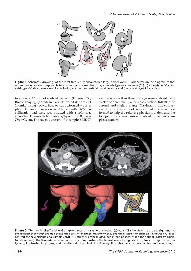
Figure 1. Schematic drawings of the most frequently encountered large bowel volvuli. Each arrow on the diagram of thenormal colon represents a possible torsion mechanism, resulting in: a) a bascule type cecal volvulus (CV), b) a loop type CV, c) anaxial type CV, d) a transverse colon volvulus, e) an organo-axial sigmoid volvulus and f) a typical sigmoid volvulus.
(a) (b)
Figure 2. The ‘‘whirl sign’’ and typical appearance of a sigmoid volvulus. (a) Axial CT slice showing a beak sign and noprogression of contrast enema beyond the obstruction site (black arrowhead) and the dilated sigmoid loop (*). (b) Axial CT slice
centred on the whirl sign of a sigmoid volvulus. Both ends of the dilated loop (*) can be seen, as can the normal upstream colon(white arrows). The three-dimensional reconstructions illustrate the lateral view of a sigmoid volvulus showing the rectum(green), the twisted loop (pink) and the efferent loop (blue). The drawing illustrates the structures involved in the whirl sign.
C Vandendries, M C Julles, I Boulay-Coletta et al
984 The British Journal of Radiology, November 2010

8/13/2019 Diagnosis of Colonic Volvulus Findings on Multidetector CT With
http://slidepdf.com/reader/full/diagnosis-of-colonic-volvulus-findings-on-multidetector-ct-with 3/8
Diagnosis and severity signs
When demonstrating an LBV the diagnosis criterion isan abrupt transition between a normal and dilated bowelcombined with the observation of convergence of bothends of the dilated loop towards the fulcrum point,creating the closed-loop obstruction. An important signto look for is the ‘‘whirl sign’’, which was first described
by Fisher [7]. This is a whirlpool pattern of concentricstructures including twisted intestinal loops, vessels andmesenteric fat that is highly suggestive of a torsionmechanism (Figure 2). The whirl sign is visible, however,
only if the view plane is orthogonal to the axis of
rotation. If the axis of the whirl were in the cranio-caudalaxis, for example, the whirl sign would be best visualisedin the axial plane. Therefore, we always review ourexams using MPR. When administered, the contrastenema does not go beyond the obstruction site and theabrupt interruption appears as a ‘‘beak sign’’.
MDCT helps radiologists to assess the severity of thecondition by analyzing the twisted loop wall and themesentery [8]. Spontaneous increased attenuation of the large bowel wall (which is related to transmuralhaemorrhagic necrosis), the absence or decreased en-hancement of the large bowel wall, pneumatosis intes-
tinalis (i.e. gas in the bowel wall) or thickening of the
(a) (b)
Figure 4. A typical sigmoid volvulus (SV) in a 51-year-old woman. (a) Coronal CT slice showing a typical SV and severity signs:dilated loop (*), rectum filled with contrast enema (+) and fully infiltrated mesosigmoid (arrowhead), which can be comparedwith normal mesentery (double arrowhead). (b) Axial CT slice at the fulcrum point (cranio-caudal axis). Pneumatosis intestinalis
of an afferent limb (white arrow), another severity sign, is seen here. The three-dimensional reconstruction shows a closedsigmoid loop (pink) with both limbs of the twisted loop converging toward the fulcrum point, the rectosigmoid junction (green)and the sigmoid-descending colon junction (blue).
(a) (b) (c)
Figure 3. Sketches illustrating the differences between (a) a normal sigmoid colon and a colon with (b) an organo-axial or (c) amesenterico-axial volvulus. Arrows represent torsion mechanisms, and the resulting mesenterico-axial axis is shown in (c).
Pictorial review: Colonic volvulus: findings on MDCT
The British Journal of Radiology, November 2010 985
8/13/2019 Diagnosis of Colonic Volvulus Findings on Multidetector CT With
http://slidepdf.com/reader/full/diagnosis-of-colonic-volvulus-findings-on-multidetector-ct-with 4/8
(a) (b)
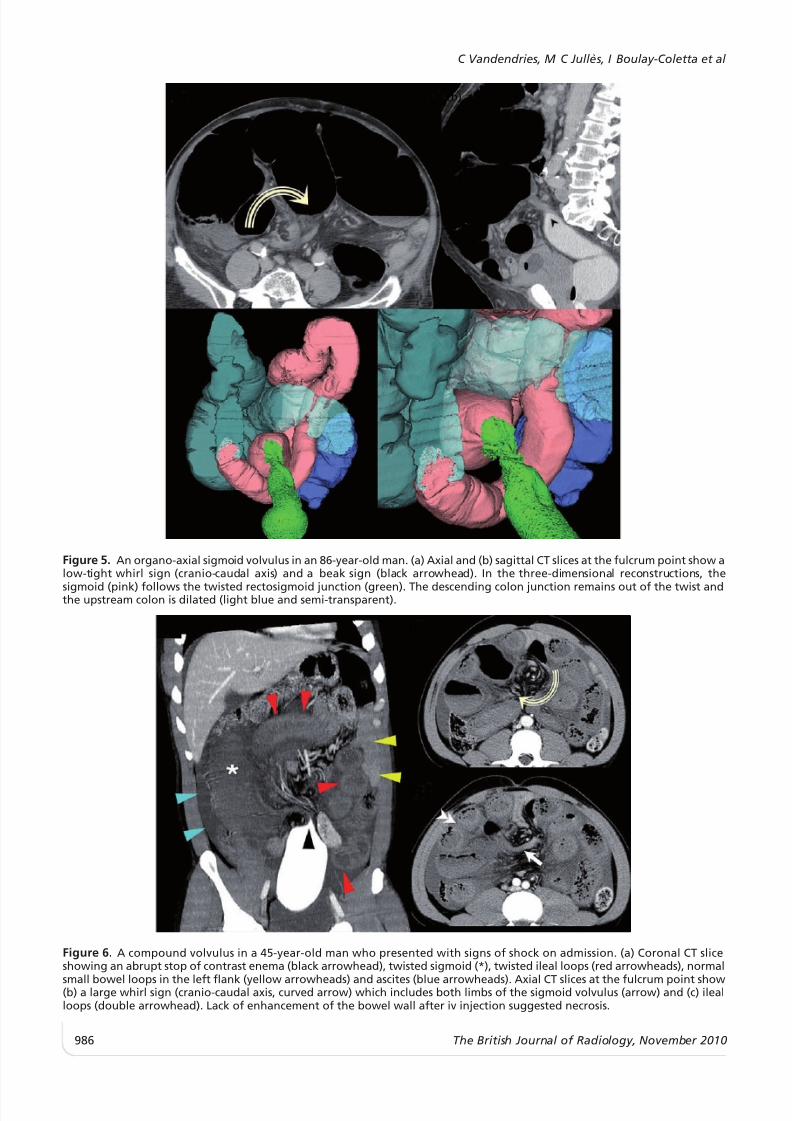
Figure 5. An organo-axial sigmoid volvulus in an 86-year-old man. (a) Axial and (b) sagittal CT slices at the fulcrum point show alow-tight whirl sign (cranio-caudal axis) and a beak sign (black arrowhead). In the three-dimensional reconstructions, the
sigmoid (pink) follows the twisted rectosigmoid junction (green). The descending colon junction remains out of the twist andthe upstream colon is dilated (light blue and semi-transparent).
(a) (b)
(c)
Figure 6. A compound volvulus in a 45-year-old man who presented with signs of shock on admission. (a) Coronal CT sliceshowing an abrupt stop of contrast enema (black arrowhead), twisted sigmoid (*), twisted ileal loops (red arrowheads), normal
small bowel loops in the left flank (yellow arrowheads) and ascites (blue arrowheads). Axial CT slices at the fulcrum point show(b) a large whirl sign (cranio-caudal axis, curved arrow) which includes both limbs of the sigmoid volvulus (arrow) and (c) ilealloops (double arrowhead). Lack of enhancement of the bowel wall after iv injection suggested necrosis.
C Vandendries, M C Julles, I Boulay-Coletta et al
986 The British Journal of Radiology, November 2010
8/13/2019 Diagnosis of Colonic Volvulus Findings on Multidetector CT With
http://slidepdf.com/reader/full/diagnosis-of-colonic-volvulus-findings-on-multidetector-ct-with 5/8
large bowel wall each suggest ischemia [6, 9] (Figure 4a,7a,b, 9a,b and 10a). MDCT readily demonstrates perfora-tion by showing even small pneumoperitoneum.
CT signs with low specificity for severe forms of LBVare the presence of ascites or haemorrhage, or theabsence of vessel enhancement within the twisted loopmesentery [6, 9].
Prompt recognition of these findings and quickdiagnosis of LBV is mandatory given the high rate of potentially lethal complications [5].
Sigmoid volvulus
Typical cases of SV (Figures 1f, 3 and 4) present asclosed loop obstructions. The loop twists around its mes-enteric axis resulting in a ‘‘mesenterico axial volvulus’’.
Both limbs of the twisted loop converge towards afulcrum point, which appears as a whirl sign when theview plane is orthogonal to the rotation axis of the loop.In most cases, the whirl sign is found in the lower leftpart of the abdomen with a cranio-caudal axis. Therectum and the upstream colon are usually flat whereasthe twisted loop is highly distended and located in the
anterior part of the abdomen.An alternative form of SV is the organo-axial volvulus(Figures 1e, 3 and 5) [10]. In these cases, there is noclosed-loop obstruction because only one transition pointis seen. Torsion occurs around the long axis of the loopand there is no more than one complete turn in theorgano-axial volvulus.
Ileosigmoid knotting, or compound volvulus, is a raremechanism of SV associated with small bowel volvulus(Figure 6). It is more frequently found in Africa, Asia and
(b)(a)
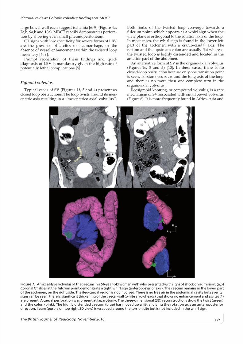
Figure 7. An axial-type volvulus of thecaecum in a 56-year-old woman with who presented with signs of shock on admission. (a,b)Coronal CT slices at the fulcrum point demonstrate a tight whirl sign (anteroposterior axis). The caecum remains in the lower partof the abdomen, on the right side. The ileo-caecal region is not involved. There is no free air in the abdominal cavity but severitysigns can be seen: there is significant thickening of the caecal wall (white arrowheads) that shows no enhancement and ascites (*)
are present. A caecal perforation was present at laparotomy. The three-dimensional (3D) reconstructions show the twist (green)and the colon (pink). The highly distended caecum (blue) has moved up a little, giving the rotation axis an anteroposteriordirection. Ileum (purple on top right 3D view) is wrapped around the torsion site but is not included in the whirl sign.
Pictorial review: Colonic volvulus: findings on MDCT
The British Journal of Radiology, November 2010 987
8/13/2019 Diagnosis of Colonic Volvulus Findings on Multidetector CT With
http://slidepdf.com/reader/full/diagnosis-of-colonic-volvulus-findings-on-multidetector-ct-with 6/8
the Middle East, at an average age of 40 years [11]. Thetwist involves a sigmoid loop as well as ileal loops. Fourlimbs converge to the fulcrum point and the whirl sign islarger and located higher in the abdomen [12]. Thisdiagnosis requires emergency surgery because ilealischemia is prone to quick perforation and leads to apoor outcome.
The initial treatment of SV is generally accepted to beendoscopic decompression by either rigid or flexiblesigmoidoscopy [2]. Urgent laparotomy is indicated in thepresence of clinical signs of peritonitis or CT signs of
bowel ischemia or perforation.
Cecal volvulus
CV is the torsion of a mobile caecum around its ownmesentery, which often results in a closed-loop obstruc-tion and a distended caecum. The terminal ileum isusually twisted along with the caecum. In most cases of CV, the whirl sign is found in the right part of the
abdomen with a lateral or an anteroposterior axis.Three forms of CV have been described: axial-type CV,
loop-type CV and cecal bascule [6, 9]. The axial type
(Figures 1c and 7), in which the caecum rotates along itsvertical axis, accounts for 45% of CV cases. In this type of CV, the distended caecum remains in the lower part of the abdomen, near the right side, which may lead tomisdiagnosis on plain radiographs.
The loop type (Figures 1b and 8) also accounts for 45%of CV cases. In these cases, the cecal rotation is associated
with an inversion secondary to an anterior displacement.Furthermore, the distended caecum is found in the upperpart of the abdomen, near the left side. Finally, the cecal
bascule (Figures 1a and 9) is encountered in 10% of CVcases. No twist occurs in this case, but anterior folding of the caecum, without rotation, is observed and there is nowhirl sign in any axis. The distended caecum is found inthe upper part of the abdomen, but it remains on theright side.
Management of CV requires surgical intervention be-cause endoscopic exsufflation is highly unpredictable [13].
Uncommon volvulus
A volvulus may involve a fixed part of the colonwhich, in theory, cannot move owing to its retroper-itoneal location, fixation by various ligaments or a broad
(a) (b) (c)
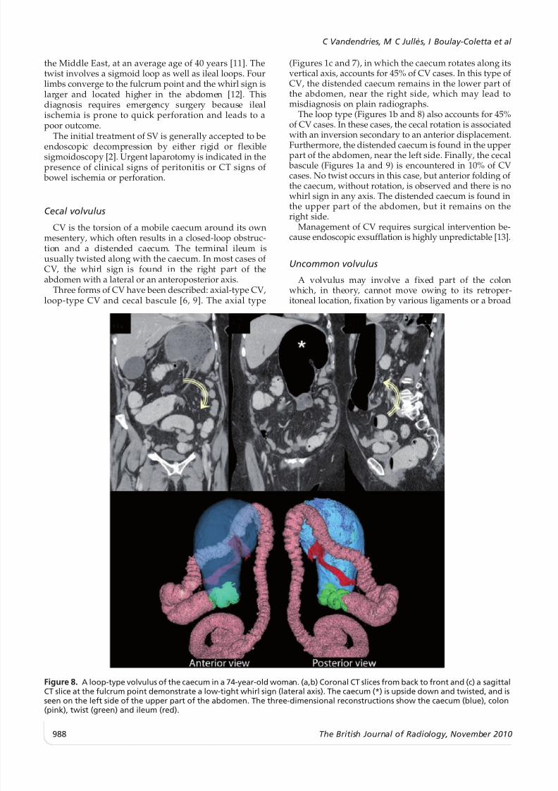
Figure 8. A loop-type volvulus of the caecum in a 74-year-old woman. (a,b) Coronal CT slices from back to front and (c) a sagittal
CT slice at the fulcrum point demonstrate a low-tight whirl sign (lateral axis). The caecum (*) is upside down and twisted, and isseen on the left side of the upper part of the abdomen. The three-dimensional reconstructions show the caecum (blue), colon(pink), twist (green) and ileum (red).
C Vandendries, M C Julles, I Boulay-Coletta et al
988 The British Journal of Radiology, November 2010
8/13/2019 Diagnosis of Colonic Volvulus Findings on Multidetector CT With
http://slidepdf.com/reader/full/diagnosis-of-colonic-volvulus-findings-on-multidetector-ct-with 7/8
base of mesenteric attachment. These segments include
the right colon, the transverse colon, the splenic flexure[14] or even the descending colon [15]. Volvulus of the
right hemicolon (Figure 10) is exceptional, requiring a
persistent mesocolon. To the best of our knowledge, thistype of LBV has never been described in the radiological
(a) (b)
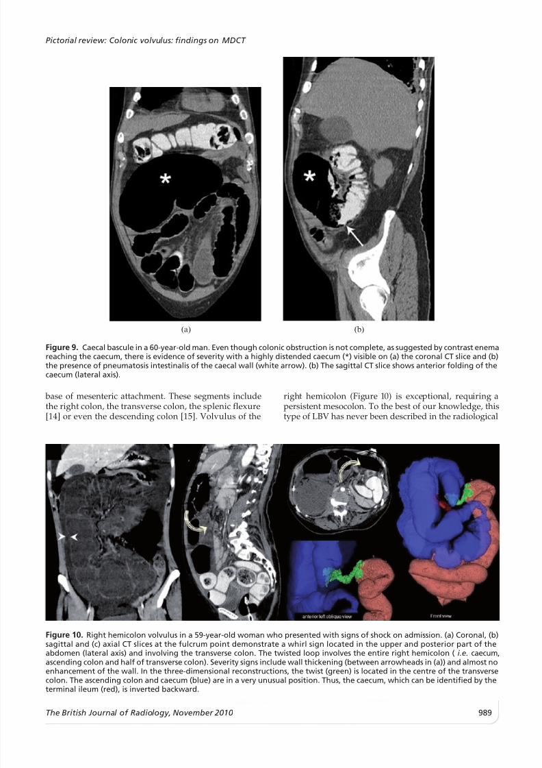
Figure 9. Caecal bascule in a 60-year-old man. Even though colonic obstruction is not complete, as suggested by contrast enemareaching the caecum, there is evidence of severity with a highly distended caecum (*) visible on (a) the coronal CT slice and (b)the presence of pneumatosis intestinalis of the caecal wall (white arrow). (b) The sagittal CT slice shows anterior folding of thecaecum (lateral axis).
(a) (b) (c)
Figure 10. Right hemicolon volvulus in a 59-year-old woman who presented with signs of shock on admission. (a) Coronal, (b)sagittal and (c) axial CT slices at the fulcrum point demonstrate a whirl sign located in the upper and posterior part of theabdomen (lateral axis) and involving the transverse colon. The twisted loop involves the entire right hemicolon ( i.e. caecum,ascending colon and half of transverse colon). Severity signs include wall thickening (between arrowheads in (a)) and almost no
enhancement of the wall. In the three-dimensional reconstructions, the twist (green) is located in the centre of the transversecolon. The ascending colon and caecum (blue) are in a very unusual position. Thus, the caecum, which can be identified by theterminal ileum (red), is inverted backward.
Pictorial review: Colonic volvulus: findings on MDCT
The British Journal of Radiology, November 2010 989

8/13/2019 Diagnosis of Colonic Volvulus Findings on Multidetector CT With
http://slidepdf.com/reader/full/diagnosis-of-colonic-volvulus-findings-on-multidetector-ct-with 8/8
literature. Although the transverse colon is a mobileloop, a volvulus in this region of colon (Figures 1d and
11) is very unusual because its base is spread widelyfrom splenic to hepatic flexure.
Conclusion
Volvulus formation is a rare but severe cause of organic obstruction of the large bowel. By establishing aprecise diagnosis including topography, mechanism andseverity, MDCT allows accurate assessment of large
bowel volvuli. Radiologists should be able to recognisetheir appearance so that the proper diagnosis can bemade and catastrophic consequences can be avoided.
Bibliography
1. Jones DJ. ABC of colorectal diseases. Large bowel volvulus.BMJ 1992;305:358–60.
2. Lau KC, Miller BJ, Schache DJ, Cohen JR. A study of large- bowel volvulus in urban Australia. Can J Surg 2006;49:203–7.
3. Ahn SH, Mayo-Smith WW, Murphy BL, Reinert SE, Cronan JJ. Acute nontraumatic abdominal pain in adult patients:abdominal radiography compared with CT evaluation.Radiology 2002;225:159–64.
4. Kellow ZS, MacInnes M, Kurzencwyg D, Rawal S, Jaffer R,Kovacina B, et al. The role of abdominal radiography in the
evaluation of the nontrauma emergency patient. Radiology2008;248:887–93.
5. Madiba TE, Thomson SR. The management of cecalvolvulus. Dis Colon Rectum 2002;45:264–7.6. Moore CJ, Corl FM, Fishman EK. CT of cecal volvulus:
unraveling the image. AJR Am J Roentgenol 2001;177:95–8.7. Fisher JK. Computed tomographic diagnosis of volvulus in
intestinal malrotation. Radiology 1981;140:145–6.8. Catalano O. Computed tomographic appearance of sigmoid
volvulus. Abdom Imaging 1996;21:314–7.9. Delabrousse E, Sarlieve P, Sailley N, Aubry S, Kastler BA.
Cecal volvulus: CT findings and correlation with patho-physiology. Emerg Radiol 2007;14:411–5.
10. Janzen DL, Heap SW. Organo-axial volvulus of the sigmoidcolon. Australas Radiol 1992;36:332–3.
11. Kotisso B, Bekele A. Ilio-sigmoid knotting in Addis Ababa:a three-year comprehensive retrospective analysis. Ethiop
Med J 2006;44:377–83.12. Lee SH, Park YH, Won YS. The ileosigmoid knot: CT
findings. AJR Am J Roentgenol 2000;174:685–7.13. Habre J, Sautot-Vial N, Marcotte C, Benchimol D. Caecal
volvulus. Am J Surg 2008;196:e48–9.14. Mittal R, Samarasam I, Chandran S, Mathew G. Primary
splenic flexure volvulus. Singapore Med J 2007;48:e87–9.
15. Chen A, Yang FS, Shih SL, Sheu CY. Case report. CTdiagnosis of volvulus of the descending colon withpersistent mesocolon. AJR Am J Roentgenol 2003;180:1003–6.
(a) (b)
(c)
Figure 11. Transverse colon volvulus in a 23-year-old woman. (a) Sagittal and (b) coronal CT slices at the fulcrum pointdemonstrate a whirl sign that is located in the upper and posterior part of the abdomen (lateral axis). Both limbs of the twistedloop (* with green outline in (b), * in (c)) converge on the transverse colon. The twisted loop involves a segment of thetransverse colon. Upstream and downstream colons are seen in the correct position. (c) Peroperative view: free round ligamentof the liver creating a strap with no adhesion to the parietal peritoneum. In the three-dimensional reconstructions, the twistedloop (green) is located on the transverse colon. The ascending colon and caecum (blue) are at their usual locations, as are thedescending colon and splenic flexure (pink).
C Vandendries, M C Julles, I Boulay-Coletta et al
990 The British Journal of Radiology, November 2010