diagnosing pancreatic disease: help from the laboratory disease 8-8-12_new… · diagnosing...
TRANSCRIPT

Diagnosing Pancreatic Disease: Help from the Laboratory
Kajsa Affolter M.D. Department of Pathology
PGY 2 Resident

University of Utah CME Statement
• The University of Utah School of Medicine adheres to ACCME Standards regarding industry support of continuing medical education.
• Speakers are also expected to openly disclose intent to discuss any off-label, experimental, or investigational use of drugs, devices, or equipment in their presentations.
• This speaker has nothing to disclose.

Objectives
1. Diagram basic gross and microscopic pancreatic anatomy 2. Given classic patient scenarios, compare and contrast pancreatic cancer and acute pancreatitis 3. When encountering a screening test in the lab, be able to evaluate advantages and disadvantages of the screening tests 4. List commonly used serum biomarkers for evaluating pancreatic disease

FIRST Let’s do a quick review:
Anatomy Histology
Physiology

http://www.hopkins-gi.org

http://www.hopkins-gi.org Major duodenal papilla = Ampulla of Vater, Sphincter of Oddi

Normal Pancreatic Histology
Islet of Langerhans
Acinar Cells
Intercalated Duct
http://www.surgpath4u.com

Conduit for acinar cell secretions: Acinar lumen intercalated ducts interlobular ducts main PD
http://www.hopkins-gi.org

Normal Pancreatic Exocrine Histology
http://www.surgpath4u.com

Pancreatic Exocrine Function
http://en.wikibooks.org

Pancreatic Exocrine Digestive Function
Adapted from: http://en.wikibooks.org
Nutrient Enzyme Product
Carbohydrates and Starch
Amylase Saccharides
Fats Lipase and Colipase Triglycerides
Proteins Trypsin (trypsinogen) Peptides
Chymotrypsin (chymotrypsinogen)
Peptides

Done with the review:
What could possibly go wrong?

Case #1 • 48-year-old female presents to her primary care
physician complaining of severe pain in her upper abdomen – Pain radiates to back – Present for the past 30 minutes
• Medical history: gallstones and obesity • Family History: Not significant • Social History: Negative tobacco and alcohol • Review of Systems: Nausea, low grade fever
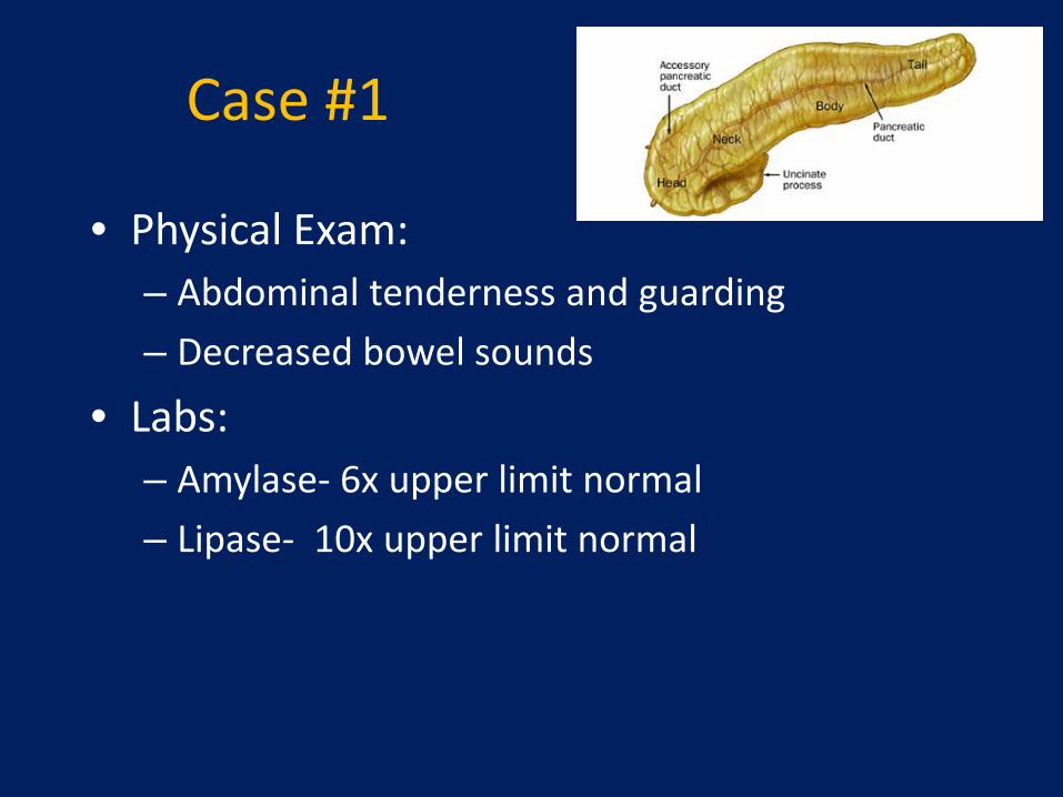
Case #1
• Physical Exam: – Abdominal tenderness and guarding – Decreased bowel sounds
• Labs: – Amylase- 6x upper limit normal – Lipase- 10x upper limit normal

DIAGNOSIS?
ACUTE PANCREATITIS

Acute Pancreatitis Pathogenesis Mechanism - Auto Digestion
Co-localization of lysosomal proteases Trypsinogen is activated to trypsin
Extensive inflammatory response Intrapancreatic and Extrapancreatic
http://www.hopkins-gi.org

Causes Acute Pancreatitis Gallstones: Duct obstruction, Reflux of bile
http://www.hopkins-gi.org
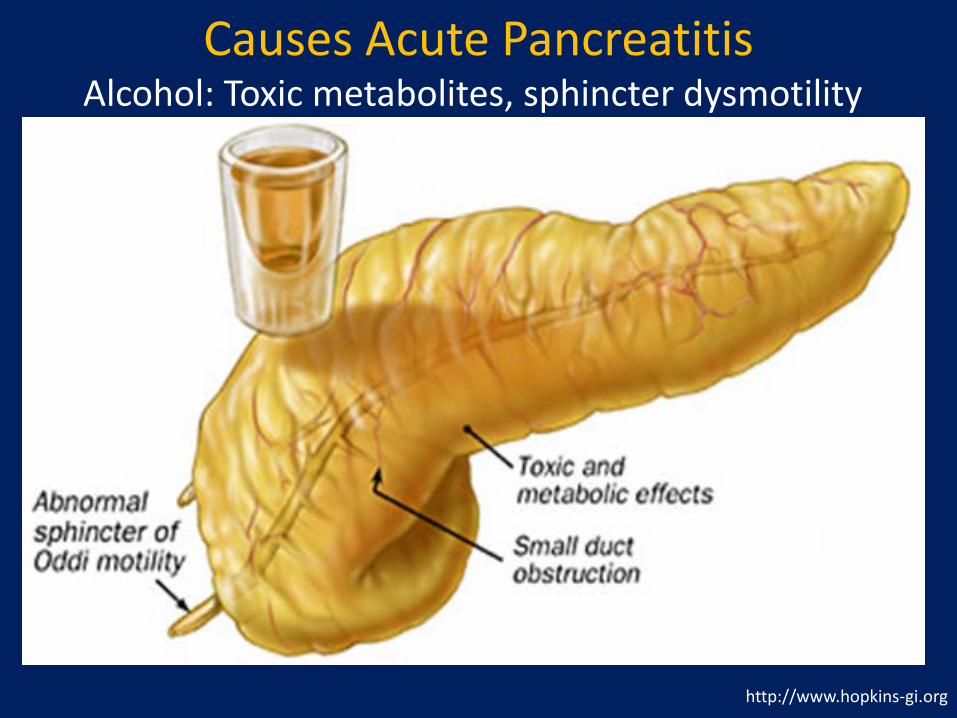
Causes Acute Pancreatitis Alcohol: Toxic metabolites, sphincter dysmotility
http://www.hopkins-gi.org
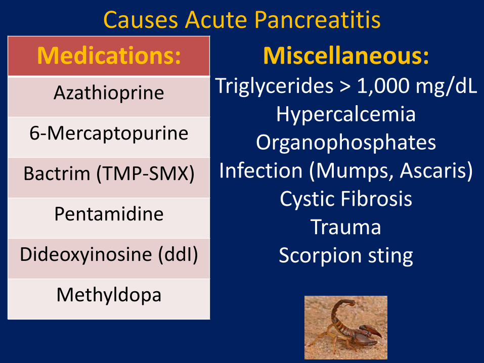
Causes Acute Pancreatitis Miscellaneous:
Triglycerides > 1,000 mg/dL Hypercalcemia
Organophosphates Infection (Mumps, Ascaris)
Cystic Fibrosis Trauma
Scorpion sting
Medications: Azathioprine
6-Mercaptopurine
Bactrim (TMP-SMX)
Pentamidine
Dideoxyinosine (ddI)
Methyldopa

Causes Acute Pancreatitis Idiopathic: Probable Microlithiasis (small stones)
http://www.hopkins-gi.org

Acute Pancreatitis • Pancreatitis ranges from mild (inflammatory
process and edema) to severe (necrotic process and secondary extra pancreatic injury)
http://www.musc.edu/pathology http://www.surgpath4u.com
Normal Histology

How could we diagnose our patient so quickly?
Severe abdominal pain
Elevated serum amylase & lipase levels
Initial diagnosis of acute pancreatitis

USEFUL LAB TEST:
Amylase
www.wikipedia.org

Amylase • Amylases are glycoside hydrolases
– Alpha amylase • Ca2+ metalloenzyme (unable to function in absence of Ca2+) • Acts at random locations along a starch chain, yielding:
– Maltotriose, maltose and limit dextrin from amylose – Maltose, glucose and limit dextrin from amylopectin
www.wikipedia.org

Amylase Sources/ Activators
• Salivary – S-amylase
• Pancreatic – P-amylase
• CALCIUM AND • Chloride
http://embryology.med.unsw.edu http://leavingbio.net www.wikipedia.org
Ca2+
Cl-

Amylase
• Plasma enzyme found in the urine (small molecule- 54,000- 62,000 MW)
• Magnitude of elevation not correlated to severity • Magnitude of elevation = greater probability acute pancreatitis
1
2
3
4
5
6
7
Symptom onset 5-8 hours 12-72 hours 3-4 days
Mul
tiple
s of B
asel
ine
Serum Total Amylase
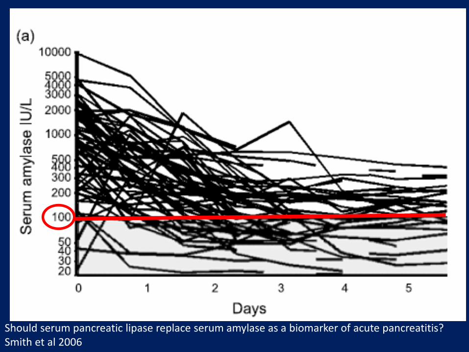
Should serum pancreatic lipase replace serum amylase as a biomarker of acute pancreatitis? Smith et al 2006

False positive test if looking for acute
pancreatitis
Lack of specificity for total AMY
↑ Specificity (90%): P-AMY and 3x the
upper ref limit Adapted from Teitz Textbook Ch 21 Enzymes
Pancreatic Disease
Pancreatitis (P-AMY)
Pancreatic Trauma (P-AMY)
Other Intraabdominal
Disease
Biliary Tract Disease (P-AMY)
Intestinal Obstruction (P-AMY)
Mesenteric Infarction (P-AMY)
Perforated peptic ulcer (P-AMY)
Gastritis, Duodenitis (P-AMY)
Ruptured Aortic Aneurysm
Acute Appendicitis
Peritonitis
Trauma
Genitourinary Disease
Ruptured Ectopic Pregnancy (S-AMY)
Salpingitis (S-AMY)
Ovarian Malignancy (S-AMY)
Renal Insufficiency (mixed)
Misc Salivary gland lesion, Acute alcoholic abuse, DKA, Macroamylasemia
Causes of Increased Amylase

Macroamylasemia
• Complexes: amylase (usually S-type) and IgG or IgA
• Cannot filter through the glomeruli (MW > 200,000); ultrafiltration assay, decreased amylase to
CrCl ratio (<1%), or urine amylase level
• No clinical symptoms associated
• 2.5% of hyperamylasemic patients and 1% of healthy subjects
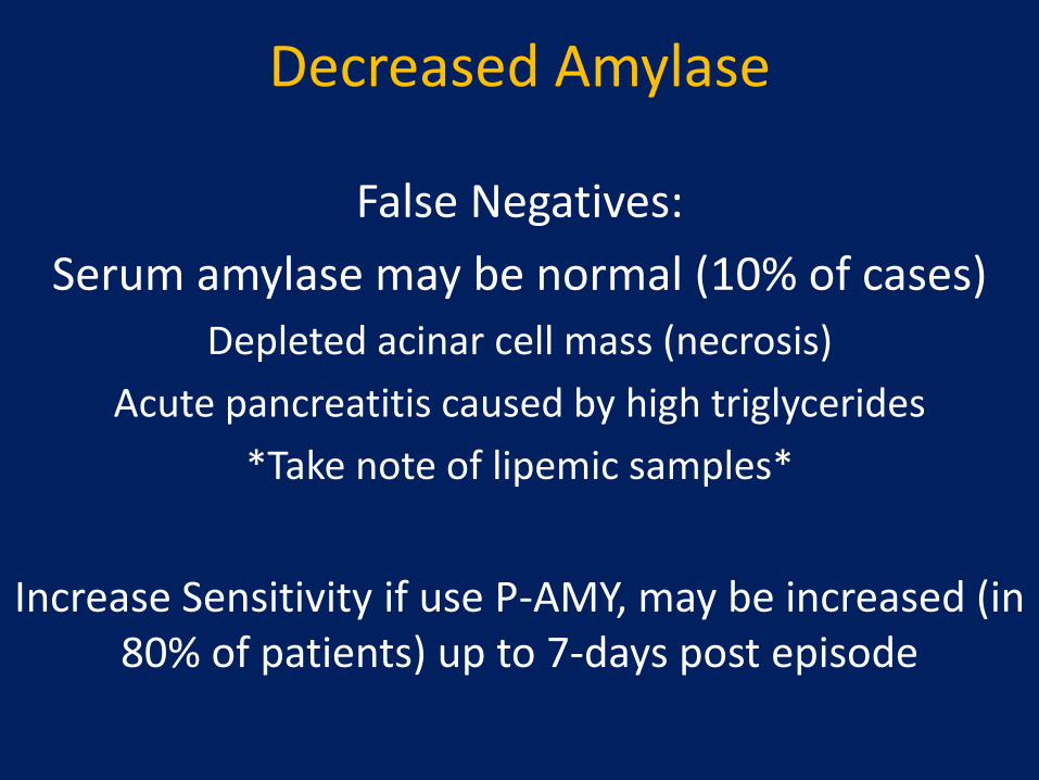
Decreased Amylase
False Negatives: Serum amylase may be normal (10% of cases)
Depleted acinar cell mass (necrosis) Acute pancreatitis caused by high triglycerides
*Take note of lipemic samples*
Increase Sensitivity if use P-AMY, may be increased (in 80% of patients) up to 7-days post episode

Amylase Method • Can measure substrate decrease viscometrically,
turbidimetrically, nephelometrically, amyloclastically • Saccharogenic and kinetic (spectrophotometric)
measurements used more commonly now – Saccharogenic assays measure glucose production
• Kinetic method correlates with HPLC measurement
Measure absorbance increase at 405 nm ET = ethylidene G = glucose PNP = p-nitrophenol
Thank you Dr. Straseski

Measuring P-Amylase Activity
Inhibit S-AMY with monoclonal antibodies
USEFUL LAB TEST:
Lipase
www.wikipedia.org

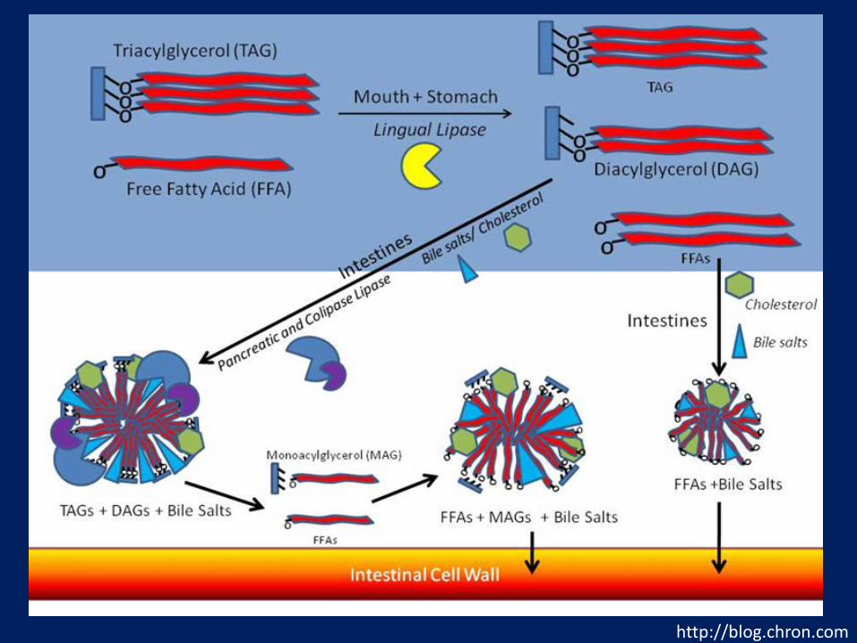
Human Pancreatic Lipase (HPL) • Lipases are a subclass of the esterases; hydrolyze
triglyceride substrates to monoglycerides and FFA
• Activated Ternary Complex – Lipase – Bile Salt Micelle – Colipase
http://blog.chron.com
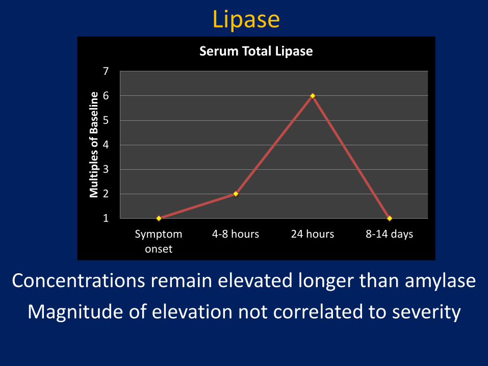
Lipase
Concentrations remain elevated longer than amylase Magnitude of elevation not correlated to severity
1
2
3
4
5
6
7
Symptomonset
4-8 hours 24 hours 8-14 days
Mul
tiple
s of B
asel
ine
Serum Total Lipase

Should serum pancreatic lipase replace serum amylase as a biomarker of acute pancreatitis? Smith et al 2006

Lipase
• Sensitivity and Specificity are 80-100% depending on patient population and diagnostic cutoff
• Increased if use guideline- likely acute pancreatitis if >5x upper limit of reference range
• False positives: Obstruction of duct (carcinoma), reduced glomerular filtration rate, Opiates (cause sphincter of Oddi to contract)

Lipase Activity: Laboratory Measurement • Enzymatic method • Cleavage of chromogenic lipase substrate emulsified with bile
acid and colipase in alkaline medium • Rate of color is directly proportional
Lipase 1,2-O-dilauryl-rac-glycero-3-glutaric acid-
(6-methylresorufin) ester
glutaric acid + methylresorufin
1,2-O-dilauryl-rac-glycerol + glutaric acid-(6-methylresorufin) ester
Spontaneous decomposition
Red dye measured at 570 nm
Thank you Dr. Straseski
Reference ranges = 16 – 63 IU/L

Diagnosis: Acute Pancreatitis
Bakerman’s ABC’s, 4th edition, 2002. Thank you Dr. Straseski

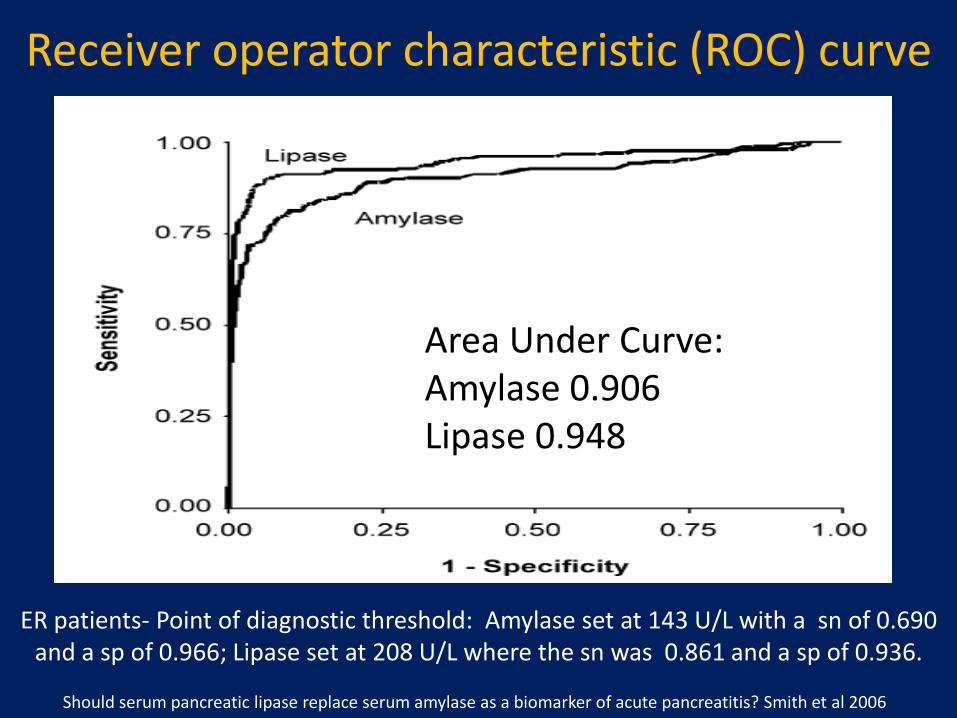
Receiver operator characteristic (ROC) curve
ER patients- Point of diagnostic threshold: Amylase set at 143 U/L with a sn of 0.690 and a sp of 0.966; Lipase set at 208 U/L where the sn was 0.861 and a sp of 0.936.
Should serum pancreatic lipase replace serum amylase as a biomarker of acute pancreatitis? Smith et al 2006
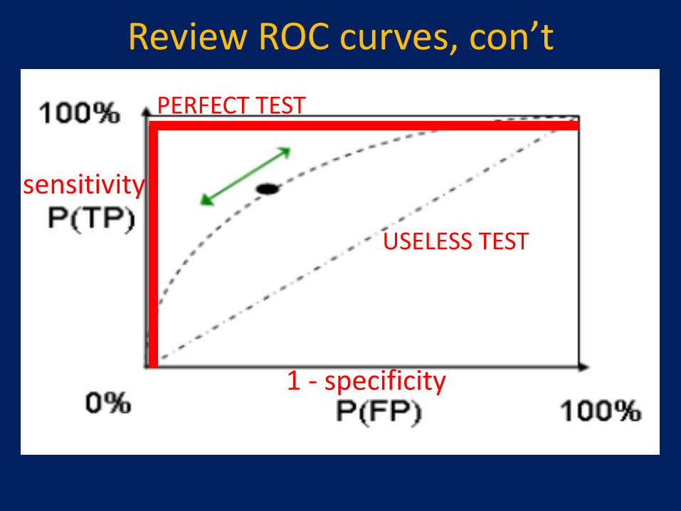
Review ROC curves, con’t
1 - specificity
sensitivity
USELESS TEST
PERFECT TEST

Review ROC curves, con’t
AUC: Increased Area = Better Test
1 - specificity
sensitivity
USELESS TEST
PERFECT TEST
0.5 or 50% or Area is filled
1.0 or 100% or the Area is filled
Receiver operator characteristic (ROC) curve
Area Under Curve: Amylase 0.906 Lipase 0.948
ER patients- Point of diagnostic threshold: Amylase set at 143 U/L with a sn of 0.690 and a sp of 0.966; Lipase set at 208 U/L where the sn was 0.861 and a sp of 0.936.
Should serum pancreatic lipase replace serum amylase as a biomarker of acute pancreatitis? Smith et al 2006

Amylase vs. Lipase Guidelines “…not necessary to measure both …lipase may be preferable …serum lipase is thought to be more sensitive and specific …in the diagnosis of acute
pancreatitis.“ -Banks et al. American GI Society and American College of Gastroenterology (2006)
Although amylase is widely available and provides acceptable
accuracy of diagnosis, where lipase is available it is preferred for the diagnosis of acute pancreatitis (recommendation grade A)“ -
UK GI Party

Acute Pancreatitis
Other Factors Contributing to the Diagnosis/ Risk:
Imaging
Additional Labs


Risk Assessment
On Admission Within 48 hours
Age >55 years Hematocrit decrease by >10%
WBC > 16,000 mm3 Urea Nitrogen increase >5 mg/dl
LDH > 350 U/L Serum calcium < 8 mg/dl
Glucose > 200 mg/dl Arterial PO2 < 60 mm Hg
AST > 250 U/L Base deficit > 4mmol/L
Estimated fluid sequestration > 6 L
Ranson’s Criteria
Temperature Arterial pH Leukocytes
Mean BP Sodium/Potassium Hematocrit
Heart Rate Glucose Albumin
Respiratory Rate Creatinine Bilirubin
Oxygenation BUN Age
APACHE III Criteria (Acute Physiology and Chronic Health Eval.

Mortality in Acute Pancreatitis
Banks et al. Practice Guidelines in Acute Pancreatitis
Treatment
• Aggressive Intravenous Fluids • Nil per os (“NPO”) = Nothing by mouth • Parenteral Narcotics • +/- Antibiotics (necrotizing pancreatitis) • Transfer to ICU • Look for Etiology
Pearls of Wisdom
Acute Pancreatitis
• Alcohol and Gallstones account for majority of cases • Amylase greater than 3x upper limit of ref range
• Lipase greater sensitivity and specificity
• False positives exist for elevated levels of enzymes

Case #2 • 61-year-old male presents to his primary care
physician complaining of gradually increasing pain in his upper abdomen – Pain radiates to his back
• Medical history is significant for Hypertension • Family History: Not significant • Social History: (+) tobacco • Review of Systems: weight loss; several week
history of “painless jaundice” prior to pain starting

Case #2 • Physical Exam (Pertinent Positives):
– Icteric sclera – Palpable left supraclavicular lymph (Virchow’s) node
• Lab – Amylase and Lipase 1.5x the upper limit of normal
• Imaging – Pancreatic Protocol CT scan
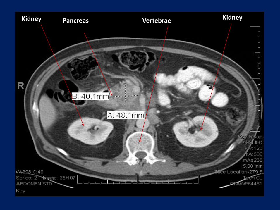
Kidney Kidney Vertebrae Pancreas
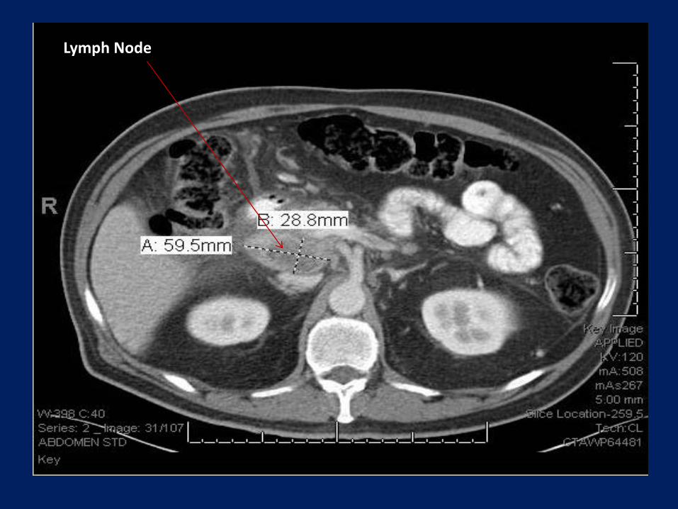
Lymph Node

Liver
Liver

Biopsy via Endoscopic Ultrasound guided Fine Needle Aspiration

Biopsy via EUS guided FNA

Normal Pancreas
Acinar cells
Ductal cells
Pancreas is near the duodenum, liver, transverse colon, stomach, spleen, and kidneys

Necrosis

“Drunken Honeycomb”

Nuclear Pleomorphism Nuclear Anisonucleosis
Clumped Chromatin

DIAGNOSIS?
PANCREATIC DUCTAL ADENOCARCINOMA

Pancreatic Ductal Adenocarcinoma (PDAC)
• 43,140 people are diagnosed annually in US – incidence 10–12 per 100,000 people
• Mortality rate of 36,800/year in US
• Mortality rate 227,000/year in World
• 4th on the list of cancer related causes of death
• 5 year survival rate is <5%
Wasif N, Ko CY, Farrell J, Wainberg Z, Hines OJ, Reber H, Tomlinson JS. Impact of tumor grade on prognosis in pancreatic cancer: should we include grade in AJCC staging?

WHO 2004 Data (incidence per 100,000)

Ahmedin Jemal, DVM, PhD et al Cancer Statistics, 2010

Ahmedin Jemal, DVM, PhD et al Cancer Statistics, 2010

Death Rate per 100,000 Males and Females
MALE FEMALE
Ahmedin Jemal, DVM, PhD et al Cancer Statistics, 2010

PDAC: Implicated Factors • Smoking (2.5-3.6 x increase risk) • Family history, a pair of first-degree relatives = familial • History of chronic pancreatitis, EtOH • Advancing age • Male sex • Diabetes mellitus • Obesity • Non-O blood group • Occupational exposures (eg chlorinated hydrocarbon solvents
and nickel) • African-American ethnicity • Diet, high fat/high meat and low in vegetables and folate • Possibly Helicobacter pylori infection • Possibly periodontal disease

Becoming Cancer
Ottenhof NA et al. Molecular characteristics of pancreatic ductal adenocarcinoma

PDAC Pathophysiology
Precursors: PanINs, MCNs, and IPMNs Vincent A et al, Pancreatic cancer
PanIN-1A PanIN-1B PanIN-2 PanIN-3

Molecular Features of PDAC
MORE THAN A DECADE TO PROGRESS… a guess (creating a window for possible early detection)
Ottenhof NA et al. Molecular characteristics of pancreatic ductal adenocarcinoma

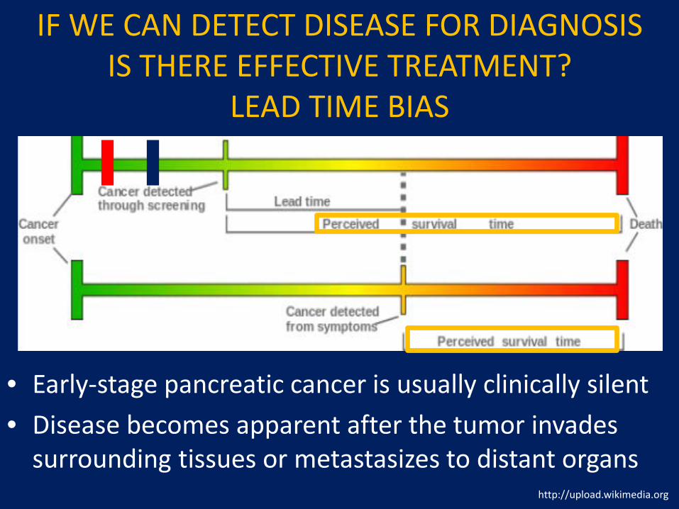
IF WE CAN DETECT DISEASE FOR DIAGNOSIS IS THERE EFFECTIVE TREATMENT?
LEAD TIME BIAS
• Early-stage pancreatic cancer is usually clinically silent • Disease becomes apparent after the tumor invades
surrounding tissues or metastasizes to distant organs – 80% of the time has already metastasized
http://upload.wikimedia.org
IF WE CAN DETECT DISEASE FOR DIAGNOSIS IS THERE EFFECTIVE TREATMENT?
LEAD TIME BIAS
• Early-stage pancreatic cancer is usually clinically silent • Disease becomes apparent after the tumor invades
surrounding tissues or metastasizes to distant organs
http://upload.wikimedia.org

World Health Organization — Principles of Screening (1968)
1. The condition should be an important health problem. 2. There should be a latent stage of the disease. 3. There should be a test or examination for the condition. 4. There should be a treatment for the condition. 5. There should be an agreed policy on whom to treat. 6. Facilities for diagnosis and treatment should be available. 7. The test should be acceptable to the population. 8. The natural history of the disease should be adequately
understood. 9. The total cost of finding a case should be economically
balanced in relation to medical expenditure as a whole. 10. Case-finding should be a continuous process, not just a
"once and for all" project.
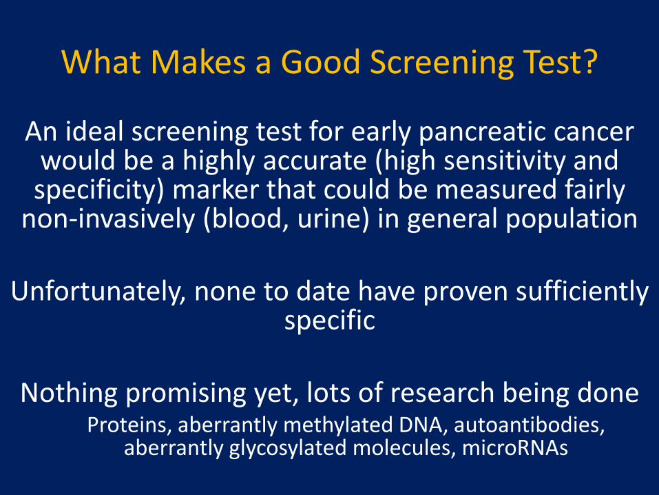
What Makes a Good Screening Test?
An ideal screening test for early pancreatic cancer would be a highly accurate (high sensitivity and specificity) marker that could be measured fairly
non-invasively (blood, urine) in general population
Unfortunately, none to date have proven sufficiently specific
Nothing promising yet, lots of research being done
Proteins, aberrantly methylated DNA, autoantibodies, aberrantly glycosylated molecules, microRNAs

Screening those with familial risk
• Families with mutated susceptibility genes – Do NOT manifest a high penetrance of PDAC – Unexplained, Under reported, Underused
• Consensus guidelines have not been established for genetic testing of those at risk for inherited PDAC
Syndrome Germline Mutations Relative Risk PDAC
Familial Atypical Multiple Melanoma and Mole Syndrome
CDKN2A 20-34
Peutz-Jeghers Syndrome LKB1 >100
Hereditary pancreatitis PRSS1/SPINK1 90
Familial Breast Cancer BRCA 2 3-10
Lynch Syndrome Mismatch repair unknown

Cancer of the Pancreas Screening Study (CAPS)- Imaging and DNA studies
• Multi-center, translational prospective controlled cohort study in high risk patients
• Pancreatic cystic lesions were detected more frequently with endoscopic ultrasound (93%) and MRI (81%) than with CT (27%) – Best sampled by EUS-FNA
• PanINs are usually not visible by imaging, research is attempting to identify markers in pancreatic fluid that could reliably identify high-grade PanINs

We need better screening tests • <20% of patients qualify for surgical resection
at diagnosis
• Surgical resection- only treatment to improve five-year survival rates – < 4% to 25–30%
• Chemo(radiation) therapy administered in
(neo)adjuvant setting

We have no good screening tests.
What about tumor markers?
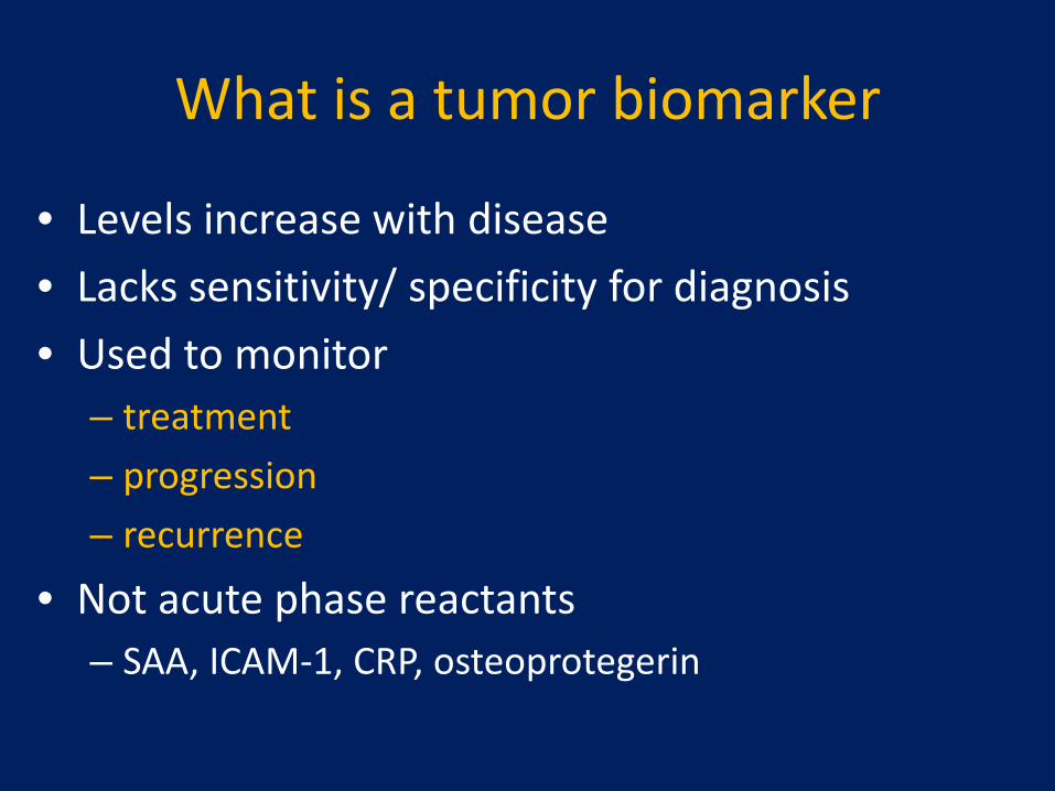
What is a tumor biomarker
• Levels increase with disease • Lacks sensitivity/ specificity for diagnosis • Used to monitor
– treatment – progression – recurrence
• Not acute phase reactants – SAA, ICAM-1, CRP, osteoprotegerin

Carcinoembryonic Antigen (CEA)
Glycoprotein involved in cell adhesion Produced during fetal life; decreases prior to birth
Can Measure in Serum or in Cyst Fluid
http://en.wikipedia.org

Carcinoembryonic Antigen (CEA) CYST
TUMOR MARKER
NON MUCINOUS CYST MUCINOUS CYST
CEA Not Elevated Elevated
Increased levels indicative of a mucinous cyst (does not distinguish benign from malignant)
ARUP- “body fluid” specimen category-off label

Carcinoembryonic Antigen (CEA) SERUM
ELEVATED LEVEL = MALIGNANT ELEVATED LEVEL = BENIGN
Colorectal Carcinoma Ulcerative Colitis
Gastric Carcinoma Crohn’s Disease
Pancreatic Carcinoma Pancreatitis
Lung Carcinoma COPD
Breast Carcinoma Cirrhosis
Medullary Thyroid Carcinoma Smokers

Carbohydrate or Cancer Antigen (CA 19-9)
• False (+): Increased in colorectal cancer, esophageal cancer, hepatocellular carcinoma, pancreatitis, cirrhosis, and diseases or obstruction of the bile ducts.
• False (-): CA 19-9 is sialylated Lewis (a) antigen
(adsorbed RBC antigens) – 10% of the Caucasian population lacks the Lewis antigen
(deficiency of a fucosyltransferase) = CA19-9 is not expressed

Carbohydrate or Cancer Antigen (CA 19-9)
• Preoperative amounts of carbohydrate antigen 19-9 (CA19-9) of more than 100–200 U/mL predict
unresectability
• Biliary drainage lowers nonspecific CA19-9 amounts, allowing for more reliable estimate of
disease burden
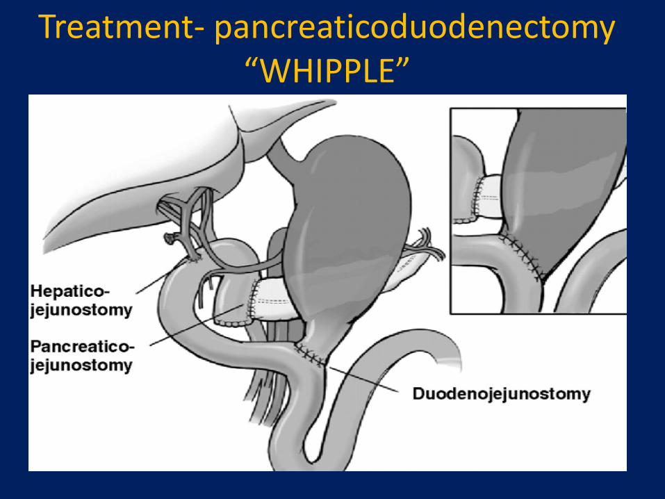
Treatment- pancreaticoduodenectomy “WHIPPLE”

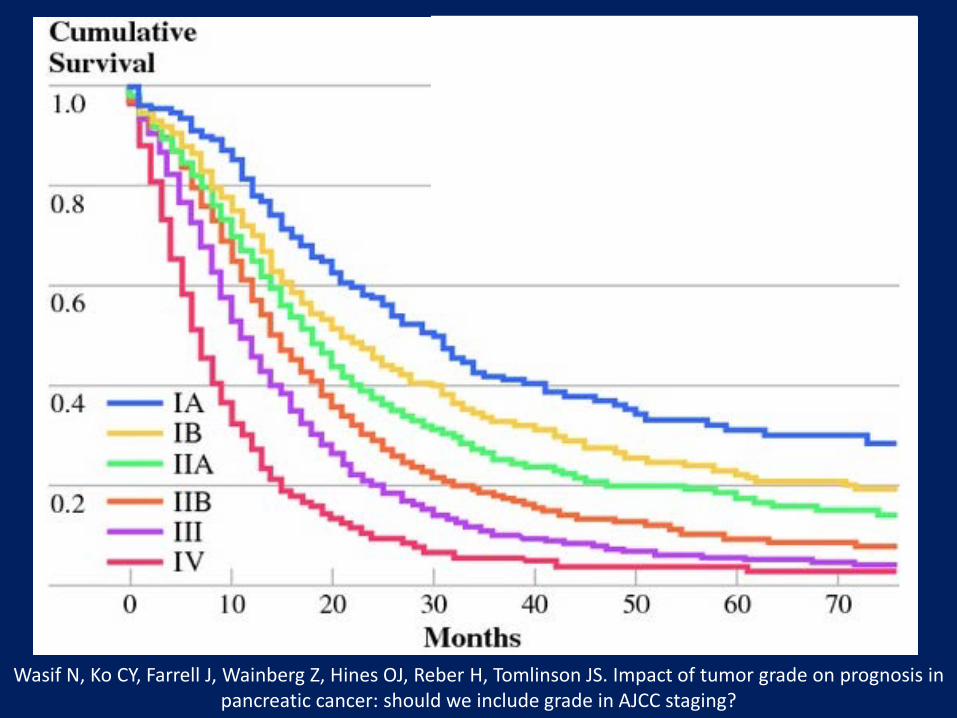
Staging of PDAC Stage Median Survival (mo) Characteristics
IA 24.1 Limited to pancreas, <2 cm
IB 20.6 Limited to pancreas, >2 cm
IIA 15.4 Locally invasive, no involvement celiac or SMA
IIB 12.7 Locally invasive, Lymph Node metastasis
III 10.6 Celiac axis or SMA involved (unresectable)
IV 4.5 Distant Metastasis (unresectable)

Wasif N, Ko CY, Farrell J, Wainberg Z, Hines OJ, Reber H, Tomlinson JS. Impact of tumor grade on prognosis in pancreatic cancer: should we include grade in AJCC staging?
Treatment
• Chemotherapy after resection- gemcitabine
• The addition of erlotinib (small molecule inhibitor of EGFR) or fluoropyrimidine have shown slight improvements of overall survival – Erlotinib - modest survival improvement and increased
level of toxicity has limited the acceptance
Pearls of Wisdom • Mortality rate is extremely high; few survivors • Research on screening for PDAC should focus of
PRE invasive lesions • Tumor markers are not synonymous with
screening tests • Much Room for Improvement
– Screening – Biomarkers – Treatment

Summary Commonly used tests for
diagnosing or evaluating pancreatic disease
Acute Pancreatitis Amylase and Lipase Imaging Pancreatic Cancer No good screens (yet) EUS, cytopathology for diagnosis CEA, CA 19-9 for monitoring Imaging

References • Ottenhof NA, de Wilde RF, Maitra A, Hruban RH,
Offerhaus GJ. Molecular characteristics of pancreatic ductal adenocarcinoma. Patholog Res Int. 2011 Mar 27;2011:620601. PubMed PMID: 21512581
• Banks P, Freeman M (2006). "Practice guidelines in acute pancreatitis". Am J Gastroenterol 101 (10): 2379–400.
• UK Working Party on Acute Pancreatitis (2005). "UK guidelines for the management of acute pancreatitis". Gut 54 Suppl 3 (Suppl 3): iii1–9.
• Smith RC, Southwell-Keely J, Chesher D. Should serum pancreatic lipase replace serum amylase as a biomarker of acute pancreatitis? ANZ J Surg. 2005 Jun;75(6):399-404. PubMed PMID: 15943725
References • Vincent A, Herman J, Schulick R, Hruban RH, Goggins M. Pancreatic cancer.
Lancet. 2011 Aug 13;378(9791):607-20. Epub 2011 May 26. Review. PubMed PMID: 21620466.
• Goggins M. Markers of pancreatic cancer: working toward early detection. Clin Cancer Res. 2011 Feb 15;17(4):635-7. Epub 2011 Feb 8. PubMed PMID: 21304000; PubMed Central PMCID: PMC3079322.
• Hidalgo M. Pancreatic cancer. N Engl J Med. 2010 Apr 29;362(17):1605-17. Review. Erratum in: N Engl J Med. 2010 Jul 15;363(3):298. PubMed PMID: 20427809.
• Jemal A, Siegel R, Xu J, Ward E. Cancer statistics, 2010. CA Cancer J Clin. 2010 Sep-Oct;60(5):277-300. Epub 2010 Jul 7. Erratum in: CA Cancer J Clin. 2011 Mar-Apr;61(2):133-4. PubMed PMID: 20610543.
• Wasif N, Ko CY, Farrell J, Wainberg Z, Hines OJ, Reber H, Tomlinson JS. Impact of tumor grade on prognosis in pancreatic cancer: should we include grade in AJCC staging? Ann Surg Oncol. 2010 Sep;17(9):2312-20. Epub 2010 Apr 27.