diag det guide detail
TRANSCRIPT
8/13/2019 Diag Det Guide Detail
http://slidepdf.com/reader/full/diag-det-guide-detail 1/54 BWC Policy and Support 1 Revised 08/05/04
Diagnosis Determination Guidelines Detail
Overview
BWC relies on Managed Care Organizations to gather pertinent medical documentation from alltreating providers to support the allowance determination. To perform this function efficiently,BWC, MCOs and providers need to know the guidelines and criteria for diagnosis determinationessential to substantiate diagnoses in claims. The medical documentation contained in the claimfile is critical as evidence for the claims determination especially when this evidence is presentedfor a hearing.
The primary objective of the Diagnosis Determination Guidelines is implementation ofconsistent criteria for diagnosis determination/coding decisions between BWC and the MCOs.These documents are to be utilized as reference tools. The document “BWC DiagnosisDetermination Guidelines” is the detailed expanded version to be utilized as a reference manualif a guideline is unclear in the abbreviated document. The document “Diagnosis Determination-Quick Reference”, listing the BWC Guidelines for Diagnosis Determination is the abbreviatedversion of the first document “BWC Diagnosis Determination Guidelines”. This lists the ICD-9code with the diagnosis narrative description, subjective and objective exam findings, diagnostictests and findings for diagnosis substantiation. The medical reports, documentation anddiagnostic tests are submitted to the customer service team to assist in the claim determination.
This first edition contains the top 30 most frequently utilized diagnoses/ICD-9 codes at BWC.Future releases addressing additional diagnoses will be forthcoming.
These documents are not intended to direct medical care or to be utilized in authorization ofmedical treatment. In determination of allowed diagnoses in a claim it is appropriate to performdiagnostic studies to determine or rule out those conditions which have specific diagnosticrequirements .
If you have any questions regarding either document, please email the Infostation email box.
8/13/2019 Diag Det Guide Detail
http://slidepdf.com/reader/full/diag-det-guide-detail 2/54
Diagnosis Determination Guidelines
BWC Policy and Support Revised 8/5/042
Health Care Provider Quality Assurance Adviso ry Commit tee
Patrick W. McCormick, M.D.Medical Director, Neurosurgical Network, Inc.
Greg Bonnoront, Rph
James Coulter, M.D.Chief, Medical AdvisorIndustrial Commission of Ohio
John W. Cunningham, M.D., M.S.President & Owner of Occupational & Preventive Medicine, Inc.
John Dunne, D.O.Occupational MedicineOhio Sports & Spine Institute, Ltd.
Barb Garwood, Ph.D.Ohio Board of Psychology
Gregory M. Jewell, M.D., M.S.BWC Medical Consultant
Ernest Johnson, M.D.Professor of Physical Medicine and RehabilitationThe Ohio State UniversityFormer Medical Advisor of the Bureau of Workers’ Compensation
John J. Larkin, M.D.Cincinnati Sports Medicine and Orthopedic Center
William S. Pease, M.D.Associate Professor & ChairpersonDept. Of Physical Medicine and RehabDodd HallThe Ohio State University
Robert Poteete, D.C.Representative, OSCA
Michael Rozen M.D.
President & CEO of the National Emergency Health Data Center
George Thomas, D.O., FACOFDOhio Osteopathic Association Representative

8/13/2019 Diag Det Guide Detail
http://slidepdf.com/reader/full/diag-det-guide-detail 3/54
Diagnosis Determination Guidelines
BWC Policy and Support Revised 8/5/043
Acknowledgements
We would like to thank all of the people who assisted in the development of this project. Robert Duran, M.D., BWC Consultant contributed to the upper extremity section of the project. Ernest Johnson, M.D., Professor of Physical Medicine and Rehabilitation, The Ohio
State University and Gregory M. Jewell, M.D., M.S., BWC Medical Consultant provided theirmedical expertise. Finally we would like to express gratitude to the members of the Health CareProvider Quality Assurance Advisory Committee for their review and feedback.
Shannon L. Lane, RRA November, 1998
Jan Dacre, RN November, 1998

8/13/2019 Diag Det Guide Detail
http://slidepdf.com/reader/full/diag-det-guide-detail 4/54
Diagnosis Determination Guidelines
BWC Policy and Support Revised 8/5/044
Table of Contents (Numerical Listing)
ICD-9 DIAGNOSIS PAGE
840 - 848 Sprain and Strain 6354.0 Carpal Tunnel Syndrome 7
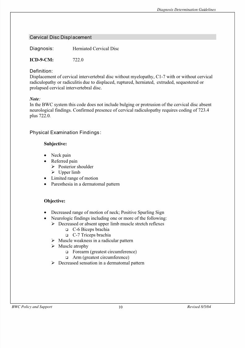
722.0 Cervical Disc Displacement 10
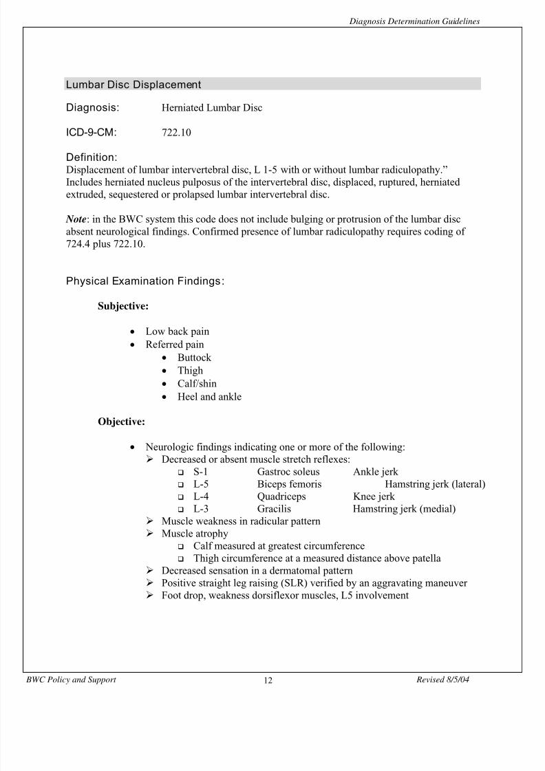
722.10 Lumbar Disc Displacement 12
722.11 Thoracic Disc Displacement 14
722.2 Disc Displacement 16
723.1 Cervicalgia 18
723.4 Cervical Radiculopathy 19
724.2 Lumbago 21
724.4 Lumbosacral Radiculopathy 22
726.10 Rotator Cuff Syndrome 24
726.2 Impingement Syndrome 27
726.31 Medial Epicondylitis 30
726.32 Lateral Epicondylitis 32
727.04 Radial Styloid Tenosynovitis 34
727.05 Tenosynovitis of Hand and Wrist 36
729.1 Myalgia and Myositis 39
739.0 Nonallopathic Lesions (Subluxation) 40
836.0 Tear Medial Meniscus of Knee 42
840.4 Rotator Cuff Tear 44
844.2 Tear Anterior Cruciate Ligament 47
no code Repetitive Motion Syndrome 49
Glossary of Tests 51
Bibliography 53

8/13/2019 Diag Det Guide Detail
http://slidepdf.com/reader/full/diag-det-guide-detail 5/54
Diagnosis Determination Guidelines
BWC Policy and Support Revised 8/5/045
Table of Contents (Alphabetical Listing)
DIAGNOSIS ICD-9 PAGE
Carpal Tunnel Syndrome 354.0 7
Cervical Disc Displacement 722.0 10
Cervicalgia 723.1 18
Cervical Radiculopathy 723.4 19
Disc Displacement 722.2 16
Impingement Syndrome 726.2 27
Lateral Epicondylitis 726.32 32
Lumbago 724.2 21
Lumbar Disc Displacement 722.10 12
Lumbosacral Radiculopathy 724.4 22
Medial Epicondylitis 726.31 30
Myalgia and Myositis 729.1 39
Nonallopathic Lesion (Subluxation) 739.0 40
Radial Styloid Tenosynovitis 727.04 34
Repetitive Motion Syndrome no code 49
Rotator Cuff Syndrome 726.10 24
Rotator Cuff Tear 840.4 44
Sprain and Strain 840 - 848 6
Tear Anterior Cruciate Ligament 844.2 47
Tear Medial Meniscus of knee 836.0 42
Tenosynovitis of hand and wrist 727.05 36
Thoracic Disc Displacement 722.11 14
Glossary of Tests 51
Bibliography 53

8/13/2019 Diag Det Guide Detail
http://slidepdf.com/reader/full/diag-det-guide-detail 6/54

8/13/2019 Diag Det Guide Detail
http://slidepdf.com/reader/full/diag-det-guide-detail 7/54
Diagnosis Determination Guidelines
BWC Policy and Support Revised 8/5/047
Carpal Tunnel Syndrome
Diagnosis: Carpal Tunnel Syndrome
ICD-9-CM: 354.0
Definition:Compression of median nerve in the carpal tunnel. There is an extensive list of medical causesfor Carpal Tunnel Syndrome along with work related causes.
Note: This is a common and potentially overused diagnosis. May suggest performingelectrodiagnostics to support this condition. Please code condition under tenosynovitis of wristor hand, 727.05 or sprain-strain of wrist, 842.00 and treat appropriately (see medical evidence
policy).
Physical Examination Findings :
Subjective:
• Numbness and tingling in the median sensory distribution and this isfrequently at night.
• Aching volar hand and wrist at the carpal tunnel.
• Grasp activities such as driving the car, holding the telephone or newspaper
aggravate the symptoms.
• Clumsiness (dropping objects) and weakness of grip are common.
Objective:
• Positive wrist flexion test (Phalen test)
• Positive Tinel sign over the median nerve at the wrist.
• Positive Median Nerve Compression Test.
• Weakness of the thenar muscles is generally an ‘early sign’.
• Atrophy of the thenar muscles is generally a ‘late sign’.
• Loss or deviation in 2 point discrimination, greater than 5-6 mm.
8/13/2019 Diag Det Guide Detail
http://slidepdf.com/reader/full/diag-det-guide-detail 8/54
Diagnosis Determination Guidelines
BWC Policy and Support Revised 8/5/048
Note:
It is not necessary to have all the symptoms listed above to make diagnosis of Carpal TunnelSyndrome.
Diagnosti c Test:
• NCV
Examine the sensory and motor conduction of the median nerve.
• Imaging
Cervical spine x-rays may be necessary to rule out peripheral cervicalnerve root signs and symptoms.
Wrist x-rays may be indicated if there is a need to rule-out bony wrist
injury or pathology. A Carpal Tunnel view may be included.
Diagnosti c Test Findings :
• Combined positive Phalen test and Tinel sign has high predictive value indiagnosis of CTS.
• Wrist shape - a square wrist dimension is a known risk factor for developmentof CTS.
Resources:
• North, ERKaul, M.P.Compression Neuropathies - MedianIn Peimer, C. (ed) SurgeryOf the Hand and Upper ExtremityMcGraw-Hill (1996)
pp. 1307-1326
• Omer G.Median Nerve Compression at the wristIn Ragan, G. (ed)
Nerve Compression SyndromesHand ClinicsW. B. Saunders Co. (1992)
pp. 317-323
8/13/2019 Diag Det Guide Detail
http://slidepdf.com/reader/full/diag-det-guide-detail 9/54
Diagnosis Determination Guidelines
BWC Policy and Support Revised 8/5/049
• Hennessey, W.J.
Johnson, E.W.Carpal Tunnel SyndromeJohnson, E.W. and Pease, W.S. (eds)
Practical Electromyography - Third EditionWilliams and Wilkins (1997) pp. 195 – 215
8/13/2019 Diag Det Guide Detail
http://slidepdf.com/reader/full/diag-det-guide-detail 10/54
8/13/2019 Diag Det Guide Detail
http://slidepdf.com/reader/full/diag-det-guide-detail 11/54
Diagnosis Determination Guidelines
BWC Policy and Support Revised 8/5/0411
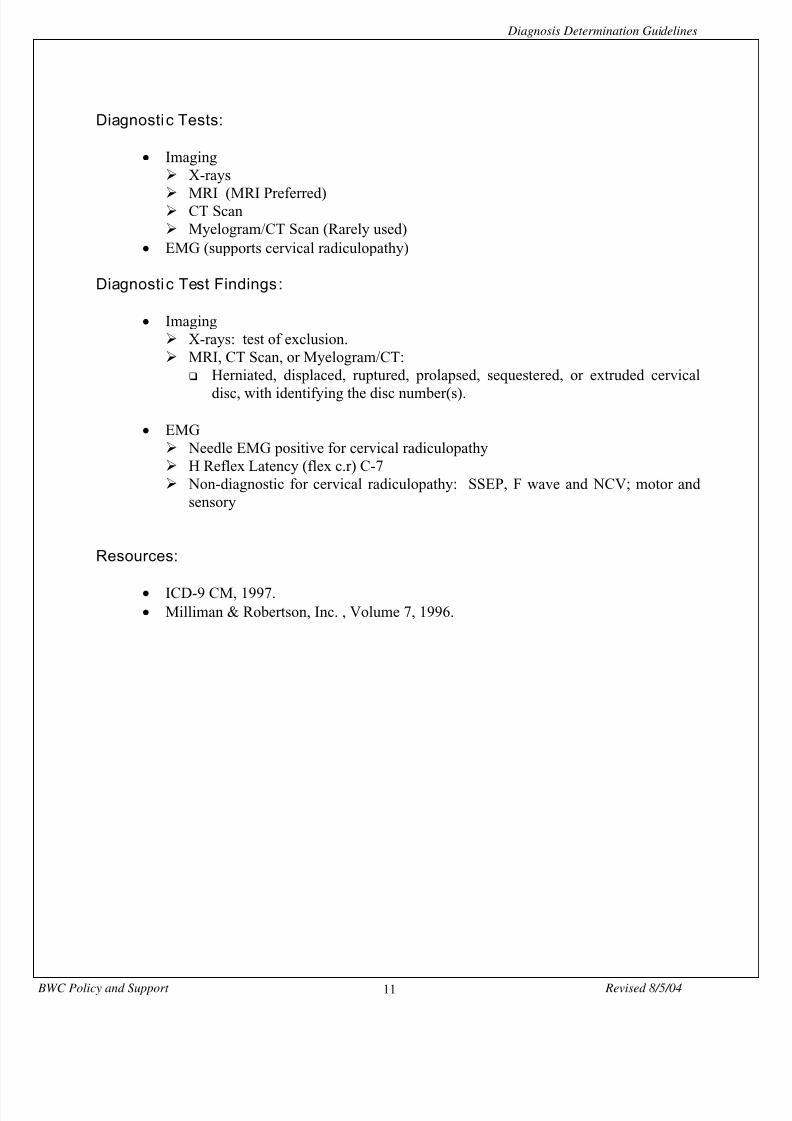
Diagnosti c Tests:
• Imaging X-rays MRI (MRI Preferred) CT Scan Myelogram/CT Scan (Rarely used)
• EMG (supports cervical radiculopathy)
Diagnosti c Test Findings :
• Imaging X-rays: test of exclusion. MRI, CT Scan, or Myelogram/CT:
Herniated, displaced, ruptured, prolapsed, sequestered, or extruded cervical
disc, with identifying the disc number(s).
• EMG Needle EMG positive for cervical radiculopathy H Reflex Latency (flex c.r) C-7 Non-diagnostic for cervical radiculopathy: SSEP, F wave and NCV; motor and
sensory
Resources:
• ICD-9 CM, 1997.• Milliman & Robertson, Inc. , Volume 7, 1996.
8/13/2019 Diag Det Guide Detail
http://slidepdf.com/reader/full/diag-det-guide-detail 12/54
8/13/2019 Diag Det Guide Detail
http://slidepdf.com/reader/full/diag-det-guide-detail 13/54
Diagnosis Determination Guidelines
BWC Policy and Support Revised 8/5/0413
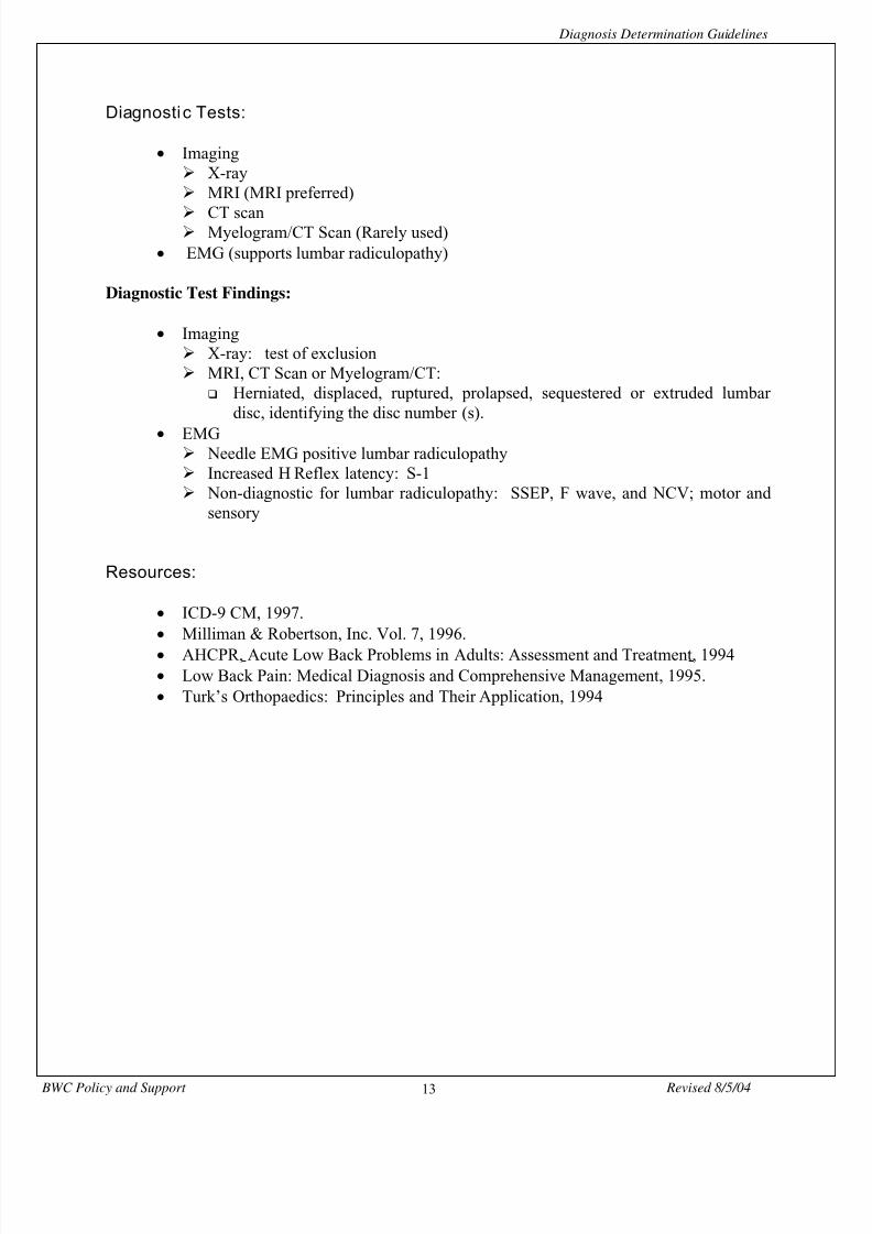
Diagnosti c Tests:
• Imaging X-ray MRI (MRI preferred) CT scan Myelogram/CT Scan (Rarely used)
• EMG (supports lumbar radiculopathy)
Diagnostic Test Findings:
• Imaging X-ray: test of exclusion MRI, CT Scan or Myelogram/CT:
Herniated, displaced, ruptured, prolapsed, sequestered or extruded lumbardisc, identifying the disc number (s).
• EMG Needle EMG positive lumbar radiculopathy Increased H Reflex latency: S-1 Non-diagnostic for lumbar radiculopathy: SSEP, F wave, and NCV; motor and
sensory
Resources:
• ICD-9 CM, 1997.• Milliman & Robertson, Inc. Vol. 7, 1996.• AHCPR, Acute Low Back Problems in Adults: Assessment and Treatment, 1994• Low Back Pain: Medical Diagnosis and Comprehensive Management, 1995.• Turk’s Orthopaedics: Principles and Their Application, 1994
8/13/2019 Diag Det Guide Detail
http://slidepdf.com/reader/full/diag-det-guide-detail 14/54
Diagnosis Determination Guidelines
BWC Policy and Support Revised 8/5/0414
Thoracic Disc Displacement
Diagnosis: Herniated Thoracic Disc
ICD-9-CM : 722.11
Definition:
Displacement of thoracic intervertebral disc without myelopathy, T 1-12 with or without thoracicradiculopathy. Included are displaced, ruptured, herniated, extruded, sequestered or prolapsedthoracic intervertebral discs.
Note : In the BWC system this code does not include bulging or protrusion of the thoracic disc absentneurological findings. Confirmed presence of thoracic radiculopathy requires coding of 724.4
plus 722.11.
Physical Examination Findings :
Subjective:
• Thoracic back pain exacerbated by flexion, extension or rotation of spine• Referred pain
Rib area
Objective:
• EMG abnormal in intercostals• Tenderness and pain in intercostals
Diagnosti c Tests:
• Imaging X-ray MRI (Preferred) CT Scan Myelogram/CT Scan (Rarely used)
• EMG (supports Thoracic Radiculopathy)

8/13/2019 Diag Det Guide Detail
http://slidepdf.com/reader/full/diag-det-guide-detail 15/54
Diagnosis Determination Guidelines
BWC Policy and Support Revised 8/5/0415
Diagnosti c Test Findings:
• Imaging X-ray: test of exclusion MRI, CT Scan, or Myelogram/CT:
Herniated, displaced, ruptured, prolapsed, sequestered, or extrudedintervertebral thoracic disc with identifying disc number(s)
• EMG Positive needle EMG thoracic radiculopathy, intercoastal abnormality. Non-diagnostic for thoracic radiculopathy: SSEP, F wave and NCV; motor and
sensory
Resources:
• ICD-9 CM, 1997.• Campbell’s Operative Orthopaedics, 1992
8/13/2019 Diag Det Guide Detail
http://slidepdf.com/reader/full/diag-det-guide-detail 16/54
Diagnosis Determination Guidelines
BWC Policy and Support Revised 8/5/0416
Disc Displacement NOS
Diagnosis : Bulging DiscDiscogenic Syndrome
ICD-9-CM: 722.2
Definition: Displacement of intervertebral disc, site unspecified, without myelopathy orradiculopathy. This code is utilized by BWC to code a bulging or protruded disc or DiscogenicSyndrome in the absence of neurologic findings.
Note: This diagnosis is no t considered eligible for surgical intervention.
Physical Examination Findings :
Subjective:• History pain consistent with level(s) affected
Objective: • Normal neurologic examination.
Diagnosti c Tests:
• Imaging X-ray MRI (MRI Preferred) CT Scan Myelogram/CT Scan
• EMG (supports radiculopathy)
Diagnosti c Tests Findings :
• Imaging X-ray: a test of exclusion. MRI, CT or Myelogram/CT:
Bulging or protruded disc without nerve root impingement, identifying thedisc level(s) and number(s).
• EMG Needle EMG negative for findings of radiculopathy

8/13/2019 Diag Det Guide Detail
http://slidepdf.com/reader/full/diag-det-guide-detail 17/54
Diagnosis Determination Guidelines
BWC Policy and Support Revised 8/5/0417
Resources :
• ICD-9 CM, 1997.• Milliman & Robertson, Inc., Volume 7, 1996.
8/13/2019 Diag Det Guide Detail
http://slidepdf.com/reader/full/diag-det-guide-detail 18/54
Diagnosis Determination Guidelines
BWC Policy and Support Revised 8/5/0418
Cervicalgia
Diagnosis: Cervicalgia
ICD-9-CM : 723.1
Definition:
Pain in neck. Refers to pain in neck usually greater than 3 months duration without morespecific diagnosis. BWC does not consider this diagnosis as eligible for allowance in claim,however, reimbursement may be eligible in selected situations.
Note: This diagnosis/symptom is not eligible for BWC coding guidelines but may be eligible forreimbursement in selected cases.
Physical Examination Findings :
Subjective:
• Complaints of pain localized to neck
Objective:
• Complaints of pain on palpation or movement• Normal neurologic examination.
Diagnosti c Tests :
• None (Most individuals may have had imaging or EMG to rule out other pathology of pain such as disc lesions)
Diagnostic Test Findings:
• None
Resources:
• ICD-9 CM, 1997.

8/13/2019 Diag Det Guide Detail
http://slidepdf.com/reader/full/diag-det-guide-detail 19/54
Diagnosis Determination Guidelines
BWC Policy and Support Revised 8/5/0419
Cervical Radiculopathy
Diagnosis: Cervical RadiculopathyCervical Neuritis or Radiculitis
ICD-9-CM: 723.4
Definition:
Inflammation and/or compression of nerve root producing symptoms of pain, tingling,numbness, or neurological deficit along the distribution of the involved spinal nerve or root.
Note: This diagnosis/symptom is not eligible for BWC coding guidelines absent the corresponding
pathology diagnosis.
Physical Examination Findings :
Subjective:
• Complaints of sclerotomal pain • Paresthesia in a dermatomal pattern • Referred pain
Posterior shoulder Upper limb
Objective:
• Neurologic findings indicating one or more of the following: Decreased or absent upper limb muscle stretch reflezes
C-6 Biceps brachis C-7 Triceps brachia
Muscle weakness in a radicular pattern Muscle atrophy
Forearm (greatest circumference) Arm (greatest circumference)
Decreased sensation in a dermatomal pattern

8/13/2019 Diag Det Guide Detail
http://slidepdf.com/reader/full/diag-det-guide-detail 20/54
Diagnosis Determination Guidelines
BWC Policy and Support Revised 8/5/0420
Diagnosti c Tests:
• Imaging X-rays MRI (MRI Preferred) CT Scan Myelogram/CT Scan (Rarely used)
• EMG (supports cervical radiculopathy)
Diagnostic Test Findings:
• Imaging X-rays: test of exclusion. MRI, CT Scan, or Myelogram/CT:
Herniated, displaced, ruptured, prolapsed, sequestered, degenerative, orextruded cervical disc, with identifying the disc number(s).
• EMG Needle EMG positive for cervical radiculopathy H Reflex Latency (flex c.r) C-7 Non-diagnostic for cervical radiculopathy: SSEP, F wave and NCV; motor and
sensory
Resources:
• ICD-9 CM, 1997.• Milliman & Robertson, Inc. , Volume 7, 1996.
8/13/2019 Diag Det Guide Detail
http://slidepdf.com/reader/full/diag-det-guide-detail 21/54
Diagnosis Determination Guidelines
BWC Policy and Support Revised 8/5/0421
Lumbago
Diagnosis : Lumbago
ICD-9-CM : 724.2
Definition :
Low back pain frequently referring to or including “low back pain”, “low back syndrome”, and“lumbalgia”. Usually used when another diagnosis does not apply and for chronic or recurrentlumbar pain.
Note: This diagnosis/symptom is not eligible for BWC coding guidelines but may be eligible forreimbursement in selected cases.
Physical Examination Findings :
Subjective:
• Complaints of low back pain without radicular symptoms usually greater than3 months duration
Objective:
• May have tenderness or complaints of pain with palpation.• Normal neurologic examination.
Diagnosti c Tests:
• None (Most individuals may have had imaging and/or EMG to rule out other pathology of pain such as disc lesions)
Diagnosti c Test Findings :
• None
8/13/2019 Diag Det Guide Detail
http://slidepdf.com/reader/full/diag-det-guide-detail 22/54
Diagnosis Determination Guidelines
BWC Policy and Support Revised 8/5/0422
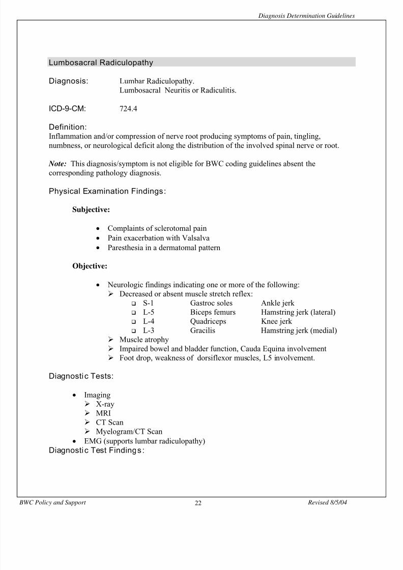
Lumbosacral Radiculopathy
Diagnosis: Lumbar Radiculopathy.Lumbosacral Neuritis or Radiculitis.
ICD-9-CM: 724.4
Definition:Inflammation and/or compression of nerve root producing symptoms of pain, tingling,numbness, or neurological deficit along the distribution of the involved spinal nerve or root.
Note: This diagnosis/symptom is not eligible for BWC coding guidelines absent thecorresponding pathology diagnosis.
Physical Examination Findings :
Subjective:
• Complaints of sclerotomal pain• Pain exacerbation with Valsalva• Paresthesia in a dermatomal pattern
Objective:
• Neurologic findings indicating one or more of the following: Decreased or absent muscle stretch reflex:
S-1 Gastroc soles Ankle jerk L-5 Biceps femurs Hamstring jerk (lateral) L-4 Quadriceps Knee jerk L-3 Gracilis Hamstring jerk (medial)
Muscle atrophy Impaired bowel and bladder function, Cauda Equina involvement Foot drop, weakness of dorsiflexor muscles, L5 involvement.
Diagnosti c Tests:
• Imaging X-ray MRI CT Scan Myelogram/CT Scan
• EMG (supports lumbar radiculopathy)Diagnosti c Test Findings :
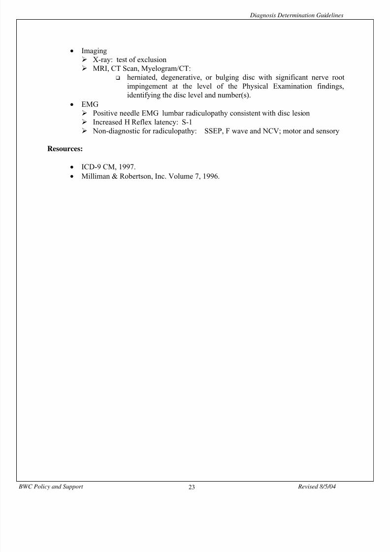
8/13/2019 Diag Det Guide Detail
http://slidepdf.com/reader/full/diag-det-guide-detail 23/54
Diagnosis Determination Guidelines
BWC Policy and Support Revised 8/5/0423
• Imaging X-ray: test of exclusion MRI, CT Scan, Myelogram/CT:
herniated, degenerative, or bulging disc with significant nerve rootimpingement at the level of the Physical Examination findings,
identifying the disc level and number(s).• EMG
Positive needle EMG lumbar radiculopathy consistent with disc lesion Increased H Reflex latency: S-1 Non-diagnostic for radiculopathy: SSEP, F wave and NCV; motor and sensory
Resources:
• ICD-9 CM, 1997.• Milliman & Robertson, Inc. Volume 7, 1996.
8/13/2019 Diag Det Guide Detail
http://slidepdf.com/reader/full/diag-det-guide-detail 24/54
8/13/2019 Diag Det Guide Detail
http://slidepdf.com/reader/full/diag-det-guide-detail 25/54

8/13/2019 Diag Det Guide Detail
http://slidepdf.com/reader/full/diag-det-guide-detail 26/54
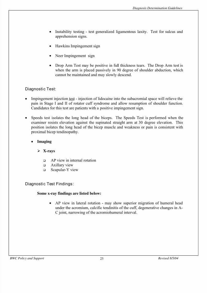
Diagnosis Determination Guidelines
BWC Policy and Support Revised 8/5/0426
• Axillary view - provides visualization of the glenoid and humeral head and
delinates the spatial relationship of the two structures.
• Scapular-Y view - to visualize acromial shape and subacromial spurs that are
often associated with rotator cuff disease.
Resources:
• Hawkins, R.J.Bokor, D.J.Clinical Evaluation of Shoulder Problemsin Rockwood Jr., C.A. andMatsen III, F.A. (eds)The Shoulder
Philadelphia, W.B. Saunders Company (1990) pp. 149 - 177
• Rockwood Jr., C.A. (et al)X-ray Evaluation of shoulder problemsin Rockwood Jr., C.A. andMatsen III, F.A. (eds)The ShoulderPhiladelphia, W.B. Saunders Company (1990)
pp. 178 - 200
• Smolinski, R.J.Leddy, J.L.Stegemann, P.M.Rotator Cuff Diseasein Peimer, C. (ed)Surgery of the Hand and Upper ExtremityMcGraw-Hill (1996)
pp. 321 - 349
8/13/2019 Diag Det Guide Detail
http://slidepdf.com/reader/full/diag-det-guide-detail 27/54
Diagnosis Determination Guidelines
BWC Policy and Support Revised 8/5/0427
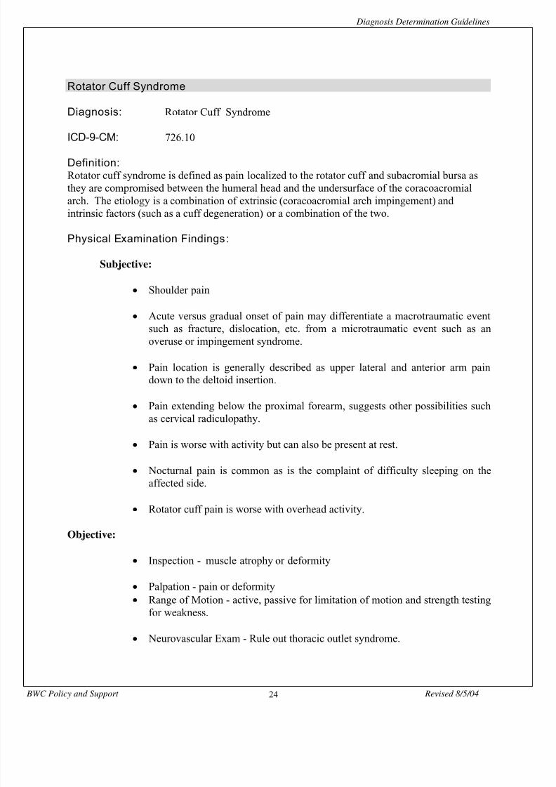
Impingement Syndrome
Diagnosis: Impingement Syndrome
ICD-9-DM: 726.2
Definition: Impingement syndrome is defined as pain localized to the rotator cuff caused by a compromise
between the humeral head and the undersurface of the coracoacromial arch during forwardelevation and rotation of the shoulder.
Physical Examination Findings:
Subjective:
• Shoulder pain in the upper lateral and anterior arm.
• Pain extending below the proximal forearm suggests other possibilities such as cervicalradiculopathy.
• Night pain is common and either interrupts sleep or does not allow the patient to lie on theinvolved side.
• Pain can be dull, toothache like which lingers for long periods or sharp, burning type whichis not well tolerated.
• May experience weakness and loss of function.
Objective:
• Palpable tenderness about the shoulder, usually centered around the greater tuberosity, butthe location varies.
• Most reliable physical sign is the Neer impingement sign or the Hawkins impingement sign.
Neer impingement sign is performed when the examiner stabilizes the scapula with one handand with the other grasps the patient’s elbow and passively elevates it fully with the arm infull internal rotation. Pain is produced by the greater tuberosity impinging the rotator cuffagainst the acromium. Hawkins impingement sign is performed when the examiner forciblyinternally rotates the 90 degree forward flexed arm, impinging the rotator cuff against thecoracoacromial ligament.
8/13/2019 Diag Det Guide Detail
http://slidepdf.com/reader/full/diag-det-guide-detail 28/54
8/13/2019 Diag Det Guide Detail
http://slidepdf.com/reader/full/diag-det-guide-detail 29/54
8/13/2019 Diag Det Guide Detail
http://slidepdf.com/reader/full/diag-det-guide-detail 30/54
Diagnosis Determination Guidelines
BWC Policy and Support Revised 8/5/0430
Medial Epicondylitis
Diagnosis: Medial Epicondylitis
ICD-9-CM: 726.31
Definition:Inflammation of the medial epicondyle of the humerus and surrounding tissues. Medialepicondylitis occurs less commonly than lateral epicondylitis; the symptoms of localized painand weakness are similar but are located at the flexor pronator tendinous origin.
Physical Examination Findings:
Subjective:
• Aching in proximal volar forearm.
Objective:
• Resisted wrist flexion and pronation will often reproduce symptoms
• Weakness in proximal volar forearm muscles.
• Repetitive valgus stress can result in medial elbow symptoms includingmedial epicondylitis. The differential diagnosis includes medial collateralligament injury or insufficiency, ulnar nerve tension-neuropraxia andintraarticular elbow pathology or any combination of these.
• To differentiate chronic medial collateral ligament injury from medialepicondylitis apply a valgus stress to the slightly flexed elbow while the wristis flexed and the forearm pronated. This eliminates the symptoms of medialepicondylitis and results in a painless valgus stress test when the medialcollateral ligament is normal.
• Medial epicondylitis and ulnar neuropraxia often occur together.

8/13/2019 Diag Det Guide Detail
http://slidepdf.com/reader/full/diag-det-guide-detail 31/54
Diagnosis Determination Guidelines
BWC Policy and Support Revised 8/5/0431
Diagnosti c Test:
• Imaging
X-rays of the elbow
Diagnosti c Test Findings :
• Imaging
X-rays of the elbow are helpful in determining if bone or elbow joint pathology is present as is the evaluation of valgus stability of the elbow.
Resource:
• Field, L. D.Altchek, D.W.Chronic elbow pain, overuse and tendonitisIn Peimer, C. (ed)Surgery of the hand and upper extremityMcGraw-Hill (1996)
pp. 499-503
8/13/2019 Diag Det Guide Detail
http://slidepdf.com/reader/full/diag-det-guide-detail 32/54
Diagnosis Determination Guidelines
BWC Policy and Support Revised 8/5/0432
Lateral Epicondylitis
Diagnosis: Lateral Epicondylitis
ICD-9-CM: 726.32
Definition:Inflammation of the lateral epicondyle of the humerus and surrounding tissues. Lateralepicondylitis also known as Tennis Elbow represents a pathologic condition at the commonextensor origin of the lateral humeral epicondyle and is characterized by pain at the epicondyle.It is one of the most common injuries among workers, musicians, and racquet sport athletes.Age of onset is generally after 30 years of age and most often around 40 years of age.
Physical Examination Findings :
Subjective:
• Aching in proximal forearm exacerbated by participation in any activity thatrequires resisted wrist extension.
• Morning stiffness along with aching throughout the day is common.
• Rest will usually improve the symptoms but the pain returns with activity.
Objective:
• Maximum tenderness is directly over or slightly distal to the lateralepicondyle.
• Application of an axial load on the forearm combined with gentle passiveforearm rotation may aid in differentiating lateral epicondylitis fromradiocapitellar joint degenerative changes. This test often causes pain whenarticular degeneration is responsible for symptoms. (If the wrist is kept inneutral position it will not exacerbate the lateral epicondylitis).
•
Typically, symptoms increase as wrist or finger extension is resisted withincreasing extension of the elbow.
8/13/2019 Diag Det Guide Detail
http://slidepdf.com/reader/full/diag-det-guide-detail 33/54
Diagnosis Determination Guidelines
BWC Policy and Support Revised 8/5/0433
• Lateral elbow symptoms due to compression neuropathy of the radial nerve
may simulate lateral epicondylitis and careful evaluation is necessary to ruleout radial tunnel syndrome. A patient with radial tunnel syndrome complainsof aching pain at the extensor-supinator muscle mass in the proximal forearm
that my radiate into the distal arm and forearm. In radial tunnel syndromemaximaltenderness is distal to the epicondyle (over the radial neck) and is morediffuse and aching in character.
Diagnosti c Tests:
• Imaging
Standard x-rays of the elbow.
Diagnostic Test Findings:
• Imaging
X-rays will help to rule out calcified deposits or other lesions in the area.Degenerative changes in the radiocapitellar joint can simulate lateral epicondylitisand any significant radial head or distal humeral osteophytes could be seen in thex-ray.
Resource:
• Field, L.Altchek, DChronic Elbow Pain, Overuse, and TendonitisIn Peimer, C. (ed)Surgery of the Hand and Upper ExtremityMcGraw - Hill (1996)
pp. 496-499

8/13/2019 Diag Det Guide Detail
http://slidepdf.com/reader/full/diag-det-guide-detail 34/54
Diagnosis Determination Guidelines
BWC Policy and Support Revised 8/5/0434
Radial Styloid Tenosynovitis (De Quervain’s Tenosynovitis)
Diagnoses: Radial Styloid Tenosynovitis(De Quervain’s Tenosynovitis)
ICD-9-CM: 727.04
Definition: Radial Styloid Tenosynovitis is defined as Tenosynovitis of the tendons in the first dorsalcompartment, the abductor pollicis longus and extensor pollicis brevis. When friction develops
between the tendon and the sheath, the natural consequence is tenosynovitis.
Physical Examination Findings :
Subjective:
• Visible swelling and aching at the radial styloid in the distal forearm.
• Thumb extension is particularly painful at the radial styloid.
Objective:
• Tenderness at the radial styloid.
• There may be crepitation over the sheath.
• Occasional “locking” similar to triggering that occurs with Tenosynovitis of adigital flexor sheath.
• When Tenosynovitis of the first dorsal compartment is severe, tendonexcursion becomes restricted, limiting active extension of the thumb.
• Finkelstein Test
Diagnosti c Test:
• None

8/13/2019 Diag Det Guide Detail
http://slidepdf.com/reader/full/diag-det-guide-detail 35/54
Diagnosis Determination Guidelines
BWC Policy and Support Revised 8/5/0435
Diagnostic Test Findings:
• Finkelstein Test is positive when the thumb is clenched firmly within the fistand then the wrist is forced into ulnar deviation producing pain. The resulting
pain will be over the radial styloid. The physician should avoid sudden
deviation because of possible excrutiating pain.
• It is necessary to rule out Trapeziometacarpal Arthritis as the cause of the pain. The tenderness in these cases will be over the trapeziometacarpal jointrather then the radial styloid. An X-ray and a positive “grind test” confirmsthis diagnosis.
• Rule out Intersection Syndrome - this represents swelling, pain, andtenderness about 4-6 cm proximal to Lister’s tubercle and is a tenosynovitis ofthe extensor carpi radialis longus and brevis (second dorsal compartment)
presenting proximal to the compartment.
Resource:
• Posner, M.Differential Diagnosis of Wrist Pain and Tendinitis, Ganglia and OtherSyndromesIn Peimer, C. (ed)Surgery of the Hand and Upper ExtremityMcGraw - Hill (1996)
pp 838 - 839

8/13/2019 Diag Det Guide Detail
http://slidepdf.com/reader/full/diag-det-guide-detail 36/54
Diagnosis Determination Guidelines
BWC Policy and Support Revised 8/5/0436
Tenosynovitis of Hand and Wrist
Diagnosis: Tenosynovitis of hand and wrist, NEC
ICD-9-CM: 727.05
Definition:Tenosynovitis, or irritation of the synovial sheath of a tendon may result from inflammation orinfection. Here, the discussion will address inflammation of the tendon sheath and not infection.The overuse syndrome may be involved in the development of tenosynovitis. This definitionapplies to all examples listed below.
Tenosynovitis at the wrist (both flexor and extensor tendons) is not uncommon.
Examples are:
Extensor Carpi Ulnaris Tenosynovitis
Subjective:
• Pain on the ulnar side of the wrist.
Objective:
• Tenderness where the tendon passes through its sheath at its insertionor along its entire length.
• Patients frequently complain of pain with resisted ulnar deviation.
Extensor Pollicis Longus Tenosynovitis
Subjective:
• Pain, which may be vague, dorsum of wrist (third dorsalcompartment).
• Active motions of the interphalangeal joint of the thumb may belimited and painful.
• Chronic Tenosynovitis may result in rupture of the extensor pollicislongus tendon.
8/13/2019 Diag Det Guide Detail
http://slidepdf.com/reader/full/diag-det-guide-detail 37/54
Diagnosis Determination Guidelines
BWC Policy and Support Revised 8/5/0437
Objective:
• Tenderness and swelling over the tendon just distal to Lister’sTubercle.
• Triggering may occur.
Flexor Carpi Radialis Tenosynovitis
Subjective:
• Pain over the flexor carpi radialis tendon just proximal to the wristflexor crease. Condition is relatively common.
Objective:
• Slight swelling and tenderness over the tendon at the wrist.
• Pain produced on resisted wrist flexion and sometimes with resistedforearm pronation.
Flexor Carpi Ulnaris Tenosynovitis
Flexor Carpi Ulnaris Tenosynovitis is often bilateral and results fromchronic repetitive trauma.
Subjective:
• Pain in the region of the pisiform or proximal to the pisiform over theflexor carpi ulnaris tendon.
Objective:
• Pain is exacerbated by resisted wrist flexion.
• Tenderness of the flexor carpi ulnaris just proximal to the pisiform.
• To rule out pisotriquetral arthritis or instability, translocating the
pisiform radially or ulnarly, pain or even crepitation may result. Diagnosti c Test:
• Imaging
Lateral x-ray.
Diagnosti c Test Findings :
8/13/2019 Diag Det Guide Detail
http://slidepdf.com/reader/full/diag-det-guide-detail 38/54
Diagnosis Determination Guidelines
BWC Policy and Support Revised 8/5/0438
• X-ray may show calcific deposit along the tendon.
• In advanced stages, x-ray will reveal narrowing of the pisotriquetral joint and subchondral sclerosis.
Flexor tenosynovitis of the wrist
Inflammation of the digital flexors as they pass through the carpal canal.
Subjective:
• Stabbing or burning pain proximal to the carpal tunnel which mayextend up to the forearm.
Objective:
• Tenderness and swelling just proximal to the wrist flexor creases. • Median nerve compression may be co-existent as evidenced by
positive Phalen Test and Tinel sign.
Diagnosti c Test:
• Electrodiagnostic studies may or may not confirm co-existent carpaltunnel syndrome. This will depend on clinical signs and symptoms ofmedian nerve compression.
Resources:
• Failla, Joseph M.Differential Diagnosis of Hand Pain: Tendonitis, Ganglia and other Syndromesin Peimer, C. (ed) Surgery of theHand and Upper ExtremityMcGraw - Hill (1996)
pp. 1223-1249
• Stern, P.J.Tendinitis, Overuse Syndromes and Tendon InjuriesIn Amadio, P. C. (ed)
Hand Injuries in Sports and Performing Arts Hand ClinicsW.B. Saunders Co. (Aug. 1990)
pp. 467-476
8/13/2019 Diag Det Guide Detail
http://slidepdf.com/reader/full/diag-det-guide-detail 39/54
8/13/2019 Diag Det Guide Detail
http://slidepdf.com/reader/full/diag-det-guide-detail 40/54
Diagnosis Determination Guidelines
BWC Policy and Support Revised 8/5/0440
Nonallopathic Lesions
Diagnosis: Nonallopathic LesionsIntersegmental Dysfunction
Subluxation
ICD-9 Code : 739.0 - 739.9
Definition: The injury/chiropractic condition of segmental dysfunction (chiropractic subluxation) is definedas an alteration in joint function in spinal segments and joints of the body which affects theneuro-musculoskeletal system as diagnosed by chiropractors.
Physical Examination Findings:
Subjective:
• Localized pain and tenderness identified through observation/percussion and palpation.
Objective:
• Asymmetry of joint function noted during examination(observational/palpation).
• Range of motion - restriction/change in normal joint function (i.e. motion palpation). Specific level of restiction should be identified.
• Localized soft tissue changes - (i.e. spasm, edema, tenderness) identifiedduring general examination.
Diagnostic Tests:
None
Diagnostic Test Findings:
None Resources:
• Philip E. Greenman, D.O., F.A.A.O.Principles of Manual Medicine (1989)
8/13/2019 Diag Det Guide Detail
http://slidepdf.com/reader/full/diag-det-guide-detail 41/54
Diagnosis Determination Guidelines
BWC Policy and Support Revised 8/5/0441
• Meridel I. Gatterman, D.C.
Chiropractic Management of Spine Related Disorders (1990)
• Meridel I. Gatterman, D.C.
Foundations of Chiropractic - Subluxation (1995)
• Topics in Clinical Chiropractic - JournalBack to Basics: Technology Assessment of the Chiropractic Subluxation’OsterbauerMarch 1996, Vol. 3, Number 1

8/13/2019 Diag Det Guide Detail
http://slidepdf.com/reader/full/diag-det-guide-detail 42/54
Diagnosis Determination Guidelines
BWC Policy and Support Revised 8/5/0442
Tear of Medial Meniscus of Knee
Diagnosis: Tear of Medial Cartilage or Meniscus of knee, current
ICD-9-CM: 836.0
Definition: Injury to cartilage attached to the medial articular surface of the tibia usually referred to as a“tear” since the cartilage is pulled away from the tibia. This diagnosis is a “current or acuteinjury” includes bucket handle tear.
Physical Examination Findings:
Subjective:
• Painful popping in knee with motion• History of twisting injury
Objective:
• Positive McMurrays test• Locking of knee• Tenderness along anteromedial joint line of knee
Diagnostic Tests:
• Arthroscopy• Imaging
MRI X-rays
Diagnostic Tests:
• Arthroscopy tear medial meniscus
Note:Arthroscopy originally authorized for diagnostic reasons may result in necessary surgical repairwhich requires submission of operative report for additional coding guideline consideration.

8/13/2019 Diag Det Guide Detail
http://slidepdf.com/reader/full/diag-det-guide-detail 43/54
Diagnosis Determination Guidelines
BWC Policy and Support Revised 8/5/0443
• Imaging MRI: “Bright signals” within the meniscus which should reach the
surface of the meniscus or report of deformity or amputation ofmeniscus
X-ray: test of exclusion
Resources :
• Turek’s Orthopaedics: Principles and Their Application, 1994.• Primary Care Orthopaedics, 1996.• Milliman & Robertson, Inc., Volume 7, 1996.• ICD-9-CM, 1997.

8/13/2019 Diag Det Guide Detail
http://slidepdf.com/reader/full/diag-det-guide-detail 44/54
Diagnosis Determination Guidelines
BWC Policy and Support Revised 8/5/0444
Rotator Cuff Tear Diagnosis: Rotator Cuff Tear
ICD-9-CM: 840.4
Definition:Rotator cuff tears represents an end stage in the development of impingement syndrome. Thetears occur most often between 45 and 65 years of age from trivial trauma to a degenerativerotator cuff, but when it occurs at a young age the trauma is usually more severe and the tearmore significant.
Physical Examination Findings :
Subjective:
• Shoulder pain over the insertion of the rotator muscles.
• Report daily activity that is producing the pain.
• Pain may be nocturnal often interfering with normal sleep.
Objective:
• Muscle atrophy, and tenderness over the insertion of rotator muscles usuallythe supraspinatus over the greater tuberosity.
• Pain can be reproduced by resisting elevation of the arm, and of the shoulderas well as resisting external rotation if the tear is in the supraspinatus tendon.
• Patients with a chronic partial tear may abduct or externally rotate the arm butit is weaker when compared to the opposite normal shoulder.
• Atrophy of the muscle is present if the tear is chronic.
• No evidence of a complete tear (drop arm test), and yet the symptoms and
disability persist or are greater than anticipated, the question of a partial tearmay be considered:
Pain is not a determining factor but active motion is. Weakness or inability to abduct the first 15 to 20 degrees from the side
implies a total tear.
8/13/2019 Diag Det Guide Detail
http://slidepdf.com/reader/full/diag-det-guide-detail 45/54
Diagnosis Determination Guidelines
BWC Policy and Support Revised 8/5/0445
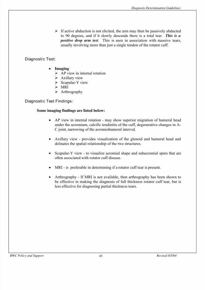
If active abduction is not elicited, the arm may then be passively abducted
to 90 degrees, and if it slowly descends there is a total tear. This is a positive drop arm test . This is seen in association with massive tears,usually involving more than just a single tendon of the rotator cuff.
Diagnosti c Test:
• Imaging AP view in internal rotation Axillary view Scapular-Y view MRI Arthrography
Diagnosti c Test Findings :
Some imaging findings are listed below:
• AP view in internal rotation - may show superior migration of humeral headunder the acromium, calcific tendinitis of the cuff, degenerative changes in A-C joint, narrowing of the acromiohumeral interval.
• Axillary view - provides visualization of the glenoid and humeral head anddelinates the spatial relationship of the two structures.
• Scapular-Y view - to visualize acromial shape and subacromial spurs that areoften associated with rotator cuff disease.
• MRI - is preferable in determining if a rotator cuff tear is present.
• Arthrography - If MRI is not available, then arthrography has been shown to be effective in making the diagnosis of full thickness rotator cuff tear, but isless effective for diagnosing partial thickness tears.

8/13/2019 Diag Det Guide Detail
http://slidepdf.com/reader/full/diag-det-guide-detail 46/54
Diagnosis Determination Guidelines
BWC Policy and Support Revised 8/5/0446
Resources:
• Matsen III, F.A.Arntz, C.T.Rotator Cuff Tendon Failure
in Rockwood Jr., C.A.and Matsen III, F.A., (eds)The ShoulderPhiladelphia, W.B. Saunders Company (1990)
pp. 647 - 677
• Rockwood Jr., C.A. (et al)X-ray Evaluation of shoulder problemsin Rockwood Jr., C.A.Matsen III, F.A. (eds)The Shoulder
Philadelphia, W.B. Saunders Company (1990) pp. 178 - 200
• Smolinski, R.J.Leddy, J.L.Stegemann, P.M.Rotator Cuff Diseasein Peimer, C. (ed)Surgery of the Hand and Upper ExtremityMcGraw-Hill (1996)
pp. 321 - 349
8/13/2019 Diag Det Guide Detail
http://slidepdf.com/reader/full/diag-det-guide-detail 47/54
Diagnosis Determination Guidelines
BWC Policy and Support Revised 8/5/0447
Tear Anterior Cruciate Ligament
Diagnosis: Tear of Cruciate Ligament of knee
ICD-9-CM: 844.2
Definition:Disruption or tearing of the anterior or posterior cruciate ligaments, the major stabilizers of theknee joint.
Note : For BWC operations this code can designate a sprain/strain or a complete tear. BWC,through the V3 computer system, has the capability of distinguishing through narrativedescription the definitive diagnosis.
Physical Examination Findings :
Subjective:• History of experiencing an injury with “pop” • Knee instability • Knee pain
Objective:• Knee effusion • Instability of knee joint with positive Drawer sign or • Positive Lachmans sign
Diagnosti c Tests:
• Imaging MRI X-ray
• Arthroscopy
Diagnosti c Test Findings :
• Imaging MRI
Disruption or tear cruciate ligament X-ray Test of exclusion
8/13/2019 Diag Det Guide Detail
http://slidepdf.com/reader/full/diag-det-guide-detail 48/54
Diagnosis Determination Guidelines
BWC Policy and Support Revised 8/5/0448
• Arthroscopy
Disruption or tear cruciate ligament
Note: Arthroscopy originally authorized for diagnostic reasons, may result in necessary surgical repairwhich requires submission of operative report for additional coding guideline consideration.
Resources:
• Primary Care Orthopaedics, 1996• Turek’s Othopaedics: Principles and Their Application, 1994• Milliman & Robertson, Inc., Volume 7, 1997• ICD-9-CM, 1997
8/13/2019 Diag Det Guide Detail
http://slidepdf.com/reader/full/diag-det-guide-detail 49/54
Diagnosis Determination Guidelines
BWC Policy and Support Revised 8/5/0449
Repetitive Motion Syndrome
Diagnoses: Repetitive Motion SyndromeCumulative Trauma Disorder Overuse Syndrome
ICD-9-CM: No specific code assigned.
Definition:Repetitive motion syndrome does not represent a new entity or type of disorder but itacknowledges that overuse may be a causative factor in many clinical problems that fall underthe category of repetitive motion syndrome. Although repetitive motion syndrome can occur innearly all tissues, the nerves, tendons, tendon sheaths and muscles of the upper extremity are themost frequently reported sites. Examples would be Carpal Tunnel Syndrome, Tendinitis andTenosynovitis at wrist and in forearm. Other names for this syndrome are Cumulative TraumaSyndrome and Overuse Syndrome.
• Work related high risk factors for repetitive motion syndrome include:
1) Repeated and sustained exertions2) Forceful exertions3) Localized contact stress4) Vibration5) Prolonged specific posture6) Low temperatures
Physical Examination Findings :
Characteristics of three types of disorders resulting from repetitive motion:
1. Tendinitis and Tenosynovitis
Subjective:
• Localized pain • Swelling
Objective:
• Pain on resisted motion • Weakness and crepitation of the tendon

8/13/2019 Diag Det Guide Detail
http://slidepdf.com/reader/full/diag-det-guide-detail 50/54
Diagnosis Determination Guidelines
BWC Policy and Support Revised 8/5/0450
2. Nerve Compression Syndromes - Symptoms may be vague (Carpal Tunnel
Syndrome, for example) but usually involve some combination of :
Subjective:• pain • numbness • tingling • weakness
3. Myofascial Pain - should be considered when there is localized soft tissue pain with or without referral. More distal site when compressed. The focal point of myofascial pain is the trigger point as a result of overuse of muscle.
Resources:
• Schuchmann, J.A. Occupational Rehabilitationin Braddom, R.L. (ed)Physical Medicine and RehabilitationW.B. Saunders (1996)
pp. 938-954
• Armstrong, T. and Ulin, S. Analysis and Design of Jobs for Control of Work Related Upper LimbDisordersRehabilatation of the Hand: Surgery and TherapyHunter, J.M., Mackin, E.J. and Callahan, A.D. (eds)Mosby (1995)
pp 1705-1723

8/13/2019 Diag Det Guide Detail
http://slidepdf.com/reader/full/diag-det-guide-detail 51/54
Diagnosis Determination Guidelines
BWC Policy and Support Revised 8/5/0451
Glossary Of Signs and Tests
Drop Arm TestThe arm is placed passively in 90 degree of shoulder abduction. In a complete tear of the rotator
cuff, the arm will slowly descend in spite of strong deltoid action. This can be considered a positive drop arm test.
Drawer’s signThe knee is flexed to a 90 degree angle; at the femoral-tibial junction, if the tibia can be drawn tofar forward there is a rupture of the anterior ligaments and if it can be drawn back too far backthere is a rupture of the posterior ligaments .
Finkelstein TestThis test is considered positive when the thumb is clenched firmly within the fist and then thewrist is forced into ulnar deviation producing pain.
Hawkins Impingement SignThe test is considered positive when the examiner forcibly internally rotates the 90 degreeforward flexed arm, impinging the rotator cuff against the coracoacromial ligament.
Lachman’s testAn anterior drawer test for cases of severe knee injury, performed at 20 degree flexion.
McMurray’s signOccurrence of a cartilage click during manipulation of the knee; indicative of meniscal injury.
Median Nerve Compression TestThis test is performed by placing the thumb over carpal tunnel, apply pressure for 30 seconds.Test is considered positive if paresthesia or numbness occurs in median nerve distribution. It is auseful test in patients that cannot flex the wrist.
Neer Impingement SignThe examiner stabilizes the scapula with one hand and with the other grasps the patient’s elbowand passively elevates it fully with the arm in full internal rotation. The test is considered
positive when pain is produced by the greater tuberosity impinging the rotator cuff against theacromium.
Phalen TestTest is performed by flexion of the wrist. Numbness or paresthesia occur within 60 seconds isdiagnostic of CTS.
8/13/2019 Diag Det Guide Detail
http://slidepdf.com/reader/full/diag-det-guide-detail 52/54
Diagnosis Determination Guidelines
BWC Policy and Support Revised 8/5/0452
Speeds TestSpeeds Test is done with elbow extended and the forearm supinated. Forward flexion of the armis resisted. Pain at the proximal portion of the long head of the biceps in the bicipital grooveindicates a positive test for the bicipital tendonitis, which is recognized as a component of
Impingement Syndrome.
Spurling SignThe neck is stressed in lateral flexion and rotation with some compression to elicit pain.Positioning the neck in this manner causes encroachment on cervical nerve roots as they exit theneural formaina; it also stresses the facet joints.
Tinel’s SignThis sign is elicited by a direct median nerve percussion just proximal to volar aspect of wrist.The nerve is tapped lightly from proximal to distal watching for paresthesia in the median nervedistribution indicating a positive test.
Valsalva ManeuverForcible exhalation effort against a closed glottis; the resultant increase in intrathoracic pressureinterferes with venous return to the heart. Forcible exhalation effort against occluded nostrils anda closed mouth; the increased pressure in the eustachian tube and middle ear causes the tympanicmembrane to move outward.

8/13/2019 Diag Det Guide Detail
http://slidepdf.com/reader/full/diag-det-guide-detail 53/54
Diagnosis Determination Guidelines
BWC Policy and Support Revised 8/5/0453
Bibliography
Acute Low Back Problems in Adults: Assessment and Treatment, Agency for Health Care Policyand Research, U.S. Department of Health and Human Services, Rockville, Maryland, (1994).
Armstrong, T. and Ulin, S.: Analysis and Design of Jobs for Control of Work Related UpperLimb Disorders, Rehabilitation of the Hand: Surgery and Therapy, Hunter, J.M., Mackin, E.J.,and Callahan, A.D. (eds), Mosby pp. 1705 - 1723 (1995).
Borenstein, DG, Wiesel, SW, and Boden, SD: Low Back Pain: Medical Diagnosis andComprehensive Management, W.B. Saunders Company, Philadelphia, (1995).
Bruckman RZ and Rasmussen H: Healthcare Management Guidelines: Workers Compensation,Volume 7, Milliman & Robertson, Inc., (1996).
“BWC ICD-9-CM Coding Policies and Guidelines”, Medical Services Division, March (1994).
Crenshaw, A.H.: Campbell’s Operative Orthopaedics, Eighth Edition, Mosby - Year Book, Inc.(1992).
Dorland’s Medical Dictionary, W.B.Saunders, Philadelphia, (1974).
Fallia, Joseph M.: Differential Diagnosis of Hand Pain: Tendinitis, Ganglia and otherSyndromes, in Peimer, C. (ed), Surgery of the Hand and Upper Extremity, McGraw-Hill pp.1223 - 1249 (1996).
Field, L.D. and Altchek, D.W.: Chronic Elbow Pain, Overuse, Tendonitis in Peimer, C. (ed),Surgery of the Hand and Upper Extremity, McGraw-Hill pp. 499 - 503 (1996).
Gatterman, Meridel: Chiropractic Management of Spine Related Disorders (1990) andFoundation of Chiropractic - Subluxation (1995).
Greenman, Philip: Principles of Manual Medicine (1989).
Hawkins, R.J. and Bokor, D.J.: Clinical Education of Shoulder Problems in Rockwood Jr., C.A.and Matsen III, F.A. (eds), The Shoulder, W.B. Saunders Co. Pp. 149 177 (1990).
Hennessey, W.J. and Johnson, E.W.: Carpal Tunnel Syndrome, Johnson, E.W. and Pease, W.S.(eds), Practical Electromyography - Third Edition, Williams and Wilkins pp. 195 - 215 (1997).
Herington, TN and Morse, LH: Occupational Injuries: Evaluation, Management, and Prevention,Mosby Publishing Co., St. Louis, (1995).

8/13/2019 Diag Det Guide Detail
http://slidepdf.com/reader/full/diag-det-guide-detail 54/54
Diagnosis Determination Guidelines
ICD–9-CM, Fifth Edition, 1997, Practice Management Information Corporation, Los Angeles,(1996).
Matsen III, F.A. and Arntz, C.T.: Rotator Cuff Tendon Failure in Rockwood Jr., C.A. and
Matsen III, F.A., (eds), The Shoulder, W.B. Saunders Company pp. 647-677 (1990).
Masear, VR: Primary Care Orthopaedics, W.B. Saunders Company, Philadelphia, (1996).
North, E.R. and Kaul, M.P.: Compression Neuropathies - Median in Peimer, C. (Ed) Surgery ofthe Hand and Upper Extremity, McGraw-Hill pp. 1307 - 1336 (1996).
Omer G.: Median Nerve Compression at the Wrist in Ragan, G. (ed) Nerve CompressionSyndromes Head Clinics, W.B. Saunders Co. pp. 317 - 324 (1992).
Osterbauer: Topics in Clinical Chiropractic Journal, ‘Back to Basics: Technology Assessment
of the Chiropractic Subluxation’, (1996).
Pope, MH, et.al.: Occupational Low Back Pain: Assessment, Treatment, and Prevention, MosbyPublishing Co., St. Louis, (1991).
Posner, M.: Differential Diagnosis of Wrist Pain and Tendinitis, Ganglia, and Other Syndromes,Peimer, C. (ed), Surgery of the Hand and Upper Extremity, McGraw-Hill pp. 838 - 839 (1996).
Rockwood Jr., C.A. (et al) X-ray Evaluation of shoulder problems in Rockwood Jr., C.A. andMatsen III, F.A. (eds), The Shoulder, W.B. Saunders Co. pp. 178 - 200 (1990).
Schuchmann, J.A., Occupational Rehabilitation in Braddom, R.L. (ed), Physical Medicine andRehabilitation, W. B. Saunders pp. 938 - 954 (1996).
Smolinski, R.J., Leddy, J.L. and Stegemann, P.M.: Rotator Cuff Disease in Peimer, C. (ed),Surgery of the Hand and Upper Extremity, McGraw - Hill pp. 321 - 349 (1996).
Stern, P.J.: Tendinitis, Overuse Syndromes and Tendon Injuries in Amadia, P.C. (ed), HandInjuries in Sports and Performing Arts Hand Clinics, W.B. Saunders Co. pp. 467 - 476 (1990).
Waylonis, George: Fibromyalgia: A Perspective for Patients (1995).
Waylonis, George and Ronan, Patrick, and Gordon Chrisanne: A Profile of Fibromyalgia inOccupational Environments, American Journal of Physical Medicine and Rehabilitation,Williams and Wilkins, (1994).