diabetic retinopathy - nicholas lee handout on... · 2015-07-13 · nicholas lee 2003 1 medical...
TRANSCRIPT
Nicholas Lee 2003 1
Medical Retina Nicholas Lee 1 Diabetic Retinopathy
� Epidemiology � 1000 registered blind each year � 2% diabetics registered as blind (8% of all Blind Registrations) � 42% with Mild Background DR will progress over 4 yrs � 65% with severe background DR will progress over 4 yrs with
14% developing NV. IRMA & Blot haemorrhages best indicators. � Importance of good Diabetic Control
� Diabetes Control and Complications trial (DCCT) “The effect of intensive treatment of diabetes on the development and progression of long-term complications in insulin-dependent diabetes mellitus” N Engl J Med 1993;977-86 � 10 yr prospective study, 1441 type I diabetics age 27yrs
randomly allocated to normal insulin regime or intensive treatment with tight control
� Diabetic Retinopathy reduced by 76% � Diabetic nephropathy reduced by 35-65% � Diabetic neuropathy reduced by 60%
� Once DR is established improving/tight diabetic control initially worsens DR, but after 18 months of treatment with tight diabetic control these patients have less progression of DR.
� Hypertension, Diabetic Nephropathy, Pregnancy aggravate DR � Lisinopril
� The effects of Lisinopril of Retinopathy in people with IDDM. Diabetologica (1997) 40: (Supppl 1) I-V � EUCLID Study group 2 yr randomised controlled trial
of Lisinopril & Placebo. � Reduced risk of progression of DR by at least one stage
by 50% � Reduced risk of progression of DR by 2+ stages by 27% � Reduced risk of progression to Proliferative disease by
18% � Reduced risk of DR forming in people with no DR by
30% � UK licence for treatment of DR in IDDM due next
month � Classification
� Background Diabetic Retinopathy � Maculopathy - Macular Oedema
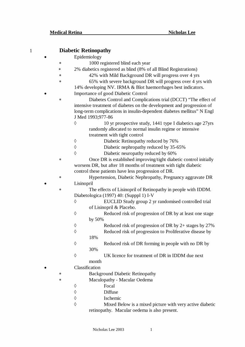
� Focal � Diffuse � Ischemic � Mixed Below is a mixed picture with very active diabetic
retinopathy. Macular oedema is also present.
Nicholas Lee 2003 2
� � Pre-Proliferative DR
� Cotton wool spots � Venous abnormalities - Tortuosity, beading, Venous
reduplication) � Arteriolar abnormalities � Intra-retinal micro vascular abnormalities (IRMA)
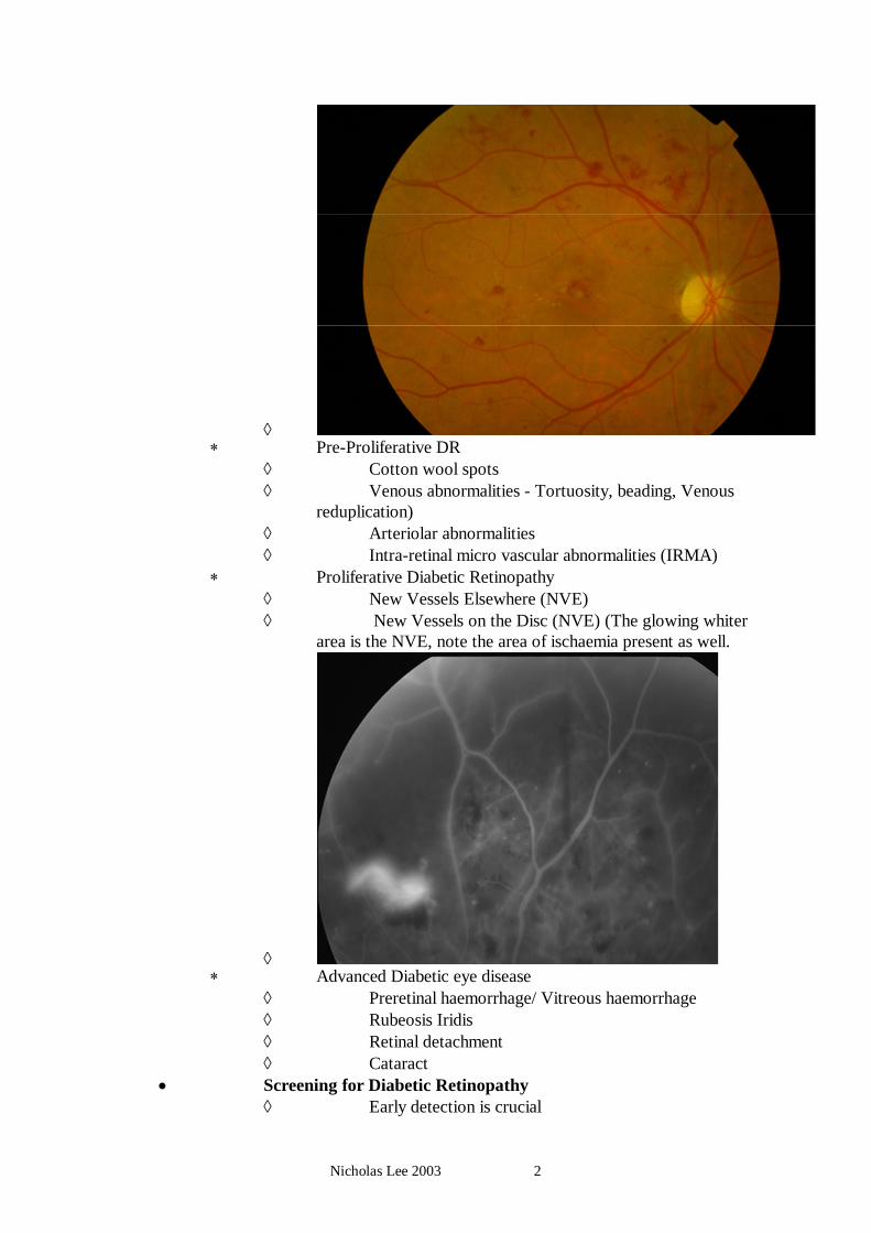
� Proliferative Diabetic Retinopathy � New Vessels Elsewhere (NVE) � New Vessels on the Disc (NVE) (The glowing whiter
area is the NVE, note the area of ischaemia present as well.
� � Advanced Diabetic eye disease
� Preretinal haemorrhage/ Vitreous haemorrhage � Rubeosis Iridis � Retinal detachment � Cataract
Screening for Diabetic Retinopathy � Early detection is crucial
Nicholas Lee 2003 3
� GP/hospital Doctor - Fundoscopy with dilated pupil � Optometric Screening � Non-mydriatic Polaroid/35mm/Digital cameras � Annual for those with no DR, 6mthly for those with any
form of DR. � Management
� Observe/record with Photography Polaroid/35mm/Digital
� Regular review according to severity � Liaise with Lead physician in charge of Diabetic care �
� Treatment � Maculopathy
� Focal - Focal laser, if no improvement - FFA � Diffuse - FFA - Rx BP/Renal function -
VA>6/60 Rx Grid or modified grid laser - Re-Rx as needed.
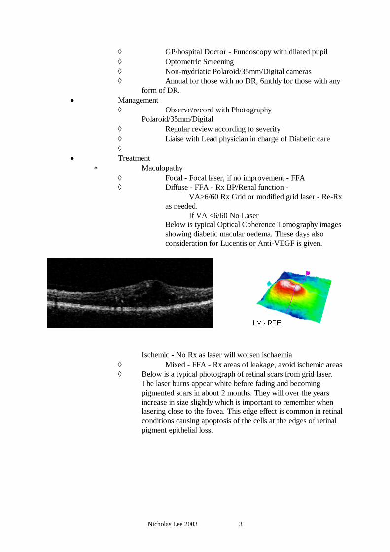
If VA <6/60 No Laser Below is typical Optical Coherence Tomography images
showing diabetic macular oedema. These days also consideration for Lucentis or Anti-VEGF is given.
Ischemic - No Rx as laser will worsen ischaemia � Mixed - FFA - Rx areas of leakage, avoid ischemic areas � Below is a typical photograph of retinal scars from grid laser.
The laser burns appear white before fading and becoming pigmented scars in about 2 months. They will over the years increase in size slightly which is important to remember when lasering close to the fovea. This edge effect is common in retinal conditions causing apoptosis of the cells at the edges of retinal pigment epithelial loss.

Nicholas Lee 2003 4
� � Proliferative Disease
� Mild NVE NIDDM - Rx around NVE, if persist - Full PRP IDDM - mild Scatter PRP
� Moderate NVE - mild Scatter PRP � Severe NVE - Full Scatter PRP � NVE/NVE & Fibrosis - Consider Vitrectomy if fibrosis
around fovea. � Pre-Retinal haemorrhages
� Look for NVE - FFA & Laser � Boat shaped - Consider Nd:YAG Opening of internal
limiting membrane � Vitreous haemorrhage
� Mild - If view PRP � No view - Ultrasound to exclude RD
Review 2 - 4 months If clears PRP If Not Vitrectomy and endolaser
� Severe Ultrasound detached retina - vitrectomy
� Cataract � 20% of UK cataracts extractions are on Diabetic patients � Benefit from small incision Phakoemulsification with less post
op uveitis � Watch careful post operatively for:
� Uveitis, Post Synaechae - damage view for laser later � advancement of DR is common.
Landmark Studies in Diabetes: N Lee 1999
1. The Diabetic Retinopathy Study

Nicholas Lee 2003 5
2. The Early Treatment Diabetic Retinopathy Study 3. The Diabetic Retinopathy Vitrectomy Study 4. The Diabetes Control and Complications Trial 5. The UKPDS Prospective Diabetes Study Trial.
The Diabetic Retinopathy Study (DRS) 1950’s - Role of Photocoagulation controversial 1967 - Anecdotal reports of benefits 1968 - Airlie House Meeting 1971 - DRS Began 1976 - 1st Preliminary reports 1976 - 1987 14 Major Publications Q - “Does Photcoagulation Surgery reduce the risk of sever visual loss in Diabetic Retinopathy ?” Studied natural history of Diabetic Retinopathy without photocoagulation and to compare Xenon & Argon. Patients 1727 from 15 centres radiomised to Indefinite Deferral, Xenon or Argon. 1976 the Indefinite Group were stopped Treatment parameters: Xenon 200-400 Spots 4.5 degree size Blue-Green Argon 800 - 1600 Spots 500 micron 0.1sec Light Blanching of Retinal Pigment Epithelium. New Vessels Elsewhere treated directly Results Severe Visual Loss 16.3% of untreated eyes 5.3% of Xenon 7.4% of Argon ie Severe Visual Loss risk reduced by half. Regression of New Vessels - 38% of treated eyes had increase in New Vessels Elsewhere compared to 63% untreated. Risk Factors for Severe Visual Loss
1. Vitreous or Preretinal Hemorrhage
2. New Vessels Elsewhere – ie in the periphery of the retina
3. New Vessels at the Optic Disc or within in one disc diameter. Below is an example of new vessels on the disc in a diabetic, and after a Pan Retinal Photocoagulation laser with 2000 burns the new vessels have resolved. The risk of the patient having a vitreous haemorrhage and loss of vision is thus dramatically reduced.

Nicholas Lee 2003 6
4. Severe New Vessels. High-risk characteristics = 3 of these. Risks of Treatment
1. Loss of 2 - 4 Lines of visual acuity 23% Xenon, (field loss) 9.8% Argon 5.6% untreated However 1 year later there was no Difference between groups. Macular oedema increased risk of visual loss.
Risk of Progression to High-Risks Characteristics in 1 Year Treated Untreated No PDR 8.8% 15% Mild New Vessels Elsewhere without Vitreous or Preretinal haemorrhages
8.5% 19.4%
Moderate - Severe New Vessels Elsewhere without Vitreous or Preretinal haemorrhages
12.2% 24.5%
Mild New Vessels at the Optic Disc without Vitreous or Preretinal haemorrhages
21.9% 58.7%
Other Findings

Nicholas Lee 2003 7
Adequate Treatment. Eyes with treatment 1 Standard Deviation above mean (61% coverage of superonasal field) have HALF the risk of Severe visual loss compared to eyes treated 1 standard deviation below mean (27% coverage). Treatment density and area both significant. Prompt Full Scatter laser in eyes with high risk features reduces risk of severe visual loss by 50% Severe NPDR & PDR with no high risk characteristics - No clear choice between treatment or Follow up waiting for high risk features. However in Non-Insulin Dependant Diabetic (NIDDM) the risk is much higher and Treatment is appropriate. ? pure local treatment to New Vessels Elsewhere.
The Early Treatment Diabetic Retinopathy Study (ETDRS)
Objectives
1. When in course of Diabetic Retinopathy is it most effective to do Laser?
2. Is Laser effective in? Diabetic maculopathy
3. Is Aspirin helpful? Patients: Mild, Moderate or severe Nonproliferative diabetic Retinopathy or early proliferative diabetic retinopathy. Groups 2707 pts
1. Moderate NPDR or Early PDR - Laser or deferred. Laser - either a. Immediate full Pan Retinal Argon Laser Photocoagulation to the Retina 1200 - 1600 0.1 sec 500 microns. 2 treatment sessions 2 weeks apart. No more than 900 burns in one session and no more than 5weeks to complete treatment.
b. Immediate mild scatter Pan Retinal Argon Laser Photocoagulation to the Retina. 400-650 burns spaced more than one burn width apart scattered uniformly. In one session.
2. Eyes with Macular edema & Less severe Retinopathy.
3. Eyes with macular edema & Severe Retinopathy Definitions Macular Edema Thickening of retina +/- hard exudates with in one disc diameter of fovea CSME One or more of:
1. Retinal thickening within 500um of macular
2. Hard Exudates with in 500um if associated with retinal thickening
3. Zone of retinal thickening one disc are in size at least part ofwhich is within one disc diameter of fovea.
Nonproliferative Diabetic Retinopathy

Nicholas Lee 2003 8
Mild NPDR >one microaneurysm Moderate NPDR Extensive Intraretinal hemorrhages +/- Microaneurysms, +/- Cotton-wool spots, venous beeding, Intra Retinal Microvascular Abnormalities (IRMA) Severe NPDR Cotton-wool spots, venous beeding, Intra Retinal Microvascular Abnormalities all in 2 quadrants or 2 in at least 2 quadrants with intraretinal hemorrhages and microaneurysms in all quadrants or 4-2-1 rule any one of
1. Intraretinal hemorrhages in 4 quadrants
2. Venous beeding in 2 quadrants
3. Severe Intra Retinal Microvascular Abnormalities (IRMA) in one. Results Early Laser in NPDR or Early PDR reduced development of High Risk Signs by 50% in Full Scatter Vs Deferred group 25% in Mild Scatter Vs Deferred Group Macular Edema & Less Severe Retinopathy Immediate Focal & Delayed Scatter initiated only when more severe Retinopathy developed. 50% reduction Eyes treated with Pan Retinal Argon Laser Photocoagulation to the Retina then focal did worse. Macular edema & Sever Retinopathy Immediate Focal & Immediate mild Scatter Visual Fields Significant loss for Full Scatter compared to Deferred or Mild scatter. Macular Edema Focal or Grid treatment reduced incidence of Moderate visual loss by 50% Visual prognosis was worse for those with worse vision at baselin. Aspirin: No effect. Also no harmful effect if required to use. Summary of ETDRS Findings.
1. Scatter Laser NOT recommended for NPDR or early PDR. Considered for eyes approaching high risk characteristics
2. Focal Laser CSME and mild-moderate NPDR, Severed NPDR & PDR
3. Type II diabetics more at risk of Progression. Lessons from study
1. Severity of intraretinal lesions (microaneurysms/hemorrhage, soft exudates, cotton-wool spots, IRMA, venous beading, arteriolar abnormalities) is important in predicting progression and the frequency of follow-up visits.

Nicholas Lee 2003 9
2. New Vessels at the Optic Disc single most prognostic feature when >standard photo 10A (new vessels greater than or equal to one-third to one fourth disc area), panretinal photocoagulation is strongly indicated. 3. NVE without preretinal vitreous hemorrhage is a weaker indicator for panretinal photocoagulation, and careful observation is reasonable. 4. Initial vitreous hemorrhage or preretinal hemorrhage is usually small and does not preclude panretinal Photocoagulation. Patients should be encouraged to report symptoms promptly. 5. Supplemental panretinal Photocoagulation is often effective in causing regression of vessels that persist or recur after initial treatment. Supplemental treatment may be concentrated near NVE, applied throughout the fundus, or extended into the posterior pole. 6. A knowledge of the tendency for neovascularisation to proliferate and then to regress is important in considering the need for additional panretinal Photocoagulation. The goal of panretinal Photocoagulation should be controlling neovascularisation rather than completely eliminating it. 7. In the presence of a complete posterior vitreous detachment, PDR rarely causes retinal traction. 8. Elevated neovascularisation is less affected by panretinal Photocoagulation than retinal surface neovascularisation. 9. Photocoagulation burn size is influenced not only by spot size but also by energy intensity. Therefore, the number of burns of a specified size is not an adequate measure of treatment applied. 10. Vascular narrowing frequently accompanies quiescence of the retinopathy. 11. Intense local Photocoagulation of NVE may produce noticeable scotomas and nerve-fiber layer defects. 12. Macular Photocoagulation improves the course of eyes with CSME, and prompt treatment is preferable to permanent non treatment. However, progression is slow, and treatment deferral with careful monitoring may be a useful strategy when thickening of the center of the macula is equivocal or when treatable lesions are close to the foveal center. 13. Macular edema should be treated prior to panretinal Photocoagulation or concurrently. 14. Hard exudates may increase when macular edema decreases and can threaten central vision. 15. ETDRS investigators emphasized careful follow up after initial treatment of CSME, with retreatment whenever CSME and treatable lesions were present. 16. Fluorescein leakage without retinal thickening does not constitute macular edema.

Nicholas Lee 2003 10
The Diabetes Control and Complications Trial Insulin Dependant Diabetic Tight control reduced incidence of Retinopathy 27% 76% reduction is 3 step progression 61% reduction of progression to severe Retinopathy 59% reduction in need for laser Goal of American Diabetes Association is all Insulin Dependant Diabetic HbA1C <7%
The UKPDS Prospective Diabetes Study Trial. Type II diabetics HbA1C <7.1 & Blood Pressure <140/80 Latest aim. Cholesterol also should be controlled some evidence helps resolve exudates. is target that should be aimed for in all diabetic patients, but of course particularly important in those with Retinopathy. Thus in the Diabetic eye clinic it is important to know at least these too parameters. This is often not difficult where the general diabetic care is in the same hospitals with the same notes, but can be very difficult or impossible without repeating the tests if the diabetic care is undertaken by the GP or at another hospital. Other risk factors to consider are microalbuminuria, smoking and dyslipidaemia. However with such good evidence that the amount of Retinopathy is less in this group this has not become an important factor. This study set out to answer the question “Does tightly controlled blood pressure reduce the risk of complications in diabetes” The study clearly showed it did as was the diabetic control. BMJ 317 Sept 1998.

Nicholas Lee 2003 11

Nicholas Lee 2003 12
Useful Web sites Www.diabeticretionpathy.org.UK www.eyetextbook.co.UK www.doh.gov.uk/ Mr. Nicholas Lee Lead Clinician at Hillingdon Hospital Consultant Ophthalmologist at The Western Eye Hospital NHS email [email protected]