diabetic foot · foot to move. your arch is a group of joints that provides stability for you...
TRANSCRIPT
Diabetic FootAYMAN MISMAR

Diabetic Foot
Definition:
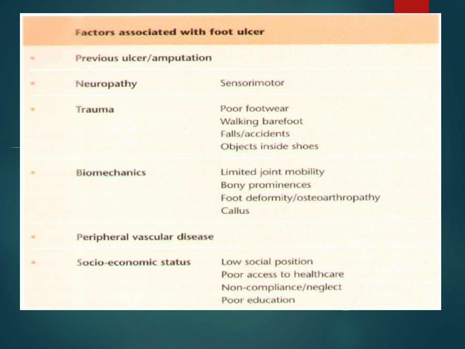
Infection, ulceration or
destruction of deep tissues
associated with neurological
abnormalities & various degrees of
peripheral vascular diseases in the
lower limb
(based on WHO definition)
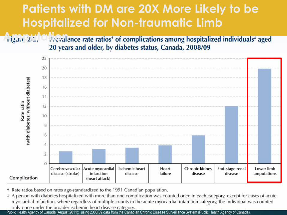
Public Health Agency of Canada (August 2011); using 2008/09 data from the Canadian Chronic Disease Surveillance System (Public Health Agency of Canada).
Patients with DM are 20X More Likely to be
Hospitalized for Non-traumatic Limb
Amputation
Epidemiology
40% - 60% of all non traumatic lower limb amputation
85% of diabetic related foot amputation are preceded by foot ulcer
4 out of 5 ulcer in diabetics are precipitated by trauma
4% -15% is the prevalence of foot ulcer in diabetics
Epidemiology
Foot ulcerations is most common cause of hospital admissions for Diabetics
Expensive to treat, may lead to amputation and need for chronic institutionalized care
After amputation 30% lose other limb in 3 years
After amputation 2/3rds die in five years
Type II can be worse
15% of diabetic will develop a foot ulcer

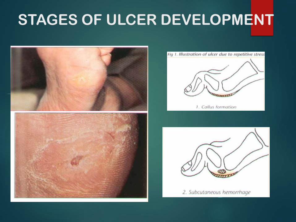
STAGES OF ULCER DEVELOPMENT

STAGES OF ULCER DEVELOPMENT

DEFINITION OF DIABETIC FOOT
SYNDROME
FOOT ABNORMALITIES CAUSED BYNEUROPATHY, ANGIOPATHY ANDINFECTION IN DIABETES MELLITUS PATIENT’S
Infection
Neuropathy Ischemia

Pathophysiology
Vascular disease
Neuropathy
Sensory
Motor
autonomic
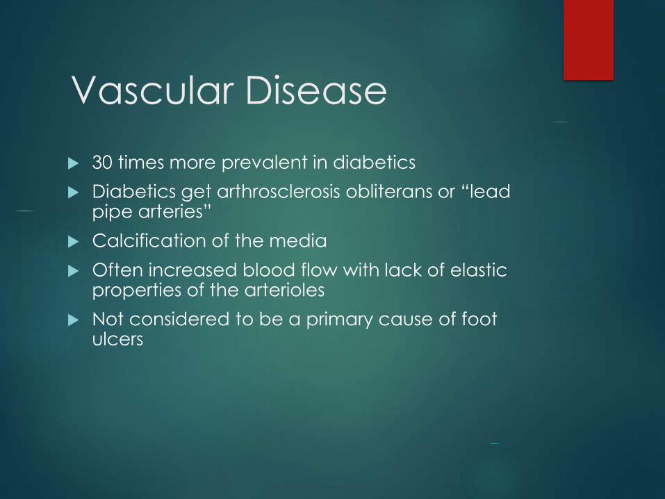
Vascular Disease
30 times more prevalent in diabetics
Diabetics get arthrosclerosis obliterans or “lead pipe arteries”
Calcification of the media
Often increased blood flow with lack of elastic properties of the arterioles
Not considered to be a primary cause of foot ulcers

Neuropathy
Changes in the vasonervorum with resulting
ischemia cause:
Increased sorbitol in feeding vessels block flow and
causes nerve ischemia
Intraneural acculmulation of advanced products of
glycosylation
Abnormalities of all three neurologic systems
contribute to ulceration

Autonomic Neuropathy
Regulates sweating and perfusion to the limb
Loss of autonomic control inhibits
thermoregulatory function and sweating
Result is dry, scaly and stiff skin that is prone to
cracking and allows a portal of entry for bacteria
Motor Neuropathy
Mostly affects forefoot ulceration
Intrinsic muscle wasting – claw toes
Equinous contracture

Sensory Neuropathy
Loss of protective sensation
Starts distally and migrates proximally in “stocking” distribution
Large fibre loss – light touch and proprioception
Small fibre loss – pain and temperature
Usually a combination of the two

Sensory Neuropathy
Two mechanisms of Ulceration
Unacceptable stress few times
rock in shoe, glass, burn
Acceptable or moderate stress repeatedly
Improper shoe ware
deformity

Ulcer Classification
Wagner’s Classification
0 – Intact skin (impending ulcer)
1 – superficial
2 – deep to tendon bone or ligament
3- osteomyelitis
4 – gangrene of toes or forefoot
5 – gangrene of entire foot
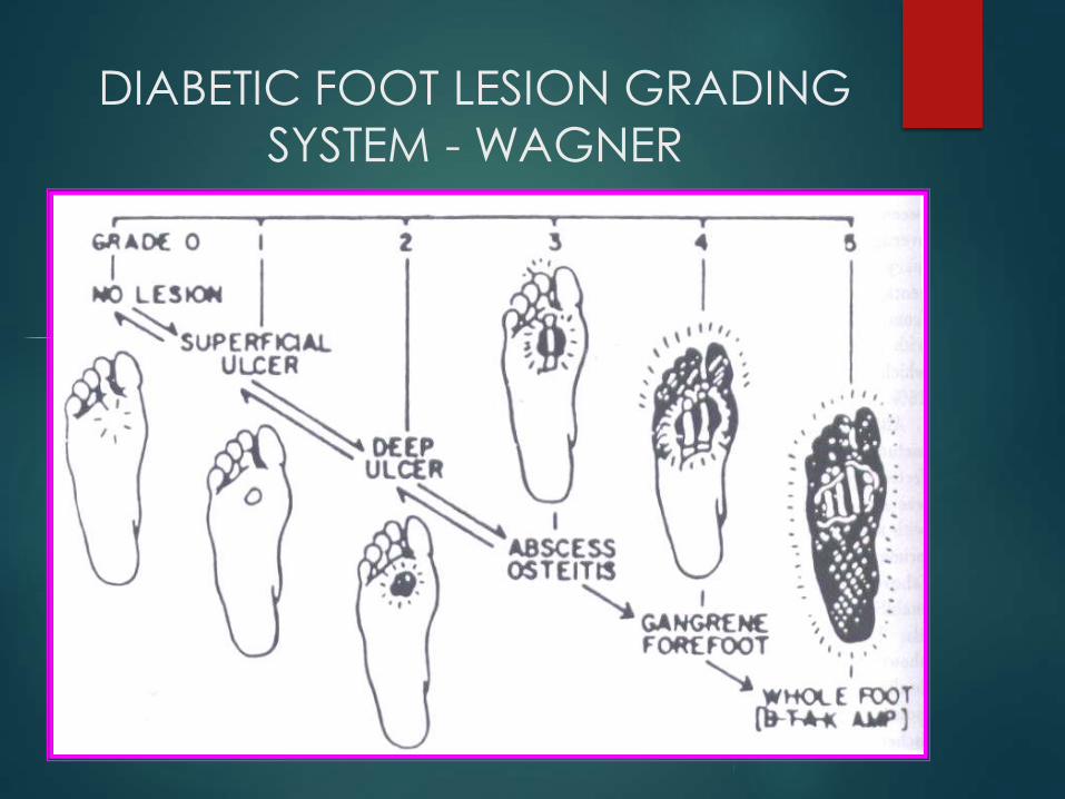
DIABETIC FOOT LESION GRADING
SYSTEM - WAGNER
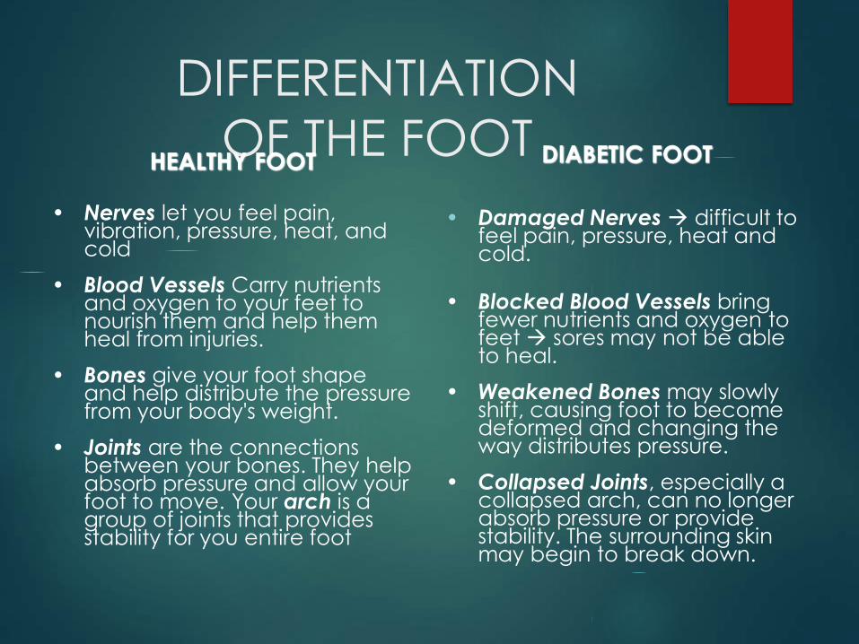
DIFFERENTIATION
OF THE FOOTHEALTHY FOOT
• Nerves let you feel pain, vibration, pressure, heat, and cold
• Blood Vessels Carry nutrients and oxygen to your feet to nourish them and help them heal from injuries.
• Bones give your foot shape and help distribute the pressure from your body's weight.
• Joints are the connections between your bones. They help absorb pressure and allow your foot to move. Your arch is a group of joints that provides stability for you entire foot
DIABETIC FOOT
• Damaged Nerves difficult to feel pain, pressure, heat and cold.
• Blocked Blood Vessels bring fewer nutrients and oxygen to feet sores may not be able to heal.
• Weakened Bones may slowly shift, causing foot to become deformed and changing the way distributes pressure.
• Collapsed Joints, especially a collapsed arch, can no longer absorb pressure or provide stability. The surrounding skin may begin to break down.

Structural
Abnormalities
Peripheral Arterial
Assessment
Skin changes
Evidence of infection
Callous or ulcer
Range of motion
Charcot foot
Temperature
Capillary refilling
Skin changes
Ankle Brachial Index
Neuropathy
Assessment10 gram monofilament
How to Perform Proper Foot
Examination
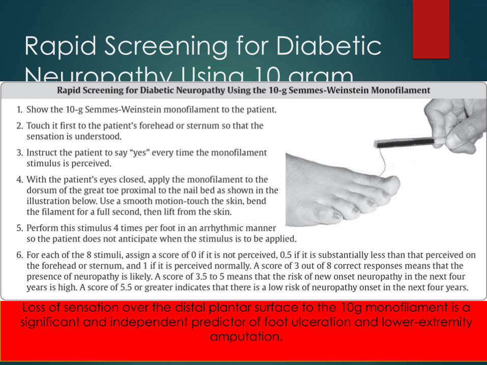
Rapid Screening for Diabetic
Neuropathy Using 10 gram
Semmes-Weinstein Monofilament
Loss of sensation over the distal plantar surface to the 10g monofilament is a
significant and independent predictor of foot ulceration and lower-extremity
amputation.
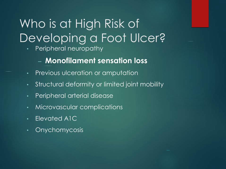
Who is at High Risk of
Developing a Foot Ulcer?• Peripheral neuropathy
– Monofilament sensation loss
• Previous ulceration or amputation
• Structural deformity or limited joint mobility
• Peripheral arterial disease
• Microvascular complications
• Elevated A1C
• Onychomycosis
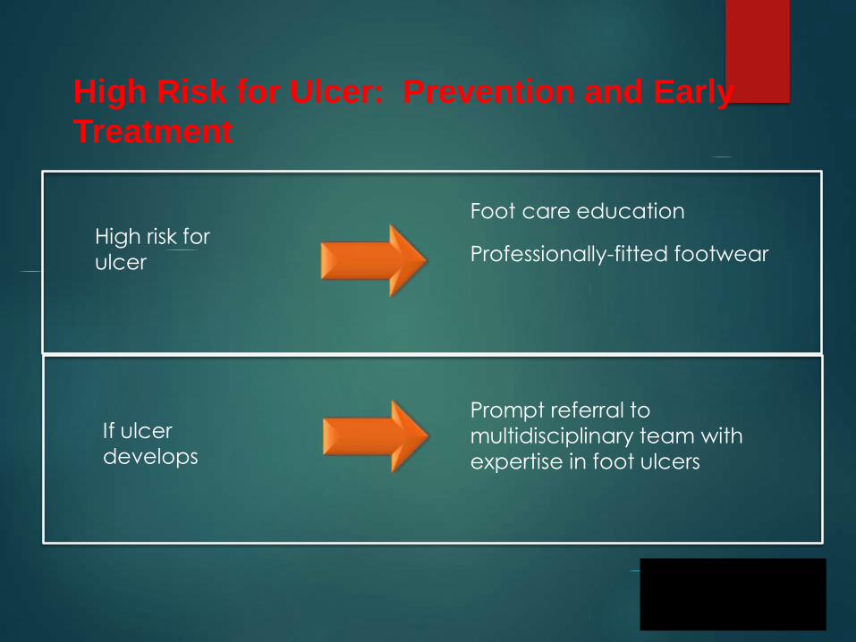
High risk for
ulcer
Foot care education
Professionally-fitted footwear
Prompt referral to
multidisciplinary team with
expertise in foot ulcers
If ulcer
develops
High Risk for Ulcer: Prevention and Early
Treatment

Local factors
Systemic factors
Wound care
Pressure offloading
Debridement (nonischemic
wounds)
Glycemic control
Treat infection
Address lower-extremity vascular
status
Foot Ulcer: Multidisciplinary Team Approach
Treatment
Patient education
Ambulation
Shoe ware
Skin and nail care
Avoiding injury
Hot water
F.B’s

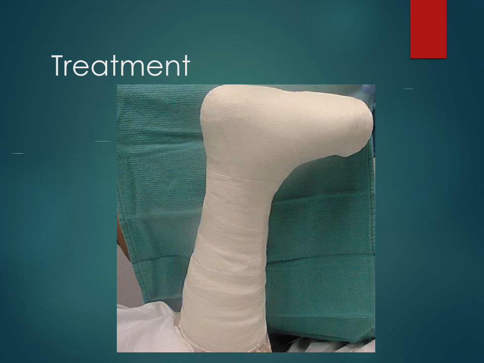
Treatment
Wagner 0-2
Total contact cast
Distributes pressure and allows patients to continue
ambulation
Antibiotics if infected
Treatment

Treatment
Wagner 0-2
Surgical if deformity present that will reulcerate
Correct deformity
exostectomy

Treatment
Wagner 3
Excision of infected bone
Wound allowed to granulate
Grafting (skin or bone) not generally effective
Wagner 4-5
Amputation
level
Treatment
After ulcer healed
Orthopedic shoes with accommodative (custom
made insert)
Education to prevent recurrence
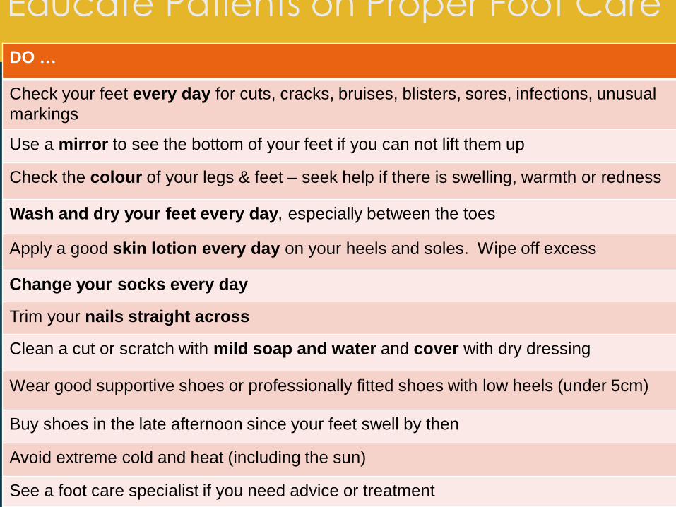
Educate Patients on Proper Foot Care
DO …
Check your feet every day for cuts, cracks, bruises, blisters, sores, infections, unusual
markings
Use a mirror to see the bottom of your feet if you can not lift them up
Check the colour of your legs & feet – seek help if there is swelling, warmth or redness
Wash and dry your feet every day, especially between the toes
Apply a good skin lotion every day on your heels and soles. Wipe off excess
Change your socks every day
Trim your nails straight across
Clean a cut or scratch with mild soap and water and cover with dry dressing
Wear good supportive shoes or professionally fitted shoes with low heels (under 5cm)
Buy shoes in the late afternoon since your feet swell by then
Avoid extreme cold and heat (including the sun)
See a foot care specialist if you need advice or treatment
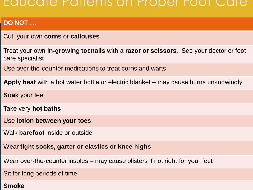
Educate Patients on Proper Foot Care
DO NOT …
Cut your own corns or callouses
Treat your own in-growing toenails with a razor or scissors. See your doctor or foot
care specialist
Use over-the-counter medications to treat corns and warts
Apply heat with a hot water bottle or electric blanket – may cause burns unknowingly
Soak your feet
Take very hot baths
Use lotion between your toes
Walk barefoot inside or outside
Wear tight socks, garter or elastics or knee highs
Wear over-the-counter insoles – may cause blisters if not right for your feet
Sit for long periods of time
Smoke

Charcot Foot
More dramatic – less common 1%
Severe non-infective bony collapse with
secondary ulceration
Two theories
Neurotraumatic
Neurovascular
Neuro-osteoarthropathy
Non- infective pathology
Should be suspected in any swollen hot erythematous foot
Differentiation from infection is important to prevent misdiagnosis & possible amputation
Treatment should aim at preventing severe deformity

Charcot Foot
Neurotraumatic
Decreased sensation + repetitive trauma = joint and bone collapse
Neurovascular
Increased blood flow → increased osteoclast activity → osteopenia → Bony collapse
Glycolization of ligaments → brittle and fail →
Joint collapse

Classification
Location
Forefoot, midfoot (most common) , hindfoot
Atrophic or hypertrophic
Radiographic finding
Little treatment implication

Classification
Eichenholtz
1 – acute inflammatory process
Often mistaken for infection
2 – coalescing phase
3 - consolidation


The Five “C’s” of Foot
Care Clean! Clean and check feet daily.
Wash with warm not hot water. Pat dry.
Condition!! Use a moisturizer daily. Use one without perfume or alcohol
Care!!! Clip normal nails straight across with a slight curve at the corners. Bathroom surgeons give up your license.
Cover!!!! Always wear shoes or slippers with a sole to protect your feet. Check your feet before and after wearing for any unusual marks or redness
Use caution and call

Indications for Amputation
Uncontrollable infection or sepsis
Inability to obtain a plantar grade, dry foot that
can tolerate weight bearing
Non-ambulatory patient
Decision not always straightforward
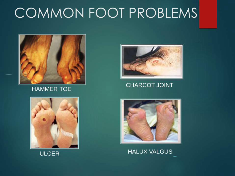
COMMON FOOT PROBLEMS
HAMMER TOECHARCOT JOINT
HALUX VALGUSULCER
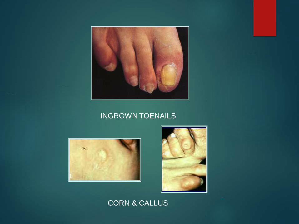
INGROWN TOENAILS
CORN & CALLUS

IRRITATIONS, SKIN LESIONS BLISTER
CUTS BETWEEN YOUR TOES
Association
Clinical
Practice
Guidelines
Foot Care

Recommendation 1
1. Assessment by healthcare providers should include the
assessment of skin changes, structural abnormalities (e.g.,
range of motion of ankles and toe joints, callus pattern,
bony deformities), skin temperature, evaluation for
neuropathy and peripheral arterial disease, ulcerations
and evidence of infection [Grade D, Level 4]
Recommendation 2
2. People at high risk of foot ulceration and amputation
should receive foot care education (including
counseling to avoid foot trauma), professionally-fitted
footwear, and early referrals to a healthcare
professional trained in foot care management if foot
complications occur [Grade B, Level 2]
Recommendation 3
3. Individuals who develop a foot ulcer should be
managed by a multidisciplinary healthcare team with
expertise in the management of foot ulcers to prevent
recurrent foot ulcers and amputation [Grade C, Level 3]

Recommendation 4
4. There is currently insufficient evidence to recommend
any specific dressing type for diabetic foot ulcers
[Grade C, Level 3]. General principles of wound
management involve the provision of a moist wound
environment, debridement of nonviable tissue
(nonischemic wounds) and offloading of pressure areas
[Grade B, Level 3]
Recommendation 5
5. Evidence is currently lacking to support the routine use
of adjunctive wound- healing therapies such as topical
growth factors, granulocyte-colony stimulating factors,
dermal substitutes in diabetic foot ulcers but they may
be considered in nonhealing, nonischemic wounds
when all other options have been exhausted [Grade D,
Level 4]