dextran derivatives in single and combination chemotherapy...
TRANSCRIPT

[CANCER RESEARCH 37, 3448-3454, September 1977]
Dextran Derivatives in Single and Combination Chemotherapyagainst Transplantable Mouse Ascites and Solid Tumors1
Masuko Suzuki, Takeshi Mikami, Minoru Kadowaki, Tatsuji Matsumoto, and Shigeo Suzuki
Department of Microbiology ¡M.S., T. M., M. K., T. M.] and Second Department of Hygienic Chemistry [S. S.], Tohku College of Pharmacy, Senda/, Japan
SUMMARY
Dextran, a typical homopolysaccharide without antitumoractivity, was modified by palmitoylation and/or phosphoryl-
ation to yield three derivatives: palmitoyldextran phosphate,dextran phosphate, and palmitoyldextran. Of these compounds, only palmitoyldextran phosphate showed growth-
inhibitory activity against Ehrlich solid tumor in mice. Incombination therapy with mitomycin C, bleomycin, cyclo-phosphamide, and 5-fluorouracil, palmitoyldextran phos
phate manifested strong synergistic effects against bothSarcoma 180 ascites and L1210 leukemic tumors. The compound is not directly cytocidal against Sarcoma 180 ascitestumor, but it appears to act via activation of peritonealmacrophage. The antitumor activity of palmitoyldextranphosphate apparently is mainly due to immunological host-
mediated mechanisms.
INTRODUCTION
A variety of plant polysaccharides possess strong growth-
inhibitory properties against several transplantable solidtumors (5, 7, 12, 27). The least toxicity and most markedantitumor effects were observed in a polysaccharide fraction isolated from the leaves of a species of bamboo grass.Previously, Nakahara et al. (16) presented experimental evidence to show that the action of this polysaccharide may behost mediated. In an earlier work, we reported that yeastmannan and glucan are able to exert adjuvant action toenhance the antibody response of mice against implantedsolid tumor cells (29).
Since some bacterial constituents, lipopolysaccharides(13, 15), endotoxin (4), glycolipid of mycobacteria (6), andcell wall skeleton of Bacillus Calmette-Guérin (3) are growth
inhibitory against some ascites tumors, we investigated theantitumor activity of the dextran derivatives POP,2 DP, and
PD, which possess functional groups similar to those of thebacterial constituents described above.
MATERIALS AND METHODS
Animals. Male dd and DBA/2 x C57BL/6 FI mice weighing 18 ±2 g were housed 5/cage in air-conditioned quarters
and were provided food and water ad libitum.
1 Supported by Grant-in-Aid for Cancer Research from the Ministry of
Health and Welfare oÃJapan.* The abbreviations used are: POP, palmitoyldextran phosphate; DP, dex
tran phosphate; PD, palmitoyldextran; Hanks' BSS, Hanks' balanced salt
solution.
Received May 21, 1976; accepted May 20, 1977.
Tumors. The tumors used were Sarcoma 180 ascites,Ehrlich solid tumor, and a L1210 line, initially supplied byDr. Fumiko Fukuoka, National Cancer Center Research Institute of Japan, and maintained in our laboratory in anascites form, dd mice inoculated with 1 x 10⁷ Sarcoma 180
or Ehrlich tumor cells, or DBA/2 x C57BL/6 F, mice inoculated with 5x10" L1210 leukemic tumor cells in our labora
tory never spontaneously recovered from the inoculation.Sarcoma 180C, a cell line of Sarcoma 180 ascites tumor,was kindly donated by Dr. Kotaro Koyama of the sameResearch Institute. Tissue culture was incubated at 37°in a
moist atmosphere containing 5% C02.Chemical Synthesis and Purification of Modified Dex-
trans. Modified dextrans were synthesized according to apreviously described method (26). Chemical analysis of DPshowed 81.0% glucose by the Molisch method (9) and18.3% phosphoric acid by the Allen-Nakamura method (17);for PD, 85.6% sugar and 0.6% palmitic acid by gas chroma-
tography; and for POP, 76.8% sugar, 19.0% phosphoricacid, and 0.6% palmitic acid. All synthesized dextran derivatives were free from nitrogen as determined by microele-
mental analysis.Assay for Antitumor Activity against Ehrlich Solid Tumor
in Vivo. About 1 x 10⁷ cells from a 7-day-old Ehrlich tumor
were transplanted s.c. into the right groin. DP, PD, or POPin 0.1 ml of 0.9% NaCI solution was administered i.p. dailyfor 10 successive days beginning 24 hr after tumor implantation. All the mice were sacrificed 20 days after implantation, and the tumors were extirpated and weighed. Relativetumor growth was calculated from the following formula (7):
Inhibition ratio (%) = [(A - B)/A] x 100
where A is the average tumor weight of the control groupand ßis the treated group. Ten mice/group were used, andthe test was repeated 3 times.
Assay for Antitumor Activity against Ascites Tumor inVivo. About 1 x 10⁷ Sarcoma 180 ascites tumor cells wereimplanted i.p. in dd mice, or 5 x 10* L1210 leukemic tumor
cells were implanted i.p. in DBA/2 x C57BL/6 F, mice. Asolution of each dextran derivative in 0.1 ml of 0.9% NaCIsolution per mouse was administered once daily for 5 successive days beginning 24 hr after tumor implantation. Aseries of combination therapies was also conducted usingthese modified dextrans and several conventional antitumoragents such as mitomycin C (Kyowa Hakko Kogyo Co.,Tokyo, Japan; Lot MIS-554AEE), bleomycin (Nihon KayakuCo., Tokyo, Japan; Lot B-126), 5-fluorouracil (Kyowa HakkoKogyo Co., Tokyo, Japan; Lot 5FU194ADD), and cyclophos-
3448 CANCER RESEARCH VOL. 37
Research. on February 12, 2019. © 1977 American Association for Cancercancerres.aacrjournals.org Downloaded from

phamide (Shionogi and Co., Osaka, Japan; Lot B210J).Each antitumor agent in 0.1 ml of 0.9% NaCI solution wasadministered at the indicated dose i.p. once daily for 5 days,1 hr after injection of dextran derivatives. The dosages ofanticancer drugs in combination treatment between theanticancer drugs and dextran derivatives were selected according to the maximum doses previously used separatelyagainst each tumor, and which had proven to be an ineffective maximum dose against each tumor. The tumor growthwas measured by the total packed cell volume method (22)on the 7th day after tumor inoculation. Life-span elongation
was observed for up to 60 days.Growth Inhibition in Vitro. About 1 x 10⁵ Sarcoma 180C
cells/culture tube were incubated in Eagle's minimum es
sential medium supplemented with 15% calf serum to whichwas added 1 mg of the dextran derivative per ml. Afterincubation, the cells were stained with safranin (21) to estimate their viability, which was compared with that of cellsincubation, the cells were stained with safranin (20) to esti-
derivatives.Peritoneal Macrophage Cultures. The methods used
were adopted from the method of Ruskin ef al. (21 ). Briefly,peritoneal macrophages were obtained from normal ddmice by washing the peritoneal cavity with 5 ml of Hanks'
BSS containing 5 units of heparin per ml. The cell suspension was centrifugea at 800 rpm at 4°for 5 min. The resulting sediment was resuspended in heparinized Hanks' BSS
to a concentration of approximately 2 x 10⁶ cells/ml. Two
ml of the cell suspension were placed in Retri dishes (A/SNUNC, Roskilde, Denmark) 3 cm in diameter and incubatedfor 1 hr. Cells that had not adhered to the dishes during thisperiod were removed by rinsing with Hanks' BSS. The cultures were then reincubated in Medium 199 with Hanks'
BSS supplemented with L-glutamine (Grand Island Biologi
cal Co., Grand Island, N. Y.) and 10% fetal calf serum.Macrophage Cytotoxicity Test. After 18 hr, the cultures
of peritoneal macrophage were challenged with 2 x 10⁵
Sarcoma 180 ascites tumor cells/ml and/or PDF and/ormitomycin C. After 24 hr of incubation, Sarcoma 180 cellswere stained with trypan blue, and the viable cells werecounted.
Plaque-forming Cells. In order to examine whether dex
tran and the dextran derivatives accelerated the antibodyproduction against heterologous antigen, the Cunninghamand Szenberg (8) modification of the Jerne plaque technique was used. Starting 1 day after Ehrlich solid tumor
Antitumor Activity of Dextran Derivatives
implantation, dextran or dextran derivatives, 100 mg/kg,were injected i.p. once a day for 10 days into duplicategroups of mice. One, 6,11, and 16 days after tumor implantation, sheep erythrocytes at a dose of 1 x 10" cells/0.1 ml
of 0.9% NaCI solution were injected into the tail vein. Forthe combination therapy, starting 1 day after Sarcoma 180ascites tumor implantation, dextran or dextran derivatives(1 mg/kg) and/or mitomycin C were injected once a day for5 days into duplicate groups of mice. Two days before and 1and 4 days after tumor implantation, sheep RBC were injected into the tail vein. Four days after the antigen injection, the spleens were removed from the mice, and thenumber of plaques formed by the antibody-forming cells per
total spleen cells were counted by the conventional method.Each time 5 mice were tested from each experimentalgroup.
Footpad Tests. The footpad tests in mice were doneaccording to the method of Katsura ef a/. (14). Briefly, 100Mg bovine serum albumin and Freund's complete adjuvant
were inoculated into the hind flank s.c. The next day, Sarcoma 180 ascites tumor inoculation and drug treatmentwere as described above. After 5 days, 12.5 /J.Qof alum-
precipitated bovine serum albumin in a volume of 0.025 mlwere injected into the right hind footpad; a similar volumeof diluent was injected into the left hind footpad. Twenty-four hr later, the difference in thickness between the pre-
and postinjection foot was recorded and used as a measureof the amount of swelling. Each time, 10 mice were testedfrom each experimental group.
Student's t test was used to evaluate the significance of
the differences between the groups given the test compound and the control group.
RESULTS
Growth-inhibitory Activity of Modified Dextrans against
Ehrlich Solid Tumor. Table 1 shows the effect of dextranand POP on solid Ehrlich tumor in dd mice at 1 and 100 mg/kg/day for 10 successive days. POP showed an inhibitionratio of 56% (p < 0.01) at 100 mg/kg/day. The other dextranderivatives, DP and PD, were ineffective. Ten mice/groupwere used, and the test was repeated 3 times.
Growth-inhibitory Activity of Modified Dextrans against
Sarcoma 180 Ascites Tumor, dd mice were implanted i.p.with 1 x 10⁷ of the Sarcoma 180 tumor cells. A series of
Table 1Antitumor effect of POP against Ehrlich tumor implanted s.c. in dd mice"
MaterialsPOPDextranControl(mg/kg/dayfor10days)11001100AvExperiment16.83.06.58.17.0.
tumor wt(g)Experi
ment215.14.29.511.910.6Experiment35.43.35.96.16.8Inhibition
ratio(%)Experi
ment10561-150Experiment2-426010-120Experiment 3p2052
<0.01''13100
" Ten mice/group.6 Versus the control group.
SEPTEMBER 1977 3449
Research. on February 12, 2019. © 1977 American Association for Cancercancerres.aacrjournals.org Downloaded from

M. Suzuki et al.
combination therapies was examined using PDP (1 mg/kg/day i.p.) and mitomycin C (0.01 mg/kg/day i.p.) once dailyfor 5 days beginning 24 hr after tumor implantation. Thetumor growth was measured by the total packed cell volumemethod on the 7th day after tumor inoculation. As seen inTable 2, the group receiving combination treatment showedabout 91% inhibition of tumor growth.
PDP-induced Life-span Elongation of Mice Bearing Sarcoma 180 Ascites or L1210 Leukemic Tumors. There hasbeen considerable interest in recent years in the use ofcombinations of drugs against experimental (11) and clinical (24) neoplasia. We tried combination therapy of dextranderivatives with mitomycin C, bleomycin, cyclophosphamide, and 5-fluorouracil. The dextran derivatives and the
other antitumor agents were injected i.p. by the methoddescribed above. First, the combination therapy of mitomycin C (0.5 mg/kg/day for 5 days) and dextran derivatives (1mg/kg/day for 5 days) was tested. PDP and DP showedsignificant synergism when combined with mitomycin C(Chart 1). Ten of the 20 mice treated with PDP and mitomycin C survived for more than 60 days after implantation withnoticeable tumor regression at that time. Similarly, 20 mice/group were used, and the following experiments were carried out: a combination therapy of PDP with bleomycin(Chart 2), with cyclophosphamide (Chart 3), and with 5-
fluorouracil (Chart 4) against Sarcoma 180 ascites tumor indd mice, all of which combinations also showed remarkablesynergism between the drugs.
Table 2Combination effect of PDP with mitomycin C on Sarcoma 180
tumor implanted i.p. in dd micePDP treatments, 1 mg/kg/day i.p. for 5 days; mitomycin C treat
ments, 0.01 mg/kg/day i.p. for 5 days.
No. ofInhibition cured/no, of
Materials ratio (%) treated mice
PDP + mitomycin CPDPMitomycin CControl
91179
10/120/120/360/36
The synergistic effect of POP and mitomycin C againstL1210 leukemic tumor in DBA/2 x C57BL/6 F, mice isshown in Chart 5. Fifty-two mice were implanted i.p. with 5x 10" L1210 cells and were then divided at random into 4
groups of 13 mice. Twenty-four hr later, a solution of POP
in 0.9% NaCI solution and/or mitomycin C was injected i.p.
30 60 Days
Chart 2. Effect of single and combination treatment with POPand bleomycin on Sarcoma 180 ascites tumor rejection in dd mice. Mice were treated asinChartl. O O, control; O O, bleomycin (7.5 mg/kg/day); D D,POP (1 mg/kg/day); D D, POP (1 mg/kg/day) + bleomycin (7.5 mg/kg/day).
100
Days
Chart 3. Effect of single and combination treatment with POP and cyclophosphamide on Sarcoma 180 ascites tumor rejection in dd mice. Mice weretreated as in Chart 1. O O, control; O O, cyclophosphamide (50 mg/kg/day); D D, POP (1 mg/kg/day); D D, PDP (1 mg/kg/day) + cyclophosphamide (50 mg/kg/day).
10 20 30 60 Days
Chart 1. Effect of single and combination treatment with dextran derivatives and mitomycin C on Sarcoma 180 ascites tumor rejection in dd mice. Micewere implanted i.p. with 1 x 10⁷cells of Sarcoma 180, and beginning 24 hr later, a solution of each dextran derivative in 0.9% NaCI solution and/or mitomycinC was injected i.p. for 5 successive days (see text). O O, control; • »,dextran (1 mg/kg/day); A A, PD (1 mg/kg/day); A A, DP (1 mg/kg/day); D D, PDP (1 mg/kg/day); O O, mitomycin C (0.5 mg/kg/day); • •,dextran (1 mg/kg/day) + mitomycin C (0.5 mg/kg/day); A A, PD(1 mg/kg/day) + mitomycin C (0.5 mg/kg/day); A A, DP (1 mg/kg/day) + mitomycin C (0.5 mg/kg/day); D D, PDP (1 mg/kg/day) + mitomycin C(0.5 mg/kg/day).
3450 CANCER RESEARCH VOL. 37
Research. on February 12, 2019. © 1977 American Association for Cancercancerres.aacrjournals.org Downloaded from

Antitumor Activity of Dextran Derivatives
f-O
10 20 30 60 Days
Chart 4. Effect of single and combination treatment with POP and 5-fluorouracil on Sarcoma 180 ascites tumor rejection in dd mice. Mice weretreated as in Chart 1. O O, control; O O, 5-fluorouracil (10 mg/kg/day); D D, POP (1 mg/kg/day); D D, POP (1 mg/kg/day) + 5-fluorouracil (10 mg/kg/day).
lOO
IO 20 30 60 Days
Chart 5. Effect of single and combination treatment with POP and mito-mycin C on leukemic L1210 ascites tumor rejection in DBA/2 x C57BL/6 F,mice. Fifty-two mice were implanted i.p. with 5 x 10⁴ L1210 cells and thendivided at random into 4 groups of 13 animals; beginning 24 hr later, asolution of POP in 0.9% NaCI solution and/or mitomycin C was injected ¡.p.for 5 successive days. O O, control; O O, mitomycin C (0.1 mg/kg/day); D D, POP (1 mg/kg/day); D D. PDP (1 mg/kg/day) + mitomycinC (0.1 mg/kg/day).
for 5 successive days. Three of 13 mice treated with PDPand mitomycin C survived for more than 60 days after tumorimplantation. These experiments were duplicated, and similar results were obtained. An abdominal incision confirmedthe absence of the remaining tumor in those mice thatsurvived for 60 days after tumor implantation.
Direct Cytocidal Action of Dextran Derivatives on Cultured Cells. In order to examine whether PDP has a directcytocidal or growth-inhibitory effect on cells in tissue cul
ture, Sarcoma 180C cells were grown in a medium containing PDP (1 mg/ml), and the viability of the cells was examined after 0, 24, 48, and 72 hr of incubation. Viability of thecells cultured in a medium containing PDP was no differentfrom that of the cells cultured without PDP. PDP has neitherdirect cytotoxicity nor growth-inhibitory activity against the
tumor cells in vitro (Chart 6).Enhancement of Mitomycin C Cytotoxicity against Sar
coma 180 Ascites Tumor Cells by PDP. The above resultsindicate that PDP is not directly cytotoxic in vitro but doesamplify the antitumor activity of several conventional anti-
cancer drugs in vivo. Therefore, we examined the combinedeffect of mitomycin C and PDP on Sarcoma 180 ascitestumor cells in vitro. Addition of mitomycin C alone (0.3 mg/ml) to the Sarcoma 180 cells had no effect at the end of a 24-
hr culture period. On the other hand, the combination of
mitomycin C (0.3 mg/ml) and PDP (0.5 mg/ml) showedcytotoxicity to the tumor cells (Table 3).
Effect of Normal Peritoneal Macrophages and PDP withMitomycin C on the Cytocidal Activity against Sarcoma180 Ascites Tumor Cells. In the preceding experiment, PDPshowed a direct enhancement of mitomycin C toxicity. Withthis in mind, we then modified our in vitro experimentalsystem by the addition of peritoneal macrophages according to the method of Ruskin ef a/. (21). Table 4 shows theresults of this experiment; the synergistic effect, not shownwith mitomycin C (0.1 mg/ml) plus PDP (0.5 mg/ml), without normal peritoneal macrophage (Table 3), is detectedupon combination of the same dosages of mitomycin C andPDP. Furthermore, the cytocidal activity against the tumorcells was detected upon PDP and the macrophage, although the activity was still weaker than in combination ofPDP with mitomycin C and the macrophage.
Effect of Dextran Derivatives on Antibody Formationagainst Sheep RBC by the Cunningham Method in MiceBearing Ehrlich Solid Tumor. Since the antitumor action ofPDP was not by direct cytotoxicity but rather by a host-mediated response or by activation of tumor-killing cells,
the effect of the PDP on various immune responses wasexamined in order to further elucidate the mechanism ofaction of the drug. The plaque-forming test was carried outon tumor-bearing mice to determine whether PDP would
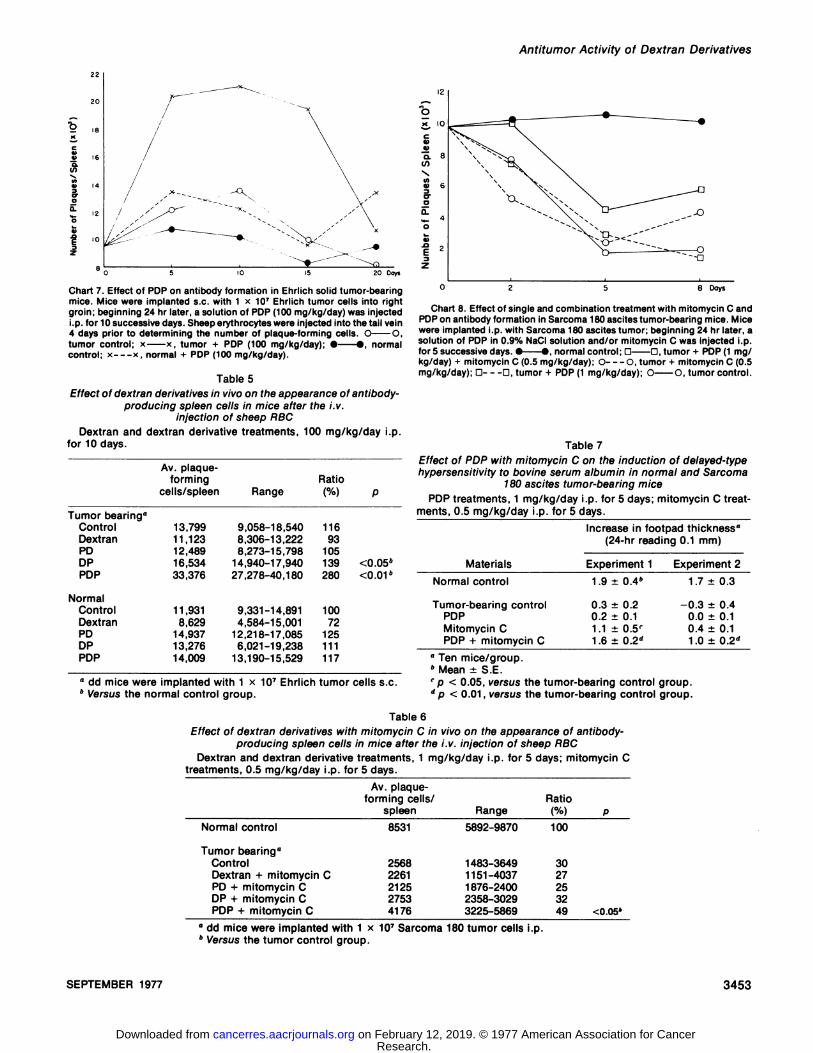
increase antibody production in the spleen against sheeperythrocytes. As seen in Table 5, the number of plaque-forming cells in PDP-treated mice 10 days after Ehrlich solid
tumor implantation showed a significant difference between those in normal and in dextran-, DP-, and PD-treated
mice. The kinetics of antibody formation by spleen cells ofPDP-treated mice with Ehrlich solid tumors is shown in
Chart 7. The increase in plaque formation was not detectable in normal PDP-treated mice but was detectable in tumor-bearing PDP-treated mice.
Effect of Dextran Derivatives on Antibody Formationagainst Sheep RBC by the Cunningham Method in MiceBearing Sarcoma 180 Ascites Tumor. Table 6 shows theplaque formation 5 days after Sarcoma 180 ascites tumorinoculation in mitomycin C and/or dextran derivative-
treated mice, and its kinetics is illustrated in Chart 8. On Day2 a decrease in plaque formation was observed in tumor-
bearing mice treated with mitomycin C (0.5 mg/kg) and PDP
IO
24 4s 72 hr
Chart 6. Growth curves of Sarcoma 180C cells in a medium containingdextran derivatives. Sarcoma 180C cells were cultured in a medium containing 1 mg/ml of each dextran derivative and viability of the cells was examinedafter 24, 48, and 72 hr. O O, control; A A, PD (1 mg/ml); A A, DP(1 mg/ml); D D, PDP (1 mg/ml); O O, endotoxin (E. coli 025:B6)(1 mg/ml).
SEPTEMBER 1977 3451
Research. on February 12, 2019. © 1977 American Association for Cancercancerres.aacrjournals.org Downloaded from

M. Suzuki et al.
Table 3Direct cytotoxicity of POP with mitomycin C against Sarcoma 180 ase/fes ce//s in vitro
% viable cells
Materials(mg/ml)NoneMitomycin
CMitomycinCMitomycinCMitomycinCMitomycinCMitomycinCMitomycinCMitomycinCMitomycinCMitomycinCPOP
(0.5)(0.10)
+(0.15)+(0.20)+(0.25)+(0.30)+(0.10)(0.15)(0.20)(0.25)(0.30)POP
(0POP(0POP(0POP(0PDP(05)5)5)5)5)Experiment
18088827578638384818384830±3.4"9±3.72±4.8•j40125744.2'1.9'3.3'3.82.61.60.63.43
2.1Experiment
284.386.383.688.977.163.084.382.881.483.380.084.6±-t-±±±±-*-±±-*-±±2.53.13.12.23.43.8'2.23.32.01.52.72.4Experiment370.4
±78.3±ND''59.4
±ND61.5
±70.4±ND70.1
±ND71.5
±80.4±8.55.710.6'5.0'7.35.16.35.1
" Mean ±S.E." ND, not done.r Versus the group treated with mitomycin C alone, p < 0.01.
Table 4Effect of normal peritoneal macrophages and POP with mitomycinC on the cytocidal activity against Sarcoma 180 ascites tumor cells
in vitro
% viable cells
Materials(mg/ml)Macrophage
onlyMacrophage + POP (0.5)Macrophage + POP (0.5) +
mitomycin C (0.1)Macrophage + mitomycin C
(0.1)180.5
±8.5"72.6 ±3.8"43.6 ±9.1'69.5
±10.7285.5
±7.864.2 ±8.9"45.5 ±9.5'75.3
±8.1
" Mean ±S.E.bp < 0.05, versus the control group (macrophage only).c p < 0.01, versus the group treated with mitomycin C alone.
(1 mg/kg) and in control mice. However, combination treatment with POP and mitomycin C prevented the decrease.Five days after the tumor implantation, plaque formation inthe group receiving combination treatment was still significantly different from that of the other treated groups.
Effect of Dextran Derivatives on the Footpad Reaction inTumor-bearing Mice. To examine the cellular reaction, we
conducted a footpad test. As shown in Table 7, the micebearing ascites tumors responded less than the normalmice, while the combination therapy group responded thesame as did the normal mice.
DISCUSSION
In the present study, 3 modified dextrans, POP, DP, andPD, were assayed for their antitumor activity against Ehrlichsolid tumor. POP inhibited growth of the solid form ofEhrlich tumor while DP, PD, and the intact dextran did not.Since the parent polysaccharide, dextran, was ineffective, itappears that both functional groups, fatty acid and phosphate, on a parent polysaccharide may be essential forgrowth inhibition against the solid tumor.
It was reported by Azuma et al. (2) that Bacillus Calmette-Guérin cell wall skeleton, a well-known and potent anti-
tumor agent, is composed of mycolic acid, polysaccharide,mucopeptide, and phosphate. POP is composed of similar
components: fatty acid, polysaccharide, and phosphate.DP, PD, and POP did not show growth inhibition against
Sarcoma 180 ascites tumor when administered alone, butPOP showed a remarkable synergistic effect against Sarcoma 180 ascites tumor and L1210 leukemic tumor in combination therapy with mitomycin C, bleomycin, cyclophos-phamide, and 5-fluorouracil (Charts 1 to 5).
POP alone did not show growth-inhibitory activity against
Sarcoma 180C strain nor did it show direct cytocidal activityagainst Sarcoma 180 ascites tumor cells in vitro. Both POPand DP are nontoxic to mice at the dose used in this work;intact mice given i.p. injections of up to 1000 mg/kg of POP,and DP showed no signs of toxicity.
The mechanism of antitumor activity of POP appears to bean indirect effect. When Sarcoma 180 ascites tumor cellswere incubated with POP and mitomycin C, there was anenhancement of cytotoxicity (Table 3), which may indicate adirect interaction of the drugs. Furthermore, POP aloneactivated peritoneal macrophage (Table 4), and this effectwas even greater in the presence of mitomycin C.
Alexander and Evans (1, 10) showed the activation ofmacrophage by endotoxin, double-strand RNA, or polyino-sinic acid-polycytidylic acid copolymer and also that the
effect was not due to a direct toxic action of the endotoxinor double-strand RNA, which under the conditions used did
not influence the growth of the target cells. Our experimentsupports these results and may provide an explanation of 1of the mechanisms of antitumor action.
POP enhanced antibody response in mice bearing solidtumors. This result shows the adjuvant action of POP fromDays 5 to 15, while the other derivatives, including dextran,did not show this effect (Table 5; Chart 7). Thus, POP has ahost-mediated antitumor action against Ehrlich solid tumor.
Differences in humoral and cellular adjuvant activities ofPOP with ascites tumors were not revealed, but statisticaldifferences were shown against the tumor-bearing control
groups (Tables 6 and 7; Chart 8). These results show thatthe combined antitumor action of POP and mitomycin C isdue to the stimulation of the immune mechanism by POP.
During the course of the study of combination therapy, itwas revealed that DP shows synergistic effect with mitomycin C. Thus, a phosphate group in both POP and DP may
3452 CANCER RESEARCH VOL. 37
Research. on February 12, 2019. © 1977 American Association for Cancercancerres.aacrjournals.org Downloaded from
Antitumor Activity of Dextran Derivatives
20 Days
Chart 7. Effect of POP on antibody formation in Ehrlich solid tumor-bearingmice. Mice were implanted s.c. with 1 x 10⁷ Ehrlich tumor cells into rightgroin; beginning 24 hr later, a solution of POP (100 mg/kg/day) was injectedi.p. for 10 successive days. Sheep erythrocytes were injected into the tail vein4 days prior to determining the number of plaque-forming cells. O O,tumor control; x x, tumor + POP (100 mg/kg/day); • •,normalcontrol; x x, normal + POP (100 mg/kg/day).
Table 5Effect of dextran derivatives in vivo on the appearance of antibody-
producing spleen cells in mice after the i.v.injection of sheep RBC
Dextran and dextran derivative treatments, 100 mg/kg/day i.p.for 10 days.
Tumorbearing"ControlDextranPDDPPOPNormalControlDextranPDDPPOPAv.
plaque-forming
cells/spleen13,79911,12312,48916,53433,37611,9318,62914,93713,27614,009Range9,058-18,5408,306-13,2228,273-15,79814,940-17,94027,278-40,1809,331-14,8914,584-15,00112,218-17,0856,021-19,23813,190-15,529Ratio(%)1169310513928010072125111117P<0.05"<0.01"
" dd mice were implanted with 1 x 10⁷ Ehrlich tumor cells s.c.6 Versus the normal control group.
ox IO
O. 8
X8 6
o.»- 4o
02 5 8 Days
Chart 8. Effect of single and combination treatment with mitomycin C andPOPon antibody formation in Sarcoma 180 ascites tumor-bearing mice. Micewere implanted i.p. with Sarcoma 180 ascites tumor; beginning 24 hr later, asolution of POP in 0.9% NaCI solution and/or mitomycin C was injected i.p.for 5 successive days. • •,normal control; D D, tumor + POP(1 mg/kg/day) + mitomycin C (0.5 mg/kg/day); O O, tumor + mitomycin C (0.5mg/kg/day); G D, tumor + POP (1 mg/kg/day); O O, tumor control.
Table 7Effect of POP with mitomycin C on the induction of delayed-typehypersensitivity to bovine serum albumin in normal and Sarcoma
180 ascites tumor-bearing mice
POP treatments, 1 mg/kg/day i.p. for 5 days; mitomycin C treatments, 0.5 mg/kg/day i.p. for 5 days.
Increase in footpad thickness"(24-hr reading 0.1 mm)
MaterialsNormal
controlTumor-bearing
controlPOPMitomycin
CPOP + mitomycin CExperiment
11.9 ±0.4"0.3
±0.20.2 ±0.11.1
±0.5'1.6 ±0.2"Experiment
21.7
±0.3-0.3
±0.4
0.0 ±0.10.4±0.1
1.0 ±0.2"
" Ten mice/group.6 Mean ±S.E.' p < 0.05, versus the tumor-bearing control group.d p < 0.01, versus the tumor-bearing control group.
Table 6Effect of dextran derivatives with mitomycin C in vivo on the appearance of antibody-
producing spleen cells in mice after the i.v. injection of sheep RBCDextran and dextran derivative treatments, 1 mg/kg/day i.p. for 5 days; mitomycin C
treatments, 0.5 mg/kg/day i.p. for 5 days.Av. plaque-
forming cells/spleen
RatioRange
Normal control 8531 5892-9870 100
Tumorbearing"ControlDextran
+ mitomycinCPD+ mitomycinCDP+ mitomycinCPOP
+ mitomycin C256822612125275341761483-36491151-40371
876-24002358-30293225-58693027253249<0.05'
" dd mice were implanted with 1 x 10⁷ Sarcoma 180 tumor cells i.p.b Versus the tumor control group.
SEPTEMBER 1977 3453
Research. on February 12, 2019. © 1977 American Association for Cancercancerres.aacrjournals.org Downloaded from

M. Suzuki et al.
play an important role in the tumor growth-inhibitory effect.Niitani et al. (18, 19, 23) reported that the synergistic
effect of dextran sulfate and mitomycin C depended uponthe increased release of lysosomal enzymes. In precedingstudies, we have investigated the interferon-including activity of DP (25, 28) and have shown that the properties of theinduced interferon were identical with that evoked by bacterial endotoxin. The phosphate residue of the polysaccha-ride phosphate is undoubtedly essential for both antitumorand interferon-inducing activities, although no interpretation has been made on the relationship between the biological activities of these phosphorylated polysaccharides.
ACKNOWLEDGMENTS
We thank Dr. Fumiko Fukuoka and Dr. Kotaro Koyama of National CancerCenter Research Institute of Japan for supplying tumor cells.
REFERENCES
1. Alexander, P., and Evans. R. Endotoxin and Double Strand RNA RenderMacrophage Cytotoxic. Nature New Biol., 232: 76-78, 1971.
2. Azuma, I., Ribi, E., Meyer, T. J., and Zbar, B. Biologically Active Components from Mycobacterial Cell Walls. I. Isolation and Composition of CellWall Skeleton and Component P3. J. Nati. Cancer. Inst., 52: 95-101,1974.
3. Azuma, I., Yamamura, Y., and Ribi, E. Preparation of Adjuvant-active,Tuberculin-free Peptidoglycolipid from Human Tubercle Bacilli. Japan.J. Microbiol., 18: 327-332, 1974.
4. Bober, L. A., Kranepool, M. J., and Hollander, V. P. Inhibitory Effect ofEndotoxin on the Growth of Plasma Cell Tumor. Cancer Res., 36: 927-929, 1976.
5. Brander, W. T., Clarke, D. A., and Stock, C. C. Stimulation of HostDefense against Experimental Cancer. I. Zymosan and Sarcoma 180 inMice. Cancer Res., Õ8:347-351, 1958.
6. Chedid, L., Lamensans, A., Parant, F., Parant, M., Adam, A., Petit, J. F.,and Lederer, E. Protective Effect of Delipidate Mycobacterial Cells andPurified Cell Walls against Ehrlich Carcinoma and Syngeneic LymphoidLeukemia in Mice. Cancer Res., 33: 2187-2195, 1973.
7. Chihara, G., Hamuro, J., Maeda, Y. Y., Arai, Y., and Fukuoka, F. Frac-tionation and Purification of the Polysaccharides with Marked AntitumorActivity, especially Lentinan, from Lentinus edodes (Berk.) Sing, (anEdible Mushroom). Cancer Res., 30: 2776-2781, 1970.
8. Cunningham, A. J., and Szenberg, A. Further Improvements in thePlaque Technique for Detecting Single Antibody Forming Cells. Immunology, 14: 599-600, 1968.
9. Devor, A. W. Improved Method of Preparing Sulfonated 1-naphthol forCarbohydrate Tests. Anal. Chem., 24: 1626, 1948.
10. Evans, R., and Alexander, P. Mechanisms of Extracellular Killing ofNucleated Mammalian Cells by Macrophage. In: D. S. Nelson (ed.),Immunobiology of the Macrophage, pp. 535-576. New York: AcademicPress, Inc., 1976.
11. Furusawa, E., Suzuki, N., Furusawa, S., and Lee, J. Y. B. CombinationChemotherapy of Rauscher Leukemia and Ascites Tumors by Narcissus
Alkaloid with Standard Drugs and Effect on Cellular Immunity. Proc.Soc. Exptl. Biol. Med., 149: 771-778, 1975.
12. Hamuro, J., Maeda, Y. Y., Arai, Y., Fukuoka, F., and Chihara, G. TheSignificance of the Higher Structure of the Polysaccharides Lentinanand Pachymaran with Regard to Their Antitumor Activity. Chem.-Biol.Interactions, 3: 69-71, 1971.
13. Kato, N., Ito, S., Yamazaki, M., and Mizuno, D. Effect of Proteus vulgarisLipopolysaccharide on Resistance of Mice Inoculated with Tumor CellsSensitized to Ehrlich Carcinoma Transplantation. Gann, 64: 111-120,1973.
14. Katsura, Y., Nakano, K., Kobara, Y., and Uesaka, I. Cell Mediated andHumoral Immune Responses in Mice. I. Necessary Conditions for theDetection of Delayed-Type Hypersensitivity. Intern Arch. Allergy Appi.lmmunol.,53: 152-161, 1977.
15. Mizuno, D., Yoshioka, 0., Akamatu, M., and Kataoka, T. AntitumorEffect of Intracutaneous Injection of Bacterial Lipopolysaccharide. Cancer Res., 28: 1531-1537, 1968.
16. Nakahara, W., Fukuoka, F.. Maeda, Y., and Aoki, K. The Host-mediatedAntitumor Effect of Some Plant Polysaccharides. Gann, 55: 283-288,1964.
17. Nakamura, M. Colorimetrie Determination of Phosphorus. Nippon NogeiKagaku Kaishi,24: 1-5. 1950.
18. Niitani, H., Suzuki, A., Shimoyama, M., and Kimura, K. The Role ofLysosomes in Cancer Chemotherapy. II. Effect of Mitomycin-C Injectionon Lysosomal Enzymic Activities of Yoshida Ascites Sarcoma Cells.Gann, 57: 193-200, 1966.
19. Niitani, H., Suzuki, A., Taniguchi, T., Saijo, N., Kawase, I., and Kimura,K. Effect of Combination Treatment with Mitomycin-C and Lysome Labi-lizers on Nodular Pulmonary Métastases.Gann, 65: 403-409, 1974.
20. Oda, A. Differential Staining Method for Dead and Live Tumor Cells.Japan. J. Exptl. Med., 29: 79-86, 1959.
21. Ruskin, J., Mclntosh, J., and Remington, J. S. Studies on the Mechanismof Resistance to Phylogenetically Diverse Intracellular Organism. J. Im-munol., 103: 252-259, 1969.
22. Sassenrath, E. N. Total Packed Cell Volume as a Measure of GrowthRetardation in Mouse Ascites Tumors. Ann. N. Y. Acad. Sci., 76: 601-609, 1958.
23. Shimoyama, M., Niitani, H., Taniguchi, T., Inagaki, J., and Kimura, K.The Role of Lysosomes in Cancer Chemotherapy. III. Influence of Plasmin on the Lysosomes of Tumor Cells and on the Cytocidal Effect ofMitomycin-C. Gann, 60: 33-39, 1969.
24. Soloway, M. S. Single and Combination Chemotherapy for PrimaryMurine Bladder Cancer. Cancer, 36: 333-340, 1975.
25. Suzuki, F., Ishida, N., Suzuki, M., Sato, T., and Suzuki, S. Effect of theInterferon Inducer, Dextran Phosphate, on Influenza Virus Infection inMice. Proc. Soc. Exptl. Biol. Med., 149: 1069-1075, 1975.
26. Suzuki, M., Mikami, T., Matsumoto, T., and Suzuki, S. Preparation andAntitumor Activity of 0-palmitoyldextran Phosphates, O-palmityldex-trans, and Dextran Phosphate. Carbohydrate Res., 53: 223-229, 1977.
27. Suzuki, S.. Suzuki, M., Hatsukaiwa, H., Sunayama, H., Suzuki, T., Uchi-yama, M., Fukuoka, F., Nakanishi, M., and Akiya, S. Antitumor Activity ofPolysaccharides. III. Growth-inhibitory Activity of Purified Mannan andGlucan Fractions from Baker's Yeast against Sarcoma-180 Solid Tumor.Gann, 60: 273-278, 1969.
28. Suzuki, S., Suzuki, M., Imaya, M., and Chaki, F. Interferon-inducingActivity of Acidic Polysaccharides. II. Induction of Rabbit Serum Interferon by Chemically Phosphorylated Polysaccharides. Japan. J. Microbiol., 16: 1-5, 1972.
29. Suzuki, S., Suzuki, M., Matsumoto, T., and Okawa, Y. Growth Inhibitionof Sarcoma-180 Solid Tumor by the Cells of Regional Lymphnode andSpleen from Mice Administered with Yeast Polysaccharides. Gann, 62:343-352, 1971.
3454 CANCER RESEARCH VOL. 37
Research. on February 12, 2019. © 1977 American Association for Cancercancerres.aacrjournals.org Downloaded from

1977;37:3448-3454. Cancer Res Masuko Suzuki, Takeshi Mikami, Minoru Kadowaki, et al. against Transplantable Mouse Ascites and Solid TumorsDextran Derivatives in Single and Combination Chemotherapy
Updated version
http://cancerres.aacrjournals.org/content/37/9/3448
Access the most recent version of this article at:
E-mail alerts related to this article or journal.Sign up to receive free email-alerts
Subscriptions
Reprints and
To order reprints of this article or to subscribe to the journal, contact the AACR Publications
Permissions
Rightslink site. Click on "Request Permissions" which will take you to the Copyright Clearance Center's (CCC)
.http://cancerres.aacrjournals.org/content/37/9/3448To request permission to re-use all or part of this article, use this link
Research. on February 12, 2019. © 1977 American Association for Cancercancerres.aacrjournals.org Downloaded from