development/plasticity/repair ... · and devor, 1992; devor, 1994; waxman et al., 1999)....
TRANSCRIPT

Development/Plasticity/Repair
Upregulation of Sodium Channel Nav1.3 and FunctionalInvolvement in Neuronal Hyperexcitability Associated withCentral Neuropathic Pain after Spinal Cord Injury
Bryan C. Hains, Joshua P. Klein, Carl Y. Saab, Matthew J. Craner, Joel A. Black, and Stephen G. WaxmanDepartment of Neurology and Paralyzed Veterans of America/Eastern Paralyzed Veterans Association Neuroscience Research Center, Yale UniversitySchool of Medicine, New Haven, Connecticut 06510, and Rehabilitation Research Center, Veterans Affairs Connecticut Healthcare System, West Haven,Connecticut 06516
Spinal cord injury (SCI) can result in hyperexcitability of dorsal horn neurons and central neuropathic pain. We hypothesized that thesephenomena are consequences, in part, of dysregulated expression of voltage-gated sodium channels. Because the rapidly reprimingTTX-sensitive sodium channel Nav1.3 has been implicated in peripheral neuropathic pain, we investigated its role in central neuropathicpain after SCI. In this study, adult male Sprague Dawley rats underwent T9 spinal contusion injury. Four weeks after injury whenextracellular recordings demonstrated hyperexcitability of L3–L5 dorsal horn multireceptive nociceptive neurons, and when pain-related behaviors were evident, quantitative RT-PCR, in situ hybridization, and immunocytochemistry revealed an upregulation ofNav1.3 in dorsal horn nociceptive neurons. Intrathecal administration of antisense oligodeoxynucleotides (ODNs) targeting Nav1.3resulted in decreased expression of Nav1.3 mRNA and protein, reduced hyperexcitability of multireceptive dorsal horn neurons, andattenuated mechanical allodynia and thermal hyperalgesia after SCI. Expression of Nav1.3 protein and hyperexcitability in dorsal hornneurons as well as pain-related behaviors returned after cessation of antisense delivery. Responses to normally noxious stimuli andmotor function were unchanged in SCI animals administered Nav1.3 antisense, and administration of mismatch ODNs had no effect.These results demonstrate for the first time that Nav1.3 is upregulated in second-order dorsal horn sensory neurons after nervous systeminjury, showing that SCI can trigger changes in sodium channel expression, and suggest a functional link between Nav1.3 expression andneuronal hyperexcitability associated with central neuropathic pain.
Key words: ion channel; sensitization; contusion; trauma; wide dynamic range; pain
IntroductionMost patients with traumatic spinal cord injury (SCI) reportmoderate to severe chronic pain that is refractory, or only par-tially responsive, to standard clinical interventions (Balazy, 1992;Turner et al., 2001). Experimental contusion SCI in rodents canproduce long-lasting central neuropathic pain (Hulsebosch et al.,2000; Lindsey et al., 2000; Hains et al., 2001; Mills et al., 2001). Inspinally injured animals, alterations in electrophysiologic prop-erties of dorsal horn neurons (Hao et al., 1991; Yezierski andPark, 1993; Drew et al., 2001; Hains et al., 2003a,b) are thought tocontribute to changes in somatosensory responsiveness.
Voltage-gated sodium channels (VGSCs) are responsible forthe generation and propagation of action potentials in neurons,
and various subtypes of these channels are expressed in specificspatial and temporal patterns at different stages of development.The TTX-sensitive (TTX-S) Nav1.3– brain type III sodium chan-nel is expressed at relatively high levels in embryonic dorsal rootganglion (DRG) neurons but is barely detectable in adult DRGneurons (Waxman et al., 1994; Felts et al., 1997). Its expression issimilarly decreased in the adult spinal cord and CNS throughoutdevelopment (Beckh et al., 1989; Brysch et al., 1991; Furuyama etal., 1993; Felts et al., 1997; Hains et al., 2002).
After peripheral nerve injury, altered regulation of VGSC ex-pression results in hyperexcitability of injured primary spinalsensory neurons that contributes to neuropathic pain (Matznerand Devor, 1992; Devor, 1994; Waxman et al., 1999). Upregu-lated expression of Nav1.3 mRNA and protein occurs in DRGneurons of adult rats after axotomy of their peripheral projec-tions (Waxman et al., 1994; Dib-Hajj et al., 1996; Black et al.,1999), after chronic constriction injury (Dib-Hajj et al., 1999),and after tight spinal nerve ligation (Kim et al., 2001), producinga rapidly repriming TTX-S current that permits neuronal firing athigher than normal frequencies (Cummins and Waxman, 1997;Cummins et al., 2001).
Although hyperexcitability of dorsal horn neurons is well doc-umented after experimental SCI and is associated with allodynia
Received June 16, 2003; revised July 30, 2003; accepted July 30, 2003.This work was supported in part by grants from the Medical Research Service and Rehabilitation Research Service,
Department of Veterans Affairs. We also thank the Eastern Paralyzed Veterans Association and the Paralyzed Vet-erans of America for their support. B.C.H. was funded by The Christopher Reeve Paralysis Foundation (HB1-0304-2)and National Institutes of Health–National Institute of Neurological Disorders and Stroke (1 F32 NS046919-01). Wethank Dr. Sulayman Dib-Hajj for guidance in design of oligodeoxynucleotides and PCR primers and Bart Toftness forexcellent technical assistance.
Correspondence should be addressed to Dr. Stephen G. Waxman, Department of Neurology, LCI-707, Yale Schoolof Medicine, 333 Cedar Street, New Haven, CT 06510. E-mail: [email protected] © 2003 Society for Neuroscience 0270-6474/03/238881-12$15.00/0
The Journal of Neuroscience, October 1, 2003 • 23(26):8881– 8892 • 8881

and hyperalgesia, little is known about the molecular mecha-nisms underlying the neuronal hyperexcitability and pain-relatedbehaviors. Thus, we hypothesized that an increase in expressionof Nav1.3, similar to the changes in DRG neurons after peripheralaxotomy, takes place in lumbar dorsal horn sensory neurons afterSCI. To test this hypothesis, we examined Nav1.3 expression inthese cells and, using targeted antisense (AS) oligodeoxynucle-otide (ODN) knock-down strategies, examined the functionalcontribution of Nav1.3 to hyperexcitability of dorsal horn neu-rons and central neuropathic pain after SCI.
Materials and MethodsAnimal care. Experiments were performed in accordance with NationalInstitutes of Health guidelines for the care and use of laboratory animals,and all animal protocols were approved by the Yale University Institu-tional Animal Care and Use Committee. Adult male Sprague Dawley rats(200 –225 gm) were used for this study. Animals were housed under a 12hr light/dark cycle in a pathogen-free area with ad libitum access to waterand food.
Spinal cord contusion injury. Rats (n � 74) were deeply anesthetizedwith ketamine/xylazine (80/5 mg/kg, i.p.). Spinal contusion injury (n �57) was produced at spinal segment T9 using the MASCIS/NYU impactinjury device (Gruner, 1992; Huang and Young, 1994). A 10 gm, 2.0 mmdiameter rod was released from a 25 mm height onto the exposed spinalcord, resulting in a mean tissue compression of 1.96 � 0.18 mm. Forsham surgery, animals (n � 17) underwent laminectomy and placementinto the vertebral clips of the impactor only. After SCI or sham surgery,the overlying muscles and skin were closed in layers with 4-0 silk suturesand staples, respectively, and the animal was allowed to recover on a 30°Cheating pad. Postoperative treatments included saline (2.0 cc, s.c.) forrehydration, and Baytril (0.3 cc, 22.7 mg/ml, s.c., twice daily) to preventurinary tract infection. Bladders were manually expressed twice dailyuntil reflex bladder emptying returned, typically by 10 d after injury.After surgery, animals were maintained under the same preoperativeconditions, and fed ad libitum.
ODN synthesis and delivery. In animals (n � 57) 28 d after SCI, underketamine/xylazine (80/5 mg/kg, i.p.) anesthesia, a sterile premeasured 32ga intrathecal catheter (ReCathCo, Allison Park, PA) was introducedthrough a slit in the atlanto-occipital membrane, threaded down to thelumbar enlargement, secured to the neck musculature with suture, andheat sealed to prevent infection and leakage of cerebrospinal fluid. Threedays after catheter placement (day 31), under brief (�1 min) halothanesedation (3% by facial mask), intrathecal administration of an AS ODNsequence corresponding to the translation initiation site of Nav1.3 (5�-CAGTGCCTGGGCCATCTTTTC-3�) (SCI � AS; n � 23), or its mis-match (MM) (5�-CGATCGCGTGCGCTATCTTCT-3�) (SCI � MM;n � 17) was initiated. For 4 d, 45 �g/5 �l twice daily of either AS or MMin artificial CSF (aCSF) containing (in mM): 1.3 CaCl2–2H2O, 2.6 KCl,0.9 MgCl, 21.0 NaHCO3, 2.5 Na2HPO4–7H2O, 125.0 NaCl, prepared insterile H2O, was injected followed by 10 �l of aCSF flush. On day 4,Cy3-tagged AS or MM was delivered in the same manner to a subset ofthese animals. In another group of animals (SCI � AS/WD; n � 6), ASinjections were stopped after 4 d, and outcome measures continued foranother 5 d. A separate group of injured animals (SCI; n � 17) under-went injection of aCSF only. Finally, a group of naive animals (N � AS;n � 6) received AS injections to test behavioral consequences of ASadministration and to determine whether Nav1.3 AS altered levels ofNav1.3 in the uninjured nervous system.
By BLAST search, the antisense sequence did not show similarity, overthe entire 21 nucleotides, to the sequences for any other sodium channelor to other genes; basic local alignment search tool (BLAST) search spe-cifically demonstrated that there was no homology to Nav1.1, Nav1.2, orNav1.6, other Na � channels that are known to be expressed in dorsalhorn neurons.
Locomotor function. Behavioral analysis was performed on animalsfrom the following groups: sham-operated (n � 10), N � AS (n � 6), SCI(n � 10), SCI � MM (n � 10), SCI � AS, and SCI � AS/WD (n � 16).Preoperative testing began 2 d before injury and was performed weekly
for 4 weeks after SCI. After this, testing was performed daily during andafter cessation of ODN administration. Locomotor function was re-corded by an observer blinded to the surgical status (or AS–MM status)of the animals using the Basso, Beattie, and Bresnahan (BBB) locomotorrating scale (Basso et al., 1995) to ensure reliability of hindlimb somato-sensory testing and to assess treatment outcome.
Mechanical behavioral testing. The time points and sample sizes formechanical testing were the same as those for locomotor testing. Afteracclimation (30 min), mechanical sensory thresholds were determinedby paw withdrawal to application of a series of von Frey filaments to theglabrous surface of the paw. Calibrated von Frey filaments (Stoelting,Wood Dale, IL) in the range of 0.4 –26 gm were applied with enoughforce to cause buckling of the filament. A modification of the “up-down”method of Dixon (1980) was used to determine the value at which pawwithdrawal occurred 50% of the time (Chaplan et al., 1994), interpretedto be the mechanical nociceptive threshold. In addition, normally nox-ious pinprick stimuli were applied to assess changes in normal nocicep-tive responsiveness. Complex supraspinal behaviors such as head turn-ing, vocalization, biting attacks on the stimulus, and whole-bodypostural changes excluded simple hyperreflexia, which is a segmentalresponse (Woolf, 1984; Vincler et al., 2001).
Thermal behavioral testing. The time points and sample sizes for ther-mal testing were the same as those for locomotor and mechanical testing.Thermal hyperalgesia was assessed by measuring the latency of paw with-drawal in response to a radiant heat source (Dirig et al., 1997). Animalswere placed in Plexiglas boxes on an elevated glass plate under which aradiant heat source (4.7 A) was applied to the glabrous surface of the pawthrough the glass plate. The heat source was turned off automatically bya photocell after limb lift (or at 20 sec to prevent tissue damage), allowingthe measurement of paw withdrawal latency. Three minutes were al-lowed between each trial, and four trials were averaged for each limb.Supraspinal responses such as whole-body attention, writhing, and vo-calizations accompanied paw withdrawals.
In situ hybridization. Tissue was collected from the lumbar enlarge-ment (L3–L5) of animals from the following groups: sham-operated(n � 10), SCI (n � 10), SCI � MM (n � 10), and SCI � AS (n � 10). Ratswere deeply anesthetized with ketamine/xylazine (80 mg/5 kg, i.p.) andperfused intracardially with 0.01 M PBS followed by 4% cold, bufferedparaformaldehyde. Tissue was postfixed for 30 min in 4% paraformalde-hyde and cryopreserved overnight at 4°C in 30% sucrose. Thin (12 �m)cryosections (n � 6 sections per animal) from different treatment groupswere processed simultaneously for in situ hybridization cytochemistry asdescribed previously (Black et al., 1996), except that incubation in 4%paraformaldehyde was increased to 12 min and permeabilization withproteinase K was reduced to 6 min. Briefly, sections were deproteinizedwith proteinase K (2.5 �g/ml), acetylated with 0.25% acetic anhydride in0.1 M triethanolamine, and incubated in hybridization buffer (50% for-mamide, 10% dextran sulfate, 20 mM Tris-HCl, pH 7.5, 5 mM EDTA, 0.3M NaCl, 0.2% SDS, 1� Denhardt’s solution, 10 mM DTT) containingdigoxigenin (DIG)-UTP-labeled Nav1.3 riboprobe (0.5 ng/�l) for 16 hrat 58°C. After rinsing in 2� SSC, 50% formamide, and RNase solution in0.2� SSC, slides were transferred into 100 mM Tris-HCl, pH 7.5, 150 mM
NaCl, incubated in alkaline phosphatase-labeled anti-DIG antibody (1:5000 dilution in Tris-NaCl buffer) overnight at 4°C, and reacted in achromagen solution containing 4-nitroblue tetrazolium chloride and5-bromo-4-chloro-3-indolyl phosphate in buffer (100 mM Tris-HCl, pH9.5, 10 mM NaCl, 50 mM MgCl2), and the reaction was stopped by wash-ing in buffer (10 mM Tris-HCl, pH 8.0, 1 mM EDTA). DIG-labeled anti-sense and sense riboprobes for Nav1.3 (nucleotides 6335– 6813; GenBankaccession number Y00766) were synthesized as described previously(Black et al., 1996). Sense riboprobes yielded no signal on in situ hybrid-ization (data not shown).
Quantitative RT-PCR. Fresh tissue was collected rapidly from the lum-bar enlargement (L3–-L5) of animals from the following groups: sham-operated (n � 10), SCI (n � 10), SCI � MM (n � 10), and SCI � AS (n �10), and flash frozen. Total tissue RNA was extracted using RNeasy mini-columns (Qiagen, Valencia, CA), and purified RNA was treated withRNase-free DNase-I and repurified using an RNeasy mini-column; RNAwas eluted in a 50 �l volume of H2O. First-strand cDNA was reverse
8882 • J. Neurosci., October 1, 2003 • 23(26):8881– 8892 Hains et al. • Nav1.3 and Nociception after SCI

transcribed in a final volume of 50 �l using 5 �l purified total RNA, 1 mM
random hexamer primer (Roche, Indianapolis, IN), 40 U SuperScript IIreverse transcriptase (Invitrogen, Carlsbad, CA), and 40 U of RNaseinhibitor (Roche). The buffer consisted of (in mM): 50 Tris-HCl, pH 8.3,75 KCl, 3 MgCl2, 10 DTT, and 5 dNTP. The reaction proceeded at 37°Cfor 90 min, 42°C for 30 min, and was terminated by heating to 95°C for 5min. A parallel reaction with identical reagents but with omission of thereverse transcriptase enzyme was performed as a negative control todemonstrate the absence of contaminating genomic DNA (data notshown).
Expression of Nav1.3 mRNA was quantified using the relative standardcurve method of PCR. Ribosomal RNA (rRNA) (18S) was used as anendogenous control to normalize expression levels. Nav1.3 cDNA wasobtained from human embryonic kidney 293 cells stably transfected witha Nav1.3 construct (Cummins et al., 2001). Standards and unknownswere amplified in quadruplets. Primer and probe sequences for theNav1.3 target were designed using Primer Express software (Applied Bio-systems, Foster City, CA) according to the specifications of the TaqManprotocol (Winer et al., 1999). Sequences for the forward primer, reverse
primer, and probe are as follows: 5�-AGGACAATGTCCAGAA-GGGTAC-3�, 5�-AGTAGTCCTGAGTCATGAGTCGAAAC-3�, and 5�-TGGGACGAAACCCCAACTACGGCTACAC-3�, respectively. The am-plicon comprises nucleotides 1442–1556 of the Nav1.3 gene. Primers andprobes were synthesized and purified by Applied Biosystems. Primersand 18S rRNA were used at a final concentration of 900 and 50 nM,respectively, whereas the probes were used at a final concentration of 200nM. Amplification was done in a 25 �l final volume, under the followingcycling conditions: 10 min at 50°C, and then 40 cycles of 95°C for 15 secfollowed by 60°C for 1 min. An ABI Prism 7700 (Applied Biosystems) ranthe PCR reaction, and values were obtained using Sequence Detectorv1.6.3. Sodium channel and 18S rRNA templates were amplified in sep-arate wells. The standard curves for sodium channel primer–probe setsand 18S rRNA endogenous control were constructed from the respectivemean critical threshold (CT) value, and the equation describing the curvewas derived using Sequence Detection System software v1.6.3 (AppliedBiosystems).
Immunocytochemistry. Tissue was collected from the lumbar enlarge-ment (L3–L5) of animals from the following groups: sham-operated(n � 10), N � AS (n � 6), SCI (n � 10), SCI � MM (n � 10), SCI � AS(n � 10), and SCI � AS/WD (n � 3). Thin (12 �m) cryosections (n � 5sections per animal) from different treatment groups were processedsimultaneously. For detection of Nav1.3 protein, slides were incubated atroom temperature in the following: (1) blocking solution (PBS contain-ing 5% NGS, 2% BSA, 0.1% Triton X-100, and 0.02% sodium azide) for30 min; (2) subtype-specific primary antibody raised in rabbit againstNav1.3 (Hains et al., 2002) overnight in blocking solution; (3) PBS, sixtimes for 5 min each; (4) goat anti-rabbit IgG-Cy3 (1:2000; AmershamBiosciences, Piscataway, NJ) in blocking solution, 2 hr; and (5) PBS, sixtimes for 5 min each. To determine whether changes in expression ofNav1.3 occurred in neurons, astrocytes, and microglia, double-labelingexperiments were performed with the addition of mouse anti-GFAP (1:50; Chemicon, Temecula, CA) or mouse anti-OX42 (CD11b/c) (1:200,BD Biosciences, San Jose, CA) antibodies. To identify expression ofNav1.3 in nociceptive afferent fibers and dorsal horn neurons, goat anti-calcitonin gene-related peptide (CGRP) (1:500, Santa Cruz Biotechnol-ogy, Santa Cruz, CA), or guinea pig anti-neurokinin-1 receptor (NK1R)(1:250, Affiniti Research Products, Exeter, UK) primary antibodies wereused. Secondary antibodies included goat anti-rabbit and anti-mouseCy3 (1:2000, Amersham Biosciences), donkey anti-goat Alexa 488 (1:
Figure 1. Behavioral changes after T9 spinal contusion SCI (n � 10 animals per group).Mean � SD. BBB open-field locomotor rating scores ( A) show spontaneous partial recovery ofmotor function after SCI, permitting nociceptive testing. Mean � SD von Frey filament (VF)threshold values to hindlimb paw withdrawal (PWD) ( B) showed that by 28 d after SCI, with-drawal thresholds significantly (*p � 0.05) decreased compared with sham-operated controls,indicating the development of mechanical allodynia. Mean � SD paw withdrawal latency(seconds) to radiant thermal stimuli for hindlimbs ( C) after SCI demonstrates significant (*p �0.05) development of thermal hyperalgesia by 28 d after injury. In sham-operated animals, nodifferences in paw withdrawal latency were observed.
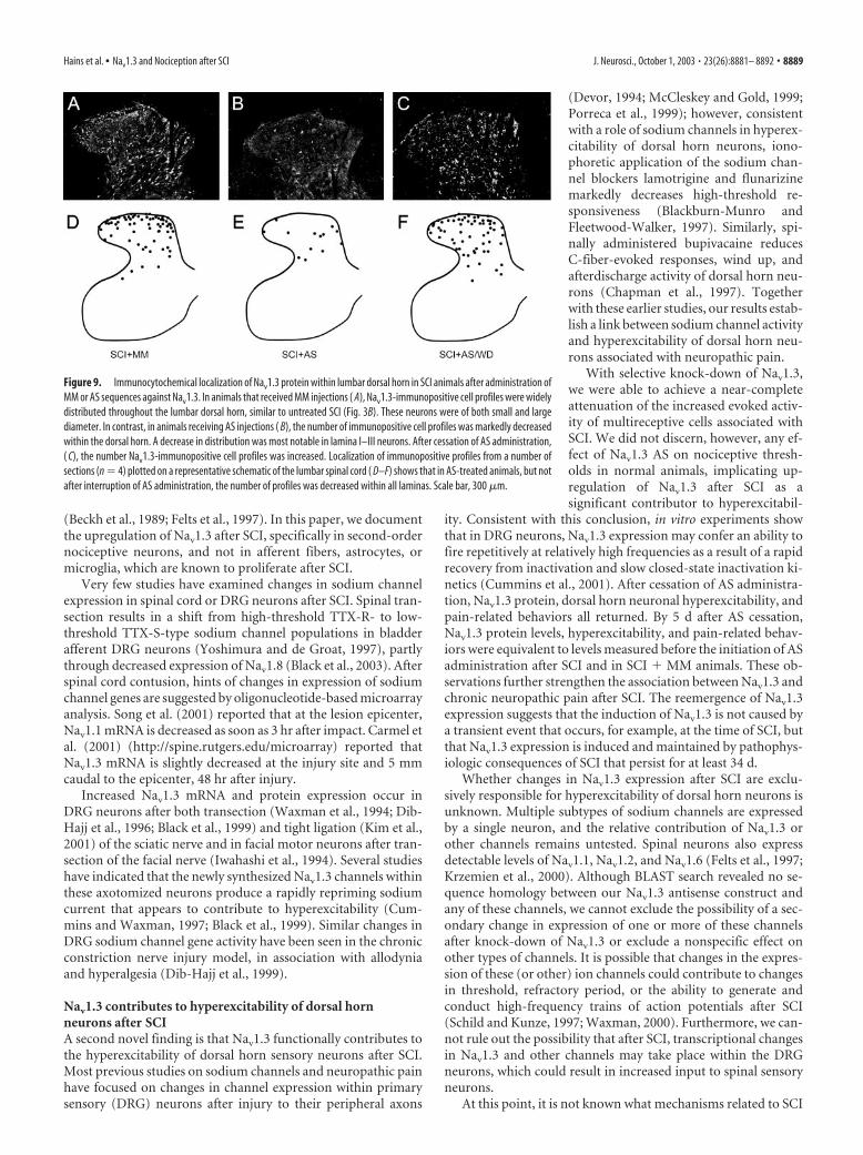
Figure 2. In situ hybridization for Nav1.3 shows very light signal within the gray matter ofthe lumbar dorsal horn of sham-operated animals ( A). In contrast, 28 d after SCI, Nav1.3 hybrid-ization signal was more intense and widely distributed throughout dorsal horn lamina I–Vneurons ( B). Higher-magnification images (B, inset boxes) of individual cells demonstrate neu-ronal morphologies and cytoplasmic staining. No labeling was observed in afferent fibers, den-dritic or axonal arborizations, or white matter. Quantitative RT-PCR amplification ( C) showed asignificant (*p � 0.05) increase in lumbar Nav1.3 mRNA in the SCI group (n � 10 animals)when compared with sham-operated controls (CTRL) (n � 10 animals). Scale bar: A, B, 300�m; insets, 10 �m.
Hains et al. • Nav1.3 and Nociception after SCI J. Neurosci., October 1, 2003 • 23(26):8881– 8892 • 8883

1000, Molecular Probes, Eugene, OR), and goat anti-guinea pig Cy2(1:250, Rockland Immunochemicals, Gilbertsville, PA). Normal horseserum replaced NGS in the blocking solution. Control experiments wereperformed without primary or secondary antibodies and blocking sub-stances, which yielded only background levels of signal.
Quantitative image analysis. Images were captured with a NikonEclipse E800 light microscope equipped with epifluorescence and No-marski optics, using a SPOT-RT camera (Diagnostic Instruments, Ster-ling Heights, MI). Quantitative microdensitometry was performed usingIPLab Spectrum software (Scanalytics, Fairfax, VA). For immunocyto-chemistry and in situ hybridization experiments, the number of posi-tively labeled neurons was counted for each dorsal horn lamina or sec-tion. Signal intensity was obtained by outlining individual neurons (n �15 cells per lamina) and using IPLab integrated densitometry functionsto determine levels of signal. Cells were sampled only if the nucleus wasvisible within the plane of section and if cell profiles exhibited distinctlydelineated borders. Background levels of signal were subtracted, andcontrol and experimental conditions were evaluated in identicalmanners.
Electrophysiologic procedures. Animals from sham-operated (n � 5),SCI (n � 5), SCI � MM (n � 4), SCI � AS (n � 5), and SCI � AS/WD(n � 2) groups underwent extracellular single unit recording accordingto established methods (Hains et al., 2003a,b). The activity of three to fiveunique cells per animal was recorded, yielding 15–25 cells per group. Inthe case of the SCI � AS/WD group, n � 11 cells were sampled. Rats wereinitially anesthetized with halothane (4% in induction chamber) andmaintained by tracheal intubation (1.1%; 2–2.5 cc tidal volume; 60 –70strokes per minute). Rectal temperature was maintained at 37°C. AT12–L6 laminectomy was performed before fixing on a stereotaxic appa-ratus (Kopf Instruments, Tujunga, CA), and the exposed spinal cord wascovered with warm (37°C) mineral oil.
Cells were isolated from L3–L5 medially near the dorsal root entryzone up to a depth of 1000 �m. Extracellular single-unit recordings weremade with a low-impedance 5 M� tungsten insulated microelectrode(A-M Systems, Carlsburg, WA). Once a cell was identified, its receptivefield was mapped and stimulated by an experimenter blinded to thesurgical status of the animal and with the audio monitor silenced. Back-ground activity was measured followed by cutaneous receptive field
mapping with von Frey filaments or brief pinches, or both. Three routinemechanical stimuli were applied: (1) brush stimulation (BR) of the skinwith a cotton brush; (2) pressure (PR), by attaching a large arterial clipwith a weak grip to a fold of skin (144 gm/mm 2); and (3) pinch (PI), byapplying a small arterial clip with a strong grip to a fold of skin (583gm/mm 2). Next, increasing-intensity von Frey filaments (0.39, 1.01, 20.8gm forces) and thermal stimuli (47°C steel probe, 1 cm 2 surface area)were applied. Multireceptive (MR) cells were identified by their respon-siveness to BR, more so to PR and PI, and with increasing responses toincrementing strength von Frey stimuli. Background activity was re-corded for 20 sec, and stimuli were applied serially for 20 sec, separatedby another 20 sec of baseline activity. Care was taken to ensure that theresponses were maximal, that each stimulus was applied to the primaryreceptive field of the cell, and that isolated units remained intact and heldfor the duration of each experiment using Spike2 template matchingroutines. Neurons responding chiefly to joint movement or to probingsubcutaneous tissue were excluded from analysis. After SCI or ODNadministration, or both, neurons were considered to be hyperexcitable ifevoked responses were �150% of control levels for BR, PR, and PIstimuli.
Electrical signals were amplified and filtered at 300 –3000 Hz (DAM80,World Precision Instruments, Sarasota, FL), processed by a data collec-tion system (CED 1401�; Cambridge Instruments, Cambridge, UK),and stored on a computer (Pentium 4 PC, Dell, Austin, TX) to constructperistimulus time histograms or wavemark files. The stored digitalrecord of individual unit activity was retrieved and analyzed off-line withSpike2 software (v3.13, Cambridge Electronic Design, Cambridge, UK).Evoked responses and afterdischarges were calculated by subtracting theprestimulus baseline activity to yield a net increase in discharge rate.
Statistical analysis. All statistical tests were performed at the � level ofsignificance of 0.05 by two-tailed analyses using parametric tests. Datawere tested for significance using one-way ANOVA to determine thedegree of variability within a sample and whether there was a differencebetween groups among the obtained means, followed by Bonferroni posthoc analysis. Tests of factors were used including pair-wise comparisonswhere appropriate with either the paired Student’s t test for before–aftercomparisons or the two sample Student’s t test to compare two groups.Data management and statistical analyses were performed using SAS(1992) statistical procedures with Jandel SigmaStat (v1.0) and graphedusing Jandel SigmaPlot (v7.0) as mean � SD.
ResultsBehavioral testingUsing the BBB locomotor rating scale, open-field motor functionwas assessed after injury (Fig. 1A). Consistent with 25 mm con-tusion spinal cord injury (SCI), rats exhibited transient parapa-resis and spontaneously recovered extensive movement of allthree hindlimb joints with limb sweeping, plantar foot place-ment, and weight support in stance, achieving a mean � SD. BBBscore was 9.90 � 1.51 at 28 d.
By 28 d after SCI, animals developed mechanical allodynia asdemonstrated by decreases in paw withdrawal thresholds to vonFrey stimulation (Fig. 1B). Mean � SD mechanical thresholds
Figure 3. Immunocytochemical localization of Nav1.3 protein within lumbar dorsal horn insham-operated controls ( A) revealed very few Nav1.3 immunopositive profiles within superfi-cial and deep laminas. Twenty-eight days after SCI ( B), positively labeled cell profiles wereobserved throughout dorsal horn lamina I–V neurons. In these sections, small neurons withinlaminas I–II and larger neurons within laminas III–V were strongly labeled. Localization ofimmunopositive profiles from a number of sections (n � 4) plotted on a representative sche-matic of the lumbar spinal cord (C, D) show that after SCI, the number of profiles was increasedwithin all laminas. The distribution of immunopositive cells was fairly homogenous and is notconfined to specific laminas. Scale bar, 300 �m. CTRL, Controls.
Table 1. Number of Nav1.3-positive neuronal profiles and Nav1.3 fluorescent signalintensity per section in lamina I–V neurons at L3–L5 segments in sham-operatedanimals and animals 4 weeks after T9 contusion SCI
Nav1.3 IF profiles Nav1.3 IF levels
Control SCI Control SCI
Lamina I 5.2 � 4.6 45.0 � 6.2* 7.8 � 2.6 49.2 � 7.3*Lamina II 3.4 � 2.8 34.9 � 4.3* 15.5 � 6.8 66.1 � 14.7*Lamina III 3.9 � 3.4 22.5 � 5.0* 18.3 � 8.5 62.0 � 10.4*Lamina IV 2.7 � 2.4 24.4 � 3.8* 20.0 � 6.8 60.9 � 7.6*Lamina V 2.5 � 1.9 20.6 � 3.6* 17.2 � 8.3 41.3 � 9.3*
Data represent mean � SD values [n � 5 sections per animal (10) per group]. *Significant difference from controlgroup (p � 0.05). IF, Immunofluorescent.
8884 • J. Neurosci., October 1, 2003 • 23(26):8881– 8892 Hains et al. • Nav1.3 and Nociception after SCI
significantly decreased from 22.1 � 3.03 gm before SCI to 4.05 �0.74 gm after SCI.
Thermal behavioral testing revealed that by 28 d after SCI,animals developed thermal hyperalgesia as indicated by decreasesin paw withdrawal latency (Fig. 1C). Before SCI, the mean � SDlatency for all animals was 10.73 � 0.79 sec. Twenty-eight daysafter SCI, latencies decreased significantly to 7.01 � 0.94 sec.
In situ hybridizationIn situ hybridization studies were performed on transverse lum-bar spinal cord sections with a riboprobe specific for sodiumchannel Nav1.3 mRNA (Fig. 2). These sections showed very littlehybridization signal for Nav1.3 in tissue collected from sham-operated animals (Fig. 2A). In contrast, extensive hybridizationsignal was observed in dorsal horn neuronal profiles after SCI(Fig. 2B). In sections in which signal was observed, labeling wasmainly cytoplasmic and could be detected within small (5–10�m) lamina I–II neurons, in addition to larger (20 – 40 �m) neu-rons deeper in lamina III–IV neurons. No labeling was observedin afferent fibers, or in dendritic or axonal arborizations. Verylittle signal was detected within white matter.
Compared with controls, the hybridization signal in neuronsof the SCI group showed significantly increased intensity. Quan-tification of signal intensity revealed that Nav1.3 in SCI sections
was significantly increased to 63.63 �12.41 arbitrary units compared with19.65 � 6.39 in sham-operated controls.In addition, the number of strongly label-ing neurons, defined by clear nuclear pro-file and strongly delineated boarders, persection in lamina I–IV neurons was signif-icantly increased in SCI to 31.72 � 3.89,compared with 3.04 � 2.65 in sham-operated controls.
Quantitative RT-PCRThe change in Nav1.3 mRNA amplifica-tion product collected from the lumbarenlargement of sham-operated and SCIgroups is shown in Figure 2C. QuantitativeRT-PCR results showed a statistically sig-nificant increase after SCI to 125% ofsham-operated Nav1.3 mRNA levels.
ImmunocytochemistryImmunocytochemistry with an isoform-specific antibody raised against Nav1.3 re-vealed light expression within the lumbardorsal horn of sham-operated animals(Fig. 3A). Twenty-eight days after T9 spi-nal contusion injury, expression of Nav1.3was increased within the L3–L5 dorsalhorn (Fig. 3B). Labeling was observed insmaller neurons within lamina I–II neu-rons as well as in deeper neurons in laminaIII–V neurons. The spatial distribution ofNav1.3 profiles from these sections is illus-trated for controls (Fig. 3C) and SCI ani-mals (Fig. 3D).
Quantification revealed that the meannumber of immunofluorescent profileswas significantly increased in all laminas(I–V) after SCI (Table 1), from an average
of 3.54 � 3.02 profiles per lamina (average for lamina I–V neu-rons) in sham-operated controls to 29.48 � 4.58 after SCI. In ad-dition, immunofluorescent intensity was increased significantly inall laminas after SCI (Table 1), from an average for lamina I–V neu-rons of 15.76 � 6.60 to 55.90 � 9.86.
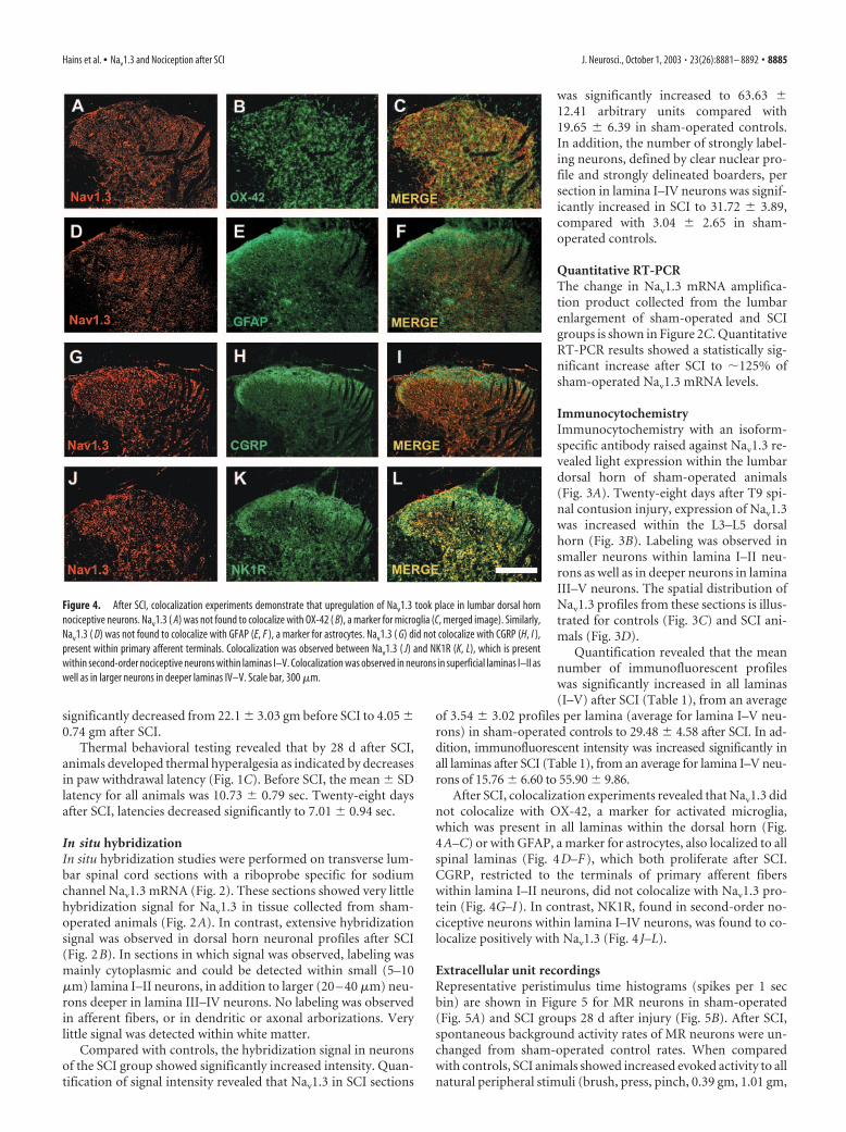
After SCI, colocalization experiments revealed that Nav1.3 didnot colocalize with OX-42, a marker for activated microglia,which was present in all laminas within the dorsal horn (Fig.4A–C) or with GFAP, a marker for astrocytes, also localized to allspinal laminas (Fig. 4D–F), which both proliferate after SCI.CGRP, restricted to the terminals of primary afferent fiberswithin lamina I–II neurons, did not colocalize with Nav1.3 pro-tein (Fig. 4G–I). In contrast, NK1R, found in second-order no-ciceptive neurons within lamina I–IV neurons, was found to co-localize positively with Nav1.3 (Fig. 4 J–L).
Extracellular unit recordingsRepresentative peristimulus time histograms (spikes per 1 secbin) are shown in Figure 5 for MR neurons in sham-operated(Fig. 5A) and SCI groups 28 d after injury (Fig. 5B). After SCI,spontaneous background activity rates of MR neurons were un-changed from sham-operated control rates. When comparedwith controls, SCI animals showed increased evoked activity to allnatural peripheral stimuli (brush, press, pinch, 0.39 gm, 1.01 gm,
Figure 4. After SCI, colocalization experiments demonstrate that upregulation of Nav1.3 took place in lumbar dorsal hornnociceptive neurons. Nav1.3 ( A) was not found to colocalize with OX-42 ( B), a marker for microglia (C, merged image). Similarly,Nav1.3 ( D) was not found to colocalize with GFAP (E, F ), a marker for astrocytes. Nav1.3 ( G) did not colocalize with CGRP (H, I ),present within primary afferent terminals. Colocalization was observed between Nav1.3 ( J) and NK1R (K, L), which is presentwithin second-order nociceptive neurons within laminas I–V. Colocalization was observed in neurons in superficial laminas I–II aswell as in larger neurons in deeper laminas IV–V. Scale bar, 300 �m.
Hains et al. • Nav1.3 and Nociception after SCI J. Neurosci., October 1, 2003 • 23(26):8881– 8892 • 8885

20.81 gm von Frey filaments, and 47°C thermal stimuli). Of 18MR cells sampled in SCI animals, 88% (16 of 18) werehyperexcitable.
Quantification of mean � SD evoked discharge rates of MRcells, over background, from sham-operated and SCI groups isshown in Figure 5C. In comparison with sham-operated animalsthat demonstrated evoked response rates of up to 20 Hz to moststimuli, SCI animals demonstrated bilaterally increased responserates of up to 55– 60 Hz. Responses were typical for MR cells,demonstrating high evoked activity to BR, PR, and PI stimuli andincreasing rates to graded von Frey stimulation. Thermal re-sponses were also increased in SCI animals, compared withsham-operated animals. All stimuli produced statistically signif-icant increases in responsiveness, demonstrating evoked hyper-excitability (Fig. 5C).
ODN deliveryIn the previous set of experiments, we have demonstrated that,after SCI, Nav1.3 mRNA and protein are upregulated in dorsalhorn sensory neurons. In the next set of experiments, we reportthe results of antisense knock-down experiments designed to de-
crease the contribution of Nav1.3 to dorsal horn hyperexcitabilityand pain-related behaviors after SCI.
One-time intrathecal injection of AS oligodeoxynucleotides(45 �g/5 �l aCSF, followed by 10 �l of aCSF flush) labeled withCy3 (to examine ODN penetration and uptake) resulted inreadily observed pink staining of the dorsal surface of the spinalcord, extending for approximately three spinal segments (fromL3 to L5) at 5 hr after injection when the animal was killed. Crosssections of spinal cord tissue from L3–L5 (Fig. 6A), observed atlow magnification, show that Cy3 penetration was very strongwithin the spinal parenchyma to a depth of 400 �m below thedorsal surface of the cord and could still be detected to a depth ofat least 800 �m below the dorsal surface, encompassing the ter-ritory of lamina I–IV neurons (Fig. 6B,C). Signal was noticeablystronger in gray matter and could be observed in individual cellsexhibiting a neuronal morphology (Fig. 6C,D). High-magnification images (Fig. 6D�,D) of two representative neu-rons at depths of 500 and 800 �m show strong cytoplasmicuptake of Cy3-labeled AS. In these larger diameter neurons, Cy3signal was also detected in their processes. Cy3 signal was notdetectable in some cells, and signal intensity was variable in Cy3-positive cells, suggesting that not all cells were exposed to Cy3after a single injection or that it was not taken up with equalefficacy.
Behavioral testing after ODN deliveryFor testing the effects of ODN, on day 28, SCI animals weredivided randomly into either AS or MM groups; before ODNadministration, the groups were matched and did not differ sig-nificantly in terms of BBB scores, mechanical thresholds, or ther-mal paw withdrawal latencies. Pre-ODN BBB scores were 9.80 �0.84 and 9.41 � 0.96 in SCI � MM and SCI � AS groups, respec-tively. Pre-ODN mechanical thresholds were 3.26 � 1.99 and3.99 � 0.78 gm in SCI � MM and SCI � AS groups, respectively.Pre-ODN thermal latencies were 6.77 � 1.00 and 6.9 � 1.29 secin SCI � MM and SCI � AS groups, respectively.
Four days after the start of AS or MM injections (day 34), BBBscores remained unchanged in all groups (Fig. 7A); scores were10.14 � 1.53, 9.90 � 1.64, and 10.13 � 1.46 for sham-operated,SCI � MM, and SCI � AS groups, respectively. Cessation of ASadministration after 4 d had no effect on BBB scores, measured5 d subsequently (9.96 � 1.59). In naive animals (N � AS), ASadministration had no effect on BBB scores, which remained at21 throughout the course of the experiment (data not shown).
Administration of Nav1.3 AS had a rapid and significant effecton mechanical thresholds and tended to reverse the decreasesthat occur with SCI (Fig. 7B). After 4 d of ODN injections, me-chanical thresholds remained unchanged in SCI and SCI � MMgroups: 3.01 � 1.54 and 2.91 � 1.64, respectively (Fig. 7B). In theSCI � AS group, paw withdrawal thresholds to von Frey stimu-lation increased to 14.36 � 1.69 by day 4, representing a signifi-cant difference when compared with SCI and SCI � MM groups.Increases in thresholds were evident and statistically significant assoon as 1 d after the start of AS administration. Responses to anormally noxious pin prick stimuli remained unchanged (datanot shown). After cessation of AS administration, mechanicalthresholds returned to levels comparable with those before ASadministration and MM levels (Fig. 7B). In naive animals (N �AS), AS administration had no effect on mechanical thresholds,which were not significantly different when measured before(21.78 � 0.93) or after (20.35 � 1.15) AS administration.
After SCI, animals receiving Nav1.3 AS exhibited increasedthermal paw withdrawal latencies. Four days after the start of
Figure 5. Representative peristimulus time histograms (spikes per 1 sec bin) of multirecep-tive neurons recorded extracellularly from L3–L5 in sham-operated control animals ( A) and inanimals 28 d after SCI ( B) in response to various innocuous and noxious peripheral stimuli: brush(BR), press (PR), pinch (PI), increasing intensity von Frey filaments (0.39, 1.01, and 20.8 gm),and thermal (47°C). Single-unit waveforms are also shown to the right. Compared with con-trols, injured animals demonstrated evoked hyperexcitability to all peripheral stimuli. Sponta-neous background activity was unchanged from control rates in injured animals. Quantification( C) of mean � SD spontaneous and evoked discharge rates of neurons sampled from sham-operated controls (n � 15 cells) and SCI (n � 15 cells) groups demonstrated significantly(*p � 0.05) increased evoked activity to all peripheral stimuli after injury. No significantchanges in background activity were detected.
8886 • J. Neurosci., October 1, 2003 • 23(26):8881– 8892 Hains et al. • Nav1.3 and Nociception after SCI

ODN injections, paw withdrawal latencies remained unchangedin SCI and SCI � MM groups: 8.41 � 1.65 and 7.22 � 1.12,respectively (Fig. 7C). In the SCI � AS group, paw withdrawallatencies increased to 12.71 � 1.36 by day 4, representing a sig-nificant difference when compared with SCI and SCI � MMgroups. Increases in latencies were evident (but not statisticallysignificant) as soon as 1 d after the start of AS administration; thedifference became significantly different by day 3 (Fig. 7C). Aftercessation of AS administration, latencies decreased to pre-AS lev-els (Fig. 7C). In naive animals (N � AS), AS administration hadno effect on thermal thresholds, which were not significantlydifferent when measured before (11.16 � 1.07) or after (10.75 �0.80) AS administration.
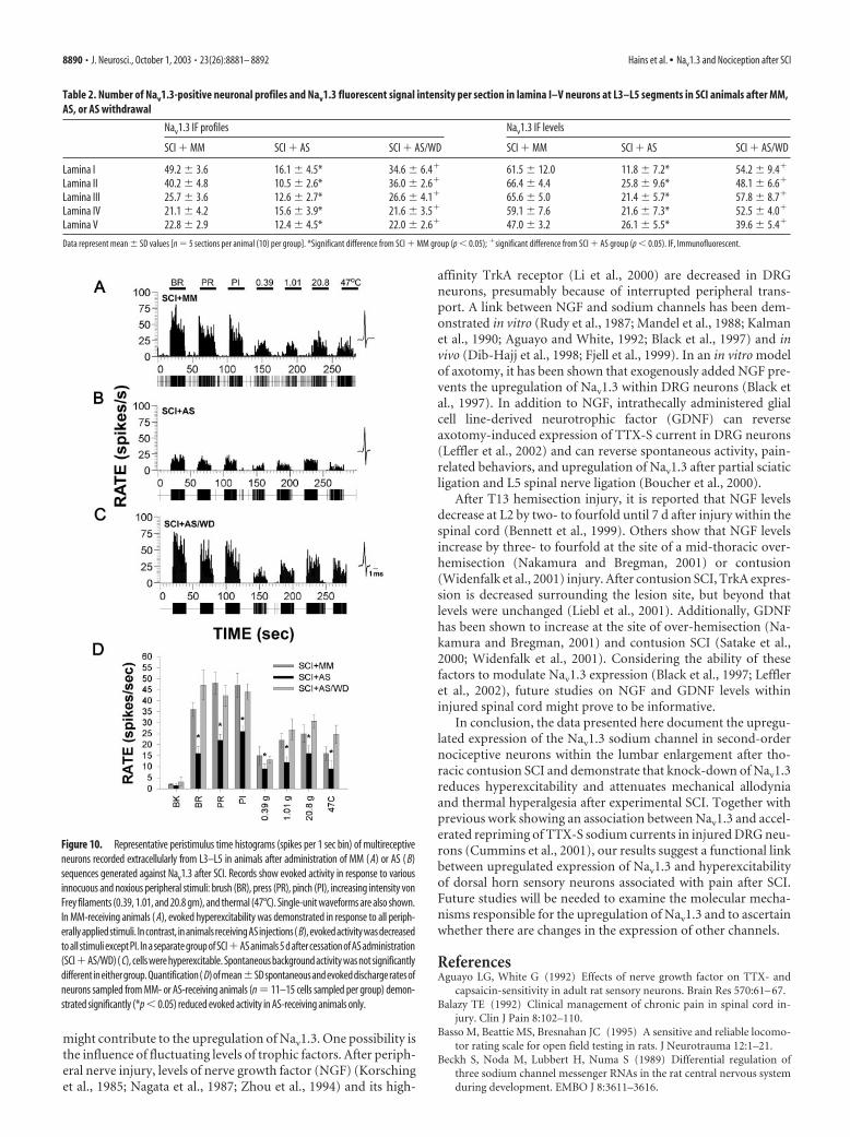
ODN in situ hybridizationAs in the SCI group, extensive in situ hybridization signal wasobserved in dorsal horn neurons 4 d after SCI � MM injections(Fig. 8A). Staining density and distribution were not noticeablydifferent between SCI and SCI � MM groups. Signal intensity forNav1.3 was substantially reduced after 4 d of AS treatment of SCIanimals (Fig. 8B). Quantification of Nav1.3 signal intensity re-vealed that in SCI � AS sections, intensity was significantly de-creased compared with SCI � MM sections: from 62.31 � 10.73to 30.34 � 13.15 arbitrary units, respectively. The number ofstrongly labeled neurons, per section, in lamina I–IV neurons wassignificantly decreased after AS treatment to 15.28 � 2.83 from37.22 � 3.65 in the SCI � MM group.
ODN quantitative RT-PCRQuantitative RT-PCR results (Fig. 8C) showed a decrease inNav1.3 mRNA 4 d after AS injections compared with SCI � MManimals. This difference was statistically significant. Nav1.3 levelsin SCI � MM animals were elevated 133% relative to sham-
operated control animals, whereas SCI �AS animals were increased 103% com-pared with controls.
ODN immunocytochemistryFour days after ODN administration, SCI� AS animals demonstrated decreasedNav1.3 labeling density and distribution(Fig. 9B), with most dramatic immunoflu-orescence decreases observed in superficiallaminas. Some immunopositive profileswere evident within the neck of the dorsalhorn at approximately lamina V that areputative neuronal profiles. Nav1.3 labelingin SCI � MM (Fig. 9A) animals was notdifferent from that observed in SCI ani-mals (Fig. 3B). In AS withdrawal animals(SCI � AS/WD), Nav1.3 expression re-emerged after cessation of AS administra-tion (Fig. 9C). Nav1.3 expression was seenin smaller neurons within lamina I–II neu-rons, as well as in deeper neurons in lam-ina III–IV neurons. The spatial distribu-tion of Nav1.3 profiles from these sectionsis illustrated for SCI � MM (Fig. 9D),SCI � AS (Fig. 9E), and SCI � AS/WDanimals (Fig. 9F).
In naive animals (N � AS), AS admin-istration had no effect on the normal levelsof expression of Nav1.3 (data not shown).
Quantification revealed that the mean number of immuno-fluorescent profiles was significantly decreased in animals receiv-ing AS compared with MM (Table 2), from 31.80 � 3.82 (averagefor lamina I–V neurons) to 13.44 � 3.64 per section. In SC �AS/WD animals, the mean number of profiles was increased to28.20 � 3.87. In addition, the average immunofluorescent inten-sity for lamina I–V neurons was significantly decreased in AS-treated animals (Table 2), down to 21.34 � 7.06 from 59.92 �6.44. In contrast, in SCI � AS/WD animals, the mean number ofimmunofluorescent profiles was not different from SCI � MManimals, at 50.44 � 6.82.
Effect of ODN on extracellular unit recordingsRepresentative peristimulus time histograms (spikes per 1 secbin) are shown in Figure 10 for MR neurons sampled in animalsfrom SCI � MM (Fig. 10A), SCI � AS (Fig. 10B), and SCI �AS/WD (Fig. 10C) groups. Four days after initiation of ODNinjections, evoked activity was markedly decreased to all stimuli(brush, press, pinch, 0.39 gm, 1.01 gm, 20.81 gm force von Freyfilaments, and 47°C thermal stimuli) in SCI � AS animals. Com-pared with SCI and SCI � MM, neurons from SCI � AS animalsexhibited a reduced incidence of hyperexcitability. Of 19 MR cellssampled in the SCI � MM group, 89% were hyperexcitable (17 of19), whereas of the 20 MR cells sampled in SCI � AS animals only20% (4 of 20) were hyperexcitable. MM treatment had no appar-ent effect on evoked discharge rates of MR cells after SCI. In theSCI � MM group, neurons remained highly responsive to pe-ripheral stimulation. In animals that had undergone SCI and ASadministration, 5 d after cessation of AS administration (SCI �AS/WD) 90% of sampled cells were hyperexcitable (10 of 11)(Fig. 10C).
Quantification of mean � SD discharge rates of MR cells, overbackground, from SCI � MM and SCI � AS groups is shown in
Figure 6. Schematic of injury and ODN delivery paradigm [i.t. ODN (CY3)] ( A) showing relative locations of T9 contusion SCI,and intrathecally delivered AS or MM in relation to the lumbar enlargement (LE). A schematic cross section of the lumbar dorsalhorn is shown illustrating field of view in C. After a one-time injection of Cy3-labeled AS (45 �g/5 �l of aCSF followed by 10 �l ofaCSF flush), Cy3 fluorescence is detectable to a depth of 800 �m below the dorsal surface of the spinal cord ( B) and can be seen topenetrate as far as lamina V ( C). Higher-magnification image of cells exhibiting neuronal profiles (C, boxes) shows strong uptakeof Cy3-labeled AS into cells with neuronal morphologies (D�, D) after a single injection. Scale bar: A, C, 300 �m; D�, D, 10 �m.
Hains et al. • Nav1.3 and Nociception after SCI J. Neurosci., October 1, 2003 • 23(26):8881– 8892 • 8887
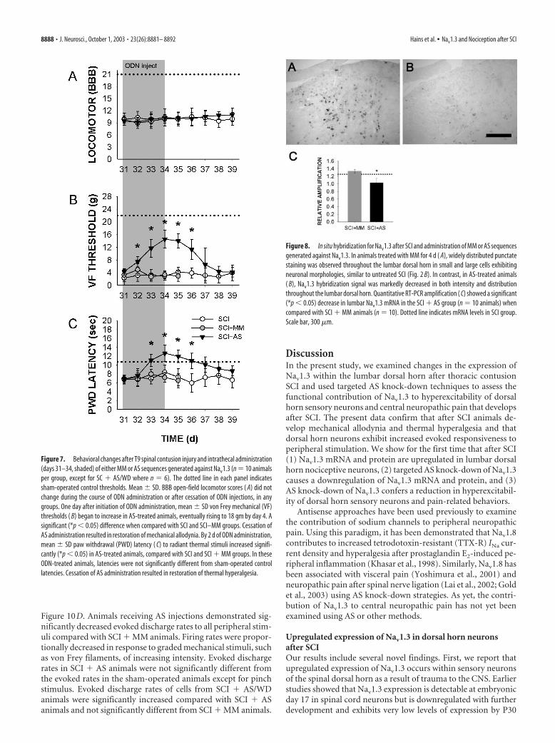
Figure 10D. Animals receiving AS injections demonstrated sig-nificantly decreased evoked discharge rates to all peripheral stim-uli compared with SCI � MM animals. Firing rates were propor-tionally decreased in response to graded mechanical stimuli, suchas von Frey filaments, of increasing intensity. Evoked dischargerates in SCI � AS animals were not significantly different fromthe evoked rates in the sham-operated animals except for pinchstimulus. Evoked discharge rates of cells from SCI � AS/WDanimals were significantly increased compared with SCI � ASanimals and not significantly different from SCI � MM animals.
DiscussionIn the present study, we examined changes in the expression ofNav1.3 within the lumbar dorsal horn after thoracic contusionSCI and used targeted AS knock-down techniques to assess thefunctional contribution of Nav1.3 to hyperexcitability of dorsalhorn sensory neurons and central neuropathic pain that developsafter SCI. The present data confirm that after SCI animals de-velop mechanical allodynia and thermal hyperalgesia and thatdorsal horn neurons exhibit increased evoked responsiveness toperipheral stimulation. We show for the first time that after SCI(1) Nav1.3 mRNA and protein are upregulated in lumbar dorsalhorn nociceptive neurons, (2) targeted AS knock-down of Nav1.3causes a downregulation of Nav1.3 mRNA and protein, and (3)AS knock-down of Nav1.3 confers a reduction in hyperexcitabil-ity of dorsal horn sensory neurons and pain-related behaviors.
Antisense approaches have been used previously to examinethe contribution of sodium channels to peripheral neuropathicpain. Using this paradigm, it has been demonstrated that Nav1.8contributes to increased tetrodotoxin-resistant (TTX-R) INa cur-rent density and hyperalgesia after prostaglandin E2-induced pe-ripheral inflammation (Khasar et al., 1998). Similarly, Nav1.8 hasbeen associated with visceral pain (Yoshimura et al., 2001) andneuropathic pain after spinal nerve ligation (Lai et al., 2002; Goldet al., 2003) using AS knock-down strategies. As yet, the contri-bution of Nav1.3 to central neuropathic pain has not yet beenexamined using AS or other methods.
Upregulated expression of Nav1.3 in dorsal horn neuronsafter SCIOur results include several novel findings. First, we report thatupregulated expression of Nav1.3 occurs within sensory neuronsof the spinal dorsal horn as a result of trauma to the CNS. Earlierstudies showed that Nav1.3 expression is detectable at embryonicday 17 in spinal cord neurons but is downregulated with furtherdevelopment and exhibits very low levels of expression by P30
Figure 7. Behavioral changes after T9 spinal contusion injury and intrathecal administration(days 31–34, shaded) of either MM or AS sequences generated against Nav1.3 (n � 10 animalsper group, except for SC � AS/WD where n � 6). The dotted line in each panel indicatessham-operated control thresholds. Mean � SD. BBB open-field locomotor scores ( A) did notchange during the course of ODN administration or after cessation of ODN injections, in anygroups. One day after initiation of ODN administration, mean � SD von Frey mechanical (VF)thresholds ( B) began to increase in AS-treated animals, eventually rising to 18 gm by day 4. Asignificant (*p � 0.05) difference when compared with SCI and SCI–MM groups. Cessation ofAS administration resulted in restoration of mechanical allodynia. By 2 d of ODN administration,mean � SD paw withdrawal (PWD) latency ( C) to radiant thermal stimuli increased signifi-cantly (*p � 0.05) in AS-treated animals, compared with SCI and SCI � MM groups. In theseODN-treated animals, latencies were not significantly different from sham-operated controllatencies. Cessation of AS administration resulted in restoration of thermal hyperalgesia.
Figure 8. In situ hybridization for Nav1.3 after SCI and administration of MM or AS sequencesgenerated against Nav1.3. In animals treated with MM for 4 d ( A), widely distributed punctatestaining was observed throughout the lumbar dorsal horn in small and large cells exhibitingneuronal morphologies, similar to untreated SCI (Fig. 2 B). In contrast, in AS-treated animals( B), Nav1.3 hybridization signal was markedly decreased in both intensity and distributionthroughout the lumbar dorsal horn. Quantitative RT-PCR amplification ( C) showed a significant(*p � 0.05) decrease in lumbar Nav1.3 mRNA in the SCI � AS group (n � 10 animals) whencompared with SCI � MM animals (n � 10). Dotted line indicates mRNA levels in SCI group.Scale bar, 300 �m.
8888 • J. Neurosci., October 1, 2003 • 23(26):8881– 8892 Hains et al. • Nav1.3 and Nociception after SCI
(Beckh et al., 1989; Felts et al., 1997). In this paper, we documentthe upregulation of Nav1.3 after SCI, specifically in second-ordernociceptive neurons, and not in afferent fibers, astrocytes, ormicroglia, which are known to proliferate after SCI.
Very few studies have examined changes in sodium channelexpression in spinal cord or DRG neurons after SCI. Spinal tran-section results in a shift from high-threshold TTX-R- to low-threshold TTX-S-type sodium channel populations in bladderafferent DRG neurons (Yoshimura and de Groat, 1997), partlythrough decreased expression of Nav1.8 (Black et al., 2003). Afterspinal cord contusion, hints of changes in expression of sodiumchannel genes are suggested by oligonucleotide-based microarrayanalysis. Song et al. (2001) reported that at the lesion epicenter,Nav1.1 mRNA is decreased as soon as 3 hr after impact. Carmel etal. (2001) (http://spine.rutgers.edu/microarray) reported thatNav1.3 mRNA is slightly decreased at the injury site and 5 mmcaudal to the epicenter, 48 hr after injury.
Increased Nav1.3 mRNA and protein expression occur inDRG neurons after both transection (Waxman et al., 1994; Dib-Hajj et al., 1996; Black et al., 1999) and tight ligation (Kim et al.,2001) of the sciatic nerve and in facial motor neurons after tran-section of the facial nerve (Iwahashi et al., 1994). Several studieshave indicated that the newly synthesized Nav1.3 channels withinthese axotomized neurons produce a rapidly repriming sodiumcurrent that appears to contribute to hyperexcitability (Cum-mins and Waxman, 1997; Black et al., 1999). Similar changes inDRG sodium channel gene activity have been seen in the chronicconstriction nerve injury model, in association with allodyniaand hyperalgesia (Dib-Hajj et al., 1999).
Nav1.3 contributes to hyperexcitability of dorsal hornneurons after SCIA second novel finding is that Nav1.3 functionally contributes tothe hyperexcitability of dorsal horn sensory neurons after SCI.Most previous studies on sodium channels and neuropathic painhave focused on changes in channel expression within primarysensory (DRG) neurons after injury to their peripheral axons
(Devor, 1994; McCleskey and Gold, 1999;Porreca et al., 1999); however, consistentwith a role of sodium channels in hyperex-citability of dorsal horn neurons, iono-phoretic application of the sodium chan-nel blockers lamotrigine and flunarizinemarkedly decreases high-threshold re-sponsiveness (Blackburn-Munro andFleetwood-Walker, 1997). Similarly, spi-nally administered bupivacaine reducesC-fiber-evoked responses, wind up, andafterdischarge activity of dorsal horn neu-rons (Chapman et al., 1997). Togetherwith these earlier studies, our results estab-lish a link between sodium channel activityand hyperexcitability of dorsal horn neu-rons associated with neuropathic pain.
With selective knock-down of Nav1.3,we were able to achieve a near-completeattenuation of the increased evoked activ-ity of multireceptive cells associated withSCI. We did not discern, however, any ef-fect of Nav1.3 AS on nociceptive thresh-olds in normal animals, implicating up-regulation of Nav1.3 after SCI as asignificant contributor to hyperexcitabil-
ity. Consistent with this conclusion, in vitro experiments showthat in DRG neurons, Nav1.3 expression may confer an ability tofire repetitively at relatively high frequencies as a result of a rapidrecovery from inactivation and slow closed-state inactivation ki-netics (Cummins et al., 2001). After cessation of AS administra-tion, Nav1.3 protein, dorsal horn neuronal hyperexcitability, andpain-related behaviors all returned. By 5 d after AS cessation,Nav1.3 protein levels, hyperexcitability, and pain-related behav-iors were equivalent to levels measured before the initiation of ASadministration after SCI and in SCI � MM animals. These ob-servations further strengthen the association between Nav1.3 andchronic neuropathic pain after SCI. The reemergence of Nav1.3expression suggests that the induction of Nav1.3 is not caused bya transient event that occurs, for example, at the time of SCI, butthat Nav1.3 expression is induced and maintained by pathophys-iologic consequences of SCI that persist for at least 34 d.
Whether changes in Nav1.3 expression after SCI are exclu-sively responsible for hyperexcitability of dorsal horn neurons isunknown. Multiple subtypes of sodium channels are expressedby a single neuron, and the relative contribution of Nav1.3 orother channels remains untested. Spinal neurons also expressdetectable levels of Nav1.1, Nav1.2, and Nav1.6 (Felts et al., 1997;Krzemien et al., 2000). Although BLAST search revealed no se-quence homology between our Nav1.3 antisense construct andany of these channels, we cannot exclude the possibility of a sec-ondary change in expression of one or more of these channelsafter knock-down of Nav1.3 or exclude a nonspecific effect onother types of channels. It is possible that changes in the expres-sion of these (or other) ion channels could contribute to changesin threshold, refractory period, or the ability to generate andconduct high-frequency trains of action potentials after SCI(Schild and Kunze, 1997; Waxman, 2000). Furthermore, we can-not rule out the possibility that after SCI, transcriptional changesin Nav1.3 and other channels may take place within the DRGneurons, which could result in increased input to spinal sensoryneurons.
At this point, it is not known what mechanisms related to SCI
Figure 9. Immunocytochemical localization of Nav1.3 protein within lumbar dorsal horn in SCI animals after administration ofMM or AS sequences against Nav1.3. In animals that received MM injections ( A), Nav1.3-immunopositive cell profiles were widelydistributed throughout the lumbar dorsal horn, similar to untreated SCI (Fig. 3B). These neurons were of both small and largediameter. In contrast, in animals receiving AS injections ( B), the number of immunopositive cell profiles was markedly decreasedwithin the dorsal horn. A decrease in distribution was most notable in lamina I–III neurons. After cessation of AS administration,( C), the number Nav1.3-immunopositive cell profiles was increased. Localization of immunopositive profiles from a number ofsections (n � 4) plotted on a representative schematic of the lumbar spinal cord ( D–F) shows that in AS-treated animals, but notafter interruption of AS administration, the number of profiles was decreased within all laminas. Scale bar, 300 �m.
Hains et al. • Nav1.3 and Nociception after SCI J. Neurosci., October 1, 2003 • 23(26):8881– 8892 • 8889
might contribute to the upregulation of Nav1.3. One possibility isthe influence of fluctuating levels of trophic factors. After periph-eral nerve injury, levels of nerve growth factor (NGF) (Korschinget al., 1985; Nagata et al., 1987; Zhou et al., 1994) and its high-
affinity TrkA receptor (Li et al., 2000) are decreased in DRGneurons, presumably because of interrupted peripheral trans-port. A link between NGF and sodium channels has been dem-onstrated in vitro (Rudy et al., 1987; Mandel et al., 1988; Kalmanet al., 1990; Aguayo and White, 1992; Black et al., 1997) and invivo (Dib-Hajj et al., 1998; Fjell et al., 1999). In an in vitro modelof axotomy, it has been shown that exogenously added NGF pre-vents the upregulation of Nav1.3 within DRG neurons (Black etal., 1997). In addition to NGF, intrathecally administered glialcell line-derived neurotrophic factor (GDNF) can reverseaxotomy-induced expression of TTX-S current in DRG neurons(Leffler et al., 2002) and can reverse spontaneous activity, pain-related behaviors, and upregulation of Nav1.3 after partial sciaticligation and L5 spinal nerve ligation (Boucher et al., 2000).
After T13 hemisection injury, it is reported that NGF levelsdecrease at L2 by two- to fourfold until 7 d after injury within thespinal cord (Bennett et al., 1999). Others show that NGF levelsincrease by three- to fourfold at the site of a mid-thoracic over-hemisection (Nakamura and Bregman, 2001) or contusion(Widenfalk et al., 2001) injury. After contusion SCI, TrkA expres-sion is decreased surrounding the lesion site, but beyond thatlevels were unchanged (Liebl et al., 2001). Additionally, GDNFhas been shown to increase at the site of over-hemisection (Na-kamura and Bregman, 2001) and contusion SCI (Satake et al.,2000; Widenfalk et al., 2001). Considering the ability of thesefactors to modulate Nav1.3 expression (Black et al., 1997; Leffleret al., 2002), future studies on NGF and GDNF levels withininjured spinal cord might prove to be informative.
In conclusion, the data presented here document the upregu-lated expression of the Nav1.3 sodium channel in second-ordernociceptive neurons within the lumbar enlargement after tho-racic contusion SCI and demonstrate that knock-down of Nav1.3reduces hyperexcitability and attenuates mechanical allodyniaand thermal hyperalgesia after experimental SCI. Together withprevious work showing an association between Nav1.3 and accel-erated repriming of TTX-S sodium currents in injured DRG neu-rons (Cummins et al., 2001), our results suggest a functional linkbetween upregulated expression of Nav1.3 and hyperexcitabilityof dorsal horn sensory neurons associated with pain after SCI.Future studies will be needed to examine the molecular mecha-nisms responsible for the upregulation of Nav1.3 and to ascertainwhether there are changes in the expression of other channels.
ReferencesAguayo LG, White G (1992) Effects of nerve growth factor on TTX- and
capsaicin-sensitivity in adult rat sensory neurons. Brain Res 570:61– 67.Balazy TE (1992) Clinical management of chronic pain in spinal cord in-
jury. Clin J Pain 8:102–110.Basso M, Beattie MS, Bresnahan JC (1995) A sensitive and reliable locomo-
tor rating scale for open field testing in rats. J Neurotrauma 12:1–21.Beckh S, Noda M, Lubbert H, Numa S (1989) Differential regulation of
three sodium channel messenger RNAs in the rat central nervous systemduring development. EMBO J 8:3611–3616.
Figure 10. Representative peristimulus time histograms (spikes per 1 sec bin) of multireceptiveneurons recorded extracellularly from L3–L5 in animals after administration of MM ( A) or AS ( B)sequences generated against Nav1.3 after SCI. Records show evoked activity in response to variousinnocuous and noxious peripheral stimuli: brush (BR), press (PR), pinch (PI), increasing intensity vonFrey filaments (0.39, 1.01, and 20.8 gm), and thermal (47°C). Single-unit waveforms are also shown.In MM-receiving animals ( A), evoked hyperexcitability was demonstrated in response to all periph-erallyappliedstimuli. Incontrast, inanimalsreceivingASinjections( B),evokedactivitywasdecreasedto all stimuli except PI. In a separate group of SCI�AS animals 5 d after cessation of AS administration(SCI�AS/WD) ( C), cells were hyperexcitable. Spontaneous background activity was not significantlydifferentineithergroup.Quantification( D)ofmean�SDspontaneousandevokeddischargeratesofneurons sampled from MM- or AS-receiving animals (n � 11–15 cells sampled per group) demon-strated significantly (*p � 0.05) reduced evoked activity in AS-receiving animals only.
Table 2. Number of Nav1.3-positive neuronal profiles and Nav1.3 fluorescent signal intensity per section in lamina I–V neurons at L3–L5 segments in SCI animals after MM,AS, or AS withdrawal
Nav1.3 IF profiles Nav1.3 IF levels
SCI � MM SCI � AS SCI � AS/WD SCI � MM SCI � AS SCI � AS/WD
Lamina I 49.2 � 3.6 16.1 � 4.5* 34.6 � 6.4� 61.5 � 12.0 11.8 � 7.2* 54.2 � 9.4�
Lamina II 40.2 � 4.8 10.5 � 2.6* 36.0 � 2.6� 66.4 � 4.4 25.8 � 9.6* 48.1 � 6.6�
Lamina III 25.7 � 3.6 12.6 � 2.7* 26.6 � 4.1� 65.6 � 5.0 21.4 � 5.7* 57.8 � 8.7�
Lamina IV 21.1 � 4.2 15.6 � 3.9* 21.6 � 3.5� 59.1 � 7.6 21.6 � 7.3* 52.5 � 4.0�
Lamina V 22.8 � 2.9 12.4 � 4.5* 22.0 � 2.6� 47.0 � 3.2 26.1 � 5.5* 39.6 � 5.4�
Data represent mean � SD values [n � 5 sections per animal (10) per group]. *Significant difference from SCI � MM group (p � 0.05); �significant difference from SCI � AS group (p � 0.05). IF, Immunofluorescent.
8890 • J. Neurosci., October 1, 2003 • 23(26):8881– 8892 Hains et al. • Nav1.3 and Nociception after SCI

Bennett AD, Taglialatela G, Perez-Polo R, Hulsebosch CE (1999) NGF levelsdecrease in the spinal cord and dorsal root ganglion after spinal hemisec-tion. NeuroReport 10:889 – 893.
Black JA, Dib-Hajj S, McNabola K, Jeste S, Rizzo MA, Kocsis JD, Waxman SG(1996) Spinal sensory neurons express multiple sodium channel alpha-subunit mRNAs. Mol Brain Res 43:117–131.
Black JA, Langworthy K, Hinson AW, Dib-Hajj SD, Waxman SG (1997)NGF has opposing effects on Na � channel III and SNS gene expression inspinal sensory neurons. NeuroReport 8:2331–2335.
Black JA, Cummins TR, Plumpton C, Chen YH, Hormuzdiar W, Clare JJ,Waxman SG (1999) Upregulation of a silent sodium channel after pe-ripheral, but not central, nerve injury in DRG neurons. J Neurophysiol82:2776 –2785.
Black JA, Cummins TR, Yoshimura N, de Groat WC, Waxman SG (2003)Tetrodotoxin-resistant sodium channels Nav1.8/SNS and Nav1.9/NaN inafferent neurons innervating urinary bladder in control and spinal cordinjured rats. Brain Res 963:132–138.
Blackburn-Munro G, Fleetwood-Walker SM (1997) The effects of Na �
channel blockers on somatosensory processing by rat dorsal horn neu-rones. NeuroReport 8:1549 –1554.
Boucher TJ, Okuse K, Bennett DL, Munson JB, Wood JN, McMahon SB(2000) Potent analgesic effects of GDNF in neuropathic pain states. Sci-ence 290:124 –127.
Brysch W, Creutzfeldt OD, Luno K, Schlingensiepen R, Schlingensiepen KH(1991) Regional and temporal expression of sodium channel messengerRNAs in the rat brain during development. Exp Brain Res 86:562–567.
Carmel JB, Galante A, Soteropoulos P, Tolias P, Recce M, Young W, Hart RP(2001) Gene expression profiling of acute spinal cord injury revealsspreading inflammatory signals and neuron loss. Physiol Genomics7:201–213.
Chaplan SR, Bach FW, Pogrel JW, Chung JM, Yaksh TL (1994) Quantitativeassessment of tactile allodynia in the rat paw. J Neurosci Methods53:55– 63.
Chapman V, Wildman MA, Dickenson AH (1997) Distinct electrophysio-logical effects of two spinally administered membrane stabilising drugs,bupivacaine and lamotrigine. Pain 71:285–295.
Cummins TR, Waxman SG (1997) Downregulation of tetrodotoxin-resistant sodium currents and upregulation of a rapidly reprimingtetrodotoxin-sensitive sodium current in small spinal sensory neuronsfollowing nerve injury. J Neurosci 17:3503–3514.
Cummins TR, Aglieco F, Renganathan M, Herzog RI, Dib-Hajj SD, WaxmanSG (2001) Nav1.3 sodium channels: rapid repriming and slow closed-state inactivation display quantitative differences after expression in amammalian cell line and in spinal sensory neurons. J Neurosci21:5952–5961.
Devor M (1994) The pathophysiology of damaged peripheral nerves. In:Textbook of pain, Ed 2 (Wall PD, Melzack R, eds), pp 79 –101. Edinburgh:Churchill Livingstone.
Dib-Hajj S, Black JA, Felts P, Waxman SG (1996) Down-regulation of tran-scripts for Na channel-SNS in spinal sensory neurons following axotomy.Proc Natl Acad Sci USA 93:14950 –14954.
Dib-Hajj SD, Black JA, Cummins TR, Kenney AM, Kocsis JD, Waxman SG(1998) Rescue of alpha-SNS sodium channel expression in small dorsalroot ganglion neurons after axotomy by nerve growth factor in vivo.J Neurophysiol 79:2668 –2676.
Dib-Hajj SD, Fjell J, Cummins TR, Zheng Z, Fried K, LaMotte R, Black JA,Waxman SG (1999) Plasticity of sodium channel expression in DRGneurons in the chronic constriction model of neuropathic pain. Pain83:591– 600.
Dirig DM, Salami A, Rathbun ML, Ozaki GT, Yaksh TL (1997) Character-ization of variables defining hindpaw withdrawal latency evoked by radi-ant thermal stimuli. J Neurosci Methods 76:183–191.
Dixon WJ (1980) Efficient analysis of experimental observations. Annu RevPharmacol Toxicol 20:441– 462.
Drew GM, Siddall PJ, Duggan AW (2001) Responses of spinal neurones tocutaneous and dorsal root stimuli in rats with mechanical allodynia aftercontusive spinal cord injury. Brain Res 893:59 – 69.
Felts PA, Yokoyama S, Dib-Hajj S, Black JA, Waxman SG (1997) Sodiumchannel alpha-subunit mRNAs I, II, III, NaG, Na6 and hNE (PN1): dif-ferent expression patterns in developing rat nervous system. Mol BrainRes 45:71– 82.
Fjell J, Cummins TR, Dib-Hajj SD, Fried K, Black JA, Waxman SG (1999)
Differential role of GDNF and NGF in the maintenance of two TTX-resistant sodium channels in adult DRG neurons. Mol Brain Res67:267–282.
Furuyama T, Morita Y, Inagaki S, Takagi H (1993) Distribution of I, II andIII subtypes of voltage-sensitive Na � channel mRNA in the rat brain. MolBrain Res 17:169 –173.
Gold MS, Weinreich D, Kim CS, Wang R, Treanor J, Porreca F, Lai J (2003)Redistribution of Na(V)1.8 in uninjured axons enables neuropathic pain.J Neurosci 23:158 –166.
Gruner JA (1992) A monitored contusion model of spinal cord injury in therat. J Neurotrauma 9:123–126.
Hains BC, Yucra JA, Hulsebosch CE (2001) Selective COX-2 inhibition withNS-398 preserves spinal parenchyma and attenuates behavioral deficitsfollowing spinal contusion injury. J Neurotrauma 18:409 – 423.
Hains BC, Black JA, Waxman SG (2002) Primary motor neurons fail toup-regulate voltage-gated sodium channel Na(v)1.3/brain type III follow-ing axotomy resulting from spinal cord injury. J Neurosci Res70:546 –552.
Hains BC, Eaton MJ, Willis WD, Hulsebosch CE (2003a) Engraftment ofserotonergic precursors amends hyperexcitability of dorsal horn neuronsafter spinal hemisection-induced central sensitization. Neuroscience116:1097–1110.
Hains BC, Willis WD, Hulsebosch CE (2003b) Temporal plasticity of dorsalhorn somatosensory neurons after acute and chronic spinal cord he-misection in rat. Brain Res 970:238 –241.
Hao JX, Xu XJ, Aldskogius H, Seiger A, Wiesenfeld-Hallin Z (1991)Allodynia-like effects in rat after ischaemic spinal cord injury photochem-ically induced by laser irradiation. Pain 45:175–185.
Huang PP, Young W (1994) The effects of arterial blood gas values on lesionvolumes in a graded rat spinal cord contusion model. J Neurotrauma11:547–562.
Hulsebosch CE, Xu GY, Perez-Polo JR, Westlund KN, Taylor CP, McAdoo DJ(2000) Rodent model of chronic central pain after spinal cord contusioninjury and effects of gabapentin. J Neurotrauma 17:1205–1217.
Iwahashi Y, Furuyama T, Inagaki S, Morita Y, Takagi H (1994) Distinctregulation of sodium channel types I, II and III following nerve transec-tion. Mol Brain Res 22:341–345.
Kalman D, Wong B, Horvai AE, Cline MJ, O’Lague PH (1990) Nervegrowth factor acts through cAMP-dependent protein kinase to increasethe number of sodium channels in PC12 cells. Neuron 4:355–366.
Khasar SG, Gold MS, Levine JD (1998) A tetrodotoxin-resistant sodiumcurrent mediates inflammatory pain in the rat. Neurosci Lett 256:17–20.
Kim CH, Oh Y, Chung JM, Chung K (2001) The changes in expression ofthree subtypes of TTX sensitive sodium channels in sensory neurons afterspinal nerve ligation. Mol Brain Res 95:153–161.
Korsching S, Auburger G, Heumann R, Scott J, Thoenen H (1985) Levels ofnerve growth factor and its mRNA in the central nervous system of the ratcorrelate with cholinergic innervation. EMBO J 4:1389 –1393.
Krzemien DM, Schaller KL, Levinson SR, Caldwell JH (2000) Immunolo-calization of sodium channel isoform NaCh6 in the nervous system.J Comp Neurol 420:70 – 83.
Lai J, Gold MS, Kim CS, Bian D, Ossipov MH, Hunter JC, Porreca F (2002)Inhibition of neuropathic pain by decreased expression of thetetrodotoxin-resistant sodium channel, NaV1.8. Pain 95:143–152.
Leffler A, Cummins TR, Dib-Hajj SD, Hormuzdiar WN, Black JA, WaxmanSG (2002) GDNF and NGF reverse changes in repriming of TTX-sensitive Na(�) currents following axotomy of dorsal root ganglion neu-rons. J Neurophysiol 88:650 – 658.
Li L, Deng YS, Zhou XF (2000) Downregulation of TrkA expression in pri-mary sensory neurons after unilateral lumbar spinal nerve transectionand some rescuing effects of nerve growth factor infusion. Neurosci Res38:183–191.
Liebl DJ, Huang W, Young W, Parada LF (2001) Regulation of Trk receptorsfollowing contusion of the rat spinal cord. Exp Neurol 167:15–26.
Lindsey AE, LoVerso RL, Tovar CA, Hill CE, Beattie MS, Bresnahan JC(2000) An analysis of changes in sensory thresholds to mild tactile andcold stimuli after experimental spinal cord injury in the rat. NeurorehabilNeural Repair 14:287–300.
Mandel G, Cooperman SS, Maue RA, Goodman RH, Brehm P (1988) Selec-tive induction of brain type II Na� channels by nerve growth factor. ProcNatl Acad Sci USA 85:924 –928.
Hains et al. • Nav1.3 and Nociception after SCI J. Neurosci., October 1, 2003 • 23(26):8881– 8892 • 8891

Matzner O, Devor M (1992) Na� conductance and the threshold for repet-itive neuronal firing. Brain Res 597:92–98.
McCleskey EW, Gold MS (1999) Ion channels of nociception. Annu RevPhysiol 61:835– 856.
Mills CD, Hains BC, Johnson KM, Hulsebosch CE (2001) Strain and modeldependence of locomotor deficits and development of chronic centralpain after spinal cord injury. J Neurotrauma 18:743–756.
Nagata Y, Ando M, Takahama K, Iwata M, Hori S, Kato K (1987) Retro-grade transport of endogenous nerve growth factor in superior cervicalganglion of adult rats. J Neurochem 49:296 –302.
Nakamura M, Bregman BS (2001) Differences in neurotrophic factor geneexpression profiles between neonate and adult rat spinal cord after injury.Exp Neurol 169:407– 415.
Porreca F, Lai J, Bian D, Wegert S, Ossipov MH, Eglen RM, Kassotakis L,Novakovic S, Rabert DK, Sangameswaran L, Hunter JC (1999) A com-parison of the potential role of the tetrodotoxin-insensitive sodium chan-nels, PN3/SNS and NaN/SNS2, in rat models of chronic pain. Proc NatlAcad Sci USA 96:7640 –7644.
Rudy B, Kirschenbaum B, Rukenstein A, Greene LA (1987) Nerve growthfactor increases the number of functional Na channels and induces TTX-resistant Na channels in PC12 pheochromocytoma cells. J Neurosci7:1613–1625.
Satake K, Matsuyama Y, Kamiya M, Kawakami H, Iwata H, Adachi K, KiuchiK (2000) Up-regulation of glial cell line-derived neurotrophic factor(GDNF) following traumatic spinal cord injury. NeuroReport11:3877–3881.
Schild JH, Kunze DL (1997) Experimental and modeling study of Na� cur-rent heterogeneity in rat nodose neurons and its impact on neuronaldischarge. J Neurophysiol 78:3198 –3209.
Song G, Cechvala C, Resnick DK, Dempsey RJ, Rao VL (2001) GeneChipanalysis after acute spinal cord injury in rat. J Neurochem 79:804 – 815.
Turner JA, Cardenas DD, Warms CA, McClellan CB (2001) Chronic painassociated with spinal cord injuries: a community survey. Arch Phys MedRehabil 82:501–509.
Vincler M, Maixner W, Vierck CJ, Light AR (2001) Effects of systemic mor-phine on escape latency and a hindlimb reflex response in rat. J Pain2:83–90.
Waxman SG (2000) The neuron as a dynamic electrogenic machine: mod-ulation of sodium channel expression as a basis for functional plasticity inneurons. Philos Trans R Soc Lond B Biol Sci 355:199 –213.
Waxman SG, Kocsis JD, Black JA (1994) Type III sodium channel mRNA isexpressed in embryonic but not adult spinal sensory neurons, and is re-expressed following axotomy. J Neurophysiol 72:466 – 471.
Waxman SG, Dib–Hajj SD, Cummins TR, Black JA (1999) Sodium chan-nels and pain. Proc Natl Acad Sci USA 96:7635–7639.
Widenfalk J, Lundstromer K, Jubran M, Brene S, Olson L (2001) Neurotro-phic factors and receptors in the immature and adult spinal cord aftermechanical injury or kainic acid. J Neurosci 21:3457–3475.
Winer J, Jung CK, Shackel I, Williams PM (1999) Development and valida-tion of real-time quantitative reverse transcriptase-polymerase chain re-action for monitoring gene expression in cardiac myocytes in vitro. AnalBiochem 270:41– 49.
Woolf CJ (1984) Long term alterations in the excitability of the flexion reflexproduced by peripheral tissue injury in the chronic decerebrate rat. Pain18:325–343.
Yezierski RP, Park SH (1993) The mechanosensitivity of spinal sensory neu-rons following intraspinal injections of quisqualic acid in the rat. Neuro-sci Lett 157:115–119.
Yoshimura N, de Groat WC (1997) Plasticity of Na� channels in afferentneurones innervating rat urinary bladder following spinal cord injury.J Physiol (Lond) 503:269 –276.
Yoshimura N, Seki S, Novakovic SD, Tzoumaka E, Erickson VL, Erickson KA,Chancellor MB, de Groat WC (2001) The involvement of thetetrodotoxin-resistant sodium channel Na(v)1.8 (PN3/SNS) in a ratmodel of visceral pain. J Neurosci 21:8690 – 8696.
Zhou XF, Zettler C, Rush RA (1994) An improved procedure for the immu-nohistochemical localization of nerve growth factor-like immunoreactiv-ity. J Neurosci Methods 54:95–102.
8892 • J. Neurosci., October 1, 2003 • 23(26):8881– 8892 Hains et al. • Nav1.3 and Nociception after SCI