developmental toxicity from exposure to various forms of ... · pdf fileand their consumption...
TRANSCRIPT

Submitted 15 January 2016Accepted 2 July 2016Published 23 August 2016
Corresponding authorWu Dong, [email protected]
Academic editorJohn Stegeman
Additional Information andDeclarations can be found onpage 11
DOI 10.7717/peerj.2282
Copyright2016 Dong et al.
Distributed underCreative Commons CC-BY 4.0
OPEN ACCESS
Developmental toxicity from exposure tovarious forms of mercury compounds inmedaka fish (Oryzias latipes) embryosWu Dong1,2, Jie Liu3, Lixin Wei4, Yang Jingfeng1, Melissa Chernick2 andDavid E. Hinton2
1 Inner Mongolia Provincial Key Laboratory for Toxicants and Animal Disease, College of Animal Science andTechnology, Inner Mongolia University for the Nationalities, Tongliao, China
2Nicholas School of the Environment, Duke University, Durham, NC, United States3Zunyi Medical College, Department of Pharmacology, Zunyi, China4Department of Tibetan Medicine, Northwest Institute of Plateau Biology, Chinese Academy of Sciences,Xining, China
ABSTRACTThis study examined developmental toxicity of different mercury compounds, in-cluding some used in traditional medicines. Medaka (Oryzias latipes) embryos wereexposed to 0.001–10 µM concentrations of MeHg, HgCl2, α-HgS (Zhu Sha), and β-HgS (Zuotai) from stage 10 (6–7 hpf) to 10 days post fertilization (dpf). Of the formsof mercury in this study, the organic form (MeHg) proved the most toxic followed byinorganic mercury (HgCl2), both producing embryo developmental toxicity. Alteredphenotypes included pericardial edema with elongated or tube heart, reduction of eyepigmentation, and failure of swim bladder inflation. Both α-HgS and β-HgS were lesstoxic than MeHg and HgCl2. Total RNA was extracted from survivors three days afterexposure to MeHg (0.1 µM), HgCl2 (1 µM), α-HgS (10 µM), or β-HgS (10 µM)to examine toxicity-related gene expression. MeHg and HgCl2 markedly inducedmetallothionein (MT ) and heme oxygenase-1 (Ho-1), while α-HgS and β-HgS failedto induce either gene. Chemical forms of mercury compounds proved to be a majordeterminant in their developmental toxicity.
Subjects Aquaculture, Fisheries and Fish Science, Developmental Biology, Toxicology, Pharma-cology, Public HealthKeywords MeHg, HgCl2, α-HgS (Zhu Sha, cinnabar), β-HgS (Zuotai), Medaka, Developmentaltoxicity, Heme oxygenase-1, Mercury, Metallothionein
INTRODUCTIONMercury-based traditional medicines are an important consideration in public health ofspecific countries. For centuries, mercury has been used as an ingredient in diuretics,antiseptics, skin ointments and laxatives, and more recently, as a dental amalgam andas a preservative in some vaccines (Clarkson, Magos & Myers, 2003; Liu et al., 2008).In traditional Indian Ayurvedic (Kamath et al., 2012), Chinese (Pharmacopeia of China,2015) and Tibetan medicines (Chen et al., 2012; Kan, 2013; Li et al., 2014; Wu et al., 2016),mercuric sulfides are frequently included in the treatment of various disorders, with theresult that health concerns for public safety are increasing (Liu et al., 2008; Kamath etal., 2012). This form of mercury, from the naturally occurring minerals, cinnabar and
How to cite this article Dong et al. (2016), Developmental toxicity from exposure to various forms of mercury compounds in medakafish (Oryzias latipes) embryos. PeerJ 4:e2282; DOI 10.7717/peerj.2282

metacinnabar, typically undergoes purification and preparation prior to use (Kamath etal., 2012; Li et al., 2016). Zuotai is primarily composed of β-HgS (metacinnabar) whilecinnabar (Zhu Sha) is α-HgS (Li et al., 2016; Wu et al., 2016). Only mercury sulfides areused in traditional remedies because they are considered to be safe at clinical dose levels(Liu et al., 2008). The Chinese Ministry of Health has closely monitored mercury contentsin these medicines and publishes allowable doses (0.1–0.5 g/day) in the Pharmacopeia ofChina (Liang & Shang, 2005; Liu et al., 2008). However, these doses can be considerablyhigher than what is considered to be safe in Western countries (Liu et al., 2008) (seeTable S1). Inorganic mercury chloride (HgCl2) and organic methylmercury (MeHg) formsare highly toxic and never used in these treatments (Kamath et al., 2012). This distinction isimportant because it is the totalmercury content rather than specific chemical forms that arecommonly used to assess risk of traditionalmedicines, and this approachmay be inaccurate.
Mercury is categorized as a nonessential metal with no biological function butconcentration-dependent toxicity (Sfakianakis et al., 2015). Envrionmental transformationrendersmercury of increased toxicologic relevance. First, the release ofmercury vapor (Hg0)occurs following evaporation from water, soil, volcanic eruption/ash, and following certainindustrial practices such as pulp and paper production, metal mining, and coal, wood andpeat burning (Morel, Kraepiel & Amyot, 1998). Inorganic mercury is converted to MeHgby anaerobic bacteria present in sediments of fresh and ocean water (Liu, Goyer & Waalkes,2008). This step is key for methylation and eventual bioaccumulation (Morel, Kraepiel& Amyot, 1998), affecting reactivity of mercury species as well as their concentration,lipid-permeability, and assimilation efficiency (Morel, Kraepiel & Amyot, 1998; Klaassen,2001). Biomagnfication of mercury occurs with consecutive passage up the food chain(Morel, Kraepiel & Amyot, 1998; Authman et al., 2015). Because of their trophic positionsas apex- or mesopredators, certain fish may contain high levels of mercury (Craig, 2003),and their consumption is the major route of human exposure to MeHg (Karimi, Fitzgerald& Fisher, 2012; Sheehan et al., 2014) (Table S1). In addition to the above dietary exposure,inorganicmercury exposure can occur via inhalation ofmercury vapor from the chlor-alkaliindustry, heat extraction of gold from amalgam, and industrial discharge as Hg2+ (Liu,Goyer & Waalkes, 2008). While this awareness has led to the use of various fish species toinvestigate toxicity of mercury, less attention has been given to developmental toxicity.
Fish tissues have a high bioaccumulation capacity and are sensitive indicators of mercurypollution. Ingested mercurials are bound, stored, and redistributed by the liver and canbe retained for long periods (Raldúa et al., 2007; Authman et al., 2015). More recently, inlaboratory model fish species, early life exposures to inorganic mercury and MeHg haveresulted in deformities, with eye, tail, and finfold alterations (Samson & Shenker, 2000).Advantages of these early life stage models are their low cost, rapid assessment, higherthroughput, and easy determination of abnormalities. Medaka (Oryzias spp.) have beenshown to be relatively sensitive to heavy metal exposure, including mercury (Dial, 1978;Ismail & Yusof, 2011;Mu et al., 2011). Their wide salinity tolerance and the development ofmarine models have led to a variety of studies of metal toxicity (Inoue & Takei, 2002; Chenet al., 2009; Mu et al., 2011). However, these studies have not tested traditional medicines(i.e., permutations of mercury ore, cinnabar).
Dong et al. (2016), PeerJ, DOI 10.7717/peerj.2282 2/17

Japanese medaka (Oryzias latipes) is a freshwater aquarium model fish with transparentembryos that allow for evaluation in ovo (Iwamatsu, 2004) as well as having a varietyof molecular tools available (Cheng et al., 2012). Measuring gene expression is useful inidentifying treatment-induced changes and mechanisms of action following exposure(Fielden & Zacharewski, 2001). Heme oxygenase-1 (Ho-1) is sensitive to a wide range oftoxicants and has a protective role in the case of oxidative stress (Voelker et al., 2008; Weilet al., 2009). Metallothioneins (MT) are cysteine-rich, metal binding proteins that detoxifyexcess heavy metal ions and play a general role in antioxidant defense (Woo et al., 2006).Their expression has been shown to increase in a concentration-dependent manner in thepresence of heavy metal contaminants; as such, they are considered to be a good biomarkerfor metal exposure in aquatic invertebrates (Amiard et al., 2006), laboratory model fish(Woo et al., 2006), and free-ranging populations of fish (Chan, 1995). These results followclosely with findings by Wu et al. (2016) in male Kunming mice exposed to organic-,inorganic mercury, and traditional medicines. The expression of these genes provides a wayfor us to evaluatemercury toxicity inmedaka with the possibility of identifyingmechanismsand commonalities with higher animal models. In this study, we determined the feasibilityof using the medaka embryo assay as a tool to detect and compare developmentaltoxicity potentials of various forms of mercury (α-HgS, β-HgS, HgCl2, and MeHg); weassessed them for mortality, morphological changes, and toxicity-related gene expression.
MATERIALS AND METHODSMercury compoundsHgCl2, MeHg (in the form of CH3HgCl), and α-HgS were obtained from Sigma-Aldrich(St. Louis, MO). Zuotai (hereafter referred to as β-HgS) was provided by the NorthwestInstitute of Plateau Biology, Chinese Academy of Sciences (Xining, China).
Medaka culture and embryo collectionOrange-red (OR) medaka (Oryzias latipes) were maintained at Duke University, Durham,NC, USA in an AHAB system (Pentair Aquatic Eco-Systems, Apopka, FL, USA) understandard recirculating water conditions. Brood stocks were housed in a charcoal-filtered,UV-treated water at 24± 2 ◦Cwith pH 7.4 and a light:dark cycle of 14:10 h. Dry food (Oto-hime β1; Pentair Aquatic Eco-Systems) was fed three times per day with supplementationof Artemia nauplii (90% GSL strain, Pentair Aquatic Eco-Systems) during the first twofeedings. Embryos were collected by siphoning approximately 30 min after feeding, cleanedby rolling on a moistened paper towel, examined under a dissecting microscope (NikonSMZ1500, Nikon Instruments, Inc., Melville, NY), and stage 10 embryos (6–7 h postfertilization (hpf)) were selected for experiments (Iwamatsu, 2004; Kinoshita et al., 2009) torepresent an early exposure window (Villalobos et al., 2000; González-Doncel et al., 2008).Breeding colonymaintenance, embryo collection, and experimental design followed animalcare and maintenance protocols approved by the Duke University Institutional AnimalCare and Use Committee (A062-15-02 and A031-15-01).
Dong et al. (2016), PeerJ, DOI 10.7717/peerj.2282 3/17

Experimental designThe experiment was conducted over a 10-day interval, from early blastula (stage 10) tohatching (Iwamatsu, 2004). All mercury compounds were dissolved in DMSO and inaddition α-HgS and β-HgS were sonicated as described in Liu et al. (2008) and He, Traina& Weavers (2007) to increase solubility. Stocks were added to the wells of 6-well tissueculture plates (Corning, VWR International) at 1:1,000 dilutions in 5 mL 0.1% (w/v)artificial seawater (ASW) to obtain final concentrations of 0 (Control), 0.001, 0.01, 0.1, 1,and 10µM,with a final DMSO concentration in each≤0.1%. A total of 10–18 embryos wereplaced in each well, with three wells per mercury compound concentration, and solutionswere not renewed during the course of the experiment. DMSO controls were chosen basedon previous studies showing it does not contribute to toxicity (e.g., Dong, Matsumura &Kullman, 2010; Dong et al., 2014). Concentrations were chosen based on preliminary rangefinding assays in the same design that produced developmental abnormalities withoutleading to 100%mortality. Plates were incubated at 25± 2 ◦C on a 14:10 h light:dark cycle.
Mortality, hatching, growth, and teratogenesisEmbryos were observed daily under a dissecting microscope for mortality, hatching,delayed growth, and teratogenic effects. The latter included skeletal malformations,pericardial edema, decreased pigmentation of eyes, and swim bladder inflation or lackthereof. The Iwamatsu (2004) atlas was used to identify timing of events in organogenesisand hatching. Mortality was defined as any embryo with a brown, opaque chorion or anyembryo with a non-beating heart. Hatching was defined as complete emergence from thechorion. Embryos that did not hatch by 10 days post fertilization (dpf) were considereddead. The experiment was terminated at 10 dpf and fish were euthanized by an overdoseof MS-222 (Dong, Matsumura & Kullman, 2010; Colton et al., 2014).
RNA extraction and real-time PCRA separate, identical exposure was run using the following concentrations: control, MeHg(0.1 µM), HgCl2 (1 µM), α-HgS (10 µM), and β-HgS (10 µM). These were the highestconcentrations of each compound that yielded sufficient embryo numbers for RT-PCRanalysis (n= 3, 15 embryos pooled per sample). Embryos were collected at 3 days postexposure, a time point selected based on survivorship in each treatment (Fig. 1). Embryoswere homogenized with 1 ml of RNAzol using a stainless steel Polytron homogenizer(Kinematica, Newark, NJ). Following homogenization, total RNA was isolated as describedin Dong, Matsumura & Kullman (2010). RNA quantity was determined using a NanoDropND-1000 spectrophotometer (ThermoScientific) and 260/280 ratios. Total RNA (500 ng)was reverse transcribed using High Capacity cDNA Reverse Transcription Kit (AppliedBiosystems, Grand Island, NY). The following medaka specific RT-PCR primers weredesigned using Primer3 software and synthesized by Integrated DNATechnologies (Skokie,IL): Metallothionein (MT, AY466516, forward primer 5′-CTGCAAGAAAAGCTGCTGTG-3′, reverse primer 5′-GGTGGAAGTGCAGCAGATTC-3′); heme oxygenase-1 (Ho-1,AB163431, forward primer 5′-TGCACGGCCGAAACAATTTA-3′, reverse primer5′-AAAGTGCTGCAGTGTCACAG-3′), and β-actin (S74868, forward primer 5′-GAGTCCTGCGGTATCCATGA-3′, reverse primer 5′-GTACCTCCAGACAGCACAGT-3′). The cDNA was amplified with SYBR Green PCR Master Mix (Applied Biosystems,
Dong et al. (2016), PeerJ, DOI 10.7717/peerj.2282 4/17

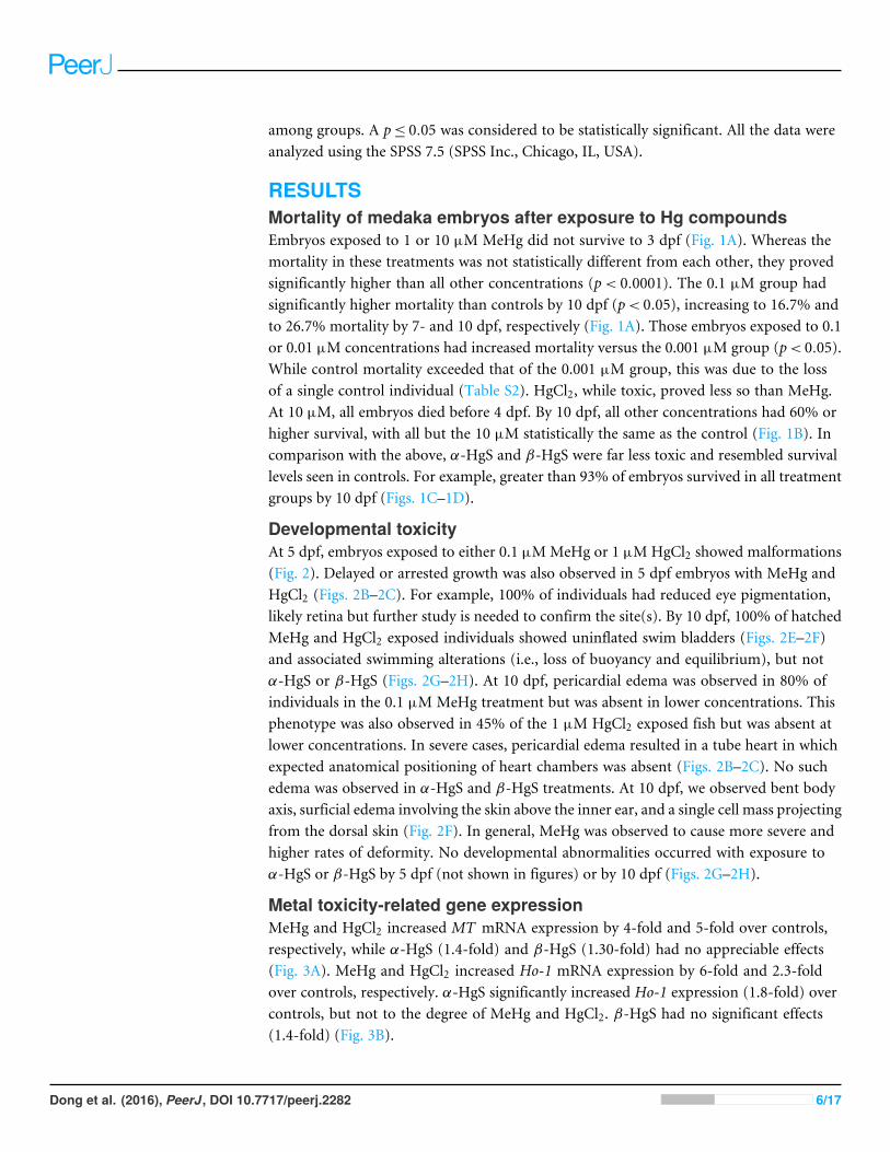
Figure 1 Survival (%) of medaka embryos following exposure toMeHg (A), HgCl2 (B), α-HgS (C) andβ-HgS (D) at 0 (control), 0.001, 0.01, 0.1, 1, or 10µM (from stage 10 to 10 dpf). Mortality was recordeddaily as the percentage of nonviable individuals. Assays were run in triplicate with each data point repre-senting the mean of n= 3 replicates of 10–18 embryos per replicate (±SD).
Grand Island, NY). RT-PCR reaction conditions were 95 ◦C for 15 min followed by 40cycles of 95 ◦C for 15 s and 60 ◦C for 60 s on the Applied Biosystems 7900HT instrumentusing their Sequence Detection System 2.0 software. For each sample, the threshold cycle(Ct) was normalized with β-actin of the same sample according to Chen et al. (2004). Theamplification was calculated using the 2−11CT method (Livak & Schmittgen, 2001; Dong,Matsumura & Kullman, 2010).
Statistical analysisFor each dpf, mean survival was calculated for each well and then used to calculate overallmean survival for each treatment group±SD. Survival data were arcsine-square root trans-formed for ANOVA. RT-PCR data were normalized to β-actin expression and presented asmean ± SD. β-actin data were analyzed by a Grubbs Outlier test; outliers did not alter theresults in subsequent tests and so were left in the analysis. For all measurements, one-wayANOVA followed by Tukey’s post-hoc test was used to assess the statistical significance
Dong et al. (2016), PeerJ, DOI 10.7717/peerj.2282 5/17
among groups. A p≤ 0.05 was considered to be statistically significant. All the data wereanalyzed using the SPSS 7.5 (SPSS Inc., Chicago, IL, USA).
RESULTSMortality of medaka embryos after exposure to Hg compoundsEmbryos exposed to 1 or 10 µM MeHg did not survive to 3 dpf (Fig. 1A). Whereas themortality in these treatments was not statistically different from each other, they provedsignificantly higher than all other concentrations (p< 0.0001). The 0.1 µM group hadsignificantly higher mortality than controls by 10 dpf (p< 0.05), increasing to 16.7% andto 26.7% mortality by 7- and 10 dpf, respectively (Fig. 1A). Those embryos exposed to 0.1or 0.01 µM concentrations had increased mortality versus the 0.001 µM group (p< 0.05).While control mortality exceeded that of the 0.001 µM group, this was due to the lossof a single control individual (Table S2). HgCl2, while toxic, proved less so than MeHg.At 10 µM, all embryos died before 4 dpf. By 10 dpf, all other concentrations had 60% orhigher survival, with all but the 10 µM statistically the same as the control (Fig. 1B). Incomparison with the above, α-HgS and β-HgS were far less toxic and resembled survivallevels seen in controls. For example, greater than 93% of embryos survived in all treatmentgroups by 10 dpf (Figs. 1C–1D).
Developmental toxicityAt 5 dpf, embryos exposed to either 0.1 µMMeHg or 1 µMHgCl2 showed malformations(Fig. 2). Delayed or arrested growth was also observed in 5 dpf embryos with MeHg andHgCl2 (Figs. 2B–2C). For example, 100% of individuals had reduced eye pigmentation,likely retina but further study is needed to confirm the site(s). By 10 dpf, 100% of hatchedMeHg and HgCl2 exposed individuals showed uninflated swim bladders (Figs. 2E–2F)and associated swimming alterations (i.e., loss of buoyancy and equilibrium), but notα-HgS or β-HgS (Figs. 2G–2H). At 10 dpf, pericardial edema was observed in 80% ofindividuals in the 0.1 µM MeHg treatment but was absent in lower concentrations. Thisphenotype was also observed in 45% of the 1 µM HgCl2 exposed fish but was absent atlower concentrations. In severe cases, pericardial edema resulted in a tube heart in whichexpected anatomical positioning of heart chambers was absent (Figs. 2B–2C). No suchedema was observed in α-HgS and β-HgS treatments. At 10 dpf, we observed bent bodyaxis, surficial edema involving the skin above the inner ear, and a single cell mass projectingfrom the dorsal skin (Fig. 2F). In general, MeHg was observed to cause more severe andhigher rates of deformity. No developmental abnormalities occurred with exposure toα-HgS or β-HgS by 5 dpf (not shown in figures) or by 10 dpf (Figs. 2G–2H).
Metal toxicity-related gene expressionMeHg and HgCl2 increased MT mRNA expression by 4-fold and 5-fold over controls,respectively, while α-HgS (1.4-fold) and β-HgS (1.30-fold) had no appreciable effects(Fig. 3A). MeHg and HgCl2 increased Ho-1 mRNA expression by 6-fold and 2.3-foldover controls, respectively. α-HgS significantly increased Ho-1 expression (1.8-fold) overcontrols, but not to the degree of MeHg and HgCl2. β-HgS had no significant effects(1.4-fold) (Fig. 3B).
Dong et al. (2016), PeerJ, DOI 10.7717/peerj.2282 6/17

Figure 2 Control morphology and common phenotypic alterations in embryos and larvae follow-ing exposure to mercury compounds: embryos at 5 dpf: (A) control; (B) 0.1µMMeHg; and (C) 1µMHgCl2. Larvae at 10 dpf: (D) control; (E) 0.1 µMMeHg; (F) 1 µMHgCl2; (G) 10 µM α-HgS; and (H) 10µM β-HgS. Arrows point to heart (h); e, eye; S, swim bladder. All images are at the same magnification,scale bar is 500 µm.
DISCUSSIONMercury-based herbo-metallic preparations have been used in traditional medicines forthousands of years (Kamath et al., 2012) and continue to see usage today. Currently, thePharmacopeia of China has 26 recipes that contain cinnabar (α-HgS). In Indian Ayurvedicmedicine, Rasasindura, which is primarily composed of mercuric sulfides (α-HgS orβ-HgS), is included in over 20 recipes (Kamath et al., 2012). In Tibetan medicine, Zuotai(β-HgS) is included in a dozen popular remedies (Kan, 2013; Li et al., 2014). By comparingMeHg and HgCl2 to α-HgS or β-HgS, we were able to assess compound related embryotoxicity and determine whether traditional medicines are toxicologically similar.
Numerous aquatic organisms have been studied with respect to the toxicity of mercury;however, most studies were focused on organic mercury (e.g., MeHg) (Liao et al., 2007;Cuello et al., 2012) and/or inorganic HgCl2 (Ismail & Yusof, 2011; Wang et al., 2011; Wanget al., 2013). The toxic potential of α-HgS and β-HgS used in traditional medicines islargely unknown. The present study demonstrated that embryo toxicity followed exposure
Dong et al. (2016), PeerJ, DOI 10.7717/peerj.2282 7/17

Figure 3 Analysis of gene expression in medaka embryos exposed to mercury compounds.Medakaembryos were sampled from control, 0.1 µMMeHg, 1 µMHgCl2, 10 µM α-HgS, and 10 µM β-HgStreatment groups (n = 3, 15 embryos pooled per replicate) at 3 days post-exposure. Total RNA was ex-tracted and subjected to RT-PCR analysis for metallothionein (MT ) and heme oxygenase-1 (Ho-1) geneexpression using β-actin expression as the reference. Data are mean± SD. *significantly different fromcontrols with p< 0.05.
to mercury, with MeHg the most toxic, followed by HgCl2, while α-HgS and β-HgS hadlittle toxicity.
In humans and rodents, MeHg is known to cross the placenta and reach the fetuswhere it is responsible for developmental toxicity (Clarkson, Magos & Myers, 2003; Gandhi,Panchal & Dhull, 2013). In laboratory studies using fish, MeHg exposure of early lifestages produced developmental toxicity. Exposures of ≤80 ppm (mg L−1) to medakaembryos have increased mortality and caused teratogenic effects including stunted growth,decreased heart rate, and small eyes with reduced pigmentation, among others (Heisinger& Green, 1975; Dial, 1978). In zebrafish (Danio rerio) larvae exposed to ≤25 mg L−1,down-regulation of >70 proteins was associated with morphological changes includingbut not limited to: smaller swim bladder, unabsorbed yolk, jaw deformities, and bent bodyaxis (Cuello et al., 2012). In the present study, 0.1 µM MeHg produced pericardial edemathat in severe cases formed a tube heart, reduced eye pigmentation, and failed swim bladderinflation. Each of these changes could impact the organism’s health and survival (Dial,1978; Hawryshyn, Mackay & Nilsson, 1982; Marty, Hinton & Cech, 1995).
Compared to MeHg, HgCl2 primarily induces kidney and liver injury in rodents andfish (Klaassen, 2001; Lu et al., 2011a; Wu et al., 2016). However, exposure of mouse- (VanMaele-Fabry, Gofflot & Picard, 1996), sea urchin- (Marc et al., 2002), and medaka embryos(Ismail & Yusof, 2011) to HgCl2 produced developmental toxicity. Wang et al. (2011),Wang et al. (2013) and Wang et al. (2015) studied HgCl2 in adult marine medaka (Oryziasmelastigma), and their proteomic analysis showed down-regulation of several dozenproteins including some related to oxidative stress after acute (1,000 µg/L for 8 h) andchronic (10 µg/L for 60 days) exposures. Subsequent work on liver and brain developeda pathway analysis for potential toxicity (Wang et al., 2013; Wang et al., 2015). However,ultrastructural changes consistent with altered cells were more apparent in brain thanin liver, where reported alterations of mitochondrial and endoplasmic reticulum werenot supported by the figures. Wester & Canton (1992) provided strong evidence for liver
Dong et al. (2016), PeerJ, DOI 10.7717/peerj.2282 8/17
toxicity following exposure of adult guppies (Poecilia reticulata) to MeHg (1–10 µg/L for1 & 3 months). Alterations involved hepatocytes (cell swelling and nuclear pyknosis) andhyperplastic biliary epithelium of the intrahepatic bile duct.
We have shown thatmedaka have good potential as amodel to investigate developmentaleffects of different forms ofmercury.However, in this study, the potential for developmentaltoxicity of α-HgS and β-HgS provedmuch lower than that ofMeHg orHgCl2. For example,at 10 dpf, less than 5% of medaka embryos died and survivors had no apparent teratogeniceffects. These results are comparable with studies of α-HgS and cinnabar-containingtraditional medicines in mice (Lu et al., 2011a; Lu et al., 2011b; Wu et al., 2016) and rats(Shi et al., 2011). Similarly, β-HgS has been shown to be much less toxic as compared toHgCl2 in mice (Zhu et al., 2013; Li et al., 2016; Wu et al., 2016). In those studies, α-HgSand β-HgS were administered orally at 1.5–6 fold (mouse studies) and 20 fold (rat study)above clinical doses, still fourfold higher than the Chinese Pharmacopoeia Allowable Limit(Shi et al., 2011). A recent study in mice showed that gestational exposure to low doseα-HgS (10 mg/kg/day, p.o. × 4 weeks) resulted in offspring with severe neurobehavioraldysfunctions (Huang et al., 2012).
The present study used aqueous exposure of embryonated eggs, which brings upbioavailability related to solubility and the role of the chorion. The solubility of cinnabaris known to be quite low (<0.001 g/L at 20 ◦C), but preparations described by Liu et al.(2008), used in the present study, can increase this value. However, future work will needto describe how formulation of the intended medicinal end product affects the solubilityof this mineral (Kamath et al., 2012). The chorion, a semi-permeable membrane, providesa degree of protection from its surrounding environment (Villalobos et al., 2000) and, neartime of hatching, becomes more permeable (Hamm & Hinton, 2000). Because xenobioticsin general, andmore recently nano-metals, have been shown to enter through chorion porecanals, this route can affect developing embryos (Villalobos et al., 2000; González-Doncel etal., 2003;Wu & Zhou, 2012). Future work is needed to compare if and how different formsof mercury penetrate the chorion.
Mercury compounds displaymultiple organ toxicity (e.g., hepatotoxicity, nephrotoxicityandneurotoxicity) in adult humans and experimental animals (Klaassen, 2001; Liu, Goyer &Waalkes, 2008; Lu et al., 2011b; Shi et al., 2011). One of the most common mechanisms fortoxicity is oxidative stress. For example, mercury induces the production of reactive oxygenspecies (ROS) by binding to intracellular thiols (GSH and sulfhydryl proteins) and byacting as a catalyst in Fenton-type reactions, producing oxidative damage (Klaassen, 2001;Liu, Goyer & Waalkes, 2008). Heme oxygenase-1 (Ho-1) is an oxidative stress biomarkerand was one of the most sensitive genes in response to toxic stimuli in a study of zebrafishembryos acutely exposed to 14 different chemicals (Weil et al., 2009). In the present study,MeHg increased Ho-1 by 6-fold and HgCl2 by 2.3-fold compared to controls, suggestingthat MeHg produced more oxidative damage to embryos. The lack of increased Ho-1expression by α-HgS and β-HgS coincided with the observed low developmental toxicity.This is in agreement with rodent studies that showed exposure to α-HgS (300 mg/kg) didnot induce Ho-1 in liver or kidney, concordant with less hepato- (Lu et al., 2011a) andnephrotoxicity (Lu et al., 2011b).
Dong et al. (2016), PeerJ, DOI 10.7717/peerj.2282 9/17
Metallothionein (MT) is thought to protect against oxidative stress and detoxifyheavy metals including mercury (Klaassen, Liu & Choudhuri, 1999). Induction of MTby mercury is a sensitive biomarker for exposure in a variety of fish species (Van Cleef-Toedt, Kaplan & Crivello, 2001; Cheung, Lam & Chan, 2004; Chan et al., 2006; Oliveira etal., 2010), including Javanese medaka (Oryzias javanicus) (Woo et al., 2006). We foundMT increased by 4–6 fold compared to controls with MeHg and HgCl2 exposure butwere unchanged following α-HgS and β-HgS. This is similar to rodent studies (Lu etal., 2011a; Shi et al., 2011). It is possible that the increases we observed were due to thetiming of our sampling (3 day post exposure). That said, considerable development occursover 3 days, including formation of many of the major organs (Iwamatsu, 2004). Inzebrafish, MT levels have been shown to have strong and ubiquitous expression duringearly embryonic development and drop off later, and it is highly susceptible tometals (Chenet al., 2004). The displacement of essential metals by Hg may compromise multiple cellularprocesses (Amiard et al., 2006), likely problematic during periods of rapid cell divisionand differentiation. At this point, we cannot directly link the morphological changes weobserved to MT, but simply state that the observed increase was a response to the addedmercury. The induction of MT by MeHg and HgCl2 reinforces the importance of thisbiomarker of mercury compounds in general, and identifies it as a potential biomarker fordevelopmental toxicity. Future work with these compounds at intermediate concentrationsmay provide enough surviving embryos to gauge stage specific gene expression changes inresponse to various mercury compounds.
Overall, this study confirmed that the medaka embryo assay is a useful tool for deter-mining and comparing potentials of developmental toxicity for various forms of mercury.We found survival even at middle concentrations of the more toxic forms, suggesting thatour ranges were acceptable given the design of our study. We did not observe changeswith α-HgS and β-HgS, possibly due to the limitations in concentrations resulting fromthe initial constraint of comparing mercurials at the same concentrations. The rapidity,repeatability, broad salinity tolerance, and precision of this model will enable assessmentof a broad range of formulations, concentrations, and mechanisms in the future.
CONCLUSIONSThe current study evaluated medaka embryo toxicity caused by exposure to MeHg andHgCl2 and compared results to mercuric sulfides (α-HgS and β-HgS) used in traditionalmedicines. MeHg and HgCl2 caused increased mortality and developmental toxicity. Thelatter presented as pericardial edema that, in severe cases, resulted in a tube heart, reducedeye pigmentation, and failure to inflate the swim bladder. Developmental toxicity appearedto be in the order of MeHg>HgCl2�α-HgS=β-HgS, indicating that the chemical formsof mercury were a major determinant of its toxicity to medaka embryos. While this workonly involved two medicinal formulations, these assays will be useful in the study of otherpermutations of cinnabar-based medicinals and their toxic mechanisms.
Dong et al. (2016), PeerJ, DOI 10.7717/peerj.2282 10/17

ADDITIONAL INFORMATION AND DECLARATIONS
FundingThis study was supported by the National Natural Science Foundation of China (21267015,81360508, 21567019); Natural Science Foundation of Inner Mongolia AutonomousRegion of China (2015MS0804), the Project of Scientific and Technical Innovation ofInner Mongolia Autonomous Region (2015–2016) and Research Program of Science andTechnology at the Universities of Inner Mongolia Autonomous Region (NJZY11202). Thefunders had no role in study design, data collection and analysis, decision to publish, orpreparation of the manuscript.
Grant DisclosuresThe following grant information was disclosed by the authors:The National Natural Science Foundation of China: 21267015, 81360508, 21567019.Natural Science Foundation of Inner Mongolia Autonomous Region of China:2015MS0804.Project of Scientific and Technical Innovation of Inner Mongolia Autonomous Region.Research Program of Science and Technology at Universities of Inner MongoliaAutonomous Region: NJZY11202.
Competing InterestsJie Liu is an Academic Editor for PeerJ.
Author Contributions• Wu Dong and Jie Liu conceived and designed the experiments, performed theexperiments, analyzed the data, contributed reagents/materials/analysis tools, wrotethe paper, prepared figures and/or tables, reviewed drafts of the paper.• Lixin Wei conceived and designed the experiments, contributed reagents/materials/-analysis tools, reviewed drafts of the paper.• Yang Jingfeng analyzed the data, contributed reagents/materials/analysis tools, revieweddrafts of the paper.• Melissa Chernick wrote the paper, prepared figures and/or tables, reviewed drafts of thepaper.• David E. Hinton contributed reagents/materials/analysis tools, wrote the paper, preparedfigures and/or tables, reviewed drafts of the paper.
Animal EthicsThe following information was supplied relating to ethical approvals (i.e., approving bodyand any reference numbers):
Medaka (Oryzias latipes) were maintained at the Duke University Aquatic ResearchFacility under standard recirculating water conditions following approved animal care andmaintenance protocols. Duke University Institutional Animal Care and Use Committee,No. A062-15-02 for breeding and No. A031-15-01 for toxicity testing protocol.
Dong et al. (2016), PeerJ, DOI 10.7717/peerj.2282 11/17

Data AvailabilityThe following information was supplied regarding data availability:
Figshare: https://figshare.com/s/23641deb4c3e10e09259;https://figshare.com/s/3ef822119d859083275d;https://figshare.com/s/7ae5a4e6a4cbfec828a6;https://figshare.com/s/469c738bbda93b93873b;https://figshare.com/s/60fad618f8beb67696b9;https://figshare.com/s/ba2608b17e612ca244b7.
Supplemental InformationSupplemental information for this article can be found online at http://dx.doi.org/10.7717/peerj.2282#supplemental-information.
REFERENCESAmiard JC, Amiard-Triquet C, Barka S, Pellerin J, Rainbow PS. 2006.Metallothioneins
in aquatic invertebrates: their role in metal detoxification and their use as biomark-ers. Aquatic Toxicology 76:160–202 DOI 10.1016/j.aquatox.2005.08.015.
AuthmanMMN, Zaki MS, Khallaf EA, Abbas HH. 2015. Use of fish as bio-indicator ofthe effects of heavy metals pollution. Journal of Aquaculture Research & Development6:328–340 DOI 10.4172/2155-9546.1000328.
Chan KM. 1995.Metallothionein: potential biomarker for monitoring heavy metalpollution in fish around Hong Kong.Marine Pollution Bulletin 31:411–415DOI 10.1016/0025-326X(95)00125-7.
Chan KM, Ku LL, Chan PCY, CheukWK. 2006.Metallothionein gene expression inzebrafish embryo-larvae and ZFL cell-line exposed to heavy metal ions.MarineEnvironmental Research 62(Supplement 1):S83–S87DOI 10.1016/j.marenvres.2006.04.012.
ChenW-Y, John JAC, Lin C-H, Lin H-F,Wu S-C, Lin C-H, Chang C-Y. 2004. Expres-sion of metallothionein gene during embryonic and early larval development inzebrafish. Aquatic Toxicology 69:215–227 DOI 10.1016/j.aquatox.2004.05.004.
Chen XP, Li L, Wong CKC, Cheng SH. 2009. Rapid adaptation of molecular resourcesfrom zebrafish and medaka to develop an estuarine/marine model. ComparativeBiochemistry and Physiology C—Toxicology & Pharmacology 149:647–655DOI 10.1016/j.cbpc.2009.01.009.
Chen C,Wu S,Wang Y, Hou J, Ma L, Sun X. 2012. Recent researches of syntheticmercury sulfide in traditional medicine system. Zhongguo Zhong Yao Za Zhi37:2968–2970.
Cheng KC, Hinton DE, Mattingly CJ, Planchart A. 2012. Aquatic models, genomics andchemical risk management. Comparative Biochemistry and Physiology C—Toxicology& Pharmacology 155:169–173 DOI 10.1016/j.cbpc.2011.06.009.
Cheung APL, Lam THJ, Chan KM. 2004. Regulation of Tilapiametallothionein geneexpression by heavy metal ions.Marine Environmental Research 58:389–394DOI 10.1016/j.marenvres.2004.03.084.
Dong et al. (2016), PeerJ, DOI 10.7717/peerj.2282 12/17
Clarkson TW,Magos L, Myers GJ. 2003. The toxicology of mercury—current exposuresand clinical manifestations. New England Journal of Medicine 349:1731–1737DOI 10.1056/NEJMra022471.
ColtonMD, Kwok KWH, Brandon JA,Warren IH, Ryde IT, Cooper EM, HintonDE, Rittschof D, Meyer JN. 2014. Developmental toxicity and DNA damage fromexposure to parking lot runoff retention pond samples in the Japanese medaka(Oryzias latipes).Marine Environmental Research 99:117–124DOI 10.1016/j.marenvres.2014.04.007.
Craig PJ. 2003.Organometallic compounds in the environment. 2nd edition. Guildford:John Wiley & Sons Ltd, p 415.
Cuello S, Ximenez-Embun P, Ruppen I, Schonthaler HB, Ashman K, Madrid Y, Luque-Garcia JL, Camara C. 2012. Analysis of protein expression in developmental toxicityinduced by MeHg in zebrafish. Analyst 137:5302–5311 DOI 10.1039/c2an35913h.
Dial NA. 1978.Methylmercury: some effects on embryogenesis in the Japanese medaka,Oryzias latipes. Teratology 17:83–91 DOI 10.1002/tera.1420170116.
DongW,Macaulay LJ, Kwok KWH, Hinton DE, Ferguson PL, Stapleton HM. 2014.The PBDE metabolite 6-OH-BDE 47 affects melanin pigmentation and THRβMRNA expression in the eye of zebrafish embryos. Endocrine Disruptors 2:e969072DOI 10.4161/23273739.2014.969072.
DongW,Matsumura F, Kullman SW. 2010. TCDD induced pericardial edema andrelative COX-2 expression in medaka (Oryzias latipes) embryos. ToxicologicalSciences 118:213–223 DOI 10.1093/toxsci/kfq254.
FieldenMR, Zacharewski TR. 2001. Challenges and limitations of gene expressionprofiling in mechanistic and predictive toxicology. Toxicological Sciences 60:6–10DOI 10.1093/toxsci/60.1.6.
Gandhi DN, Panchal GM, Dhull DK. 2013. Influence of gestational exposure on theeffects of prenatal exposure to methyl mercury on postnatal development in rats.Central European Journal of Public Health 21:30–35.
González-Doncel M, Larrea M, Sánchez-Fortún S, Hinton DE. 2003. Influence of waterhardening of the chorion on cadmium accumulation in medaka (Oryzias latipes)eggs. Chemosphere 52:75–83 DOI 10.1016/S0045-6535(03)00227-3.
González-Doncel M, OkihiroMS, Torija CF, Tarazona JV, Hinton DE. 2008. Anartificial fertilization method with the Japanese medaka: implications in earlylife stage bioassays and solvent toxicity. Ecotoxicology and Environmental Safety69:95–103 DOI 10.1016/j.ecoenv.2006.12.009.
Hamm JT, Hinton DE. 2000. The role of development and duration of exposure to theembryotoxicity of diazinon. Aquatic Toxicology 48:403–418DOI 10.1016/S0166-445X(99)00065-X.
Hawryshyn CW,MackayWC, Nilsson TH. 1982.Methyl mercury induced visual deficitsin rainbow trout. Canadian Journal of Zoology 60:3127–3133 DOI 10.1139/z82-397.
He Z, Traina SJ, Weavers LK. 2007. Sonochemical dissolution of cinnabar (α-HgS).Environmental Science & Technology 41:773–778 DOI 10.1021/es0613299.
Dong et al. (2016), PeerJ, DOI 10.7717/peerj.2282 13/17

Heisinger JF, GreenW. 1975.Mercuric chloride uptake by eggs of the ricefish andresulting teratogenic effects. Bulletin of Environmental Contamination and Toxicology14:665–673 DOI 10.1007/BF01685240.
Huang CF, Hsu CJ, Liu SH, Lin-Shiau SY. 2012. Exposure to low dose of cinnabar(a naturally occurring mercuric sulfide (HgS)) caused neurotoxicological effectsin offspring mice. Journal of Biomedicine and Biotechnology 2012:Article 254582DOI 10.1155/2012/254582.
Inoue K, Takei Y. 2002. Diverse adaptability in Oryzias species to high environmentalsalinity. Zoological Science 19:727–734 DOI 10.2108/zsj.19.727.
Ismail A, Yusof S. 2011. Effect of mercury and cadmium on early life stages of Javamedaka (Oryzias javanicus): a potential tropical test fish.Marine Pollution Bulletin63:347–349 DOI 10.1016/j.marpolbul.2011.02.014.
Iwamatsu T. 2004. Stages of normal development in the medaka Oryzias latipes.Mechanisms of Development 121:605–618 DOI 10.1016/j.mod.2004.03.012.
Kamath SU, Pemiah B, Sekar RK, Krishnaswamy S, Sethuraman S, Krishnan UM. 2012.Mercury-based traditional herbo-metallic preparations: a toxicological perspective.Archives of Toxicology 86:831–838 DOI 10.1007/s00204-012-0826-2.
Kan Z. 2013. An introduction of Zuotai in Tibetan patent medicine. Zhongguo Zhong YaoZa Zhi 38:1621–1623.
Karimi R, Fitzgerald TP, Fisher NS. 2012. A quantitative synthesis of mercury in com-mercial seafood and implications for exposure in the United States. EnvironmentalHealth Perspectives 120:1512–1519 DOI 10.1289/ehp.1205122.
Kinoshita M, Murata K, Naruse K, TanakaM. 2009.Medaka: biology, management, andexperimental protocols. Singapore: John Wiley & Sons, Ltd.
Klaassen CD. 2001. Heavy metals and heavy-metal antagonists. In: Hardman JG,Limbird LE, Gilman AG, eds. The pharmacological basis of therapeutics. New York:McGraw-Hill, 1851–1876.
Klaassen CD, Liu J, Choudhuri S. 1999.Metallothionein: an intracellular protein toprotect against cadmium toxicity. Annual Review of Pharmacology and Toxicology39:267–294 DOI 10.1146/annurev.pharmtox.39.1.267.
Li H, LiW-K, Lu Y-F, Wei L-X, Liu J. 2016. The Tibetan medicine Zuotai influencesclock gene expression in the liver of mice. PeerJ 4:e1632 DOI 10.7717/peerj.1632.
Li C,Wang D, Duo J, Duojie L, Chen X, Du Y, Yang H, Zheng Z, YuM,Wei L. 2014.Study on safety of Tibetan medicine zuotai and preliminary study on clinical safety ofits compound dangzuo. Zhongguo Zhong Yao Za Zhi 39:2573–2582.
Liang A, ShangM. 2005. General situation of the study on the toxicity of Cinnabaris.Zhongguo Zhong Yao Za Zhi 30:249–252.
Liao C-Y, Zhou Q-F, Fu J-J, Shi J-B, Yuan C-G, Jiang G-B. 2007. Interaction ofmethylmercury and selenium on the bioaccumulation and histopathology in medaka(Oryzias latipes). Environmental Toxicology 22:69–77 DOI 10.1002/tox.20236.
Liu J, Goyer RA,Waalkes MP. 2008. Toxic effects of metals. In: Klaassen CD, ed.Casarett & Doull’s toxicology: the basic science of poisons. New York: McGraw-Hill,931–979.
Dong et al. (2016), PeerJ, DOI 10.7717/peerj.2282 14/17

Liu J, Shi J-Z, Yu L-M, Goyer RA,Waalkes MP. 2008.Mercury in traditional medicines:is cinnabar toxicologically similar to common mercurials? Experimental Biology andMedicine 233:810–817 DOI 10.3181/0712-MR-336.
Livak KJ, Schmittgen TD. 2001. Analysis of relative gene expression data using real-timequantitative PCR and the 2-11CT method.Methods 25:402–408DOI 10.1006/meth.2001.1262.
Lu Y-F, WuQ, Liang S-X, Miao J-W, Shi J-S, Liu J. 2011a. Evaluation of hepatotoxicitypotential of cinnabar-containing An-Gong-Niu-Huang Wan, a patent traditionalChinese medicine. Regulatory Toxicology and Pharmacology 60:206–211DOI 10.1016/j.yrtph.2011.03.007.
Lu Y-F, WuQ, Yan J-W, Shi J-Z, Liu J, Shi J-S. 2011b. Realgar, cinnabar and An-Gong-Niu-Huang Wan are much less chronically nephrotoxic than common arsenicals andmercurials. Experimental Biology and Medicine 236:233–239DOI 10.1258/ebm.2010.010247.
Marc J, Maguer C, Bellé R, Mulner-lorillon O. 2002. Sharp dose- and time-dependenttoxicity of mercuric chloride at the cellular level in sea urchin embryos. Archives ofToxicology Archiv für Toxikologie 76:388–391 DOI 10.1007/s00204-002-0368-0.
Marty GD, Hinton DE, Cech JJ. 1995. Oxygen consumption by larval Japanese medakawith inflated or uninflated swim bladders. Transactions of the American FisheriesSociety 124:623–627 DOI 10.1577/1548-8659(1995)124<0623:NOCBLJ>2.3.CO;2.
Morel FMM, Kraepiel AML, Amyot M. 1998. The chemical cycle and bioaccumulation ofmercury. Annual Review of Ecology and Systematics 29:543–566DOI 10.1146/annurev.ecolsys.29.1.543.
Mu J,Wang Y,Wang X,Wang J. 2011. Toxic effects of cadmium, mercury, chromiumand lead on the early life stage of marine medaka (Oryzias melastigma). Asian Journalof Ecotoxicology 6:352–360.
Oliveira M, Ahmad I, Maria VL, Serafim A, BebiannoMJ, PachecoM, Santos MA. 2010.Hepatic metallothionein concentrations in the golden grey mullet (Liza aurata)—relationship with environmental metal concentrations in a metal-contaminatedcoastal system in Portugal.Marine Environmental Research 69:227–233DOI 10.1016/j.marenvres.2009.10.012.
Pharmacopeia of China. 2015. Pharmacopeia of the People’s Republic of China. Pharma-copoeia Commission China: Chemical Industry Press.
Raldúa D, Díez S, Bayona JM, Barceló D. 2007.Mercury levels and liver pathology inferal fish living in the vicinity of a mercury cell chlor-alkali factory. Chemosphere66:1217–1225 DOI 10.1016/j.chemosphere.2006.07.053.
Samson JC, Shenker J. 2000. The teratogenic effects of methylmercury on early develop-ment of the zebrafish, Danio rerio. Aquatic Toxicology 48:343–354DOI 10.1016/S0166-445X(99)00044-2.
Sfakianakis DG, Renieri E, Kentouri M, Tsatsakis AM. 2015. Effect of heavy metals onfish larvae deformities: a review. Environmental Research 137:246–255DOI 10.1016/j.envres.2014.12.014.
Dong et al. (2016), PeerJ, DOI 10.7717/peerj.2282 15/17
SheehanMC, Burke TA, Navas-Acien A, Breysse PN, McGready J, FoxMA. 2014.Global methylmercury exposure from seafood consumption and risk of develop-mental neurotoxicity: a systematic review. Bulletin of the World Health Organization92:254–269F DOI 10.2471/BLT.12.116152.
Shi J-Z, Kang F,WuQ, Lu Y-F, Liu J, Kang YJ. 2011. Nephrotoxicity of mercuricchloride, methylmercury and cinnabar-containing Zhu-Sha-An-Shen-Wan in rats.Toxicology Letters 200:194–200 DOI 10.1016/j.toxlet.2010.11.015.
Van Cleef-Toedt KA, Kaplan LAE, Crivello JF. 2001. Killifish metallothionein messengerRNA expression following temperature perturbation and cadmium exposure. CellStress & Chaperones 6:351–359DOI 10.1379/1466-1268(2001)006<0351:KMMREF>2.0.CO;2.
VanMaele-Fabry G, Gofflot F, Picard JJ. 1996. Defects in the development of branchialnerves and ganglia induced by in vitro exposure of mouse embryos to mercuricchloride. Teratology 53:10–20DOI 10.1002/(SICI)1096-9926(199601)53:1<10::AID-TERA2>3.0.CO;2-D.
Villalobos SA, Hamm JT, Teh SJ, Hinton DE. 2000. Thiobencarb-induced embryotox-icity in medaka (Oryzias latipes): stage-specific toxicity and the protective role ofchorion. Aquatic Toxicology 48:309–326 DOI 10.1016/S0166-445X(99)00032-6.
Voelker D, Stetefeld N, Schirmer K, Scholz S. 2008. The role of cyp1a and heme oxy-genase 1 gene expression for the toxicity of 3,4-dichloroaniline in zebrafish (Daniorerio) embryos. Aquatic Toxicology 86:112–120 DOI 10.1016/j.aquatox.2007.10.007.
Wang Y,Wang D, Lin L,WangM. 2015. Quantitative proteomic analysis reveals proteinsinvolved in the neurotoxicity of marine medaka Oryzias melastigma chronicallyexposed to inorganic mercury. Chemosphere 119:1126–1133DOI 10.1016/j.chemosphere.2014.09.053.
WangM,Wang Y,Wang J, Lin L, Hong H,Wang D. 2011. Proteome profiles in medaka(Oryzias melastigma) liver and brain experimentally exposed to acute inorganicmercury. Aquatic Toxicology 103:129–139 DOI 10.1016/j.aquatox.2011.02.020.
WangM,Wang Y, Zhang L,Wang J, Hong H,Wang D. 2013. Quantitative proteomicanalysis reveals the mode-of-action for chronic mercury hepatotoxicity to marinemedaka (Oryzias melastigma). Aquatic Toxicology 130–131:123–131.
Weil M, Scholz S, ZimmerM, Sacher F, Duis K. 2009. Gene expression analysis inzebrafish embryos: a potential approach to predict effect concentrations in the fishearly life stage test. Environmental Toxicology and Chemistry 28:1970–1978DOI 10.1897/08-627.1.
Wester PW, Canton HH. 1992.Histopathological effects in Poecilia reticulata (guppy)exposed to methyl mercury chloride. Toxicologic Pathology 20:81–92DOI 10.1177/019262339202000110.
Woo S, Yum S, Jung JH, ShimWJ, Lee C-H, Lee T-K. 2006.Heavy metal-induced dif-ferential gene expression of metallothionein in Javanese Medaka, Oryzias javanicus.Marine Biotechnology 8:654–662 DOI 10.1007/s10126-006-6046-0.
WuQ, LiW-K, Zhou Z-P, Li Y-Y, Xiong T-W, Du Y-Z,Wei L-X, Liu J. 2016. TheTibetan medicine Zuotai differs from HgCl2 and MeHg in producing liver injury in
Dong et al. (2016), PeerJ, DOI 10.7717/peerj.2282 16/17

mice. Regulatory Toxicology and Pharmacology 78:1–7DOI 10.1016/j.yrtph.2016.03.017.
WuY, Zhou Q. 2012. Dose- and time-related changes in aerobic metabolism, chorionicdisruption, and oxidative stress in embryonic medaka (Oryzias latipes): underlyingmechanisms for silver nanoparticle developmental toxicity. Aquatic Toxicology 124–125:238–246 DOI 10.1016/j.aquatox.2012.08.009.
ZhuH,Wei L, Du Y,Wang D, Li C. 2013. The chronic toxicity study of Tibetan medicineZuotai in mice. Shizheng Guoyi Guoyao 24:2022–2024.
Dong et al. (2016), PeerJ, DOI 10.7717/peerj.2282 17/17