developmental cell, vol. 4, 941–952, june, 2003, … colon... · yeshiva university transcription...
TRANSCRIPT

Developmental Cell, Vol. 4, 941–952, June, 2003, Copyright 2003 by Cell Press
Asymmetric Distribution of Nuclear Pore Complexesand the Cytoplasmic Localization of �2-Tubulin mRNAin Chlamydomonas reinhardtii
bodies, define nuclear compartments with distinct gene,transcript, and protein composition (reviewed in Dundrand Misteli, 2001; Gall, 2001). In addition, chromosomesoccupy distinct territories within the nucleus (Marshallet al., 1996).
Daniel A. Colon-Ramos,1,4 Jeffrey L. Salisbury,2
Mark A. Sanders,2,5 Shailesh M. Shenoy,3
Robert H. Singer,3 and Mariano A. Garcıa-Blanco1,*1Department of Molecular Genetics and
Microbiology andNuclear architecture has significant effects on tran-Department of Medicine
scription and processing of nascent transcripts. NuclearDuke University Medical Centercomplexes containing RNA polymerase II localize toDurham, North Carolina 27710sites of gene transcription, and splicing factor com-2 Tumor Biology Programplexes (SFCs) localize as nuclear “speckles” to distinctMayo Clinicdomains separated from sites of transcription (reviewedRochester, Minnesota 55905in Dundr and Misteli, 2001). Additionally, cotranscrip-3 Department of Cell Biology andtional processing such as capping, splicing, and poly-Department of Anatomy and Structural Biologyadenylation occurs within the vicinity of active geneAlbert Einstein College of Medicinetranscription, implicating nuclear architecture in theYeshiva Universityregulation of transcript processing (reviewed in Golds-1300 Morris Park Avenuetrohm et al., 2001 and Maniatis and Reed, 2002; ZhangBronx, New York 10461et al., 1994).
Whereas these observations suggest that nuclear ar-chitecture affects gene expression, it is not clearSummarywhether nuclear organization is linked to cytoplasmicevents such as transcript targeting and cell polarity.Although it is generally accepted that nuclear architec-For example, whereas mRNA stability and cytoplasmicture is an important determinant of nuclear activity,localization depend on nuclear factors (reviewed init is not clear whether cytoplasmic events, such asBrennan and Steitz, 2001; Gu et al., 2002; Long et al.,transcript localization and cell polarity, are affected2001), there is little evidence to directly link nuclearby this architecture. Characterization of the nucleararchitecture with the cytoplasmic fate of transcripts.architecture of the single-cell alga ChlamydomonasBlobel’s “gene gating” hypothesis suggested that cyto-reinhardtii revealed a polarized nucleus, with nuclearplasmic localization of new transcripts may result frompore complexes preferentially concentrated at thedirected nucleocytoplasmic transport, such that active
posterior side of the nucleus. Nuclear asymmetry wasgenes located near the nuclear periphery, adjacent to
greatly exaggerated during the upregulation of genes the nuclear pore complexes (NPCs), may help targetencoding flagellar proteins, when nuclear pore com- the mRNA to its cytoplasmic destination (Blobel, 1985).plexes (NPCs) were observed to hyperpolarize to the Although certain active genes have been found near theposterior side of the nucleus while heterochromatin nuclear periphery, their localization has not been foundpolarized to the anterior side. Interestingly, prior to to influence gene activity or cytoplasmic transcript local-deflagellation, the �2-tubulin gene was preferentially ization (Bullock and Ish-Horowicz, 2001; Lawrence etlocated in the posterior region of the nucleus, and al., 1989; Mahy et al., 2002; Wilkie et al., 1999).following deflagellation, �2-tubulin transcripts accu- In the present study, we used Chlamydomonas rein-mulated posteriorly in polysome-rich cytoplasmic re- hardtii, a biflagellated unicellular alga with an asymmet-gions adjacent to the highest concentration of NPCs, ric cellular structure to study the relationship betweensuggesting a connection between nuclear architec- nuclear architecture and cytoplasmic processes. Cellu-ture and cytoplasmic transcript localization. lar asymmetry in Chlamydomonas is determined by the
anterior position of the flagella and the posterior positionof the chloroplast, with the nucleus nested betweenIntroductionthese two conspicuous organelles (Harris, 1989; seealso Figures 1 and 2A). The nucleus is about 3–5 �m inThe nucleus is highly compartmentalized into functionaldiameter and is surrounded by a double nuclear mem-subnuclear domains (reviewed in Dundr and Misteli,brane envelope interrupted by numerous nuclear pores2001). The most prominent of these is the nucleolus,(�500 A in diameter; Johnson and Porter, 1968; Sagerwhich consists of an assembly of rRNA gene repeats andand Palade, 1957; Weiss et al., 2000). A spherical nucleo-rRNA and proteins involved in the synthesis, processing,lus consisting of tightly packed particles surroundingand assembly of ribosomal subunits (Garcıa-Blanco etfiner particles and light inclusions is located near itsal., 1995; Leger-Silvestre et al., 1999). Other nucleoplas-center (Harris, 1989; Holmes and Dutcher, 1989; John-mic compartments, such as the Cajal, Gemini, and PMNson and Porter, 1968; see also Figure 1). The C. rein-hardtii nucleus is tethered to the two basal bodies and
*Correspondence: [email protected] flagella by a cytoskeletal fiber complex known as the4 Present address: University Program in Genetics, Duke University,nucleus-basal body connector (NBC) that contains theDurham, North Carolina 27710.calcium binding protein centrin (20 kDa; Wright et al.,5 Present address: College of Biological Sciences, University of Min-
nesota, St. Paul, Minnesota 55108. 1985). We previously reported that contraction of the

Developmental Cell942
Figure 1. Flagellar Excision Leads to Asymmetric Distribution of Heterochromatin
Electron micrographs of medial longitudinal sections of Chlamydomonas reinhardtii before (A), immediately following (B), and 90 min after (C)pH shock-induced flagellar excision. Note that the distance between the nucleus and flagellar apparatus is reduced at the time of flagellarexcision (B) and the nucleus has undergone a subtle shape change. Higher magnification of nuclear profiles (D–F) shows a redistribution ofheterochromatin immediately adjacent to the nuclear envelope. Before flagellar excision (D), heterochromatin is uniformly dispersed in patchesaround the nuclear periphery. Immediately following flagellar excision (E), heterochromatin has redistributed toward the flagellar apparatusand becomes cleared from the posterior side of the nucleus. By 90 min of recovery from pH shock (F), heterochromatin has redistributed toagain show a relatively even distribution around the nuclear periphery. At the 90 min time point, the heterochromatin at the nuclear envelopeappears to be somewhat reduced in thickness. Diagrammatic representation (G–I) of the redistribution of heterochromatin (red) and locationof nuclear pores (blue arrowheads) seen in the micrographs illustrated in (D–F) is shown.
NBC following deflagellation causes a dramatic change the nucleolus, and the nucleus-basal body connector.We also used fluorescent in situ hybridization to visual-in nuclear shape (from oval to pyriform or pear-shaped)
and its displacement toward the anterior end of the cell ize the location of the �2-tubulin gene and the cyto-plasmic localization of �2-tubulin transcripts. We found(Salisbury et al., 1987; Wright et al., 1985). Others have
shown that following deflagellation there is a dramatic that NPCs were asymmetrically distributed toward theposterior pole of the Chlamydomonas nucleus. Thisupregulation of genes encoding flagellar proteins re-
quired for the complete regeneration of the flagella asymmetry in NPC distribution was greatly exaggeratedfollowing deflagellation, when dramatic changes in over-(Baker et al., 1984; Keller et al., 1984). In this study,
we explored changes in nuclear architecture following all nuclear shape and redistribution of heterochromatinnear the nuclear envelope was also observed. Impor-deflagellation using electron microscopy and confocal
immunofluorescence, which allowed simultaneous visu- tantly, newly synthesized �2-tubulin transcripts werelocalized in the posterior cytoplasm adjacent to the nu-alization of the NPCs, the core (Sm) snRNP proteins,

Asymmetric Nuclear Architecture in C. reinhardtii943
Figure 2. Nuclear Architecture in InterphaseCells
(A) Diagram of the nuclear architecture of C.reinhardtii cells in the context of the rest ofthe cellular structure (based on studies con-ducted by Johnson and Porter, 1968; Salis-bury et al., 1987; Schotz et al., 1972; Weisset al., 2000; also discussed in Harris, 1989).The Chlamydomonas reinhardtii nucleus(clear) is nested between the two flagella atthe anterior end (F) and the chloroplast at theposterior end ([C], in green). The flagella areconnected to the nucleus via the nucleus-basal body connector, which contains centrinfibers (blue). The spherical nucleolus (dotted)has been previously visualized in electron mi-croscopy studies.(B) Triple confocal immunofluorescence wasconducted in interphase C. reinhardtii cellsusing antibodies against centrin fibers (blue),the core Sm proteins of spliceosomal snRNPs(red), nucleolar fibrillarin (red), and NPC pro-teins (green). Dotted lines outline the cell wall,which was visualized by differential interfer-ence contrast (DIC) microscopy. For conve-nience and comparison, the cells were placedin the same orientation as the diagram, withanterior facing up (F, flagellum) and the pos-terior end facing down (C, chloroplast).
clear area with the highest NPC density. Thus, these coding for flagellar proteins (Baker et al., 1984, 1986;observations suggest that an asymmetric and dynamic Bernstein et al., 1994; Keller et al., 1984; Schloss etnuclear architecture can affect transcript localization in al., 1984). In order to determine whether the nuclearthe cytoplasm. architecture was affected during this process of in-
creased transcription activity, we analyzed electron mi-crographs of medial longitudinal sections of C. rein-Resultshardtii before, immediately following, and 90 min afterpH shock-induced flagellar excision.Heterochromatin Redistribution during
In undisturbed cells, the nucleus showed a centrallyFlagellar Regenerationpositioned nucleolus and a narrow rim of heterochroma-C. reinhardtii sheds its flagella when exposed to adversetin evenly distributed near the nuclear periphery, justenvironmental conditions, such as pH change, throughbeneath the nuclear envelope (Figures 1A, 1D, and 1G).an active process known as flagellar excision or auto-NPCs appeared to be asymmetrically localized towardtomy (Rosenbaum et al., 1969). Upon return to favorablethe posterior of the nucleus. Immediately following de-conditions, cells regenerate their flagella within 2 hr fol-
lowing increased transcription and stability of mRNAs flagellation, the nucleus became pyriform in shape and

Developmental Cell944
Figure 3. Changes in Nuclear Architecture during Flagellar Regeneration
Cells were deflagellated by pH shock, and allowed to recover in neutral pH media. Samples were collected during recovery and flagellarregeneration. Triple confocal immunofluorescence as described above was carried to visualize changes in nuclear architecture. The anteriorend of the cells is labeled F for flagellum, and the posterior part is labeled C for chloroplast.(A and B) Cells visualized prior to flagellar autotomy.(C and D) Cells visualized immediately after pH shock.(E and F) Reflagellating cells visualized 40 min after pH shock.
was displaced toward the flagellar apparatus as pre- posterior end of the nucleus, their asymmetric distribu-tion was not as pronounced as in cells immediately fol-viously reported (Salisbury et al., 1987; Wright et al.,lowing deflagellation (Figures 1C, 1F, and 1I).1985). A dramatic redistribution of heterochromatin was
also apparent in nuclei following deflagellation, suchthat the narrow rim of heterochromatin near the nuclear NPCs Are Asymmetrically Distributedenvelope appeared to be displaced toward the anterior in C. reinhardtii Nucleiside of the nucleus (Figures 1B, 1E, and 1H). Interest- In order to confirm and extend the electron microscopeingly, the asymmetric distribution of the NPCs toward observations, we used immunofluorescence and confo-the posterior side of the nucleus noted in unperturbed cal microscopy to visualize nuclear proteins known tocells became more pronounced immediately following localize in distinct subnuclear compartments. In thisdeflagellation and in cells during the period of flagellar study, we utilized mAb 414 antibody (BabCO), a mono-regrowth (Figures 1B, 1E, and 1H). clonal antibody that recognizes the nucleoporins (Nup),
By about 90 min following deflagellation, cells fully protein components of the NPC; a human antiserumrecovered full-length flagella and showed an even distri- (AW) against the core Sm protein of the spliceosomalbution of the heterochromatin rim near the nuclear enve- snRNPs (Lerner et al., 1981); and a human antiserum
against the nucleolar protein fibrillarin (Ochs et al., 1985).lope. Whereas NPCs remained concentrated toward the
Asymmetric Nuclear Architecture in C. reinhardtii945
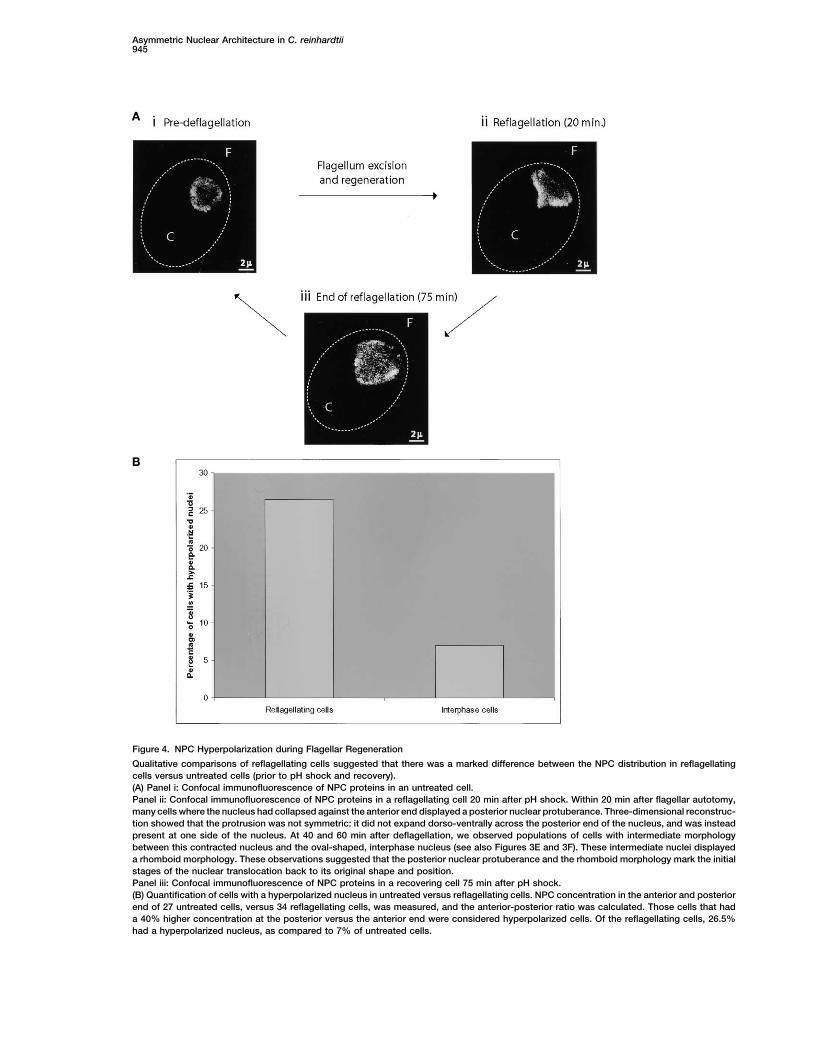
Figure 4. NPC Hyperpolarization during Flagellar Regeneration
Qualitative comparisons of reflagellating cells suggested that there was a marked difference between the NPC distribution in reflagellatingcells versus untreated cells (prior to pH shock and recovery).(A) Panel i: Confocal immunofluorescence of NPC proteins in an untreated cell.Panel ii: Confocal immunofluorescence of NPC proteins in a reflagellating cell 20 min after pH shock. Within 20 min after flagellar autotomy,many cells where the nucleus had collapsed against the anterior end displayed a posterior nuclear protuberance. Three-dimensional reconstruc-tion showed that the protrusion was not symmetric: it did not expand dorso-ventrally across the posterior end of the nucleus, and was insteadpresent at one side of the nucleus. At 40 and 60 min after deflagellation, we observed populations of cells with intermediate morphologybetween this contracted nucleus and the oval-shaped, interphase nucleus (see also Figures 3E and 3F). These intermediate nuclei displayeda rhomboid morphology. These observations suggested that the posterior nuclear protuberance and the rhomboid morphology mark the initialstages of the nuclear translocation back to its original shape and position.Panel iii: Confocal immunofluorescence of NPC proteins in a recovering cell 75 min after pH shock.(B) Quantification of cells with a hyperpolarized nucleus in untreated versus reflagellating cells. NPC concentration in the anterior and posteriorend of 27 untreated cells, versus 34 reflagellating cells, was measured, and the anterior-posterior ratio was calculated. Those cells that hada 40% higher concentration at the posterior versus the anterior end were considered hyperpolarized cells. Of the reflagellating cells, 26.5%had a hyperpolarized nucleus, as compared to 7% of untreated cells.
Developmental Cell946
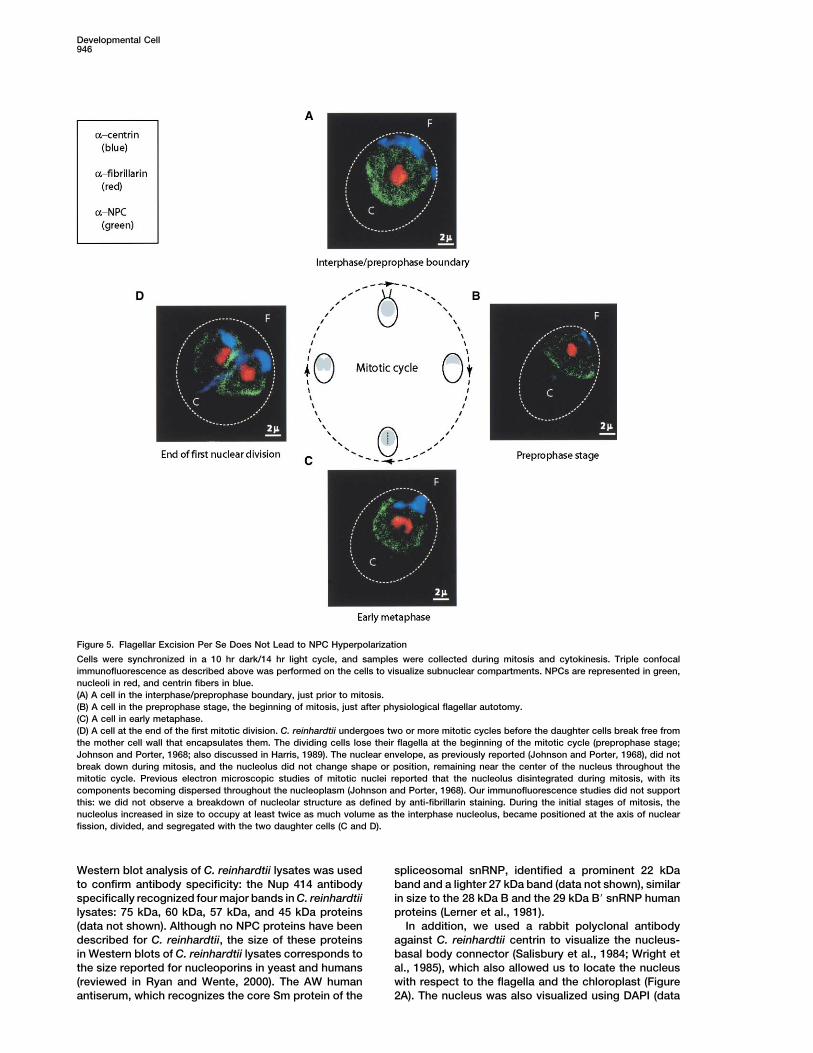
Figure 5. Flagellar Excision Per Se Does Not Lead to NPC Hyperpolarization
Cells were synchronized in a 10 hr dark/14 hr light cycle, and samples were collected during mitosis and cytokinesis. Triple confocalimmunofluorescence as described above was performed on the cells to visualize subnuclear compartments. NPCs are represented in green,nucleoli in red, and centrin fibers in blue.(A) A cell in the interphase/preprophase boundary, just prior to mitosis.(B) A cell in the preprophase stage, the beginning of mitosis, just after physiological flagellar autotomy.(C) A cell in early metaphase.(D) A cell at the end of the first mitotic division. C. reinhardtii undergoes two or more mitotic cycles before the daughter cells break free fromthe mother cell wall that encapsulates them. The dividing cells lose their flagella at the beginning of the mitotic cycle (preprophase stage;Johnson and Porter, 1968; also discussed in Harris, 1989). The nuclear envelope, as previously reported (Johnson and Porter, 1968), did notbreak down during mitosis, and the nucleolus did not change shape or position, remaining near the center of the nucleus throughout themitotic cycle. Previous electron microscopic studies of mitotic nuclei reported that the nucleolus disintegrated during mitosis, with itscomponents becoming dispersed throughout the nucleoplasm (Johnson and Porter, 1968). Our immunofluorescence studies did not supportthis: we did not observe a breakdown of nucleolar structure as defined by anti-fibrillarin staining. During the initial stages of mitosis, thenucleolus increased in size to occupy at least twice as much volume as the interphase nucleolus, became positioned at the axis of nuclearfission, divided, and segregated with the two daughter cells (C and D).
Western blot analysis of C. reinhardtii lysates was used spliceosomal snRNP, identified a prominent 22 kDaband and a lighter 27 kDa band (data not shown), similarto confirm antibody specificity: the Nup 414 antibody
specifically recognized four major bands in C. reinhardtii in size to the 28 kDa B and the 29 kDa B� snRNP humanproteins (Lerner et al., 1981).lysates: 75 kDa, 60 kDa, 57 kDa, and 45 kDa proteins
(data not shown). Although no NPC proteins have been In addition, we used a rabbit polyclonal antibodyagainst C. reinhardtii centrin to visualize the nucleus-described for C. reinhardtii, the size of these proteins
in Western blots of C. reinhardtii lysates corresponds to basal body connector (Salisbury et al., 1984; Wright etal., 1985), which also allowed us to locate the nucleusthe size reported for nucleoporins in yeast and humans
(reviewed in Ryan and Wente, 2000). The AW human with respect to the flagella and the chloroplast (Figure2A). The nucleus was also visualized using DAPI (dataantiserum, which recognizes the core Sm protein of the

Asymmetric Nuclear Architecture in C. reinhardtii947
Figure 6. �2-Tubulin Transcripts Localize to the Polysome-Rich Cytoplasmic Area Adjacent to the Posterior End of the Nucleus during FlagellarRegeneration
FISH was conducted against C. reinhardtii cells as indicated in the Experimental Procedures. DAPI signals are represented in blue, FISHsignals are represented in red.(A) A field of cells prior to deflagellation, hybridized with �2-tubulin probes.(B) Panel i: Deconvolved image of an unmanipulated cell hybridized with �2-tubulin probes. Deconvolution removed and reassigned scatteredlight to its point sources, enhancing signal within the image volume that was then visualized in three dimensions, permitting the digital analysisof the spatial position of the transcription sites/transcripts.Panel ii: Deconvolved image of an unmanipulated cell hybridized with LacZ probes.Panel iii: A cell 40 min after deflagellation, hybridized with �2-tubulin probes.Panel iv: Deconvolved image of a cell 40 min after deflagellation, hybridized with LacZ probes.Panels v–viii: Deconvolved images of cells 40 min after deflagellation, hybridized with �2-tubulin probes.(C) Electron micrograph of a medial longitudinal section of Chlamydomonas reinhardtii, showing ribosome-rich areas at the cytoplasmic siteadjacent to the posterior hemisphere of the nucleus. Polysomes were visualized as tightly packed dark particles.
not shown; see below). Triple confocal immunofluores- (Figure 2B). In order to determine whether this distribu-tion was due to the shape of the nucleus, we analyzedcence analysis of interphase cell nuclei using these anti-
bodies allowed us to characterize the distribution of optical Z-sections acquired by confocal microscopy.Analysis of the three-dimensional reconstruction dem-the NPCs at the nuclear envelope, the spliceosomal
snRNPs, and the nucleoli, and their position relative to onstrates that the asymmetric distribution of NPCs to-ward the posterior region of the nucleus was not simplythe flagellar apparatus and chloroplast (Figure 2B). As
previously reported, interphase nuclei are oval shaped, due to nuclear geometry (data not shown). Quantifica-tion of NPC staining showed that, on average, thereslightly wider at the posterior or chloroplast end (Figure
2C), and narrower at the anterior or flagellar end (Figure were 20% more NPCs localized to the posterior side ofthe nucleus as compared to the anterior side, suggesting2F), where they attach to the centrin fibers (Salisbury et
al., 1988; Figure 2B). Nup 414 staining corroborated our a genuine nuclear structural asymmetry.We tested whether other nuclear structures alsoEM observations showing that NPCs were asymmetri-
cally distributed in the nuclear envelope: NPC staining showed asymmetry in C. reinhardtii nuclei. Although wedid not observe the typical “speckled” snRNP patternwas consistently higher at the posterior pole of the nuclei

Developmental Cell948
described for nuclei of some mammalian cell lines (Ler- steadily increasing until almost doubling its volume priorto mitotic entry (Figure 5A). The NPCs in mitotic cellsner et al., 1981), snRNP proteins were found to localize
in a compartment that appeared as a granular ring in were more prevalent in the posterior nuclear hemi-sphere, as observed in interphase nuclei. At the begin-cross-sections (Figure 2B). The ring staining did not
extend to the nuclear envelope, suggesting that there ning of mitosis, during the preprophase stage, the cellslost their flagella (data not shown). Flagellar excisionwas, as in the case of other eukaryotes, a cortical lamina,
which unfortunately we could not visualize with several was accompanied by a contraction in the centrin fibersand a dramatic change in nuclear shape and positionanti-lamin antisera. Interestingly, the snRNPs were com-
pletely excluded from the center of the nucleus, sug- within the cell (Salisbury et al., 1988; Figure 5B). Thenucleus moved to the anterior end of the cell and itsgesting a centrally located nucleolus. Indeed, anti-fibril-
larin antisera stained the central nucleolus precisely in shape changed from oval to pyriform. These changeswere similar to those observed during flagellar excisionthe nuclear region that excluded snRNP staining, con-
sistent with our electron micrograph observations (Fig- due to pH shock (Salisbury et al., 1987, 1988; Figure 2).The nucleus remained in a pyriform shape until laterures 1 and 2B). These observations suggest that the
nucleolus and the snRNP compartment did not have the mitotic stages, when it adopted a rhomboid shape fol-lowed by nuclear fission along the anterior-posteriorsame asymmetric distribution observed for NPCs.axis (Figure 5D). Although the shape and volume of thenucleus and nucleolus changed during mitosis, the dis-NPCs Hyperpolarize during Flagellartribution of the NPCs did not vary during the cell cycle,Regenerationsuggesting that NPC hyperpolarization was specific toElectron microscopy demonstrated dramatic changes inflagellar regeneration.nuclear architecture following deflagellation and during
flagellar regrowth. In order to examine these changesin more detail, we stained nuclei in cells following defla- Upregulated �2-Tubulin Transcripts Accumulate
in the Polysome-Rich Cytoplasm Adjacentgellation and during flagellar regrowth by triple confocalimmunofluorescence using the antibody cocktail de- to the Posterior Nuclear Hemisphere
Our observations of increased surface area of the nu-scribed above. Consistent with previous reports (Salis-bury et al., 1987; Wright et al., 1985), flagellar autotomy cleus at the posterior hemisphere, hyperpolarization of
the NPC to the posterior side, and polarization of hetero-was immediately followed by a contraction of the centrinfibers and movement of the nucleus toward the anterior chromatin to the anterior side of the nucleus suggested
that nuclear architecture dramatically changed duringof the cell (Figures 3A–3D). The movement of the nucleusis accompanied by a dramatic change in nuclear shape: flagellar biosynthesis. These observations led us to hy-
pothesize that changes in nuclear architecture couldthe posterior side of the nucleus was flattened, resultingin a pyriform shape (Figures 3C and 3D). The snRNP- affect the cytoplasmic fate of mRNAs. In order to test
this idea, we adapted the FISH methods of Femino etcontaining nucleoplasm and the nucleolus also becamedistorted by the dramatic anterior-posterior shortening al. and Long et al. to C. reinhardtii (Femino et al., 1998;
Long et al., 1995). We designed and labeled five 50-of the nucleus (Figures 3C and 3D). The movement andchange in nuclear shape resulted in a relative increase nucleotide probes against C. reinhardtii �2-tubulin
mRNA, which is highly upregulated during flagellar bio-in the surface area at the posterior side of the nucleus.The most dramatic difference noted during reflagella- synthesis (Bernstein et al., 1994; Keller et al., 1984). Prior
to flagellar autotomy, our probes decorated a singletion was the hyperpolarization of the NPCs to the poste-rior nuclear hemisphere (Figure 4A). These results con- bright point of fluorescence that was frequently ob-
served in the posterior half of the nucleus (Figure 6A;firmed the EM observations that NPC hyperpolarizationwas more dramatic in cells following deflagellation and Figure 6B, panel i). This FISH signal corresponds to a
single gene locus for �2-tubulin in this haploid organismduring flagellar regrowth as compared to untreatedcells. In order to quantify this difference, we counted (Femino et al., 1998). We also observed very low cyto-
plasmic �2-tubulin RNA signal in these untreated cellsthe number of untreated and reflagellating cells withhyperpolarized nuclei, which we defined as nuclei with (Figure 6A; Figure 6B, panel i). The transcription site and
cytoplasmic signal were not observed when FISH was40% higher NPC concentration at the posterior side ofthe nucleus. Twenty minutes after pH shock (deflagella- done on cells using a cocktail of probes against the lacZ
gene (Figure 6B, panels ii and iv), in cells that weretion), 26.5% of cells were hyperpolarized, as comparedto 7% in untreated cells (Figure 4B). These changes treated with RNase A (data not shown), or in cells that
were hybridized with a cocktail mix that included a 1000-in NPC polarity, combined with the reorganization ofheterochromatin, suggested that the nucleus in C. rein- fold excess of unlabeled �2-tubulin probe (data not
shown). Deflagellated cells that were allowed to regener-hardtii became highly asymmetric following deflagel-lation. ate flagella for 20 and 40 min showed a single nuclear
transcription site and a high level of �2-tubulin tran-C. reinhardtii cells also shed their flagella during thecell cycle; however, unlike reflagellating cells, mitotic scripts in the cytoplasm (Figure 6B, panels iii and v–viii).
This observation is consistent with previous reports ofcells do not regenerate their flagella. In order to deter-mine which changes in nuclear architecture were solely increased �2-tubulin mRNA transcription and stability
during flagellar biosynthesis (Baker et al., 1984). It wasdue to flagellar excision, we examined the changes innuclear architecture during the cell cycle using triple very clear that the cytoplasmic �2-tubulin transcripts
localized in clusters adjacent to the posterior end of theconfocal immunofluorescence microscopy. The size ofthe interphase nucleus varied through the cell cycle, nucleus. Interestingly, and concordant with a prior report

Asymmetric Nuclear Architecture in C. reinhardtii949
(Triemer and Brown, 1974), we observed that ribosome- 2001; Maniatis and Reed, 2002), it is not clear whetherthere is coordination between nuclear architecture andlike structures are very abundant in the cytoplasmic area
adjacent to the posterior side of the nucleus (Figure 6C). cytoplasmic processes. A structural relationship cannotbe easily discerned unless the cell cytoplasm is polar-Thus, the newly synthesized �2-tubulin RNAs localized
in the polysome-rich cytoplasm adjacent to where NPCs ized and thus provides a spatial frame of reference forthe nuclear architecture. Prior to our findings, there wereaccumulated. Although we have not proven a causal
relationship for these observations, we interpret them suggestions that nuclei of blastoderm embryos of Dro-sophila melanogaster were polar, with the nucleolus andin terms of a careful choreographing of nuclear architec-
ture and cytoplasmic function. the chromocenters consistently located apically (Foeand Alberts, 1985; Hiraoka et al., 1989), and it was hy-pothesized that the correct cytoplasmic location couldDiscussionbe determined by nuclear targeting (Davis et al., 1993).However, the initial work that suggested that nuclearThe data presented here reveal extensive organizationpolarity might play a role in asymmetric paired rule tran-in the nucleus of C. reinhardtii and dramatic alterationscript exit has not been supported by recent data (Bul-in its organization during flagellar regeneration. The nu-lock and Ish-Horowicz, 2001). The posterior concentra-clear architecture of Chlamydomonas is organized as ation of NPCs in the C. reinhardtii nucleus directlyseries of concentric spheroids, the innermost being thesuggests a way in which nuclear architecture could af-nucleolus. Surrounding the nucleolus is a spherical com-fect transcript targeting. Our data imply that the in-partment that presumably contains the sites of synthesiscreased density of NPCs in the posterior nuclear hemi-and processing of pre-messenger RNAs, as evidencedsphere following flagellar autotomy leads to theby anti-snRNP staining. This partition is circumscribedobserved accumulation of �2-tubulin transcripts in theby an area devoid of staining, which we presume is theadjacent cytoplasmic region where polysomes reside.laminar periphery, itself surrounded by the outermostThe lack of hyperpolarization during mitosis suggestscortex—the nuclear envelope with punctate NPC stain-that hyperpolarization of NPCs is most likely a responseing. Heterochromatin is homogeneously distributed asto reflagellation, a process that involves massive upreg-a narrow rim close to the nuclear periphery, adjacent toulation and mobilization of gene products involved inthe nuclear envelope. It is in this outermost cortex whereflagellar biogenesis (Lefebvre et al., 1978; Remillard andwe noted evidence for a unique architecture: NPCs wereWitman, 1982; Weeks and Collis, 1976). We must as-not distributed evenly, but were more prevalent in thesume that newly synthesized �2-tubulin protein is trans-posterior hemisphere of the C. reinhardtii nucleus, sug-ported around the nucleus to the flagella, where theygesting nuclear polarity.are shunted to the growing flagellar tips (Kozminski etThe uneven arrangement of NPCs was dramaticallyal., 1993).augmented during reflagellation, a period of robust tran-
Although we have not characterized the mechanismsscription necessary for the de novo synthesis of flagellarthat could lead to a hyperpolarization of the NPC duringcomponents (Schloss et al., 1984). Interestingly, NPCsreflagellation, at least four distinct, but not mutually ex-were not the only nuclear components that were dramat-clusive, mechanisms can be invoked to account for ourically reorganized during reflagellation. Whereas theobservations. Novel assembly of NPCs and disassemblyNPC became preferentially distributed toward the pos-followed by reassembly of NPCs are two mechanismsterior side of the nucleus, the narrow rim of heterochro-that we consider unlikely given the rapidity of observedmatin near the nuclear periphery redistributed to theaccumulation of NPCs in the posterior hemisphere. Re-anterior side of the nucleus. These observations sug-duction of surface area of the posterior hemisphere ofgested to us that nuclear polarity was dynamic andthe nucleus, perhaps by dynamic interactions betweenregulated in response to physiological stimuli.the nuclear envelope and the lamina, could lead to theThe observed redistribution of heterochromatin repre-increased NPC density observed during reflagellation.sents a dramatic reorganization of the underlying chro-Given the contraction of the nucleus-basal body con-matin architecture. Chromosomes occupy distinct re-nector and changes in overall nuclear morphology ob-gions within the nucleus, and this organization affectsserved immediately after deflagellation and during re-the synthesis and processing of transcripts (Marshallflagellation, it is conceivable that mechanical forceset al., 1996). Chromosomal domains are organized intocreate local changes at the nuclear envelope that coulddistinct euchromatic and heterochromatic regions, andperhaps be responsible for NPC and heterochromatinthe positioning of genes in these regions is a tightlyredistribution. Finally, NPC movement could account forregulated process that affects gene expression: activetheir uneven distribution in the nuclear envelope; thisgenes preferentially localize to the euchromatic areashas been shown to occur in S. cerevisiae (Bucci andand silenced genes to the heterochromatic ones (re-Wente, 1997). S. cerevisiae NPCs are not only capableviewed by Carmo-Fonseca, 2002; Labrador and Corces,of moving dynamically within the nuclear envelope, but2002). Our data show that the position of heterochroma-are also known to distribute nonrandomly in high-den-tin in Chlamydomonas is regulated. Given its exclusionsity clusters (Bucci and Wente, 1997). Whereas it hasfrom the area of increased NPC and �2-tubulin mRNAbeen hypothesized that the clusters may arise from highconcentration, it is possible that its regulated relocaliza-local transport activity between the nucleus and thetion allows active genes in the euchromatic regions ac-cytoplasm, this has not been addressed experimentally.cess to the NPC at the posterior side of the nucleus.
Further studies in C. reinhardtii should unravel theWhereas it is clear that nuclear architecture increasesthe efficiency of nuclear processes (Goldstrohm et al., mechanistic underpinnings for the hyperpolarization of
Developmental Cell950
was lowered to 4.5 by adding 0.5 N acetic acid. After less than aNPCs and the redistribution of heterochromatin. It re-minute, the pH was raised to 7.0 by the dropwise addition of 0.5mains to be determined whether the change in chroma-N KOH, and the cells were harvested in a clinical centrifuge andtin distribution during flagellar biogenesis causes anresuspended in fresh medium to allow flagellar regeneration. During
increase in the transcription of selected genes or con- the process of flagellar regeneration, aliquots were removed andversely, whether the upregulation of transcription fixed at the specified time points.causes a change in the heterochromatic distribution.Regardless of the mechanism, our data strongly suggest Immunofluorescence Microscopy
Chlamydomonas cells were prepared for immunofluorescence bythat the changes in nuclear architecture and the upregu-modifying the protocol described by Sanders and Salisbury (1995).lation of transcription are tightly linked in C. reinhardtii.Briefly, cells were allowed to adhere to poly-L-lysine- (Sigma) orThe causal relationship between NPC redistribution andpolyethylenimine- (Sigma) coated coverslips, and were then simulta-
the localization of �2-tubulin transcripts also remains to neously fixed and permeabilized by submerging the coverslips inbe determined. It is possible that NPCs actively redis- �20�C methanol for 10 min. The samples were rehydrated with threetribute to the posterior end of the nucleus to drive the changes of phosphate-buffered saline (PBS), and blocked in normal
donkey serum (Jackson ImmunoResearch Laboratories) for 30 min.localization of �2-tubulin transcripts and other mRNAsThe blocking buffer was then substituted by the primary antibody.to the most propitious site of translation. Conversely,The mAb 414 antibody (BabCO), a monoclonal antibody against anprocesses that direct transcript localization could leadNPC protein, was used at a dilution of 1:400 in normal donkey serum
to NPC polarization. Regardless of the model, we can at room temperature. The human antiserum (AW; Dr. J. Keene, Dukeconclude with certainty that the nucleus and cyto- University), with specificity against core Sm proteins of snRNPs,plasmic organization are well coordinated in Chlamydo- was used at a dilution of 1:1000 in normal donkey serum (Lerner et
al., 1981). The human antiserum, (Serum 1875hv; Dr. J. Craft, Yalemonas.University), with specificity against fibrillarin, was used at a dilutionThis study provides several insights into the regulationof 1:1000 in normal donkey serum (described in Ochs et al., 1985).of nuclear architecture in eukaryotes: to our knowledge,The rabbit polyclonal anti-centrin antibody (MC1) was used at ait is the first demonstration that NPC distribution mightdilution of 1:750 in normal donkey serum. For triple immunofluores-
be directly linked to the site of transcript localization, cence, a cocktail of the three primary antibodies was preparedthat NPC and heterochromatin distribution can be re- according to the dilutions mentioned above, and the samples werearranged in response to physiological stimuli, and that left incubating for 1 hr at room temperature. Incubation was followed
by three washes of PBS and incubation in the secondary antibody.NPC partitioning can be choreographed with cyto-Cy2-conjugated AffiniPure donkey anti-mouse IgG (Jackson Immu-plasmic polarity. The FISH and nuclear confocal immu-noResearch Laboratories) was used at a dilution of 1:50 in normalnofluorescence techniques we adapted for C. reinhardtiidonkey serum. Cy5-conjugated AffiniPure donkey anti-rabbit IgGshould allow the simultaneous visualization of multiple(Jackson ImmunoResearch Laboratories) was used at a dilution of
subnuclear compartments, the nuclear transcription 1:50 in normal donkey serum. Rhodamine red-X-conjugated Affini-site, and the cytoplasmic transcript localization. These, Pure donkey anti-human IgG (Jackson ImmunoResearch Labora-in combination with genetic analysis, should be very tories) was used at a dilution of 1:100 in normal donkey serum.
For triple immunofluorescence, a cocktail of the three secondaryuseful in studying the functional consequences of nu-antibodies was prepared according to the dilutions mentionedclear architecture and polarity.above, and the samples were left incubating for 45 min at roomtemperature. Samples were then stained with 4�,6-diamidino-2-phe-Experimental Proceduresnylindole hydrochloride (DAPI) nucleic acid stain (Molecular Probes),followed by washes in PBS and water. After washing, slides wereCell Culturemounted using the ProLong Antifade kit (Molecular Probes) andChlamydomonas reinhardtii wild-type cells (CC-125) used in theobserved in a Zeiss LSM-410 confocal microscope. Image analysisimmunofluorescence studies were obtained from the Chlamydomo-on nuclear pore concentration was performed on confocal sectionsnas Genetics Center (Duke University). The cells used in the FISHof 29 unmanipulated cells and 34 reflagellating cells by quantifyingstudies were kindly provided to us by Dr. Mitch Bernstein (Albertthe concentration of fluorescence produced by Nup 414 staining atEinstein College of Medicine). The cells were grown in 100 ml ofthe anterior versus the posterior side of the nucleus. The concentra-high-salt (HS) medium (Sueoka, 1960) in 500 ml Erlenmeyer flaskstion of fluorescence was quantified using Carl Zeiss LSM Firmwareunder 14/10 hr light/dark cycle at 24�C, and bubbled with 5% CO2
and Adobe Photoshop by calculating the average pixel value inin air while shaking. Cells were used at a density of 1–4 � 106 cells/ml during the third hour of the light cycle. random samples of equivalent surface area at the posterior and
anterior end of the nuclear envelope.Electron MicroscopyCells were fixed according to a modification of McDonald (1984) in Fluorescent In Situ Hybridization3% glutaraldehyde buffered with 10 mM HEPES (pH 7.2) for 2 hr at The fluorescent in situ hybridization protocol optimized by the Singer4�C. After a buffer wash (10 mM HEPES [pH 7.2]), samples were lab for mammalian and yeast cells (Femino et al., 1998; Long et al.,fixed in 1% osmium tetroxide and 0.8% K3Fe(CN)6 in 4 mM phos- 1995) was adapted to Chlamydomonas reinhardtii. Five 50-nucleo-phate buffer (pH 7.2) for 30 min at 4�C. Samples were washed in tide probes were designed against C. reinhardtii �2-tubulin tran-deionized water, mordanted with 0.15% aqueous tannic acid for 1 scripts:min at room temperature, washed with deionized water, and stainedwith 2% aqueous uranyl acetate for 2 hr in the dark. After washing (1) 5�-CGA TCA CAA GCT CGA GTG GCC TGT GTA GAA GTG GTAwith deionized water, the samples were dehydrated through an etha- GTG ATC TAG GTG TT-3�nol series, cleared with propylene oxide, and embedded in Poly/ (2) 5�-AAA CCA TGA CGG CAA AAA CAT TAT CAA GCA TTG GCTBed 812. Blocks were polymerized at 60�C for 48 hr. Silver sections GGG AAC GGC GGT GC-3�were collected on copper grids and poststained with aqueous 2% (3) 5�-TAC GAA GAG TTC TTG TTC TGC ACG TTC AGC ATC TGCuranyl acetate for 15 min, followed by Reynold’s lead citrate for 15 TCG TCC ACC TCC TT-3�min. Samples were observed and photographed on a Jeol 1200 EX (4) 5�-GCC TCC ACA CCA AAG CGT CAA ATG GCA ATC ACA TGTelectron microscope. CAA GTT GTC TTC AG-3�
(5) 5�-CAG CTG CTA TGG CCT ATC ACA CAA GAG CTA ATC CGAFlagellar Amputation and Regeneration CGA GAT GAA TGT CC-3�Deflagellation by pH shock and flagellar regeneration were per-formed as described by Lefebvre (1995). The pH of the HS medium The criteria for probe design were a 50% GC content and minimal

Asymmetric Nuclear Architecture in C. reinhardtii951
crossreactivity. Probes were synthesized with five modified thy- Bucci, M., and Wente, S.R. (1997). In vivo dynamics of nuclear porecomplexes in yeast. J. Cell Biol. 136, 1185–1199.mines (amino-modified C6-dT from Glen Research; shown in the
sequences in bold) spaced six to ten base pairs from each other. Bullock, S.L., and Ish-Horowicz, D. (2001). Conserved signals andUp to 20 �g of the five-probe cocktail were labeled with a monofunc- machinery for RNA transport in Drosophila oogenesis and em-tional reactive Cy3 dye (Amersham Pharmacia Biotech). The labeling bryogenesis. Nature 414, 611–616.reaction was allowed to proceed overnight at room temperature,
Carmo-Fonseca, M. (2002). The contribution of nuclear compart-and the probes were then purified by gel filtration on a G50 column,mentalization to gene regulation. Cell 108, 513–521.at all times using RNase-free reagents. Twenty nanograms of probeDavis, I., Francis-Lang, H., and Ish-Horowicz, D. (1993). Mechanismscocktail was used per hybridization reaction. One hundred-fold ofof intracellular transcript localization and export in early Drosophilacompetitor (4 mg/ml of a cocktail of salmon sperm DNA and E. coliembryos. Cold Spring Harb. Symp. Quant. Biol. 58, 793–798.tRNA) was added to the probe preparation, and the cocktail was
then dried in a speed vacuum machine. The dried probe cocktail Dundr, M., and Misteli, T. (2001). Functional architecture in the cellwas resuspended in 10 �l of formamide, and heated for 10 min at nucleus. Biochem. J. 356, 297–310.85�C prior to hybridization. Chlamydomonas cells were prepared by Femino, A.M., Fay, F.S., Fogarty, K., and Singer, R.H. (1998). Visual-allowing them to attach to poly-L-lysine-coated coverslips and fixing ization of single RNA transcripts in situ. Science 280, 585–590.them in 4% formaldehyde (from 8% stock solution, EM grade, Elec-
Foe, V.E., and Alberts, B.M. (1985). Reversible chromosome conden-tron Microscopy Science) to preserve nuclear structure. Fixed cellssation induced in Drosophila embryos by anoxia: visualization ofwere subsequently permeabilized by immersion in �20�C methanolinterphase nuclear organization. J. Cell Biol. 100, 1623–1636.for 10 min. This step was repeated with fresh methanol to quenchGall, J.G. (2001). A role for Cajal bodies in assembly of the nuclearall remaining autofluorescence. Samples were then rehydrated andtranscription machinery. FEBS Lett. 498, 164–167.washed twice in PBS with 5 mM MgCl2. The samples were then
washed with 0.5% Triton PBS with 5 mM MgCl2, followed by two Garcıa-Blanco, M.A., Miller, D.D., and Sheetz, M.P. (1995). Nuclearmore washes in PBS and 5 mM MgCl2. Cells were then equilibrated spreads: I. Visualization of bipartite ribosomal RNA domains. J. Cellin 2� SSC with 50% formamide solution and allowed to hybridize Biol. 128, 15–27.overnight at 37�C with the hybridization cocktail (labeled probe with
Goldstrohm, A.C., Greenleaf, A.L., and Garcıa-Blanco, M.A. (2001).hybridization buffer, consisting of 40 �l of DEPC water, 20 �l of BSA
Co-transcriptional splicing of pre-messenger RNAs: considerations[20 mg/ml], 20 �l of 20� SSC, and 20 �l of vanadyl/ribonucleoside
for the mechanism of alternative splicing. Gene 277, 31–47.complex). Following hybridization, samples were washed twice in
Gu, W., Pan, F., Zhang, H., Bassell, G.J., and Singer, R.H. (2002). Aprewarmed (37�C) 1� SSC with 50% formamide solution. This waspredominantly nuclear protein affecting cytoplasmic localization offollowed by a series of washes of 1� SSC, 0.5� SSC, and PBS with�-actin mRNA in fibroblasts and neurons. J. Cell Biol. 156, 41–51.5 mM MgCl2. The cells were then stained with DAPI nucleic acid
stain (Molecular Probes), followed by a wash in PBS and 5 mM Harris, E.H. (1989). The Chlamydomonas Sourcebook (San Diego,MgCl2. Coverslips were mounted in 90% glycerol, PBS, 1 mg/ml CA: Academic Press).p-phenylenediamine, and the slides were stored at �20�C until im- Hiraoka, Y., Minden, J.S., Swedlow, J.R., Sedat, J.W., and Agard,aging. Cell imaging and analysis for FISH was done as described D.A. (1989). Focal points for chromosome condensation and decon-previously (Femino et al., 1998; Matthiesen et al., 2001). densation revealed by three-dimensional in vivo time-lapse micros-
copy. Nature 342, 293–296.Acknowledgments Holmes, J.A., and Dutcher, S.K. (1989). Cellular asymmetry in Chla-
mydomonas reinhardtii. J. Cell Sci. 94, 273–285.This work was supported by grant AI40875 to M.A.G.-B. (supplement
Johnson, U.G., and Porter, K.R. (1968). Fine structure of cell divisionfor training of D.A.C.-R.); grant GM54887 to R.H.S.; grant CA72836 toin Chlamydomonas reinhardtii. J. Cell Biol. 38, 403–425.J.L.S.; a Josiah Macy Foundation fellowship of the Marine BiologicalKeller, L.R., Schloss, J.A., Silflow, C.D., and Rosenbaum, J.L. (1984).Laboratory to M.A.G.-B.; and an Anita Zorzoli, Ph.D. Memorial FundTranscription of �- and �-tubulin genes in vitro in isolated Chlamydo-research award of the Marine Biological Laboratory to D.A.C.-R.monas reinhardi nuclei. J. Cell Biol. 98, 1138–1143.M.A.G.-B. also thanks Mr. and Mrs. Arthur D. Lionberger for support.
We thank Steve Braut and Jeff Levsky for help with FISH and im- Kozminski, K.G., Johnson, K.A., Forscher, P., and Rosenbaum, J.L.aging. We thank Rick Fehon, Elizabeth Harris, Charles Hauser, Mitch (1993). A motility in the eukaryotic flagellum unrelated to flagellarBernstein, Eric Wagner, Bryan Cullen, and Javier Irazoqui for stimu- beating. Proc. Natl. Acad. Sci. USA 90, 5519–5523.lating discussions and reagents. We also thank Wallace Marshall
Labrador, M., and Corces, V.G. (2002). Setting the boundaries ofand Joel Rosenbaum for enlightening discussions.
chromatin domains and nuclear organization. Cell 111, 151–154.
Lawrence, J.B., Singer, R.H., and Marselle, L.M. (1989). Highly local-Received: August 6, 2002ized tracks of specific transcripts within interphase nuclei visualizedRevised: March 5, 2003by in situ hybridization. Cell 57, 493–502.Accepted: April 16, 2003Lefebvre, P.A. (1995). Flagellar amputation and regeneration in Chla-Published: June 2, 2003mydomonas. Methods Cell Biol. 47, 3–7.
Lefebvre, P.A., Nordstrom, S.A., Moulder, J.E., and Rosenbaum, J.L.References(1978). Flagellar elongation and shortening in Chlamydomonas. IV.Effects of flagellar detachment, regeneration, and resorption on theBaker, E.J., Schloss, J.A., and Rosenbaum, J.L. (1984). Rapidinduction of flagellar protein synthesis. J. Cell Biol. 78, 8–27.changes in tubulin RNA synthesis and stability induced by deflagel-
lation in Chlamydomonas. J. Cell Biol. 99, 2074–2081. Leger-Silvestre, I., Trumtel, S., Noaillac-Depeyre, J., and Gas, N.(1999). Functional compartmentalization of the nucleus in the bud-Baker, E.J., Keller, L.R., Schloss, J.A., and Rosenbaum, J.L. (1986).ding yeast Saccharomyces cerevisiae. Chromosoma 108, 103–113.Protein synthesis is required for rapid degradation of tubulin mRNA
and other deflagellation-induced RNAs in Chlamydomonas rein- Lerner, M.R., Boyle, J.A., Hardin, J.A., and Steitz, J.A. (1981). Twohardi. Mol. Cell. Biol. 6, 54–61. novel classes of small ribonucleoproteins detected by antibodies
associated with lupus erythematosus. Science 211, 400–402.Bernstein, M., Beech, P.L., Katz, S.G., and Rosenbaum, J.L. (1994).A new kinesin-like protein (Klp1) localized to a single microtubule Long, R.M., Elliott, D.J., Stutz, F., Rosbash, M., and Singer, R.H.of the Chlamydomonas flagellum. J. Cell Biol. 125, 1313–1326. (1995). Spatial consequences of defective processing of specific
yeast mRNAs revealed by fluorescent in situ hybridization. RNA 1,Blobel, G. (1985). Gene gating: a hypothesis. Proc. Natl. Acad. Sci.1071–1078.USA 82, 8527–8529.
Brennan, C.M., and Steitz, J.A. (2001). HuR and mRNA stability. Cell. Long, R.M., Gu, W., Meng, X., Gonsalvez, G., Singer, R.H., andChartrand, P. (2001). An exclusively nuclear RNA-binding proteinMol. Life Sci. 58, 266–277.

Developmental Cell952
affects asymmetric localization of ASH1 mRNA and Ash1p in yeast. Zhang, G., Taneja, K.L., Singer, R.H., and Green, M.R. (1994). Local-ization of pre-mRNA splicing in mammalian nuclei. Nature 372,J. Cell Biol. 153, 307–318.809–812.Mahy, N.L., Perry, P.E., Gilchrist, S., Baldock, R.A., and Bickmore,
W.A. (2002). Spatial organization of active and inactive genes andnoncoding DNA within chromosome territories. J. Cell Biol. 157,579–589.
Maniatis, T., and Reed, R. (2002). An extensive network of couplingamong gene expression machines. Nature 416, 499–506.
Marshall, W.F., Dernburg, A.F., Harmon, B., Agard, D.A., and Sedat,J.W. (1996). Specific interactions of chromatin with the nuclear enve-lope: positional determination within the nucleus in Drosophila mela-nogaster. Mol. Biol. Cell 7, 825–842.
Matthiesen, S.H., Shenoy, S.M., Kim, K., Singer, R.H., and Satir, B.H.(2001). A parafusin-related Toxoplasma protein in Ca2�-regulatedsecretory organelles. Eur. J. Cell Biol. 80, 775–783.
McDonald, K. (1984). Osmium ferricyanide fixation improves micro-filament preservation and membrane visualization in a variety ofanimal cell types. J. Ultrastruct. Res. 86, 107–118.
Ochs, R.L., Lischwe, M.A., Spohn, W.H., and Busch, H. (1985). Fibril-larin: a new protein of the nucleolus identified by autoimmune sera.Biol. Cell 54, 123–133.
Remillard, S.P., and Witman, G.B. (1982). Synthesis, transport, andutilization of specific flagellar proteins during flagellar regenerationin Chlamydomonas. J. Cell Biol. 93, 615–631.
Rosenbaum, J.L., Moulder, J.E., and Ringo, D.L. (1969). Flagellarelongation and shortening in Chlamydomonas reinhardtii. J. CellBiol. 41, 600–619.
Ryan, K.J., and Wente, S.R. (2000). The nuclear pore complex: aprotein machine bridging the nucleus and cytoplasm. Curr. Opin.Cell Biol. 12, 361–371.
Sager, R., and Palade, G.E. (1957). Structure and development of thechloroplast in Chlamydomonas. I. The normal green cell. J. Biophys.Biochem. Cytol. 3, 463–487.
Salisbury, J.L., Baron, A., Surek, B., and Melkonian, M. (1984). Stri-ated flagellar roots: isolation and partial characterization of a cal-cium-modulated contractile organelle. J. Cell Biol. 99, 962–970.
Salisbury, J.L., Sanders, M.A., and Harpst, L. (1987). Flagellar rootcontraction and nuclear movement during flagellar regeneration inChlamydomonas reinhardtii. J. Cell Biol. 105, 1799–1805.
Salisbury, J.L., Baron, A.T., and Sanders, M.A. (1988). The centrin-based cytoskeleton of Chlamydomonas reinhardtii: distribution ininterphase and mitotic cells. J. Cell Biol. 107, 635–641.
Sanders, M.A., and Salisbury, J.L. (1995). Immunofluorescence mi-croscopy of cilia and flagella. Methods Cell Biol. 47, 163–169.
Schloss, J.A., Silflow, C.D., and Rosenbaum, J.L. (1984). mRNAabundance changes during flagellar regeneration in Chlamydomo-nas reinhardtii. Mol. Cell. Biol. 4, 424–434.
Schotz, F., Bathelt, H., Arnold, C.G., and Schimmer, O. (1972). Proto-plasma 75, 229–254.
Sueoka, N. (1960). Mitotic replication of deoxyribonucleic acid inChlamydomonas reinhardtii. Proc. Natl. Acad. Sci. USA 46, 83–91.
Triemer, R.E., and Brown, R.M.J. (1974). Cell division in Chlamydo-monas moewusii. J. Phycol. 10, 419–433.
Weeks, D.P., and Collis, P.S. (1976). Induction of microtubule proteinsynthesis in Chlamydomonas reinhardi during flagellar regeneration.Cell 9, 15–27.
Weiss, D., Schneider, G., Niemann, B., Guttmann, P., Rudolph, D.,and Schmahl, G. (2000). Computed tomography of cryogenic biolog-ical specimens based on X-ray microscopic images. Ultramicros-copy 84, 185–197.
Wilkie, G.S., Shermoen, A.W., O’Farrell, P.H., and Davis, I. (1999).Transcribed genes are localized according to chromosomal positionwithin polarized Drosophila embryonic nuclei. Curr. Biol. 9, 1263–1266.
Wright, R.L., Salisbury, J., and Jarvik, J.W. (1985). A nucleus-basalbody connector in Chlamydomonas reinhardtii that may function inbasal body localization or segregation. J. Cell Biol. 101, 1903–1912.