development of pelagic larvae and postlarva of squilla
TRANSCRIPT
Old Dominion UniversityODU Digital Commons
OEAS Faculty Publications Ocean, Earth & Atmospheric Sciences
1979
Development of Pelagic Larvae and Postlarva ofSquilla empusa (Crustacea, Stomatopoda), with anAssessment of Larval Characters within theSquillidaeSteven G. MorganOld Dominion University
Anthony J. Provenzano Jr.Old Dominion University
Follow this and additional works at: https://digitalcommons.odu.edu/oeas_fac_pubs
Part of the Aquaculture and Fisheries Commons, and the Biology Commons
This Article is brought to you for free and open access by the Ocean, Earth & Atmospheric Sciences at ODU Digital Commons. It has been accepted forinclusion in OEAS Faculty Publications by an authorized administrator of ODU Digital Commons. For more information, please [email protected].
Repository CitationMorgan, Steven G. and Provenzano, Anthony J. Jr., "Development of Pelagic Larvae and Postlarva of Squilla empusa (Crustacea,Stomatopoda), with an Assessment of Larval Characters within the Squillidae" (1979). OEAS Faculty Publications. 191.https://digitalcommons.odu.edu/oeas_fac_pubs/191
Original Publication CitationMorgan, S. G., & Provenzano, A. J. (1979). Development of pelagic larvae and postlarva of Squilla empusa (crustacea, stomapoda),with an assessment of larval characters within the squillidae. Fishery Bulletin, 77(1), 61-90.

DEVELOPMENT OF PELAGIC LARVAE AND POSTLARVA OF
SQUILLA EMPUSA (CRUSTACEA, STOMATOPODA), WITH AN
ASSESSMENT OF LARVAL CHARACTERS WITHIN THE SQUILLIDAE
STEVI':N G. MOH(;AN' AND ANTHONY J. PH()VI':NZ,\NO, JH."
ABSTRACT
Larvae of the predatory crustacean Squilla empusa were collected from the plankton in ChesapeakeBay and reared in the laboratory topermit description of the pelagic stages before the postlarval stage.
Characters such as rostral length and spinulation, carapace spinulation, relative size of telson,overall body size, and appearance probably are of more value for specific than for generic identification.The presence or absence of teeth on the dactylus of the second maxilliped, the presence or absence of aspine on the basis of the second maxilliped, and the number of epipods may be useful characters indetermining generic alliances of larvae belonging to the Squillidae, but present data are not adequatefor construction of generic keys to stomatopod larvae
Mantis shrimps are formidable predators, able toslice a swimming shrimp in half or smash open abivalve with enlarged second maxillipeds (MacGinitie and MacGinitie 1965). Even though thestrike occurs under water, it is one of the fastestmovements known in the animal kingdom taking4 to S ms to complete and traveling at a velocity of1,000 cm/s (Burrows 1969), Basically, any animalof appropriate size may fall prey to a stomatopodincluding fish, shrimp, crabs, annelids, clams,mussels, snails, squids, and echinoderms (Piccinetti and Manfrin 1970).
Stomatopod larvae are often met with in greatswarms, particularly in tropical waters. Theplanktonic larval stages constitute a considerableportion of the diet of reef fishes and commerciallyimportant pelagic fishes such as the tunas, skipjack tuna, mackerel, herring, and snapper (Sunier1917; Fish 1925; Reintjes and King 1953; Randall1967; Dragovich 1970).
Squilla empusa Say ISIS is found in the westernAtlantic Ocean and ranges from Massachusetts,U.S.A. to northern South America, includingTrinidad, Venezuela, Surinam, and FrenchGuiana (Manning 1969). It is abundant throughout its range, but is especially prevalent in commercial shrimp beds, and is believed to be a seriouspredator of the shrimp. Hildebrand (1954) ob-
'Institute of Oceanography, Old Dominion University, Norfolk, Va.; present address: Institute of Marine Science, University of North Carolina, Morehead City, NC 28557.
"Institute of Oceanography, Old Dominion University, Nor·folk, VA 23508.
Manuscript alTl·ptl·d August 1H7H.FISHEI,Y BULLETIN: VOL. 77. NO. I, lfl7n.
served that it is the most abundant crustacean inthe offshortt trawl fishery of the Gulf of Mexicoexcept for Penaeus sp. and Callinectes sp.Stomatopods are fished and eaten in mostMediterranean countries, Japan, and the tropicalPacific (Kaestner 1970).
Like the adult, the larvae of S. empusa arerapacious predators. Able to attain a length of 17mm, they can capture zooplankters as large asthemselves by using their second maxillipeds(Lebour 1924). Squ ilia empusa larvae not only filla role as predators, but also as prpy. To date verylittle has been published on the ecology of thelarvae nor has the sequence of pelagic stages beenestablished for this species.
Of the 350 known species of stomatopods, only 1in 10 can be identified with their larvae and only 1has been reared from hatching to metamorphosis.Difficulty in rearing the larvae has forced investigators to base their accounts on reconstructionsmade from preserved specimens or from holdingplanktonic larvae through one ecdysis to connectsuccessive stages. The species of stomatopods forwhich larvae are definitely known by the rearingof late stage larvae through metamorphosis werelisted by Provenzano and Manning (197S).Stomatopod species for which larvae are definitelyknown by the hatching of eggs also are listed byProvenzano and Manning.
The only description of the developmental sequence of S. empusa was made almost 100 yr agoby Brooks (IS7S) who captured larvae at Fort Woolnear the mouth of the Chesapeake Bay. Because
61

none of Brooks' larvae metamorphosed into postlarvae, he could not be sure of their identity. However, ofthe thousands he collected, all appeared toBrooks to be "specifically identical" and the seriesof forms were so complete with the differencesbetween the successive stages so slight that heconcluded there was "no reason to doubt that theyare all of the same species, and that species theonly one which is known to occur in theChesapeake Bay." However, we now know thatearly larval stages of at least one other species ofstomatopod occur at the mouth of Chesapeake Bay(Provenzano and Goy, pers. obs.) Brooks' description is inadequate to permit stages to beadequately assigned to larvae.
Brooks apparently was unable to obtain successful molting in his larvae, but instead had to rely onreconstruction to describe the larval history. Heprovided no conjecture as to the number of pelagicstages, and only partially described four stages.Furthermore, the illustrations which Brooks included were of whole specimens only; detailedfigures of appendages, necessary for accuratespecies identification, were not made.
Faxon (1882), working in Rhode Island, heldwhat he considered to be the last pelagic stage ofS.empusa until it metamorphosed and could be identified. However, the last stage larva and postlarvaappear to belong to another species, notS. empusa(see Discussion).
In this paper we describe the pelagic larval development and postlarval stage ofS. empusa. Because egg masses were not collected, hatched, andreared, the propelagic stages remain undescribed.Furthermore, because larvae were obtained fromthe plankton, we are not positive that the larvaedescribed as stage I are the true first pelagic stage.However, of the hundreds oflarvae collected thesestage I larvae are the least developed and closelyresemble stage I larvae of other species rearedfrom eggs.
A brief review of previous efforts to associatestomatopod larvae with adults and a discussion ofpossible specific and generic larval characterswithin the Squillidae is presented.
METHODS
Larval specimens of S. empusa were collectedweekly 1 to 2 km north ofCape Henry at the mouthofthe Chesapeake Bay where we have determinedthat a population of adults exists. A 1/2-m planktonnet (153-p.m mesh) was used to make 10-min
62
FISHERY BULLETIN: VOL. 77. NO. I
stepped oblique tows, as the ship circled the collection site at idle speed.
Each plankton sample was placed into one ortwo 1.9-1 (%-gal) jars filled with seawater untilstomatopod larvae could be separated from thesample. Separation oflarvae from the samples wasstarted aboard ship and completed in the laboratory. Larvae were sorted according to size tominimize cannibalism, and held temporarily inaerated 1.9-1 jars filled with seawater.
The larvae were then placed in compartmentalized plastic trays, one per compartment. Eachtray contained 18 compartments measuring 4.5 x5 x 4 em. Medium for rearing the larvae was madefrom Instant Ocean Synthetic Sea Salts2 (Aquarium Systems, Inc., Eastlake, Ohio) and tapwater.
Larvae were reared over a range of temperatures (10° to 25°C) and salinities (10 to 35%0) in anattempt to insure survival of at least some larvaesince optimum conditions were unknown. Becausethe larvae were not hatched in the laboratoryunder the temperature-salinity combination atwhich they were reared, some larvae had to beacclimated to the test conditions. Lavae werenever acclimated to temperature changes of morethan 5°C and lO%oIday. The larvae were maintained in total darkness except for briefperiods (15to 20 min/day) when they were examined andtransferred to newly prepared trays.
Each larva was reared in 25 ml of water andgiven approximately 30 Artemia salina nauplii/mldaily. Decapod larvae and A. salina, grown onyeast or an algal culture ofDunaliella, were fed tolarvae that became too large to capture or obtainsubstantial nutrition from the A. salina nauplii.Larvae were transferred daily, early stages bymeans of a pipette, later stages with a spoon, intocompartments containing freshly prepared seawater and food. During this transfer, frequency ofmolting, duration oflarval development, survival,and the stage of development were recorded. Deadlarvae were preserved in 70% ethyl alcohol and10% glycerin. Preserved larvae were heated in a5% potassium hydroxide solution to dissolve thetissue so that only the exoskeleton remained. Thelarvae were then stained in acid fuchsin red tofacilitate description and illustration. Larvae andexuviae were dissected in lactic acid. All larvalappendages were illustrated using a Tasco cameralucida on a Unitron binocular compound scope,
'Reference to trade names does not imply endorsement by theNational Marine Fisheries Service, NOAA.

MORGAN and PROVENZANO, DEVELOPMENT OF SQUILLA EMPUSA LARVAE AND POSTLARVA
while the postlarval appendages were illustratedwith the aid ofa Bausch and Lomb microprojector.
The descriptions of all larval stages were basedon five specimens; four specimens were used todescribe the postlarva. Carapace length (CL) indicates the distance from the tip ofthe rostrum to themedian posterior margin of the carapace, exclusive of the median spine; telson length (tL), distance from the articulation with the sixth abdominal somite to the median posterior margin of thetelson. The term pleotelson (Kaestner 1970) isused to refer to the telson before the sixth abdominal somite has become completely articulated.Pleotelson length (PL) indicates the distance fromthe articulation with the fifth abdominal somite tothe median posterior margin of the pleotelson; telson width (TW) and pleotelson width (PW), distance across the widest portion; rostral length(RL) of the larva, distance from the tip of the rostrum to the base of the anterolateral spines of thecarapace; rostral length ofthe postlarva, distancefrom the tip of the rostrum to the posterior marginof the rostrum; rostral width (RW) of the postlarva, distance from the tip of the rostrum to themedian posterior margin of the telson; and totallength (TL) of the postlarva, distance from theanterior margin of the rostrum to the medianposterior margin of the telson.
Maxillules, maxillae, pleopods, distal spinulesof the rostrum, and most epipods were omittedfrom illustrations of the whole animal for the sakeof clarity.
RESULTS
The pelagic larval development of S. empusawas found to include nine stages before the postlarval stage. Juvenile stages reared for severalmonths after metamorphosis attained sizes of approximately 30 to 40 mm TL and could be positively identified with the adult. Although none ofthe 576 larvae reared at the 16 different temperature and salinity combinations was rearedthrough the entire pelagic development tometamorphosis, larvae survived well and moltedfrequently at two of the test combinations. Oflarvae kept at 20°C, 25%0 salinity, or at 25°C, 25%0salinity, 47% molted three or more times, 24%underwent at least five ecdyses, and 3% moltedseven times over a 6-wk period. Thirty-four animals molted to postlarva. Great increases in sizefrom the first to the last stage necessitated adjustments in food size and quantity. Detailed re-
suIts of effects of various experimental conditionsare being prepared by Morgan for publicationelsewhere.
The difficulty in rearing the larvae through thelengthy pelagic development made it necessary toreconstruct the developmental sequence by observing the larvae molt through successive stages.
Stage I (Figure 1A)
Measurements (mm): RL, 0.70 to 1.10; PL, 0.4to 0.8; TL, 2.9 to 3.3; CL, 0.80 to 1.30; PW, 0.3 to0.6.
Rostrum deflexed, extending slightly beyondantennular flagella, ventral spinules absent.
Carapace with one pair of supraorbital spines.Lateral margins of carapace convex, armed ventrally with four spinules. Posterior margin ofcarapace deeply notched, armed with a mediandorsal spine. Posterolateral spines, armed withone ventral spinule, extending to fourth abdominal somite.
Ocular somite articulated, armed with one spineon median ventral margin. Antennular somite notarticulated.
Antennule (Figure 2A) with long twosegmented inner flagellum, apical segment armedwith one strong and one weak seta distally and apair of weak setae medially, proximal segmentarmed with one strong and two weak setae distally. Outer flagellum armed with one large andone small seta distally, followed by one weak setaand six aesthetascs arranged 1-2-3 along innermargin. Median flagellum absent.
Antenna (Figure 3A) armed with nine plumosesetae, endopod absent.
Mandible (Figure 4A) serrate, mandibular palpabsent.
Maxillule (Figure 5A) with coxal endite bearingthree spinules and one seta, basal endite with onestrong tooth flanked by two strong setae, palp withone long seta, endopod absent.
Maxilla (Figure 6A) unsegmented, armed withsix setae.
First maxilliped (Figure 7A) with dactylussmall, pointed. Propodus with two extremely minute spinules, inner margin bearing seven strongsetae arranged 2-3-2. Carpus with one strong setadistally, merus without setae, epipod absent.
Second maxilliped (Figure 8A) large, basis withstout proximal spine. Propodus with 1 strong terminal tooth followed by 17 to 20 denticles on innermargin. Dactylus armed with spinules, distal
63

64
f
FISHERY BULLETIN: VOL. 77, NO. I
1.0mm
FIGURE l.-Squilla empuBa, A-C: stages I-IIIrespectively, ventral views.
MORGAN and PROVENZANO: DEVELOPMENT OF SQUILLA EMPUSA LARVAE AND POSTLARVA
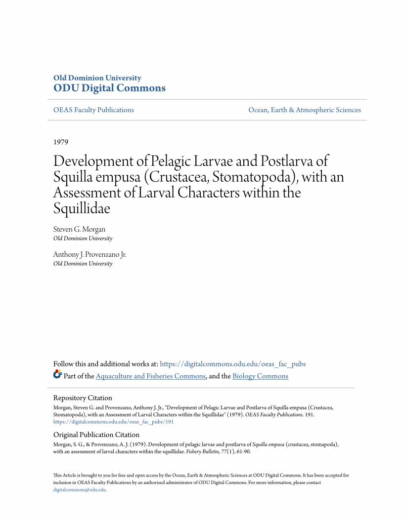
FIGURE 2.-Squilla empusa. A-I:stages I-IX respectively. antennules.
65

FISHERY BULLETIN: VOL. 77. NO.1
G
A
E-'O.5mm
O.25mm
B·P IO.25mm
B
FIaURE 3.-Squilla empusa, A-I: stages I-IX respectively, antennae.
66

MORGAN and PROVENZANO: DEVELOPMENT OF SQUILLA EMPUSA LARVAE AND POSTLARVA
~O.l mm
LE.:!JO.1mm
FIGURE 4.-Squilla empusa, A-I: stages I-IX respectively. mandibles.
FIGURE 5.-Squilla empusa, A-I: stages I-IX respectively, maxillu1es
o
E
H
A-EO.lmm
LL:.LJO.tmm
67

FISHERY BULLETIN: VOL. 77, NO. I
B
D/
E
1 A-E !
O.lmm
L£-UO.lmm
FIGUJm 6.-Squilla empusa, A-I: stages I-IX respect.ively, maxillae.
portion of cutting edge minutely serrate. Epipodpresent.
Third, fourth, and fifth maxillipeds absent.Pereiopods absent.Posterolateral angles of pleomeres rounded.
Pleopods (Figure 9A to 9D) one through four present with appendix interna. Pleopod setation ofthisand other larval stages presented in Table 1.
Sixth pleomere not articulated, submedianspines absent, uropods absent.
Pleotelson (Figure lOA) with paired lateral, intermediate, and posterolateral spines, 4 pairs ofintermediate denticles, 15 submedian dentidesbearing spinules.
Stage II (Figure IB)
Measurements (mm): RL, 1.35 to 1.55; PL,0.70 to 0.90; TL, 4.20 to 4.60; CL, 1.05 to 1.:30; PW,0.55 to 0.75.
Rostrum extending well beyond end ofantennular flagellum, armed with one to four ventralspinules.
Carapace slightly convex, posterolateral spinesextend to anterior section of telson.
Antennule (Figure 2B) as in previous stage.Antenna (Figure 3B) with 10 to 13 plumose
setae, endopod present as incipient bud.
68

MORGAN and PROVENZANO: DEVELOPMENT OF SQUILLA EMPUSA LARVAE AND POSTLARVA
o
A-C ----lO.25mm
O.2Smm
LLLJO.25mm
FIGURE 7.-Squilla empusa, A·I: stages I·IX respectively, first maxillipeds.
69

H
70
F-I1.0mm.
FISHERY BULLETIN: VOL. 77, NO.1
c
FIGURE 8,-Squilla empusa, A-I: stages I-IXrespectively, second maxillipeds.

MORGAN and PROVENZANO; DEVELOPMENT OF SQUILLA EMPUSA LARVAE AND POSTLARV A
O.25mm
{\
G
\
F
H \lI
o
FIGURE 9.-Squilla empusa. A-D: stage I, first to fourth pleopods respectively, E-I: stage II, first to fifthpleopods respectively.
71

FISHERY BULLETIN: VOL. 77, NO. I
TABLE I.-Number of setae on margins ofpIeopods for pelagic larval stages I to IX ofSquilla empusa.Fifth pleopod does not appear until stage V.
Abdominal sOlTliteEndopod Exopod
Stage 2 3 4 5 1 2 3 4 5
I 6 6 6-7 6-7 7 7 7-9 7-9II 6 7-8 7-8 7 7-8 9-10 9-11 9-10III 8-10 9-13 10-14 9-14 9-10 12-13 13-14 11-14IV 12-13 14-15 15-16 15-17 12 16-17 17-18 16-17V 14-15 15-18 16-18 16-20 14-15 17-19 19-20 18-19 7-8VI 16-20 18-25 20-27 20-27 3-8 15-20 21-26 23-29 21-27 12-16VII 22-28 26-28 26-31 26-32 23-28 22-30 26-31 29-33 28-31 20-23VIII 25-29 27-32 28-36 31-38 30-39 24-30 32-36 32-38 31-38 24-31IX 27-31 32-39 35-42 36-45 40-47 36-41 36-41 38-46 37-45 33-39
Mandible (Figure 4B), maxillule (Figure 5Bl,maxilla (Figure 6Bl, and first maxilliped (Figure7B) as in previous stage.
Second maxilliped (Figure 8A) with propodusbearing proximal tooth followed by another largetooth and 19 to 21 denticles along inner margin.
Pleopods one through four (Figure 9E to 9H)increase setation slightly (Table 1). Fifth pleopod(Figure 91l present as bifurcated bud.
Pleotelson (Figure lOB) as in previous stage.
Stage III (Figure 20
Measurements (mm): RL, 1.90 to 2.10; PL,1.00 to 1.10; TL, 5.80 to 6.50; CL, 1.50 to 1.78; PW,0.75 to 0.95.
Rostrum with four to eight spinules ventrallyfor stages III to VIII.
Carapace with lateral margins straight, armedwith two anterior and three posterior spinules allventrally directed, and one median spinule laterally directed. Posterolateral spines extend to posterior region of pleotelson.
Antennule (Figure 2C) with two-segmentedinner flagellum, proximal segment armed withone strong distal seta. Outer flagellum twosegmented with 9 or 10 mesial aesthetascs arranged in three groups of2 or 3, each group with aweak seta.
Antenna (Figure 3C) with 13 to 16 plumosesetae, length of unsegmented endopod increased.
One median ventral spine situated betweenbase of antennules and antennae.
Mandible (Figure 4C) essentially unchanged.Maxillule (Figure 5C) with coxal endite bearing
four to eight teeth.Macilla (Figure 6C) with six to eight setae.First maxilliped (Figure 7C) with 9 or 10 strong
setae arranged in four groups of2 or 3 on propodus,carpus with 1 or 2 strong setae distally.
Second maxilliped (Figure 8C) with propodusbearing 23 to 27 denticles, basis with proximal
72
spine well developed.Third maxilliped present as bud.Pleopods one through four (Figure lIA to lID)
increase setation (Table 1). Fifth pleopod (FigurelIE) biramous, nonsetose.
Sixth pleomere partially articulated, one pair ofsmall submedian spines present. Uropods (Figure10C) present as buds.
Pleotelson (Figure 10C) with 8 to 10 pairs ofintermediate denticles, including 1 pair in axis ofintermediate spines 15 to 27 submedian denticles.
Stage IV (Figure 12)
Measurements (mm): RL, 2.50 to 2.90; PL,1.20 to 1.40; TL, 6.50 to 6.90; CL, 1.80 to 2.25; PW,1.00 to 1.20.
Rostrum with a row of minute spinules distallyfor stages IV to VIII.
Antennule (Figure 2D) with three-segmentedinner flagellum armed with two strong setae ondistal half of proximal segment. Outer flagellumdivided into a two-segmented median flagellum,and a broader outer flagellum with 10 or 11 aesthetascs arranged in four groups of 2 or 3, a smallseta with each group. Antennular somite articulated.
Antenna (Figure 3D) with 17 to 19 plumosesetae, length of unsegmented endopod increasedslightly.
Mandible (Figure 4D) essentially unchanged.Maxillule (Figure 5D) with coxal endite bearing
seven to nine teeth, palp with two setae.Maxilla (Figure 6D) with eight to nine setae.First maxilliped (Figure 7D) with 12 or 13
strong setae arranged in five groups of 2 or 3 onpropodus, carpus with 4 distal setae, merus with 1strong distal seta.
Second maxilliped (Figure 8D) with propodusbearing proximal tooth followed by 2 strong teethin opposition and 25 to 33 dentides along innermargin.
MORGAN and PROVENZANO: DEVELOPMENT OF SQUlLLA EMPUSA LARVAE AND POSTLARVA
',U
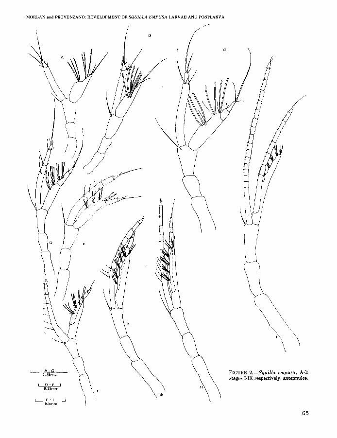
FIGURE lO.-Squilla empusa, A-I, stagesI-IX respectively, tail fans. Setae omittedfrom uropods of stage IX.
F -I1.0mm
A-CO,5mm
L D-EO,5mm
73

F GL----!
O.25mmH
FISHERY BULLETIN: VOL. 77, NO.1
FIGURE lI.-Squilla empusa, A·E: stage III, first to fifth pleopods respectively, F·J: stage IV, first to fifth pleopods respectively.
Third, fourth, and fifth maxiIlipeds (Figure 13Ato 13C) unsegmented buds.
Pereiopods (Figure 14A to 14C) present as buds.Posterolateral angles of pleomeres acute.
Pleopods one through four (Figure 11F to 111) increase setation. Fifth pleopod (Figure 11J) setoseand possessing appendix interna.
Sixth abdominal somite articulated. Uropods(Figure 1OD) biramous.
Pleotelson (Figure 10D) with 1 denticle in axisof lateral spine, 8 or 9 pairs of intermediate denticles, 23 to 30 submedian denticles.
Stage V (Figure 15)
Measurements (mm): RL, 2.50 to 3.25; PL,1.50 to 1.65; TL, 8.50 to 9.00; CL, 2.30 to 2.50; PW,1.40 to 1.60.
Antennule (Figure 2E) with three-segmentedinner flagellum armed with two to four setae ondistal halfofproximal segment; median flagellumalso three-segmented.
74
Antenna (Figure 3E) with 19 to 23 plumosesetae, length of unsegmented endopod now nearlyequal to segments bearing it.
Epistome with small apical spine.Mandible (Figure 4E) essentially unchanged.Maxillule (Figure 5E) with coxal endite bearing
8 to 12 teeth.Maxilla (Figure 6E) with 10 or 11 setae.First maxilliped (Figure 7E) with 13 to 17
strong setae arranged in five or six groups of2 to 4,carpus with 5 to 8 distal setae.
Second maxilliped (Figure 8E) with propodusarmed with 30 to 43 denticles.
Third, fourth, and fifth maxillipeds (Figure 13Dto 13F) still unsegmented, but increased in length.
Pereiopods (Figure 14D to 14F) present as bifurcated buds.
Uropods (Figure IOE) with basal prolongationpresent.
Pleotelson (Figure 10E) with 8 to 10 pairs ofintermediate denticles, 28 to 33 submedian denticles.
MORGAN and PROVENZANO: DEVELOPMENT OF SQUILLA EMPUSA LARVAE AND POSTLARVA
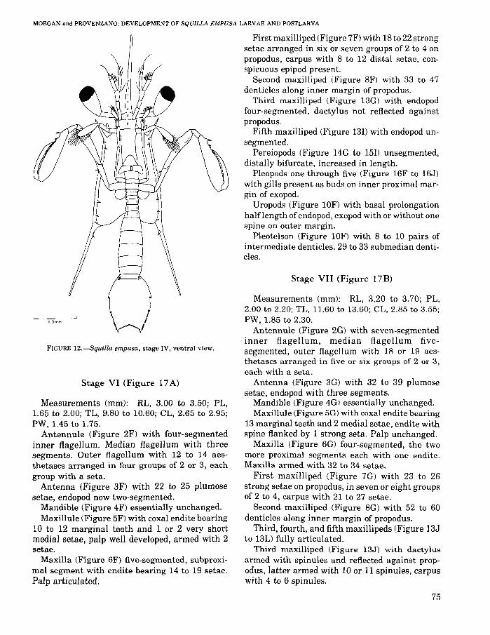
Stage VII (Figure 17B)
Measurements (mm); RL, 3.20 to 3.70; PL,2.00 to 2.20; TL, 11.60 to 13.60; CL, 2.85 to 3.55;PW, 1.85 to 2.30.
Antennule (Figure 2G) with seven-segmentedinner flagellum, median flagellum fivesegmented, outer flagellum with 18 or 19 aesthetascs arranged in five or six groups of 2 or 3,each with a seta.
Antenna (Figure 3G) with 32 to 39 plumosesetae, endopod with three segments.
Mandible (Figure 4G) essentially unchanged.Maxillule (Figure 5G) with coxal endite bearing
13 marginal teeth and 2 medial setae, endite withspine flanked by 1 strong seta. Palp unchanged.
Maxilla (Figure 6G) four-segmented, the twomore proximal segments each with one endite.Maxilla armed with 32 to 34 setae.
First maxilliped (Figure 7G) with 23 to 26strong setae on propodus, in seven or eight groupsof 2 to 4, carpus with 21 to 27 setae.
Second maxilliped (Figure 8G) with 52 to 60denticles along inner margin of propodus.
Third, fourth, and fifth maxillipeds (Figure 13Jto 13L) fully articulated.
Third maxilliped (Figure 13J) with dactylusarmed with spinules and reflected against propodus, latter armed with 10 or 11 spinules, carpuswith 4 to 6 spinules.
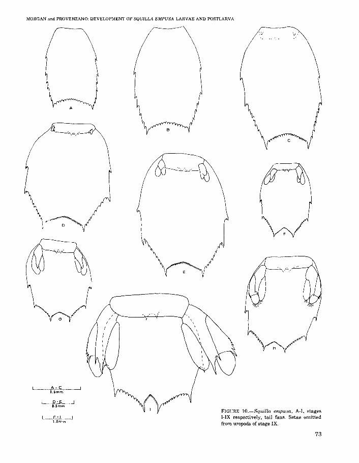
FIGURE 12.--Squilla empusa, stage IV, ventral view.
First maxilliped (Figure 7F) with 18 to 22 strongsetae arranged in six or seven groups of 2 to 4 onpropodus, carpus with 8 to 12 distal setae, conspicuous epipod present.
Second maxilliped (Figure 8F) with 33 to 47denticles along inner margin of propodus.
Third maxilliped (Figure 13G) with endopodfour-segmented, dactylus not reflected againstpropodus.
Fifth maxilliped (Figure 13I) with endopod unsegmented.
Pereiopods (Figure 14G to 15I) unsegmented,distally bifurcate, increased in length.
Pleopods one through five (Figure 16F to 16J)with gills present as buds on inner proximal margin of exopod.
Uropods (Figure 10F) with basal prolongationhalflength of endopod, exopod with or without onespine on outer margin.
Pleotelson (Figure 10F) with 8 to 10 pairs ofintermediate denticles. 29 to 33 submedian denticles.
Stage VI (Figure 17A)
Measurements (mm); RL, 3.00 to 3.50; PL,1.65 to 2.00; TL, 9.80 to 10.60; CL, 2.65 to 2.95;PW, 1.45 to 1.75.
Antennule (Figure 2F) with four-segmentedinner flagellum. Median flagellum with threesegments. Outer flagellum with 12 to 14 aesthetascs arranged in four groups of 2 or 3, eachgroup with a seta.
Antenna (Figure 3F) with 22 to 25 plumosesetae, endopod now two-segmented.
Mandible (Figure 4F) essentially unchanged.Maxillule (Figure 5F) with coxal endite bearing
10 to 12 marginal teeth and 1 or 2 very shortmedial setae, palp well developed, armed with 2setae.
Maxilla (Figure 6F) five-segmented, subproximal segment with endite bearing 14 to 19 setae.Palp articulated.
75

FISHERY BULLETIN: VOL. 77. NO.1
o
R
NM
Q
E
P
O.5mm
L~O.25mm
P-R
~O.Imrn
FIGURE 13.-Squilla empusa, A-C: stage IV,third to fifth maxillipeds respectively; D-F:stage V, third to fifth maxillipeds respectively; G-I: stage VI, third to fifth maxillipedsrespectively; J-L: stage VII, third to fifthmaxillipeds respectively; M-O: stage VIII,third to fifth maxillipeds respectively; P-R:stage IX, third to fifth maxillipeds respectively.
76

MORGAN and PROVENZANO: DEVELOPMENT OF SQUlLLA EMPUSA LARVAE AND POSTLARVA
/."B
P
L
O.5mm
J-Q IO.26mm
~O.1mm
l&.EJO.05mm
P-R
FIGURE 14.-Squilla empusa, A-C: stage IV, first to third pereioJXlds respectively; D-F: stage V,first to third pereiopods respectively; G-I: stage VI, first to third pereiopods respectively; J-L: stageVII, first to third pereiopods respectively; M-O: stage VIII, first to third pereiopods respectively;P-R: stage IX, first to third pereiopods respectively.
Fourth maxilliped (Figure 13K) with dactylusarmed with spinules and partially reflectedagainst propodus, latter armed with four to eightspinules, carpus with four to six spinules, epipodpresent.
Fifth maxilliped (Figure 13L) segmented, dactylus unarmed and not reflected against unarmedpropodus, carpus with zero to three spinules.
Pereiopods (Figure 14J to 14L) with clearly differentiated endopods and exopods, though segmentation not yet distinct.
Pleopods (Figure 18A to 18E) with bilobedrudimentary gills.
Sixth pleomere partially separated from telson.Uropods (Figure lOG) with basal prolongationacute. Exopod with one or two spines on outermargin, armed with zero to five plumose setae.Endopod with zero to two plumose setae.
Pleotelson (Figure lOG) with 9 or 10 pairs ofintermediate denticles, 32 to 35 submedian denticles.
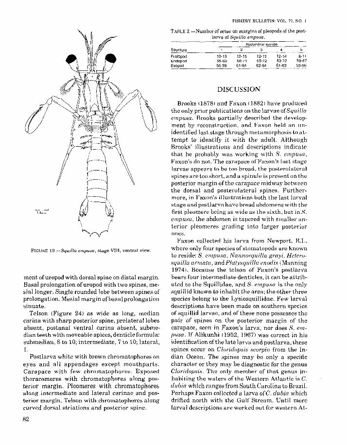
Stage VIII (Figure 19)
Measurements (mm): RL, 3.25 to 3.45; tL, 2.30to 2.50; TL, 13.90 to 15.30; eL, 3.90 to 4.35; TW,2.25 to 2.65.
Antennule (Figure 2H) with inner flagellumbearing 9 to 11 distinctly articulated segments.Median flagellum with five to seven distinct segments. Outer flagellum with 21 to 23 aesthetascsarranged in six or seven groups of 2 or 3, eachgroup with a seta in addition.
Antenna (Figure 3H) with 40 to 55 plumoseseta.
Mandible (Figure 4H) essentially unchanged.Maxillule (Figure 5H) with coxal endite bearing
15 to 19 marginal teeth and 2 to 4 medial setae,distal margin of basis with 1 median seta.
Maxilla (Figure 6H) armed with 40 to 58 setae.First maxilliped (Figure 7H) with 29 to 31
strong setae on propodus arranged in eight groupsof 2 to 5, carpus with 37 to 44 setae.
77

FIGURE 15.-Squilla empusa, stage V, ventral view.
Second maxilliped (Figure 8H) with 61 to 75denticles on propodus.
Third, fourth, and fifth maxillipeds (Figure 13Mto 130) with dactylus armed with spinules andreflected against propodus, epipods present.
Third maxilliped (Figure 13M) with propodusarmed with 12 to 21 spinules, carpus with 6 to 9spinules.
Fourth maxilliped (Figure 13N) with propodusarmed with 12 to 16 spinules, carpus with 5 to 8spinules.
Fifth maxilliped (Figure 130) with propodusarmed with 6 to 11 spinules, carpus with 4 to 7spinules.
78
FISHERY BULLETIN: VOL. 77, NO.1
Pereiopods (Figure 14M to 140) with twosegmented exopods, endopods shorter, unsegmented.
Pleopods (Figure 20A to 20E) with trilobed gills.Sixth abdominal somite completely separated
from telson. Uropods (Figure 10H) with twosegmented exopod. Basal segment with 2 to 4spines on outer distal margin, apical segment with4 to 14 plumose setae on distal margin. Endopod ofuropod with four to eight plumose setae on distalmargin.
Telson (Figure 10H) with 10 pairs of intermediate denticles and 31 to 35 submedian dentides.
Stage IX (Figure 21)
Measurements (mm): RL, 2.40 to 4.50; tL, 2.30to 2.60; TL, 13.00 to 17.50; CL, 3.00 to 3.90; TW,2.10 to 2.80.
Rostrum with decrease in number of spinules,now with two to six spinules; with or without minute distal spinules.
Antennule (Figure 21) with inner flagellumbearing 14 to 20 distinctly articulated segments.Median flagellum with 8 to 11 distinctly articulated segments. Outer flagellum with 23 to 33aesthetascs arranged in seven to nine groups of 2to 7, each group with a seta.
Antenna (Figure 31) with 48 to 60 plumosesetae, endopod with 6 to 9 segments.
Mandible (Figure 41) essentially unchanged.Maxillule (Figure 51) with coxal endite bearing
20 to 25 marginal teeth and 1 or 2 medial setae,margin of basal endite with 1 or 2 setae.
Maxilla (Figure 6U) with 78 to 131 setae.First maxilliped (Figure 71) with 35 to 44 setae
arranged in 9 or 10 groups of2 to 5, carpus with 59to 107 setae, dactylus with or without duster ofsetae on median outer margin, propodus with orwithout cluster of setae on distal outer margin.
Second maxilliped (Figure 81) with 71 to 92 dentides on propodus.
Dactylus of third, fourth, and fifth maxillipeds(Figure 13P to 13R) with or without regularlyspaced setae along outer margin, propodus with orwithout a cluster of setae on distal outer margin.
Third maxilliped (Figure 13P) with propodusbearing 19 to 56 spinules, carpus with 11 to 22spinules.
Fourth maxilliped (Figure 13Q) with propodusbearing 17 to 51 spinules, carpus with 10 to 28spinules.

'FIGURE 16.-Squilla empusa, A·E: stage V, first to fifth pleopods respectively; F·J: stage VI: first to fifth pleopods respectively.

Fifth maxilliped (Figure 13R> with propodusbearing 18 to 40 spinules, carpus with 9 to 20spinules,
Pereiopods (Figure 14P to 14R) slender with orwithout distal segment of exopods setose,
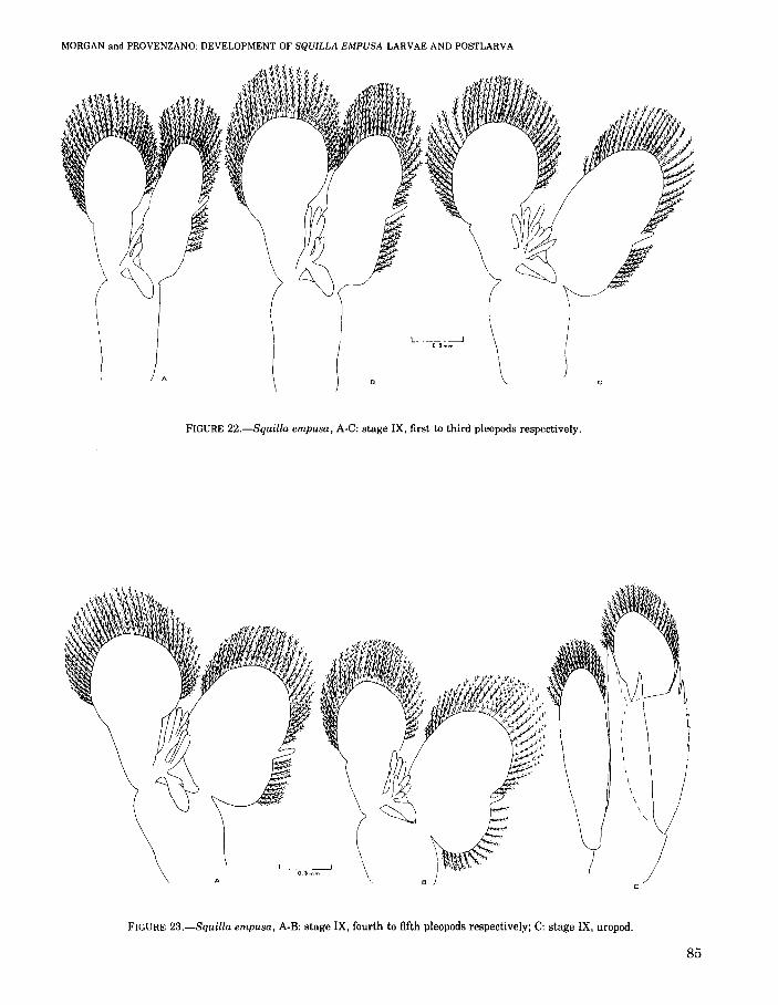
Pleopods (Figures 22A to 22C; 23A, 2:38) withdistal lobe of gill pinnate,
Uropod (Figure 26C) with basal segment ofexopod armed with 6 to 8 spines, apical segment ofexopod with 17 to 60 plumose setae. Endopod ofuropod with 10 to 38 plumose setae. Inner spine ofbasal prolongation with blunt spine on outer proximal margin. Basal uropod segment with a dorsalspine on distal margin.
Telson (Figure 14Il with 8 to 10 pairs of intermediate denticles, 26 to 34 submedian denticles.
Postlarva (Figure 24)
Measurements (mm): RL, 0.50 to 0.60; CL,2.90 to 3.30; TW, 2.55 to 3.10; RW, 0.65 to 0.75; tL,1.95 to 2.55; TL, 12.3 to 14.20.
Eyes large, extending to middle of second segment of antennular peduncle. Cornea bilobed, set
80
VISHEHY BULLETll'I: VOL. 77, NO.1
FIGURE 17.-Squilla empusa, A-B:
stages VI and VII respectively. ventralviews,
obliquely on stalk. Ocular scales rounded, anteriormargin of opthalmic somite evenly rounded.
Antennular process produced into blunt spinedirected anterolaterally, antennular peduncleslightly shorter than carapace, antennule (Figure25A) with inner flagellum bearing 34 segments,median flagellum with 30 segments, outer flagellum with 15 segments and 22 aesthetascs arranged in eight groups of 2 or 3.
Antenna (Figure 25B) with 63 to 75 plumosesetae, endopod with 16 segments.
Rostral plate wider than long, lateral marginstapering to rounded apex. Median carina present.
Anterolateral angle of carapace without spine,almost forming right angle, posterolateral margins broadly rounded, carinae poorly developed,median carina not bifurcate anteriorly or posteriorly, intermediate and lateral carinae present,reflected carinae absent.
Mandible (Figure 25C) serrate, mandibularpalp absent.
Maxillule (Figure 25D) with coxal endite bearing 26 to 27 strong marginal teeth and 6 to 9 smallmedial teeth. Basal endite with one spine flanked

MORGAN and PROVENZANO: DEVELOPMENT OF SQUILLA EMPUSA LARVAE AND POSTLARVA
FIGURE 18.-Squilla empusa, A-E:
stage VII, first to fifth pleopods respectively.
by one strong seta. Distal margin of basis withthree setae. Endopod present as palp on distalmargin of basis, armed with two setae.
Maxilla (Figure 25E) four-segmented, two proximal segments with endites, second bilobed.
Five pairs of maxillipeds (Figure 25F to 25J)each maxilliped with one epipod. First maxilliped(Figure 25F) with distal margin of propodus bearing 14 teeth, inner margin with 48 to 50 strongsetae arranged in 10 transverse rows, 2 or 3 mostdistal setae spatulate with strong setules.
Second maxilliped (Figure 25G) with dactylusbearing six teeth, pectinate propodus with threemoveable proximal spines, dorsal ridge of carpusundivided.
Pereiopods (Figure 26A) with setose endopodand exopod.
Last three thoracic somites with unarmed submedian and intermediate carinae. Lateral processof fifth thoracic somite subacute, sloping pos-
teriorly. Lateral processes of next two somitesbilobed each with a small anterior lobe and a largebroadly rounded posterior lobe. Median ventralkeel of eighth somite with rounded apex.
Abdomen broad, depressed, Submedian, intermediate, lateral, and marginal carinae present.Abdominal spines in submedian carinae of sixthsomite, intermediate and lateral carinae of fifthand sixth somites, and marginal carinae of fifthsomite, formula: submedian 6; intermediate 5 to 6;lateral, 5 to 6; marginal, 5. Sixth abdominal somite with sharp ventral spine anterior to uropodarticulation.
Pleopods (Figure 26B to 26F) with gills. Pleopodsetation presented in Table 2.
Uropod (Figure 26G) with eight graded moveable spines on outer margin of proximal segmentof exopod, last extending to middle of apical segment. Apical segment of exopod extending posteriorly to apex of intermediate spine. Basal seg-
81
FISHERY BULLETIN: VOL. 77, NO.1
TABLE 2,-Number of setae on margins of pleopods of the post·larva of Squilla empusa.
Brooks (1878) and Faxon (1882) have producedthe only prior publications on the larvae ofSquillaempusa. Brooks partially described the development by reconstruction, and Faxon held an unidentified last stage through metamorphosis to attempt to identify it with the adult. AlthoughBrooks' illustrations and descriptions indicatethat he probably was working with S. empusa,Faxon's do not. The carapace of Faxon's last stagelarvae appears to be too broad, the posterolateralspines are too short, and a spinule is present on theposterior margin of the carapace midway betweenthe dorsal and posterolateral spines. Furthermore, in Faxon's illustrations both the last larvalstage and postlarva have broad abdomens with thefirst pleomere being as wide as the sixth, but inS.elllpusa, the abdomen is tapered with smaller anterior pleomeres grading into larger posteriorones.
Faxon collected his larva from Newport, R.I.,where only four species of stomatopods are knownto reside: S. elllpusa. Nannosquilla grayi, Helemsquilla armata, and P!atysqu ilia enod is (Manning1974). Because the telson of Faxon's postlarvabears four intermediate denticles, it can be attributed to the Squillidae, and S. empusa is the onlysquillid known to inhabit the area; the other threespecies belong to the Lysiosquillidae. Few larvaldescriptions have been made on southern speciesof squillid larvae, and of these none possesses thepair of spines on the posterior margin of thecarapace, seen in Faxon's larva, nor does S. elllpusa. If Alikunhi (1952, 1967) was correct in hisidentification of the late larva and postlarva, thesespines occur on Cloridopsis scorpio from the Indian Ocean. The spines may be only a specificcharacter or they may be diagnostic for the genusCloridopsis. The only member of that genus inhabiting the waters of the Western Atlantic is C.dubia which ranges from South Carolina to Brazil.Perhaps Faxon collected a larva ofC. dubia whichdrifted north with the Gulf Stream. Until morelarval descriptions are worked out for western At-
DISCUSSION
8-1159-6753-56
4
12·1463·7261·63
3
12·1565·7262-64
Abdominal somite
2
12·1560-7161-64
12-1555-6055-59
ProtopodEndopodExopod
Structure
FIGURE 19.-Squilla empusa, stage VIII, ventral view.
ment ofuropod with dorsal spine on distal margin.Basal prolongation ofuropod with two spines, mesial longer. Single rounded lobe between spines ofprolongation. Mesial margin ofbasal prolongabonsinuate.
Telson (Figure 24) as wide as long, mediancarina with sharp posterior spine, prelaterallobesabsent, postanal ventral carina absent, submedian teeth with moveable apices, denticle formula:submedian, 8 to 10; intermediate, 7 to 10; lateral,1.
Postlarva white with brown chromatophores oneyes and all appendages except mouthparts.Carapace with few chromatophores. Exposedthoracomeres with chromatophores along posterior margin. Pleomeres with chromatophoresalong intermediate and lateral carinae and posterior margin. Telson with chromatophores alongcurved dorsal striations and posterior spine.
82

MORGAN and PROVENZANO: DEVELOPMENT OF SQUILLA EMPUSA LARVAE AND POSTLARVA
D
FIGURE 20.-Squilla empusa, A-E:stage VIII, first to fifth pJeopods respectively.
lantic species of stomatopods, the identity of Faxon's larva will remain uncertain.
To identify larvae of S. empusa the spinules ofthe carapace and dentides of the telson should beexamined. Stages I and II possess four spinules onthe lateral margin of the carapace and four intermediate denticles. The third to ninth stages arearmed with six spinules on the lateral margin of
the carapace. There are two anterior and threeposterior spinules all ventrally directed, and onemedian spinule laterally directed. The telsons ofstages III to IX have 8 to 10 intermediate dentides.
Except for Provenzano and Manning (1978),who reared Gonadactylus oerstedii from hatchingto metamorphosis, experimenters who have at-
83

FIGURE 21.-Squilla empusa, stage IX, ventral view.
tempted to hatch and rear larvae either to linkthem with an adult or to describe the entire larvaldevelopment have been unsuccessful at rearinglarvae past the first pelagic stage because the larvae could not be induced to feed (Manning andProvenzano 1963). Pyne (1972) was unable to rearPterygosquilla armata schizodontia eggs past thefirst pelagic stage, but did hold stages I to VIIlarvae taken from the plankton for periods as longas 10 to 16 days wherein the larvae passed throughat least one ecdysis. Pyne also found it possible tokeep later stage larvae for very much longerperiods of up to 165 days during which time theymolted as many as six times. Pyne reared his larvae in mass culture using 4-in (10.2-cm) finger
L-__-------J
1,Omm
FISHERY BULLETIN: VOL, 77. NO. I
bowls. Alikunhi (1975) reared planktonic larvae ofOratosquilla nepa in aquaria through metamorphosis until they reached adulthood, bred, andproduced eggs.
The manner in which all species of Squillidaedevelop is similar. All Squillidae hatch aspseudozeae with four pairs ofpieopods and developinto the alima form. Some, ifnot all, pass throughtwo propelagic stages before the first trulyplanktonic stage. The alima is characterized by atelson with four or more intermediate dentides,the distance between the submedian spines inlater stages being not larger than that betweenthe intermediate and submedian spines, the propodus ofthe second maxilliped bearing three basalspines, the antennular somite generally having amedian spine, the posterolateral spine of thecarapace having a basal accessory spine, the eyestalks long, and the exopod of the uropod beinglonger than the endopod (Gurney 1942, 1946).Alikunhi (1952) added that alima larvae possesscarapaces armed with a varying number ofspinules on the lateral margins, the sixth abdominal somite usually being equipped with a pair ofsubmedian dorsal spines, and in advanced larvae,the posterolateral angles of the abdominal somitesending in acute or subacute spines.
Alikunhi (1952) noted that between alliedspecies, the specific differences are often "trivial"but remarkably constant. He determined thatsome features, such as the size of the final pelagicstage, the shape and spinulation of the carapace,telson, and uropods, and the presence or absence ofteeth other than the terminal on the dactylus ofthe second maxilliped, hardly show any variationwithin a species. These characters may be used forspecific determinations but are presently of littleaid in defining generic alliances for three reasons.
First, relatively few stomatopods have been associated definitely with the adult of the species.
Second, most of these have had described onlyone larval stage of the entire development. Only19 of the Squillidae have been definitely connectedwith their larval forms. Provenzano and Manning(1978) listed 17 species of identified stomatopodlarvae, but O. massavensis was omitted and S.empusa has now been added to the list. Of the 19species, only 2, P. armata schizodentia and S. empusa, have been reared in the laboratory throughessentially their entire pelagic development. Twoadditional species have been hatched from eggsobtained from a known adult and the first pelagicstage described, i.e., Clorida choprai by Gurney
84
MORGAN and PROVENZANO: DEVELOPMENT OF SQUlLLA EMPUSA LARVAE AND POSTLARVA
L_. -----lQ.5mm
A
FIGURE 22.-Squilla empusa, A-C: stage IX, first to third pleopods respectively.
O.5mmA
c
I\\
\\\
\ ,
Jc
FIGURE 23.-Squilla empusa, A-B: stage IX, fourth to fifth pleopods respectively; C: stage IX, uropod.
85
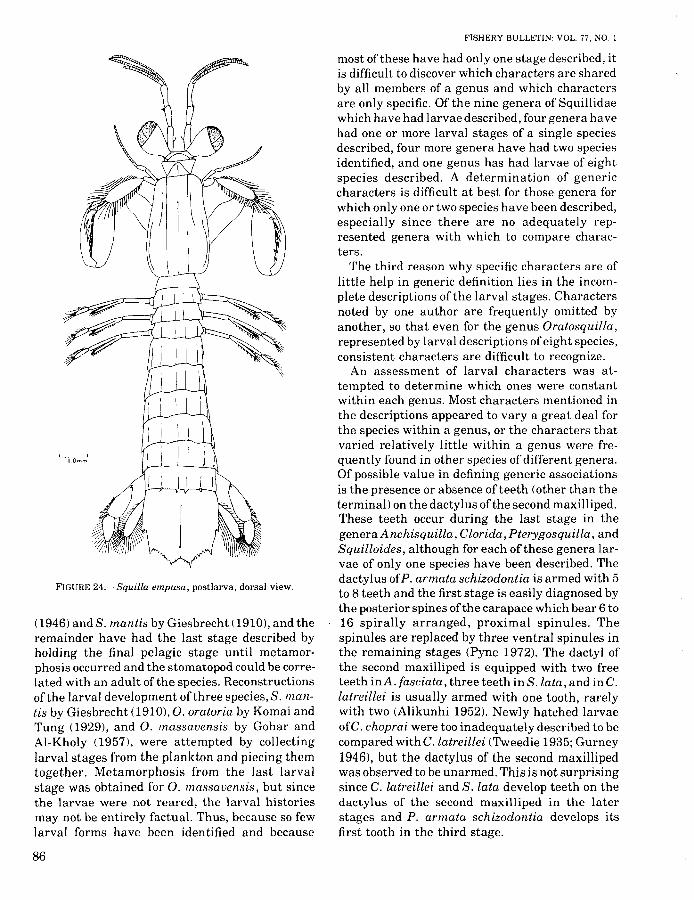
FIGURE 24.-Squilla empusa, postlarva, dorsal view.
(1946) andS. mantis by Giesbrecht (1910), and theremainder have had the last stage described byholding the final pelagic stage until metamorphosis occurred and the stomatopod could be correlated with an adult of the species. Reconstructionsof the larval development of three species, S. mantis by Giesbrecht (1910), O. oratoria by Komai andTung (1929), and O. massavensis by Gohar andAI-Kholy (1957), were attempted by collectinglarval stages from the plankton and piecing themtogether. Metamorphosis from the last larvalstage was obtained for O. massavensis, but sincethe larvae were not reared, the larval historiesmay not be entirely factual. Thus, because so fewlarval forms have been identified and because
86
FISHERY BULLETIN: VOL. 77. NO.1
most of these have had only one stage described, itis difficult to discover which characters are sharedby all members of a genus and which charactersare only specific. Of the nine genera of Squillidaewhich have had larvae described, four genera havehad one or more larval stages of a single speciesdescribed, four more genera have had two speciesidentified, and one genus has had larvae of eightspecies described. A determination of genericcharacters is difficult at best for those genera forwhich only one or two species have been described,especially since there are no adequately represented genera with which to compare characters.
The third reason why specific characters are oflittle help in generic definition lies in the incomplete descriptions of the larval stages. Charactersnoted by one author are frequently omitted byanother, so that even for the genus Oratosquilla,represented by larval descriptions of eight species,consistent characters are difficult to recognize.
An assessment of larval characters was attempted to determine which ones were constantwithin each genus. Most characters mentioned inthe descriptions appeared to vary a great deal forthe species within a genus, or the characters thatvaried relatively little within a genus were frequently found in other species of different genera.Of possible value in defining generic associationsis the presence or absence of teeth (other than theterminal) on the dactylus of the second maxilliped.These teeth occur during the last stage in thegenera Anchisquilla, Clorida, Pterygosquilla, andSquilloides, although for each of these genera larvae of only one species have been described. Thedactylus ofP. armata schizodontia is armed with 5to 8 teeth and the first stage is easily diagnosed bythe posterior spines ofthe carapace which bear 6 to16 spirally arranged, proximal spinules. Thespinules are replaced by three ventral spinules inthe remaining stages (Pyne 1972). The dactyl ofthe second maxilliped is equipped with two freeteeth inA. fasciata, three teeth inS. lata, and inC.latreillei is usually armed with one tooth, rarelywith two (Alikunhi 1952). Newly hatched larvaeofC. choprai were too inadequately described to becompared withG.latreillei (Tweedie 1935; Gurney1946), but the dactylus of the second maxillipedwas observed to be unarmed. This is not surprisingsince C. latreillei and S. lata develop teeth on thedactylus of the second maxilliped in the laterstages and P. armata schizodontia develops itsfirst tooth in the third stage.

MORGAN and PROVENZANO: DEVELOPMENT OF SQUILLA EMPUSA LARVAE AND POSTLARVA
A-Bf-J I1.0m01
C-EO.25mm
FIGURE 25.-Squilla empusa, postlarva, A. antennule; B.antenna; C, mandible; D. maxillule; E. maxilla; F-J. first tofifth maxillipeds respectively.
87

88
FISHERY BULLETIN: VOL. 77, NO.1
FIGURE 26.-Squilla empusa, postlarva, A, first pereipod; B-F, first to fifth pleopods respectively; G, uropod.

MORGAN and PROVENZANO: DEVELOPMENT OF SQUILlA EMPUSA LARVAE AND POSTLARVA
The second maxilliped of the remaining described larvae is unarmed throughout the larvaldevelopment. To distinguish these genera, othercharacters, such as the presence or absence of aspine on the basis of the second maxilliped, mustbe relied on. The spine is definitely born by sevenof the eight species of Oratosquilla, but was notmentioned for O. massauensis. Other species,Squilla empusa, P. armata schizodontia, Alimahyalina, and Meiosquilla lebouri have the spine,while Harpiosquilla harpax and A. fasciatadefinitely do not.
The development of epipods on five pairs ofmaxillipeds in older larvae appears to be a genericcharacter ofSquilla as most other genera bear fourpairs of epipods.
Characters such as rostral length and spinulation, carapace and telson shape, size, and spinulation, and overall body size and appearance havebeen too variable within the limited number ofspecies presently described to use them in defininggeneric associations of the larvae. Derivingcharacters which apply to the youngest larvae aswell as the old will be difficult since far fewercharacters are present in the early stage larvae,and the gross appearance of the young larvae isvery similar due to the small degree of differentiation. Other characters such as antennular segmentation, mouthpart morphology, setation, spination of the maxillipeds, or the presence ofocular, antennular, epistomal, or basal uropodalspines may also need to be examined. The setationand spination of the first maxilliped may be ofgreat value in defining alliances of the species aswell as in making specific determinations. However, many more complete descriptions of the larval developments undergone by the variousspecies must be accomplished before larvalcharacters can be used in establishing generic relationships.
The postlarva of Squilla empusa exhibited thebasic features of first stage postlarva as determined for other species by Alikunhi (1967). Theseinclude the absence of anterolateral spines on thecarapace, the extremely poorly developed carination of the carapace, acutely pointed marginaldenticles of the telson, and moveable apices of thesubmedian spines of the telson. As with the adult,the postlarva possesses the full complement ofteeth on the raptorial dactylus, just as Alikunhi(1967) found. Furthermore, the five pairs ofepipods found in the adult are also possessed bythe postlarva. Other adult characters were de-
veloped upon the next molt. The dorsal carinationsof the carapace were developed, the lateral processes of the exposed thoracic somites five througheight resembled those of the adult, the marginaldenticles of the telson were not as acute, and thesubmedian spines were fixed. The abdominal spinal formula was still not equal to that ofthe adult.Nevertheless, after the postlarva had undergoneits first molt more than enough characters wereshared with the adult to make a definite determination of the species.
CONCLUSIONS
1. Squilla empusa undergoes nine pelagicstages before attaining the postlarval stage.
2. The last stage stomatopod larva and postlarva described by Faxon (1882) are not S.empusa.
3. Larvae ofS. empusa may be identified by thespinules of the carapace and the intermediate denticles of the telson. Stages I andII possess four spinules on the lateral marginof the carapace and four intermediate denticles. The third to ninth stages are armed withsix spinules on the lateral margin of thecarapace. There are two anterior and threeposterior spinules all ventrally' directed, andone median spinule laterally directed. Thetelsons of stages III to IX have 8 to 10 intermediate denticles.
4. Rostral length and spinulation, carapace andtel son size and spinulation, and overall bodysize and appearance probably are specificrather than generic characters.
5. The presence or absence of teeth on the dactylus of the second maxilliped, the presenceor absence of a spine on the basis of the second maxilliped, and the number of epipodsmay all be useful characters in determininggeneric status of larvae belonging to theSquillidae. However, many more completedescriptions of the larval developments undergone by the various species are neededbefore larval characters can be used in establishing generic relationships.
ACKNOWLEDGMENTS
We are indebted to the National Science Foundation for its support of this work under grantDEB76-11716 to the Old Dominion University Research Foundation.
89

LITERATURE CITED
ALIKUNHI, K. H.1952. An account of the stomatopod larvae of the Madras
plankton. Rec. Indian Mus. (Calcutta) 49:239-319.1967. An account of the post-larval development, moult
ing, and growth ofthe common stomatopods ofthe Madrascoast. In Symposium on Crustacea, Ernakulam, India,1965, p. 824-939. Mar. Bio\. Assoc. India, Symp. Ser. 2.
1975. Studies on Indonesian stomatopods 1. Growth,maturity and spawning of Squilla nepa. Bul\. ShrimpCult. Res. Cent. 1(1):27-32.
BROOKS. W. K.1878. The larval stages of Squilla empusa Say. Johns
Hopkins Univ., Chesapeake Zoo\. Lab. Sci. Res.1878:143-170.
BURROWS. M.1969. The mechanics and neural control of the prey cap
ture strike in the mantid shrimpsSquilla andHemisquillao Z. vg\. Physio\. 62:361-381.
DRAGOVICH. A.1970. The food of skipjack and yellowfin tunas in the At
lantic Ocean. U.S. Fish Wild\. Serv., Fish. Bul\. 68:445460.
FAXON. W.1882. I.-Crustacea. In A. Agassiz, W. Faxon, and E. L.
Mark (compilers), Selections from embryological monographs. Mem. Mus. Compo Zoo\. (Harv. Univ.) 9(1), 14plates.
FISH, C. J.1925. Seasonal distribution of the plankton of the Woods
Hole region. Bul\. [U.S.] Bur. Fish. 41:91-179.GIESBRECHT. W.
1910. Stomatopoden. Erster Thei\. Fauna Flora Golfesvon Neapel, Monogr. 33,239 p.
GOHAR. H. A. F., AND A. A. AL-KHOLY.
1957. The larval stages of three stomatopod Crustacea(from the Red Sea). Pub\. Mar. Bio\. Stn., Al-Ghardaqa(Red Sea) 9:85-130.
GURNEY, R.1942. Larvae of decapod Crustacea. Ray Soc. (Lond.)
Pub\. 129, 306 p.1946. Notes on stomatopod larvae. Proc. Zoo\. Soc. Lond.
116(1):133-175.HILDEBRAND, H. H.
1954. A study of the fauna of the brown shrimp (J>enaeusaztecus Ives) grounds in the western Gulf ofMexico. Pub\. Inst. Mar. Sci., Univ. Tex. 3:233-366.
90
FISHERY BULLETIN: VOL. 77, NO. I
KAESTNER. A.1970. Invertebrate zoology. Vo\. 3, Crustacea. Wiley In
terscience, N.Y., 523 p.KOMAI. T., AND Y. M. TUNG.
1929. Notes on the larval stages of Squilla oratoria, withremarks on some other stomatopod larvae found in theJapanese Seas. Annot. Zoo\. Jpn. 12:187-214.
LEBOUR, M. V.1924. Young anglers in captivity and some of their
enemies. A study in a plunger jar. J. Mar. Bio\. Assoc.U.K. 13:721-734.
MACGINITlE, G. E., AND N. MACGINITIE.
1968. Natural history of marine animals. 2ded. McGraw-Hill, N.Y., 523 p.
MANNING, R. B.1969. Stomatopod Crustacea of the western Atlan
tic. Stud. Trop. Oceanogr. (Miami) 8, 380 p.1974. Marine flora and fauna of the northeastern United
States. Crustacea: Stomatopoda. U.S. Dep. Commer.,NOAA Tech. Rep. NMFS CIRC-387, 6 p.
MANNING. R. B., AND A. J. PROVENZANO. JR.
1963. Studies on development of stomatopod Crustacea I.Early larval stages of Gonodactylus oerstedii Hansen. Bull. Mar. Sci. Gulf Caribb. 13:467-487.
PICCINETTI, C., AND G. P. MANFRIN.
1970. Prime osservazioni sull'alimentazione di Squillamantis L. Bologna Univ. Inst. Zoo\. Note 3(10):251-263.
PROVENZANO. A. J., JR., AND R. B. MANNING.
1978. Studies on development of stomatopod Crustacea II.The later larval stages of Gonodactylus oerstedii Hansenreared in the laboratory. Bull. Mar. Sci. 28:297-315.
PYNE, R. R.1972. Larval development and behaviour of the mantis
shrimp Squilla armata Milne Edwards (Crustacea:Stomatopoda). J. R. Soc. N.Z. 2:121-146.
RANDALL. J. E.1967. Food habits of reef fishes of the West Indies. Stud.
Trop. Oceanogr. (Miami) 5:665-847.REINTJES, J. W., AND J. E. KING.
1953. Food ofyellowfin tuna in the central Pacific. U.S.Fish Wild\. Serv., Fish. Bull. 54:91-110.
SUNIER, A.
1917. The Stomatopoda of the collection "Visscherijstation" at Batavia. Contrib. Faune lndes Neerl. 1(4):6275.
TWEEDIE. M. W. F.1935. Two new species of Squilla from Malayan waters.
Bull. Raffles Mus. 10:45-52.