development of methods for identification of hemoglobin - enerca
TRANSCRIPT

Development of methods for identification of hemoglobin disorders (f P li t l l bi l )”(from Pauling to molecular biology)”
H i W jHenri Wajcman
INSERM U955INSERM U955 Hôpital Henri Mondor Créteil

The first identifications of Hb disorders1904-1925
In 1925 Cooley observedIn 1904, Herrick observed sickle-shaped red blood cells on the blood film of
In 1925, Cooley observed a specific anemia in children from Italian origin.cells on the blood film of
Walter Clement Noel a medical student from Grenada.
In 1943 Bianco and Silvestroni described a test based on decreasedtest based on decreased osmotic fragility which, together with RBC morphology, and RBC
t ll d fcount allowed for diagnosis of thalassemia.

Hematological and clinical observationsHematological and clinical observations
Electrophoretical studies Chromatographic studies
Moving boundary electrophoresis (1937 Tiselius)
Electrophoretical studies Chromatographic studies
Chromatography on CM or DEAE cellulose Zone electrophoresis ( adapted to Hb around 1955) (on paper, starch gel, or cellulose acetate)
Isoelectric focusing ( around 1960)
columns (around 1975)
CE-HPLC (1993)Isoelectric focusing ( around 1960)
Capillary electrophoresis (around 1980)RP-HPLC (2000)
Mass spectrometry studies
Electrospray mass spectrometry
Molecular biology studies
DOT blot, RFLP…
MALDI-TOF MS DNA sequencing

Moving boundary electrophoresis (1949)
Hb phenotype studies started in 1949 with Linus Pauling h i h h bili f HbS diff f h fshowing that the mobility of HbS was different from that of
HbA during liquid electrophoresis. At this time very few apparatus were available.
Using this approach it was clamed that SCD was a « molecular disease » but this test was much too laborious to be of practical use. Rapidly other methods, electrophoretical or chromatographical were developed
Hb A Hb S
electrophoretical or chromatographical were developed.
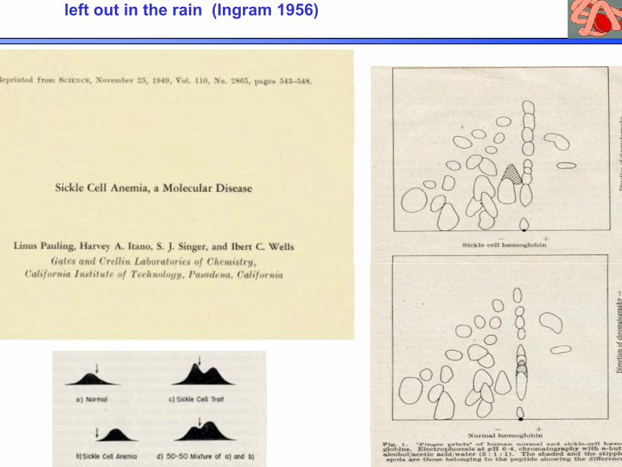
…our first fingerprints looked like a modern watercolour left out in the rain (Ingram 1956)

Typical Hb analysis in 1955
Examples of zone electrophoresis on paper:
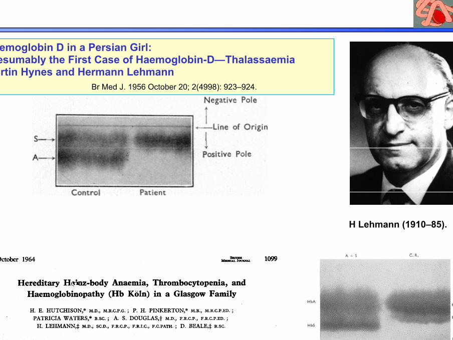
Haemoglobin D in a Persian Girl: Presumably the First Case of Haemoglobin-D—ThalassaemiaPresumably the First Case of Haemoglobin-D—ThalassaemiaMartin Hynes and Hermann Lehmann
Br Med J. 1956 October 20; 2(4998): 923–924.
H Lehmann (1910–85).

Zone electrophoresis on starch gel
Principle of ZE
Hb AHb FHb S
Hb C

Zone electrophoresis on Cellulose Acetate at Alkaline pH
+ Hb A 0+Hb F
Hb A
Hb S, Hb D
0
Hb C, Hb E,Hb A2
Carbonicanhydrase
- 10
Scale to compare mobilities
Cellulose acetate is a highly pure medium described in 1957. It was li d thi f il L t f t i li d St i i
Scale to compare mobilities
applied on very thin foil. Low amounts of protein were applied. Staining and drying were easy.At pH 8.5, Hb is negatively charged and migrates towards the cathode.All the Hbs carrying an identical charge have the same migrationAll the Hbs carrying an identical charge have the same migration. This technique has not a high resolution: variants with an identical difference in charge move on an almost similar way.

Determination of the HbA2 level by densitometry scanning
1958: from starch gel 2009 f S bi H d l1958: from starch gel 2009: from Sebia HydragelUsing GELSCAN
Goldberg Clinical Chemistry 1958

1961-1990 : Isoelectricfocusing
IEF 2011

Capillary electrophoresis (Sebia)
Normaladult
HbA/HbS
Mixture of Hbs
HbA/Hb J-Tongarikiα 115Ala>Asp
HbA/HbC HbA/Hb Matsue Okiα 75 Asp>Asn
A few examples

The chromatographic approach ….
1903Invention of chromatography 1903
1941Partition chromatography
1941
Gradient elution 1952
First commercial LC instruments 1969
First commercial HPLC instruments 1973

The chromatographic approach
19031903

The chromatographic approach to diagnosis of Hb disorders
First tentatives

Column 1 x 25 cmFlow rate 15 ml/h> 48 h per analysis
Hb J-Paris α12 Ala>Asp
Hb I α16 Lys>Glu

α Chain variants
Column 1 x 25 cmFlow rate 15 ml/h> 48 h per analysis
Hb G-Georgia α95 Pro>Leu
Hb G Phil d l hi 68 A >LHb G-Philadelphia α68 Asn>Lys

1975-1980 :Microcolumn chromatography for measurement of minor Hb fractions
Hb A2 DEAE ll l lHb A2 : DEAE-cellulose columnseluted with 0.2 M glycine-0.01% KCN at pH around 7.5
Hb F : CM-cellulose columns
Drawbacks of these methods:
• manual techniqueHb F : CM cellulose columnseluted with Tris-HCl or bis tris buffer
Hb A1c : Bio-Rex 70 columns
• sensitive to minimal pH differences
iti t t teluted with phosphate buffers • sensitive to temperature

Evidence for Thal
ββ
α
CM-cellulose chromatography of globin in 8M urea
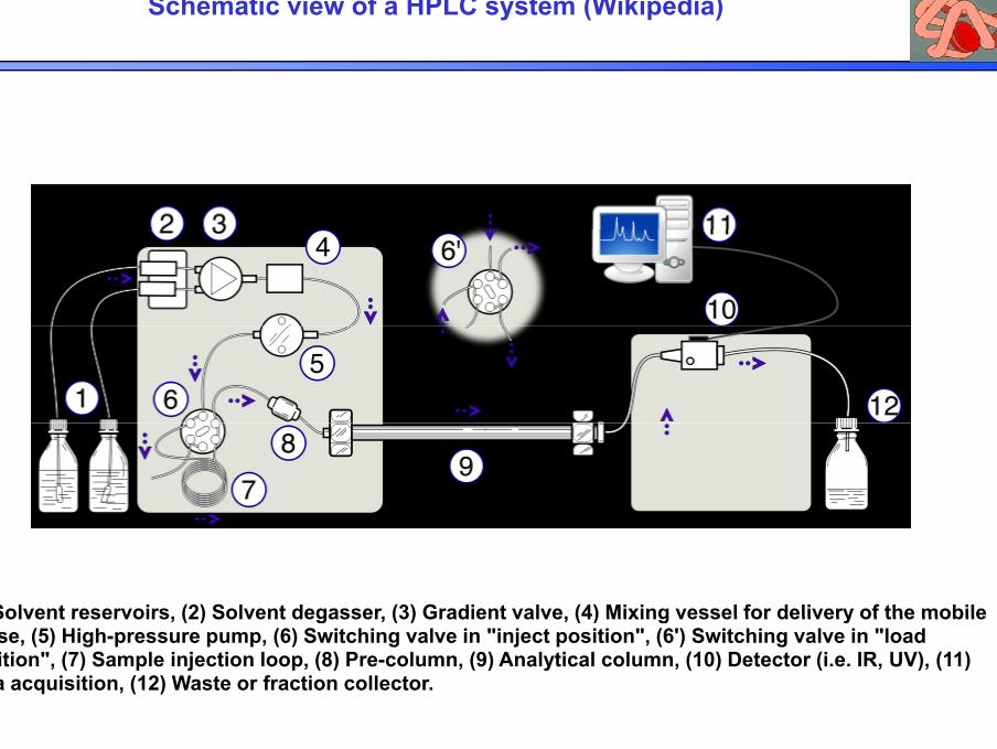
Schematic view of a HPLC system (Wikipedia)
(1) Solvent reservoirs, (2) Solvent degasser, (3) Gradient valve, (4) Mixing vessel for delivery of the mobile(1) Solvent reservoirs, (2) Solvent degasser, (3) Gradient valve, (4) Mixing vessel for delivery of the mobile phase, (5) High-pressure pump, (6) Switching valve in "inject position", (6') Switching valve in "load position", (7) Sample injection loop, (8) Pre-column, (9) Analytical column, (10) Detector (i.e. IR, UV), (11) Data acquisition, (12) Waste or fraction collector.
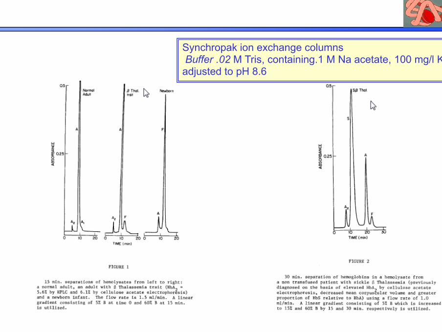
TThe first HPLC separations of Hbs have been performed on AE-columns
Synchropak ion exchange columnsBuffer 02 M Tris containing 1 M Na acetate 100 mg/l KCNBuffer .02 M Tris, containing.1 M Na acetate, 100 mg/l KCNadjusted to pH 8.6
S.M. Hanash and D.N. Shapiro,Hemoglobin 5(2),65-175 (1981)

HPLC separations of Hbs on CE-columns
Description of the polyCAT Asp columns (1983)Description of the polyCAT Asp columns (1983)Practical use of these columns for measurement of glycated Hbs (1984)
U i 13 i di t b t t bil h th th dOu C-N, Rognerud CLCLIN. CHEM. 39/5, 820-824 (1993)
Using a 13-min gradient between two mobile phases, the method could separate more than 35 commonly encountered hemoglobin variants within 12 min.

Today analysis of the hemolysate byCE-HPLCCE HPLC

Characterization of a Hb variant
Finger print on paperFinger print on paper followed by elution of the spot, amino-acid composition, Edman degradation
Finger print on silicagel plates
1960
Finger print on silicagel platesfollowed by elution of the spot, amino-acid composition, Edman degradation
Ion-exchange chromatography of peptides
1970
Ion exchange chromatography of peptidesfollowed by collection of the peptide, amino-acid composition, Edman degradation
RP-HPLC of peptides
1980
p pfollowed by collection of the peptide, amino-acid composition, Edman degradationOr mass-spectrometry studies
1990
DNA sequencing 2000

Home made cation exchange chromatography instrument for separation of tryptic peptides (RT Jones, 1967)
• Column: Dowex WX8 (strong acid cation resin)• Elution: pyridine-aceticElution: pyridine acetic acid gradient• Ninhydrin staining of a part of the eluted volume• 12h chromatography g p y
Example of profile obtained

Separation of tryptic peptides
Fingerprint on silicagel plates
RP- HPLC

Mass spectrometry analysis of a Hb varianty y
Isolation of the abnormal globin chain
by semi-preparative RP-LC
Collecting and dryness (no salts)
ESI-MS of abnormal Interpretation : abnormal mass anddryness (no salts)
evaporationglobin chain abnormal mass and
mass variation
Tryptic digestion
Peptide mass Interpretation :
Tryptic digestPeptide mass
fingerprint by MALDI-TOF MS
abnormal peptide/abnormal
cleavage
Separation of tryptic peptides by
RP LC
nanoLC-MS/MS of peptides
Interpretation : peptide sequencing
RP-LC

Molecular characterization of β-thalassemia mutations
C t l l t h i i l dCurrent molecular techniques include:
1- Methods to confirm a suggested abnormality:amplification refractory mutation system (ARMS)amplification refractory mutation system (ARMS), dot blot hybridization, restriction enzyme analysis ,reverse dot blot hybridizationreverse dot blot hybridization, allele specific oligonucleotide (ASO) hybridization, GAP PCR,
2- Methods to identify an unknown abnormality
DNA Sequencing.

Further identification of beta-thalassemia by DOT blot: the use of Viennalab stripsthe use of Viennalab strips
MED IME SEA
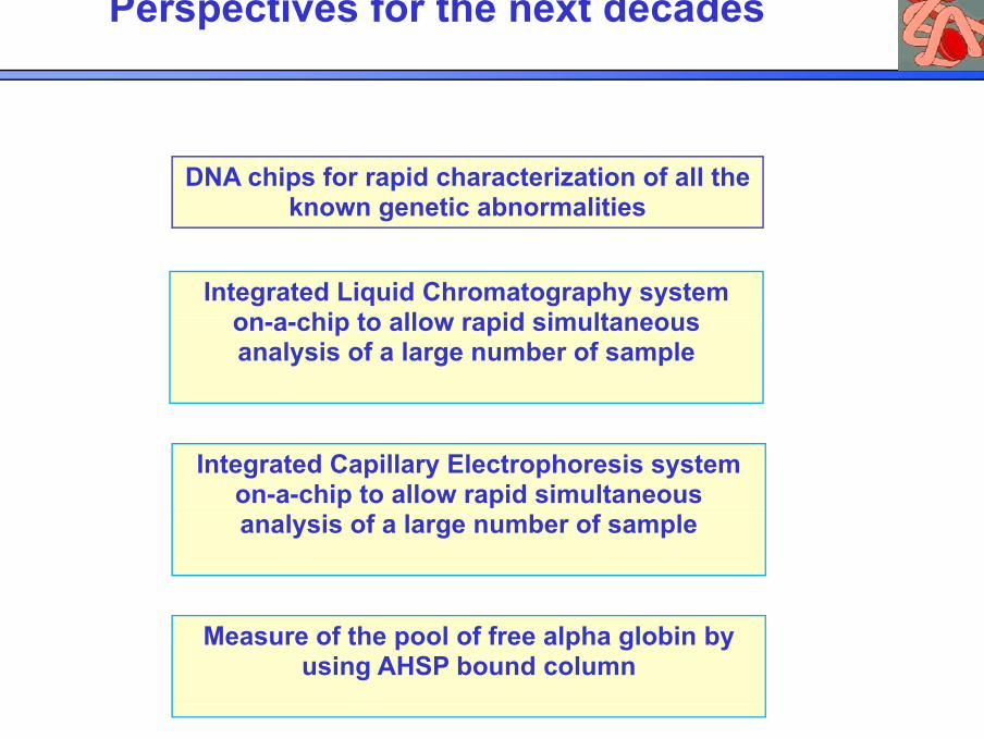
Perspectives for the next decades
DNA chips for rapid characterization of all the known genetic abnormalities
Integrated Liquid Chromatography system on a chip to allow rapid simultaneouson-a-chip to allow rapid simultaneous analysis of a large number of sample
Integrated Capillary Electrophoresis system on-a-chip to allow rapid simultaneous analysis of a large number of sample
Measure of the pool of free alpha globin by using AHSP bound column
