development of locust bean gum nanoparticles for protein ... · na abordagem inicial, tentou-se...
TRANSCRIPT

Universidade do Algarve
Departamento de Ciências Biomédicas e Medicina
Development of Locust Bean Gum Nanoparticles for Protein Delivery
Master dissertation in Biomedical Sciences
Pedro Raimundo
Supervisor: Professora Doutora Ana Margarida Grenha
Faro 2014
ii
Universidade do Algarve
Departamento de Ciências Biomédicas e Medicina
Development of Locust Bean Gum Nanoparticles for Protein Delivery
Master dissertation in Biomedical Sciences
Pedro Raimundo
Supervisor: Professora Doutora Ana Margarida Grenha
Faro 2014

iii
Development of Locust Bean Gum Nanoparticles for Protein Delivery
Declaração de autoria de trabalho
Declaro ser o autor deste trabalho, que é original e inédito. Autores e trabalhos
consultados estão devidamente citados no texto e constam da listagem de referências
incluída.
Copyright © 2014
Pedro Raimundo
______________________________________________________
A Universidade do Algarve tem o direito, perpétuo e sem limites geográficos, de arquivar
e publicitar este trabalho através de exemplares impressos reproduzidos em papel ou de
forma digital, ou por qualquer outro meio conhecido ou que venha a ser inventado, de o
divulgar através de repositórios científicos e de admitir a sua cópia e distribuição com
objetivos educacionais ou de investigação, não comerciais, desde que seja dado crédito
ao autor e editor.

iv
Agradecimentos
Primeiramente queria agradecer à Professora Ana Grenha por me ter aceitado na sua
equipa durante quase um ano e pela sua disponibilidade, dedicação, pela transmissão de
conhecimentos e na orientação deste trabalho.
Quero agradecer ao Professor Luís Braz pela ajuda na parte prática com especial destaque
na síntese dos polímeros, cultura de células e microfotografias TEM.
Quero agradecer à Susana Rodrigues pela sua disponibilidade e paciência em ensinar-me
as técnicas essenciais à execução da parte prática deste trabalho e pela ajuda com software
e pelo brainstorming de ideias que por vezes surgia.
Agradeço às minhas colegas Ana Dias, Carla Arruda, Filipa Pereira e Ludmylla Cunha
pela camaradagem e boa disposição no laboratório ao longo deste ano de trabalho.
Agradeço há minha família pelo apoio importante que me deram não só na execução deste
trabalho como em todo o meu percurso académico.
Agradeço há minha namorada Stephany Martins pelo grande apoio que me deu nos
momentos mais difíceis e por apoiar e respeitar as minhas decisões.
Agradeço também a todos os amigos e colegas de universidade e residência por terem
estado sempre presentes e pelas constantes conversas na tentativa de explicar o trabalho
que estava a desenvolver no laboratório.

v
Resumo
O interesse pela goma de alfarroba tem vindo a crescer ao longo dos últimos tempos.
Inicialmente na região do Algarve utilizava-se a goma de alfarroba para alimentar animais
de pecuária, contudo, por conter um polissacárido com propriedades importantes em
variados setores económicos, passou a ser utilizada na indústria de transformação
alimentar como espessante e na indústria farmacêutica como agente gelificante. Além das
aplicações referidas anteriormente, a goma de alfarroba pode ser utilizada para muitas
mais finalidades. Por se tratar de um polissacárido biodegradável e potencialmente não
tóxico, revela-se interessante explorar as suas capacidades como material formador de
matriz de sistemas de administração de fármacos e, nomeadamente, de proteínas, por vias
mucosas. São vários os sistemas de administração, mas as nanopartículas têm chamado
particularmente a atenção no âmbito da administração de proteínas. Para que uma
administração por vias mucosas seja efetiva, os sistemas nanoparticulados devem ter um
tamanho entre 50 e 500 nm (para maximizar a sua interação), e um potencial zeta positivo
(para poderem interagir com o ácido siálico que tem carga negativa e se encontra na
superfície das mucosas). Além disso, outras características são requeridas para que se
possam utilizar estes sistemas para administração proteica, nomeadamente uma eficácia
de encapsulação razoável e ausência de toxicidade.
Este trabalho teve como objetivo o desenvolvimento de nanopartículas de goma de
alfarroba, para entrega de proteínas com fins sistémicos, através das vias mucosas. De
modo a ir de encontro ao objetivo, o desenvolvimento do sistema nanoparticulado de
goma de alfarroba proposto teve por base a utilização de um método de complexação
polieletrolítica, segundo o qual as nanopartículas se formam por interação eletrostática
entre grupos químicos dos polímeros que apresentam cargas opostas. Dada a neutralidade
deste polissacárido, um trabalho anterior do grupo de investigação consistiu numa síntese
química para produção de derivados carregados de goma de alfarroba. Foi assim
produzido um derivado aminado e um derivado sulfatado, os quais exibem cargas opostas,
permitindo a formação das nanopartículas por complexação polieletrolítica. As
nanopartículas resultantes foram caraterizadas relativamente ao seu tamanho, índice de
polidispersão, potencial zeta, rendimento de produção, eficácia de encapsulação, bem
como quanto ao perfil de citotoxicidade no âmbito de administração por vias mucosas.

vi
Para preparar as nanopartículas foram utilizados três rácios de massa de goma de alfarroba
aminada (A-LBG) e goma de alfarroba sulfatada (S-LBG), respetivamente 2/1, 1/1 e 1/2.
Depois de obter as duas soluções respetivas, o S-LBG é adicionado ao A-LBG, sob
agitação magnética moderada, em concentrações diferentes de modo a ter as três razões
de massa. A formação das nanopartículas ocorre de forma imediata, mas as suspensões
permanecem em agitação durante 10 minutos. Imediatamente após a mistura dos dois
polímeros, é possível observar o efeito de Tyndall, que evidencia a formação de
nanopartículas. As suspensões destas são centrifugadas a 16000xg, a 15 ºC durante 30
minutos, sobre uma camada de 10 μL de glicerol, a qual visa facilitar a ressuspensão das
nanopartículas. Após a centrifugação, o sobrenadante é descartado e as nanopartículas
são ressuspendidas em 100 μL de água para utilização nos ensaios subsequentes.
A formulação A-LBG/S-LBG = 2/1 apresentou um diâmetro de aproximadamente 560
nm, um índice de polidispersão de 0,432 e um potencial zeta de +43,5 mV. A formulação
A-LBG/S-LBG = 1/2 apresentou um diâmetro de aproximadamente 359 nm, um índice
de polidispersão de 0,381 e um potencial zeta de -49,7 mV. A formulação A-LBG/S-LBG
= 1/1 precipitou no momento em que o S-LBG foi adicionado ao A-LBG e, como tal, esta
formulação foi abandonada. Atendendo a estes valores, em termos de diâmetro, a
formulação 2/1 apresenta um diâmetro acima do que seria ideal e a formulação 1/2
apresenta um diâmetro entre os valores pretendidos. Relativamente à polidispersão,
ambas as formulações apresentam valores abaixo do limite máximo aceite (0,5) no
entanto estão acima do valor ideal (0,2). É no entanto importante assinalar que para
nanopartículas produzidas com polímeros naturais é muito difícil obter índices de
polidispersão abaixo de 0,2. Analisando os valores de potencial zeta, a formulação 2/1
apresenta um potencial positivo, que é mais coincidente com os objetivos da
administração transmucosa, e a formulação 1/2 apresenta um potencial negativo. Os
valores de potencial observados estão de acordo com a carga exibida pelo polímero
predominante em cada formulação. O potencial negativo, apesar de não ser ideal para o
objetivo de administração transmucosal, não é desfavorável, porque pode ser utilizado
para outro tipo de terapias.
Para o cálculo do rendimento de produção, a preparação das nanopartículas é feita como
descrito anteriormente, com exceção do facto de não se utilizar glicerol, nem haver
ressuspensão. Após a centrifugação o sobrenadante é descartado e o sedimento de
nanopartículas é sujeito a congelação e posterior liofilização. A formulação 2/1 teve um

vii
rendimento de produção de aproximadamente 17% e a formulação 1/2 de 30%, os quais
são considerados baixos.
Para testar a capacidade das nanopartículas como transportadores de proteínas, a insulina
foi selecionada como proteína modelo. Dado o seu ponto isoelétrico (pI), que é de 5.3, a
proteína não dissolve em água. Para a sua solubilização usam-se o NaOH ou o HCl, ambos
à concentração de 0,01 M, ficando a proteína com uma carga negativa ou positiva,
respetivamente. Na abordagem inicial, tentou-se fazer a associação de uma quantidade de
insulina equivalente a 30% do total da massa de polímeros utilizados na preparação das
nanopartículas, mas não houve sucesso, já que se verificava precipitação. Após
otimização do processo, assumiu-se a quantidade de insulina correspondente a 10% da
massa do polímero que contribui com maior massa na preparação de cada formulação.
Como a formulação A-LBG/S-LBG = 2/1 tinha mais quantidade de polímero positivo
(conferindo um potencial zeta positivo), é maior o número de grupos carregados
positivamente que estão disponíveis para interação com outras moléculas, em
comparação com os grupos carregados negativamente. Assim, para esta formulação a
insulina foi dissolvida em NaOH para assumir uma carga negativa e ter desta forma
maiores probabilidades de interação e, consequentemente, de associação às
nanopartículas. Após solubilização, a insulina foi adicionada à S-LBG, que tem
igualmente carga negativa, evitando-se a interação eletrostática entre ambos os polímeros.
Esta solução mista foi posteriormente adicionada à solução de A-LBG para formação das
nanopartículas, havendo uma competição entre a insulina e a S-LBG pelos grupos
carregados positivamente da A-LBG. Ao contrário, a formulação A-LBG/S-LBG = 1/2
tinha mais polímero de S-LBG, que tem carga negativa, o que proporciona em princípio
maior capacidade de interação com grupos carregados positivamente. Neste caso, então,
a estratégia consistiu em atribuir à insulina um predomínio de cargas positivas no
momento da preparação das nanopartículas. Para isso, a proteína foi dissolvida em HCl e
adicionada ao A-LBG, antes da adição desta mistura à S-LBG.
A eficácia de encapsulação de insulina observada foi de 15% para ambas as formulações,
um valor considerado reduzido. Com vista a aumentar a eficácia de encapsulação, foi feita
uma otimização que teve por base a hipótese de alterar o pH do meio da insulina de forma
a ficar próximo do seu ponto isoelétrico. A literatura disponibiliza pelo menos um
trabalho que mostra que há uma maior adsorção das proteínas aos polímeros quando estas
se encontram em meios com pH perto do seu ponto isoelétrico. No valor de pH

viii
equivalente ao ponto isoelétrico, a insulina ficará com um equilíbrio de cargas positivas
e negativas e deixará livres as cargas dos polímeros, para interagirem entre si,
maximizando a sua interação e a insulina ficará adsorvida aos polímeros na matriz das
nanopartículas formadas. De forma a testar a hipótese anterior, para a formulação 2/1, a
solução mãe de insulina foi preparada em NaOH, mas na preparação das diluições de A-
LBG e S-LBG a partir das correspondentes soluções mãe, a água utilizada nas diluições
foi substituída por tampão citrato (pH 5,0) por ter um pH próximo do ponto isoelétrico da
insulina, fazendo com que esta exibisse um equilíbrio de cargas negativas e positivas após
solubilização. Para a formulação 1/2 a insulina foi dissolvida em HCl e depois foi
inicialmente realizado o mesmo procedimento com o tampão citrato, como para a
formulação 2/1, mas ocorria precipitação. A otimização para a formulação 1/2 foi então
realizada substituindo a água da solução S-LBG por uma solução de HCl a 0,1 M. As
otimizações que foram operadas acolheram sucesso, tendo conduzido a um aumento das
eficácias de encapsulação, na formulação 2/1 de 15% para 22% e na formulação 1/2 de
15% para 96%.
As nanopartículas carregadas com insulina foram caraterizadas quanto às propriedades
físico-químicas. Para a formulação 2/1 registou-se um diâmetro de 740 nm (PDI de 0,389)
e um potencial zeta de +22,8 mV. Para a formulação 1/2 o diâmetro foi de 400 nm (PDI
de 0,404) e o potencial zeta -33,6 mV. No entanto, na formulação 1/2 observaram-se
alguns agregados de difícil ressuspensão, mesmo após alteração da quantidade de glicerol
utilizada.
Dado o objetivo de aplicação das nanopartículas desenvolvidas na administração por vias
mucosas, considerou-se relevante avaliar a toxicidade das formulações. Realizou-se
assim um ensaio de citotoxicidade em duas linhas celulares epiteliais, Caco-2 e A549. A
primeira linha representa o epitélio intestinal, enquanto a segunda representa o epitélio
pulmonar e, mais especificamente, alveolar. As concentrações testadas foram de 0,1
mg/mL, 0,5 mg/mL e 1 mg/mL para ambas as formulações. Os tempos de incubação das
células com as nanopartículas foram de 3 horas e 24 horas para ambas as formulações, e
linhas celulares, bem como para todas as concentrações. Em ambas as linhas celulares a
viabilidade ficou acima de 70% para as duas formulações, indicando que as
nanopartículas possuem baixa citotoxicidade.
Em conclusão, os derivados de LBG sulfatado e aminado permitem a formulação de
nanopartículas com capacidade para associar insulina. No entanto, a formulação 2/1

ix
apresenta um tamanho inadequado para o objetivo de administração por vias mucosas,
além de baixo rendimento de produção e eficácia de encapsulação. A formulação 1/2
apresenta um tamanho aceitável para o objetivo descrito, bem como uma eficácia de
encapsulação muito significativa. No entanto, o rendimento da formulação é algo baixo e
o seu potencial zeta negativo, possivelmente registando uma menor propensão para
interação com a superfície epitelial em comparação com uma formulação de carga
positiva. Este tipo de nanopartículas, pode ser utilizado em superfícies com carga positiva
que requeiram nanopartículas com potencial zeta negativo, ou para encapsular moléculas
de carga positiva, maximizando a sua interação.
Palavras-chave: administração de proteínas, complexação polieletrolítica, goma de
alfarroba, nanopartículas, polissacáridos

x
Abstract
The interest of Locust Bean Gum (LBG) for biomedical applications have been increasing
in the last few years. Due to its biodegradability and non-toxic characteristics, LBG
became an interesting material regarding the production of drug and protein delivery
systems. Our aim was to develop a delivery system based on LBG nanoparticles for
mucosal protein delivery. Since LBG displays no charged groups, aminated LBG (A-
LBG) and sulphated LBG (S-LBG) were synthetized in our laboratory in order to allow
the use of a method of polyelectrolyte complexation for the production of the carriers.
Using this method enabled forming LBG nanoparticles with distinct properties depending
on the weight ratio. A-LBG/S-LBG nanoparticles with three different mass ratios were
prepared, namely, 2/1, 1/1 and 1/2. Mucosal delivery of drugs is known to be potentiated
by the use of nanoparticles with sizes varying between 50 and 500 nm, in order to
maximize the interaction with the epithelium. Moreover, nanoparticles should have
positive zeta potential to interact with the negative charges of syalic acid residues present
on epithelial surfaces.
In this work, LBG nanoparticles were characterised in terms of size, polidispersion index
(PDI), zeta potential (ZP) and production yield (PY). The ability of the carriers to
associate the model protein insulin was determined and cell viability tests were also
performed to obtain a first indication on the nanoparticle biocompatibility profile.
Formulation 1/1 was rejected due to precipitation, the work thus focusing on formulations
2/1 and 1/2. According to the initial objectives, formulation 2/1 showed excessively high
size, and low association efficiency (AE, 22%). In turn, formulation 1/2 showed a suitable
size and high AE (96%), but it was difficult to ressuspend. Regarding the cytotoxic
profile, in a general manner both formulations of LBG nanoparticles showed low
cytotoxicity.
In conclusion, after the optimizations, the formulation that appears the most suitable for
protein delivery is A-LBG/S-LBG = 1/2, in spite of its negative zeta potential, which does
not favour the interaction with epithelial surfaces.
Keywords: locust bean gum, nanoparticles, polysaccharide, polyelectrolyte
complexation, protein delivery

xi
List of Abbreviations
AE – association efficiency
A-LBG - aminated locust bean gum
BCA – bicinchoninic acid
DMSO – dimethyl sulfoxide
FBS – foetal bovine serum
G - galactose
LBG - locust bean gum
LC – loading capacity
M - mannose
MTT - Thiazolyl blue tatrazolium bromide
NP’s – nanoparticles
PBS – Phosphate buffer solution
PDI - Polydispersion index
PY – production yield
S-LBG - sulphated locust bean gum
SD – standard deviation
SEM – standard error of the mean
SDS – sodium dodecyl sulphate
ZP - zeta potential

xii
Index of contents
1. Introduction ........................................................................................................... 1
1.1. Nanotechnology applied to biomedicine .......................................................... 1
1.2. Nanoparticles for protein delivery ................................................................... 1
1.2.1. Non-parenteral routes of administration ................................................... 3
1.3. Polymeric materials as carrier matrix .............................................................. 4
1.3.1. Polysaccharides ....................................................................................... 5
1.3.2. Locust Bean Gum as nanoparticle matrix material ................................... 6
1.3.2.1. General features ................................................................................... 6
1.3.2.2. Chemical structure, physicochemical properties and enzymatic
degradation ............................................................................................................ 7
3. Materials and cell lines ........................................................................................ 11
3.1. Materials ....................................................................................................... 11
3.2. Cell lines ....................................................................................................... 11
4. Methods ............................................................................................................... 12
4.1. Preparation of A-LBG/S-LBG nanoparticles ................................................. 12
4.2. Characterisation of nanoparticles .................................................................. 13
4.3. Association of insulin to A-LBG/S-LBG nanoparticles ................................. 14
4.4. Evaluation of the cytotoxicity of nanoparticles .............................................. 15
4.5. Statistical analysis ......................................................................................... 16
5. Results and discussion ......................................................................................... 17
5.1. Preparation and characterisation of A-LBG/S-LBG nanoparticles ................. 17
5.2. Association of the model protein ................................................................... 19
5.3. Evaluation of cytotoxicity of the nanoparticles .............................................. 21
6. Conclusion ........................................................................................................... 26
7. Bibliography ........................................................................................................ 27

xiii
Index of figures
Figure 1.1 Schematic representation of barriers to overcome in insulin delivery by oral
route [6] ...................................................................................................................... ..2
Figure 1.2. Routes of administration of nanoparticles: advantages and disadvantages [9]
..................................................................................................................................... 3
Figure 1.3. Polymers from renewable and non-renewable resources [15] ....................... 5
Figure 1.4. Locust Bean Gum Chemical Structure (1-4) – linked β-D-mannose backbone
with (1-6)- linked side chains of α-D-galactose (from [31])……………………………..8
Figure 1.5. Scheme of LBG enzymatic degradation (Adapted from [22]). ..................... 9
Figure 4.1. Schematic representation of the preparation of A-LBG/S-LBG nanoparticles.
................................................................................................................................... 13
Figure 5.1. TEM microphotograph of representative A-LBG/S-LBG nanoparticles ..... 19
Figure 5.2. Caco-2 cell viability of raw materials determined by the MTT assay after 3 h
and 24 h exposure to increasing concentrations of A-LBG and S-LBG. Data represent
mean ± SEM (n = 3, three replicates per experiment at each concentration) ................ 23
Figure 5.3. Caco-2 cell viability of nanoparticles determined by MTT assay after 3 h and
24 h exposure to increasing concentrations of A-LBG/S-LBG = 2/1 and A-LBG/S-LBG
= 1/2 nanoparticles. Data represent mean ± SEM (n = 3, three replicates per experiment
at each concentration). ................................................................................................ 24
Figure 5.4. A549 cell viability of nanoparticles determined by MTT assay after 3 h and
24 h exposure to increasing concentrations of A-LBG/S-LBG = 2/1 and A-LBG/S-LBG
= 1/2 nanoparticles. Data represent mean ± SEM (n = 3, three replicates per experiment
at each concentration). ................................................................................................ 25

xiv
Index of tables
Table 5.1. Physicochemical characteristics and production yield of LBG unloaded
nanoparticles (mean ± SD, n = 3). ............................................................................... 18
Table 5.2 Encapsulation efficiency (EE) of A-LBG/S-LBG nanoparticles before and
after optimisations and loading capacity (LC) (mean ± SD, n = 3) ............................... 20
Table 5.3. Physicochemical characteristics and production yield of LBG insulin loaded
nanoparticles (mean ± SD, n = 3) ................................................................................ 21

1
Development of Locust Bean Gum Nanoparticles for Protein Delivery
Pedro Raimundo - 2014
1. Introduction
1.1. Nanotechnology applied to biomedicine
In the last few years, the interest in nanotechnology has increased considerably due its
potential towards many applications such as informatics, electronics, pharmaceutics and
biomedicine, among others. The prefix nano refers to a scale in the order of 1x10⁻⁹ m,
more precisely between 1-1000 nm. Nanotechnology is now being explored in more depth
in many fields and namely in the biomedical area. The creation of nanochips to monitor
blood chemistry and the nanocarriers for drug delivery, are valuable and representative
examples. Nanotechnologies are very useful in drug delivery because the encapsulation
of a drug in a carrier designed at the nanoscale may increase its bioavailability and
improve its release profile. These are prominent aspects, as they might reduce the amount
of administered drug and the number of needed administrations, thereby reducing the side
effects [1].
1.2. Nanoparticles for protein delivery
Designing therapeutic carriers for proteins is a challenging task. Parenteral route is the
most commonly employed method of administration, however, requirement of frequent
injections due to short in vivo half-life results in poor patient compliance. Attending to
the previous issues become an urgent scientific challenge being the transmucosal
administration the first option for protein systemic delivery [2].
Nanoparticles have been proposed as suitable protein carriers, overcoming many of the
limitations of these macromolecules. Proteins show physicochemical characteristics that
make them susceptible to degradation and limit their absorption due the high molecular
weight and, sometimes, hydrophilicity [3, 4]. In the present work, the chosen protein
model was insulin. Like other peptides, insulin shows a poor physical and chemical
stability and a relatively short plasma half-time [5]. Per oral bioavailability of insulin is
relatively low mainly due to high proteolytic activity in the gut and low permeability of
the intestinal epithelium as shown in figure 1.1 [6]. To overcome these barriers, several
strategies are underway for peroral administration of insulin, those strategies including
the addition of enzyme inhibitors and/ or permeation enhancers, chemical modification,
cell penetration peptides, vitamin B12 or cyclodextrin conjugation, polymeric carriers,
liposomes or a colon targeting of the drug delivery system where the enzymatic activity
is relatively low.

2
Development of Locust Bean Gum Nanoparticles for Protein Delivery
Pedro Raimundo - 2014
Our aim is to use nanoparticulate systems to avoid the previous issues. A promising
strategy is the use of multifunctional polymers exhibiting permeation enhancing and
mucoadhesive properties [7].
Figure 1.1: Schematic representation of barriers to overcome in insulin delivery by oral
route [6].
Previous studies have shown that nanoparticles potentiate the improvement of drug
pharmacokinetic profile, providing their stabilisation and, in some cases, permitting
controlled release and enhancing drug absorption [8]. Furthermore, the high surface-to-
volume ratio displayed by nanoparticles increases the drug loading capacity [9]. Scientists
have demonstrated that nanoparticle contact with epithelial surfaces is maximised when
their size is between 50 and 500 nm [10, 11], but also when the carriers exhibit a strongly
positive zeta potential [12].

3
Development of Locust Bean Gum Nanoparticles for Protein Delivery
Pedro Raimundo - 2014
1.2.1. Non-parenteral routes of administration
Therapeutic molecules may enter the body via oral ingestion, inhalation, dermal
penetration and intravascular injection, among others, being thus distributed through the
body and reaching the various organs. Figure 1.2 shows the advantages and disadvantages
of each route of administration [13].
Figure 1.2: Routes of administration of nanoparticles: advantages and disadvantages [9]
Pulmonary, oral and transdermal routes are non-invasive. Transdermal route shows
limitations for translocation of the nanoparticles from the dermal surface to the systemic
circulation. According to the previous figure, the most suitable routes for protein delivery
to the systemic circulation appear to be the pulmonary and oral due to their advantages
and disadvantages regarding to the other routes. Nevertheless, other non-parenteral routes
are suitable for protein delivery as ocular and nasal [2].

4
Development of Locust Bean Gum Nanoparticles for Protein Delivery
Pedro Raimundo - 2014
1.3. Polymeric materials as carrier matrix
Different classes of materials are available that permit preparing nanoparticulates, such
as polymers, lipids, metals and so on. Nevertheless, polymeric nanomaterial-based
therapeutics have been increasingly used in biomedicine, namely in areas such as tissue
engineering and drug delivery. In the latter case, cancer, diabetes, and neurodegenerative
diseases are examples of diseases that might find great improvements with the application
of nanotechnologies. In this context, the main advantages of polymers over other
materials for nanomedicine include increased functionality, design flexibility, improved
processability and, in some cases, biocompatibility [14].
Polymers can be obtained from different sources. The majority of known polymers are
derived from petroleum, which represents a huge environmental problem. Additionally,
petroleum sources are finite. Consequently, the use of polymers from other sources,
eventually more environment-friendly, became necessary. Polymers from renewable
resources can be divided into three major groups, as shown in figure 1.3: natural polymers
such as starch and proteins, synthetic polymers from natural monomers such as polylatic
acid, and polymers from microbial fermentation such as polyhidroxybutirate [15].
Polymer scientists have performed extensive research in the development of
biodegradable polymers. In turn, biopolymer research has found several applications in
the area of biomedical science. Despite the apparent proliferation of these materials in the
biomedical science, the science and technology of biopolymers is still in its early stages
of development. Great opportunities exist and will continue to exist for the engagement
of biopolymers in every facet of medical science through intensive research and
development [16].

5
Development of Locust Bean Gum Nanoparticles for Protein Delivery
Pedro Raimundo - 2014
Figure 1.3: Polymers from renewable and non-renewable resources [15].
Natural and synthetic polymers have been used in biomedicine, in pharmaceutical
formulations, imaging, drug delivery and targeting, prosthetics and tissue engineering.
Synthetic polymers are often used, but most of them lack bioactivity and biocompatibility.
Natural polymers have a better profile regarding these features. According to their
structure, natural polymers can be ordered into three main classes: polysaccharides;
polypeptides and polynucleotides [17]. In the next section a more detailed description of
polysaccharides is provided.
1.3.1. Polysaccharides
Polysaccharides can be obtained from different sources: vegetal (i.e. starch), animal (i.e.
chitosan) and microbial (i.e. dextran and gellan). These macromolecules present several
advantages in therapeutic uses in comparison with the synthetic polymers. Some
polysaccharides are very similar with glycosaminoglycans (GAGs) such chondroitin
sulphate, dermatan sulphate, keratin sulphate, heparin, heparin sulphate and hyaluroran.
The advantage of the chemical similarity of some polysaccharides with heparin, is the
fact that those polysaccharides had potential hemocompatibility properties. Another
relevant advantage of the polysaccharides are their propensity to be non-toxic and
biocompatible and, with some exceptions, their low cost in comparison with other
biopolymers like, for example, collagen [18].
Polysaccharides are a class of biopolymers constituted of simple sugar monomers [19].
The monomers are linked together by O-glycosidic bonds that can be made to any of the

6
Development of Locust Bean Gum Nanoparticles for Protein Delivery
Pedro Raimundo - 2014
hydroxyl groups of a monosaccharide, giving polysaccharides the ability to form both
linear and branched polymers. Differences in the monosaccharide composition, chain
shapes and molecular weight are important to understand their physical properties
including solubility, gelation and surface properties[20].
Owing to the above mentioned properties, polysaccharides are widely used in several
fields such as food and cosmetic industries, biomedicine and pharmaceutics, assuming
relevant roles that include thickening, gelling, emulsifying, hydrating and suspending
actions [21].
1.3.2. Locust Bean Gum as nanoparticle matrix material
1.3.2.1. General features
Locust Bean Gum (LBG) is a neutral polysaccharide composed of mannose and galactose
monomers, thus belonging to the category of galactomannans. Besides Ceratonia siliqua,
other plants might be used as sources of galactomannans, such as Cyamopsis
tetragonoloba (guar gum) and Caesalpinia spinosa (tara gum) [22]. The polysaccharide
is obtained from the crush of the endosperm of seeds, which represent 10% of the weight
of the fruit of carob tree (Ceratonia siliqua L.), has a powdered appearance and assumes
a white to yellowish white colour. The seeds have a composition of 80% galactomannan,
the remaining 20% are proteins and impurities [23, 24]. The protein content of LBG is
represented by approximately 32% albumin and globulin and the glutelin content is
approximately 68% [25]. Crude galactomannan can be purified in order to eliminate
proteins and other impurities isolating the polysaccharide. The purification process could
include one of the procedures: enzymatic and alkaline hydrolysis, precipitation with
ethanol or isopropanol, and purification by methanol, or by copper or barium complexes.
The performance of purification procedures has shown to result in higher
mannose/galactose ratio and decreased amount of proteins and impurities [22].
The Carob tree is very abundant in the Mediterranean region, but it can also be found in
North Africa, South America and Asia. The literature provides other expressions referring
to locust bean gum, such as carob bean gum, carob seed gum, carob flour and ceratonia
[26].

7
Development of Locust Bean Gum Nanoparticles for Protein Delivery
Pedro Raimundo - 2014
LBG is largely used as a thickener, stabilizer, emulsifier and gelling agent in food and
cosmetic industries [27]. The use of LBG in drug delivery is already reported, namely
associated with formulations designed for colonic delivery, thus benefiting from the
ability of colonic microflora to degrade LBG [28, 29]. Other applications involve topical,
ocular and buccal delivery systems. The large majority of formulations are based on
tablets, but some hydrogels and multiparticulate systems are described in the literature
[22]. LBG use as tablet matrix is related with the fact that polysaccharides are generally
considered to play important role in drug release mechanisms from matrixes [30]. It is
observed, in most cases, that LBG associated with another polymer, affords an improved
effect in controlled drug release systems.
1.3.2.2. Chemical structure, physicochemical properties and
enzymatic degradation
As mentioned above, LBG is a galactomannan, evidencing a chemical structure
consisting of a (1-4)–linked β-D-mannose backbone with (1-6)-linked side chains of α-
D-galactose [21, 26] (figure 1.4). Mannose (M) and galactose (G) are both neutral
monomers, thus attributing neutrality to LBG. This polysaccharide has little change in
viscosity and solubility in a wide pH range (between 3-11) [31]. Galactomannans differ
in their M/G ratio, which in turn depends on the distribution of galactose units over
mannose backbone. This ratio is approximately 4:1 for LBG [32], 3:1 for tara gum and
2:1 for guar gum [33]. Substitution patterns of side-chain units and their molecular weight
are influenced by harvesting and manufacturing practices, among other factors [34].
Therefore, the previous ratios are approximated and dependent on the material origin and
plant growth conditions during production [31]. An important note is the fact that the
galactose grafts are not spaced regularly in the mannose backbone but, instead of that,
galactose is randomly placed [35]. Importantly, the M/G ratio is the characteristic most
affecting galactomannan solubility [21], due to the fact that mannose chains are relatively
hydrophobic and galactose units are more hydrophilic. In this manner, LBG has limited
solubility, forming aggregates in cold water, as the long segments of unsubstituted
mannose are likely to undergo aggregation [34, 36].

8
Development of Locust Bean Gum Nanoparticles for Protein Delivery
Pedro Raimundo - 2014
Figure 1.4: Locust Bean Gum Chemical Structure (1-4) – linked β-D-mannose backbone
with (1-6)- linked side chains of α-D-galactose (from [31]).
For biopharmaceutical applications, an important issue to assess is the in vivo
biodegradability of the polymer-based materials. This question is very important since
the elimination of the systems by the organism, after administration, is required without
the need for additional interventions. The biodegradation of natural polymers occurs by
the action of enzymes, microorganisms and pH action, being complex in a biological,
physical and chemical way. The mentioned processes lead to a breakdown of polymer
chains, resulting in decreased molecular weight and the modification of other properties
such as solubility. The biodegradation of LBG is mostly driven by enzymatic processes
and there are several enzymes in the human organism with the ability to cleave the LBG
molecule. The oral route is the one offering the most effective degradation of the
polysaccharide, due to the presence of the enzyme β-mannanase in the human colonic
region [22, 37, 38]. This enzyme acts on β-D-(1,4) links of mannose chains converting
LBG in three metabolites as shown in figure 1.5. According to this figure, the metabolic
processing of LBG is also performed by two other enzymes, β-mannosidase and α-
galactosidase, which act on mannose and galactose residues, respectively. These enzymes
were also detected in the human colonic region [39].

9
Development of Locust Bean Gum Nanoparticles for Protein Delivery
Pedro Raimundo - 2014
Figure 1.5: Scheme of LBG enzymatic degradation (Adapted from [22]).
The interest of using natural materials as part of drug development has increased in the
past two decades [5]. In this work, Locust Bean Gum will be explored as a potential
matrix material in the preparation of nanocarriers for insulin delivery.

10
Development of Locust Bean Gum Nanoparticles for Protein Delivery
Pedro Raimundo - 2014
2. Objectives
The aim of this work was to develop a nanodelivery system based on LBG for
transmucosal delivery of proteins. LBG nanoparticles were produced by a method of
polyeletrolyte complexation, using two LBG-derivatives previously synthesized by the
research group (aminated LBG and sulphated LBG). For the approach of transmucosal
delivery to be successful, the nanoparticles should display a size between 50 and 500 nm,
apart from a positively charged surface, which permit a more intimate contact between
the protein carrier and the epithelial surface, thus contributing for a favourable release of
the protein.
The conditions for the preparation of the nanoparticles were optimised, regarding the A-
LBG and S-LBG ratios to be used, and the selected formulations characterised in terms
of physicochemical properties and production yield. In order to test the ability of LBG
nanoparticles as protein carriers, insulin was used as model protein and associated to the
nanocarriers, to determine the encapsulation efficiency and loading capacity. Finally, an
evaluation of the cytotoxic profile of the developed LBG nanoparticles was performed in
two different epithelial cell lines, one representative of the intestinal epithelium (Caco-2
cells) and another one, a model of the respiratory epithelium, namely the alveolar zone
(A549 cells). These two cell lines represent the environment of both the oral and the
pulmonary route of administration.

11
Development of Locust Bean Gum Nanoparticles for Protein Delivery
Pedro Raimundo - 2014
3. Materials and cell lines
3.1. Materials
A-LBG and S-LBG were synthesised by our group from LBG, kind gift from Industrial
Farense (Portugal). Buffer solution citric acid/sodium hydroxide pH 5.0 was purchased
from Fluka (Germany). Hydrochloric acid (HCl) (37%), sodium hydroxide (NaOH)
tablets, insulin Mw 5777.6, penicillin-streptomycin solution (10000 units/mL, 10000
µg/mL), trypsin-EDTA solution, Dulbeco’s Modified Eagle’s Medium (DMEM), trypan
blue solution (0.4%), 3-(4,5-Dimethylthiazol-2-yl)-2,5-diphenyltetrazolium bromide
(MTT), dimethyl sulfoxide (DMSO), sodium dodecyl sulphate (SDS) and phosphate
buffer saline (PBS) tablets pH 7.4 were purchased from Sigma (Germany). Foetal bovine
serum (FBS) from Gibco was purchased from Life Technologies (USA). Ultrapure water
(Milli-Q plus, Millipore Iberica (Spain)) was used throughout. Micro BCA (bicinchoninic
acid) protein assay kit was purchased from Pierce (USA).
3.2. Cell lines
The Caco-2 and A549 cell lines were obtained from the American Type Culture
Collection (Rockville, USA) and used between passages 77-90 and 27-40, respectively.
Cell cultures were grown in 75 cm2 flasks in a humidified 5% CO2/95% atmospheric air
incubator at 37ºC. For both cell lines, cell culture medium was DMEM supplemented
with 10% (v/v) FBS, 1% (v/v) L-glutamine solution, 1% (v/v) non-essential amino acids
solution and 1% (v/v) penicillin/streptomycin. Medium was changed every 2-3 days and
cells were subcultured weekly.

12
Development of Locust Bean Gum Nanoparticles for Protein Delivery
Pedro Raimundo - 2014
4. Methods
4.1. Preparation of A-LBG/S-LBG nanoparticles
Nanoparticles (NPs) were prepared by polyelectrolyte complexation method [40] which
consists in the electrostatic interaction between the positive and negative charges of A-
LBG and S-LBG, respectively. As LBG has as neutral charge, a previous work of the
group consisted in the amination and sulfation of the polymer in order to obtain
derivatives with positive and negative charge, respectively. FTIR analysis was performed
to production of aminated and sulphated derivatives of LBG (data not shown).
Three mass ratios of A-LBG/S-LBG were used to prepare the NPs by polyelectrolyte
complexation, in particular 2/1, 1/1 and 1/2. The stock solution of A-LBG was prepared
to reach a final concentration of 1 mg/mL, while that of S-LBG had a final concentration
of 2 mg/mL. The solutions were filtered with a 0.45 µm filter prior to use. The
formulations 2/1, 1/1 and 1/2 were prepared by adding 1.8 mL of S-LBG to 1 mL of A-
LBG, as depicted in Figure 4.1. The concentration of A-LBG was kept constant at 0.5
mg/mL for the preparation of all formulations, but that of S-LBG was modified to obtain
different ratios.
The addition of S-LBG to A-LBG was done by dripping during approximately 15
seconds. After that, it was possible to see, almost immediately, the Tyndall effect
evidencing the formation of nanoparticles. The suspensions of nanoparticles were mixed
by magnetic stirring for 10 minutes and then centrifuged in eppendorfs. Each eppendorf
had a layer of 10 µL of glycerol, in order to facilitate the following step of ressuspension.
The isolation of nanoparticles was performed by centrifugation (Thermo Scientific-
Heraeus Fresco 17, Germany) at 16000xg, for 30 min at 15 ºC. After discarding the
supernatants, the nanoparticles were ressuspended with 200 µL of ultrapure water.

13
Development of Locust Bean Gum Nanoparticles for Protein Delivery
Pedro Raimundo - 2014
Figure 4.1- Schematic representation of the preparation of A-LBG/S-LBG nanoparticles.
4.2. Characterisation of nanoparticles
The physicochemical characterisation of nanoparticles was performed on freshly
prepared samples. Size and polidispersion index (PDI) were measured by dynamic light
scattering and zeta potential was measured by laser Doppler anemometry, using a
Zetasizer Nano ZS (Malvern instruments, Malvern, UK). To prepare the samples, 20 µL
of each formulation were diluted in 1 mL of ultrapure water.
For determination of nanoparticle production yield, the nanoparticles were prepared as
described in the previous section but without the use of the 10 µL of glycerol. After
discarding the supernatant of each formulation, the pellets were frozen and then dried on
a freeze-dryer (Alpha RVC, Christ, Germany). The yield of nanoparticle production (PY)
was calculated as follows:
PY = (Nanoparticle sediment weight/Total solids weight) x 100
where nanoparticle sediment weight is the weight after freeze-drying and total solids
weight is the total amount of solids added for nanoparticle formation.
The morphological examination of A-LBG/S-LBG nanoparticles was conducted by
transmission electron microscopy (TEM; JEM-1011, JEOL, Japan). The samples were
stained with 2% (w/v) phosphotungstic acid and placed on copper grids with carbon films
(Ted Pella, USA) for TEM observation.

14
Development of Locust Bean Gum Nanoparticles for Protein Delivery
Pedro Raimundo - 2014
4.3. Association of insulin to A-LBG/S-LBG nanoparticles
Insulin was chosen as model protein for the association to LBG nanoparticles. Insulin is
insoluble in water and in solutions of pH near its isoelectric point (5.0). Two insulin stock
solutions were prepared, one with pH < 5.0 (insulin dissolved in HCl 0.01M) and another
one with pH > 5.0 (insulin dissolved in NaOH 0.01M).
When insulin is dissolved in NaOH it becomes deprotonated and acquires negative
charge, ideal to interact with positively charged groups. In turn, when it is dissolved in
HCl, the protonation that occurs attributes positive charges, favouring the interaction with
negatively charged groups. In formulation A-LBG/S-LBG = 2/1 the polymer in higher
amount was A-LBG which is positively charged. To associate insulin to this formulation,
the protein was dissolved in NaOH to display negative charges, thus favouring the
interaction with the positively charged amino groups of A-LBG. In contrary, in the
formulation A-LBG/S-LBG = 1/2 the polymer in higher amount was S-LBG. Therefore,
insulin was dissolved in HCl to be mostly positively charged, favouring the interaction
with negatively charged groups of S-LBG.
Objectively, for the preparation of the formulation A-LBG/S-LBG = 2/1, insulin was
dissolved in NaOH and the obtained solution was mixed with that of the polymer present
in lower amount in the formulation (S-LBG), prior to the addition to A-LBG for the
formation of nanoparticles. As such, for the formulation 1/2, insulin was dissolved in HCl
and the solution was mixed with that of the polymer in lower amount (A-LBG) prior to
the formation of nanoparticles. Insulin stock solutions in NaOH and HCl were prepared
at a concentration of 0.9 mg/mL.
The amount of insulin added to the formulations was defined as a function of the mass of
the polymers used in the preparation of the nanoparticles. The association of different
amounts of insulin was attempted as follows: 1) an amount of insulin corresponding to
30% of the total amount of polymers; 2) an amount of insulin corresponding to 20% of
the total amount of polymers; and 3) an amount of insulin corresponding to 10% of the
polymer present in higher quantity in each formulation (A-LBG for formulation 2/1 and
S-LBG for formulation 1/2).
Insulin was quantified in each sample using the Micro BCA Protein Assay Kit, which
provides a colorimetric method optimized to quantify reduced amounts of protein (0.5-
20 μg/mL). The method utilizes bicinchoninic acid (BCA) as the detection reagent for

15
Development of Locust Bean Gum Nanoparticles for Protein Delivery
Pedro Raimundo - 2014
Cu+1, which is formed when Cu+2 is reduced by protein in an alkaline environment [41].
A purple-coloured water-soluble reaction product is formed by the chelation of two
molecules of BCA with one cuprous ion (Cu+1), which exhibits a strong absorbance at
562 nm that is linear with increasing protein concentrations. High absorbance is therefore
interpreted as high insulin concentration.
Different calibration curves were performed for each formulation using the adequate
solvents (HCl or NaOH). After reacting with the MicroBCA, samples were analysed by
spectrophotometry (Infinite M200 Tecan, Austria) at 562 nm.
The protein association efficiency (AE) and the nanoparticle loading capacity (LC) were
calculated as follows:
AE (%) = [(Total insulin amount – Free insulin amount)/Total insulin amount] x 100
LC (%) = [Total insulin amount – Free insulin amount)/Nanoparticle weight] x 100
4.4. Evaluation of the cytotoxicity of nanoparticles
The in vitro cytotoxicity of A-LBG/S-LBG nanoparticles, as well as that of the raw
materials involved in nanoparticle production, was assessed by the metabolic assay
thiazolyl blue tetrazolium bromide (MTT) test. Two cell lines representative of
pulmonary and intestinal epithelia (A549 and Caco-2 cells, respectively) were used.
Caco-2 and A549, used between the passages 77-90 and 27-40, respectively, were seeded
at a density of 1×10⁴ cells/well in 96-well plates, in 100 µL of the same medium used for
culture in cell culture flasks. The cells were grown at 37 °C in a 5% CO2 atmosphere for
24 h before use.
Three different concentrations (0.1, 0.5 and 1.0 mg/mL) of unloaded nanoparticles, as
well as that of raw materials involved in nanoparticle production, were evaluated for
cytotoxicity over 3 and 24 h. Sodium dodecyl sulphate (SDS, 2%, w/v) was used as a
positive control of cell death. All formulations and controls were prepared as
solution/suspensions in pre-warmed cell culture medium without FBS immediately
before application to the cells.
To initiate the assay, culture medium of cells at 24 h in culture was replaced by 100 µL
of fresh medium without FBS containing the test samples or controls. After 3 or 24 h of

16
Development of Locust Bean Gum Nanoparticles for Protein Delivery
Pedro Raimundo - 2014
cell exposure, samples/controls were removed and 30 µL of the MTT solution (0.5
mg/mL in PBS, pH 7.4) were added to each well. After 2 h, any generated formazan
crystals were solubilised with 50 µL of DMSO. Upon complete solubilisation of the
crystals, the absorbance of each well was measured by spectrophotometry (Infinite M200,
Tecan, Austria) at 540 nm and corrected for background absorbance using a wavelength
of 650 nm [42].
The relative cell viability (%) was calculated as follows:
Viability (%) = (A – S)/(CM – S)× 100
Where A is the absorbance obtained for each of the concentrations of the test substance,
S is the absorbance obtained for the 2% SDS and CM is the absorbance obtained for
untreated cells (incubated with cell culture medium). The latter reading was assumed to
correspond to 100% cell viability. The assay was performed between three and six
occasions with three replicates at each concentration of test substance in each instance.
4.5. Statistical analysis
The t-test and the one-way analysis of variance (ANOVA) with the pairwise multiple
comparison procedures (Student–Newman–Keuls method) were performed to compare
two or multiple groups, respectively. All analyses were run using the SigmaStat statistical
program (Version 3.5, USA) and differences were considered to be significant at a level
of P < 0.05.

17
Development of Locust Bean Gum Nanoparticles for Protein Delivery
Pedro Raimundo - 2014
5. Results and discussion
5.1. Preparation and characterisation of A-LBG/S-LBG
nanoparticles
Three formulations of LBG-based nanoparticles were produced by polyelectrolyte
complexation, according to a procedure detailed in a previous section. Polyelectrolyte
complexation is a process that involves electrostatic interaction between oppositely
charged groups. This procedure takes the advantage of occurring in a hydrophilic
environment with mild preparation conditions, avoiding the use of organic solvents or
high shear forces that might compromise the stability of the encapsulated material [40,
43]. LBG is a natural polymer with neutral charge, which hinders the application of
polyelectrolyte complexation. In order to overcome that relevant limitation, aminated and
sulphated derivatives of LBG were produced in a previous work of the group, thus
obtaining positively and negatively charged polymers. The three formulations of A-
LBG/S-LBG nanoparticles were designed to have polymeric mass ratios of 2/1, 1/1 and
1/2. After the preparation procedures, the nanoparticles were characterised in terms of
size, polydispersion index, zeta potential and production yield. The detailed results are
shown in table 5.1.
The formulation A-LBG/S-LBG = 1/1 precipitated immediately upon adding S-LBG to
A-LBG. This is probably due to a positive to negative (+/-) charge ratio around 1, which
permits a strong interaction between the two LBG derivatives. If anionic charges from S-
LBG neutralize A-LBG positive charges, a reduction or elimination of electrostatic
repulsion will occur, leading to precipitation [44, 45]. Therefore, this 1/1 formulation was
disregarded and the work proceeded only with the two remaining formulations. The
formulation A-LBG/S-LBG = 2/1, displayed a size of approximately 560 nm, a
polydispersion index (PDI) of 0.432 and a positive zeta potential around +44 mV. The
size of the formulation A-LBG/S-LBG = 1/2 was significantly lower (P < 0.05), 359 nm,
the formulation further registering a PDI of 0.381 and a negative zeta potential of -50 mV.
18
Development of Locust Bean Gum Nanoparticles for Protein Delivery
Pedro Raimundo - 2014
Table 5.1 – Physicochemical characteristics and production yield of LBG unloaded
nanoparticles (mean ± SD, n = 3).
A-LBG/S-LBG
(w/w)
Size (nm) PDI Zeta potential
(mV)
Production
yield (%)
2/1 560.2 ± 108.3 0.432 ± 0.050 +43.5 ± 5.8 16.7 ± 3.8
1/1
1/2
n.d.
359.1 ± 69.2
n.d.
0.381 ± 0.063
n.d.
-49.7 ± 5.0
n.d.
30.0 ± 8.6
n.d. - not determined
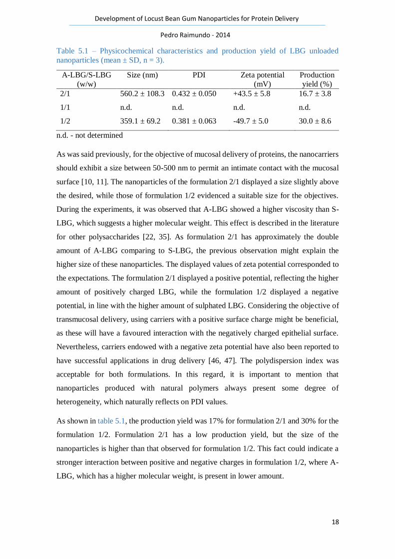
As was said previously, for the objective of mucosal delivery of proteins, the nanocarriers
should exhibit a size between 50-500 nm to permit an intimate contact with the mucosal
surface [10, 11]. The nanoparticles of the formulation 2/1 displayed a size slightly above
the desired, while those of formulation 1/2 evidenced a suitable size for the objectives.
During the experiments, it was observed that A-LBG showed a higher viscosity than S-
LBG, which suggests a higher molecular weight. This effect is described in the literature
for other polysaccharides [22, 35]. As formulation 2/1 has approximately the double
amount of A-LBG comparing to S-LBG, the previous observation might explain the
higher size of these nanoparticles. The displayed values of zeta potential corresponded to
the expectations. The formulation 2/1 displayed a positive potential, reflecting the higher
amount of positively charged LBG, while the formulation 1/2 displayed a negative
potential, in line with the higher amount of sulphated LBG. Considering the objective of
transmucosal delivery, using carriers with a positive surface charge might be beneficial,
as these will have a favoured interaction with the negatively charged epithelial surface.
Nevertheless, carriers endowed with a negative zeta potential have also been reported to
have successful applications in drug delivery [46, 47]. The polydispersion index was
acceptable for both formulations. In this regard, it is important to mention that
nanoparticles produced with natural polymers always present some degree of
heterogeneity, which naturally reflects on PDI values.
As shown in table 5.1, the production yield was 17% for formulation 2/1 and 30% for the
formulation 1/2. Formulation 2/1 has a low production yield, but the size of the
nanoparticles is higher than that observed for formulation 1/2. This fact could indicate a
stronger interaction between positive and negative charges in formulation 1/2, where A-
LBG, which has a higher molecular weight, is present in lower amount.

19
Development of Locust Bean Gum Nanoparticles for Protein Delivery
Pedro Raimundo - 2014
The morphological analysis of nanoparticles was performed by TEM. Figure 5.1 displays
the microphotograph of representative A-LBG/S-LBG nanoparticles. It can be seen that
the carriers generally have a spherical shape, apparently corresponding to a compact
structure. This morphology is very similar to that normally reported for polysaccharide-
based nanoparticles produced by polyelectrolyte complexation [44, 48-51].
Figure 5.1- TEM microphotograph of representative A-LBG/S-LBG nanoparticles.
5.2. Association of the model protein
Insulin was the selected protein model to test the encapsulation capacity of LBG
nanoparticles. As described previously in the methodology section, the association of the
protein to the nanoparticles required several optimisations, namely related with the initial
amount of insulin to be included in the formulations. The initial approaches included the
addition of an amount of insulin corresponding to 30% (at first) and 20% (secondly) of
the total amount of polymer, but these options were unsuccessful because severe
precipitation was observed. This precipitation was certainly due to the presence of an
excess of anionic charges, which neutralize positive charges and, thus, reduce or eliminate
electrostatic repulsion, leading to precipitation [44, 45]. The attempt of associating an
amount of insulin corresponding to 10% of the polymer present in higher quantity in each
formulation was the chosen one, as it led to the production of nanoparticles. However,
the encapsulation efficiency was around 15% in both formulations 2/1 and 1/2, which was
considered unsatisfactory. In order to improve the encapsulation efficiency, an

20
Development of Locust Bean Gum Nanoparticles for Protein Delivery
Pedro Raimundo - 2014
optimization was made which relied on changing the pH of the insulin medium so that it
was close to its isoelectric point. This hypothesis was constructed based on information
available on the literature which refers that there is a higher adsorption of proteins to
polymers when proteins are in medium with pH close to their isoelectric point [52]. When
the pH is equivalent to the isoelectric point (pI), insulin will display a net neutral charge
because the number of positive charges is equal to the number of negative charges.
Therefore, the charges of polymers will be free to interact with each other maximizing
their interaction and insulin will have a favoured adsorption to the nanoparticles. To test
this hypothesis, for the formulation 2/1, the stock solution of insulin was prepared with
NaOH and the water used to prepare the nanoparticles (mainly in dilutions of the
polymers) was replaced by a citrate buffer solution (pH = 5.0). As insulin pI is 5.3, using
a buffer of pH 5.0 will allow a final pH value close to the protein pI. Similarly, for the
formulation 1/2, insulin was dissolved previously in HCl and the water used to prepare
the nanoparticles was replaced by the citrate buffer solution. However, in the latter case
strong precipitation was observed, this formulations demanding further optimisations to
improve its encapsulation efficiency. A second approach consisted in substituting the
water used to prepare the nanoparticles by HCl solution 0.1 M, which revealed successful.
As shown in table 5.2, the association efficiency was thus increased in both formulations.
For the formulation 2/1 an improvement from 15% to 22% was observed, while for the
formulation 1/2 a very significant improvement from 15% to 96% was verified. The
loading capacity was significantly low for both formulations in particular for formulation
2/1. This values are low probably due to a little amount of insulin used for both
formulations.
Table 5.2 – Encapsulation efficiency (EE) of A-LBG/S-LBG nanoparticles before and
after optimisations and loading capacity (LC) (mean ± SD, n = 3).
A-LBG/S-LBG
(w/w)
EE before
optimisations (%)
EE after
optimisations (%)
LC (%)
2/1 15.0 ± 7.8 22.2 ± 8.1 2.78 ± 1.01
1/2 15.0 ± 5.4 96.1 ± 8.1 12.01 ± 1.02
21
Development of Locust Bean Gum Nanoparticles for Protein Delivery
Pedro Raimundo - 2014
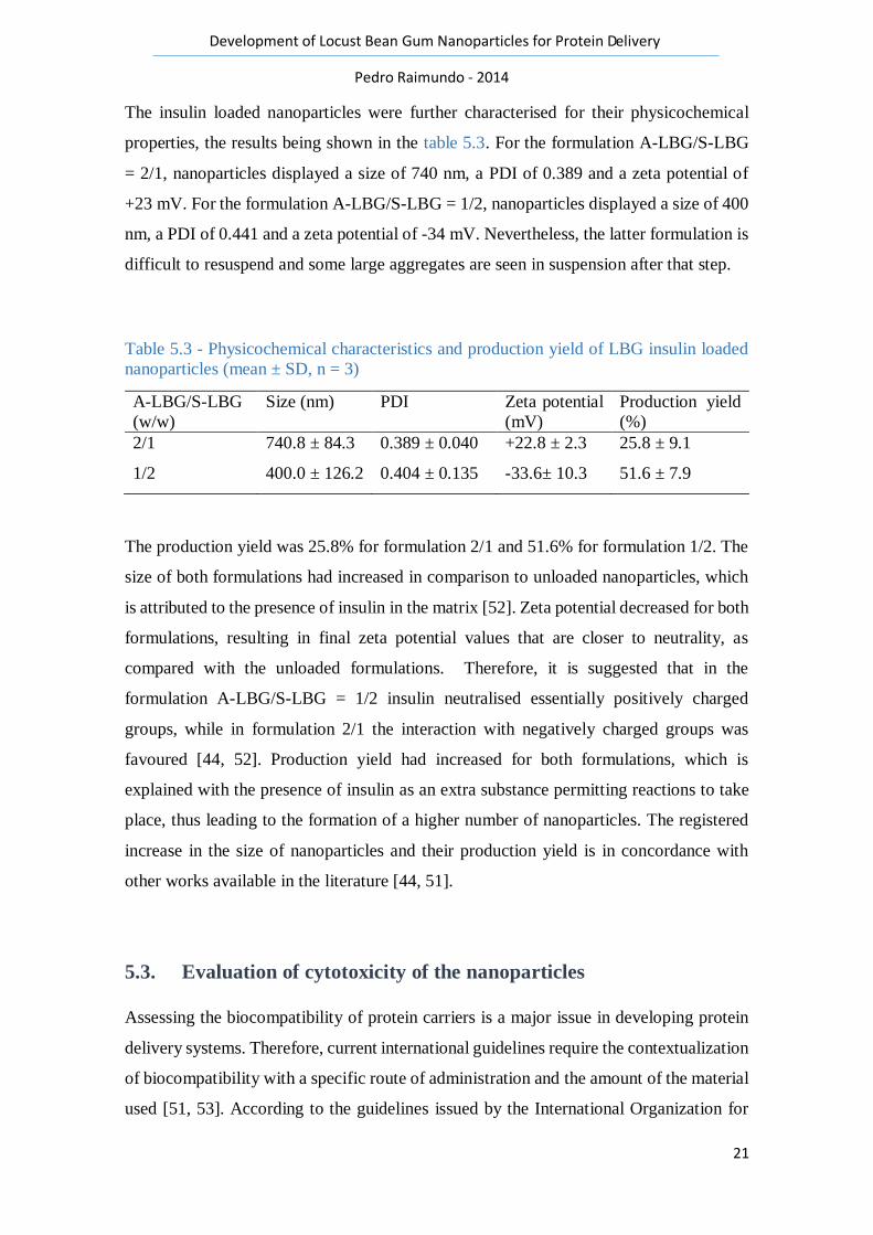
The insulin loaded nanoparticles were further characterised for their physicochemical
properties, the results being shown in the table 5.3. For the formulation A-LBG/S-LBG
= 2/1, nanoparticles displayed a size of 740 nm, a PDI of 0.389 and a zeta potential of
+23 mV. For the formulation A-LBG/S-LBG = 1/2, nanoparticles displayed a size of 400
nm, a PDI of 0.441 and a zeta potential of -34 mV. Nevertheless, the latter formulation is
difficult to resuspend and some large aggregates are seen in suspension after that step.
Table 5.3 - Physicochemical characteristics and production yield of LBG insulin loaded
nanoparticles (mean ± SD, n = 3)
A-LBG/S-LBG
(w/w)
Size (nm) PDI Zeta potential
(mV)
Production yield
(%)
2/1 740.8 ± 84.3 0.389 ± 0.040 +22.8 ± 2.3 25.8 ± 9.1
1/2 400.0 ± 126.2 0.404 ± 0.135 -33.6± 10.3 51.6 ± 7.9
The production yield was 25.8% for formulation 2/1 and 51.6% for formulation 1/2. The
size of both formulations had increased in comparison to unloaded nanoparticles, which
is attributed to the presence of insulin in the matrix [52]. Zeta potential decreased for both
formulations, resulting in final zeta potential values that are closer to neutrality, as
compared with the unloaded formulations. Therefore, it is suggested that in the
formulation A-LBG/S-LBG = 1/2 insulin neutralised essentially positively charged
groups, while in formulation 2/1 the interaction with negatively charged groups was
favoured [44, 52]. Production yield had increased for both formulations, which is
explained with the presence of insulin as an extra substance permitting reactions to take
place, thus leading to the formation of a higher number of nanoparticles. The registered
increase in the size of nanoparticles and their production yield is in concordance with
other works available in the literature [44, 51].
5.3. Evaluation of cytotoxicity of the nanoparticles
Assessing the biocompatibility of protein carriers is a major issue in developing protein
delivery systems. Therefore, current international guidelines require the contextualization
of biocompatibility with a specific route of administration and the amount of the material
used [51, 53]. According to the guidelines issued by the International Organization for

22
Development of Locust Bean Gum Nanoparticles for Protein Delivery
Pedro Raimundo - 2014
Standardization (ISO) 10993-3, a complete set of assays should be performed to test
biocompatibility, addressing firstly cellular morphology, membrane integrity, and
metabolic efficiency [54].
In this work, the cytotoxicity was assessed by means of the MTT assay, which assesses
cell metabolic efficiency, relying on the evaluation of an enzymatic function. To perform
the assay, after the exposure to the test formulations and raw materials, cells are incubated
with yellow tetrazolium (MTT) salts which are reduced to purple-blue formazan crystals
by active mitochondrial dehydrogenase [44, 51]. A higher concentration of the formazan
crystals corresponds to a higher amount of metabolically active cells, which is usually
interpreted as higher cell viability.
Two cell lines representative of pulmonary and intestinal epithelia (A549 and Caco-2
cells, respectively) were used. Three different concentrations (0.1, 0.5 and 1.0 mg/mL) of
both unloaded nanoparticles and raw materials involved in nanoparticle production were
evaluated for cytotoxicity over 3 h and 24 h. A statistical analysis was performed in order
to compare the obtained values and its meaning in terms of significance.
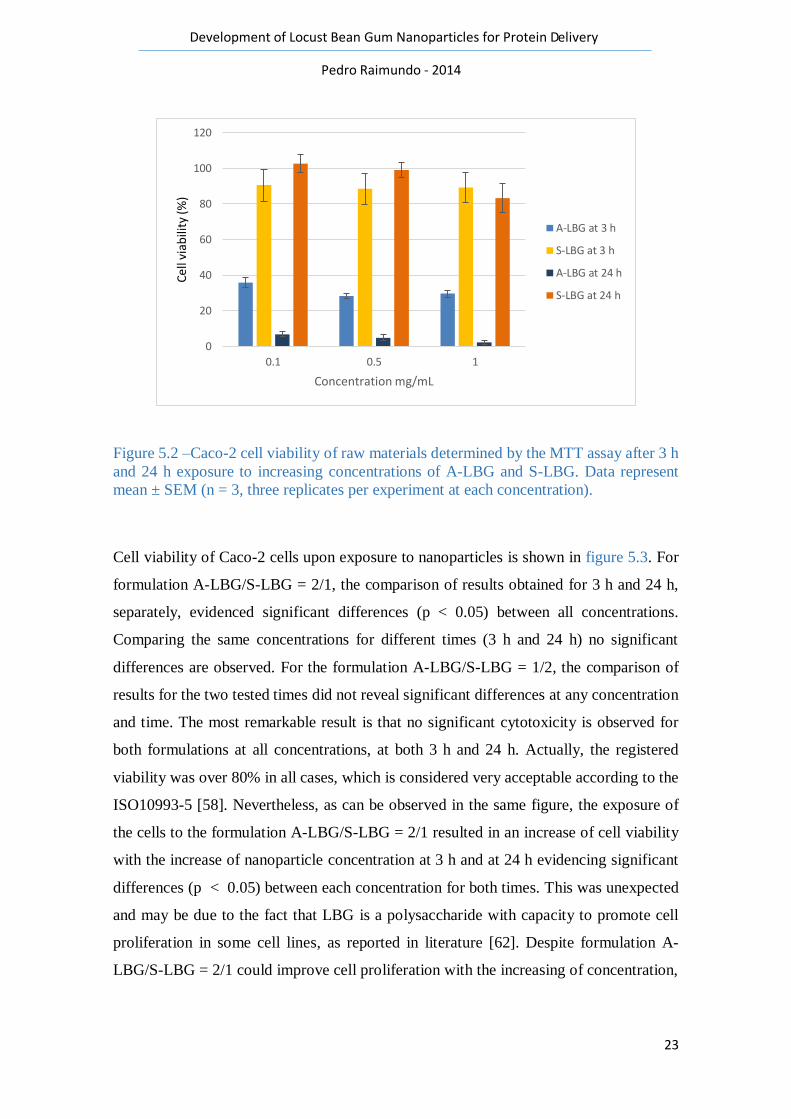
The results obtained for raw materials in Caco-2 cells are shown in figure 5.2. For both
polymers A-LBG and S-LBG, comparing the results obtained for 3 h and 24 h, separately
for each polymer, indicates that no significant differences are observed. In turn,
comparing each concentration of A-LBG for different exposure times (3 h and 24 h)
reveals significant differences (p < 0.05), which mainly comprise a decrease of cell
viability from 3 h to 24 h. This trend is found in many other works as the prolonged
exposure to an aggressive agent usually produces effects at prolonged times [55-57]. On
the contrary, this effect was not observed for S-LBG. All concentrations of positively
charged polymer reveal values of cell viability below 40%. According to the ISO 10993-
5 [58] A-LBG evidences cytotoxic potential as cell viability values below 70% are
indicative of toxicity. The influence of charges in cell viability remains largely
unresolved, however there are some indications in the literature suggesting some
hypotheses [51]. One of the explanations could be the fact that positively charged
polymers tend to be more cytotoxic due to a stronger interaction of the positive charges
with the negative cell surface charges, sometimes culminating with the internalization of
the material and leading to cell death [59-61]. The negatively charged polymer tested in
this study (S-LBG) evidences values of cell viability above 70%, which represents low
cytotoxicity.
23
Development of Locust Bean Gum Nanoparticles for Protein Delivery
Pedro Raimundo - 2014
Figure 5.2 –Caco-2 cell viability of raw materials determined by the MTT assay after 3 h
and 24 h exposure to increasing concentrations of A-LBG and S-LBG. Data represent
mean ± SEM (n = 3, three replicates per experiment at each concentration).
Cell viability of Caco-2 cells upon exposure to nanoparticles is shown in figure 5.3. For
formulation A-LBG/S-LBG = 2/1, the comparison of results obtained for 3 h and 24 h,
separately, evidenced significant differences (p < 0.05) between all concentrations.
Comparing the same concentrations for different times (3 h and 24 h) no significant
differences are observed. For the formulation A-LBG/S-LBG = 1/2, the comparison of
results for the two tested times did not reveal significant differences at any concentration
and time. The most remarkable result is that no significant cytotoxicity is observed for
both formulations at all concentrations, at both 3 h and 24 h. Actually, the registered
viability was over 80% in all cases, which is considered very acceptable according to the
ISO10993-5 [58]. Nevertheless, as can be observed in the same figure, the exposure of
the cells to the formulation A-LBG/S-LBG = 2/1 resulted in an increase of cell viability
with the increase of nanoparticle concentration at 3 h and at 24 h evidencing significant
differences (p < 0.05) between each concentration for both times. This was unexpected
and may be due to the fact that LBG is a polysaccharide with capacity to promote cell
proliferation in some cell lines, as reported in literature [62]. Despite formulation A-
LBG/S-LBG = 2/1 could improve cell proliferation with the increasing of concentration,
0
20
40
60
80
100
120
0.1 0.5 1
Cel
l via
bili
ty (%
)
Concentration mg/mL
A-LBG at 3 h
S-LBG at 3 h
A-LBG at 24 h
S-LBG at 24 h

24
Development of Locust Bean Gum Nanoparticles for Protein Delivery
Pedro Raimundo - 2014
formulation A-LBG/S-LBG = 1/2, induced constant cell viability near 100%, irrespective
of the concentration.
Figure 5.3 – Caco-2 cell viability of nanoparticles determined by MTT assay after 3 h and
24 h exposure to increasing concentrations of A-LBG/S-LBG = 2/1 and A-LBG/S-LBG
= 1/2 nanoparticles. Data represent mean ± SEM (n = 3, three replicates per experiment
at each concentration).
When the developed nanoparticles were tested on the cell line representing the pulmonary
epithelium (A549 cells), the results were somewhat different (figure 5.4). For formulation
A-LBG/S-LBG = 2/1, the comparison of results obtained for each concentration at 3 h
and 24 h, evidenced a tendency to a slight increase of cell viability with the increase of
concentration, although no significant differences were found between the groups. For
the formulation A-LBG/S-LBG = 1/2, this comparison did not reveal significant
differences for any concentration at 3 h. However, at 24 h significant differences were
found between concentrations (p < 0.05). Nevertheless, comparison between 3 h and 24
h for same formulation for each concentration revealed no significant differences.
Generally, the tested conditions resulted in viabilities above 70%, with the exception of
formulation 1/2 for the concentration of 1 mg/mL at 24 h, which is below 60%. For the
formulation 1/2, while the 3 h exposure did not lead to significant differences in viability,
at 24 h the differences are significant among nanoparticle concentrations. In that case it
0
20
40
60
80
100
120
0,1 0,5 1
Cel
l via
bili
ty (%
)
Concentrations mg/mL
Formulation 2/1 at 3 h
Formulation 1/2 at 3 h
Formulation 2/1 at 24 h
Formulation 1/2 at 24 h

25
Development of Locust Bean Gum Nanoparticles for Protein Delivery
Pedro Raimundo - 2014
is possible to observe a decrease from 100% for concentration 0.1 mg/mL, to 80% for
concentration 0.5 mg/mL and 60% for concentration 1 mg/mL.
A549 cell line seems to be more sensitive than Caco-2 cell line, which is probably due to
the origin of the cells. Since Caco-2 are from intestinal epithelium they are exposed to
higher mechanical forces and pH changes than A549 cells and, consequently, they present
a more resistant pattern.
Figure 5.4 – A549 cell viability of nanoparticles determined by MTT assay after 3 h and
24 h exposure to increasing concentrations of A-LBG/S-LBG = 2/1 and A-LBG/S-LBG
= 1/2 nanoparticles. Data represent mean ± SEM (n = 3, three replicates per experiment
at each concentration).
Taking into account the objectives delineated for the LBG nanoparticles, which concern
the transmucosal delivery of proteins, the two developed formulations (2/1 and 1/2) are
considered to display adequate cytotoxic profile, as low cytotoxicity was evidenced in
cell lines representative of the respiratory and intestinal epithelia. Nevertheless, as
proposed by the ISO 10993-5, these results of cytotoxicity should be complemented with
those of genotoxicity, acute and sub-acute toxicity to give further indications regarding
the biocompatibility profile of the drug delivery systems [58].
0
20
40
60
80
100
120
0,1 0,5 1
Cel
l via
bili
ty (%
)
Concentration mg/mL
Formulation 2/1 at 3 h
Formulation 1/2 at 3 h
Formulation 2/1 at 24 h
Formulation 1/2 at 24 h

26
Development of Locust Bean Gum Nanoparticles for Protein Delivery
Pedro Raimundo - 2014
6. Conclusion
The combination of the two tested LBG derivatives (sulphated and aminated) enables the
formation of nanoparticles by the method of polyelectrolyte complexation. Varying
several parameters of the procedure permits obtaining nanoparticles with different
physicochemical characteristics (variable size, opposite zeta potential). The developed
nanocarriers evidence capacity to associate insulin, which is dependent on several
variables of the process, such as the pH of the reaction media. Nevertheless, according to
the objectives proposed for the work, which comprise the transmucosal delivery of
proteins, formulation A-LBG/S-LBG = 2/1 does not show suitable properties.
Formulation A-LBG/S-LBG = 1/2 shows acceptable characteristics, in spite of the
evidenced negative charge, which does not favour the interaction with epithelial surfaces
in a particular manner. A very positive indication was obtained regarding the cytotoxic
profile of the developed carriers, as both formulations evidenced low cytotoxicity upon
incubation with two cell lines representative of both the intestinal (Caco-2) and the
alveolar (A549) epithelia.
27
Development of Locust Bean Gum Nanoparticles for Protein Delivery
Pedro Raimundo - 2014
7. Bibliography
1. Ratner, M. and D. Ratner, Nanotechnology: a gentle introduction to the next big
idea. 2002: Prentice Hall Press. p. 208-210.
2. Patel, A., et al., Recent Advances in Protein and Peptide Drug Delivery: A Special
Emphasis on Polymeric Nanoparticles. Protein Peptide Letters, 2014.
3. Antosova, Z., et al., Therapeutic application of peptides and proteins: parenteral
forever? Trends Biotechnology, 2009. 27(11): p. 628-635.
4. Liu, Z., et al., Polysaccharides-based nanoparticles as drug delivery systems.
Advanced Drug Delivery Reviews, 2008. 60(15): p. 1650-1662.
5. Reis, C.P., et al., Nanoparticulate biopolymers deliver insulin orally eliciting
pharmacological response. Journal of Pharmaceutical Sciences, 2008. 97(12): p.
5290-5305.
6. Delie, F. and Blanco-Prieto, M.J. Polymeric particulates to improve oral
bioavailability of peptide drugs. Molecules, 2005. 10(1): p. 65-80.
7. PATIL, P., et al., Formulation and in vitro evaluation of mucoadhesive tablets of
ofloxacin using natural gums. International Journal of Current Pharmaceutical
Research. 3(2): p. 93-98.
8. Hartig, S.M., et al., Multifunctional nanoparticulate polyelectrolyte complexes.
Pharmaceutical Research, 2007. 24(12): p. 2353-2369.
9. de la Fuente, M., et al., Nanoparticles as protein and gene carriers to mucosal
surfaces. Nanomedicine (Lond), 2008. 3(6): p. 845-857.
10. Desai, M.P., et al., Gastrointestinal uptake of biodegradable microparticles:
effect of particle size. Pharmaceutical Research, 1996. 13(12): p. 1838-1845.
11. Jani, P., et al., Nanoparticle uptake by the rat gastrointestinal mucosa:
quantitation and particle size dependency. Journal of Pharmacy and
Pharmacology, 1990. 42(12): p. 821-826.
12. Bogataj, M., et al., The correlation between zeta potential and mucoadhesion
strength on pig vesical mucosa. Biological and Pharmaceutical Bulletin, 2003.
26(5): p. 743-746.
13. Yildirimer, L., et al., Toxicology and clinical potential of nanoparticles. Nano
Today, 2011. 6(6): p. 585-607.
14. Banik, B.L. and Brown, J.L. Chapter 23 - Polymeric Biomaterials in
Nanomedicine, in Natural and Synthetic Biomedical Polymers, S.G. Kumbar, C.T.
Laurencin, and M. Deng, Editors. 2014, Elsevier: Oxford. p. 387-395.
15. Yu, L., K. Dean, and Li, L.. Polymer blends and composites from renewable
resources. Progress in Polymer Science, 2006. 31(6): p. 576-602.
16. Kariduraganavar, M.Y., Kittur, A.A., and Kamble, R.R. Chapter 1 - Polymer
Synthesis and Processing, in Natural and Synthetic Biomedical Polymers,
Kumbar, S.G., Laurencin, C.T. and Deng, M. Editors. 2014, Elsevier: Oxford. p.
1-31.
17. Aravamudhan, A., et al., Chapter 4 - Natural Polymers: Polysaccharides and
Their Derivatives for Biomedical Applications, in Natural and Synthetic
Biomedical Polymers, Kumbar S.G., Laurencin, C.T. and Deng, M. Editors. 2014,
Elsevier: Oxford. p. 67-89.
18. Cascone, M.G., et al., Bioartificial polymeric materials based on polysaccharides.
Journal of Biomaterials Science, Polymer Edition, 2001. 12(3): p. 267-281.
19. Nishinari, K. and Takahashi, R. Interaction in polysaccharide solutions and gels.
Current Opinion in Colloid & Interface Science, 2003. 8(4–5): p. 396-400.

28
Development of Locust Bean Gum Nanoparticles for Protein Delivery
Pedro Raimundo - 2014
20. Malafaya, P.B., Silva, G.A. and Reis, R.L. Natural–origin polymers as carriers
and scaffolds for biomolecules and cell delivery in tissue engineering
applications. Advanced Drug Delivery Reviews, 2007. 59(4–5): p. 207-233.
21. Rinaudo, M., Main properties and current applications of some polysaccharides
as biomaterials. Polymer International, 2008. 57(3): p. 397-430.
22. Dionisio, M. and Grenha, A. Locust bean gum: Exploring its potential for
biopharmaceutical applications. Journal of Pharmacy and Bioallied Sciences,
2012. 4(3): p. 175-185.
23. Dakia, P.A., et al., Composition and physicochemical properties of locust bean
gum extracted from whole seeds by acid or water dehulling pre-treatment. Food
Hydrocolloids, 2008. 22(5): p. 807-818.
24. Bouzouita, N., et al., The analysis of crude and purified locust bean gum: A
comparison of samples from different carob tree populations in Tunisia. Food
Chemistry, 2007. 101(4): p. 1508-1515.
25. Smith, B.M., et al., Composition and molecular weight distribution of carob germ
protein fractions. Journal of Agricultural and Food Chemistry, 2010. 58(13): p.
7794-7800.
26. Rowe R, S.P., Owen S. , Handbook of Pharmaceutical Excipients. 5 th ed. 2006,
London: Pharmaceutical Press.
27. H. El Batal, A.H., Ouatmane, A., Boulli, A., Dehbi, F., Jaouad A. Yield and
composition of carob bean gum produced from different Moroccan populations
of carob (Ceratonia siliqua L.). Journal of Materials and Environmental Science,
2013. 4(2): p. 309-314.
28. Hirsch, S., et al., Lauroyldextran and crosslinked galactomannan as coating
materials for site-specific drug delivery to the colon. European Journal of
Pharmaceutics and Biopharmaceutics, 1999. 47(1): p. 61-71.
29. Raghavan, C.V., et al., An in vitro and in vivo investigation into the suitability of
bacterially triggered delivery system for colon targeting. Chemical
Pharmaceutical Bulletin (Tokyo), 2002. 50(7): p. 892-895.
30. Hoffman, A.S., Hydrogels for biomedical applications. Advanced Drug Delivery
Reviews, 2002. 54(1): p. 3-12.
31. Beneke, C.E., Viljoen, A.M. and Hamman, J.H. Polymeric plant-derived
excipients in drug delivery. Molecules, 2009. 14(7): p. 2602-2620.
32. Coviello, T., et al. Two galactomannans and scleroglucan as matrices for drug
delivery: preparation and release studies. European Journal of Pharmaceutics and
Biopharmaceutics, 2007. 66(2): p. 200-209.
33. Andrade, C.T., et al., Solution properties of the galactomannans extracted from
the seeds of Caesalpinia pulcherrima and Cassia javanica: comparison with
locust bean gum. International Journal of Biological Macromolecules, 1999. 26(2-
3): p. 181-185.
34. Picout, D.R., et al., Pressure cell assisted solution characterization of
polysaccharides. 2. Locust bean gum and tara gum. Biomacromolecules, 2002.
3(4): p. 761-767.
35. Mathur, V. and Mathur, M.N. Fenugreek and other less known legume
galactomannan-polysaccharides: Scope for developments. Journal of Scientific
and Industrial Research, 2005. 64: p. 475-481.
36. Pollard, M., et al. Investigation of molecular weight distribution of LBG
galactomannan for flours prepared from individual seeds, mixtures, and
commercial samples. Food Hydrocolloids, 2008. 22: p. 1596-1606.

29
Development of Locust Bean Gum Nanoparticles for Protein Delivery
Pedro Raimundo - 2014
37. Alonso-Sande, M., et al., Glucomannan, a promising polysaccharide for
biopharmaceutical purposes. European Journal of Pharmaceutics and
Biopharmaceutics, 2009. 72(2): p. 453-462.
38. Nakajima, N. and Matsuura, Y. Purification and characterization of konjac
glucomannan degrading enzyme from anaerobic human intestinal bacterium,
Clostridium butyricum-Clostridium beijerinckii group. Bioscience,
Biotechnology and Biochemistry, 1997. 61(10): p. 1739-1742.
39. Jain, A., Gupta, Y. and Jain, S.K. Perspectives of biodegradable natural
polysaccharides for site-specific drug delivery to the colon. Journal of Pharm
Pharmaceutical Sciences, 2007. 10(1): p. 86-128.
40. Grenha, A., Chitosan nanoparticles: a survey of preparation methods. Journal of
Drug Targeting, 2012. 20(4): p. 291-300.
41. Smith, P.K., et al., Measurement of protein using bicinchoninic acid. Anal
Biochemistry, 1985. 150(1): p. 76-85.
42. Carmichael, J., et al., Evaluation of a tetrazolium-based semiautomated
colorimetric assay: assessment of chemosensitivity testing. Cancer Research,
1987. 47(4): p. 936-42.
43. Prego, C., Torres, D. and Alonso, M.J. The potential of chitosan for the oral
administration of peptides. Expert Opinion on Drug Delivery, 2005. 2(5): p. 843-
854.
44. Rodrigues, S., et al., Biocompatibility of chitosan carriers with application in drug
delivery. Journal of Functional Biomaterials, 2012. 3(3): p. 615-641.
45. Crouzier, T. and Picart, C. Ion pairing and hydration in polyelectrolyte multilayer
films containing polysaccharides. Biomacromolecules, 2009. 10(2): p. 433-442.
46. Chandratre, S.S. and Dash, A.K. (in press) Multifunctional Nanoparticles for
Prostate Cancer Therapy. AAPS PharmSciTech, 2014. DOI: 10.1208/s12249-
014-0202-z
47. Lee, J.Y., et al., Nanocomplexes Based on Amphiphilic Hyaluronic Acid
Derivative and Polyethylene Glycol-Lipid for Ginsenoside Rg3 Delivery. Journal
of Pharmaceutical Sciences, 2014. 103(10): p. 3254–3262
48. Goycoolea, F.M., et al., Chitosan-alginate blended nanoparticles as carriers for
the transmucosal delivery of macromolecules. Biomacromolecules, 2009. 10(7):
p. 1736-1743.
49. Oyarzun-Ampuero, F.A., et al., Chitosan-hyaluronic acid nanoparticles loaded
with heparin for the treatment of asthma. International Journal of Pharmacy, 2009.
381(2): p. 122-129.
50. Zorzi, G.K., et al., Hybrid Nanoparticle Design Based on Cationized Gelatin and
the Polyanions Dextran Sulfate and Chondroitin Sulfate for Ocular Gene
Therapy. Macromolecular Bioscience, 2011. 11(7): p. 905-913.
51. Dionisio, M., et al., Pullulan-based nanoparticles as carriers for transmucosal
protein delivery. European Journal of Pharmaceutical Sciences, 2013. 50(1): p.
102-113.
52. Peng, Z.G., Hidajat, K. and Uddin, M.S. Adsorption of bovine serum albumin on
nanosized magnetic particles. Journal of Colloid and Interface Science, 2004.
271(2): p. 277-283.
53. Gaspar, R. and Duncan, R. Polymeric carriers: preclinical safety and the
regulatory implications for design and development of polymer therapeutics.
Advanced Drug Delivery Reviews, 2009. 61(13): p. 1220-1231.

30
Development of Locust Bean Gum Nanoparticles for Protein Delivery
Pedro Raimundo - 2014
54. ISO, 2003, Biological evaluation of medical devices Part 3: Tests for genotoxicity,
carcinogenicity, and reproductive toxicity. In: Standardization, I.O.f. (Ed.),
10993-3.
55. Martins, A.F., et al., Preparation and cytotoxicity of N,N,N-trimethyl
chitosan/alginate beads containing gold nanoparticles. International Journal of
Biological Macromolecules, 2014. 72C: p. 466-471.
56. Kang, T., et al., In vitro toxicity of different-sized ZnO nanoparticles in Caco-2
cells. Nanoscale Research Letters, 2013. 8(1): p. 496.
57. Bhattacharjee, S., et al., Cytotoxicity of surface-functionalized silicon and
germanium nanoparticles: the dominant role of surface charges. Nanoscale,
2013. 5(11): p. 4870-4883.
58. ISO, 2009, Biological evaluation of medical devices Part 5: Tests for in vitro
cytotoxicity. In: Standardization. I.O.f. (Ed.), 10993-5.
59. Bhattacharjee, S., et al., Role of surface charge and oxidative stress in cytotoxicity
of organic monolayer-coated silicon nanoparticles towards macrophage NR8383
cells. Particle and Fibre Toxicology, 2010. 7(25) p. 1-12.
60. Ilinskaya, O.N., et al., Changing the net charge from negative to positive makes
ribonuclease Sa cytotoxic. Protein Science, 2002. 11(10): p. 2522-2525.
61. Turcotte, R.F., Lavis, L.D. and Raines, R.T. Onconase cytotoxicity relies on the
distribution of its positive charge. FEBS Journal, 2009. 276(14): p. 3846-3857.
62. Perestrelo, A.R., et al., Locust bean gum as an alternative polymeric coating for
embryonic stem cell culture. Material Sciences and Engeneering C: Materials for
Biological Applications, 2014. 40: p. 336-344.