development of kits for - international atomic energy agency · development of kits for 99mtc...
TRANSCRIPT

IAEA-TECDOC-1414
Development of kits for99mTc radiopharmaceuticals
for infection imaging
Report of a co-ordinated research project2000–2003
September 2004
IAEA-TECDOC-1414
Development of kits for99mTc radiopharmaceuticals
for infection imaging
Report of a co-ordinated research project2000–2003
September 2004

The originating Section of this publication in the IAEA was:
Industrial Applications and Chemistry Section International Atomic Energy Agency
Wagramer Strasse 5 P.O. Box 100
A-1400 Vienna, Austria
DEVELOPMENT OF KITS FOR 99MTc RADIOPHARMACEUTICALSFOR INFECTION IMAGING
IAEA, VIENNA, 2004 IAEA-TECDOC-1414 ISBN 92–0–111304–8
ISSN 1011–4289 © IAEA, 2004
Printed by the IAEA in Austria September 2004
FOREWORD
Infectious diseases remain a major health problem and cause of death worldwide, particularly in developing countries. Nuclear medicine imaging, because of its sensitivity, offers an attractive option for diagnosis of focal infections. This needs a reliable radiopharmaceutical that can selectively concentrate in sites of infection. Over the years 67Ga and other radiopharmaceuticals that localize in inflammation associated with infection sites, also known as “non-specific agents” have been used for infection imaging. However, experience has shown that an “infection specific agent” that concentrates selectively at sites of infection and not inflammation would have several advantages. The first such agent developed more than two decades ago was 111In-leucocytes which is still considered a “gold standard”. Considerations of cost, availability, and superior properties for imaging make 99mTc a better label than 111In. 99mTc white blood cell (WBC) was developed subsequently and used for infection imaging. However, both 111In and 99mTc WBCs have a number of drawbacks, in particular: each patient’s blood sample has to be collected and individually radiolabelled; well-trained staff and suitable facilities for separating and labelling the patient’s blood are needed; the risk of infection and cross-contamination associated with potential blood-borne microorganisms; and cost of materials. Because of these, considerable efforts have been continuously made towards developing convenient replacements for 99mTc WBCs with limited success, 99mTc antigranulocyte antibody being a good example. However, these radiopharmaceuticals still have many disadvantages, related to either their cost and availability or their performance. In view of the large potential for applications in patients, the development of new and improved 99mTc labelled infection specific imaging agents was considered as a very worthwhile aim for scientific research in general and, in particular, for the establishment of a Co-ordinated Research Project (CRP) by the IAEA. The CRP could investigate alternate biochemical pathways, promising recent advances in 99mTc labelling methodologies and recent progress in evaluation methods. Based on recommendations of two consultants meetings, the IAEA initiated a CRP entitled Development of Kits for 99mTc Radiopharmaceuticals for Infection Imaging in 2000. Twelve laboratories from Asia, Europe, North America, and South America participated in the CRP, which was concluded in 2003.
Among the objectives of this CRP was the development of different 99mTc labelling strategies in participating laboratories that would be useful in the development of 99mTc labelled infection imaging agents. In addition, techniques were to be developed for the in vitro and in vivo testing of label stability. Finally, it was hoped that one or more of the identified agents would prove to localize in infection by a specific mechanism. The CRP may be said to be successful in all three measures. Finally, with the identification of 99mTc ubiquicidine fragment (UBI 29-41) as a radiolabelled agent with potential clinical utility, this CRP can be considered to have made a major contribution by providing the first validated specific 99mTc labelled infection imaging agent.
The IAEA wishes to thank all participants in the CRP and, in particular, E. Pauwels of Leiden University, Netherlands and D. Hnatowich of Massachusetts University, USA for providing material support and guidance to the participants.
The IAEA officer responsible for this publication was D.V.S. Narasimhan of the Division of Physical and Chemical Sciences.

EDITORIAL NOTE
This publication has been prepared from the original material as submitted by the authors. The views expressed do not necessarily reflect those of the IAEA, the governments of the nominating Member States or the nominating organizations.
The use of particular designations of countries or territories does not imply any judgement by the publisher, the IAEA, as to the legal status of such countries or territories, of their authorities and institutions or of the delimitation of their boundaries.
The mention of names of specific companies or products (whether or not indicated as registered) does not imply any intention to infringe proprietary rights, nor should it be construed as an endorsement or recommendation on the part of the IAEA.
The authors are responsible for having obtained the necessary permission for the IAEA to reproduce, translate or use material from sources already protected by copyrights.
CONTENTS
Summary ................................................................................................................................................. 1
Publications and Presentations arising out of the CRP ........................................................................... 7
Reports by Participants in the CRP
In vitro and in vivo evaluation of 99mTc-labelled peptides for infection imaging ................................. 11 J. Crudo, A. Zapata, N. Nevares, E. Obenaus, S.G. de Castiglia
99mTc-labelled ligands for inflammation and infection imaging............................................................ 23L. Korosi, L. Balogh, D. Mathe, A. Polyak, R. Kiraly, Gy.A. Janoki
Preparation and evaluation of 99mTc labelled radiopharmaceuticals and formulation of kits for imaging infection/inflammation ................................................................................. 31A. Bhelose, J.S. Sarnaik, A. Raju, K.S. Mehra, Y. Singh, P. Kumar, A. Chanda, D.K. Ranganatha, S.H. Joshi, N. Ramamoorthy
99mTc labelling of ubiquicidine (UBI 29-41) and EDTA-biotin monomer (EB1) ................................. 41 W. Widjaksana, F. Yunita, L. Andriastuti, Yunilda, A. Ariyanto, G. Mondrida, A. Roseliana, S. Bagiawati, E. Sovilawati, A. Mutalib
Preparation of 99mTc kits for infection imaging..................................................................................... 55G. Ferro-Flores, C. Arteaga de Murphy, L. Melendez-Alafort, M. Pedraza-Lopez, P. Altamirano-Bustamante
Development of kits for 99mTc radiopharmaceuticals for infection imaging ......................................... 65M. Jehangir, M. Bashar, S. Pervez
Development of kits for 99mTc radiopharmaceuticals for infection imaging ......................................... 79 R. Mikolajczak, A. Korsak, B. Gorska, A. Markiewiz, E. Zakrzewska, W. Zulczyk, U. Karczmarczyk, J. Michalik
Development of 99mTc labelled infection imaging agents based on peptides ........................................ 87N. Poramatinuli, J. Sangsuriyan, M. Dangprasert, S. Laloknam, S. Pumkhem , N. Mudsomboon
99mTc radiopharmaceuticals for infection imaging: Kits development and validation .......................... 99 B. Souto Pais, G. Rodriguez, H. Balter, S. Lanzzeri, P. Oliver, A. Lopez, Z. Gonçalvez, A. Nappa, S. Verdera, J. Berbejillo, L. Mallo
List of Participants .............................................................................................................................. 113
SUMMARY
1. INTRODUCTION
The most significant advancement in public health in the last century is undoubtedly the
introduction of vaccines and antibiotics that have effectively eradicated or controlled most of the
infectious diseases. In spite of the great strides in management of infectious diseases, infections
remain among the most frequently encountered and costly causes of death and disease worldwide,
particularly in developing countries. Since most of the infectious diseases can be diagnosed by simple
laboratory tests and effectively treated with drugs, it can be perceived that a significant proportion of
those resulting in mortality could be due to conditions difficult to diagnose. Timely diagnosis in such
cases could help in instituting effective treatment and reduce morbidity and mortality.
Nuclear medicine imaging, because of its sensitivity, offers an attractive option for imaging
focal infections. Some examples of conditions which can benefit from imaging are deep-seated
muscular or orthopaedic infections especially those resulting from previous surgery; acute life-
threatening infections which require immediate effective treatment such as acute appendicitis, severe
chronic infections arising from drug-resistance; and opportunistic infections in immune-compromised
individuals. The questions often answered by imaging are presence or absence of infection and its
location, severity and probable cause. In order to answer these questions, the most important
requirement is a reliable radiopharmaceutical that can selectively concentrate in sites of infection. The
ideal radiopharmaceutical for imaging should be able to answer the clinical questions posed above, but
in addition be non-toxic, of low radiation burden to the patient, showing minimal uptake in non target
tissues, be inexpensive, easy to prepare and widely available.
Radiopharmaceuticals for imaging infection have generally been divided into two types —
‘specific’ and ‘non-specific’. The distinction between them relates to the mechanism of their action.
‘Non-specific’ agents work solely by their ability to localize at the site of inflammation that is often
accompanied with infection. ‘Specific’ agents, while also exhibiting some degree of non-specific
localization show an additional interaction with either the host immune system or the agent causing
the inflammation that increases the efficiency of delivery. Both ‘specific’ and ‘non-specific’
radiopharmaceuticals may, depending on the nature of the condition under study is equally effective.
For example, a ‘non-specific’ agent, 111
In-hIgG has shown high accuracy in diagnosis of number of
infections by localizing in associated inflammations. However, in some cases, particularly with regard
to identifying the cause of an inflammation, a ‘specific’ product may be more appropriate.
The most well established ‘specific’ agent that is still regarded as the ‘gold-standard’ for
infection imaging is 111
In labelled WBCs. In view of the cost, limited availability, and not so
favourable nuclear properties for imaging of 111
In, techniques for 99m
Tc labelling of WBCs have also
been developed and used. However, both these products have a number of drawbacks, in particular:
the need for labelling individual patient’s blood sample and reinjection; need for well-trained staff and
suitable facilities; the risk of infection and cross-contamination; and the considerable cost of the
materials required for cell labelling. Because of these, WBC labelling procedure has not become so
extensively used commensurate with its potential and considerable efforts have gone towards
developing convenient replacements. They include 99m
Tc labelled anti neutrophil antibodies,
chemotactic peptides, and platelet factor. However, these radiopharmaceuticals retain many
disadvantages related to either their cost and availability or their performance.
To achieve a more desirable 99m
Tc radiopharmaceutical for infection imaging, one displaying a
faster blood clearance, less non- target uptake and better concentration in infection sites, it will be
necessary to exploit alternate biochemical pathways. Some of these pathways like binding of anti
microbial peptides for example ubiquicidine, have been recently explored with promising results.
Recent years have also witnessed significant advances in 99m
Tc radiochemistry. Rational approaches to
99m
Tc labelling of different molecules have also been introduced. These can be fruitfully exploited for
incorporating 99m
Tc into biomolecules of promise for infection imaging without altering the biological
1
properties. Considerable progress has also been made in methodologies for in vitro and in vivo
evaluation and characterization of the 99m
Tc labelled agents. The labelling of various receptor specific
peptides with 99m
Tc is a good example of such advances. These also can be very useful in objective
evaluation of potential infection specific agents.
In view of the large potential for applications in patients, the development of new and improved
infection imaging agents labelled with the most commonly used radioisotope for imaging, namely
99m
Tc, is still considered a very worthwhile aim for scientific research and development particularly in
developing Member States. Development of 99m
Tc infection imaging agents was also identified as a
relevant topic for a Co-ordinated Research Project (CRP) of the IAEA by two consultants meetings
organized in 1995 and 1998. Based on their recommendations and considering the need for a superior
99m
Tc labelled infection imaging agent and advances in 99m
Tc radiopharmaceutical chemistry, a
Coordinated Research Project entitled Development of kits for 99m
Tc radiopharmaceuticals for
infection imaging was initiated in 2000.
Twelve laboratories from Asia, Europe, North America, and South America participated in the
CRP that was concluded in 2003. The first Research Co-ordination meeting (RCM) held in Budapest,
Hungary, took stock of the recent advances in 99m
Tc infection imaging agents, and decided on the
potential agents to work on. Among the several 99m
Tc agents considered, three were identified for
further study in the CRP. These were 99m
Tc ubiquicidine (UBI) as a ‘specific’ agent and 99m
Tc-
ethylenediaminetetraacetic acid biotin monomer (EB1) and 99m
Tc-human neutrophil elastase inhibitor
(HNE2) as superior ‘non specific’ agents. The available literature on all three agents suggested that
their further study and evaluation could be useful for arriving at a desirable infection agent. The
second RCM held in Mexico City reviewed the progress and decided to focus further efforts on UBI
and EB1 in view of the promising results obtained and uncertainty in future availability of HNE2. The
third and final RCM was held in Vienna to consolidate and document the results of all participants.
2. ACHIEVEMENTS OF THE CRP
In the three years of this CRP, a strong collaboration among participants was established as is
evident by the exchange of information among participants and especially in the distribution of
reagents. These collaborations are expected to continue even beyond the CRP.
Among the objectives of the CRP was the development of 99m
Tc labelling strategies in
participating laboratories that would be useful in the preparation of infection imaging agents. In
addition, techniques were to be developed for the testing of label stability in vitro and in vivo. Finally,
it was hoped that one or more of the identified agents would prove to localize in infection by what
appears to be a specific mechanism. The CRP can be considered successful in all three measures. In
general, each participating laboratory became proficient in labelling the chosen molecules with 99m
Tc
using a variety of techniques. By the final RCM, in vivo and in vitro quality assurance measurements
were standardized by all participants and applied such that the 99m
Tc agents were viewed as reliably
labelled. Finally, with the identification of 99m
Tc UBI as a radiolabelled agent with potential clinical
utility, this CRP can be considered to have made a major contribution to nuclear medicine by
providing the first convenient 99m
Tc labelled ‘specific’ infection imaging agent.
3. SUMMARY OF RESULTS
The results obtained in the different laboratories with each of the identified compound are summarized
below
3.1.99m
Tc human neutrophil elastase inhibitor (HNE2)
Source: HNE2 is available from a single source (provided from Dynax Corporation, USA) and
its binding protein, human neutrophil elastase, is not available. The later makes quality assurance of
radiolabelled HNE2 difficult. However, HNE2 is very similar in size and structure to BPTI (Aprotinin)
2
and BPTI binds effectively to trypsin. Both BPTI and trypsin are readily available. Therefore, BPT1
was used to standardize the conjugation and radiolabelling of HNE2. BPTI in sufficient quantities was
provided by Dr. Hnatowich, USA, to be used as a model for the radiolabelling of the much less
available HNE2
Labelling: HNE2 and BPTI were both radiolabelled using HYNIC/tricine, MAG3, and DTPA.
Purification after labelling was necessary and was accomplished on either P4 columns or using a
Sep-pak.
Quality assurance: Adequate quantities of trypsin were also supplied by Dr. Hnatowich, USA,
to use in a shift assay with BPTI. Shift assays performed in Argentina and India with open column gel
chromatography (Sephadex G75-100) showed the expected shift and, therefore, provided evidence that
the BPTI was properly labelled.
Stability studies: Argentina showed by cysteine challenge assay of 99m
Tc DTPA-BPTI and 99m
Tc
HYNIC–BPTI, that both were very stable. Both were also stable in PBS but both also showed binding
to serum proteins, especially in the case of 99m
Tc HYNIC/tricine. Serum stability studies in India of
99m
Tc MAG3-HNE2 also showed stability.
Animal studies: In Argentina, labelled BPTI showed high uptake in kidneys at 2 h post injection
in mice. In India, MAG3-BPTI provided high kidney uptake in normal mice. Monkey studies in
Hungary with DTPA-HNE2 showed high kidney uptake, low liver, and a somewhat less than optimum
T/NT ratio between 1.2-1.7 for inflammation vs. bacterial infection.
Summary: The 99m
Tc labelling of HNE2 or BPTI was accomplished satisfactorily. Due to the
difficulty in procuring HNE2 after the first year, further studies to evaluate its usefulness in infection
imaging had to be postponed.
3.2.99m
Tc ethylenediaminetetraacetic acid biotin monomer (EB1)
Source: EB1 was provided by Dr. Hnatowich of USA initially while, later in the CRP, solid
EB1 was synthesized and distributed from Dr. M. Jehangir of Pakistan and Dr. N. Ramamoorthy of
India.
Labelling: 99m
Tc labelling of EB1 could be achieved with specific activities as high as
40 GBq/mgm.
Quality Assurance: ITLC with acetone was used to determine pertechnetate. Sep-pak could be
used to measure 99m
Tc colloids. In this, 99m
Tc EDTA is eluted in the first wash with 0.001 M HCl and
99m
Tc EB1 in the second wash with 50% ethanol/saline. 99m
Tc colloids remain on the column. Typical
labelling efficiencies were greater than 95%. The shift assay using sephadex G25 open column
chromatography or HPLC were both used: the molar ratio of avidin was shown to be critical to
achieve a complete shift. Kits containing tin for preparing 99m
Tc EB1 were produced in Pakistan, India,
and Poland. The kit preparation from Poland was freeze dried using mannitol. There was general
agreement that the tin content was not critical in these kits.
Stability studies: Analysis of 99m
Tc EB1 incubated for 24 h in human serum at 37o
C showed no
evidence of instabilities.
Animal studies: 99m
Tc EB1 cleared rapidly through the kidneys showing some accumulation in
this organ but otherwise very low backgrounds everywhere. The infection/inflammation ratio in mice
was 1.4 and T/NT was about 2-3. However, uniform numbers of bacteria were not used to prepare the
infection sites making comparison difficult. For example, an excellent T/NT ratio of five was achieved
in Thailand using high concentrations of bacteria.
Summary: 99m
Tc EB1, regardless of source of EB1or whether labelled from a kit or instantly, is
easily radiolabelled at high specific activity and the radiolabel is stable in vitro and in vivo. The
3

participants agreed that 99m
Tc EB1 probably provides superior T/NT ratios compared to 99m
Tc UBI but
unlike the later is non-specific.
3.3.99m
Tc ubiquicidine (UBI)
Source: The 29-41 amino acid fragment of Ubiquicidine (UBI 29-41) was used for 99m
Tc
labelling in this CRP. Scrambled peptide of the same amino acids but different sequencing was used as
control. Both were synthesized and distributed to participants by Dr. Pauwels of Netherlands. The
peptide was found to be stable to transport. In addition, the peptide was also synthesized in Poland for
HYNIC conjugation and for kit preparations in that country.
Labelling: 99m
Tc labelling of UBI 29-41 was carried out both by direct and indirect methods.
Direct method: As demonstrated in several participant laboratories, UBI 29-41 may be
radiolabelled directly without the need for an exogenous chelator. The mechanism of direct labelling
was investigated by molecular mechanics and quantum mechanical calculations by Mexico. The
arginine and lysines appear to provide a cage for the reduced technetium. While the initial method
developed in The Netherlands involved potassium borohydride and pyrophosphate, investigators in
Mexico showed that both were not essential and might have functioned only to raise pH above 8.
Thus, the final method excludes these reagents avoiding concerns regarding the toxicity of
borohydride. Kits for the direct labelling of 99m
Tc UBI 29-41 have been developed in Poland, India,
and Pakistan. The compositions of the three different formulations are listed in the relevant reports and
all appear to work well.
Indirect method: MAG3, DTPA, and HYNIC were considered for conjugation of UBI 29-41 and
indirect labelling with 99m
Tc, in part, to determine their influence on the pharmacokinetic properties of
the labelled agent. While DTPA conjugation was shown in Poland to provide a useful method of
labelling UBI 29-41 with 111
In, this chelator gave too low a specific activity with 99m
Tc. In Argentina,
99m
Tc MAG3-UBI 29-41 and
99m
Tc HYNIC/tricine UBI 29-41 were found to be similar in stability but
the former gave lower binding to S. aureus. However, the difference in pharmacokinetics was dramatic
in that kidney accumulations were much higher with the latter. While using HYNIC, between tricine
and EDDA as coligands, labelling efficiencies at room temperature were better (>90%) with tricine
and lower with EDDA. In the later case heating in boiling water bath improved labelling but the
stability of the peptide under these conditions is uncertain. As shown in Poland, combined
EDDA/tricine provides a poor label compared to tricine alone but the label is more stable in serum and
is expected to show improved pharmacokinetics. UBI 29-41 was synthesized in Poland with the
HYNIC attached but the properties appear to be exactly the same with the conjugated UBI 29-41. In
all cases, the coupling ratio was 3:1 but groups attached per molecule were not measured. Two UBI
29-41 labelling kits were made in Poland, one with tricine and another with tricine/EDDA and kits
were made in Thailand and Pakistan differing in the amount of tricine. However there appears to be no
significant differences in pharmacokinetics, labelling yield (approx. 95%), stability of either the kit or
the end product. The in vitro binding to S. aureus varied between 25–40%.
Quality assurance: Labelling efficiency was established using reverse phase HPLC, C18-Sep-
pak, ITLC, and paper chromatography. Sep-pak was judged the method of choice for the analysis of
99m
Tc UBI 29-41. Labelled peptide is eluted with acidic methanol, pertechnetate with 0.001 M HCl
and 99m
Tc colloids remain on the column. Nevertheless, HPLC analysis is essential to look for non-
radioactive impurities such as tricine. Serum stabilities under physiological conditions were measured
along with cysteine challenge as a further measure of quality with what can be considered as
satisfactory results. The only quality assurance procedure of integrity of the peptide is bacterial
binding.
Animal studies: Biodistribution in normal mice showed that the labelled peptide is
predominantly excreted by the kidneys. 99m
Tc HYNIC/tricine UBI 29-41 exhibited much more liver
radioactivity than 99m
Tc UBI 29-41 prepared by direct labelling which exhibited much higher kidney
4
activity. Stability in vivo of the peptide was shown in The Netherlands in that intact UBI 29-41 was
identified in urine. The T/NT ratio was approximately 1.5–1.8 for indirectly labelled peptide compared
to 2.5-3.5 for the direct labelled one, both after 2 h
Patients: Even though patient studies were not a primary objective of the CRP, one laboratory
performed preliminary clinical trials. Seven patients in Mexico with suspected bone infections were
studied with direct labelled 99m
Tc UBI 29-41, with the approval of the appropriate institutional and
national regulatory committees. The T/NT ratio was about 2.2+0.7. Radioactivity levels were found at
4 h to be decreasing and at 24 hr no activity remained at the site of infection. Overall, the
biodistributions look similar to that seen in mice. It appeared that the best imaging time is 1-2 h.
Clinicians evaluated the images as “good” and “useful” and concluded that chronic infection showed
slight uptake but acute infection showed intense accumulation. Several infections were confirmed by
biopsy and there were no evidence of false positive results.
Summary: There seems to be no outstanding advantages to indirect labelling so that the
participants agreed that direct labelling was preferred. There was also agreement that 99m
Tc UBI 29-41
appears to show specific binding to infection.
4. CONCLUSIONS AND RECOMMENDATIONS
The work done under the CRP has demonstrated that it is feasible to have 99m
Tc labelled
compounds specific for infection imaging, as shown with 99m
Tc UBI 29-41. This can be considered to
have paved the way for further development of more such 99m
Tc radiopharmaceuticals using other
mechanisms as well. A novel approach to infection imaging using radiolabelled bacteriophage was
proposed by Dr. Hnatowich of USA based on preliminary promising results obtained in his laboratory
with a 99m
Tc labelled phage. The participants are expected to continue the collaboration established in
the CRP to distribute M13 phage, study its labelling with 99m
Tc and compare its potential for infection
imaging with that of 99m
Tc UBI.
The participants identified the absence of a uniform infection model that is available to all
laboratories and that permits reliable cross comparisons. Further work in developing a reliable and
reproducible animal infection model will be very useful in the pursuit of other 99m
Tc labelled infection
imaging agents.
5

PUBLICATIONS AND PRESENTATIONS ARISING OUT OF THE CRP
CRUDO, J.L., EDREIRA, M., OBENAUS, E.R., CASTIGLIA, S.G., “Radiomarcación de Peptidos Catiónicos Antimicrobianos con 99mTc por un Método Directo”, Estudios, ALASBIMN Journal 4 (15) 2002.
CRUDO, J.L., et al., “99mTc-UBI 29-41 seems to be an efficient agent for imaging infected mice”, ALASBIMN Journal 5 (17) 2002.
CRUDO, J.L., et al., “99mTc-UBI y 99mTc-IgG: experiencias con S.a. no viables obtenidos por irradiación gamma”, ALASBIMN Journal 5 (2003).
FERRO-FLORES, G., et al., “Evaluation of 99mTc-UBI recognition for bacteria”, Abstract Book of the 11th European Symposium on Radiopharmacy and Radiopharmaceuticals, Innsbruck, Austria (2003) 27.
FERRO-FLORES, G., et al., “In vitro and in vivo assessment of 99mTc-UBI specificity for bacteria”, Nucl. Med. Biol. 30 (2003) 597–603.
FERRO-FLORES, G., et al., “Synthesis of 99mTc-IDA-bis-Biotin and 99mTc-DTPA-bis-Biotin”, J. Labelled Cpd. Radiopharm. 44 Suppl. 1 (2001) 541–543.
K RÖSI, L., ANDÓCS, G., MÁTHÉ, D., BALOGH, L., JÁNOKI, G.A., “In vitro and in vivo evaluation of new 99mTc-labelled inflammation and infection agents”, Nucl. Med. Review 6 (2003) 72.
K RÖSI, L., BALOGH, L., ANDÓCS, G., JÁNOKI, G.A., “Labelling analytical evaluation and biodistribution of 99mTc-UBI and 99mTc-EB1”, Abstract Book of the 11th European Symposium on Radiopharmacy and Radiopharmaceuticals, Innsbruck, Austria (2003) 75.
KUMAR, P., BHELOSE, A., SING, Y., et al., “Synthesis of EDTA-biotin monomer (EB1) and radiolabelling with 99mTc for inflammation imaging”, Abstract in Ind. J. Nucl. Med. 16(2001) 185.
MEHRA, K.S., BHELOSE, A., SARNAIK, J.S., et al., “Studies on the radiolabelling of antimicrobial peptide ubiquicidine (UBI)-An agent of choice for infection imaging”, Abstract in Ind. J. Nucl. Med. 16 (2001) 185.
MELENDEZ-ALAFORT, L., et al., “A new direct method under alkaline conditions to prepare 99mTc-UBI”, Abstract Book of the 11th European Symposium on Radiopharmacy and Radiopharmaceuticals, Innsbruck, Austria (2003) 74.
MELENDEZ-ALAFORT, L., et al., “Lys and Arg in UBI: A specific site for a stable 99mTc complex”, Nucl. Med. Biol. 30 (2003) 605–615.
MIKO AJCZAK, R., et al., “99mTc-UBI Complexes obtained by direct labelling method and BFCA approach, a preclinical investigation of a potential infection specific imaging agent”, Sixth International Symposium on Technetium in Chemistry and Nuclear Medicine, 4-7 September 2002, Bressanone, Italy.
RODRÍGUEZ, G., et al., “99mTc labelled antimicrobial peptides for infection imaging: in vitro and in vivo studies”, Poster in Advances in Nuclear Medicine and in Radiopharmaceuticals, First International Meeting, Cabo Frío, Brasil (2002) 16.
RODRIGUEZ-CORTES, J., et al., “99mTc-UBI Biokinetics: A specific peptide for infection detection”, Mexican Symposium on Medical Physics, Conference Proc., American Institute of Physics AIP (2003) in press.
7
SARNAIK, J.S., RAJU, A., BHELOSE, A., et al., “Studies with human neutrophil elastase inhibitor (HNE2) with 99mTc for inflammation imaging”, Abstract in Ind. J. Nucl. Med. 16(2001) 149.
SOUTO. B., et al., “Experimental model for infection”, Poster in the XIII Congreso Argentino de Biología y Medicina Nuclear, 1ra. Jornada de Países del Mercosur de Biología y Medicina Nuclear, Buenos Aires, Argentina, Abstract Book (2001) 5.
WELLING, M., et al., “99mTc labelled antimicrobial peptides for detection of bacterial and Candida albicans infections”, J. Nucl. Med. 42 (2001) 788–794.
WELLING, M., et al., “Radiochemical and biological characteristics of 99mTc-UBI 29-41 for imaging of bacterial infections”, Nucl. Med. Biol. 29 (2002) 413–422.
ZAKRZEWSKA, E., et al., “99mTc labelling and quality control of potential infection imaging radiopharmaceuticals. Preliminary results”, COST Conference on Advances and Perspectives in Radiotracer Development, 7–8 March 2002, Dresden, Germany.
ZAKRZEWSKA, E., et al., “Badania nad radiofarmaceutykiem do diagnozowania stanów zapalnych z wykorzystaniem peptydu antybakteryjnego znakowanego technetem-99m”, Instytut Chemii i Techniki J drowej, Warszawa 2002, Raporty IChTJ. Seria A nr 2 (2002) 349–355.
8

REPORTS BY PARTICIPANTS IN THE CRP


IN VITRO AND IN VIVO EVALUATION OF 99m
Tc-LABELLED PEPTIDES FOR
INFECTION IMAGING
J. CRUDO, A. ZAPATA, N. NEVARES, E. OBENAUS, S.G. DE CASTIGLIA
Centro Atómico Ezeiza, Comisión Nacional de Energía Atómica,
Buenos Aires, Argentina
Abstract
The aim of the present work was to evaluate the different in vitro and in vivo behaviours of UBI 29-41
labelled with 99m
Tc by direct and indirect methods, in order to examine its specificity for detection of S. aureus
infected sites. The UBI 29-41 was labelled with 99m
Tc by a direct method (98% labelling yield), using KBH4 and
stannous pyrophosphate in order to reduce the 99m
Tc. The conjugation of UBI 29-41 with NHS-MAG3 and with
NHS-HYNIC, for labelling with 99m
Tc was also studied. The conjugates were purified by Sephadex G-15 column
and labelled with 99m
Tc using tricine as coligand for HYNIC conjugate and sodium tartrate for MAG3 conjugate.
Chromatographic studies were performed using ITLC and reverse phase and gel permeation HPLC.
Radiochemical purities higher than 98% were obtained in all cases. Biodistribution studies and digital
autoradiography in normal and S. aureus infected NIH mice were performed. Results were correlated with
chromatographic and in vitro bacteria binding assays.
The purpose of this investigation was to select the best method for labelling UBI 29-41 in order to obtain
images with the highest infected site / normal site ratio and a favourable biodistribution in mice.
1. INTRODUCTION
Radiolabelled peptides are an emerging class of radiopharmaceuticals that share chemical and
biological properties. The study of peptides/receptor systems provides a novel means by which, using
nuclear medicine imaging, one can characterize cellular structures and tissues. Although radiopeptides
have potential for application across the whole range of nuclear medicine investigations, their intial
focus was in oncology and the present interest has focussed especially on the field of inflammation
and infection. Particular attention has been devoted to the use of cytokines to study acute and chronic
inflammation [1], chemotactic peptides [2], platelet factors, monoclonal antibody derivatives, and non-
specific tracers.
A radiolabelled human neutrophil elastase inhibitor has recently been investigated in a monkey
model. It shows rapid plasma clearance and specifically binds to neutrophil elastase released at
inflammatory sites by activated neutrophils providing early high-quality images [3]. Recently,
radiolabelled antimicrobial peptides derived from ubiquidicin protein have been introduced in attempts
to distinguish infection from sterile inflammation [4,5].
Direct or indirect labelling methods have been used to radiolabel these peptides with 99m
Tc. A
widely used method for labelling small peptides is by conjugation of bifunctional chelators to the
peptide and several attempts have been made using hydrazinonicotinamide (HYNIC) and N3S
compounds (S-benzoyl MAG3). The purpose of this investigation was to label different peptides (UBI
29-41, BPTI and HNE-2) with 99m
Tc and evaluate its suitability as agents for in vivo use. Labelling
with 99m
Tc was carried out using a direct method and after conjugation of the peptides with different
chelating agents, such as NHS-HYNIC, c-DTPA and NHS-acetyl-MAG3.
2. MATERIALS
UBI 29-41 (TGRAKRRMQYNRR) and UBI 29-41 scrambled (KRNQRMARYRRGT)
peptides were provided by E. Pauwels, NHS-HYNIC was provided by R. Mikolajczak and NHS-
MAG3 was provided by D. Hnatowich. Other chemicals were purchased from Sigma Chemical Co.
Staphylococcus aureus (S. aureus) ATCC 25923 was provided by the Bacteriological Section of
Instituto A. Roffo. Mice were provided by the Div. Bioterio of Ezeiza Atomic Center.
11
3. METHODS
3.1. Labelling of UBI 29-41 with 99m
Tc by direct method
3.1.1. Labelling procedure
10 µl of UBI peptide solution (1 mM in 0.01 M of acetic acid pH 4) was added to 2 µl of an
aseptic solution of 0.5 mg/ml of stannous pyrophosphate. Immediately thereafter, 4 µl of a solution of
10 mg of KBH4 per ml of 0.1 M NaOH was added. After addition of 0.1 ml of freshly eluted
99m
Tc-
sodium pertechnetate, the mixture, having a final pH between 5 and 6, was gently stirred at room
temperature for 15 minutes (min) and then used as such.
A similar radiollabelling was done with UBI 29-41 scrambled as a control. Likewise 99m
Tc
labelled IgG was used as a control for inflammation.
3.1.2. Quality control
ITLC studies: Radiochemical purity was determined by instant thin-layer chromatography
(ITLC) using saline or methyl ethyl ketone as solvent.
HPLC studies: The composition of the samples (1-15 µL) was analysed by Reverse Phase (RP)
HPLC (Delta Pack column C18, Waters) and Gel Permeation (GP) HPLC (Protein Pack 60 column,
Waters), and the recovery of the activity was checked in each case. The gradient for RP-HPLC was
performed as follows: 0% B for 3 min, 0-100% B in 10 min, 100% B for 5 min, 100-0% B in 5 min,
0% B for 5 min; Solvent A (water/ TFA 0.1%) and solvent B (water/ TFA 0.1%- acetonitrile 40:60).
Chromatography was carried out at a flow rate of 1.0 mL / min.
3.1.3. In vitro assays
Challenge with cysteine: 99m
Tc labelled peptide was tested for instability toward cysteine at
different molar ratios of cysteine : compound (5:1, 50:1 and 500:1) in 0.2 M PBS (pH 7.2). Samples
were incubated for 1 h at 37°C. After incubation, the percentage dissociation of 99m
Tc was measured
by ITLC in saline.
Stability in serum and PBS: Approximately 15 µg of the labelled peptide were added to 200 µL
of diluted human serum (1:20) and incubated at 37° C. The percentage of radioactivity bound to
peptide was evaluated at 0, 2, and 24 h by GP-HPLC and ITLC.
Binding to bacteria: About 2 µg of 99m
Tc-labelled peptide each were transferred to Eppendorf
vials. Next, 0.8 ml of 50% (v/v) of 0.01 M acetic acid in Na-PB containing 0.01% (v/v) Tween-80 was
added to each. 0.1 ml of Na-PB containing 2.5×10⁶
, 5×10⁶
, 1×10⁷
and 3×10⁷
viable S. aureus
respectively was added. The samples, with a final pH of 5, were incubated for 1 h at 4°C and
thereafter, the vials were centrifuged in a pre-cooled centrifuge at 2000g for 5 min.
3.1.4. In vivo assays
Biodistribution studies were carried out following the general protocol described below:
Biodistribution in normal mice: The radiopharmaceutical (50 µL of labelled peptide) was
administered by the tail vein of the mouse. Two hours (h) later, the animals were sacrificed by cervical
dislocation and tissues of interest isolated. Percent of injected dose per gram in all major tissues were
calculated.
12

Biodistribution in S. aureus infected mice: 50 and 80 µ L of S. aureus. suspension containing
about 3×10⁸
CFU/mL were injected i.m. in the mice thigh. 18 h later, the radiopharmaceutical was
administered for the biodistribution study.
Biodistribution in mice injected with heat killed S. aureus: 1 mL of the above S. aureus
suspension was heated at 100°C for 2 h to obtain killed S. aureus. 50 µL of this suspension were
injected i.m. in the right mice thigh.
Biodistribution in mice injected with irradiated S. aureus: 1ml of S.aureus suspension
containing about 3×10⁸
CFU/mL was gamma irradiated with a 2.5 KGy dose to obtain the irradiated S.
aureus. Non-viability was tested by cultivating bacteria in solid and liquid medium. 50 µL of S. aureus
suspension were injected i.m. in the right thigh of mice.
Biodistribution in inflamed mice: Biodistribution studies were carried out in mice with a sterile
turpentine-induced inflammation in the right thigh.
Digital autoradiographies: Accumulation of the tracers in normal, S. aureus infected or inflamed
thighs of mice was assessed by digital autoradiography. Likewise, the thighs of mice injected with
heat killed or irradiated bacteria were also assessed by this method. Data acquisitions were made on a
digital equipment (Instant Imager, Packard) with a detector area of 20cm×24 cm (Oxford positron
detector). Normal resolution was 0.5 mm pixels. Images of normal and injected thighs were acquired
simultaneously at 2 h p.i. ROIs were generated over the injected site (target) and contralateral site
(non-target) by drawing the region on the monitor. Target to non-target (T/NT) ratios were calculated
without background correction.
3.2. Labelling of UBI 29-41 with 99m
Tc by indirect methods
3.2.1. Conjugation and labelling with NHS-MAG3
A solution of NHS-MAG3
(40 mg / mL in DMF) was added dropwise with agitation to a
5 mg/mL solution of UBI 29-41 in HEPES 0.1M pH 8.0. The final chelator/peptide molar ratio was 3:
1. After 30 min incubation, the conjugated peptide was purified on a Sephadex G-15 size-exclusion
column eluted with 0.25 M ammonium acetate buffer, pH 5.2. Fractions were collected and analysed
by UV absorbance at 280 nm (U-2001, UV/VIS spectrophotometer, Hitachi).
200 µl of MAG3-UBI 29-41 were mixed with 40 µl of a buffer consisting of 50 mg/ mL sodium
tartrate in 0.5 M sodium bicarbonate, 0.25 M ammonium acetate and 0.18 M ammonium hydroxide at
pH 9.2. Finally, 105 µl (89.9 MBq) of 99m
TcO4
-
and 5 µl of a fresh solution of SnCl2.2H2O (1mg/mL
in 10 mM HCl) were added. The incubation time was 30 min at room temperature. A similar
radiolabelling was done with UBI 29-41 without conjugation in order to use as a control.
3.2.2. Conjugation and labelling with NHS-HYNIC
The conjugation of UBI 29-41 with NHS-HYNIC was done at a chelator/peptide molar ratio of
3:1. Sephadex G-15 purification and analysis by UV spectrophotometry were carried out as in 3.2.1.
100 µL of a 10mg/mL of tricine solution in water was added to 25 µg of HYNIC-UBI dissolved
in 25 µL of 0.25 M ammonium acetate, pH 5.2. After that 8.1 MBq of 99m
TcO4
-
and 10 µl of a fresh
solution of SnCl2.2H2O (1mg/mL in 10 mM HCl) were added. The incubation time was 30 min at
room temperature.
3.2.3. Quality control
See details in 3.1.2.
13
3.2.4. In vitro assays
See details in 3.1.3
3.2.5. In vivo assays
Biodistribution in S. aureus infected mice: The biodistribution studies were carried out in S.
aureus infected mice according to the general protocol described in 3.1.4.
Digital autoradiographies: Accumulation of the tracers in S. aureus infected areas in mice was
assessed by digital autoradiography according to the protocol described in 3.1.4.
3.3. Labelling of BPTI and HNE-2 with 99m
Tc by indirect methods
The methods used in this part of the research were taken directly from the literature (6). Only
little changes on the protocol are mentioned below.
3.3.1. BPTI Conjugation and labelling with NHS-HYNIC and cDTPA
In the conjugation reaction, the quantities used were more than six times of BPTI, NHS-
HYNIC, and cDTPA compared to literature (6). After Biogel P4 purification, the first four purified
fractions of the HYNIC-BPTI and the first three of DTPA-BPTI were labelled with 99m
Tc. The labelled
fraction with the best labelling efficiency was selected for each conjugated product.
DTPA-BPTI was labelled with 99m
Tc at pH 5.2 and HYNIC-BPTI was labelled with 99m
Tc using
only tricine as coligand. 99m
Tc-DTPA-BPTI was purified by C18 Sep-Pack cartridge.
3.3.2. HNE-2 Conjugation and labelling with NHS- MAG3
The chelator to peptide ratio was 20:1.
3.3.3. Quality control
See details in 3.1.2.
3.3.4. In vitro assays
See details in 3.1.3.
3.3.5. In vivo assays
See details in 3.1.4. Biodistributions in normal mice were carried out for 99m
Tc-
[(Tricine)HYNIC-BPTI] and 99m
Tc-DTPA-BPTI (pH 5.2) after C18 Sep-Pack purification.
4. RESULTS
4.1. Labelling of UBI 29-41 with 99m
Tc by direct method
4.1.1. Labelling procedure
No particular difficulty was experienced in radiolabelling antimicrobial peptides with this
method. Therefore, no post-labelling purification was required. The ranges of specific activity for
99m
Tc-UBI 29-41 and 99m
Tc-UBI 29-41 scrambled were 1-47.9 MBq/µg and 1.1–7.6 MBq/µg,
respectively.
14

4.1.2. Quality control
ITLC studies: Radiochemical purity of 99m
Tc-UBI 29-41 and 99m
Tc-UBI 29-41 scrambled
determined by ITLC was 95.5 ± 4.9% (n = 6) and 96.2 ± 3.1% (n = 3) respectively.
HPLC studies: The RP-HPLC profile of the 99m
Tc-UBI 29-41 showed one major peak (95.8 ±
2.9%, n = 22) at RT (retention time) = 10.87 min and a second peak (3.2%) at RT = 1.89 min
(corresponding to pertechnetate). The percentage of recovery was 51.0–91.6% of the total activity and
results were related to the number of HPLC injections.
Likewise, the RP-HPLC profile of the 99m
Tc-UBI 29-41 scrambled showed one major peak at
RT = 10.35 min (96.2 ± 3.1%, n = 3) and the pertechnetate peak (2%). The percentage of recovery was
92% of the total activity. When these products were analysed by GP-HPLC the major peak was at
RT=10.13 min and the pertechnetate peak at RT=16.93 min. The total recovery was 88.9%.
4.1.3. In vitro assays
Challenge with cysteine: Cysteine challenge showed that 99m
Tc-UBI 29-41 was less
transchelated than 99m
Tc- UBI 29-41 scrambled (6.8% vs 25.2% of total activity at ratio 500:1).
TABLE I. STABILITY TO CYSTEINE CHALLENGE
% of 99m
Tc transchelated to cysteine
Molar ratio (cysteine to peptide) 5 to 1 50 to 1 500 to1
99m
Tc-UBI 29-41 1,2 3,3 6,8
99m
Tc-UBI 29-41 Scrambled 1,6 5,4 25,2
Stability in serum and PBS:
TABLE II. SERUM STABILITY (% OF TOTAL ACTIVITY BOUND TO PEPTIDE)
Time (h) 3 24
99m
Tc-UBI 29-41 26,7 21,42
Stability studies in PBS showed that 99m
Tc-UBI 29-41 was more stable than 99m
Tc- UBI 29-41
scrambled (92.4% vs 79.7% at 48 h).
TABLE III. PBS STABILITY % (OF TOTAL ACTIVITY BOUND TO PEPTIDE)
Time (h) 99m
Tc-UBI 29-41 99m
Tc-UBI 29-41 Scrambled
0 99,40 96,9
24 97,7 92,2
48 92,4 79,7
Binding to serum protein:
TABLE IV. PERCENTAGE OF TOTAL ACTIVITY BOUND TO SERUM PROTEIN
Time (h) 0 24
99m
Tc-UBI 29-41 49,6 73,7
Binding to bacteria: The results obtained by binding to S. aureus at different CFU per tube
showed no significant differences between both labelled peptides.
15

TABLE V. PERCENTAGE OF TOTAL ACTIVITY BOUND TO S. AUREUS
Number of S. aureus(CFU) 99m
Tc-UBI 29-41 99m
Tc-UBI 29-41 Scrambled
2,50E+06 8,3 7,0
5,00E+06 28,9 30,6
1,00E+07 38,9 -
3,00E+07 41,4 40,9
4.1.4. In vivo assays
Biodistribution of 99m
Tc-UBI 29-41 in normal mice at 15min, 4 and 24 h p.i.: Biodistribution
studies have shown main elimination via kidney for 99m
Tc-UBI 29-41.
TABLE VI. PERCENTAGE I.D./G FOR 99m
Tc -UBI29-41 AT 15 MIN, 4H AND 24 H
15 min 4 h 24 h
Organ average s.d. average s.d. average s.d.
Blood 2,13 0,82 0,16 0,03 0,03 0,01
Liver 7,70 0,01 4,35 1,67 1,22 0,42
Spleen 2,52 0,81 1,31 1,34 0,37 0,06
Kidney 19,13 1,14 12,72 3,57 1,17 0,14
Stomach 1,54 0,54 0,83 0,38 0,07 0,02
Intestine 0,63 0,08 0,84 0,40 0,25 0,35
Lungs 2,85 0,97 0,31 0,12 0,06 0,01
NT 0,35 0,23 0,08 0,06 0,00 0,00
Biodistribution of 99m
Tc-UBI 29-41 and 99m
Tc-UBI 29-41 scrambled (control agent) in S. aureus
infected mice at 2 h p.i.: There were big differences between results corresponding at T/NT (Act/g)
ratio of 99m
Tc-UBI 29-41 (2,56) and 99m
Tc-UBI 29-41 scrambled (0.99).
TABLE VII. PERCENTAGE I.D./G FOR 99m
Tc -UBI 29-41 AND 99m
Tc -UBI 29-41 SCRAMBLED
AT 2H P.I.
99m
Tc-UBI 50 µl S.a. 99m
Tc-UBI 80 µl S.a. 99m
Tc-UBI-scrambled
Organ average s.d. average s.d. average s.d
NT 0,03 0,02 0,03 - 0,09 0,01
T 0,11 0,07 0,16 - 0,09 0,05
T/NT 3,03 0,70 5,83 - 0,99 0,53
Biodistribution of 99m
Tc-UBI 29-41 in mice injected with heat killed S. aureus. (2 h p.i.):
TABLE VIII. PERCENTAGE I.D./G. FOR 99m
Tc -UBI 29-41 AT 2H P.I.
Organ average s.d.
NT 0,04 0,02
T 0,05 0,00
T/NT 1,37 0,48
16
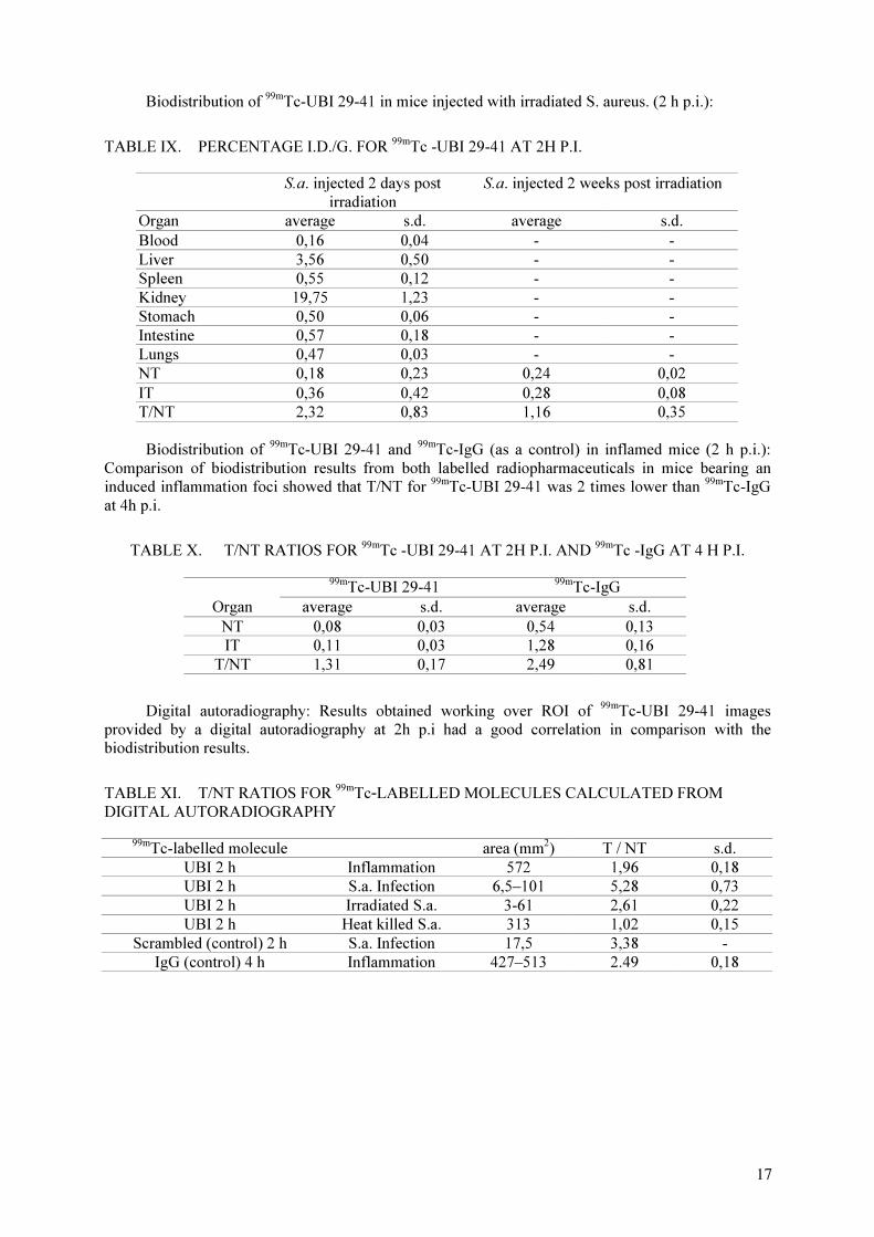
Biodistribution of 99m
Tc-UBI 29-41 in mice injected with irradiated S. aureus. (2 h p.i.):
TABLE IX. PERCENTAGE I.D./G. FOR 99m
Tc -UBI 29-41 AT 2H P.I.
S.a. injected 2 days post
irradiation
S.a. injected 2 weeks post irradiation
Organ average s.d. average s.d.
Blood 0,16 0,04 - -
Liver 3,56 0,50 - -
Spleen 0,55 0,12 - -
Kidney 19,75 1,23 - -
Stomach 0,50 0,06 - -
Intestine 0,57 0,18 - -
Lungs 0,47 0,03 - -
NT 0,18 0,23 0,24 0,02
IT 0,36 0,42 0,28 0,08
T/NT 2,32 0,83 1,16 0,35
Biodistribution of 99m
Tc-UBI 29-41 and 99m
Tc-IgG (as a control) in inflamed mice (2 h p.i.):
Comparison of biodistribution results from both labelled radiopharmaceuticals in mice bearing an
induced inflammation foci showed that T/NT for 99m
Tc-UBI 29-41 was 2 times lower than 99m
Tc-IgG
at 4h p.i.
TABLE X. T/NT RATIOS FOR 99m
Tc -UBI 29-41 AT 2H P.I. AND 99m
Tc -IgG AT 4 H P.I.
99m
Tc-UBI 29-41 99m
Tc-IgG
Organ average s.d. average s.d.
NT 0,08 0,03 0,54 0,13
IT 0,11 0,03 1,28 0,16
T/NT 1,31 0,17 2,49 0,81
Digital autoradiography: Results obtained working over ROI of 99m
Tc-UBI 29-41 images
provided by a digital autoradiography at 2h p.i had a good correlation in comparison with the
biodistribution results.
TABLE XI. T/NT RATIOS FOR 99m
Tc-LABELLED MOLECULES CALCULATED FROM
DIGITAL AUTORADIOGRAPHY
99m
Tc-labelled molecule area (mm2
) T / NT s.d.
UBI 2 h Inflammation 572 1,96 0,18
UBI 2 h S.a. Infection 6,5–101 5,28 0,73
UBI 2 h Irradiated S.a. 3-61 2,61 0,22
UBI 2 h Heat killed S.a. 313 1,02 0,15
Scrambled (control) 2 h S.a. Infection 17,5 3,38 -
IgG (control) 4 h Inflammation 427–513 2.49 0,18
17
4.2. Labelling of UBI 29-41 with 99m
Tc by indirect methods
4.2.1. Conjugation and labelling procedure
The ranges of specific activity for 99m
Tc-MAG3-UBI 29-41 and
99m
Tc-((Tricine)HYNIC-UBI
29-41) were 0.8–1.3 MBq/µg and 0.5-1.9 MBq/µg, respectively.
4.2.2. Quality control
ITLC studies: Radiochemical purity of 99m
Tc-[(Tricine)HYNIC-UBI 29-41] determined by
ITLC was 96.2 ± 3.1% (n = 3).
HPLC studies: The RP-HPLC profile of the 99m
Tc -MAG3-UBI 29-41 showed one major peak
(98.4 ± 0.9%, n = 5) at RT = 10.45 min and a second peak (1.6%) at 1.6 min corresponding to
pertechnetate. Likewise, the RP-HPLC profile of the 99m
Tc - [(Tricine)HYNIC-UBI 29-41] showed
one major peak (86.7% , n = 19) at RT = 10.47 min. The percentage of recovery was between 65–95%
of the total activity for both radiopharmaceuticals. Due to its low radiochemical purity, 99m
Tc-
[(Tricine) HYNIC-UBI 29-41] was purified by a Sep-Pack C18 cartridge before doing in vitro and in
vivo assays. The final radiochemical purity after purification was 99%. The labelling yield of native
UBI 29-41 used as a control in the MAG3-UBI 29-41 labelling method was 42%, showing a high non-
specific binding for this indirect method.
4.2.3. In vitro assays
Challenge with cysteine: It showed that 99m
Tc-MAG3-UBI 29-41 was less transchelated than
99m
Tc-[(Tricine) HYNIC-UBI 29-41] (1.7% vs 6.1% of total activity at ratio 500:1).
TABLE XII. % OF 99m
TC TRANSCHELATED TO CYSTEINE
Molar ratio (cysteine to peptide) 5 to 1 50 to 1 500 to 1
99m
Tc-MAG3-UBI 29-41 0,8 1,1 1,7
99m
Tc-[(Tricine)HYNIC-UBI 29-41] 8,8 9,4 6,1
Stability in serum:
TABLE XIII. SERUM STABILITY (% OF TOTAL ACTIVITY BOUND TO PEPTIDE)
Time (h) 3 24
99m
Tc-MAG3-UBI 29-41 - 93.8
99m
Tc-[(Tricine)HYNIC-UBI 29-41] 91,2 80,6
Binding to serum protein:
TABLE XIV. PERCENTAGE OF TOTAL ACTIVITY BOUND TO SERUM PROTEIN
Time (h) 0 24
99m
Tc-MAG3-UBI 29-41 40 49,1
99m
Tc-[(Tricine)HYNIC-UBI 29-41] 32 43
Binding to bacteria: The results of binding to bacteria of 99m
Tc-[(Tricine)HYNIC-UBI 29-41]
were better than those corresponding to 99m
Tc-MAG3-UBI 29-41 and were worse than those of UBI
29-41 labelled by direct method.( see 4.1.3.4.)
18
TABLE XV. PERCENTAGE OF TOTAL ACTIVITY BOUND TO S. AUREUS
Number of S. aureus
(CFU)
99m
Tc-MAG3-UBI 29-41
99m
Tc-[(Tricine)HYNIC-UBI 29-41]
2,50E+06 6,6 3.9
5,00E+06 6,7 11.9
1,00E+07 12,5 31.4
3,00E+07 15,9 -
4.2.4. In vivo assays
Biodistribution of 99m
Tc-MAG3-UBI 29-41 and
99m
Tc-[(Tricine)HYNIC-UBI 29-41] in S. aureus
infected mice (2 h p.i.):
TABLE XVI. % I.D./G FOR 99m
Tc -MAG3-UBI 29-41 AND 99m
Tc-((TRICINE)HYNIC-UBI 29-41)
AT 2H P.I.
99m
Tc-MAG3-UBI 29-41
99m
Tc-[(Tricine)HYNIC-UBI 29-41]
Organ average s.d. average s.d.
Blood 0,20 0,03 1,71 0,18
Liver 0,63 0,17 16,89 1,50
Spleen 0,08 0,01 0,90 0,12
Kidney 0,77 0,13 45,46 7,45
Stomach 0,37 0,49 4,08 1,81
Intestine 3,43 0,77 6,22 2,76
Lungs 0,16 0,05 2,36 0,23
NT 0,04 0,01 0,26 0,07
IT 0,06 0,02 0,45 0,09
T/NT 1,60 0,56 1,76 0,10
Digital autoradiography:
TABLE XVII. T/NT RATIOS FOR 99m
TC -LABELLED MOLECULES CALCULATED FROM
DIGITAL AUTORADIOGRAPHIES
Biomolecule n T/NT error area (mm2
)
99m
Tc-MAG3-UBI 29-41. S.a. Infection 3 3,08 1,11 184-237
99m
Tc-[(Tricine)HYNIC-UBI 29-41] S.a. Infection 3 2,15 0,53 186-247
4.3. Labelling of BPTI and HNE-2 with 99m
Tc by indirect methods
4.3.1. BPTI Conjugation and labelling with NHS-HYNIC and cDTPA
Radiochemical purity of 99m
Tc-[(Tricine) HYNIC-BPTI] and 99m
Tc-DTPA-BPTI determined by
RP-HPLC was higher than 95% and 55%, respectively. The range of specific activity of these products
was between 0.07-0.6-MBq/µg. Fraction 2 had the highest labelling yield for HYNIC-BPTI with
tricine. Fraction 1 had the highest labelling yield for DTPA-BPTI at pH 5.2. The radiochemical purity
of the C18 Sep-Pack purified 99m
Tc-DTPA-BPTI was 77%.
19

TABLE XVIII. LABELLING EFFICIENCY AND % OF RECOVERY FOR 99m
Tc-LABELLED
BPTI AND HNE-2
Fraction HYNIC-BPTI DTPA-BPTI MAG3-HNE-2 HYNIC-BPTI DTPA-BPTI MAG
3-HNE-2
1 86,1 60,9 0 -
2 94,6 ± 4,2 57,1 ± 5,0 99.0 95,8% 88% 95.7%
3 38,7 2,4 -
4 54 -
4.3.2. HNE-2 Conjugation and labelling with NHS- MAG3
Radiochemical purity of 99m
Tc-MAG3-HNE-2 determined by RP-HPLC was 99.0%. The
specific activity was range of 1.4 MBq/µg. Fraction 2 had the highest labelling yield.
4.3.3. Quality control
HPLC studies:
TABLE XIX. RETENTION TIMES AND RADIOCHEMICAL PURITIES OF 99m
Tc-LABELLED
BPTI AND HNE-2
RT(min) 99m
Tc-[(Tricine)
HYNIC-BPTI]
99m
Tc-DTPA-BPTI Sep-Pack
purified 99m
Tc-
DTPA-BPTI
99m
Tc-MAG3-
HNE-2
2.0 1.8% 37.7% 2.4% 1.0%
10.0 99.0%
11.7 98.2%
11.9 77.0%
12.3 62.3%
13.4 20.6%
4.3.4. In vitro assays
Challenge with cysteine: The results of cysteine challenge assay of labelled BPTI are similar to
labelled HNE2 and different from HNE4. The anomalous behaviour of HYNIC-BPTI was very
similar to HYNIC-HNE2. (6)
TABLE XX. % OF 99m
Tc TRANSCHELATED TO CYSTEINE
Molar ratio (cysteine to peptide) 0.5 5 50
99m
Tc-DTPA-BPTI 0 0.2 1.0
99m
Tc-[(Tricine) HYNIC-BPTI] 5.7 21.2 5.3
Stability in PBS: Both radiollabelled products show very good stability in PBS. The dissociation
of 99m
Tc-[(Tricine) HYNIC-BPTI] was less than 10% at 24 h. (analysed by RP-HPLC)
TABLE XXI. PBS STABILITY (% OF TOTAL ACTIVITY NOT BOUND TO PEPTIDE)
Time (h) 0 24
99m
Tc-DTPA-BPTI 48.0 48.6
99m
Tc-((tricine)HYNIC-BPTI) 1.0 6.2
20

Binding to serum protein: The activity bound to serum protein of 99m
Tc-[(Tricine) HYNIC-
BPTI] was 20% higher than the value of 99m
Tc-DTPA-BPTI.
TABLE XXII. % OF TOTAL ACTIVITY BOUND TO SERUM PROTEIN
Time (h) 0
99m
Tc-DTPA-BPTI 78.3
99m
Tc-[(Tricine) HYNIC-BPTI] 97.9
Binding to trypsin: Sephadex G75 radiometric elution profile of 99m
Tc-[(Tricine) HYNIC-
BPTI]: trypsin (molar ratio 1:10) showed the biggest maximum activity in the fraction number
8 corresponding to the complex and other maximum in fraction number 12 corresponding at
99m
Tc-[(Tricine) HYNIC-BPTI] (Fig. 1).
G 7 5 R a d io m e t r ic E l u t io n P r o f i l e o f
9 9 m T c -H y n ic -B P T I + T r y p s in
0
2 00 0 0 0
4 00 0 0 0
6 00 0 0 0
8 00 0 0 0
1 0 00 0 0 0
1 2 00 0 0 0
0 2 4 6 8 1 0 1 2 1 4 1 6
m l
cp
m
c p m/m l
FIG. 1. Shift in radioactivity profile of 99m
Tc-[(Tricine) HYNIC-BPTI] on binding with trypsin.
4.3.5. In vivo assays
Biodistribution of 99m
Tc-[(Tricine) HYNIC-BPTI] and 99m
Tc-DTPA-BPTI in normal mice: The
highest radioactivity levels were found in liver and spleen following administration of 99m
Tc-DTPA-
BPTI at pH 5.2. The highest radioactivity levels were found in kidneys following administration of
99m
Tc-[(Tricine) HYNIC-BPTI]. The two compounds showed different patterns of biodistribution.
First biodistribution pattern was similar to HYNIC-HNE2 and HYNIC-HNE4 and the second was
similar to DTPA-HNE2 and DTPA-HNE4 (pH 5.2) (6).
TABLE XXIII. % I.D./G FOR 99m
Tc-[(TRICINE) HYNIC-BPTI] AND 99m
Tc -DTPA-BPTI (PH 5.2)
AT 2H P.I.
99m
Tc-[(Tricine) HYNIC-BPTI] 99m
Tc-DTPA-BPTI (pH 5.2)
Organ average s.d. average s.d.
Blood 1,54 0,66 1,35 0,14
Liver 14,05 5,22 6,25 2,31
Spleen 7,02 4,96 2,22 0,66
Kidney 72,50 23,71 171,70 13,02
Stomach 1,31 0,72 0,88 0,20
Intestine 2,33 1,04 0,53 0,09
Lungs 4,32 2,28 2,81 0,67
21
5. CONCLUSIONS
Labelling UBI 29-41 by direct method showed a stable product with high yield and good in
vitro properties.
The labelled UBI 29-41 showed the highest target to non target ratio when injected in S. aureus.
infected mice compared with the labelled UBI 29-41 scrambled peptide and labelled IgG. On the other
hand, 99m
Tc-IgG showed the highest T / NT ratio in inflamed mice.
The T/NT ratio for the 99m
Tc-UBI 29-41 increased from 3.0 to 5.8 when using 80 µL of bacteria
suspension instead of 50 µL.
In the experiments with viable, irradiated and heat killed bacteria, the results showed a
decreasing T/NT ratio (3.0, 2.3 and 1.4). These data could suggest that the envelope of the non-viable
bacteria (irradiated) has not suffered modifications 48 h post irradiation since the electrostatic binding
still appear to be functioning. On the other hand, in the case bacteria two weeks post irradiation, the
T/NT ratio had no significant differences (LC = 5%) with T/NT for heat killed bacteria.
Comparing the T/NT ratios of 99m
Tc UBI 29-41 labelled by direct and two indirect methods
(HYNIC and MAG3), a decreasing uptake at the infected site was observed (3.03 ± 0.70, 1.76 ± 0.10
and 1.60 ± 0.56 respectively), although the two indirect methods did not show significant differences
(LC=5%) . These data are in agreement with the in vitro binding results. 99m
Tc-((tricine)HYNIC-UBI
29-41 showed a higher kidney elimination than the other ones. 99m
Tc-MAG3-UBI 29-41 showed a
different pattern of biodistribution.
The labelling procedures for BPTI and HNE-2 were carried out with good yields except in the
case of using cDTPA. Different patterns of biodistribution were obtained for labelled BPTI conjugated
with cDTPA and HYNIC respectively.
REFERENCES
[1] VAN DER LAKEN, C.J., et al., “Radiolabelled interleukin-8: specific scintigraphic detection of
infection within a few hours”, J. Nucl. Med. 41 (2000) 436–469.
[2] HARTWIG, W., et al., “Chemotactic peptide uptake in acute pancreatitis: correlation with tissue
accumulation of leukocytes”, J. Appl. Physiol. 87 (1999) 743–749.
[3] RUSCKOWSKI, M., et al., “Inflammation and infection imaging with a 99m
Tc-neutrophil
elastase inhibitor in monkeys”, J. Nucl. Med. 41 (2000) 363–374.
[4] WELLING, M.M., et al., “Technetium-99m labelled antimicrobial peptides discriminate
between bacterial infections and sterile inflammations”, Eur. J. Nucl. Med. 27 (2000) 292–301.
[5] WELLING, M.M., et al., “Imaging of bacterial infections with 99m
Tc-labelled human neutrophil
peptide-1”, J. Nucl. Med. 40 (1999) 2073–2080.
[6] QU, T., WANG, Z., RUSCKOWSKI, M., HNATOVICH, D.J., “Different chelators and
different peptides together influence the in vitro and mouse in vivo properties of 99m
Tc”, Nucl.
Med. Comunications 22 (2001) 202–215.
22
99m
Tc-LABELLED LIGANDS FOR INFLAMMATION AND INFECTION IMAGING
L. KOROSI, L. BALOGH, D. MATHE, A. POLYAK, R. KIRALY, GY.A. JANOKI
“Fodor József” National Center of Public Health,
National “Frédéric Joliot-Curie” Research Institute for Radiobiology and Radiohygiene,
Budapest, Hungary
Abstract
Detection of inflammation and infection foci at an early phase, non-invasivelly, sensitively and
specifically is a great challenge for nuclear medicine specialists even today. We investigated 99m
Tc-labelled EB1,
UBI and HNE2 for imaging infections. All three agents showed high labelling efficiency. The label proved to be
stable for 24 h in physiological saline and human serum. Animal examinations revealed a fast renal excretion,
low background activity for all the three molecules; 99m
Tc EB1 showed the best T/NT ratio in inflammation
models, 99m
Tc UBI proved to be specific for infection imaging and 99m
Tc HNE2 was found to be useful only in
the primate model.
1. INTRODUCTION
To achieve a more desirable radiopharmaceutical for infection imaging, one displaying a faster
blood clearance, less non-target uptake and better concentration in infection/inflammation sites, it will
be necessary to exploit alternate biochemical pathways. Some of these pathways like antimicrobial
peptides have been recently explored with promising results. The recent years have also witnessed
significant advances in 99m
Tc radiochemistry and rational approaches to 99m
Tc labelling of different
molecules have been introduced.
In our study different specific and non-specific agents were evaluated in normal and
infection/inflammation induced animals following the protocols finalized in the 1st
RCM.
2. MATERIALS AND METHODS
In this work we labelled and tested the following agents:
- ethylendiaminetetraacetic acid biotin monomer (EB1, kit form, Polatom, Poland))
- ubiquicidin (UBI, kit form, Polatom, Poland)
- DTPA– HNE2 conjugate
- HYNIC- UBI conjugate
- DTPA –UBI conjugate
- HNE2
2.1. Radiolabelling
All the radiolabelling reactions were performed as described in the reports of 1st
and 2nd
Research Coordination Meeting of the CRP and the Leiden Protocol.
2.1.1.99m
Tc EB1
The kit obtained from POLATOM was labelled with 99m
Tc as per protocol recommended.
Quality control: The yield of kit containing EB1 was determined by ITLC using 0.9% saline
(ITLC-SG will assess the percent of 99m
Tc present as colloid Rf=0.0) and acetone (ITLC-SG or
Whatman No.1, will assess the percent as pertechnetate Rf=0.9) as eluent. Samples of 1-2 µL were
applied at 3 cm from the bottom. After developing, the chromatography sheeet was sliced into 1 cm
sections and radioactivity in each measured by gamma counter (Table I). Size exclusion HPLC
(BIORAD 800) runs were performed as follow: linear gradient, Solvent: 0.1 M phosphate buffer, Flow
rate: 1mL/min, UV:280 nm, Column: TSK-6-3000 SW
23
2.1.2.99m
Tc UBI
To 10 µL 1mM peptide solution in 0.01M acetic acid pH 4, was added 4 µL of 0.5 mg/mL of
stannous pyrophosphate; immediately thereafter 4µL of a solution of 10 mg of KBH4
(SIGMA) per
mL of 0.1M NaOH was added. Solution was freeze-dried and kit form was provided from Polatom,
Poland.
After addition of 0.1 mL of 99m
Tc-sodium-pertechnetate (200-800 MBq/mL), the mixture stirred
at room temperature for 60 min.
2.1.3. 99m
Tc DTPA-HNE-2
Conjugation: To 0.1 mL HNE-2 solution was added 0.1 mL 0.5 M NaHCO 3 solution. 0.1 mL
suspension of 0.1 mg DTPA in DMF was then added with shaking. After 30 min. incubation at room
temperature the coupled peptide was purified on a 0.7×20 cm G-25 column with 0.25 M ammonium
acetate buffer, pH 5.2 as eluent. The elution was monitored at U.V. 280 nm. The conjugated peptide
eluted in 2-5 mL range.
Labelling: The conjugated peptide concentration was 0.5 ug/uL. To the peptide 18 ul buffer
consisting of 0.5 M sodium bicarbonate, 0,25 M ammonium acetate and 0,18 M ammonium hydroxide
at pH 9.2 were added. Finally, 0.1 mL /500 MBq of 99m
Tc -pertechnetate and 40 µL fresh solution of
SnCl2.2H
2O solution (1 mg/mL in 10 mM HCl) were added. The final pH was 7.6. After incubation at
room temperature for 1h the labelled peptide was purified over Sephadex G-25 column.
Radiochemical analysis: ITLC-SG in saline-hydrolysed 99m
Tc activity remained at the origin;
ITLC-SG in acetone-all the hydrolized and labelled 99m
Tc activity remain at origin; HPLC analysis
with Chrompack-SS RP-18 column (250×4 mm) was used with elution programme as given in the
Leiden Protocol.
2.1.4. 99m
Tc HYNIC –UBI
Conjugation: 1 mg UBI was dissolved in 0.4 mL bicarbonate buffer pH 8.5 and 1.2.mg NHS
HYNIC in 0.1 mL dry DMF was added with shaking. After 2 h at room temperature the mixture was
purified with Sephadex G-25 column 0.7× 20 cm with 0.25 M ammonium acetate buffer pH 5.2 . The
peak fractions were collected and dispensed in 5 vials each containing ~0.2 mg peptide.
Radiolabelling employed tricine as the coligand. To 50 µg of conjugated peptide in 0.25 M
ammonium acetate, pH 5.2 were added 99m
Tc pertechnetate (500 MBq / 0.1 mL) and 50 µL of tricine
solution in water (100 µg/µL). Finally 20 µl of fresh SnCl2.2H
2O solution (1 mg/mL) was added. The
final pH was 5.3. After incubation at room temperature for 60 min, the labelled peptide was purified
over Sephadex G-25 column with 50 mM PBS, pH 7.2.
Radiochemical Analysis: ITLC-SG in saline, ITLC SG in acetone and HPLC as described above
2.1.5. 99m
Tc DTPA–UBI
Conjugation: 1.0 mg peptide was dissolved in 0.1 mL water, and then 0.1 mL 0.5 M NaHCO3
solution was added. 3 .0 mg DTPA anhydride dissolved 0.1 mL dry DMF. From DTPA suspension 0.1
mL was added to protein with shaking. After 1 h incubation at room temperature, the coupled peptide
was purified on a 0.7×20 cm G-25 column, with 0.25 M ammonium acetate buffer, pH 5.2.The
elution was monitored at U.V. 280 nm. The eluted conjugated peptide was kept frozen in 1 mL lots
Labelling: To 0.1 mg DTPA–UBI in 20 µL buffer pH 9.2, 0.1 mL (500 MBq) of 99m
Tc-pertechnetate
and 40 µL SnCl2 × 2H
2O fresh solution (1 mg/mL in 10 mM HCl) were added. The final pH was 7.6.
After incubation at room temperature for 2 h labelled peptide was purified over Sephadex G-25
column.
24
Radiochemical analysis by ITLC and HPLC was carried about as described earlier.
2.2. Pathological animal models
2.2.1. Sterile inflammation induction
Heat killed E. coli (0101-RG/W) bacteria (endotoxin, lipopolysaccharide – LPS) were used for
inducing sterile muscle inflammation. Two hundred microgramm LPS in 100 µL physiological saline
was deeply injected into the right thigh muscle in C57/Black mice 24 h before further examinations.
The injected LPS in the recommended dose did not cause detectable signs of inflammation in
the animals. On that basis we choose a 4 times higher concentration of injected LPS. Even that model
showed different (from mild to hard hemorrhagic) severity of muscle inflammation.
2.2.2. Bacterial infection induction
Staphylococcus aureus (OKI-110003) was chosen as microorganism for muscle infection
induction. Frozen bacteria suspension was cultured by 24 h incubation at 37o
C in shaking waterbath.
Staphylococcus aureus bacterial suspension in 107
CFU / 100 µL was injected deeply into the right
thigh muscle 24 h before further examinations.
2.2.3. Bioassay in mice
Animals were injected into the tail veins with labelled compounds. The same dose was injected
in a 50 mL glass for estimating the injected dose. Fifteen minutes, 2 h and 24 h after injection, three
mice were sacrificed in each group. Organs e.g.: tail, blood, muscle, bone, heart, larynx with thyroids,
lungs, liver, spleen, kidneys, gastric, small intestines, large intestines, and muscle with induced
infection/inflammation were removed, weights and activities were measured, induced / normal muscle
ratios and standard biodistribution were calculated as I.D. / whole organ and I.D. / gram tissue.
2.2.4. Imaging of infectio/inflammation in dogs
Infection and inflammation were similarly induced in Beagle dogs. Static gamma camera
imaging was done 20 mts post injection of 280 MBq/1.5 ml 99m
Tc EB1 and 15 min after 320 MBq/1.6
ml 99m
Tc UBI from kit, respectively
3. RESULTS AND DISCUSSIONS
The results of ITLC and HPLC are given in Tables I, II and III. All the examined agents were
found to show good (>90%) labelling efficiency. 99m
Tc EB1 was stable up to 24 h after labelling using
700 MBq added activity.
Labelling of the kit form of UBI revealed high and stable labelling efficiency only up to 10 – 20
MBq activity. Higher (500 – 700 MBq) activities and greater labelling volume (over 1 mL) revealed
not satisfactory labelling yield (app. 80%) and stability. The labelled componds were found to be
stable also in human serum up to 24 h.
Basically all agents showed a rapid clearance via kidneys resulted low background activities in
the blood and other organs. Selected results on animal distribution are given in Tables IV and V.
Normal biodistribution studies may not be necessary, because induced animal model biodistributions
show very similar to data of “other” organs. Induced muscle inflammation and infections vary on a
wide scale from mild to severe. Other sterile inflammation models are necessary.
Fifteen (15) min, 2 h, 4 and/or 6 h, 24 h (12 h and 18 h also) would be recommended for
biodistribution and imaging studies. This will also help in calculation of mean residency times and
25

internal dosimetry data. Labelled HNE-2 was investigated only by scintigraphy in Rhesus monkey.
Images showed high thyroid and stomach activity which may be due to poor labelling efficiency or in
vivo instability.
Kit form of EB1 and UBI (both from Polatom) showed very similar results on 99m
Tc labelling
and in animal experiments than the earlier in-house forms. 99m
Tc EB1 imaging showed high uptake in
inflammation site in dog (Fig.1). 99m
Tc UBI showed lower but specific uptake in infection site in dog
(Fig. 2.)
TABLE I. PAPER CHROMATOGRAPHY RESULTS (N=3)
TABLE II. ITLC ANALYSIS RESULTS(N=3)
99m
Tc - EB1 99m
Tc - UBI
Conjugation of
HNE2 with
DTPA
Conjugation of
UBI with
Hynic
Conjugation of
UBI with
DTPA
Labelling efficiency (in %)
After labelling 93,6 ± 0,1 99,0 ± 0,1 97,5 ± 0,5 92,0 ± 0,9 92,1 ± 0,2
After 3 h 92,4 ± 0,3 97,9 ± 0,4 97,5 ± 0,4 91,7 ± 0,5 87,7 ± 0,2
After 6 h 90,7 ± 0,4 92,5 ± 0,5 94,4 ± 0,6 86,9 ± 0,2 82,9 ± 0,2
TABLE III. REVERSE-PHASE HPLC ANALYSIS RESULTS
99m
Tc - EB1 99m
Tc – UBI
Conjugation of
HNE2 with
DTPA
Conjugation of
UBI with Hynic
Conjugation of
UBI with DTPA
After labelling 99,0% ± 0,1 96,2% ± 0,1 97,3 ± 0,1 92,6 ± 0,3 93,6 ± 0,8
After 3 h 98,3% ± 0,1 94,6% ± 0,1 96,6 ± 0,7 91,5 ± 0,9 88,3 ± 0,1
After 6 h 93,2% ± 0,2 92,8% ± 0,2 93,9 ± 0,1 86,9 ± 06 84,2 ± 0,1
99m
Tc - EB1 99m
Tc - UBI
Conjugation of
HNE2 with
DTPA
Conjugation of
UBI with Hynic
Conjugation of
UBI with
DTPA
Labelling efficiency (in %)
After labelling 97,8 ± 0,2 98,3 ± 0,2 96,3 ± 0,1 87,0 ± 0,2 88,7 ± 0,5
After 3 h 94,4 ± 0,9 97,1 ± 0,3 96,1 ± 0,1 83,2 ± 0,3 85,9 ± 0,9
After 6 h 94,2 ± 0,6 95,5 ± 0,1 95,3 ± 0,7 82,6 ± 0,6 86,8 ± 0,5
26

TABLE IV. 99m
Tc EB1 BIODISTRIBUTION IN INDUCED MICE
No. 1 No. 2 No. 3
I.D.% / I.D.% / I.D.% / I.D.% / I.D.% / I.D.% / Average SD+
1g tissue w.organ 1g tissue w.organ 1g tissue w.organ
15 min
Tail 3,6 1,52 3,97 1,76 3,42 1,82 3,66 0,28
Blood 12,33 13,48 9,04 15,32 12,10 11,72 11,16 1,84
Control muscle 0,98 5,32 1,42 6,12 1,28 4,56 1,23 0,22
Induced muscle 4,01 0,48 2,99 0,28 2,76 0,32 3,25 0,67
Bone 1,02 1,98 0,96 1,98 1,06 2,34 1,01 0,05
Heart 1,52 0,33 1,54 0,24 1,62 0,22 1,56 0,05
Thyroid 5,26 0,23 5,52 0,32 4,83 0,23 5,20 0,35
Lungs 3,61 0,62 3,88 0,93 3,95 0,91 3,81 0,18
Liver 4,62 6,22 4,58 5,32 4,48 4,92 4,56 0,07
Spleen 1,58 0,21 1,99 0,20 1,71 0,23 1,76 0,21
Kidneys 14,13 4,49 12,10 4,01 13,01 4,36 13,08 1,02
Stomach 14,85 2,78 14,45 2,69 13,28 2,92 14,19 0,82
Small intestine 4,96 4,32 4,01 3,98 4,66 4,56 4,54 0,49
Large intestine 1,58 1,92 1,23 1,97 1,97 0,99 1,59 0,37
Activity % 43,9 % 45,12 % 40,10 % 43,04 % 2,62
Ind./Cont. M. 4,09 X 2,11 X 2,16 X 2,78 X 1,13
2 h
Tail 2,6 1,58 1,87 1,23 2,56 1,84 2,34 0,41
Blood 3,59 3,2 1,86 2,22 2,92 2,88 2,79 0,87
Control muscle 1,12 1,96 0,67 2,98 0,34 1,24 0,71 0,39
Induced muscle 3,55 0,18 3,22 0,24 1,25 0,22 2,67 1,24
Bone 0,58 1,12 0,58 1,41 1,12 1,89 0,76 0,31
Heart 0,45 0,12 0,92 0,21 1,23 0,15 0,87 0,39
Thyroid 1,9 0,11 0,23 0,12 2,98 0,34 1,70 1,39
Lungs 2,33 0,54 2,43 0,75 4,23 0,65 3,00 1,07
Liver 4,78 4,99 4,22 4,96 5,12 5,33 4,71 0,45
Spleen 2,11 0,51 1,99 0,22 2,12 0,34 2,07 0,07
Kidneys 8,68 3,11 8,45 4,04 9,12 1,99 8,75 0,34
Stomach 7,22 0,98 16,56 1,54 8,45 2,01 10,74 5,07
Small intestine 6,54 5,99 7,02 5,45 9,34 6,76 7,63 1,50
Large intestine 3,32 1,67 3,45 2,81 3,65 1,88 3,47 0,17
Activity % 26,06 % 28,18 % 27,52 % 27,25 % 1,08
Ind./Cont. M. 3,17 X 4,81 X 3,68 X 3,88 X 0,84
24 h
Tail 3 1,72 2,12 1,49 3,67 2,00 2,93 0,78
Blood 0,44 0,34 0,55 0,31 0,61 0,67 0,53 0,09
Control muscle 0,22 0,64 0,22 0,56 0,15 0,42 0,20 0,04
Induced muscle 0,78 0,21 0,87 0,31 0,87 0,25 0,84 0,05
Bone 0,44 0,86 0,67 0,71 0,56 0,91 0,56 0,12
Heart 0,23 0,02 0,65 0,09 0,67 0,08 0,52 0,25
Thyroid 1,78 0,05 1,45 0,04 1,24 0,05 1,49 0,27
Lungs 1,91 0,56 0,65 0,19 0,86 0,45 1,14 0,68
Liver 3,98 2,99 3,55 3,29 2,96 2,12 3,50 0,51
Spleen 0,67 0,06 0,88 0,06 0,81 0,06 0,79 0,11
Kidneys 4,56 1,41 5,34 1,88 4,01 1,96 4,64 0,67
Stomach 3,23 0,96 2,99 0,16 4,24 0,78 3,49 0,66
Small intestine 1 1,02 1,23 0,76 0,98 0,66 1,07 0,14
Large intestine 19,56 6,56 17,34 5,45 21,45 6,91 19,45 2,06
Activity % 17,4 % 15,30 % 17,32 % 16,67 % 1,19
Ind./Cont. M. 3,55 X 3,95 X 5,80 X 4,43 X 1,20
27

TABLE V. 99m
Tc UBI BIODISTRIBUTION IN INDUCED MICE
No. 1 No. 2 No. 3
I.D.% / I.D.% / I.D.% / I.D.% / I.D.% / I.D.% / Average SD+
1g tissue w. organ 1g tissue w. organ 1g tissue w. organ
15 min
Tail 13,99 7,83 7,99 3,95 5,96 2,67 9,31 4,18
Blood 4,56 4,12 3,89 2,98 3,45 4,01 3,97 0,56
Control muscle 0,39 1,66 0,66 3,18 0,44 1,98 0,50 0,14
Induced muscle 1,58 0,44 1,20 0,33 0,79 0,13 1,19 0,40
Bone 2 3,98 2,13 4,00 2,86 5,23 2,33 0,46
Heart 1,23 1,23 1,45 0,24 1,34 0,16 1,34 0,11
Thyroid 0,12 0,01 0,92 0,02 0,24 0,03 0,43 0,43
Lungs 7,51 0,65 6,54 1,34 5,92 1,20 6,66 0,80
Liver 59,76 48,23 59,25 52,13 59,23 50,12 59,41 0,30
Spleen 23,12 2,56 40,12 2,87 40,12 3,01 34,45 9,81
Kidneys 12,23 3,94 19,12 3,29 20,26 5,12 17,20 4,34
Stomach 1,2 0,34 1,56 0,90 1,65 0,45 1,47 0,24
Small intestine 0,9 1,11 1,84 1,41 1,02 0,65 1,25 0,51
Large intestine 0,87 0,55 0,93 0,55 2,02 0,97 1,27 0,65
Activity % 76,65 77,19 % 75,73 % 76,52 % 0,74
Ind./Cont. M. 4,05 X 1,82 X 1,80 X 2,55 X 1,30
2 h
Tail 15,02 2,01 2,02 0,2 3,02 1,06 6,69 7,23
Blood 0,87 0,86 0,92 0,98 0,87 0,99 0,89 0,03
Control muscle 0,22 0,65 0,23 0,78 0,26 0,99 0,24 0,02
Induced muscle 0,33 0,19 0,7 0,13 0,82 0,12 0,62 0,26
Bone 0,62 1,24 1,67 2,2 1,88 2,11 1,39 0,68
Heart 0,56 0,09 0,88 0,06 0,89 0,07 0,78 0,19
Thyroid 0,59 0,01 0,86 0,01 0,56 0,02 0,67 0,17
Lungs 6,34 0,3 6,23 0,32 5,92 1,34 6,16 0,22
Liver 48,97 46,78 52,54 50,12 49,78 46,43 50,43 1,87
Spleen 21,45 3,23 27,65 2,11 29,78 2,79 26,29 4,33
Kidneys 19,86 4,58 15,78 3,96 16,23 3,67 17,29 2,24
Stomach 0,44 0,07 0,66 0,34 0,98 0,08 0,69 0,27
Small intestine 1,45 1,9 1,66 1,87 1,45 0,92 1,52 0,12
Large intestine 0,45 0,25 0,44 0,19 1,23 0,98 0,71 0,45
Activity % 62,16 63,27 % 61,57 % 62,33 % 0,86
Ind./Cont. M. 1,50 X 3,04 X 3,15 X 2,57 X 0,92
24 h
Tail 2,78 1,98 4,33 1,28 3,87 1,21 3,66 0,80
Blood 0,45 0,65 0,43 0,33 0,34 0,42 0,41 0,06
Control muscle 0,13 0,55 0,13 0,23 0,14 0,66 0,13 0,01
Induced muscle 0,28 0,03 0,44 0,14 0,22 0,04 0,31 0,11
Bone 1,56 2,98 1,45 1,94 1,23 1,92 1,41 0,17
Heart 0,34 0,04 0,32 0,03 0,32 0,02 0,33 0,01
Thyroid 0,31 0,01 0,44 0,02 0,32 0,01 0,36 0,07
Lungs 1,93 0,11 0,72 0,44 1,82 0,09 1,49 0,67
Liver 6,58 4,12 5,92 5,24 4,23 3,98 5,58 1,21
Spleen 34,23 2,65 31,23 1,99 29,34 2,12 31,60 2,47
Kidneys 12,45 1,87 11,67 2,14 16,78 2,34 13,63 2,75
Stomach 1,45 1,56 1,12 1,97 1,45 0,97 1,34 0,19
Small intestine 0,45 0,12 0,44 0,67 0,65 0,56 0,51 0,12
Large intestine 2,04 0,98 2,91 1,24 2,56 1,91 2,50 0,44
Activity % 17,65 % 17,66 % 16,25 % 17,19 % 0,81
Ind. / Cont. M. 2,15 X 3,38 X 1,57 X 2,37 X 0,93
28

FIG.1.99m
Tc EB1 imaging in inflammation induced beagle dog.
FIG.2.99m
Tc UBI imaging in infection induced beagle dog.
29
4. CONCLUSIONS
On the basis of IAEA supported CRP we can conclude that 99m
Tc EB1 and 99m
Tc UBI have the
potential for a diagnostic kit in the field of inflammation and infection imaging. Further studies are
needed to evaluate available agents in term of differential diagnosis of bacterial and non-bacterial
(sterile) inflammation.
ACKNOWLEDGEMENTS
The authors would like to thank the IAEA for the generous support, R. Mikolajczak,
D.J. Hnatowich and E.K.J. Pauwels for sending test materials and M. Pállai, Z. Suhajda, C. Dirner,
K. Haller, N. Fésűs, Z.K. Gombos and I. Dékány for their excellent technical assistance and
preparation of this manuscript.
BIBLIOGRAPHY
BRITTON, K.E., et al., “Imaging bacterial infection with (99m
)Tc-ciprofloxacin (Infecton)”, Clin.
Pathol. 55 (2002) 817–823.
FERRO-FLORES, G., et al., “In vitro and in vivo assessment of 99m
Tc-UBI specificity for bacteria”,
Nucl. Med. Biol. 30 (2003) 597–603.
HNATOWICH, D.J., et al., “Labelling peptides with technitium-99m using a bifunctional chelator of a
N-hydroxysuccinimide ester of mercaptoacetriglycine”. J. Nucl. Med. 39 (1998) 56–64.
KŐRÖSI, L., BALOGH, L., ANDÓCS, G., JÁNOKI, GY.A., “Labelling analytical evaluation and
biodistribution of 99m
Tc-UBI and 99m
Tc-EB1”, Book of Abstracts, 11th
European Symposium on
Radiopharmacy and Radiopharmaceuticals, Innsbruck, Austria (2003) 75.
KŐRÖSI, L., et al., “In vitro and in vivo evaluation of new 99m
Tc-labelled inflammation and infection
agents”, Nucl. Med. Review 6 (2003) 72.
LUPETTI, A., PAUWELS, E.K., NIBBERING, P.H., WELLING, M.M., “99m
Tc antimicrobial
peptides: promising candidates for infection imaging”, Q.J. Nucl. Med. 47 (2003) 238–245.
NIBBERING, P.H., et al., “99m
Tc-Labelled UBI 29-41 peptide for monitoring the efficacy of
antibacterial agents in mice infected with Staphylococcus aureus”, J. Nucl. Med. 45 (2004) 321–326.
PAUWELS, E.K.J, WELLING, M.M., FEITSMA, R.I.J., ATSMA, D.E., NIEUWENHUIZEN, W.,
“The labelling of proteins and LDL with Tc-99m: a new direct method employing KBH4 and stannous
chloride”, Nucl. Med. Biol. 20 (1993) 825–833.
RUSCKOWSKI, M., et al., “Imaging osteomyelytis with streptavidin and indium-111 labelled biotin”,
J. Nucl. Med. 37 (1996) 1655–1662.
30
PREPARATION AND EVALUATION OF 99m
Tc LABELLED
RADIOPHARMACEUTICALS AND FORMULATION OF KITS FOR IMAGING
INFECTION/INFLAMMATION
A. BHELOSE, J.S. SARNAIK, A. RAJU, K.S. MEHRA, Y. SINGH, P. KUMAR,
A. CHANDA, D.K. RANGANATHA, S.H. JOSHI, N. RAMAMOORTHY
Board of Radiation and Isotope Technology, Department of Atomic Energy,
BARC Vashi Complex, Mumbai, India
Abstract
Three agents were studied for infection/inflammation imaging under the CRP. EDTA-biotin monomer
(EB1) offers advantages of ease of in-house synthesis, being inexpensive and showing better pharmacokinetics,
as compared to currently used radiopharmaceuticals for inflammation imaging. Hence we had placed additional
emphasis on this product starting from the synthesis step. The synthesized product was also provided under CRP.
Lyophilized kit for 99m
Tc-EB1 was standardized and evaluated to satisfaction in terms, of >95% yield, >95% RC
purity and ratio of uptake in inflamed tissue to normal being around 1.5–2 in mice. Among the other products,
Ubiquicidine fragment, UBI 29-41, an antimicrobial peptide (made available under CRP) was radiolabelled by
both direct method as well as by indirect method using DTPA as bifunctional chelating agent. Direct labelling of
UBI resulted in product of high purity and stability (>90% labelling yield and >95% RC purity was obtained),
but radiolabelling of UBI using DTPA as bifunctional chelating agent was not satisfactory. The biological
efficacy of 99m
Tc-UBI was shown by both bacterial (S.aureus) binding studies (>30%) and higher uptake in
muscle with induced infection, as compared to the results with 99m
Tc-scrambled peptide of UBI analog. The
radiolabelling of UBI using HYNIC as bifunctional chelating agent was not fully optimized for obtaining high
labelling efficiency. Human neutrophil elastase inhibitor (HNE2) (made available under CRP) was radiolabelled
with 99m
Tc using MAG3 as bifunctional chelating agent. The procedures of conjugation with NHS-MAG3 and
radiolabelling of the conjugate with 99m
Tc were first authenticated using the suggested reference compound,
BPTI. The studies on radiolabelling of BPTI were also performed with DTPA as bifunctional chelating agent.
However, the studies with HNE2 could not be completed due to non-availability of HNE2.
1. INTRODUCTION
The ability to image infection specifically is a very useful tool in the management of patients,
especially suffering from fever (pyrexia) of unknown origin (FUO/PUO) and in such other suspected
cases of occult infection. As inflammatory process is always associated with any active infection,
many procedures depend on imaging inflammation more often. Since differential diagnosis from
sterile inflammation would be more informative, the search for products for infection imaging based
on sound rationale has been continued by many groups world over. IAEA accordingly supported the
present CRP in an attempt to channel efforts towards the accomplishment of a specific infection
imaging agent. The features of the existing products such as 67
Ga-citrate, leucocytes labelled with 111
In
/99m
Tc, 11I
In/99m
Tc-human immunoglobulin (HIG), 99m
Tc-ciprofloxacin, etc. are well known, as are
their limitations. Based on detailed project formulation discussions held in the First RCM of the
present CRP, three compounds were chosen for detailed investigation by the participant groups who
were to follow a harmonized protocol in preparation and evaluation (Report of First RCM of the
CRP). The three agents studied for infection/inflammation imaging under the CRP are: EDTA-biotin
monomer (EB1), Ubiquicidine fragment, UBI 29-41, an antimicrobial peptide and human neutrophil
elastase inhibitor (HNE2). The salient details of the studies carried out and the major findings are
reported here.
31

2. METHODS
2.1. HNE-2 - Conjugation of BPTI and HNE2 with NHS-MAG3
Conjugation of BPTI and HNE2 with NHS-MAG3 was carried out at the molar ratio of 4:1 of
NHS-MAG3: BPTI and the reaction mixture purified over P4 column (20×0.7cm). 100 µl aliquots of
suitable fractions were dispensed and stored at -200
C until use.
2.2. UBI - Conjugation of UBI with cDTPA
Conjugation of UBI with cDTPA was carried out at the molar ratio of cDTPA : UBI:: 5:1. The
conjugation of scrambled peptide for UBI was also carried out using the same procedure.
2.3. EB1 - Synthesis of biotinyl hydrazine-EDTA (EDTA biotin monomer, EB1)
To a stirred solution of biotin hydrazide (50 mg, 0.194 mM) in 20 mL of mixture of dry DMF:
CHCl3, 1:1 (v/v) at 65
0
C, was added 20 mL of mixture of dry DMF: CHCl3, 1:1 (v/v) containing
EDTA bicyclic anhydride (65.0 mg, 0.254 mM). The mixture was stirred for 30 min, hydrolyzed by
the addition of 0.5 mL of water and stirred for additional 10 min. The reaction mixture was poured
into 300 mL of dry dichloromethane and extracted twice with 15 mL of distilled water. Water was
removed by rotary evaporation. To the small amount of liquid that remained in the round bottom flask,
dry dichloromethane was added. The precipitated EB1 was filtered, washed with dry acetonitrile and
dried by vacuum desiccation. Crude EB1 was redissolved in 1 mL of 0.2 M sodium bicarbonate pH
8.5 and purified over P2 (120×1.4 cm) column using water as eluent. 1 mL fractions were collected
and absorbance was measured at 220 nm. Suitable fractions were pooled and pure EB1 was recovered
by rotary evaporation. White shiny crystals obtained were characterized for MP (186–1920
C dec), IR
(IR cm-1
(KBr pellets): 1660 (-CO- of amide); 3300 (-NH- of amide)) and NMR. A sample of this EB1
was distributed to CRP participants in the 2nd
RCM held in Mexico.
Kit formulation for EB1: Kit formulations with 200µg of EB1 and different stannous tin
contents of 20, 50, 75 and 100 µg were prepared, lyophilized and evaluated. Kit containing 200µg
EB1, 20µg SnCl2.2H
2O, 2mg NaCl was finally chosen and lyophilized kits were made. Kit samples
were distributed to all the CRP participants at the 2nd
RCM held in Mexico.
2.4. Radiolabelling with 99m
Tc
Labelling of conjugates of BPTI-MAG3, HNE2-MAG3, UBI-DTPA and scrambled (UBI)
peptide-DTPA was carried out and the labelling efficiency (LE) determined. In case of UBI,
radiolabelling was also carried out by direct method.
Radiolabelling of EB1 with 99m
Tc: The kit vial was allowed to attain the ambient temperature.
2mL of 99m
Tc-pertechnetate was added to the kit vial. After mixing, the reaction vial was allowed to
stand for 20 min at room temperature and the labelling efficiency determined.
2.5. Quality assurance and quality control testing
2.5.1. 99m
Tc HNE2
The radiolabelled conjugates of BPTI and HNE2-MAG3 were tested for kinetics and stability,
stability in human serum and cysteine challenge test. The test for trypsin binding described below was
carried out with BPTI-MAG3 conjugate labelled with 99m
Tc in order to ensure the retention of the
biological activity of the molecule
32
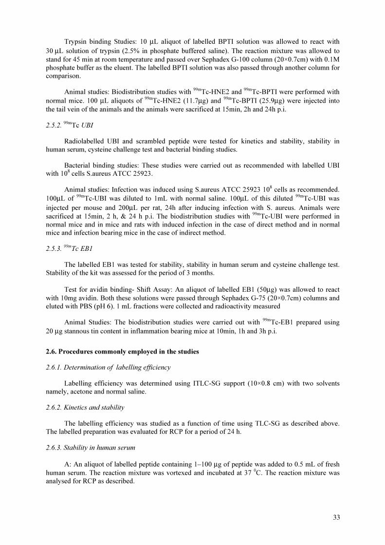
Trypsin binding Studies: 10 µL aliquot of labelled BPTI solution was allowed to react with
30 µL solution of trypsin (2.5% in phosphate buffered saline). The reaction mixture was allowed to
stand for 45 min at room temperature and passed over Sephadex G-100 column (20×0.7cm) with 0.1M
phosphate buffer as the eluent. The labelled BPTI solution was also passed through another column for
comparison.
Animal studies: Biodistribution studies with 99m
Tc-HNE2 and 99m
Tc-BPTI were performed with
normal mice. 100 µL aliquots of 99m
Tc-HNE2 (11.7µg) and 99m
Tc-BPTI (25.9µg) were injected into
the tail vein of the animals and the animals were sacrificed at 15min, 2h and 24h p.i.
2.5.2.99m
Tc UBI
Radiolabelled UBI and scrambled peptide were tested for kinetics and stability, stability in
human serum, cysteine challenge test and bacterial binding studies.
Bacterial binding studies: These studies were carried out as recommended with labelled UBI
with 108
cells S.aureus ATCC 25923.
Animal studies: Infection was induced using S.aureus ATCC 25923 108
cells as recommended.
100µL of 99m
Tc-UBI was diluted to 1mL with normal saline. 100µL of this diluted 99m
Tc-UBI was
injected per mouse and 200µL per rat, 24h after inducing infection with S. aureus. Animals were
sacrificed at 15min, 2 h, & 24 h p.i. The biodistribution studies with 99m
Tc-UBI were performed in
normal mice and in mice and rats with induced infection in the case of direct method and in normal
mice and infection bearing mice in the case of indirect method.
2.5.3. 99m
Tc EB1
The labelled EB1 was tested for stability, stability in human serum and cysteine challenge test.
Stability of the kit was assessed for the period of 3 months.
Test for avidin binding- Shift Assay: An aliquot of labelled EB1 (50µg) was allowed to react
with 10mg avidin. Both these solutions were passed through Sephadex G-75 (20×0.7cm) columns and
eluted with PBS (pH 6). 1 mL fractions were collected and radioactivity measured
Animal Studies: The biodistribution studies were carried out with 99m
Tc-EB1 prepared using
20 µg stannous tin content in inflammation bearing mice at 10min, 1h and 3h p.i.
2.6. Procedures commonly employed in the studies
2.6.1. Determination of labelling efficiency
Labelling efficiency was determined using ITLC-SG support (10×0.8 cm) with two solvents
namely, acetone and normal saline.
2.6.2. Kinetics and stability
The labelling efficiency was studied as a function of time using TLC-SG as described above.
The labelled preparation was evaluated for RCP for a period of 24 h.
2.6.3. Stability in human serum
A: An aliquot of labelled peptide containing 1–100 µg of peptide was added to 0.5 mL of fresh
human serum. The reaction mixture was vortexed and incubated at 37 0
C. The reaction mixture was
analysed for RCP as described.
33
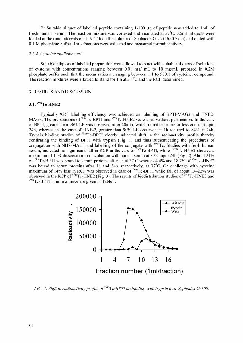
B: Suitable aliquot of labelled peptide containing 1-100 µg of peptide was added to 1mL of
fresh human serum. The reaction mixture was vortexed and incubated at 370
C. 0.5mL aliquots were
loaded at the time intervals of 1h & 24h on the column of Sephadex G-75 (16×0.7 cm) and eluted with
0.1 M phosphate buffer. 1mL fractions were collected and measured for radioactivity.
2.6.4. Cysteine challenge test
Suitable aliquots of labelled preparation were allowed to react with suitable aliquots of solutions
of cysteine with concentrations ranging between 0.01 mg/ mL to 10 mg/mL prepared in 0.2M
phosphate buffer such that the molar ratios are ranging between 1:1 to 500:1 of cysteine: compound.
The reaction mixtures were allowed to stand for 1 h at 37 0
C and the RCP determined.
3. RESULTS AND DISCUSSION
3.1.99m
Tc HNE2
Typically 93% labelling efficiency was achieved on labelling of BPTI-MAG3 and HNE2-
MAG3. The preparations of 99m
Tc-BPTI and 99m
Tc-HNE2 were used without purification. In the case
of BPTI, greater than 90% LE was observed after 20min, which remained more or less constant upto
24h, whereas in the case of HNE-2, greater than 90% LE observed at 1h reduced to 84% at 24h.
Trypsin binding studies of 99m
Tc-BPTI clearly indicated shift in the radioactivity profile thereby
confirming the binding of BPTI with trypsin (Fig. 1) and thus authenticating the procedures of
conjugation with NHS-MAG3 and labelling of the conjugate with 99m
Tc. Studies with fresh human
serum, indicated no significant fall in RCP in the case of 99m
Tc-BPTI, while 99m
Tc-HNE2 showed a
maximum of 11% dissociation on incubation with human serum at 370
C upto 24h (Fig. 2). About 21%
of 99m
Tc-BPTI was bound to serum proteins after 1h at 370
C whereas 4.4% and 18.7% of 99m
Tc-HNE2
was bound to serum proteins after 1h and 24h, respectively, at 370
C. On challenge with cysteine
maximum of 14% loss in RCP was observed in case of 99m
Tc-BPTI while fall of about 13–22% was
observed in the RCP of 99m
Tc-HNE2 (Fig. 3). The results of biodistribution studies of 99m
Tc-HNE2 and
99m
Tc-BPTI in normal mice are given in Table I.
0
50000
100000
150000
200000
1 4 7 10 13 16
Fraction number (1ml/fraction)
Ra
dio
ac
tiv
ity
. Without
trypsin
With
i
FIG. 1. Shift in radioactivity profile of 99m
Tc-BPTI on binding with trypsin over Sephadex G-100.
34
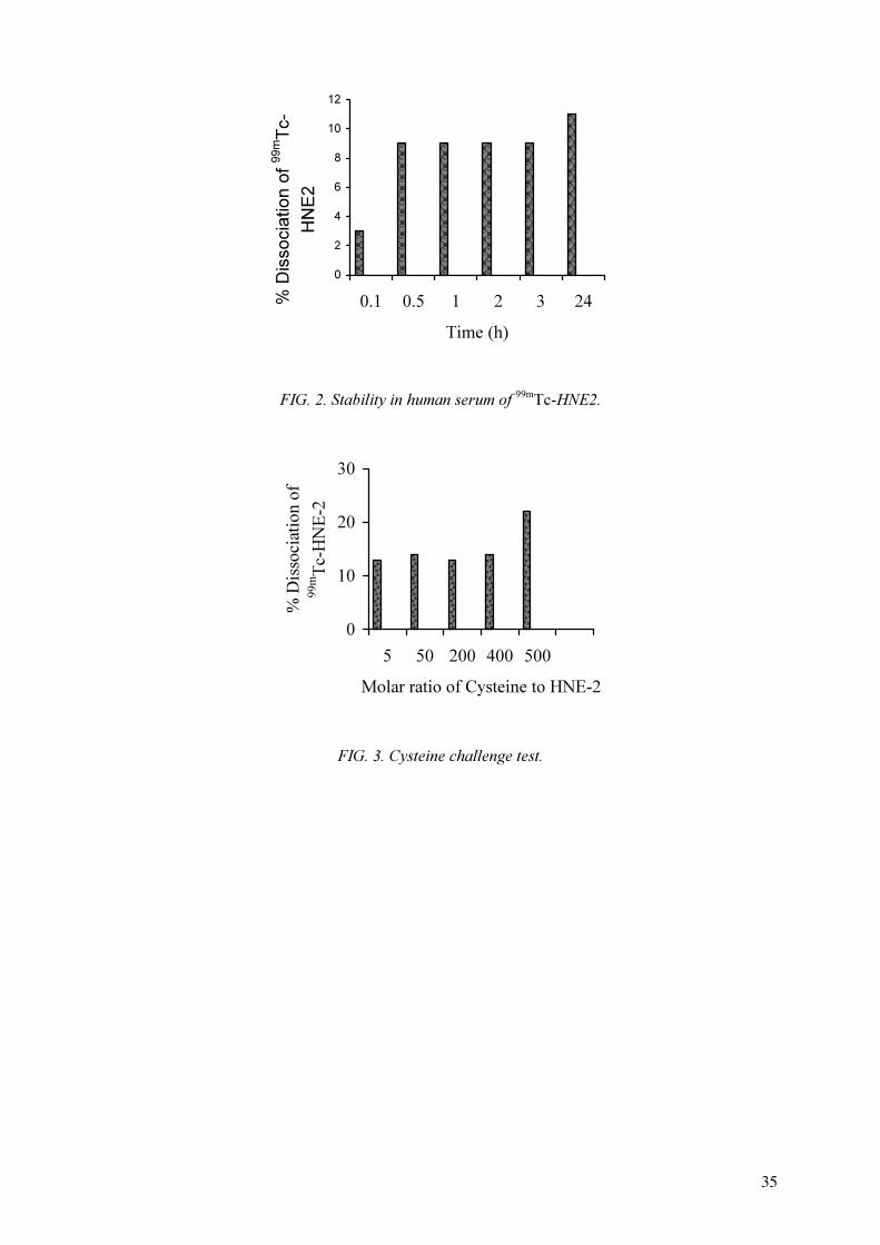
0
2
4
6
8
10
12
0.1 0.5 1 2 3 24
Time (h)
% D
isso
cia
tio
n o
f
99m
Tc-
HN
E2
FIG. 2. Stability in human serum of 99m
Tc-HNE2.
0
10
20
30
5 50 200 400 500
Molar ratio of Cysteine to HNE-2
% D
isso
ciatio
n o
f
99m
Tc-H
NE
-2
FIG. 3. Cysteine challenge test.
35
TABLE I. BIODISTRIBUTION RESULTS OF 99m
Tc-MAG3-HNE2 AND 99m
Tc-MAG3-BPTI IN
NORMAL MICE (MEAN % I.D.)
99m
Tc-BPTI 99m
Tc-HNE2
Tissue 15 min 2 h 15 min 2 h
Blood/g 2.7 0.5 2.4 1.33
Lungs 0.95 0.14 0.63 0.23
Stomach 0.61 0.28 0.46 0.57
NM/g 1.52 0.4 2.26 2.3
Liver 5.41 4.8 2.5 4.8
Spleen 0.16 0.03 0.14 0.07
Kidneys 33.35 48.2 12.6 14.9
Bladder +
Urine
4.63 9.47 5.9 9.96
3.2.99m
Tc UBI
3.2.1. Direct method
The LE of greater than 90% of 99m
Tc-UBI was observed in different solvent systems
(Table II). Acetone (LE: 98%) was selected as a solvent for further studies. 99m
Tc-UBI was found to be
stable at RT upto 2h. Significant fall in RCP was observed at 4h (81%) and further at 24h (70%).
However it was observed that on incubation with serum at 370
C the RCP of 99m
Tc-UBI remained
constant at about 94% upto 24h. Serum binding and bacterial binding studies indicated that about
8.9% of 99m
Tc-UBI was bound to the serum proteins and 30% was bound to bacteria, S.aureus at 37 0
C
after 1h. The observed ratio of uptake of 99m
Tc-UBI in infected muscle to normal muscle (IM/NM) at
15 min, 2 h and 24 h p.i. were 1.7, 2.8 and 6.7 respectively in mice and 1.3, 2.0 and 2.0 respectively in
rats (Table III).
3.2.2. Indirect method
Relatively higher labelling efficiencies were obtained at pH 5.2 (>95%) as compared to pH 7.6
(~90%). For further studies, labelling was carried out at pH 5.2. There was no significant change in the
RCP upto 24h. On incubation with serum at 370
C, the RCP of 99m
Tc-UBI gradually reduced to 71% at
24h (Table IV). About 68% of 99m
Tc-UBI was bound to the serum proteins after 1 h, at 370
C; 67% and
7% of 99m
Tc-UBI and 99m
Tc-scrambled peptide, respectively, were bound to bacteria, S.aureus at 370
C
after 1h. On challenge with cysteine, a fall of about 4-26% was observed in the RCP of 99m
Tc-UBI
(Table V). The ratios of uptake in infected muscle to normal muscle (IM/NM) at 15 min, 2h and 24h
p.i. for 99m
Tc-UBI were slightly higher as compared to that for 99m
Tc-labelled scrambled peptide
36
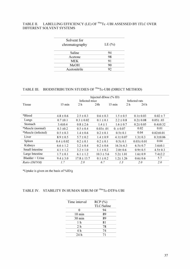
TABLE II. LABELLING EFFICIENCY (LE) OF 99m
Tc -UBI ASSESSED BY ITLC OVER
DIFFERENT SOLVENT SYSTEMS
Solvent for
chromatography LE (%)
Saline 94
Acetone 98
MEK 91
MeOH 90
Acetonitrile 92
TABLE III. BIODISTRIBUTION STUDIES OF 99m
Tc-UBI (DIRECT METHOD)
Tissue
Injected dDose (% ID)
Infected mice Infected rats
15 min 2 h 24h 15 min 2 h 24 h
*Blood 4.8 ± 0.6 2.5 ± 0.3 0.6 ± 0.3 1.5 ± 0.5 0.1± 0.03 0.02 ± 7
Lungs 0.7 ±0.1 0.3 ± 0.02 0.1 ± 0.1 2.2 ± 0.8 0.2± 0.08 0.05± .03
Stomach 3.4±0.4 0.8 ± 2.6 1.4 ± 1 1.6 ± 0.7 0.2± 0.05 0.4±0.32
*Muscle (normal) 0.3 ±0.2 0.5 ± 0.4 0.03± .01 0. ± 0.07 0.02 0.01
*Muscle (infected) 0.5 ± 0.3 1.4 ± 0.6 0.2 ± 0.1 0.5± 0.1 0.04 0.02±0.01
Liver 8.9 ± 0.5 5.7 ± 0.2 1.4 ± 0.9 4.1± 0.07 1.3± 0.3 0.3±0.06
Spleen 0.4 ± 0.02 0.2 ± 0.1 0.2 ± 0.1 0.5± 0.1 0.03± 0.01 0.04
Kidneys 6.6 ± 1.2 3.2 ± 0.4 0.2 ± 0.6 16.3± 6.3 6.5± 0.7 3.6±0.1
Small Intestine 4.1 ± 1.2 3.2 ± 1.0 1.1 ± 0.2 2.0± 0.6 0.9± 0.5 4.5± 0.3
Large Intestine 1.7 ± 0.1 6.1 ± 1.2 10.3 ± 5.6 5.2± 1.01 1.6± 0.9 7.4±2.2
Bladder + Urine 9.4 ± 3.0 17.8 ± 13.7 0.1 ± 0.2 1.2± 1.26 0.6± 0.6 5.7
Ratio (IM/NM) 1.7 2.8 6.7 1.3 2.0 2.0
*Uptake is given on the basis of %ID/g
TABLE IV. STABILITY IN HUMAN SERUM OF 99m
Tc-DTPA-UBI
Time interval RCP (%)
TLC/Saline
0 94
10 min 89
30 min 89
1 h 81
2 h 78
4 h 74
24 h 71
37

TABLE V. CYSTEINE CHALLENGE TEST OF 99m
Tc-DTPA-UBI
3.3.99m
Tc-EB1
Labelling efficiency of >95% was achieved. No significant difference in the LE was observed
with 99m
Tc-EB1 prepared with different stannous tin content. The labelled preparation was found to be
stable up to 24 h at RT. The kit was found to be stable for not less than about 3 months when stored at
40
C. The studies with human serum indicated stability of the labelled preparation with no significant
fall in the RCP up to 2 h and 67% was bound to human serum. On challenge with cysteine the fall of
about 6–29% was observed in the RCP of 99m
Tc-EB1 (Fig. 4). The studies on avidin binding indicated
shift in the radioactivity profile thereby confirming the binding of 99
Tc m
-EB1 with avidin (Fig. 5) The
ratios of uptake of 99m
Tc-EB1 (prepared with 20 µg stannous tin content) in inflamed muscle to normal
muscle at 10min, 1h and 3h were 2.0, 1.5 and 1.7, respectively, with EB1 obtained under CRP and 1.9,
1.2 and 2.0, respectively, with in-house synthesized EB1 in mice (Table VI). The 99m
Tc-EB1 prepared
from formulations with higher stannous tin (II) content resulted in higher liver uptake as expected.
0
5
10
15
20
25
30
35
0 0.01 0.1 1 10
Conc of cysteine, mg/ml
% D
issociation of
99m
Tc-E
B1 .
FIG. 4. Cysteine challenge test.
Cysteine concentration
(mg/ml)
RCP (%)
TLC/Saline
Control 99
0.01 95
0.1 91
1 87
10 73
38

0
200000
400000
600000
800000
1000000
1200000
1400000
1600000
0 5 10 15 20
Fraction number / ml
Ra
dio
activ
ity .
99m
Tc-EB1-Avidin
99m
Tc-EB1
FIG. 5. Radiochromatogram of 99m
Tc-EB1-avidin and 99m
Tc-EB1.
TABLE VI. BIODISTRIBUTION RESULTS WITH 99m
Tc-EB1
% I.D. (Mean±SD)
Tissue / Organ In-house synthesized EB1 EB1 obtained under CRP
10 min 1 h 3 h 10 min 1 h 3 h
Blood / g* 4.0±0.6 1.8±0.4 0.8±0.01 2.5±0.3 1.2±0.4 0.7±0.4
S. intestine 1.4 ± 0.5 2.7± 0.4 2.3 ± 0.6 1.3± 0.3 1.1± 0.3 0.5±0.1
L. intestine 1.5 ± 0.4 0.7 ± 0.1 2.4±0.4 0.5±0.01 2.0±0.2 2.7±0.7
Stomach 1.6±0.2 1.1±0.2 0.5± 0.2 0.8±0.2 1.4±0.1 0.3±0.03
Liver 10.6 ±0.7 12.3 ±1.3 10.1±2.2 7.8±0.2 9.8±2.2 6.6±0.6
Kidneys 2.1±0.8 1.1±0.2 0.8±0.03 0.9±0.1 0.6±0.1 0.5±0.03
NM/g* 0.7±0.1 0.5±0.2 0.2±0.1 0.7±0.1 0.2 ±0.04 0.7± 0.03
IM/g* 1.3±0.3 0.6±0.2 0.4±0.1 1.4±0.1 0.3±0.2 1.2 (-)
IM/NM 1.9 1.2 2.0 2.0 1.5 1.7
*Uptake is given on the basis of %ID/g.
4. SUMMARY AND HIGHLIGHTS
Radiolabelling of HNE2 through MAG3 as BCA has demonstrated good labelling efficiency,
stability and integrity of the molecule as indirectly determined by trypsin binding studies with BPTI.
Radiolabelling of UBI by direct method has demonstrated good labelling efficiency, stability,
adequate bacterial binding and significant uptake in the infection bearing tissue.
Radiolabelling of UBI through DTPA as BCA has demonstrated good labelling efficiency,
stability and bacterial binding as compared to the control scrambled peptide. However no significant
advantage over direct labelling was noted.
39

Synthesis of EB1 and the formulation of the kit for labelling with 99m
Tc were successfully
accomplished. The labelled product showed high labelling efficiency, stability and significant uptake
in the inflamed muscle as compared with the normal muscle.
BIBLIOGRAPHY
HNATOWICH, D.J., et al., “Labelling peptides with 99m
Tc using an NHS-MAG3 bifunctional
chelator”, J. Nucl. Med. 39 (1998) 56–64.
VIRZI, F., et al., “New Indium–111 labelled biotin derivatives for improved immunotargeting”, Nucl.
Med. Biol. 18 (1991) 719–726.
WELLING, M.M, NIBBERING, P.H., PAULUSMA-ANNEMA, A., et al., “Antibacterial activity of
human neutrophil defensins in experimental infections in mice is accompanied by increased leukocyte
accumulation”, J. Clin. Invest. 102 (1998) 1583–1590.
WELLING, M.M., HIEMSTRA, P.S., VAN DEN BARSELAAR, M.T., et al., “Imaging of
experimental bacterial infections with a technetium-99m labelled antimicrobial human neutrophil
peptide”, J. Nucl. Med. 40 (1999) 2073–2080.
40
ACKNOWLEDGEMENTS
We thank our colleagues, Mr. S.K. Sarkar and Mr. G. Arjun, for providing 99m
Tc-pertechnetate
for some of the CRP studies and Dr. M.R.A. Pillai, Senior General Manager (Medical and Biological
Products Programme, MBPP), Dr. (Mrs.) Meera Venkatesh, General Manager, (Quality Control,
MBPP) for their keen interest in the work.
99m
Tc LABELLING OF UBIQUICIDINE (UBI 29-41) AND EDTA-BIOTIN
MONOMER (EB1)
W. WIDJAKSANA, F. YUNITA, L. ANDRIASTUTI, YUNILDA, A. ARIYANTO,
G. MONDRIDA, A. ROSELIANA, S. BAGIAWATI, E. SOVILAWATI, A. MUTALIB
Center for Development of Radioisotopes and Radiopharmaceuticals,
National Nuclear Energy Agency,
Tangerang, Indonesia
Abstract
Ubiquicidine (UBI 29-41) and EDTA-Biotin (EB1) labelled with 99m
Tc have been reported to have
capability as infection and inflammation imaging agent, respectively. UBI 29-41 was labelled with 99m
Tc
following direct and indirect methods using cyclic DTPA anhydride and NHS-HYNIC as bifunctional chelators
with tricine and EDDA as coligands, and analysed by thin layer chromatography (TLC) and reversed phase
HPLC (RP-HPLC). The stability of 99m
Tc-UBI and 99m
Tc-EB1 was assessed in human serum and by cysteine
challenge. The efficacy of 99m
Tc-UBI for infection imaging was evaluated using in vitro bacterial binding assay
and in vivo studies in infected mice. 99m
Tc-EB1 was subjected to shift assay after reacting with avidin, while its
efficacy for inflammation imaging was assessed by in vivo study in inflammation induced mice and rats. The
labelling efficiency (LE) of UBI and scrambled UBI using both direct and indirect methods did not show any
significant difference (>85%), and EB1 also giae high labelling yield (>90%). Stability of 99m
Tc-HYNIC-UBI in
cysteine seemed to be higher than that of 99m
Tc-UBI both in human serum and in cysteine. Bacterial binding of
99m
Tc UBI and 99m
Tc scrambled UBI prepared using direct and indirect methods was 40-75% and did not show
any significant difference. Bioassay in infected mice showed higher radioactivity in infected thigh than in the
normal thigh for both direct and indirect labelled UBI, and T/NT ratio 1.5 and 3 for 99m
Tc-HYNIC-UBI and
99m
Tc-UBI, respectively, at two h post injection. The 99m
Tc-scrambled UBI unexpectedly showed higher T/NT
ratio than 99m
Tc-UBI, and it needs further evaluation. 99m
Tc EB1 showed binding for avidin (Gel permeation
assay), and higher uptake in inflammation site in mice (T/NT ratio 2.3).
1. INTRODUCTION
Infectious diseases still pose a serious problem in developing countries, especially in tropical
countries such as Indonesia, that need accurate and proper diagnosis for treatment and to minimize the
risk of resistence to antibiotics by microorganisms. Nuclear medicine imaging has been used to
localize infection sites and attempts have been continued to develop improved infection specific
radiopharmaceuticals [1,2]. Synthesized ubiquicidine 29-41 (UBI) derived from human antimicrobial
peptide has been reported to be able to bind with gram positive bacteria (Staphylococcus Aureus and
Klebsiella pneumonia) and fungi either in vitro or in vivo after labelling with 99m
Tc, and furthermore it
is reported to distinguish between infection and sterile inflammation [3,4,5].
99m
Tc labelling methods for small peptides should be carefully optimized to retain biological
activity and achieve high labelling efficiency. High pharmacological activity often exhibited by
peptides in vivo requires that the amount of peptides administered should be as small as possible.
Peptide-based radiopharmaceuticals for imaging purpose should have high specific activity and
stability. In addition to the originally reported direct labelling method for 99m
Tc UBI, bifunctional
chelating agent (BFC) approach was considered interesting for investigation under the CRP
programme. One of the agents commonly used now for 99m
Tc labelling of peptides is
hydrazinonicotinamide (HYNIC) with tricine and EDDA as coligands [6]. In this project, 99m
Tc
labelling of UBI 29-41 and a control peptide having scrambled sequence of same amino acids but
poorer bacterial binding, was investigated by both direct and BFC methods and compared.
In addition, 99m
Tc complex of EDTA-biotin (EB1) reported to have good prospect as
inflammation imaging agent, was also investigated.
41
2. MATERIALS
Ubiquicidine 29-41 (UBI) and Scrambled Ubiquicidine 29-41 (Scr.UBI) were supplied from
Leiden University Medical Center (LUMC), Netherlands. EDTA-Biotin (EB1) was supplied from
PINSTECH, Pakistan. NHS-hydrazinonicotinamide (NHS-HYNIC) was supplied from POLATOM,
Poland. P-4 column (Biorad) and cyclic-DTPA anhydride (Sigma) were donated by Prof.D.Hnatowich
(Massachussets Univ., USA). 99m
Tc generator was supplied by BATAN TECH, Indonesia. Basic
chemicals such as potassium borohydride, stannous chloride dihydrate, sodium pyrophosphate etc.
were purchased from Sigma, Aldrich and Merck. Staphylococcus Aureus was provided by P3TIR-
BATAN, Indonesia.
3. METHODS
3.1. Synthesis of HYNIC-UBI and DTPA-UBI
100 µL of UBI or Scrambled UBI solution (3 mg/mL in bicarbonate buffer pH 8.5) was reacted
with 10 µL of NHS-HYNIC solution (in dry DMF) at room temperature for 1 h, the ratio of NHS-
HYNIC to peptide was 3. The HYNIC-peptide was purified through P-4 column using 0.05 M
ammonium acetate buffer pH 5.2 as eluant. The amount of pure conjugate was measured using UV
spectrometry. HYNIC-peptides solution was stored at -40°C. DTPA-UBI was synthesized in the same
way.
3.2. Labelling UBI and Scrambled UBI with 99m
Tc using direct method
10 µL UBI or Scr.UBI solution (1 mM in 0.01 M acetic acid) was added with 4 µL stannous
pyrophosphate solution (containing 0.95 mg/mL SnCl2.2 H
2O and 2 mg/mL sodium pyrophosphate), 2
µL KBH4 solution (10 mg/mL in 0.1 M NaOH) and 0.1 mL pertechnetate
99m
Tc solution (∼10
mCi/mL), mixed and allowed to stand at room temperature for 10 min.
3.3. Labelling UBI and Scrambled UBI with 99m
Tc using indirect method
NHS-HYNIC and cDTPA were used as BFC. Tricine and EDDA were selected as coligands for
labelling HYNIC-UBI.
To a solution of HYNIC-UBI or HYNIC-Scr.UBI (containing 10 µg of HYNIC-UBI or
HYNIC-Scr.UBI) were added 20 µL tricine solution (100 mg/mL in water), 0.1 mL pertechnetate
99m
Tc solution (∼10 mCi/mL), and 10 µL stannous chloride solution (1 mg/mL in 0.01 M HCl
previously purged with nitrogen), mixed and allowed to stand at room temperature for 30 min.
Another method using mixture of tricine and EDDA (2:1) as coligand was also carried out. 500
µL of a solution containing 10 mg tricine and 5 mg EDDA was added to the the reaction vial
containing 10 µg of HYNIC-UBI prior to adding 0.5 mL pertechnetate 99m
Tc solution (∼10 mCi/mL)
and 10 µL stannous chloride solution, followed by heating at 70°C for 1 h.
Solution of DTPA-UBI or DTPA-Scr.UBI (containing 10 µg of DTPA-UBI or DTPA-Scr.UBI)
was mixed with 0.1 mL pertechnetate 99m
Tc solution (∼10 mCi/mL), and 10µL stannous chloride
solution (1 mg/mL in 0.01 M HCl previously purged with nitrogen), mixed and allowed to stand at
room temperature for 30 min.
42

3.4. Labelling EB1 with 99m
Tc
3 µL of EB1 solution (10 mg/mL in sodium acetate) was mixed with 0.1 mL pertechnetate 99m
Tc
solution (∼10 mCi/mL), and 2 µL stannous chloride solution (1 mg/mL in 0.01 M HCl previously
purged with nitrogen), and allowed to react at room temperature for 30 min.
3.5. Analysis of radiolabelled UBI, Scr.UBI and EB1
Radiolabelled UBI, Scr.UBI and EB1 were analysed using RP-HPLC (Shimadzu) and TLC. RP-
HPLC was run using C-18 column (Microbondapack C-18, Waters), and acetonitrile and water
containing 0.1 % TFA as eluants with gradient, i.e 0–3 min TFA/water, 3–13 min 0–50% ACN,
13-23 min 50% ACN, 23–26 min 50–70% ACN, 27 min 70–0% CAN and UV detection at 280 nm as
well as radioactivity detection.
TLC was carried out in three separated chambers, i.e. the 1st
chamber with acetone and
Whatman-1 paper to determine % free pertechnetate, 2nd
chamber with mixture of 0.05 M ammonium
acetate pH 5.2 and methanol (1:1) and ITLC-SG strips to determine % Tc colloid and 3rd
chamber
with PBS pH 7.4 and TLC silica strips to determine % Tc-coligand and free pertechnetate. The
radioactivity was measured using a TLC scanner (Veenstra Instrument), and % radiochemical purity of
99m
Tc peptides was calculated by substracting from 100 the sum of percentage of measured impurities.
3.6. Bacterial binding assay
To 0.1 mL of phosphate buffer containing 10 µL of 99m
Tc-UBI or 99m
Tc-Scr.UBI, were added
0.8 mL of phosphate buffer containing 50% acetic acid 0.01M and 0.01% Tween-80 and 0.1 mL of
phosphate buffer containing about 2×10⁷
CFU Staphylococcus Aureus. The mixture was incubated at
37ºC for 1 h followed by centrifugation for 5 min at 1000 rpm. The precipitate was separated and
washed with 2×1 mL of phosphate buffer, and resuspended with 1 mL of phosphate buffer. Bacterial
precipitate was measured for radioactivity, and % bacterial binding was calculated.
3.7. In vivo assay
In vivo assay was carried out using healthy mice and mice infected with bacterial culture. Assay
on healthy mice was done to see the normal pharmacokinetics of radiolabelled UBI, while assay on
Staphylococcus Aureus infected mice was done to see its effectiveness for marking infection. Mice
were infected by injecting 0.1 mL of saline containing 107
CFU bacteria into right thigh. 18 h later,
mice were injected with 0.2 ml saline containing 10% of 99m
Tc-UBI or 99m
Tc-Scr.UBI solution through
tail vein. Injection of 99m
Tc-UBI or 99m
Tc-Scr.UBI was also done to normal mice as control. Mice were
sacrificed after 30 min, 1 h and 2 h, and the organs including blood and the thigh were taken and
measured for radioactivity.
3.8. Stability test
Stability test was carried out using fresh human serum and cysteine solution. Stability in serum
was tested by incubating solution of 1-10 µg 99m
Tc-UBI or 99m
Tc-EB1 in 1 mL serum at 37ºC for 1 h,
and the radiochemical purity was analysed with TLC. Stability against cysteine (cysteine challenge
study) was tested by adding 20 µL of cysteine solution (1 mg/mL in 0.1 M PBS pH 7) into 90 µL of
99m
Tc-UBI solution (2.2 µM in 0.2 M PBS pH 7), incubating at 37ºC for 1 h, and analysing with TLC
for radiochemical purity.
43
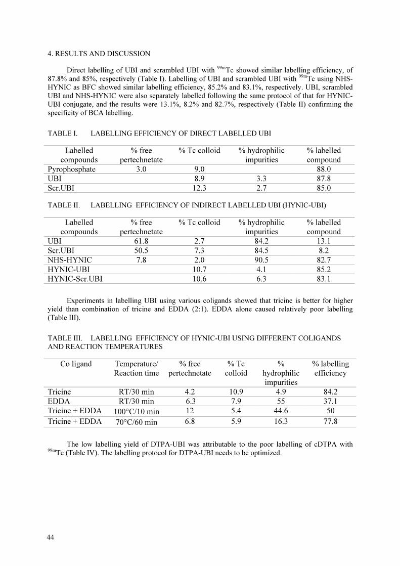
4. RESULTS AND DISCUSSION
Direct labelling of UBI and scrambled UBI with 99m
Tc showed similar labelling efficiency, of
87.8% and 85%, respectively (Table I). Labelling of UBI and scrambled UBI with 99m
Tc using NHS-
HYNIC as BFC showed similar labelling efficiency, 85.2% and 83.1%, respectively. UBI, scrambled
UBI and NHS-HYNIC were also separately labelled following the same protocol of that for HYNIC-
UBI conjugate, and the results were 13.1%, 8.2% and 82.7%, respectively (Table II) confirming the
specificity of BCA labelling.
TABLE I. LABELLING EFFICIENCY OF DIRECT LABELLED UBI
Labelled
compounds
% free
pertechnetate
% Tc colloid % hydrophilic
impurities
% labelled
compound
Pyrophosphate 3.0 9.0 88.0
UBI 8.9 3.3 87.8
Scr.UBI 12.3 2.7 85.0
TABLE II. LABELLING EFFICIENCY OF INDIRECT LABELLED UBI (HYNIC-UBI)
Labelled
compounds
% free
pertechnetate
% Tc colloid % hydrophilic
impurities
% labelled
compound
UBI 61.8 2.7 84.2 13.1
Scr.UBI 50.5 7.3 84.5 8.2
NHS-HYNIC 7.8 2.0 90.5 82.7
HYNIC-UBI 10.7 4.1 85.2
HYNIC-Scr.UBI 10.6 6.3 83.1
Experiments in labelling UBI using various coligands showed that tricine is better for higher
yield than combination of tricine and EDDA (2:1). EDDA alone caused relatively poor labelling
(Table III).
TABLE III. LABELLING EFFICIENCY OF HYNIC-UBI USING DIFFERENT COLIGANDS
AND REACTION TEMPERATURES
Co ligand Temperature/
Reaction time
% free
pertechnetate
% Tc
colloid
%
hydrophilic
impurities
% labelling
efficiency
Tricine RT/30 min 4.2 10.9 4.9 84.2
EDDA RT/30 min 6.3 7.9 55 37.1
Tricine + EDDA 100°C/10 min 12 5.4 44.6 50
Tricine + EDDA 70°C/60 min 6.8 5.9 16.3 77.8
The low labelling yield of DTPA-UBI was attributable to the poor labelling of cDTPA with
99m
Tc (Table IV). The labelling protocol for DTPA-UBI needs to be optimized.
44
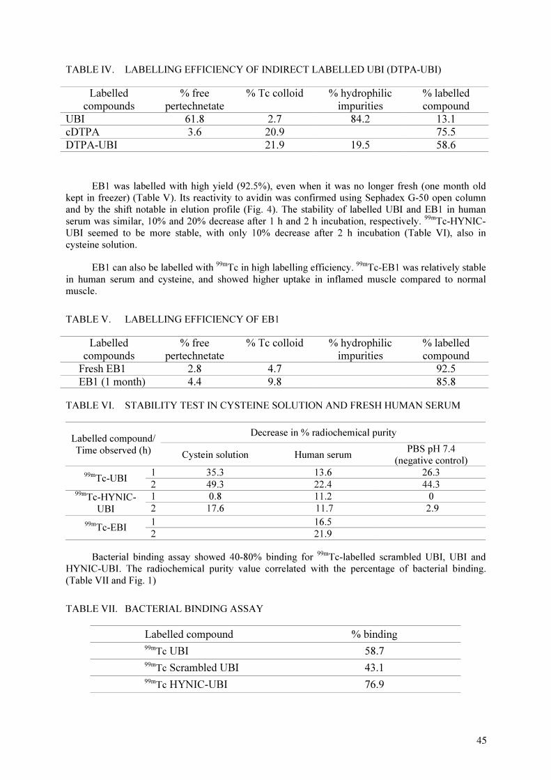
TABLE IV. LABELLING EFFICIENCY OF INDIRECT LABELLED UBI (DTPA-UBI)
Labelled
compounds
% free
pertechnetate
% Tc colloid % hydrophilic
impurities
% labelled
compound
UBI 61.8 2.7 84.2 13.1
cDTPA 3.6 20.9 75.5
DTPA-UBI 21.9 19.5 58.6
EB1 was labelled with high yield (92.5%), even when it was no longer fresh (one month old
kept in freezer) (Table V). Its reactivity to avidin was confirmed using Sephadex G-50 open column
and by the shift notable in elution profile (Fig. 4). The stability of labelled UBI and EB1 in human
serum was similar, 10% and 20% decrease after 1 h and 2 h incubation, respectively. 99m
Tc-HYNIC-
UBI seemed to be more stable, with only 10% decrease after 2 h incubation (Table VI), also in
cysteine solution.
EB1 can also be labelled with 99m
Tc in high labelling efficiency. 99m
Tc-EB1 was relatively stable
in human serum and cysteine, and showed higher uptake in inflamed muscle compared to normal
muscle.
TABLE V. LABELLING EFFICIENCY OF EB1
Labelled
compounds
% free
pertechnetate
% Tc colloid % hydrophilic
impurities
% labelled
compound
Fresh EB1 2.8 4.7 92.5
EB1 (1 month) 4.4 9.8 85.8
TABLE VI. STABILITY TEST IN CYSTEINE SOLUTION AND FRESH HUMAN SERUM
Decrease in % radiochemical purity Labelled compound/
Time observed (h) Cystein solution Human serum
PBS pH 7.4
(negative control)
1 35.3 13.6 26.3 99m
Tc-UBI 2 49.3 22.4 44.3
1 0.8 11.2 0 99m
Tc-HYNIC-
UBI 2 17.6 11.7 2.9
1 16.5 99m
Tc-EBI 2 21.9
Bacterial binding assay showed 40-80% binding for 99m
Tc-labelled scrambled UBI, UBI and
HYNIC-UBI. The radiochemical purity value correlated with the percentage of bacterial binding.
(Table VII and Fig. 1)
TABLE VII. BACTERIAL BINDING ASSAY
Labelled compound % binding
99m
Tc UBI 58.7
99m
Tc Scrambled UBI 43.1
99m
Tc HYNIC-UBI 76.9
45
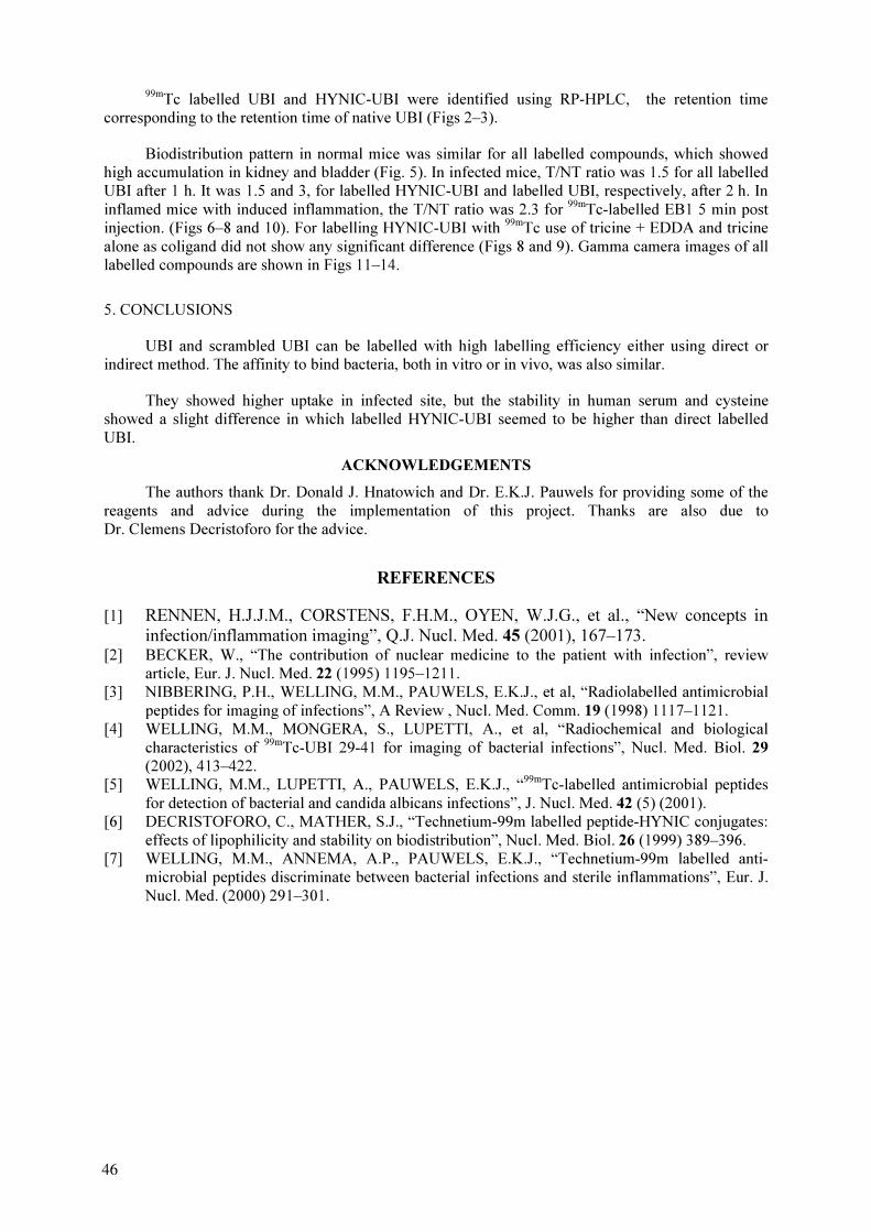
99m
Tc labelled UBI and HYNIC-UBI were identified using RP-HPLC, the retention time
corresponding to the retention time of native UBI (Figs 2–3).
Biodistribution pattern in normal mice was similar for all labelled compounds, which showed
high accumulation in kidney and bladder (Fig. 5). In infected mice, T/NT ratio was 1.5 for all labelled
UBI after 1 h. It was 1.5 and 3, for labelled HYNIC-UBI and labelled UBI, respectively, after 2 h. In
inflamed mice with induced inflammation, the T/NT ratio was 2.3 for 99m
Tc-labelled EB1 5 min post
injection. (Figs 6–8 and 10). For labelling HYNIC-UBI with 99m
Tc use of tricine + EDDA and tricine
alone as coligand did not show any significant difference (Figs 8 and 9). Gamma camera images of all
labelled compounds are shown in Figs 11–14.
5. CONCLUSIONS
UBI and scrambled UBI can be labelled with high labelling efficiency either using direct or
indirect method. The affinity to bind bacteria, both in vitro or in vivo, was also similar.
They showed higher uptake in infected site, but the stability in human serum and cysteine
showed a slight difference in which labelled HYNIC-UBI seemed to be higher than direct labelled
UBI.
ACKNOWLEDGEMENTS
The authors thank Dr. Donald J. Hnatowich and Dr. E.K.J. Pauwels for providing some of the
reagents and advice during the implementation of this project. Thanks are also due to
Dr. Clemens Decristoforo for the advice.
REFERENCES
[1] RENNEN, H.J.J.M., CORSTENS, F.H.M., OYEN, W.J.G., et al., “New concepts in
infection/inflammation imaging”, Q.J. Nucl. Med. 45 (2001), 167–173.
[2] BECKER, W., “The contribution of nuclear medicine to the patient with infection”, review
article, Eur. J. Nucl. Med. 22 (1995) 1195–1211.
[3] NIBBERING, P.H., WELLING, M.M., PAUWELS, E.K.J., et al, “Radiolabelled antimicrobial
peptides for imaging of infections”, A Review , Nucl. Med. Comm. 19 (1998) 1117–1121.
[4] WELLING, M.M., MONGERA, S., LUPETTI, A., et al, “Radiochemical and biological
characteristics of 99m
Tc-UBI 29-41 for imaging of bacterial infections”, Nucl. Med. Biol. 29
(2002), 413–422.
[5] WELLING, M.M., LUPETTI, A., PAUWELS, E.K.J., “99m
Tc-labelled antimicrobial peptides
for detection of bacterial and candida albicans infections”, J. Nucl. Med. 42 (5) (2001).
[6] DECRISTOFORO, C., MATHER, S.J., “Technetium-99m labelled peptide-HYNIC conjugates:
effects of lipophilicity and stability on biodistribution”, Nucl. Med. Biol. 26 (1999) 389–396.
[7] WELLING, M.M., ANNEMA, A.P., PAUWELS, E.K.J., “Technetium-99m labelled anti-
microbial peptides discriminate between bacterial infections and sterile inflammations”, Eur. J.
Nucl. Med. (2000) 291–301.
46

0.0%
10.0%
20.0%
30.0%
40.0%
50.0%
60.0%
70.0%
80.0%
90.0%
52.7% 56.3% 61.3% 67.6% 70.3% 76.2% 84.6% 87.0%
% Labeling
% B
acterial b
in
din
g
Ubi Hynic Ubi
FIG. 1. Correlation between % labelling of 99m
Tc-UBI and % bacterial binding.
0
50,000
100,000
150,000
200,000
250,000
1 3 5 7 9 11 13 15 17 19 21 23 25 27 29
retention time, min
rad
io
activity, cp
m
FIG. 2. Reversed phase HPLC of radiolabelled UBI.
47

0
100,000
200,000
300,000
400,000
500,000
600,000
700,000
800,000
900,000
1 3 5 7 9 11 13 15 17 19 21 23 25 27 29
retention time, min
rad
io
activity, cp
m
FIG. 3. Reversed phase HPLC of radiolabelled HYNIC-UBI.
……
……
……
……
FIG. 1. (Figure caption should be in italics, Font 11, and should end with a period).
……
……
……
FIG. 4. Shift assay of radiolabelled EB1 — (1) 99m
Tc-EB1-avidin, (2) 99m
Tc-EB1.
1
2
0
500,000
1,000,000
1,500,000
2,000,000
2,500,000
3,000,000
3,500,000
1 3 5 7 9 11 13 15 17 19 21 23 25 27 29
Fraction no.
rad
io
activity, cp
m
48
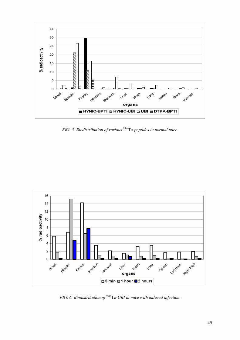
0
5
10
15
20
25
30
35
Blood
Bladder
Kidney
Intestine
Stomach
Liver
Heart
Lung
Spleen
Bone
Muscles
organs
% rad
io
activity
HYNIC-BPTI HYNIC-UBI UBI DTPA-BPTI
FIG. 5. Biodistribution of various 99m
Tc-peptides in normal mice.
0
2
4
6
8
10
12
14
16
Blo
od
Bla
dder
Kid
ney
Inte
stine
Sto
m
ach
Liver
Heart
Lung
Sple
en
Left
thigh
Rig
ht th
igh
organs
% rad
io
activity
5 min 1 hour 2 hours
FIG. 6. Biodistribution of 99m
Tc-UBI in mice with induced infection.
49

0
4
8
12
16
20
24
28
32
Blood
Bladder
Kidney
Intestine
Stomach
Liver
Heart
Lung
Spleen
Leftthigh
Rightthigh
organs
% rad
io
activity
5 min. 1 hour 2 hours
FIG. 7. Biodistribution of 99m
Tc-scrambled UBI in mice with induced infection.
0
5
10
15
20
25
30
Blo
od
Bla
dder
Kid
ney
Inte
stine
Sto
m
ach
Liver
Heart
Lung
Sple
en
Left thigh
Rig
ht th
igh
organs
% rad
io
activity
5 min 1 hour 2 hours
FIG. 8. Biodistribution of 99m
Tc-HYNIC-UBI with tricine in mice with induced infection.
50
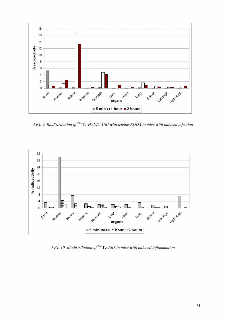
0
2
4
6
8
10
12
14
16
18
Blo
od
Bla
dder
Kid
ney
Inte
stine
Sto
m
ach
Liver
Heart
Lung
Sple
en
Left
thigh
Rig
ht th
igh
organs
% ra
dio
ac
tiv
ity
5 min 1 hour 2 hours
FIG. 9. Biodistribution of 99m
Tc-HYNIC-UBI with tricine/EDDA in mice with induced infection.
0
4
8
12
16
20
24
28
32
Blood
Bladder
Kidney
Intestine
Stom
ach
Liver
Heart
Lung
Spleen
Leftthigh
Rightthigh
organs
% rad
io
activity
5 minutes 1 hour 2 hours
FIG. 10. Biodistribution of 99m
Tc-EB1 in mice with induced inflammation.
51

FIG. 11. Gamma camera imaging of 99m
Tc-EB1 in Wistar rat with induced inflammation.
FIG. 12. Gamma camera imaging of 99m
Tc-HYNIC-UBI/tricine in mice with induced infection 1 h p.i.
52

FIG. 13. Gamma camera imaging of 99m
Tc-HYNIC-UBI/EDDA in mice with induced infection 1 h p.i.
FIG. 14. Gamma camera imaging of 99m
Tc-UBI in mice with induced infection 1 h p.i.
53


PREPARATION OF 99m
Tc KITS FOR INFECTION IMAGING
G. FERRO-FLORES, C. ARTEAGA DE MURPHY, L. MELENDEZ-ALAFORT,
M. PEDRAZA-LOPEZ, P. ALTAMIRANO-BUSTAMANTE
Instituto Nacional de Investigaciones Nucleares,
Instituto Nacional de Ciencias Médicas y Nutrición Salvador Zubirán,
Universidad Autónoma del Estado de México,
México
Abstract
The aim of this study was to label ubiquicidin fragment 29-41 (UBI) and biotin with 99m
Tc and evaluate
their feasibility as infection imaging agents for in vivo use. 99m
Tc-UBI, labelled by a direct method, showed high
in vitro and in vivo stability, specific uptake at the site of infection, rapid background clearance, minimal
accumulation in non-target tissues and rapid detection of infection sites. 99m
Tc labelled biotin showed in vitro
and in vivo stability, fast renal clearance and ability to detect infection and sterile inflammation processes in
mice. Because of their stability and biological properties, both agents could be used in clinical applications.
1. INTRODUCTION
Recently technetium-99m labelled ubiquicidin peptide 29-41 fragment (99m
Tc-UBI) has been
proposed as a new radiopharmaceutical for infection imaging [1,2]. UBI is a cationic human
antimicrobial peptide fragment (MW 1.69 kDa) with the amino acid sequence Thr-Gly-Arg-Ala-Lys-
Arg-Arg-Met-Gln-Tyr-Asn-Arg-Arg, therefore, with 6 positively charged residues (5 Arg + 1 Lys).
Studies of the biodistribution of 99m
Tc-UBI in mice have shown a fast renal clearance with minimal
hepatobiliary excretion. 99m
Tc-UBI is bound to bacteria in vitro and is accumulated at sites of infection
in experimental animals [1,2].
Considering that five of the thirteen amino acids in UBI 29-41 fragment are arginine (with free
–NH2 groups), and 3 of these are close to a lysine with its own –NH
2 group, it is possible that some of
these groups could form a stable complex with reduced technetium, such as the mono-oxo Tc(V).
Therefore, the first aim of this study was to help establish if ubiquicidin peptide 29-41 fragment (UBI)
contains a specific site for 99m
Tc labelling by a direct method under alkaline conditions. Since this
peptide does not have cysteine residues, it is possible that neighbouring arginine and lysine in the
peptide amino acid sequence (Thr-Gly-Arg-Ala-Lys-Arg-Arg-Met-Gln-Tyr-Asn-Arg-Arg) could be a
coordination site to form a stable 99m
Tc-UBI complex. Following direct labelling, the in vitro stability
of 99m
Tc-UBI was compared to UBI radiolabelled by one indirect method using HYNIC/tricine [3].
A second aim was to help determine if 99m
Tc-UBI is bound to the bacterial cell envelope by a
simple nonspecific electrostatic interaction. A comparative study of the in vitro binding of 99m
Tc-UBI
and two different 99m
Tc labelled cationic peptides (99m
Tc-Tat-1-Scr and 99m
Tc-Tat-2-Scr) to bacteria
and to two tumor cell lines (LS174T and ACHN) was accomplished [4]. The in vivo specificity of
99m
Tc-UBI for infection in mice was also evaluated using dual labels in the same animal and
comparing the target/non-target ratio for 67
Ga-citrate and 99m
Tc-UBI at sites of induced infection and
sterile inflammation [4]. In addition, sequential gammagraphic images of 99m
Tc-UBI biodistribution in
patients were obtained to calculate biokinetic parameters as residence time useful in absorbed dose
calculations [5].
As a third aim, EDTA-biotin (EB1) and DTPA-bis-biotin were labelled with 99m
Tc and
evaluated as non-specific infection imaging agents in mice [6].
2. MATERIALS
Ubiquicidin 29-41 fragment peptide (UBI) was synthesized and characterized at the Leiden
University Medical Center, The Netherlands. Cationic control peptides with amino acid sequences
Cys-Arg-Lys-Arg-Gly-Lys-Arg-Arg-Gln-Arg-Gly-Arg (Tat-1-Scr) and Arg-Lys-Arg-Lys-Arg-Gly-
55
Arg-Arg-Gln-Arg (Tat-2-Scr) were solid-phase synthesized and characterized at the University of
Massachusetts Medical School (UMASS), USA. EDTA-biotin was supplied by the Pakistan Institute
of Nuclear Science and Technology and NHS-HYNIC by POLATOM Radioisotope Centre and
UMASS. Staphylococcus aureus microorganism, human renal adenocarcinoma cell line (ACHN) and
the human colon cancer cell line (LS174T) were obtained from American Type Culture Collection.
DTPA-bis-Biotin, tricine and other chemicals were purchased from Sigma Chemical Co.
3. METHODS
3.1. Direct techenetium-99m UBI labelling
For 99m
Tc labelling, 10 µL of a UBI aqueous solution (1 mg/0.2 mL) was added to a vial
containing 5 µL of a fresh 1 mg/mL solution of stannous chloride in 10 mM HCl. The pH was
adjusted to 9 by addition of 10 µL of 0.1 M NaOH, followed by 100 µL of 99m
Tc-pertechnetate (185-
370 MBq). The resulting solution (pH 9-10) was left to react for 5 min at room temperature and
finally, 1 mL of 0.9% NaCl solution was added to obtain a final pH 6.5-7.0. The KBH4
and
pyrophosphate used in an earlier direct labelling method were found to be unnecessary.
3.2. Indirect 99m
Tc UBI labelling using hydrazinonicotinamide (HYNIC)
Conjugation: UBI peptide was prepared at a concentration of 5 mg/mL in 0.1 M HEPES (N-(2-
hydroxyethyl)piperazine-N’-(2-ethanesulfonic acid)) buffer, pH 8.0, to which a fresh 10 mg/mL
solution of NHS-HYNIC in dry dimethylformamide (DMF) was added dropwise with agitation. The
final HYNIC-to-peptide molar ratio was 3:1, and the volume of DMF added was always <10% of the
total volume. The mixture was reacted at room temperature for 30-60 min before purification on a
0.7×20 cm P4 size-exclusion open column (BioRad, Hercules, CA) with 0.25 M ammonium acetate,
pH 5.2 or 0.05 M phosphate buffer pH 6.0 as eluants. Fractions were collected and analysed by
ultraviolet (UV) absorbance at 280 nm (U-20000; Hitachi Instruments, Inc., Danbury, CT) using an
extinction coefficient of 0.756 for UBI. The conjugated peptide was diluted to approximately 0.4-0.5
mg/mL with 0.25 M ammonium acetate pH 5.2 or 0.1 M phosphate buffer pH 6.0 (conjugated peptide
solution).
99m
Tc-HYNIC-UBI/tricine: Approximately 20 µL of a 100 mg/mL tricine solution in water was
added to 0.05 mg of HYNIC-UBI conjugate dissolved in 0.1 mL of 0.25 M ammonium acetate, pH
5.2. Then 111 MBq 99m
Tc-pertechnetate generator eluant was added followed by 6 µL of a fresh
SnCl2
.
2H2O (1 mg/mL in 10 mM HCl) solution. After reacting for 30-60 min at room temperature, the
labelled peptide was purified over the P4 column as above.
3.3. Quality control
Radiochemical purity analyses were performed by Sep-Pak C-18 cartridge open column
chromatography, instant thin layer chromatography (ITLC) and reverse phase and size exclusion high-
performance liquid chromatography (HPLC). The Sep-Pak cartridges were preconditioned with 5 mL
of ethanol followed by 5 mL of 1 mM HCl. An aliquot of 0.1 mL of the labelled peptide was loaded
on the cartridge followed by 5 mL of 1 mM HCl to elute 99m
Tc-pertechnetate. The labelled peptide was
eluted with 3 mL of ethanol:saline (1:1) followed by 3 mL of 0.1 M HCl: methanol (15:85). The
hydrolyzed-reduced 99m
Tc remainded on the column.
ITLC silica gel glass fiber 10 cm strips (Gelman Sciences) were used as stationary phase. Saline
solution was the mobile phase to determine the amount of free 99m
TcO4
-
(Rf= 1). In this system the
radiopeptide migrates when it is diluted in human serum.
Reverse Phase HPLC Method: HPLC analyses were carried out with a Waters instrument
(Waters, Milford, MA, USA) running Millenium software with both radioactivity and UV-photodiode
56
array in-line detectors. YMC ODS-AQ S5 column (5µm, 4.6×250 mm) at a flow rate of 1 mL/min
using 0.1% trifluoroacetic acid (TFA)/water (solvent A) and 0.1% TFA/acetonitrile (solvent B). The
gradient started at 100% solvent A for 3 min, changed to 50% solvent A over 10 min, held constant for
10 min, changed to 30% solvent A over 3 min and finally back to 100% solvent A over 4 min.
Size-exclusion HPLC Method: Superdex-peptide column (1×30 cm, Pharmacia,
Piscataway, NJ), at a flow rate of 1.0 mL/min with 0.1 M sodium phosphate, pH 7.0, as
mobile phase.
3.4. Serum stability
Size exclusion HPLC analysis was used to estimate the serum stability of UBI labelled with
99m
Tc by the direct method and the indirect method using HYNIC. A volume of 100 µL of the labelled
peptide solution (5µg/100µL) was incubated at 37o
C with 1 mL of fresh human serum. Radiochemical
stability was determined taking samples of 10 µL at different times from 5 min to 24 h for analysis. A
shift of the radioactivity profile to higher molecular weight indicates protein binding, while lower-
molecular weight peaks indicate labelled catabolites or serum cysteine binding. Recovery of
radioactivity was routinely determined.
3.5. Dilution stability
To determine the stability of 99m
Tc-UBI after dilution, 50 µL of radiolabelled peptide
(25 µg/50 µL) was diluted to a ratio 1:20 with saline solution (0.9% NaCl), and incubated for 24 h at
room temperature at pH 6.5 and pH 9. The reaction mixtures were analysed by C-18 Sep-Pak and
reverse phase HPLC at various time intervals between 5 min and 24 h.
3.6. Cysteine challenge
The 99m
Tc-UBI, 99m
Tc-HYNIC-UBI/tricine and 99m
Tc-HYNIC-UBI/tricine/EDDA were tested
for instability toward cysteine. A fresh cysteine solution was prepared (10 mg/mL in 0.1M PBS, pH 7)
and diluted to different concentrations. Then 12 µL of each cysteine solution was mixed with 90 µL of
the 2.2 µM of the labelled peptide solutions. The molar ratios of cysteine to peptide were between 5:1
and 500:1. Each test tube was incubated at 37°C and radiochemical purity analysed 1 h later by ITLC.
3.7. Semi-empirical and theoretical methodology
Molecular mechanics (MM) and quantum-mechanical calculations were performed on a PC
using BioMedCAChe and CAChe Work System Pro 5.02 or 5.04 (Fujitsu America Inc., USA). UBI
has thirteen amino acids including five arginines. Arginine contains a strongly basic guanidino group
(pKa >12.0) that is protonated at neutral pH and therefore carries a positive charge.
The molecules of UBI peptide and protonated-UBI peptide were built considering different
hybridization geometries, technetium oxidation state (V, IV, and III), bond type and coordination bond
from the most probable coordination sites of the peptide to the mono-oxo-Tc(n+) cation. Also,
geometries were optimized after adding water molecules from the reaction media into the coordination
core, with MM calculations using Augmented MM3 force field. The energy minimization was
performed until the energy change was <0.001 kcal/mol, or until the molecule had been updated 300
times. For Tc-UBI complex, several structures were calculated to identify donor sites able to
coordinate technetium as (Tc(O))n+
and the minimum energies found fluctuated between -100 and
1000 kcal/mol.
The optimum geometrical structures of UBI peptide and protonated-UBI peptide found by MM
were confirmed by quantum-mechanical methods using the Schrödinger equation with MOPAC
57
(Molecular Orbital Package) which calculates heat of formation. Tc-UBI complexes were not
calculated by MOPAC (technetium parameters are not available in MOPAC).
3.8. In vitro specificity test
Bacteria: Staphylococcus aureus ATCC 25923 microorganism, was used for in vitro and in vivo
studies. The bacteria, previously preserved frozen (-800
C) and after overnight culture incubation at
370
C in a shaking water bath, were washed (0.1 M phosphate buffer, pH 7.5), and aliquoted at a
concentration of about 4×10⁷
cells per mL.
Tumor cells: The human renal adenocarcinoma cell line (ACHN) and the human colon cancer
cell line (LS174T) were obtained from American Type Culture Collection (Manassas, VA). Cells were
grown in Minimum Essential Medium (MEM, Gibco BRL Products, Gaithersburg, MD) with 2 mM
L-glutamine, 1.5 mg/L sodium bicarbonate, 0.1 mM non-essential amino acids and 1.0 mM sodium
pyruvate supplemented with 10% fetal bovine serum (FBS) and 100 mg/mL of penicillin-streptomycin
(Gibco). Cells were maintained as monolayers in a humidified 5% CO2 atmosphere, normally in T75
flasks (Falcon, Becton Dickinson, Lincoln Park, NJ)
The cells were trypsinated in the T75 flasks at 80-90% confluence using 0.05% trypsin/ 0.02%
EDTA and were then suspended in MEM with 10% FBS to the desired density, normally 2×10⁵
cells/mL. The cells were seeded in wells of 24-well flat-bottomed plates at 1 mL/well and incubated at
37°C in 5% CO2for 24 h for adherence and growth. The wells of the plates were pre-wetted with 1 mL
of Dulbecco’s phosphate buffered saline (DPBS, Gibco) to ensure homogeneity of cell adherence to
the well bottom. The medium was changed to MEM with 1% FBS to slow cell growth and the cells
were cultured further for another 10 h or until 80% confluence was reached. Cell growth status and
viability was monitored by inverted phase contrast microscopy. Trypan blue exclusion was also used
to assess viability. About 30 min before in vitro studies, tumor cells were washed using 0.1 M
phosphate buffer, pH 7.5, and aliquoted at a concentration of about 4×10⁷
cells per mL (tumor cell
suspension).
In vitro competition assay for the binding of 99m
Tc-labelled cationic peptides: To 0.8 mL of the
incubation buffer (0.05 M PBS containing 0.1% Tween 80 and 0.2% acetic acid, pH 5), was added 0.1
mL of the preparation containing the labelled cationic peptide and 0.1 mL of the cell suspension. The
active suspensions were gently mixed in a vortex and incubated at 370
C. In competition experiments,
bacteria or tumor cells were pre-incubated for 1 h at 370
C with 100 fold excess of unlabelled cationic
peptides before the addition of a radiolabelled peptide. After incubation of the bacteria or tumor cells
with radiolabelled peptides for 1 h, the tubes were centrifuged at 1000 g for 5 min. The supernatant
was removed and the pellet resuspended in 1 mL of incubation buffer and re-centrifuged as described
above. The supernatant was removed and the radioactivity in the pellet was counted in a NaI(Tl)
detector. The radioactivity associated with cells was expressed as percentage of the total 99m
Tc-activity
added.
3.9. In vivo specificity test
Biodistribution studies in balb/c mice were performed according to the Official Mexican Norm
(NOM): rules and regulations for safe and adequate animal handling. 67
Ga-citrate was purchased at a
radiochemical concentration of 37 MBq/mL.
Inflammation and infection induction in mice: Heat killed bacteria [endotoxin,
lipopolysaccharide (LPS)] was used for creating inflammation. Gram negative bacteria (Klebsiella
pneumoniae, sp) in saline solution were heated at 1000
C for 2 h and aseptically dispensed in 1 mL
aliquots into sterile vials. These were subjected to several freeze-thaw cycles. Samples were checked
for absence of bacterial growth after culturing. Finally 0.1 mL of the heat killed bacterial suspension
was injected (i.m) at the same concentration in the left thigh of 5 mice. To create infection, 0.1 mL of
58

the bacterial suspension (S. aureus) was immediately injected i.m. in the right thigh of the same 5 mice.
Uptake of 67Ga-citrate and 99mTc-UBI at sites of infection and inflammation in the same animal:After 24 h of inflammation and infection induction in mice, 1.5-2.3 MBq (0.05 mL) of 99mTc-UBI and 67Ga-citrate were together administered intravenously in a tail vein. After 2 h post-injection the animals were sacrificed in a CO2 chamber. Infected, inflamed and normal muscles (taken from the left anterior limb) were extracted. Selected tissues (blood, heart, liver, kidney, spleen, stomach, intestine, muscle and bone) were also removed. The organs were weighed in tarred test tubes. The radioactivity in the tissues was analysed by -spectrometry using a multichannel HPGe detector (Canberra). The 140 keV -rays of 99mTc and 300 keV -rays of 67Ga were used for detection. The 67Ga and 99mTc counts per gram of tissue were used to calculate the target/non target (T/NT) ratios for 99mTc-UBI and 67Ga-citrate with respect to normal tissues at sites of infection and inflammation. For all tissues radioactivity was expressed as percentage of the injected dosage per gram of tissue (% I.D./g).
Infection and Inflammation Imaging: Imaging was performed 2 h after the radiopharmaceutical injection on an E-CAM, Siemens scintillation camera with a pinhole and the window adjusted for 67Ga and 99mTc to avoid cross talk between radiolabels. The anesthetized live mouse was placed in a prone position with limbs spread out and secured with surgical tape. The image was taken for 35 min and stored in a 256×256 matrix. Two regions of interest (ROIs) were drawn over the infected muscle and inflamed muscle. Accumulation at damaged muscles was expressed as infection/inflammation ratio for 99mTc-UBI and 67Ga-citrate. Differences between the in vivo data were evaluated with the Student pair t test.
3.10. 99mTc-UBI biokinetics
Whole-body images from 6 children with suspected bone infection were acquired at 1, 30, 120, 240 min and 24 h after 99mTc-UBI administration. To evaluate biokinetics, total-body images were analysed with the geometric mean of the anterior and posterior views (256×1024 pixel matrix). ROIs were drawn around source organs (heart, liver, kidneys and bladder) on each time frame. The same set of ROIs was used for all 6 scans and the counts per minute of each ROI were converted to activity using the system calibration factor. Counts were corrected by physical decay and by the best background correction factor derived from preclinical phantom studies. The image sequence was used to extrapolate 99mTc time activity curves in each organ and calculate the cumulated activity ( A~ ). Urine samples were used to obtain the cumulative percent of injected activity (% I.A.) versus time eliminated via kidney. To establish the biokinetic model the % I.A. for each organ and the remainder of the body was fitted to a bi-exponential function using the Biexp 2.0 software. Blood activity curve was derived from the heart activity.
A~ is the cumulated activity in the source organ and is given to the area under the time-activity curve for a source organ or region. As activity is the number of disintegrations per unit time, integrating this over time gives the total number of disintegrations:
),0(~ ∞hA =∞
0
)( dttAh
residence time ( ) was calculated as:
o
hh A
A~=τ
The absorbed dose to organs was evaluated according to the general equation described in the MIRD formalism. In this study, absorbed dose estimates were obtained by entering the experimental values for all source organs in the MIRDOSE 3.1 software using a reference 10 years old child phantom.
59

3.11.99m
Tc labelled EB1 and DTPA-bisBiotin
Diethylenetriaminepentaacetic-α,ω-bis(biocytinamide) (DTPA-bis-Biotin) or EDTA-Biotin, 1
mg, was dissolved in 100 µL of 1 M sodium acetate buffer pH 6. For labelling, 50 µL was added to a
vial containing 37-185 MBq of 99m
Tc-pertechnetate eluant in a volume of 10-20 µL. To this was added
2 µL of a fresh 1 mg/mL solution of stannous chloride in 10 mM HCl. The reaction mixture was
incubated at room temperature for 10 min. Radiochemical purity of the 99m
Tc-biotin complexes was
assessed using SepPak C-18 cartridges (Waters), ITLC-SA/ 20% sodium chloride and ITLC-SG /85%
methanol systems.
Size exclusion HPLC radiochromatograms (ProteinPak 125, Waters, phosphate buffer 0.1 M,
1.0 mL/min) were obtained of the radiolabelled EDTA-Biotin and DTPA-Biotin with and without the
addition of avidin in a five molar excess. For measurement of the in vitro stability in human serum of
the 99m
Tc-biotin conjugates, 10 µL of a solution containing 10 nmol of the labelled conjugate in saline
was added to 1 mL of fresh human serum. At various intervals samples were taken and diluted with
avidin solution (5 mg/mL). The stability was expressed as the percentage of Tc-Biotin bound to an
excess of avidin as determined by HPLC and ITLC. 99m
Tc-biotin uptake at site of infection and
inflammation in mice was accomplished as described above for 99m
Tc-UBI.
4. RESULTS AND DISCUSSION
Radiochemical purity of 99m
Tc-UBI averaged 97% compared to 88% for 99m
Tc-HYNIC
BI/tricine. Both 99m
Tc-HYNIC-UBI and 99m
Tc-UBI showed stability in human serum and solutions of
cysteine (Figs 1 and 2).
Time
5.00 10.00 15.00 20.00 25.00 30.005.00 10.00 15.00 20.00 25.00 30.00
Time
0.00 5.00 10.00 15.00 20.00 25.00 30.00 35.00
Time
0.00 5.00 10.00 15.00 20.00 25.00 30.00 35.00
99m
Tc-HYNIC-UBI
1 h
99m
Tc-HYNIC-UBI
24 h
99m
Tc-UBI
1 h
99m
Tc-UBI
24 h
FIG. 1. Size-exclusion HPLC: Dash line represents the UV-chromatogram (280 nm) of human serum
proteins and solid line the radiochromatogram of 99m
Tc-HYNIC-UBI (tR=
19 min) and 99m
Tc-UBI (tR=
19 min) after 4 and 24 h incubation in human serum at 37°C.
60
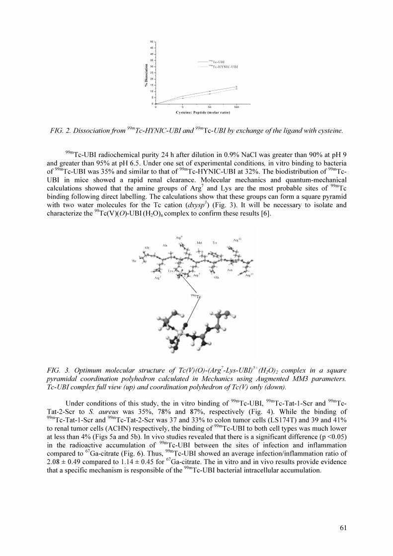
0 5 50 500
0
5
10
15
20
25
30
35
40
45
50
% D
isso
cia
tio
n
Cystein e: Peptide (m olar ratio)
99m
Tc-UBI
99m
Tc-HYNIC-UBI
FIG. 2. Dissociation from 99m
Tc-HYNIC-UBI and 99m
Tc-UBI by exchange of the ligand with cysteine.
99m
Tc-UBI radiochemical purity 24 h after dilution in 0.9% NaCl was greater than 90% at pH 9
and greater than 95% at pH 6.5. Under one set of experimental conditions, in vitro binding to bacteria
of 99m
Tc-UBI was 35% and similar to that of 99m
Tc-HYNIC-UBI at 32%. The biodistribution of 99m
Tc-
UBI in mice showed a rapid renal clearance. Molecular mechanics and quantum-mechanical
calculations showed that the amine groups of Arg7
and Lys are the most probable sites of 99m
Tc
binding following direct labelling. The calculations show that these groups can form a square pyramid
with two water molecules for the Tc cation (dxysp3
) (Fig. 3). It will be necessary to isolate and
characterize the 99
Tc(V)(O)-UBI (H2O)
ncomplex to confirm these results [6].
Arg3
Arg6
Arg7
Ala
Lys
Met
Gly
Thr
Gln
Tyr
Arg13
Arg12
Asn
99m
Tc
FIG. 3. Optimum molecular structure of Tc(V)(O)-(Arg7
-Lys-UBI)5+
(H2O)
2complex in a square
pyramidal coordination polyhedron calculated in Mechanics using Augmented MM3 parameters.
Tc-UBI complex full view (up) and coordination polyhedron of Tc(V) only (down).
Under conditions of this study, the in vitro binding of 99m
Tc-UBI, 99m
Tc-Tat-1-Scr and 99m
Tc-
Tat-2-Scr to S. aureus was 35%, 78% and 87%, respectively (Fig. 4). While the binding of
99m
Tc-Tat-1-Scr and 99m
Tc-Tat-2-Scr was 37 and 33% to colon tumor cells (LS174T) and 39 and 41%
to renal tumor cells (ACHN) respectively, the binding of 99m
Tc-UBI to both cell types was much lower
at less than 4% (Figs 5a and 5b). In vivo studies revealed that there is a significant difference (p <0.05)
in the radioactive accumulation of 99m
Tc-UBI between the sites of infection and inflammation
compared to 67
Ga-citrate (Fig. 6). Thus, 99m
Tc-UBI showed an average infection/inflammation ratio of
2.08 ± 0.49 compared to 1.14 ± 0.45 for 67
Ga-citrate. The in vitro and in vivo results provide evidence
that a specific mechanism is responsible of the 99m
Tc-UBI bacterial intracellular accumulation.
61
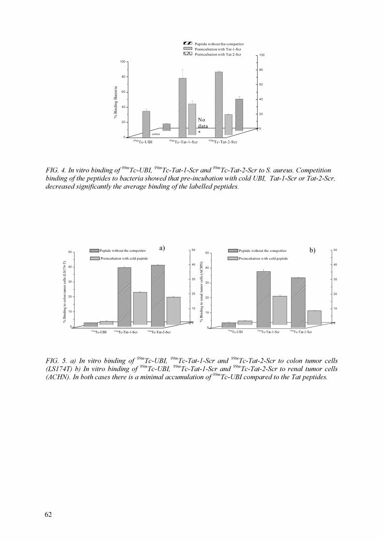
0
20
40
60
80
100
% B
in
din
g B
acteria
0
20
40
60
80
100
99m
Tc-UBI
99m
Tc-Tat-1-Scr
Peptide without the competitor
Preincubation with Tat-1-Scr
Preincubation with Tat-2-Scr
99m
Tc-Tat-2-Scr
*
No
data
FIG. 4. In vitro binding of 99m
Tc-UBI,99m
Tc-Tat-1-Scr and 99m
Tc-Tat-2-Scr to S. aureus. Competition
binding of the peptides to bacteria showed that pre-incubation with cold UBI, Tat-1-Scr or Tat-2-Scr,
decreased significantly the average binding of the labelled peptides.
FIG. 5. a) In vitro binding of 99m
Tc-UBI, 99m
Tc-Tat-1-Scr and 99m
Tc-Tat-2-Scr to colon tumor cells
(LS174T) b) In vitro binding of 99m
Tc-UBI,99m
Tc-Tat-1-Scr and 99m
Tc-Tat-2-Scr to renal tumor cells
(ACHN). In both cases there is a minimal accumulation of 99m
Tc-UBI compared to the Tat peptides.
0
10
20
30
40
50
Preincubation with cold peptide
0
10
20
30
40
50
% B
inding to colon tum
or cells (L
S174-T
)
Peptide without the competitor
99m
Tc-UBI99m
Tc-Tat-1-Scr99m
Tc-Tat-2-Scr
0
10
20
30
40
50
Preincubation with cold peptide
0
10
20
30
40
50
% B
in
din
g to
ren
al tu
mo
r cells (A
CH
N)
Peptide without the competitor
99m
Tc-UBI
99m
Tc-Tat-1-Scr
99m
Tc-Tat-2-Scr
a) b)
62

FIG. 6. Whole body images of the same mouse with induced infection (right thigh) and sterile
inflammation (left thigh) 2 h after administration of both 99m
Tc-UBI and 67
Ga-citrate.
FIG. 7. Patient with chronic bone infection on right arm. Anterior and posterior views at 1 min,
30 min 2 h and 24 h after 99m
Tc-UBI administration.
99m
Tc-UBI showed a fast renal clearance in patients (Fig. 7). Two hours after administration a
small percentage of the injected activity was removed by the liver in comparison with the kidney.
Residence times and absorbed dose calculated from patients are shown in Table I and target non-target
ratio at the infection sites are shown in Table II.
Radiochemical yield for 99m
Tc-EDTA-Biotin and 99m
Tc-DTPA-bis-Biotin was 93% and >98%
respectively. A quantitative shift of the radioactivity profile to shorter retention times (avidin retention
time) in the presence of the avidin demonstrated a good radiolabel and the avidity of the 99m
Tc -biotin
complexes for avidin. Specifically 99m
Tc labelled biotin showed an average infection/inflammation
ratio of 1.36 ± 0.27.
1 min 30 min 24 h 2 h
63

TABLE I. ABSORBED DOSE, RESIDENCE TIMES AND EFFECTIVE DOSE OF 99m
Tc-UBI
CALCULATED FROM PATIENTS
Patient
No.
Absorbed Dose, mGy/MBq
[Residence time, h]
Effective Dose
mSv/MBq
Kidney Liver
1 3.94E-02 [0.74] 3.94E-03 [0.38] 1.01E-02
2 6.79E-02 [1.27] 5.43E-03 [0.52] 7.62E-03
3 3.97E-02 [0.74] 4.51E-03 [0.43] 4.21E-03
4 5.32E-02 [0.99] 3.06E-03 [0.29] 7.89E-03
5 7.56E-02 [1.41] 5.37E-03 [0.51] 6.68E-03
6 3.86E-02 [0.72] 7.92E-03 [0.76] 3.53E-03
TABLE II. TARGET/ NON-TARGET RATIO OF 99m
Tc-UBI AT INFECTION SITES IN
PATIENTS
5. CONCLUSIONS
99m
Tc-UBI, labelled by a direct method, showed high in vitro and in vivo stability, specific
uptake at the site of infection, rapid background clearance, minimal accumulation in non-target tissues
and rapid detection of infection sites. 99m
Tc labelled biotin showed to be stable in vitro and in vivo,
fast renal clearance and ability to detect infection and sterile inflammation processes in mice. Because
of their stability and biological properties, both agents could be used in clinical applications.
REFERENCES
[1] WELLING, M., PAULUSMA-ANNEMA, A., BALTER, H.S., PAUWELS, E.K.J.,
NIBBERING, P.H., “Technetium-99m labelled antimicrobial peptides discriminate between
bacterial infections and sterile inflammations”, Eur. J. Nucl. Med. 27 (2000) 291–301.
[2] WELLING, M., et al., “Radiochemical and biological characteristics of 99m
Tc-UBI 29-41 for
imaging of bacterial infections”, Nucl. Med. Biol. 29 (2002) 413–422.
[3] FERRO-FLORES, G., et al., “In vitro and in vivo assessment of 99m
Tc-UBI specificity for
bacteria”, Nucl. Med. Biol. 30 (2003) 597–603.
[4] MELENDEZ-ALAFORT, L., et al., “Lys and Arg in UBI: A specific site for a stable Tc-99m
complex?”, Nucl. Med. Biol. 30 (2003) 605–615.
[5] RODRIGUEZ-CORTES, J., et al., 99m
Tc-UBI Biokinetics: a Apecific Peptide for Infection
Detection, Mexican Symposium on Medical Physics, Conference Proceedings, American
Institute of Physics AIP (2003) in press.
[6] FERRO-FLORES, G., et al., “Synthesis of 99m
Tc-IDA-bis-Biotin and 99m
Tc-DTPA-bis-Biotin”,
J. Labelled Cpd. Radiopharm. 44 Suppl. 1 (2001) 541–543.
Patient No. 99m
Tc-UBI
Target/non-target ratio
1 2.83
2 3.09
3 1.58
4 1.45
5 1.02 (negative)
6 1.97
64

DEVELOPMENT OF KITS FOR 99m
Tc RADIOPHARMACEUTICALS FOR
INFECTION IMAGING
M. JEHANGIR, M. BASHAR, S. PERVEZ
Isotope Production Division,
Pakistan Institute of Nuclear Science and Technology,
Islamabad, Pakistan
Abstract
The aim of the present work was to evaluate three agents selected in this CRP for their possible role as
99m
Tc labelled infection imaging agents: human neutrophil elastase inhibitor (HNE2), Ubiquicidin (UBI 29-41)
and Ethylene diamine tetraacetic acid biotin monomer (EB1). 99m
Tc labelling of EB-1 was carried out by direct
labelling method with a yield of 90–95%. UBI was labelled by direct labelling as well as indirect labelling
method using cyclic DTPA and HYNIC as conjugates with labelling efficiency of >95% in both cases, while
HNE-2 was labelled only by indirect method and >96% labelling with 99m
Tc could be achieved.
Chromatographic studies like ITLC, paper chromatography and HPLC were carried out for radiochemical
separation and for determining the labelling yields. Kit formulations were also developed for these agents. Since
availability of HNE-2 was doubtful detailed study of 99m
Tc-UBI (29-41) involving cysteine challenge, in vitro
binding with bacteria as well as infection imaging in mice and rabbits was carried out. Experimental thigh
muscle infection was induced by injecting 2×10⁸ CFU of live S. aureus or E. coli bacteria into right thigh muscle
of rabbits while turpentine oil and formalin killed S.aureus were utilized for inducing sterile thigh muscles
inflammation. Significantly higher (p<0.05) accumulation of 99m
Tc-UBI (29-–41) was seen at sites of S.aureus
infected subjects (T/NT ratio 2.2 ± 0.5) as compared to E. coli (T/NT ratio 1.7±0.4). Maximum tracer
accumulation was seen at 60 min post injection followed by gradual decline. No accumulation of 99m
Tc-UBI was
observed in thighs of rabbits injected with either turpentine oil or killed S.aureus with significant lower T/NT
ratios (p<0.05) compared with S. aureus and E. coli infected thighs. Hence, it can be concluded that 99m
Tc-UBI
29-41 scintigraphy is a good technique for differentiating infection with S. aureus and E. coli. This study can be
extended to clinical study after toxicity clearance of 99m
Tc UBI 29-41.
1. INTRODUCTION
One of the best studied antimicrobial peptides is UBI (29-41) [TGRAKRRMQYNRR, 1,693
Da) which was originally isolated from the cytosolic fraction of IFN-γ-activated cells of mouse
macrophage cell line RAW264.7. Later on, an identical UBI was isolated from human, H 292 airway
epithelial cells [1]. Similarly, another microbial peptide which has been studied for antibacterial
therapy in experimental infections is human neutrophil elastase inhibitor (HNE2) [2]. The antibacterial
effect was found to be associated with an increased influx of neutrophils in the infected area [3]. Use
of this agent in experimental thigh muscle infection in mice allowed rapid visualization of bacterial
infection, but abscess to background ratios were low and decreased with time [4,5].
The various methods of labelling peptides with 99m
Tc including indirect labelling using the
preformed chelate approach or bifunctional chelating agents and the direct labelling method have been
discussed extensively [6]. The direct labelling method is a simple procedure in which the peptide is
labelled in absence of exogenous chelators. EB1 and UBI can be labelled by direct labelling with
99m
Tc in presence of reducing agent. The labelling of UBI is rapid and labelling yield quite good
(>95%) [7]. UBI can also be labelled through indirect labelling while HNE2 can only be labelled
through bifunctional chelating agent procedures [8]. Welling et al [9] have shown significant
accumulation of 99m
Tc-UBI (29-41) in rabbits with experimental thigh muscle infection induced by
two different types of S.aureus (ATCC25923) and MRSA. They found mean target to non-target ratio
of 3.3+0.2 in both cases and found similar results in mice [10–11].
The purpose of the present investigation was to study in detail 99m
Tc labelling conditions for
three agents HNE2, EB1 and UBI and formulate them into easily available freeze-dried kit form. The
work on HNE2 and EB1 was limited to determining the labelling conditions only. However, UBI was
found quite promising. So a detailed study of this antimicrobial peptide was carried out which
included: (i) Direct labelling of UBI with 99m
Tc in in-situ form, (ii) Indirect labelling, (iii) Formulation
65
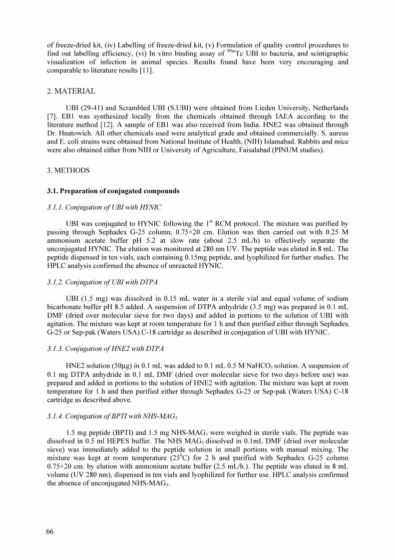
of freeze-dried kit, (iv) Labelling of freeze-dried kit, (v) Formulation of quality control procedures to
find out labelling efficiency, (vi) In vitro binding assay of 99m
Tc UBI to bacteria, and scintigraphic
visualization of infection in animal species. Results found have been very encouraging and
comparable to literature results [11].
2. MATERIAL
UBI (29-41) and Scrambled UBI (S.UBI) were obtained from Lieden University, Netherlands
[7]. EB1 was synthesized locally from the chemicals obtained through IAEA according to the
literature method [12]. A sample of EB1 was also received from India. HNE2 was obtained through
Dr. Hnatowich. All other chemicals used were analytical grade and obtained commercially. S. aureus
and E. coli strains were obtained from National Institute of Health, (NIH) Islamabad. Rabbits and mice
were also obtained either from NIH or University of Agriculture, Faisalabad (PINUM studies).
3. METHODS
3.1. Preparation of conjugated compounds
3.1.1. Conjugation of UBI with HYNIC
UBI was conjugated to HYNIC following the 1st
RCM protocol. The mixture was purified by
passing through Sephadex G-25 column, 0.75×20 cm. Elution was then carried out with 0.25 M
ammonium acetate buffer pH 5.2 at slow rate (about 2.5 mL/h) to effectively separate the
unconjugated HYNIC. The elution was monitored at 280 nm UV. The peptide was eluted in 8 mL. The
peptide dispensed in ten vials, each containing 0.15mg peptide, and lyophilized for further studies. The
HPLC analysis confirmed the absence of unreacted HYNIC.
3.1.2. Conjugation of UBI with DTPA
UBI (1.5 mg) was dissolved in 0.15 mL water in a sterile vial and equal volume of sodium
bicarbonate buffer pH 8.5 added. A suspension of DTPA anhydride (3.5 mg) was prepared in 0.1 mL
DMF (dried over molecular sieve for two days) and added in portions to the solution of UBI with
agitation. The mixture was kept at room temperature for 1 h and then purified either through Sephadex
G-25 or Sep-pak (Waters USA) C-18 cartridge as described in conjugation of UBI with HYNIC.
3.1.3. Conjugation of HNE2 with DTPA
HNE2 solution (50µg) in 0.1 mL was added to 0.1 mL 0.5 M NaHCO3solution. A suspension of
0.1 mg DTPA anhydride in 0.1 mL DMF (dried over molecular sieve for two days before use) was
prepared and added in portions to the solution of HNE2 with agitation. The mixture was kept at room
temperature for 1 h and then purified either through Sephadex G-25 or Sep-pak (Waters USA) C-18
cartridge as described above.
3.1.4. Conjugation of BPTI with NHS-MAG3
1.5 mg peptide (BPTI) and 1.5 mg NHS-MAG3 were weighed in sterile vials. The peptide was
dissolved in 0.5 ml HEPES buffer. The NHS MAG3
dissolved in 0.1mL DMF (dried over molecular
sieve) was immediately added to the peptide solution in small portions with manual mixing. The
mixture was kept at room temperature (250
C) for 2 h and purified with Sephadex G-25 column
0.75×20 cm. by elution with ammonium acetate buffer (2.5 mL/h.). The peptide was eluted in 8 mL
volume (UV 280 nm), dispensed in ten vials and lyophilized for further use. HPLC analysis confirmed
the absence of unconjugated NHS-MAG3.
66
3.2. Procedure for kit formulation
3.2.1. Direct labelling kit for 99m
Tc UBI
The labelling procedure recommended by Leiden University was slightly amended as
lyophilized pyrophosphate kit was used as source of stannous ions and sodium borohydride as
reducing agent. Various formulations have been tried as shown in Table I.
The following composition has given more than 90% labelling efficiency in repeated tests: UBI
100µg dissolved in 10 µL 0.01 M acetic acid; 5 µl from pyrophosphate lyophilized kit consisting of 10
mg sodium pyrophosphate, 2.5 mg SnCl2.2H
2O and reconstituted with 5.7 mL distilled water; 8µL
from a solution of 7 mg/mL sodium borohydride in 0.1N NaOH; 0.15 mL activity (99m
Tc 2~3 mCi)
freshly eluted from Generator; pH 6~7
Lyophilized kits with 4 times above amount were also prepared in order to use the higher
amounts of activity for studies in higher animals.
3.2.2. Direct labelling kit for 99m
Tc S-UBI
The procedure and formulation developed for 99m
Tc UBI was tested for 99m
Tc S-UBI and found
that the same can be used without any change. Therefore the S-UBI kits used in our experimental work
were prepared by the same procedure as for UBI kits.
3.2.3. 99m
Tc HYNIC-UBI kit
Following procedures were investigated:
Ligand exchange procedure: The lyophilized kit of heptagluconate was used containing 100 mg
calcium heptagluconate and 0.12 mg stannous chloride. The kit was labelled by addition of 4 mL
activity (99m
Tc, 5~7 mCi) at room temperature. The kit was labelled within 20 min and 0.1mL peptide
conjugate solution was then added. After further half an hour the kit was analysed for quality control
Co-ligand procedure: Tricine was used as co-ligand for labelling of UBI-HYNIC. In order to
determine the optimum quantity of the reagents to get labelling yield >90%, the amount of peptide,
tricine and tin have been varied as shown in Table II. The first four experiments were carried out at
room temperature while in 5 and 6, the reaction mixture vial was heated in boiling water for 15 min.
The following composition was considered the best for kit production: 50µg UBI-HYNIC in
50µL ammonium acetate buffer pH 5.2; 5µL from 1 mg/ml SnCl2.2H
2O, solution in HCl; 50µl from 1
mg/50µL aqueous solution of tricine; 0.1 mL activity (99m
Tc 3~4 mCi) (with heating); pH 6~7
3.2.4. 99m
Tc DTPA -UBI kit
The following formulation provided labelling efficiency in the range of 80-90%: 150 µg UBI-
DTPA; 18µL from 0.5 M sodium bicarbonate buffer pH 9.2; 5 µL from SnCl2.2H
2O 1 mg/mL solution
in HCl; 0.1mL activity (99m
Tc 3~4 mCi) from generator; pH 7.5.
However the formulation was not much investigated and the labelled solution used after
removal of the hydrolyzed activity by elution through the Sephadex G-25 column (3×0.75 cm).
3.2.5. 99m
Tc DTPA -HNE2 kit
The following formulation provided above 90% labelling: 5µg DTPA-HNE2 (lyophilized in the
vial); 18µL from buffer consisting of 0.5 M NaHCO3, 0.25 M ammonium acetate and ammonium
67
hydroxide upto pH 9.2; 5 µL from 1 mg/mL SnCl2.2H
2O in HCl; 0.1 mL activity (
99m
Tc, 2~3 mCi)
from generator; pH 7.6. The solution was analysed for labelling after 30 min.
3.2.6.99m
Tc MAG3-BPTI kit
The following formulation has been used for labelling the conjugated peptide: 20µg MAG3-
BPTI in 0.1 mL ammonium acetate buffer; 50µL tartarate buffer pH 9.2; 10µL from 0.15 mg/mL
SnCl2.2H
2O in HCl; 0.2 mL activity (
99m
Tc 4~5 mCi); pH 7.0–7.5
However the above formulation gave excessive amount of hydrolyzed activity (about 30–40%)
and purification through Sephadex on Sep-pak was necessary.
3.2.7. 99m
Tc EB1 kit
Various protocols were tried as given in Table III. Finally, following protocol was adopted. EB1
(1mg) was dissolved in acetate buffer 10mL of pH~6. Then to this solution freshly prepared Sn Cl2.
2H2O solution was added (10 µg/0.01mL). This solution was dispensed into 10 vials and lyophilized.
Each vial contains 100 µg EB1 in 1mL acetate buffer of pH~6 and 1µg SnCl2.
3.3. Labelling procedures
3.3.1. 99m
Tc EB1
Each time a single vial was taken and 1ml of 99m
Tc added and heated in boiling water for
10 min and 15 min. Labelling efficiency was determined by paper chromatography with two solvents,
acetone and saline. The results show that 92–96% labelling is obtained with 15 min heating while 80%
labelling was obtained with 10 min heating.
3.3.2. 99m
Tc UBI and 99m
Tc HNE2
The lyophilized kits of direct labelled UBI, scrambled UBI, UBI-DTPA conjugate and HNE2-
DTPA conjugate were labelled by addition of 0.1~0.15 mL 99m
Tc activity freshly eluted from
generator and was kept at room temperature for 30 min. When a kit was prepared by taking multiple
amount of the formulation, the activity was increased in the same ratio. In case of UBI-HYNIC kit
with tricine as co-ligand, heating in boiling water bath for 10 mts followed addition of 99m
Tc activity.
3.4. Quality control
For determination of free activity (TcO4
-
), labelled activity and reduced/hydrolyzed activity in
direct labelled peptide kits as well as conjugated peptide kits, the following systems were used.
Whatman 3-acetone/ethanol: The chromatographic system of Whatman-3 paper with acetone
solvent was very reliable to differentiate TcO-
4from other species. The free TcO
-
4moves with the front
while other species remain at the origin. In ethanol only hydrolysed Tc stayed at the origin while
TcO-
4 and labelled peptide moved.
ITLC-SG-85% Ethanol, pH 3: The system is very reliable to differentiate hydrolyzed activity
from other species because all other species move while hydrolyzed activity remains at the origin.
However some times the movement of labelled compound was so small that it was difficult to decide
whether the activity was at the origin or moved.
HPLC: The gradient programme as given in Leiden University protocol was adopted by minor
adjustment by using 0.01 M TFA (A) and acetonitrile (B) as mobile phase as follows:
0-5 min.(Constant) 100% A, 0% B; 5–15 min (Gradient) 20% A, 80% B; 15–20 min
68

(Constant) 20% A, 80% B; 20–25 min (Gradient)100% A,0% B; 25-30 min (Constant)100%
A,0% B. In the above mentioned system Bonda PAK RP-columns 0.5×25 cm was used. The
elution time for free TcO4
was 3.6 min. and for peptide 12–16 min. with peak splitting.
3.5. Room temperature stability of labelled kits
The quality control of all the labelled kits was performed after 30 min of labelling. The analysis
repeated after the intervals of 2 h in order to determine the stability of the label. It was found that all
the kits were stable up to 6.0 h. However the analysis of the next day showed that the labelled kit was
not stable on the next day.
3.6. Cysteine challenge
The cysteine challenge test was performed only with direct abeled 99m
Tc UBI. The instability
towards cysteine was checked according to the protocol recommended in first RCM report. The results
are given in Table IV.
3.7. In vitro binding studies with bacteria
Two different strains of bacteria have been used in the subject studies. The standard strain of
S.aureus ATCC 25923 as recommended has been obtained from National Institute of Health (NIH)
Islamabad. The standard strain of E.coli ATCC No. 25922 was also obtained from NIH Islamabad.
The primary culture provided was Mac’Conky agar culture petri dish. Microscopic slides were
prepared and stained with grahm’s strain to check the purity of the culture.
The binding of labelled compounds to bacteria (S.aureus ATCC 25923) was assessed at 4o
C
following 1st
RCM protocol. The supernatant was removed and the bacterial pellet resuspended in 1
mL PBS and re-centrifuged. The supernatant was recombined and the activity associated with bacterial
pellet was determined in the activity meter.
3.8. Animal studies
The animal studies can be subdivided in three categories (I) mice studies (II) rabbit studies at
PINSTECH (III) rabbit studies at PINUM, Faisalabad.
3.8.1. Mice studies
Induction of infection in mice: The standard culture containing 2×10⁷
viable bacteria per mL
was thawed 2 h before injection. The mice were anaesthetized given anesthesia by exposure to vapors
of anesthetic ether. 0.1 mL culture injected was into left or right thigh muscle. After 24 h the
symptoms of infection were obvious.
Testing the labelled kit: The standard kit of UBI(29-41) was labelled with addition of 0.1 mL
99m
Tc elution from the generator as standard procedure. After 1 h the kit was diluted to 2 mL and
analysed for radiochemical purity as standard procedure. The mice were anaesthesized with
anaesthetic ether and 0.1 mL (5 µg UBI content) was injected through the tail vein. Three groups of
three mice each were sacrificed after 1 h, 5 h and 24 h, respectively, for biodistribution. The uptake in
infected compared with the normal thigh muscle are given in Table V.
3.8.2. Rabbit studies at PINSTECH
In order to confirm the findings in mice, New Zealand white rabbits (weighing from 1.5 kg to
2.0 kg and fed on fresh vegetables) have been used for imaging to assess the accumulation of
radiotracer in infected lesions, induced by injecting 2×10⁹
CFU fresh cultured S.aureus ATCC 25923
69
or 2×10⁸
CFU of ATCC 25922 E. coli. The bacteria cultured in BHI broth, were resuspended in
normal saline before injection in order to avoid the effect of accumulated toxins. Right thigh muscle
was infected using 0.4 mL suspension two to four days before the imaging studies. The development
of infection was confirmed by physical examination and collection of blood/puss from the lesion. The
imaging was performed using Siemens Spect Gamma Camera using parallel hole low energy
collimator and 256×256 matrix. The rabbit was laid anaesthesized with surgical tapes on legs under
the gamma camera and images taken at 15, 30, 45 min and 1 h post injection with time interval 2 min
and 5 min. The sterile inflammation was induced by injecting 0.3 mL sterilized solution of E.coli in
saline (containing the endotoxins). The rabbit was injected 2 mL diazepam in the muscles of fore leg.
The anesthesia remained effective for about 1 h, sufficient for our imaging purpose.
Infection imaging: Following kits have been used for infection imaging after labelling with
99m
Tc: UBI 29-41 lyophilized kit, UBI Scrambled lyophilized kit, Infecton (labelled ciprofloxacin)
lyophilized kit, UBI-DTPA lyophilized kit, UBI-HYNIC lyophilized kit
Summary of the protocols: Lyophilized kit of UBI or DTPA-UBI or HUNIC-UBI or S.UBI
containing 0.4 mg peptide was reconstituted with 0.5 mL 99m
Tc (about 3 mCi). After about 30 min the
quality control was performed and 0.4 mL injected in the ear vein of the rabbit. Separate kits were
used for imaging gram positive, gram negative or sterile inflammation. The Infecton kit, consisting of
2 mg ciprofloxacin and 1 mg FSA adjusted to pH 7.5 was labelled by addition of 1 mL 99m
Tc (8 mCi).
After Q.C. 0.1 mL was injected in the ear vein of the rabbits. The imaging was performed as described
above after 45 min.
3.8.3. Rabbit studies at PINUM
Thirty three rabbits were used in the study. Twenty-five were females and eight males. Weight
of each animal ranged from 1.2 – 1.6 kg, with an average of 1.4 kg.
Induction of inflammation in rabbits: sterile inflammation was induced by injecting 0.3 mL of
saline containing 2×10⁸
formalin killed S.aureus. into the right thigh muscle. After 48 h, when
swelling of thigh muscles was present, 99m
Tc UBI 29-41 imaging was carried out. Group of the
animals and agents used to induce infection and sterile inflammation are summarized below.
Group No. of animals Infection/Inflammation inducing agent
I (control) 3 Nil
II IIa 10 S.aureus (2×10⁸
CFU/animal)
IIb 10 E.coli (2×10⁸
CFU/animal)
III IIIa 5 Killed S.aureus (2×10⁸
CFU/animal)
IIIb 5 Turpentine oil (0.3 ml/animal
Biodistribution of 99m
Tc UBI 29-41 in rabbits: Total body counts were determined by drawing
ROI over the image of entire animal. To find out the uptake (counts) of tracer in different organs,
ROI’s were drawn over the liver, both kidneys and urinary bladder in the images taken at 5 min, 30
min, 60 min and 120 min.
70
4. RESULTS
4.1. Direct labelling of UBI with 99m
Tc
Various protocols were studied for formulation of UBI in kit form. For direct labelling the
products are given in Table I. Radiochemical analysis showed >95% labelled fraction. The stability of
the labelled kit was studied up to 6 h during which it remained intact.
TABLE I. DIRECT LABELLING OF UBI WITH 99m
TC
Peptide Sn+2
Borohydride Results after 1.5 h
Volume From Volume From Volume
(Weight)
From Free
TcO4
Hydro-
lysed
Peptide
labelled
10µL
(10µg)
Acetate buffer
pH 2.5,1µ g/µL
5µL SnCl2.2H
2O 0.1
mg/mL in HCl
5µL
(15µg)
3mg/mL in 0.1
NaOH
84.6% 7.0% 8.4%
10µL -do- 5µL -do- 5µL
(25µg)
5mg/mL in
0.1N NaOH
5.4% 45.6% 49.0%
10µL -do- 5µL 10mL water to
pyrophosphate
kit
5µL
(25µg)
5mg/mL NaBH4
in 0.1N NaOH
45% 0.0% 45.03%
20µL 0.01 N acetic
acid 1µg/µL
5µL 5.7mL water to
pyrophosphate
kit
6µL
(42µg)
7mg/mL NaBH4
in 0.1N NaOH
3.1% 30.8% 66.1%
10µL 0.01 N acetic
acid 10µg/µL
5µL 5.7mL water to
pyrophosphate
kit
8µL
(56µg)
7mg/mL NaBH4
in 0.1N NaOH
10.4% 0.0% 89.6%
Lyophilized kit sample 1 8.4% 0.0% 91.6%
Lyophilized kit sample 2 7.3% 0.0% 92.7%
10µl 0.01 N acetic
acid 10µg/µl
5µL SnCl2.2H
2O
0.44 mg/mL in
HCl
8µL
(56µg)
7mg/mL NaBH4
in 0.1N NaOH
2.6% 0.0% 97.4%
4.2. Indirect labelling of UBI with 99m
Tc
For indirect labelling two techniques ligand exchange procedure with Hepta gluconate
kit as well as co-ligand procedure using tricine as co-ligand with UBI-HYNIC has been
studied as given in Table II.
TABLE II. 99m
TC LABELLING OF HYNIC-UBI USING TRICINE CO-LIGAND
Peptide Sn+2
(SnCl2.2H
2O) Tricine Result after 1.5 h
Vol. From Vol. From Vol. From Free
TcO-
4
Hydro-
lysed
Peptide
labelled
0.1mL Acetate buffer
100µg/0.1mL
5µL 1mg/mL in HCl 50µL 4mg/50µL 0% 0% 56.%
0.1mL Acetate buffer
100µg/0.1mL
5µL 1mg/mL in HCl 50µL 2mg/50µL 0% 0% 60%
0.1mL Acetate buffer
100µg/0.1mL
5µL 1mg/mL in HCl 50µL 1mg/50µL 67% 0% 33%
0.1mL Acetate buffer
100µg/0.1mL
5µL 1mg/mL in HCl 50µL 0.5mg/50µL 91% 1% 8%
50µL Acetate buffer
200µg/0.1mL
5µL 1mg/mL in HCl 50µL 1mg/50µL
(heating for
15 min)
0% 0% 96%
50µL Acetate buffer
100µg/0.1mL
5µL 1mg/mL in HCl 50µL 1mg/50µL
(heating for
15 min)
0% 0% 93%
71
UBI was labelled with 99m
Tc efficiently (>95%) by both direct and indirect labelling
techniques. Since the direct labelling method is convenient this was followed for the production of kits
and further studies.
4.3. Cysteine challenge
The results of cysteine challenge studies given in Table III show that labelled peptide
is sufficiently stable toward cysteine.
TABLE III. CYSTEINE CHALLENGE OF DIRECT LABELLED 99m
TC UBI(29-41)
99m
TcO4
− in
standard kit
99m
TcO4
−+
99m
Tc Cysteine after 1.5 h at Cysteine ratio to
peptide
5:1 50:1 500:1
1.3% 14.5% 26.7% 34.5%
4.7% 21.4% 30.2% 44.1%
4.4. In vitro binding assay of 99m
Tc UBI to bacteria
The in vitro bacterial binding study at 4o
C is found to be a good indicator for the development
of infection imaging agents. The kits having better binding properties in vitro, have shown also the
good in vivo binding. The results revealed the highest binding of 99m
Tc direct labelled UBI peptides to
bacteria. The results are presented in Table IV.
TABLE IV. BACTERIAL BINDING PROPERTY OF DIFFERENT 99m
TC UBI KITS
Description % Activity bound n=5
99m
Tc UBI Direct labelled 31 ± 5%
99m
Tc UBI Scrambled 9 ± 3%
99m
Tc DTPA- UBI 22 ± 5%
99m
Tc HYNIC -UBI 25 ± 4%
4.5. Biodistribution and accumulation of 99m
Tc UBI in infection in mice
The results as given in Table V show that the activity is accumulated in infected site within 1 h,
which is useful for rapid scanning. The IM/NM activity ratio is 4.1, 4.9 and 5.8 after 1, 5 and 24 h,
respectively. The activity is rapidly excreted mainly through the urine. The IM/NM ratio is improved
with the passage of time but the total activity in the IM is rapidly decreased.
72

TABLE V. BIODISTRIBUTION STUDIES OF 99m
TC UBI IN MICE (%ID/ORGAN, SD N=3)
Tissue 1 h 5 h 24 h
Liver 3.6 +3.0 2.7 + 2.0 1.4 + 0.4
Spleen 0.12 + 0.1 0.06 + 0.02 0.02 + 0.01
Stomach 1.0 + 0.5 0.1 + 0.05 0.01 + 0.04
Intestine 13.9 + 5.0 7.3 + 4.0 6.1 + 2.0
Lungs 0.1 + .02 0.003 + 0.01 0.003 + 0.02
Kidneys 17.5 + 7 6.1 + 2.0 2.1 + 1.2
Urine//Bladder 42.8 + 10 78.1 + 12 94.3 + 5.0
Heart 0.6 + 0.3 0.003 + 0.01 0.002 + 0.01
Head 2.1 + 1.5 0.68 + 0.2 0.12 + 0.05
Normal Muscle(NM) 0.3 + 0.1 0.04 + .02 0.01 + 0.05
Infected Muscle (IM) 1.3 + 0.3 0.19 + 0.05 0.058 + 0.01
IM/NM 4.1 4.9 5.8
4.6. Infection imaging studies in rabbits (PINSTECH)
A summary of the results is, as follows (Table VI):
99m
Tc UBI from lyophilized kit for direct labelling showed highest uptake (T/NT ratio >2) in
gram positive, S. aureus infection and comparative uptake in gram negative E.coli. The uptake in
sterile inflammation was negligible. 45 min is the appropriate time for imaging.
99m
Tc scrambled UBI from lyophilized kit for direct labelling showed negligible uptake of
activity in the lesion.
The imaging properties 99m
Tc DTPA -UBI and 99m
Tc HYNIC- UBI were inferior to the direct
labelled 99m
Tc UBI.
TABLE VI. INFECTION IMAGING STUDIES IN RABBITS
Target T/NT ratio (Average) n=3 Kit for 99m
Tc
labelling 15 min. 30 min. 45 min. 60 min.
UBI-29-41 S.aureus Inf 1.20±0.20 2.00±0.12 2.10±0.22 1.25±0.07
UBI-29-41 E.coli Inf 1.15±0.04 1.90±0.31 1.95±0.24 1.30±0.11
Scrambled UBI S.aureus Inf 1.01±0.03 1.15±0.12 1.15±0.11 1.07±0.05
UBI-29-41 Sterile Infl. 1.03±0.01 1.10±0.05 1.12±0.04 1.10±0.03
Infecton S.aureus Inf 1.10±0.04 1.51±0.21 2.21±0.20 2.20±0.15
UBI-DTPA S.aureus Inf 1.07±0.01 1.23±0.12 1.87±0.16 1.31±0.10
Infl= Inflammation
Inf= Infection
4.7. Rabbits studies at PINUM
The biodistribution of 99m
Tc UBI in various organs of rabbit determined scintigraphically are
presented in Table VII. Kidney displayed gradual excretion of radioactivity with mean percent uptake
values of 10.6 + 2.1 and 4.2 + 0.3 at 5 and 120 min p.i., respectively. Liver also showed gradual
decline in uptake values with the passage of time having mean percent uptake values of 6.6 + 1.6 and
2.5 + 0.8 at 5 and 120 min p.i., respectively. Rapid accumulation of radioactivity was seen in urinary
bladder with almost 80% of injected tracer present in bladder at 120 min p.i.
73
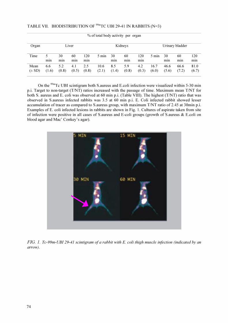
TABLE VII. BIODISTRIBUTION OF 99m
TC UBI 29-41 IN RABBITS (N=3)
% of total body activity per organ
Organ Liver Kidneys Urinary bladder
Time 5
min
30
min
60
min
120
min
5 min 30
min
60
min
120
min
5 min 30
min
60
min
120
min
Mean
(± SD)
6.6
(1.6)
5.2
(0.8)
4.1
(0.5)
2.5
(0.8)
10.6
(2.1)
8.5
(1.4)
5.9
(0.8)
4.2
(0.3)
16.7
(6.0)
46.6
(5.6)
66.6
(7.2)
81.0
(6.7)
On the 99m
Tc UBI scintigram both S.aureus and E.coli infection were visualized within 5-30 min
p.i. Target to non-target (T/NT) ratios increased with the passage of time. Maximum mean T/NT for
both S. aureus and E. coli was observed at 60 min p.i. (Table VIII). The highest (T/NT) ratio that was
observed in S.aureus infected rabbits was 3.5 at 60 min p.i. E. Coli infected rabbit showed lesser
accumulation of tracer as compared to S.aureus group, with maximum T/NT ratio of 2.45 at 30min p.i.
Examples of E. coli infected lesions in rabbits are shown in Fig. 1. Cultures of aspirate taken from site
of infection were positive in all cases of S.aureus and E-coli groups (growth of S.aureus & E.coli on
blood agar and Mac’ Conkey’s agar).
FIG. 1. Tc-99m-UBI 29-41 scintigram of a rabbit with E. coli thigh muscle infection (indicated by an
arrow).
74

TABLE VIII. TARGET TO NON-TARGET RATIOS FOR 99m
Tc UBI IN INFECTED THIGH
MUSCLES IN RABBITS
Time 5 min 30 min 60 min 2 h 3 h 4 h Culture
S. aureus Mean (±
SD)
1.7
(0.6)
2.0 (0.6) 2.2 (0.5) 1.8 (0.5) 1.7 (0.3) 1.5 (0.4) +
E. coli Mean
(± SD)
1.2 (0.2) 1.5 (0.4) 1.7 (0.4) 1.4 (0.3) 1.5 (0.4) 1.4 (0.4) +
In rabbits with sterile inflammation induced by turpentine oil and formalin killed S.aureus 99m
Tc
UBI did not show significant accumulation in the lesions. Target to non-target ratios calculated in case
of inflammation induced by turpentine oil were (1.2 + 0.14), (1.25 + 0.08), (1.16 + 0.15) and (1.03 +
0.17) at 5, 30, 60 and 120 min p.i. respectively. These ratios are lower than T/NT ratios in cases of
both S.aureus and E.coli infected rabbits. In the same way, lower T/NT ratios were seen in rabbit with
formalin killed S.aureus induced inflammation. Results are shown in Table IX. 99m
Tc-UBI scintigram
of a rabbit with turpentine oil induced thigh muscle inflammation is depicted in Fig. 2.
FIG. 2. 99m
Tc-UBI 29-41 scintigram of a rabbit with turpentine oil induced thigh muscle inflammation
(arrow).
75

TABLE IX. TARGET-TO-NON-TARGET RATIOS FOR 99m
Tc UBI IN INFLAMED THIGH
MUSCLES IN RABBITS
Time (min) p.i 5 30 60 120
Turpentine Ind, Mean (± SD) 1.20(0.14) 1.25 (0.08) 1.16 (0.15) 1.03 (0.17)
Formalin Ind, Mean (± SD) 1.05(0.09) 1.11 (0.35) 1.01 (0.27) 1.11 (0.39)
4.8.99m
Tc HNE2
HNE2 was with >90% yield through indirect labelling after making conjugate with cyclic
DTPA. Since, the availability of HNE2 was not certain further work could not be extended.
4.9.99m
Tc EB1
In order to develop lyophilized kit with EB1, locally produced various parameters were
studied as shown in Table X. Finally a protocol giving >95% yield was adopted. Radiochemical
analysis with paper chromatography (10 cm) in acetone was carried out to assess the percentage of
99m
Tc present as pertechnetate (Rf ~ 0.9) while using saline as eluent the percentage of
99m
Tc present as
colloid was assessed.
TABLE X. 99m
TC LABELLING OF EB1
EB1 SnCl2
99m
Tc
(~2mCi)
Free99m
Tc
%
Colloid
%
Labelled
%
30 µg 1 µg
(shaking for 30 min)
0.5 mL 5–15 10–26 58–80
100 µg 1 µg
(Heating for 30 min)
0.5 mL 5–15 1–5 80–94
30 µg (1 µg–1.5 µg)
Mannitol (5–15mg)
(Shaking for 30 min)
0.5 mL 0–1 0–11 88–93
90 µg 1 µg
Mannitol =15mg
Heating for 15 min
0.5 mL nil nil 100
25 µg 1.5 µg
Hepta gluconate (5–15 mg)
0.5 mL nil 11–15 85–89
90 µg 1.5 µg
Hepta gluconate (5–15 mg)
Heating for 15 min
0.5 mL nil 0–5 95–100
25 µg 1.5 µg ascorbic acid = 10mg 0.5 mL 48 16 36
5. CONCLUSIONS
Out of the three agents selected for possible use as infection imaging agent after labelling with
99m
Tc. UBI looked quite promising. UBI has been formulated into kit form, it can be labelled with
99m
Tc by direct labelling as well as through bifunctional chelates with a labelling yield of >95%.
Radiochemical analysis technique is quite easy and rapid. It binds well to the bacteria during in vitro
studies. 99m
Tc UBI scintigraphy can be used for differentiating infections with S.aureus and E.coli with
significantly higher scintigraphic intensity as compared to sterile inflammatory sites. The optimum
time for infection imaging using 99m
Tc UBI was found to be 60 min post injection in this study. The
76
encouraging results of 99m
Tc-UBI (29–41) warrant detailed study in other types of bacteria than
S.aureus and E.coli). Once toxicology studies of UBI are complete, clinical trials will be quite
challenging and interesting.
ACKNOWLEDGEMENTS
We are thankful to the IAEA for awarding us project PAK-11263R. Guidance by
Dr. Hnatowich and Dr. Pauwels is very much appreciated.
The biodistribution and scintigraphic studies on rabbits were carried out in collaboration with
Punjab Institute of Nuclear Medicine (PINUM) Faisalabad, Pakistan.
REFERENCES
[1] HIEMSTRA, P.S., VAN DEN BARSELAAR, M.T., ROEST, M., NIBBERING, P.H., VAN
FURTH, R., “Ubiquicidin, a novel murine microbicidal protein in the cytosolic fraction of
activated macrophages”, J. Leukocyte Biol. 66 (1999) 423–428.
[2] RUSCKOWSKI, M., et al., “Inflammation and infection imaging with a Tc-99m-neutrophil
elastase inhibitor in monkeys”, J. Nucl. Med. 41 (2000) 363–374.
[3] WELLING, M.M., HIEMSTRA, P.S., VAN DEN BARSELAAR, M.T., et al., “Antibacterial
activity of human neutrophil defensins in experimental infections in mice is accompanied by
increased leukocyte accumulation”, J. Clin. Invest. 102 (1998) 1583–1590.
[4] WELLING, M.M., et al., “Imaging of bacterial infections with 99m
Tc-labelled human neutrophil
peptide-1”, J. Nucl. Med. 40 (1999) 2073–2080.
[5] NIBBERING, P.H., WELLING, M.M., PAULUSMA-ANNEMA, A., VAN DEN
BARSELAAR, M.T., PAUWELS, E.K.J., “Monitoring the efficacy of antibacterial treatments
of infections with 99m
Tc-labelled anrtimicrobial peptides”, Nucl. Med. Commun. 21 (2000),
575-576 (abstract).
[6] OKARVI, S.M., “Recent developments in Tc-labelled peptide-based radiopharmaceuticals: an
overview”, Nucl. Med. Commun 20 (1999), 1093–1112.
[7] PAUWELS, E.K.J., WELLING, M.M., FEITSMA, R.I.J., NIEUWENHUIZEN, W., “The
labelling of proteins and LDL with Tc-99m: a new direct method employing KBH4 and
stannous chloride”, Nucl. Med. Biol. 20 (1993) 825–833.
[8] RUSCKOWSKI, M., et al., “Imaging osteomyelitis with streptavidin and indium-111-labelled
biotin”, J. Nucl. Med. 37 (1996) 1655–1662.
[9] WELLING, M.M., LUPETTI, A., BALTER, H.S., et al., “Technetium-99m labelled
antimicrobial peptides for detection of bacterial and Candida albicans infections”, J. Nucl. Med.
42 (2001) 788–794.
[10] WELLING, M.M., PAULUSMA-ANNEMA, A., BALTER, H.S., PAUWELS, E.K.J.,
BIBBERING, P.H., “Technetium-99m labelled antimicrobial peptides discriminate between
bacterial infections and sterile inflammations”, Eur. J. Nucl. Med. 27 (2000) 292–301.
[11] LUPETTI, A., WELLING, M.M., MAZZI, U., NIBBERING, P.H., PAUWELS, E.K.J.,
“Technetium-99m labelled fluconazole and antimicrobial peptides for imaging of Candida
albicans and Aspergillus fumigatus infection”, Eur. J. Nucl. Med. 29 (2002) 674–679.
[12] VIRZI, F., et al., “New Indium-111 labelled biotin derivatives for improved
immunotargeting”, Nucl. Med. Biol. 18 (1991) 719–724.
77

DEVELOPMENT OF KITS FOR 99m
Tc RADIOPHARMACEUTICALS FOR
INFECTION IMAGING
R. MIKOLAJCZAK, A. KORSAK, B. GORSKA, A. MARKIEWICZ,
E. ZAKRZEWSKA, W. ZULCZYK, U. KARCZMARCZYK, J. MICHALIK
Radioisotope Centre POLATOM,
Otwock-Swierk, Poland
Abstract
UBI (a derivative of antimicrobial peptide ubiquicidin) labelled with 99m
Tc is reported to discriminate
between bacterial infections and sterile inflammatory processes. In this study we compared UBI labelled with
99m
Tc by a direct method and using hydrazinonicotinamide (HYNIC) as a bifunctional chelator. We tested four
dry kits, two of them based on the direct labelling method, with or without KBH4 and two based on HYNIC
conjugation via BOC (one of them contained EDDA). After addition of 99m
TcO4
-
solution the kits were labelled
under specific conditions and ITLC, HPLC analyses (reverse-phase and ion exchange) were performed. The
binding of labelled UBI to bacteria, S. aureus, was estimated and stability experiments in human serum were
performed. The accumulation in infected tissues was studied using ex vivo countings and scintigraphy. Data
were compared to the UBI 29-41 labelled with 99m
Tc in solution (control). Conjugate of UBI 29-41 with HYNIC
was prepared with high purity (>97%). Radiochemical analysis indicated rapid and high labelling yield (90-95%)
for all four kits, the labelled compounds being stable (RCP>90%) for 24 h in vitro in human serum. The kit
containing HYNIC-UBI 29-41 and the control labelling showed highest binding of 40% of the added activity to
bacteria, being significantly higher than the direct labelling kits (30%). The range of pH was from 6 to 9 for
direct labelling kits and 4 for the kits containing HYNIC. After injection into infected mice, all tracers were
rapidly removed from the circulation by renal excretion (50–200%ID/g), but blood activity of kits containing
HYNIC-UBI 29-41 remained higher than that of directly labelled UBI 29-41. Specific accumulation in infected
thigh muscles as indicated by a T/NT ratios between 2.3 and 2.9 until 2 h after injection was observed for all the
kits. For all preparations we calculated good correlations between the number of microorganisms and T/NT ratio.
The UBI 29-41 directly labelled in presence of SnCl2 and the HYNIC-UBI 29-41 kit without EDDA showed the
most promising results for further in vivo evaluation.
The native human neutrophil elastase inhibitor (HNE-2) radiolabelled with 99m
Tc is reported to localized
specifically in inflammations in experimental animals and provide early images of diagnostic quality. The aim of
the present work was to study the radiochemical behaviour of the peptide HNE-2 and BPTI when radiolabelled
with 99m
Tc by indirect method using HYNIC as a bifunctional chelator. The conjugation of HNE-2 and BPTI
with HYNIC for labelling with 99m
Tc was previously described. Chromatographic studies were performed using
size-exclusion HPLC.
1. INTRODUCTION
In several previous papers, the 99m
Tc-labelled cationic antimicrobial peptide derived from
human ubiquicidine (UBI) was introduced for the detection of bacterial and fungal infections but not
sterile inflammatory processes in mice and rabbits [1]. Originally, UBI was isolated from murine
macrophages [2,3] and later from human airway epithelial cells. 99m
Tc-labelled synthetic UBI 29-41
was successful at imaging the infections in animal studies [4]. Furthermore this labelled peptide
allowed monitoring the efficacy of antimicrobial treatment in mice having an experimental infection
[5]. Moreover, it was reported that 99m
Tc-UBI peptide can be useful in infection detection in both
immunocompetent and immunocompromised animals [6]. UBI-derived peptides can be good
candidates for radiolabelling and studies on detection of bacterial infections in humans.
Application in humans requires standardization of the radiolabelling. Diverse dry kits of this
peptide were developed in our work for 99m
Tc labelling. The aim of the present study was to
evaluate the radiochemical and biological characteristics of four kits for 99m
Tc UBI 29-41.
In recent years radiolabelled polyclonal IgG, has been shown to be useful in the diagnosis of
infection and inflammation as a ‘non-specific’ agent [7]. In animal model streptavidin may be a more
useful agent to detect infection [8]. Unlabelled streptavidin was administered and allowed sufficient
time to accumulate non-specifically in the leison. Thereafter, radiolabelled biotin (EB1) derivative was
administered. The labelled EB1 may be retained in the lesion because biotin has extremely high
79
affinity for streptavidin. Radioscintigraphy with 111
In-biotin in this approach was successfully used in
15 patients with chronic osteomyelitis [9]. In this study we report the radiochemical characteristics and
stability of two kits for preparing 99m
Tc EB1.
The radiolabelled peptide HNE-2, is a potential radiopharmaceutical with properties permitting
specific localization in inflammation/infection and rapid pharmacokinetics. The peptide HNE-2 has a
molecular weight of 6.7 kDa and 2 lysines plus the terminal amine available for conjugation. The
peptide was conjugated with the N-hydroxysusuccinimide ester of mercaptoacetyltriglycine (MAG3),
mercaptoacetyltriserine (MAS3), HYNIC, and the cyclic anhydride of diethylenetriaminepentaacetic
acid (DTPA). The ability of 99m
Tc labelled conjugates to image inflammation/infection was evaluated
in studies in monkeys [10].
This report describes the small-scale preparation of conjugate of HNE-2 with HYNIC for
labelling with Tc-99m. Bovine pancreatic trypsin inhibitor (BPTI), which differs from HNE-2 in only
8 amino acid changes at the 58 BPTI-homologous positions plus 4 additional residues at the amino
terminus, was radiolabelled with 99m
Tc in identical manner.
2. MATERIALS AND METHODS
2.1.99m
Tc- UBI
2.1.1. UBI peptide
Synthetic peptide UBI 29-41 in powder form was received from Prof. Ernest Pauwels
(Department of Radiology, Division of Nuclear Medicine, LUMC, Leiden, The Netherlands). Peptide
was dissolved to get a 1 mM solution in 0.01 M acetic acid, pH 4 and stored at –20°C.
2.1.2. Synthesis of peptide conjugate
The method for peptide conjugation with 6-BOC hydrazinopiridine-3-carboxylic acid (BOC-
HYNIC) and synthesis of HYNIC has been previously described [11]. A total of 8.31 mg of BOC-
HYNIC (Radioisotope Centre POLATOM, Otwock-Swierk, Poland), 12.49 mg of
O-(7-azabenzotriazolyl)-1,1,3,3-tetra-methyluronium hexafluorophosphate (HATU) (Merck KGaA
Darmstadt, Germany), in 5.60 µL of N-ethyldiisopropylamine (DIPEA) (Merck KGaA Darmstadt,
Germany) and 150 µL of N-methyl-2-pyrrolidone (NPM) (Merck KGaA Darmstadt, Germany) were
reacted for 30 min. This solution was added to 100 mg of UBI 29-41 in 5.60 µL of DIPEA and 150 µL
of NPM and allowed to react for 2 h. To this solution 1mL of 5% aqueous NaHCO3 solution and 1mL
of ethyl acetate were added. The NaHCO3 solution was extracted three times with 1 mL ethyl acetate.
The combined organic phases were washed four times with water and evaporated to dryness. The
deprotection was achieved by dissolving the white solid in 5% H2O 89% TFA (Merck KGaA
Darmstadt, Germany), 3% thioanisole, 3% TIS, solution and reacted for 4 h at room temperature. After
incubation the conjugate was precipitated with ethyl ether and centrifuged. Reversed-phase HPLC
with an on-line UV detector set at 220 nm was used for analysis and purification of crude conjugate
HYNIC-UBI 29-41. A gradient with two different eluents, i.e. 0.1% TFA/H2O (A) and 0.1%
TFA/CAN (B), at a flow rate of 15 mL/min was used. The gradient was performed as follows: 0-9 min
40%B, 9-15 min 40%B, 15-16 min 0%B, 16-22 min 0%B. The purity of the conjugate amounted to
approximately 97%. The molecular weight of the purified conjugate was confirmed by mass
spectrometry.
2.1.3. Preparation of kits
The compositions of the 4 types of lyophilized kits per vial were as follows - kit A: 50 µg UBI,
5 µg SnCl2 (Fluka Chemie GmbH, Busch); kit B: 50 µg UBI, 10 µg stannous pyrophosphate (Aldrich
Chemical Company, Inc, Milwaukee WI, USA), 100 µg KBH4 (Sigma Chemical Co. St. Louis, MO
80
USA); kit C: 20 µg HYNIC-UBI, 20 µg SnCl2, 20 mg tricine, 50 mg mannitol (Calbiochem); kit D: 20
µg HYNIC-UBI, 20 µg SnCl2,
20 mg tricine, 10 mg ethylenediamine-N,N’-diacetic acid (EDDA)
(Fluka Chemie GmbH, Buchs, Siwtzerland), 50 mg mannitol. Parameters of lyophilization: -45°C:
21 h, -45°C to + 40°C: 10.3 h, + 40°C: 14.3 h, total time: 46 h, vacuum: 0.12 mbar, final temperature:
16 °C. Kits were sealed in N2 atmosphere.
2.1.4. Labelling procedure
UBI 29-41 peptide in solution was directly labelled with 99m
Tc as control. Briefly, 10 µL of a
peptide stock solution was added to 4 µL of an aseptic solution of 950 mg/L SnCl2
.
2H2O and 2g/L
sodium pyrophosphate decahydrate (Department of Clinical Pharmacy and Toxicology, LUMC).
Immediately thereafter, 2 µL of a solution of KBH4 (crystalline, Sigma Chemical Company, St. Louis,
Mo., USA) 10 mg/mL in 0.1 M NaOH was added. After addition of 0.1 mL of 99m
Tc-sodium
pertechnetate solution (200–700 MBq/mL) obtained from a 99m
Tc generator (Ultratechnekow,
Mallinckrodt Medical, Petten, The Netherlands), the mixture, having a final pH between 8 and 9, was
gently stirred at room temperature for at least 10 min.
Kits were reconstituted and labelled with 200 µL (kit A and B) or 500 µL (kit C and kit D) of
99m
Tc-sodium pertechnetate solution (200-700 MBq/mL) obtained as above. The mixture (A,Band C)
was gently stirred at room temperature for at least 1h. Kit D was incubated for 10 min at 100°C and
then cooled at room temperature for 30 min. The range of pH was between 6 and 9 for direct labelling
kits (kit A and kit B) and 4 for the kits containing HYNIC (kit C and kit D).
2.1.5. HPLC analysis
The reaction mixtures after labelling of UBI or HYNIC-UBI were analysed by anion exchange
HPLC to evaluate charged species (method 1) and reverse-phase HPLC to evaluate lipophilic species
(method 2). Method 1: samples (10–50 µL) were applied on a MONO-S column attached to a
chromatography apparatus equipped with an on line UV detector set at 220 nm and NaI (Tl) crystal
gamma detection system. A gradient with two different eluents, i.e. 0.01% (v/v) Tween in PBS
(PBST) (A) and 2M NaCl in PBST (B), at a flow rate of 1mL/min was used. The gradient was
performed as follows: 0–5 min 0%B, 5–15 min 0-100%B, 15-20 min 100%B, 20–25 min 100–0%B,
25–30 min 0%B. Method 2: samples (5–10 µL) were applied on a 4.6×250 mm C18
column (Vydac,
The Separations Group, Hesperia, CA) attached to a chromatography apparatus equipped with an on
line UV detector set at 220 nm and NaI (Tl) crystal gamma detection system. A gradient with two
different eluents, i.e. 0.1% TFA in H2O (A) and, 0.1% TFA in methanol/ H
2O [(80/20,v/v),(B)] at a
flow rate of 1mL/min was used. The gradient was performed as in method 1.
2.1.6. ITLC analysis
Labelling yields of 99m
Tc-UBI 29-41 and 99m
Tc-HYNIC-UBI 29-41 were determined using
instant thin-layer chromatography (ITLC). 1×6.5 cm ITLC/SG strips (Gelman Sciences, Ann Arbor,
MI), were dried in a microwave at 500 W for 1 min directly before use. Samples of the preparations
containing labelled peptides (2 µL) were applied at approximately 1 cm from the bottom (baseline) of
the ITLC strips and were immediately placed in air tight containers with methyl ethyl ketone or saline
solvents. With both methods 99m
Tc in the form of pertechnetate can be quantitated. After 2 min of
development, strips were dried in a microwave oven, cut into 2 fragments and radioactivity in the front
section or remaining on the baseline was determined in a dose-calibrator (VDC 101, Veenstra
Instruments, Joure, the Netherlands).
2.1.7. Stability of 99m
Tc-labelled peptides and 99m
Tc-labelled conjugate in neat human serum
Stability 99m
Tc-labelled UBI 29-41 and HYNIC-UBI 29-41 was challenged by incubating one
volume of the labelling solution with one volume of neat human serum for 1 h and 24 h at 37°C. Next,
81

the amounts of free pertechnetate and 99m
Tc-peptide in the samples were determined by ITLC using
methyl ethyl ketone and saline as solvent as described before.
2.1.8. In vitro binding of 99m
Tc-UBI and 99m
Tc-HYNIC-UBI to microorganisms
The multi-drug-(including vancomicine) resistant S. aureus type 2141 (MRSA) was a clinical
isolate (Department of Infectious Diseases, LUMC). Overnight culture of bacteria was prepared in
brain heart infusion broth (BHI, Oxoid, Basingstoke, UK) in a shaking waterbath at 37°C. The next
day, bacteria were washed, counted and aseptically aliquoted. Stocks of 0.5 mL containing 1.2×10⁹
viable bacteria were stored at -20°C. Before use the bacteria were washed twice in saline.
Binding of 99m
Tc-labelled UBI 29-41 and HYNIC-UBI 29-41 to bacteria was assessed at 4°C. In
short, 0.1 mL PBS containing 1/10 of the mixture containing 99m
Tc-labelled peptides was transferred
to an Eppendorf vial. Next, 0.8 mL of 50% (v/v) of 0.1% (v/v) acetic acid in PBS containing
0.01% (v/v) Tween-80 (PBST/HAc) and 0.1 mL of PBS containing approximately 2×10⁸
of viable
bacteria were added. The mixture, with a final pH of 5, was incubated for 1 h at 4°C and then the vials
were centrifuged in a pre-cooled centrifuge at 2000xg for 5 min. The supernatant was removed and the
bacterial pellet was gently resuspended in 1 mL PBST/HAc and centrifuged as above. The supernatant
was removed and the activity of bacterial pellet was determined in dose calibrator (VDC 101, Veenstra
Instruments, Joure, The Netherlands). The radioactivity related with bacteria was expressed as % of
added 99m
Tc activity bound to viable bacteria.
2.1.9. Studies of 99m
Tc-labelled UBI 29-41 in mice with induced infection
Specific pathogen-free, female, Swiss mice, weighing 23–30 g were used in this study. The
animals were housed in the animal housing facilities of the LUMC for at least 1 week before the onset
of the experiments. Food and water were given ad libitum. All animal studies were done in compliance
with the National laws related to the conduct of animal experiments and approved by the local
Committee for Animal Experiments.
Mice were anaesthetized with a single intraperitoneal injection of a freshly prepared 0.1 mL of
saline containing 1 mg of fluanisone and 0.03 mg fentanyl citrate (Hypnorm, Janssen Pharmaceutics,
Tilburg, The Netherlands). Immediately thereafter, 2×10⁷
of viable MRSA in water were injected
aseptically into the right thigh muscle. After 18-24 h the mice were anaesthetized as above and
preparations containing 1/10 (v/v in saline) of the radiolabelled peptide injected into tail vein, plus a
subcutaneous injection of 0,1 ml of 10-fold (v/v) diluted in saline of valium to induce muscle
relaxation. After injection of 99m
Tc-UBI 29-41 (2-7 MBq), accumulation of radioactivity in various
organs was determined using scintigraphy.
Whole body images for each duplicate of mice were made until 2 h post-injection. Radioactivity
counts in infected thigh muscles were assessed by means of anatomically fitted ROI over the entire
infected (target) and equivalent size non-infected (non-target) thigh muscle and data are expressed as
target-to-non-target (T/NT) ratios. Thereafter, the mice were sacrificed with an intraperitoneal
injection of 0,50 ml of sodium pentobarbiturate (60mg/ml, Nembutal, Sanofi BV, Division Algin,
Maassluis, The Netherlands). Blood was immediately collected by cardiac puncture, followed by
removal of urine by a syringe from the bladder. Infected and non-infected thigh muscles and various
organs were dissected, weighed and counted for radioactivity. Data are expressed as the percentage of
total 99m
Tc activity (%ID) and then calculated as %ID per gram of tissue (%ID/g). To confirm the
bacterial infection, the number of viable bacteria in the thigh muscle was determined
microbiologically and expressed as CFU per gram of tissue (CFU/g) or CFU/thigh muscle (CFU/thigh
muscle).
82
2.1.10.111
In-DTPA-UBI 29-41
The coupling of the cyclic anhydride of diethylenetriaminepentaacetic acid (DTPA) (Aldrich
Chemical Company, Gillingham, Dorset, England) to the UBI 29-41 was prepared by the same
method as described above in 2.2. The conjugate DTPA-UBI 29-41 was labelled with 111
In.
2.2.99m
Tc EB1
2.2.1. Preparation of kits
Synthetic peptide EB1 in powder form was received from Dr Mustansar Jehangir (Pakistan
Institute of Nuclear Science and Technology — PINSTECH, Islamabad, Pakistan)
The compositions of 2 types of lyophilized kits per vial was as follows - kit I: 25 µg EB1, 1 µg
SnCl2; kit II: 25 µg EB1, 2 µg SnCl
2 10 mg mannitol. Mannitol was added to the kit as an agent for
improving the quality of the freeze-dried pellet. Parameters of lyophilization were same as that given
for UBI kits.
2.2.2. Labelling procedure
Kits were dissolved in 100µL water and labelled with 74-220 MBq (200 µL)
99m
Tc-sodium pertechnetate obtained from a 99m
Tc generator. The mixture was incubated for 15 min at
80°C and then cooled at room temperature for 15 min
2.2.3. Quality control
Samples of 10-50 µL were applied on size exclusion HPLC (gradient pump, Merk Hitachi)
column (BioSep-SEC-S 4000, size: 300×7.80 mm, Phenomenex) and eluted with 0.1 M phosphate
buffer at a flow-rate of 1 mL/min. UV and radioactivity profile recorded as before.
Labelling yield and stability of 99m
Tc-EB1 with time was assessed by instant thin layer
chromatography (ITLC) and mini paper chromatography. Samples of the preparations containing
labelled peptides (2 µL) were applied at approximately 1.5 cm from the baseline of the 1×10 cm
ITLC-SG strips and were placed in airtight containers with saline. The Whatman1 paper strips (1×10
cm) were placed in airtight containers with acetone. With both systems, only pertechnetate would
migrate to solvent front while other 99m
Tc species are retained at origin.
2.2.4. Stability in human serum
Stability experiments in neat human serum were performed with two methods:
Method 1: Incubating one volume of the labelling solution with one volume of human serum for
1 h and 24 h at 37°C challenged stability of kit containing EB1. Next, the amounts of 99m
Tc-EB1 in the
samples were determined by ITLC using saline and acetone as eluent.
Method 2: After incubation 100 µl aliquots (prepared as in method 2) were taken and treated
with 200 µl of ethanol. Samples were cooled (4°C) and centrifuged for 15 min at 3000 rpm to
precipitate serum protein. 100 µl of supernatant was removed for activity measurement.
The sediment was washed twice with 1 ml of ethanol and measured. The activity in the supernatant
was compared with the activity in the pellet to give the percent peptide not bound to proteins. Next,
the amounts of 99m
Tc-EB1 in the samples were determined by ITLC using saline as solvent.
83
2.2.5. Shift assay with avidin
Shift assay was carried out by reacting 99m
Tc-EB1 with an excess of avidin (molar ratio 1:20)
and followed by size exclusion HPLC analysis (as described before). The shift in retention time was
assessed by comparing with that of 99m
Tc-EB1 without avidin. 99m
Tc-EB1 was also checked by
reacting with excess of avidin (molar ratio 1:1 and 1:2) followed by by open column P4
chromatography (0.7×20 cm) using ammonium acetate as eluent.
2.2.6. Biodistribution in mice with strile inflammation
Pathogen free, female mice (Balb/c) were used in the study. The animals were housed in the
animal housing facilities of the Military Institute of Hygiene and Epidemiology.
Mice were anaesthetized with single intraperitoneal injection of 0.2 ml of vetbutal 20-fold (v/v)
diluted in saline. Immediately thereafter, 0,3 of Oleum Terebinthnae was injected aseptically into the
right thigh muscle to induce sterile inflammation. After 48 h, the mice with typical symptoms of
inflammation and healthy mice (as control) were anaesthetized as above and 0.2 ml solution of 99m
Tc-
EB1 (approximately 37MBq), injected into tail vein.
Accumulation of radioactivity in various organs was determined using scintigraphy. Whole
body images for at least 2 mice were made 6 h post-injection. Radioactivity counts in inflamed thigh
muscles were carried out by means of anatomically fitted ROI over the entire inflamed (target) and
equivalent size non- inflamed (non-target) thigh muscle and data are expressed as target to non target
(T/NT) ratios.
2.3.
99m
Tc-HNE2
2.3.1. Conjugation with HYNIC
The peptide HNE-2 was received from Professor D. Hnatowich (University of Massachusetts
Medical School, USA). The HYNIC-conjugated peptide was prepared following a method previously
described [11].
To 9 µL of a 1.1 mg/mL HNE-2 solution in water 86 µL of 0.1 M phosphate buffer, pH 7.3 was
added. Next, a 3 mM solution of HYNIC (4,88 µg) in dimethylforamide (DMF) was added dropwise
to final HYNIC-to-peptide molar ratio of 5:1. The reaction mixture was incubated at room temperature
for 2.5 h before purification on a Sephadex G-25 column with 0.1 M phosphate buffer, pH 7.3 eluant.
The conjugated peptide eluted from the column was collected in the fractions between 5 and 7 mL.
2.3.2. 99m
Tc labelling procedure
To 100 µL of conjugated peptide in 0.1 M phosphate buffer, pH 7.3 was added 5 µl SnCl2
solution (1mg/mL in 0.1 M HCL) and approximately 7.4 MBq 99m
Tc-pertechnetate (50 µL). The
solution was incubated at 37°C for 30 min.
2.3.3. Quality control
99m
Tc labelled peptide was analysed by size-exclusion HPLC using a BioSep
SEC-S-2000 [300×7.80 mm (Phenomenex)], with 0.1 M phosphate buffer, pH 7.3,
as eluant at a flow-rate of 1 ml/min and with on-line radioactivity and UV detection (absorbance
280 nm).
84
RESULTS
2.4.
99m
Tc-UBI
The results for kits were compared to the UBI 29-41 labelled with 99m
Tc in solution (control)
[1]. Radiochemical analyses showed high labelling yield (90-95%) for all kits except D, as assessed by
ITLC and HPLC. ITLC analysis with MEK as solvent showed, that the different preparations of 99m
Tc-
UBI 29-41 revealed small amounts of relased/free 99m
Tc after 1 h incubation in human serum (~2% of
the total activity) and after 24 h (~6% of the total activity). The kit prepared 99m
Tc-HYNIC-UBI 29-41
and the 99m
Tc- UBI 29-41 control showed highest binding of ~40% of the added activity to bacteria,
being significantly higher than the direct labelling kits 30%. The range of pH was from 6 to 9 for
direct labelling kits and 4 for the kits containing HYNIC. After injection into infected mice, all tracers
were rapidly removed from circulation by renal excretion (50-200%ID/g) but blood activity of kit
prepared 99m
Tc-HYNIC-UBI 29-41 remained high. Specific accumulation in infected thigh muscles as
indicated by a T/NT ratios between 2.3 and 2.9 for the direct labelling kits until 2 h after injection was
observed. For all preparations good correlations between the number of microorganisms and T/NT
ratio were observed.
The results with 111
In-DTPA-UBI 29-41 indicated that the peptide preparation was >98% pure
and stable in human serum.
2.5.
99m
Tc EB1
Radiochemical analyses showed high labelling yield (93-99%) for both kits, as assessed by
ITLC and HPLC. The labelled peptide with avidin was eluted out earlier than the one without avidin
from both the size exclusion HPLC column and open column P4. ITLC analysis showed that 99m
Tc-
EB1, after 1 h incubation in human serum, showed very small amounts of released/free 99m
Tc. But
~13% of other 99m
Tc complexes for both kits II and I at 1 h and ~64% for kit I and ~44% for kit II
after 24 h were observed,. The range of pH for kits was from 5.0 to 7.7. The accumulation of 99m
Tc-
EB1 in inflammed thigh muscle resulted in the T/NT ratio of 3.4 for the labelling kit II after 6 h p.i.
2.6.
99m
Tc-HNE2
The size-exclusion HPLC analysis of 99m
Tc-HYNIC-HNE-2 revealed a single peak at retention
time of approximately 9.5 min. The recovery from HPLC was higher than 99%.
3. CONCLUSIONS
Both peptides UBI and HYNIC-UBI can be prepared in the dry kit form for labelling with
99m
Tc. All four kits containing UBI 29-41 or HYNIC-UBI 29-41 can be easily and quickly labelled
with99m
Tc with high yields. All tracers can be used without further purification. All the labelled
preparations were stable in human serum up to 24 h. On the basis of these results, the UBI 29-41
directly labelled in presence of SnCl2 and the HYNIC-UBI 29-41 kit without EDDA seem to be the
most promising candidates for infection imaging.
EB1 can be easily and rapidly labelled with 99m
Tc using dry kit form wothout need for post-
labelling purification. The shift in elution proflie towards higher molecular weights of 99m
Tc EB1 with
excess avidin was proved. 99m
Tc EB1 was slightly less stable than 99m
Tc UBI 29-41 on 24 h in vitro
incubation in human serum
ACKNOWLEDGEMENTS
The studies were made with help and courtesy of M.M. Welling, H. Feitsma, T. Hagendoorn
and E.K.J. Pauwels (Department of Radiology, Leiden University Medical Center).
85
REFERENCES
[1] WELLING, M.M., et al., “Technetium-99m labelled antimicrobial peptides discriminate
between bacterial infections and sterile inflammations”, Eur. J. Nucl. Med. 27 (2000) 292–301.
[2] HIEMSTRA, P.S., “Antimicrobial proteins of murine macrophages”, Infect. Immun. 61 (1993)
3038–3046.
[3] HIEMSTRA, P.S., et al., “Ubiquicidin, a novel murine microbicidal protein present in the
cytosolic fraction of macrophages”, J. Leukocyte Biol. 66 (1999) 423–428.
[4] WELLING, M.M, et al., “Radiochemical and biological characteristics of 99m
Tc-UBI 29-41 for
imaging of bacterial infections”, Nucl. Med. Biol. 29 (2002) 413–422.
[5] NIBBERING, P.H., et al., “Monitoring the efficacy of antibacterial treatments of infections with
99m
Tc-labelled antimicriobial peptides”, Nucl. Med. Commun. 21 (2000) 575–576 (abstract).
[6] WELLING, M.M., et al., “Antibacterial activity of human neutrophil defensin in experimental
infections in mice is accompanied by increased leukocyte accumulation”, J. Clin. Invest. 102
(1998) 1583–1590.
[7] RUBIN, R.H., FISCHMAN, A.J., NEEDLEMAN, M., et al., “Radiolabelled non-specific
human immunoglobulin in the detection of focal inflammation by scintigraphy: comparison
with gallium-67 citrate and technetium-99m labelled albumin”, J. Nucl. Med 30 (1989)
385–389.
[8] RUCKOWSKI, M, FRITZ, B., HNATOWICH, D.J., “Localization of infection using
streptavidin and biotin: an alternative to non-specific policlonal immonoglobulin”, J. Nucl.
Med. 33 (1992) 1810–1815.
[9] RUCKOWSKI, M., et al., “Imaging osteomyelitis with streptavidin and indium-111-labelled
biotin”, J. Nucl. Med. 37 (1996) 1655–1662.
[10] RUCKOWSKI, M., QU, T., GUPTA, S., LEY, A., HNATOWICH, D.J., “A comparison in
monkeys of 99m
Tc labelled to a peptide by 4 methods”, J. Nucl. Med. 42 (2001) 1870–1877.
[11] ABRAMS, M.J., et al., “Technetium-99m human polyclonal IgG radiolabelled via the
hydrazino nicotinamide derivative for imaging focal sites of infection in ratios”, J. Nucl. Med.
31 (1990) 2022–2028.
86
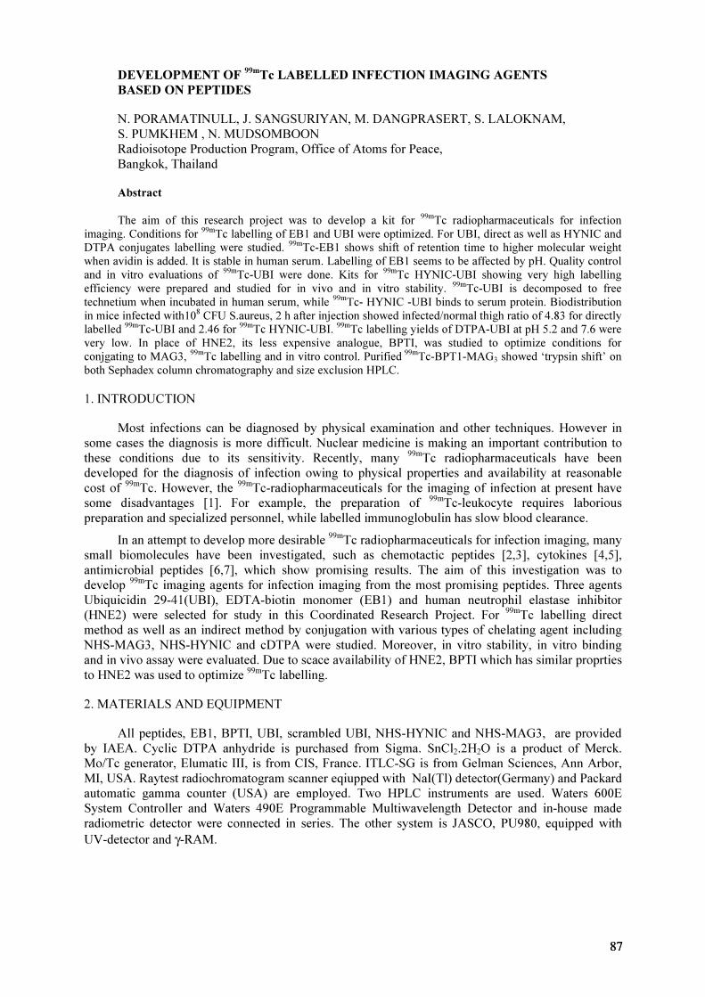
DEVELOPMENT OF 99m
Tc LABELLED INFECTION IMAGING AGENTS
BASED ON PEPTIDES
N. PORAMATINULL, J. SANGSURIYAN, M. DANGPRASERT, S. LALOKNAM,
S. PUMKHEM , N. MUDSOMBOON
Radioisotope Production Program, Office of Atoms for Peace,
Bangkok, Thailand
Abstract
The aim of this research project was to develop a kit for 99m
Tc radiopharmaceuticals for infection
imaging. Conditions for 99m
Tc labelling of EB1 and UBI were optimized. For UBI, direct as well as HYNIC and
DTPA conjugates labelling were studied. 99m
Tc-EB1 shows shift of retention time to higher molecular weight
when avidin is added. It is stable in human serum. Labelling of EB1 seems to be affected by pH. Quality control
and in vitro evaluations of 99m
Tc-UBI were done. Kits for 99m
Tc HYNIC-UBI showing very high labelling
efficiency were prepared and studied for in vivo and in vitro stability. 99m
Tc-UBI is decomposed to free
technetium when incubated in human serum, while 99m
Tc- HYNIC -UBI binds to serum protein. Biodistribution
in mice infected with108
CFU S.aureus, 2 h after injection showed infected/normal thigh ratio of 4.83 for directly
labelled99m
Tc-UBI and 2.46 for 99m
Tc HYNIC-UBI. 99m
Tc labelling yields of DTPA-UBI at pH 5.2 and 7.6 were
very low. In place of HNE2, its less expensive analogue, BPTI, was studied to optimize conditions for
conjgating to MAG3, 99m
Tc labelling and in vitro control. Purified 99m
Tc-BPT1-MAG3 showed ‘trypsin shift’ on
both Sephadex column chromatography and size exclusion HPLC.
1. INTRODUCTION
Most infections can be diagnosed by physical examination and other techniques. However in
some cases the diagnosis is more difficult. Nuclear medicine is making an important contribution to
these conditions due to its sensitivity. Recently, many 99m
Tc radiopharmaceuticals have been
developed for the diagnosis of infection owing to physical properties and availability at reasonable
cost of 99m
Tc. However, the 99m
Tc-radiopharmaceuticals for the imaging of infection at present have
some disadvantages [1]. For example, the preparation of 99m
Tc-leukocyte requires laborious
preparation and specialized personnel, while labelled immunoglobulin has slow blood clearance.
In an attempt to develop more desirable 99m
Tc radiopharmaceuticals for infection imaging, many
small biomolecules have been investigated, such as chemotactic peptides [2,3], cytokines [4,5],
antimicrobial peptides [6,7], which show promising results. The aim of this investigation was to
develop 99m
Tc imaging agents for infection imaging from the most promising peptides. Three agents
Ubiquicidin 29-41(UBI), EDTA-biotin monomer (EB1) and human neutrophil elastase inhibitor
(HNE2) were selected for study in this Coordinated Research Project. For 99m
Tc labelling direct
method as well as an indirect method by conjugation with various types of chelating agent including
NHS-MAG3, NHS-HYNIC and cDTPA were studied. Moreover, in vitro stability, in vitro binding
and in vivo assay were evaluated. Due to scace availability of HNE2, BPTI which has similar proprties
to HNE2 was used to optimize 99m
Tc labelling.
2. MATERIALS AND EQUIPMENT
All peptides, EB1, BPTI, UBI, scrambled UBI, NHS-HYNIC and NHS-MAG3, are provided
by IAEA. Cyclic DTPA anhydride is purchased from Sigma. SnCl2.2H
2O is a product of Merck.
Mo/Tc generator, Elumatic III, is from CIS, France. ITLC-SG is from Gelman Sciences, Ann Arbor,
MI, USA. Raytest radiochromatogram scanner eqiupped with NaI(Tl) detector(Germany) and Packard
automatic gamma counter (USA) are employed. Two HPLC instruments are used. Waters 600E
System Controller and Waters 490E Programmable Multiwavelength Detector and in-house made
radiometric detector were connected in series. The other system is JASCO, PU980, equipped with
UV-detector and γ-RAM.
8787
3. METHODS
3.1. Labelling of EB1 with 99m
Tc
40 µg of EB1 in 4 µL of 1 M sodium acetate buffer pH 6 is added to a vial containing 37 to 150
MBq of 99m
Tc-pertechnetate, followed by 1 µL of stannous chloride dihydrate solution ( 1 mg/mL in
10 mM HCl). The radiolabelled of EB1 is purified by P4 column (0.7 mL P4 in 1 ml syringe) with
0.05 M phosphate buffer pH 7 as eluant. Methods for radiochemical purity analysis and optimum
conditions for labelling are studied. Quality assurance of 99m
Tc-EB1 is done by Avidin Shift by
addition of 120 µg of avidin into 17 µg of the labelled EB1. 99m
Tc-EB1 and 99m
Tc-EB1-avidin are
analysed by both size exclusion HPLC column and P-4 column (0.7×15 cm.). After incubation for 30
min, the labelled product is transferred into P-4 column, and the HPLC analysis was done after 1 h
incubation. Phosphate buffer (0.05M. pH 7.2) is used as mobile phase for both systems (flow rate 1
ml/min for HPLC). In vitro stability of the purified product was studied. To maximize labelling
efficiency, EB1 kits with higher amount of EB1 (50-80 µg) at various pH values have been
evaluated.
3.2.99m
Tc labelling of UBI
3.2.1. Direct labelling
UBI 29-41 is labelled using 99m
Tc pyrophosphate kit (Office of Atomic Energy for Peace,
Bangkok, Thailand). Each kit consisted of tetrasodium pyrophosphate 4.17 mg and stannous chloride
100 µg). Briefly, 10 µL of UBI solution (1 mM in 0.01M acetate buffer pH 4.0) is added 12 µL of a
solution of pyrophosphate kit (dissolved in 550 µl sterile water for injection). Then 2 µL of a 10
mg/mL solution of KBH4
(crystalline, Sigma Chemical Company, St. Louis, MO, USA) in 0.1M
NaOH is added. After addition of 100 µL of 99m
Tc sodium pertechnetate solution (approximately 200
MBq/mL), the mixture is gently swirled and let stand at room temperature for 30 min. The labelling
yield of 99m
Tc-UBI is determined by ITLC-SG strips. Saline solution and 35%ACN in 0.1%TFA/H2O
are used as mobile phases. The 99m
Tc activity in ITLC strips are quantified by the radiochromatogram
scanner.
99m
Tc UBI is also analysed by reverse-phase HPLC on a C18 column (Ultremex 4.6×250 mm,
Phenomenex,) and eluted with a 0.1%TFA/H2O (A) and acetonitrile (B) gradient as follows : 0%B for
5 min, 0-100%B in 10 min, 100%B for 5 min, 100-0%B in 5 min and 0%B for 5 min at 1.0 mL/min
flow rate. 99m
Tc UBI is monitored by NaI(Tl) detector and UV detector at 280 nm.
Effect of the quantity of KBH4 and
99m
Tc on the labelling of UBI is also studied by varying
amounts of KBH4from 1.25 to 20 µg and of
99m
Tc from about 20 MBq to 0.2 GBq in 100 µL.
3.2.2. 99m
Tc labelling of HYNIC-UBI
Conjugation of UBI to HYNIC: UBI is dissolved in 0.1M pH 8.0 HEPES buffer to a
concentration of 3 mg/mL. To the UBI solution is added freshly made NHS-HYNIC in dry DMF (20
mg/mL) at a 5:1 HYNIC/UBI molar ratio. The amount of DMF is kept less than 10% of the total
volume. The reaction mixture is purified on P4 column with 0.25M ammonium acetate pH 5.2 as
eluant. The peak fractions were collected and lyophilized to dryness and then stored at -20 ºC.
99m
Tc labelling: 37-185 MBq 99m
Tc-pertechnetate is added into a tricine kit ( 5 mg tricine, 10 ug
SnCl2.2H
2O, pH 5.5). To solution of 50 µg of UBI-HYNIC is added various amounts of
99m
Tc-tricine.
The labelled peptide is incubated at room temperature. Labelling efficiency is determined using instant
thin-layer chromatography (ITLC) in 0.9% NaCl, Whatman#3 in acetone as well as Sep Pak and
HPLC. Optimum amount of 99m
Tc-tricine for 50 µg. UBI-HYNIC is studied for kit formulation.
88

3.2.3.99m
Tc labelling of DTPA -UBI
Conjugation of UBI to DTPA: The conjugate was prepared according to a procedure previously
described [8]. A 10 µg/µl solution of UBI in water is mixed with an equal volume of 0.2M HEPES
buffer pH 8 and a suspension of cyclic DTPA anhydride in DMF is added dropwise with agitation.
The final DTPA/peptide molar ratio is 5:1 and the addition of DMF is kept less than 10% of total
volume. After 30 minute incubation at room temperature, the conjugated peptide is purified by size
exclusion HPLC (Superdex, Pharmacia biotech) and eluted with a 0.1%TFA/H2O (A) and acetonitrile
(B) gradient as follow : 0%B for 5 min, 0-50%B in 10 min, 50%B for 5 min, 50-0%B in 5 min and
0%B for 5 min at 1.0 mL/min flow rate. The peak at 10.6 min is collected and the concentration of the
fractions is determined by UV absorption at 280 nm. The solution of the coupled peptide is dispensed
to 20 µg/vial and lyophilized for further use.
99m
Tc labelling: The DTPA-conjugated peptide was radiolabelled with 99m
Tc at both pH 5.2 and
7.6. The concentration of conjugated peptide is 0.5-1.0 µg/µL in 0.25M ammonium acetate buffer at
pH 5.2. When labelled at pH 7.6, to 100 µL of the DTPA-peptide solution is added 18 µL of a buffer
consisting of 0.5M sodium bicarbonate, 0.25M ammonium acetate and 0.18M ammonium hydroxide
at pH 9.2. Finally, about 10 µL (about 37-74 MBq) of the 99m
Tc-pertechnetate solution is added.
Immediately thereafter, 4 µl of a fresh solution of SnCl2.2H
2O (1 mg/mL in 10 mM HCl) is added and
the solution is agitated. Labelling efficiency is determined using instant thin-layer chromatography
(ITLC) in 0.9% NaCl and 35% acetonitrile in 0.1%TFA water.
3.3.99m
Tc labelling of BPTI with using NHS-MAG3
3.3.1. Conjugation of BPTI to NHS-MAG3
Freshly made 3 to 5 fold molar excessive NHS-MAG3 in dry DMF solution (12-40 mg/mL) is
added to 0.2-1.0 mg BPTI solution (3-5 mg/mL in 0.1 M HEPES buffer, pH 8.0) The addition of DMF
is kept less than 10% of the total volume. The reaction mixture is placed at room temperature for 30 to
60 min, and then purified by HPLC using BioSep-SEC S2000 Column (Phenomenex), 7.8×300 mm.
Phosphate buffer 0.05 M, pH 7.2 is used as mobile phase with flow rate 1.0 mL/min. The peak
fractions are pooled. The concentration of MAG3-BPTI conjugate is determined by UV absorpbance
at 280 nm. The purified MAG3-BPTI is stored at –20o
C before use.
3.3.2. 99m
Tc labelling
To 100 µL of purified MAG3-BPTI (10-80 µg) are added 100 µL PBS, 25µL sodium tartrate
buffer, pH 9.2 (0.5M NaHCO3, 0.25 M NH
4OAc, 0.175 M NH
4OH, and 50 mg/mL sodium tartrate),
0.1-10 mCi of 99m
Tc-pertechnetate, and 3-4 µL of freshly made SnCl2 solution (1 mg/mL SnCl
2 in 0.01
N HCl). After incubation for 30 min, the radiolabelled MAG3-BPTI is purified by either column
chromatography (Sephadex G-50, 0.05M PBS, pH 7.2 as eluant) or HPLC (Waters 600E System
Controller, Waters 490E Programmable Multiwavelength Detector, 215 nm, NaI detector, BioSep-
SEC 2000 Column (Phenomenex), 7.8×300 mm, 0.05 M PB, pH 7.2, flow rate 1.0 mL/min). The
radioactive fractions monitored by UV detector are pooled and tested by thin layer and paper
chromatography (SG-ITLC/methanol (MeOH) and Whatman 1/acetone), then stored for further
experiments. Trypsin shift is performed to assure quality of the labelled product: A ten fold molar
excess of Trypsin (1 mg/mL in saline) is added to radiolabelled MAG3-BPTI in 0.2 M PBS before or
after purification. The mixture is incubated at room temperature for 30 min before analysis by HPLC.
8989
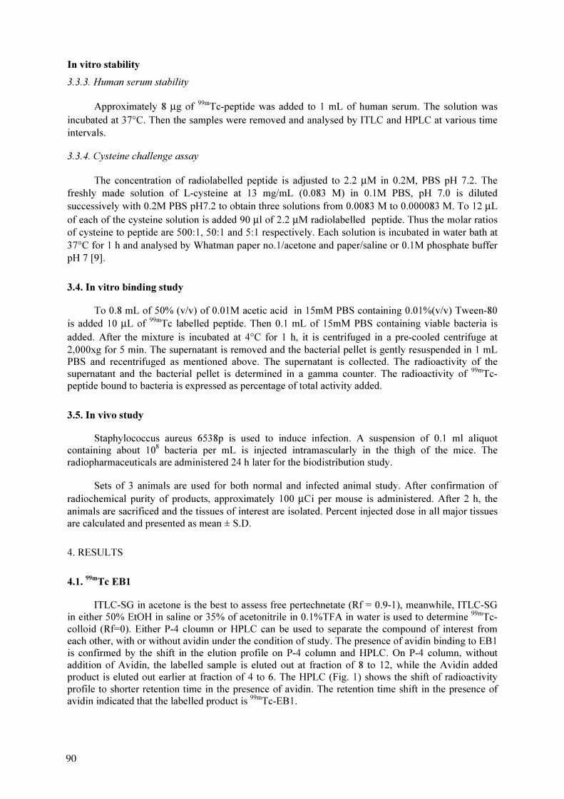
In vitro stability
3.3.3. Human serum stability
Approximately 8 µg of 99m
Tc-peptide was added to 1 mL of human serum. The solution was
incubated at 37°C. Then the samples were removed and analysed by ITLC and HPLC at various time
intervals.
3.3.4. Cysteine challenge assay
The concentration of radiolabelled peptide is adjusted to 2.2 µM in 0.2M, PBS pH 7.2. The
freshly made solution of L-cysteine at 13 mg/mL (0.083 M) in 0.1M PBS, pH 7.0 is diluted
successively with 0.2M PBS pH7.2 to obtain three solutions from 0.0083 M to 0.000083 M. To 12 µL
of each of the cysteine solution is added 90 µl of 2.2 µM radiolabelled peptide. Thus the molar ratios
of cysteine to peptide are 500:1, 50:1 and 5:1 respectively. Each solution is incubated in water bath at
37°C for 1 h and analysed by Whatman paper no.1/acetone and paper/saline or 0.1M phosphate buffer
pH 7 [9].
3.4. In vitro binding study
To 0.8 mL of 50% (v/v) of 0.01M acetic acid in 15mM PBS containing 0.01%(v/v) Tween-80
is added 10 µL of 99m
Tc labelled peptide. Then 0.1 mL of 15mM PBS containing viable bacteria is
added. After the mixture is incubated at 4°C for 1 h, it is centrifuged in a pre-cooled centrifuge at
2,000xg for 5 min. The supernatant is removed and the bacterial pellet is gently resuspended in 1 mL
PBS and recentrifuged as mentioned above. The supernatant is collected. The radioactivity of the
supernatant and the bacterial pellet is determined in a gamma counter. The radioactivity of 99m
Tc-
peptide bound to bacteria is expressed as percentage of total activity added.
3.5. In vivo study
Staphylococcus aureus 6538p is used to induce infection. A suspension of 0.1 ml aliquot
containing about 108
bacteria per mL is injected intramascularly in the thigh of the mice. The
radiopharmaceuticals are administered 24 h later for the biodistribution study.
Sets of 3 animals are used for both normal and infected animal study. After confirmation of
radiochemical purity of products, approximately 100 µCi per mouse is administered. After 2 h, the
animals are sacrificed and the tissues of interest are isolated. Percent injected dose in all major tissues
are calculated and presented as mean ± S.D.
4. RESULTS
4.1.99m
Tc EB1
ITLC-SG in acetone is the best to assess free pertechnetate (Rf = 0.9-1), meanwhile, ITLC-SG
in either 50% EtOH in saline or 35% of acetonitrile in 0.1%TFA in water is used to determine 99m
Tc-
colloid (Rf=0). Either P-4 cloumn or HPLC can be used to separate the compound of interest from
each other, with or without avidin under the condition of study. The presence of avidin binding to EB1
is confirmed by the shift in the elution profile on P-4 column and HPLC. On P-4 column, without
addition of Avidin, the labelled sample is eluted out at fraction of 8 to 12, while the Avidin added
product is eluted out earlier at fraction of 4 to 6. The HPLC (Fig. 1) shows the shift of radioactivity
profile to shorter retention time in the presence of avidin. The retention time shift in the presence of
avidin indicated that the labelled product is 99m
Tc-EB1.
90
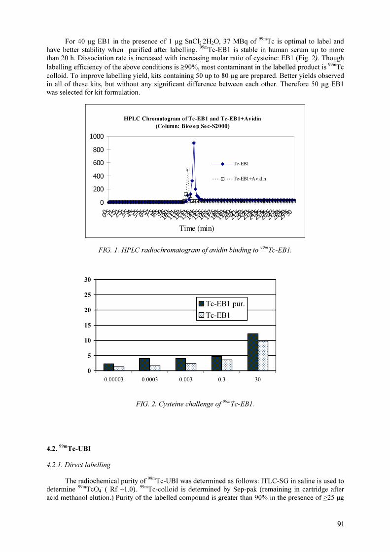
For 40 µg EB1 in the presence of 1 µg SnCl2
.
2H2O, 37 MBq of
99m
Tc is optimal to label and
have better stability when purified after labelling. 99m
Tc-EB1 is stable in human serum up to more
than 20 h. Dissociation rate is increased with increasing molar ratio of cysteine: EB1 (Fig. 2). Though
labelling efficiency of the above conditions is ≥90%, most contaminant in the labelled product is 99m
Tc
colloid. To improve labelling yield, kits containing 50 up to 80 µg are prepared. Better yields observed
in all of these kits, but without any significant difference between each other. Therefore 50 µg EB1
was selected for kit formulation.
HPLC Chromatogram of Tc-EB1 and Tc-EB1+Avidin
(Column: Biosep Sec-S2000)
0
200
400
600
800
1000
0
0.5 1
1.52
2.5 3
3.5 4
4.5 5
5.5 6
6.5 7
7.5 8
8.5 9
9.510
10.511
11.512
12.513
13.514
14.515
15.516
16.517
17.518
18.519
19.520
20.521
21.522
22.523
23.524
24.525
25.526
26.527
27.528
28.529
29.530
Time (min)
Tc-EB1
Tc-EB1+Avidin
FIG. 1. HPLC radiochromatogram of avidin binding to 99m
Tc-EB1.
0
5
10
15
20
25
30
0.00003 0.0003 0.003 0.3 30
Tc-EB1 pur.
Tc-EB1
FIG. 2. Cysteine challenge of 99m
Tc-EB1.
4.2.99m
Tc-UBI
4.2.1. Direct labelling
The radiochemical purity of 99m
Tc-UBI was determined as follows: ITLC-SG in saline is used to
determine 99m
TcO4
-
( Rf ~1.0). 99m
Tc-colloid is determined by Sep-pak (remaining in cartridge after
acid methanol elution.) Purity of the labelled compound is greater than 90% in the presence of >25 µg
9191

of KBH4 (in 100 µL reaction volume) and stable up to 5 h. Effect of KBH
4 and
99m
Tc activity on
labelling are studied. The results showed that 40 µg of KBH4 provided highest effective labelling of
UBI with greater than 90% purity with the 99m
Tc concentration up to 1 GBq/ml. HPLC is successfully
utilized to purify 99m
Tc-UBI with retention time of 15.6 min (Fig. 3). However, 99m
Tc-UBI is poorly
stable in human serum. HPLC chromatogram (Fig. 4a) shows decomposition to free pertechnetate at
about 3 h after incubation. Fig 5. shows dissociation of 99m
Tc in cysteine challenge . Furthermore,
there is no binding of 99m
Tc-UBI to either S. aureus or K. pneumonia at 107
CFU, but the binding at
FIG. 3. HPLC profile of 99m
Tc-UBI, [left]: UV detector at 280 nm, [right] NaI(Tl) detector.
FIG. 4. Serum stability study on size exclusion column- [A]: 99m
Tc -UBI [B]: 99m
Tc -HYNIC-UBI.
92
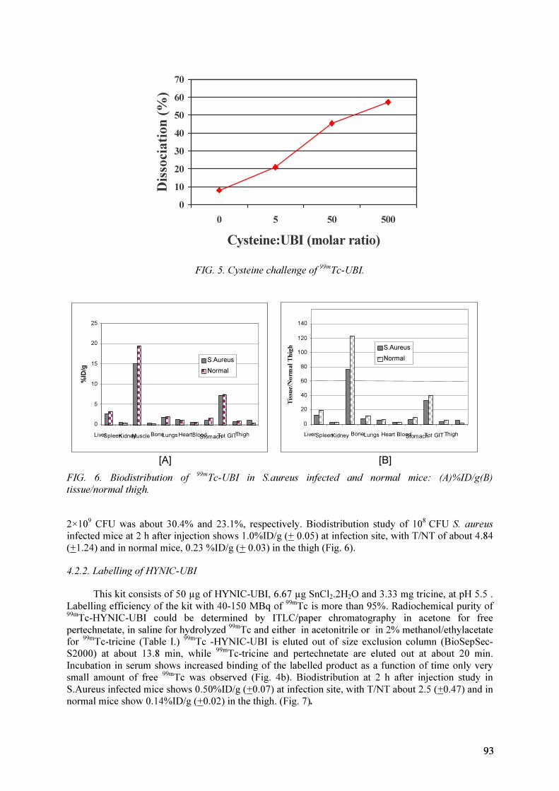
0
10
20
30
40
50
60
70
0 5 50 500
Cysteine:UBI (molar ratio)
Dissociation
(%
)
FIG. 5. Cysteine challenge of 99m
Tc-UBI.
0
5
10
15
20
25
LiverSpleenKidneyMuscleBoneLungsHeartBlood
StomachTot GITThigh
%ID/g
S.Aureus
Normal
0
20
40
60
80
100
120
140
LiverSpleenKidneyBoneLungs Heart Blood
StomachTot GIT Thigh
Tissu
e/N
orm
al T
high S.Aureus
Normal
[A] [B]
FIG. 6. Biodistribution of 99m
Tc-UBI in S.aureus infected and normal mice: (A)%ID/g(B)
tissue/normal thigh.
2×10⁹
CFU was about 30.4% and 23.1%, respectively. Biodistribution study of 108
CFU S. aureus
infected mice at 2 h after injection shows 1.0%ID/g (+ 0.05) at infection site, with T/NT of about 4.84
(+1.24) and in normal mice, 0.23 %ID/g (+ 0.03) in the thigh (Fig. 6).
4.2.2. Labelling of HYNIC-UBI
This kit consists of 50 µg of HYNIC-UBI, 6.67 µg SnCl2.2H
2O and 3.33 mg tricine, at pH 5.5 .
Labelling efficiency of the kit with 40-150 MBq of 99m
Tc is more than 95%. Radiochemical purity of
99m
Tc-HYNIC-UBI could be determined by ITLC/paper chromatography in acetone for free
pertechnetate, in saline for hydrolyzed 99m
Tc and either in acetonitrile or in 2% methanol/ethylacetate
for 99m
Tc-tricine (Table I.) 99m
Tc -HYNIC-UBI is eluted out of size exclusion column (BioSepSec-
S2000) at about 13.8 min, while 99m
Tc-tricine and pertechnetate are eluted out at about 20 min.
Incubation in serum shows increased binding of the labelled product as a function of time only very
small amount of free 99m
Tc was observed (Fig. 4b). Biodistribution at 2 h after injection study in
S.Aureus infected mice shows 0.50%ID/g (+0.07) at infection site, with T/NT about 2.5 (+0.47) and in
normal mice show 0.14%ID/g (+0.02) in the thigh. (Fig. 7).
9393

TABLE I. MIGRATION OF SAMPLES OF 99m
TC UBI-HYNIC IN ITLC WITH VARIOUS
SOLVENTS
Eluant Tc-colloid Tc-HYNIC-UBI Tc-tricine NaTcO4
Saline Origin Solvent front Solvent front Solvent front
Acetone Origin Origin origin Solvent front
Acetonitrile Origin Origin Solvent front Solvent front
MeOH Origin Solvent front Solvent front Solvent front
2%MeOH/EtOAc Origin Origin Solvent front Solvent front
0.5M PB pH 7.4 Origin Solvent front Solvent front Solvent front
0
0.5
1
1.5
2
2.5
3
Liver
S
pleen
Kidney
Muscle
Bone
Lungs
H
eart
Blood
Stom
ach
Tot G
IT
Thigh
%ID/g
S.Aureus
Normal
0
2
4
6
8
10
12
14
16
Live
r
S
ple
en
K
idn
eys
Bo
ne
L
un
gs
H
ea
rt
B
lo
od
S
to
ma
ch
To
t G
IT
T
high
Tissu
e/N
orm
al T
hig
hS.Aureus
Normal
[A] [B]
FIG. 7. Biodistribution of 99m
Tc-HYNIC-UBI in S.aureus infected and normal mice: (A) %ID/g
(B) tissue/normal thigh.
4.3.99m
Tc -MAG3-BPTI
Elution profile of samples on Sephadex G-50 column is better than on Sephadex G-100. Hence
the former is chosen for purification or identification of the non-radioactive or radioactive compounds.
The column can separate Trypsin, BPTI, NHS-MAG3, BPTI-MAG3, and pertechnetate from one
another (Figs 8 and 9).
On a reverse phase column (Crest Pak), BPTI and NHS-MAG3, monitored at 235 nm, are
separated with retention times of 11 and 6 mins respectively. Comparing the purified samples (peak A
and peak B in Fig. 9) to the retention time of BPTI and NHS-MAG3 (Fig. 8), peak B has retention
time corresponding to NHS-MAG3, but peak A showed different retention time than that of BPTI or
NHS-MAG3. It could hence be assumed that peak A is that of conjugate of MAG3-BPTI and
therefore, the pooled fraction of peak A is used for radiolabelling studies.
The MAG3-BPTI is labelled with 99m
Tc at room temperature complete in 30 min and purified
by either column chromatography or HPLC. By column chromatography, it is found that 99m
Tc-
MAG3-BPTI is eluted earlier (fraction 20–30), followed by free 99m
TcO4
-
, (fraction 45-50) with
confirmation by ITLC (ITLC-SG/MeOH, Whatman#1/acetonitrile). By HPLC (Fig. 10), the retention
times of 99m
TcO4
-
, 99m
Tc-tartrate, and 99m
Tc-BPTI-MAG3 are 21.86, 12.27, and 9.87 min, respectively.
94

FIG. 8. Elution profile of trypsin, BPTI, NHS-MAG3, and 99m
Tc on Sephadex G-50 column.
FIG. 9. Chromatogram of MAG3-BPTI and NHS-MAG3on Sephadex G-50 column.
0.0
0.1
0.2
0.3
0.4
0.5
0.6
0.7
0.8
0.9
1.0
0 10 20 30 40 50 60 70 80
Fraction no.
OD280
0.00
0.01
0.02
0.03
0.04
0.05
0.06
0.07
0.08
0.09
0.10
Radioactivity
Trypsin
BPTI
NHS-MAG3
Tc
9595
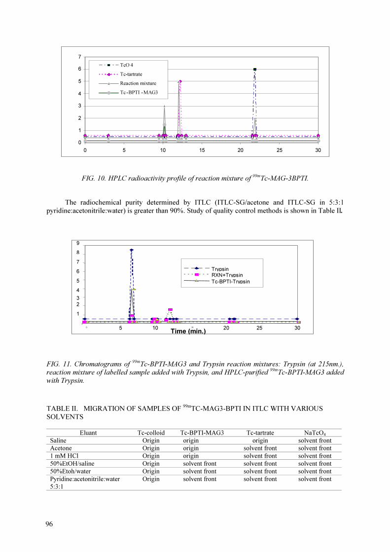
0
1
2
3
4
5
6
7
0 5 10 15 20 25 30
TcO 4
Tc-tartrate
Reaction mixture
Tc -BPTI -MAG3
FIG. 10. HPLC radioactivity profile of reaction mixture of 99m
Tc-MAG-3BPTI.
The radiochemical purity determined by ITLC (ITLC-SG/acetone and ITLC-SG in 5:3:1
pyridine:acetonitrile:water) is greater than 90%. Study of quality control methods is shown in Table II.
0
1
2
3
4
5
6
7
8
9
0
5 1015
20 25 30
Time (min.)
Trypsin
RXN+Trypsin
Tc-BPTI-Trypsin
FIG. 11. Chromatograms of 99m
Tc-BPTI-MAG3 and Trypsin reaction mixtures: Trypsin (at 215nm.),
reaction mixture of labelled sample added with Trypsin, and HPLC-purified 99m
Tc-BPTI-MAG3 added
with Trypsin.
TABLE II. MIGRATION OF SAMPLES OF 99m
TC-MAG3-BPTI IN ITLC WITH VARIOUS
SOLVENTS
Eluant Tc-colloid Tc-BPTI-MAG3 Tc-tartrate NaTcO4
Saline Origin origin origin solvent front
Acetone Origin origin solvent front solvent front
1 mM HCl Origin origin solvent front solvent front
50%EtOH/saline Origin solvent front solvent front solvent front
50%Etoh/water Origin solvent front solvent front solvent front
Pyridine:acetonitrile:water
5:3:1
Origin solvent front solvent front solvent front
96

Affinity of the conjugated compound to trypsin is tested and verified on size-exclusion HPLC
by the shift in retention time to 6.58 min (Fig. 11).
99m
Tc-MAG3-BPTI is stable up to 24 h in human serum while ~ 60% dissociation is noticed in
cysteine solution (300:1).
5. DISCUSSION AND CONCLUSION
EB1 (40 µg) has been successfully labelled with 37 MBq of 99m
Tc in the presence of 1 µg
SnCl2.2H
2O. The labelled compound is confirmed by the shorter retention time of avidin-biotin
complex (99m
Tc-EB1-avidin) compared to 99m
Tc-EB1 on P4 column and size exclusion column. The
shift in retention time confirmed EB1 labelling to 99m
Tc. Even though 99m
Tc-EB1 is stable in human
serum, dissociation is high with the increasing molar ratio of cysteine: EB1.
Direct labelling of UBI with 99m
Tc was studied. Radiochemical purity of 99m
Tc-UBI, determined
by ITLC-SG in saline and 35% acetonitrile in 0.1%TFA/water, is greater than 90%. HPLC using
reverse phase column can be used to identify 99m
Tc-UBI. The radiolabelling yield is not affected by the
concentration of Na99m
TcO4. However,
99m
Tc-UBI is unstable in human serum and cysteine solution.
Affinity of 99m
Tc-UBI to bind the bacteria could be proved by very high binding with 109
CFU.
Biodistribution at 2h post injection was encouraging for infection imaging.
High labelling efficiency kit for 99m
Tc HYNIC-UBI is formulated. 99m
Tc HYNIC-UBI shows
lower ratio of uptake of target/nontarget than the product of direct labelling.
99m
Tc labelling efficiency of DTPA-UBI at pH about 5.2 and 7.6 are rather low but yield at pH
7.6 seem to be higher than at pH 5.2.
99m
Tc is labelled to BPTI by indirect method using MAG3 as bifunctional chelating agent. BPTI
has been conjugated to NHS-MAG3 via active ester. The conjugation can be completed within 1 h. It
can be purified by either by Sephadex G-50 or size exclusion HPLC. 99m
Tc labelling to MAG3- BPTI
can be completed within 30 min. Radiochemical purity of 99m
Tc-MAG3-BPTI is greater than 90% by
ITLC-SG in acetone and solvent mixture of pyridine:acetonitrile:water (5:3:1). Affinity measurement
of 99m
Tc-MAG3-BPTI to trypsin proved that 99m
Tc is labelled to BPTI-MAG3. 99m
Tc-MAG3- BPTI is
quite stable in both human serum and cysteine solution.
REFERENCES
[1] RENNEN, H.J.J.M., BOERMAN, O.C., OYEN, W.J.G., CORSTENS, F.H.M., “Imaging
infection /inflammation in the new millennium”, Eur. Nucl. Med. 28 (2001) 241–252.
[2] BABICH, J.W., SOLOMON, H., PIKE, M.C., et al., “Technetium-99m-labelled hydrazine-
nicotiamide derivatized chemotactic peptide analogs for imaging focal sites of bacterial
infection”, J. Nucl. Med. 34 (1993) 1964–1974.
[3] FISCHMAN, A.J., PIKE, M.C., KROON, D., et al., “Imaging focal sites of bacterial infection
in rats with indium-111-labelled chemotactic peptide analogs”, J. Nucl. Med. 32 (1991)
483-491.
[4] VAN DER LAKEN, C.J., BOERMAN, O.C., OYEN, W.J.G., et al., “Recombinant human
interleukin-1: potential agent to image infections foci”, Eur. J. Nucl. Med. 21 (1994) 790.
[5] RENNEN, H.J.J.M., BOERMAN, O.C., OYEN, W.J.G., VAN DER LAKEN, C.J.,
CORSTENS, F.H.M., “Specific and rapid scintigraphic detection of infection with Tc-99m-
labelled interleukin-8”, J. Nucl. Med. (2001) 42.
[6] WELLING, M.M., NIBBERING, P.H., PAULUSMA-ANNEMA, A., et al., “Imaging of
bacterial infections with 99m
Tc-labelled human neutrophil peptide-1”, J. Nucl. Med. 40 (1999)
2073–2080.
9797
[7] WELLING, M.M., PAULUSMA-ANNEMA, A., BALTER, H.S., et al., “Technetium-99m
labelled antimicrobial peptides discriminate between bacterial infections and sterile
inflammations”, Eur. J. Nucl. Med. 27 (2000) 292–301.
[8] ZHANG, Y., LIU, N., ZHU, Z., et al., “Influence of different chelators (HYNIC, MAG3 and
DTPA) on tumor cell accumulation and mouse biodistribution of technetium-99m labelled to
antisense DNA”, Eur. J. Nucl. Med. 271 (2000) 1700–1707.
[9] HNATOWICH, D.J., VIRZI, F., FOGARACI, M., et al., “Can a cysteine challenge assay
predict the in vivo behavior of 99m
Tc-labelled antibodies?”, Nucl. Med. Biol. 21 (1994)
1037-1039.
98

99m
Tc RADIOPHARMACEUTICALS FOR INFECTION IMAGING:
KITS DEVELOPMENT AND VALIDATION
B. SOUTO PAIS, G. RODRIGUEZ, H. BALTER, S. LANZZERI, P. OLIVER, A. LOPEZ,
Z. GONÇALVEZ, A. NAPPA, S. VERDERA, J. BERBEJILLO, L. MALLO
Departamento de Radiofarmacia, Centro de Investigaciones Nucleares,
Facultad de Ciencias, Montevideo, Uruguay
Abstract
This research concerns the preparation and evaluation of various radioactive agents for infection imaging.
Our investigations concentrate on the development of a radiopharmaceutical for specific infection imaging. The
molecules UBI 29-41, (TGRAKRRMQYNRR), EDTA-Biotin (EB1) and Human Neutrophil Elastase 2 (HNE2)
were labelled with direct /or indirect methods with 99m
Tc by reducing pertechnectate. Quality control was done
with chromatographic methods, including HPLC and Seppak. The binding studies to bacteria and biodistribution
in normal and infected mice were performed. The labelling yield of UBI 29-41 was higher than 90% with a
direct method. Target to non- target ratio of 2.1 at 1 h after injection of 99m
Tc-UBI 29-41 was obtained. For
99m
Tc-EB1 a yield higher than 90% was observed and the biological activity was illustrated by binding the
radioactive compound to avidin. In the case of the HNE2 conjugation with cDTPA
[Diethylenetriamineepntaacetic acid α, ω- bis ( Biocytinamide)] was achieved.
1. INTRODUCTION
Various 99m
Tc labelled compounds have been developed for the scintigraphic detection of
infection and sterile inflammation in man [1,2]. Unfortunately, these radiopharmaceuticals do not
allow discrimination between infections with pathogens and sterile inflammatory processes [3], which
is often of clinical importance. In an attempt to develop such a tracer, our research has been focussed
on peptide antibiotics [4], which could be promising radiopharmaceuticals to distinguish between
bacterial infections and sterile inflammatory processes [5].
Our group performed experiments with labelled 99m
Tc-IgG and peptide derivates from
ubiquicidine (UBI). 99m
Tc-IgG is an agent that accumulates at sites of infection and inflammation
whereas 99m
Tc-UBI has shown specific binding to bacteria. Our investigations were extended with
studies on 99m
Tc- EB1 and 99m
Tc-HNE2. The latter compounds are known to accumulate at both sites
of infection and inflammation. This report summarizes our findings as far as radiochemical and
biological characteristics of these compounds are concerned with enphasis on a radiolabelled peptide
derived from ubiquicidine: 99m
Tc- UBI 29-41.
2. MATERIALS AND METHODS
2.1. Direct 99m
Tc labelling and control of Ubiquicidin derivatives (UBI-29-41 and UBI-
Scrambled)
The UBI 29-41 peptide, corresponding to residues 29-41 of a cationic α-helical domain of
human ubiquicidin (TGRAKRRMQYNRR, 1,693 Da) was synthesized as described before [6]. A UBI
scrambled peptide (Sc; KRNQRMARYRRGT, 1,693 Da) was included as a control [7,11].
The peptides were directly labelled according to an earlier developed method [5] by mixing 10
uL of the peptide solution (1 mM in 0.01 M acetic acid pH 4.0) with 4 uL of a solution of stannous
pyrophosphate (0.5 mg/mL), 2 uL of a solution containing 10 mg/mL potassium borohydride in 0.1 M
sodium hydroxide (both as reducing agents), and 0.1 mL of 99m
Tc-sodium pertechnetate (200-700
MBq/mL, CIS, Paris, France). Continuous and gentle agitation during 30 min at room temperature was
done.
Physicochemical controls for determination of radiochemical species as well as stabiliy of the
labelled peptides were carried out using the following methods:
9999

Instant thin layer chromatography (ITLC-SG) using 0.9% NaCl, metyl ethyl ketone (MEK), or
water-trifluoroacetic acid 0.1% (H20/TFA 0.1%) as solvents.
Reverse-phase high performance chromatography (RP-HPLC) on a C 18 reverse phase column
(Varian, Ireland) with 0.1% (w/w) trifluoroacetic acid in water (A) and 0.1% (w/w) trifluoroacetic acid
in acetonitrile (B) as eluents. The run with a flow of 1 mL/min was performed as follows: 0-3 min
100% A, 3-13 min from 100% A to 100% B, 13-18 min 100% B, 18-23 min from 100% B to 100% A.
Sep-pak: extraction on a cartridge (Waters, Milford, Mass., USA) previously activated with 5
mL methanol, 5 mL water and 5 mL 0.1% acetic acid, and airdried. On the cartridge, 20 to 100 ul of
sample was applied and the cartridge was then rinsed with 5 mL of 0.1% acetic acid and the trapped
peptide was eluted with 5 mL of methanol.
Stability studies: the chromatographic system described above was used to assess the stability of
labelled peptide at room temperature for 6 h. ITLC-SG was used to estimate the stability of the
labelled peptides in (1/5) diluted human serum at 37ºC. at various time intervals (5, 30, 60, 120 min,
and 24 hs). Around 30 µg (in 200 uL) of each radiolabelled peptide was added to 200 µL of diluted
serum.
Cysteine Challenge: each labelled peptide was tested for instability towards sulfhydryl groups.
A solution of 10 mg/mL cysteine was diluted in PBS 0.2 M, pH 7.0 to 1, 0.1, 0.01 and 0.001 mg/mL.
Mixtures of 10 µL of each solution and 90 µL of radiolabelled peptide solution were incubated for 1 h
at 37ºC and were compared with a blank sample. After this time the mixtures were controlled by
chromatography.
In vitro binding of S.aureus and S.aureus virulen: studies were done with a modification of the
techniques recommended by the group [8]. S.aureus ATCC 25923 (American type cell culture Catalog
number 25923) and S. aureus virulent were used. Bacteria were thawed and let at RT for 10 min, and
centrifugued for 5 min at 3500 rpm, 4º C. Two washings of the pellet were done resuspending each
time in 10 mL of buffer A (PB 14 mM pH 7,5) and homogenizing (Politron). The final pellet was
resuspended in 0.4 mL of the same buffer (contains about 109
CFU in 0.4 mL). In order to prepare
working preparations, made a 1:1solutions in Buffer A, serially seven times of the suspended pellet.
To 0.1 mL of each dilution was added 0.1mL of labelled peptide (1nmol/tube) with 0,8 mL of a
solution of 0.05% acetic acid and 0.005% Tween 80 in buffer A, incubated at 4ºC for 1 h, and total
activity counted. Centrifuged in the same conditions as above and washed twice with 1 mL of the
previous buffer and finally counted pellet activity. Specific binding of activity was calculated as
activity in pellet minus activity in blank.
In vivo experiments: Biodisribution studies were performed in mice having a bacterial infection,
sterile inflammation, or none. Infection was induced by an intramuscular injection of 0.1 mL
containing around 108
CFU /mL of Staphylococcus aureus ATCC 25923 in the right hind leg of the
mice, 24 h before the biodistribution study. Sterile inflammation was induced by intramuscular
injection of 0.1 mL containing about 109
CFU /mL of heat killed multiresistant Klebsiella
pneumoniae (K.p.R) (LPS). Labelled peptide (0.1 mL containing 20 to 70 MBq) was injected
intravenously, and the activity uptake in different organs/tissues was determined after cervical
dislocation at 1h and 2 h post injection. Data were expressed as % of injected dose/gram (%ID/g). In
mice with an infection or with sterile inflammation target (infected/inflamed thigh muscle)-to-non-
target (contralateral thigh muscle) ratios (T/NT) were calculated. The presence or absence of viable
bacteria was determined microbiologically, in the muscle of sacrificiced mice from the biodistribution
studies. These homogenized tissues were plate counted (both muscle inoculated and not inoculated, as
blank). Finally, we calculated correlations between the T/NT ratio and the presence of viable
bacteria/g tissue.
100

2.2.99m
Tc labelling and control of BPTI and HNE-2 – Indirect method
According with the protocol suggested [8] we made the initial experiments with the control
peptide Binding Pancreatic Trypsin Inhibitor (BPTI), with a view to tranfer the conditions
standardized to the peptide of interest, HNE-2. Conjugation of BPTI with NHS-MAG3 (N-
hydroxysuccinimide-mercaptoacetyltriglycine) was done as follows: Molar ratios of NHS-MAG3 /
BPTI, were 20:1, 7:1 and 4:1, incubation of reagents during 1 h at RT, purifications of the conjugates
by means of gel-filtration in a P4 column and elution with NH4AcO 0.25 M, pH 5,2. Measurement of
peptide concentration of the eluted fractions was done by UV spetrophotometry at 280 nm (extinction
coefficient: 0.80 for BPTI [9]). Fractions with absorbances higher than 0,050 were stored at –80ºC for
further use.
Labelling of the conjugates with 99m
Tc was done on 100-250 uL of the fractions with peptide
concentration between 0.2-0.4 mg/mL, and adding 100-250 uL PBS 0.1 M, pH 7,2; 25-62 uL tartrate
buffer 50 mg/mL, pH 9 (in sodium acid carbonate 0,5 M , ammonium acetate 0.25 M and ammonium
hydroxide 0.175 M), 200-500 uL pertechnetate solution (1.5-4.4 MBq/mL) and 4-40 uL SnCl2.2H
20
freshly prepared solution (1mg/mL) in 10 mM HCl.
The mixture was incubated for 30–60 min at RT. The labelled conjugate was purified using a P4
column with PBS 0.2 M ( pH 7,2 ) as eluent. 1 mL Fractions were collected and activity measured in a
dose calibrator.
The initial reaction mixture as well as all the fractions showing significant activities were
evaluated using the following methods:
ITLC-SG with various solvent systems (NaCl 0.9%, MEK, ACN:H20 60:40, 0.2 M Sodium
citrate.
HPLC in two column systems: a) C18 Reverse Phase: TFA 0.1%/ H20 (A), TFA 0.1%/- ACN
(B), gradient from 100% A at 0 min, to 33% A– 7% B at 60 min. b) Superdex peptide column, sodium
buffer phosphate 0,1 M, pH 7,2, isocratic mode.
Sep-pak (C18) previously activated with 10 mL ethanol and 10 mL HCl 0,001 M, washing with
HCl 0.001 M to release pertechnetate and tartrate, and eluting with ethanol:saline (1:1) to release the
labelled peptide, the 99m
TcO2 remained trapped in the cartridge.
Conjugation of HNE2 with α,ω-bis-(biocytinamide) acid and cDTPAa was done by the
following assay conditions: molar ratio cDTPAa:HNE 25:1, incubation of reagents for 1 h at room
temperature, purification of the conjugate by gel permeation in P4 column and elution with NH4
AcO
0.25 M, pH 5.2. Peptide concentration was measured in the collected fractions by UV
spectrophotometry at λ: 280 nm (extinction coefficient: 0.89 for HNE2 [9]. One peak was collected
and stored at –80º C for further use.
Labelling with 99m
Tc of the conjugate was done on 250 uL of pooled fractions (peptide
concentration 0.35 mg/mL), adding 45 uL tartrate buffer 50 mg/mL, pH 9 (in sodium acid carbonate
0,5 M, ammonium acetate 0.25 M and ammonium hydroxide 0.175 M), 250 uL pertechnetate solution
(6.4 MBq/mL) and 10 uL of SnCl2.2H
20 freshly prepared solution (1mg/mL) in 10 mM HCL. The
mixture was incubated for 1 h at RT. The 99m
Tc conjugate was purified by P4 column, eluting with
PBS 0,2 M, pH 7,2. Fractions of 1 mL each were collected. All fractions showing significant activities
were controlled by ITLC-SG, HPLC and Seppak as was described for BPTI conjugate.
101101

2.3.99m
Tc labelling and control of EB1
According to the protocol recommended [10] the labelling of EB1 was done by mixing 100 uL
of EB1 (10mg/mL in acetate buffer 1 M, pH 6), 1000 uL( 0.1-0.6 MBq/mL) of 99m
TcO4
-
, and 10, 35,
100 uL of SnCl2.2
H20 freshly prepared solution (1mg/mL) in 10 mM HCL, incubated either at RT for
30 min, heating in a water bath at 60-65ºC 15 min; 90-95ºC, 5 min or immediately heating at 90–95ºC
for 5 min. Physicochemical methods for determination of radiochemical purity of the 99m
Tc-EB1 were
evaluated: ITLC-SG in 0,9% NaCl and MEK and HPLC in TSK G3000 SW, UV at λ: 280nm and
radiometric on line detection, isocratic elution with phosphate buffer 0.1M, pH: 7, at flow 0.5 mL/min.
Stability at RT until 48 hs was evaluated by chromatography in saline and MEK.
Stability in human serum: Incubation of 200 uL labelled peptide with 200 uL diluted human
serum (1/5) at 37ºC for 5´,30´, 60´ 120´ and 24 hs, controlling with the chromatographic systems.
Cysteine challenge: Incubation of 1000 uL of different solutions of cysteine with concentrations
of 10mg/mL, 1mg/mL, 0,1 mg/mL, 0,01 mg/mL and 0,001 mg/mL, with 4,5 uL of labelled peptide for
1 h at 37ºC, and controlling by chromatographic systems. The highest molar ratio was 500/1
(cysteine:EB1).
Specific binding of 99m
Tc-EB1 to avidin was evaluated by gel permeation (PD10) using
phosphate buffer 0,05 M , pH 7.2, SAB 0,2 % and NaCl 0.9%, as eluent. One mL fractions were
collected and counted for activity. Molar ratio of 99m
Tc-EB1 and 99m
Tc-EB1 to avidin was 1:50.
3. RESULTS
3.1. Direct 99m
Tc labelling and control of Ubiquicidin derivatives ( UBI-29-41 and UBI-
Scrambled)
According to ITLC analysis the labelling efficiency for 99m
Tc-UBI-29-41 was 92,0±7.7% (n=11)
and 92.5±7.4% (n=11) determined using MEK and 0.9% NaCl respectively. For 99m
Tc-UBI-Scrambled
the results were 87.7±7.7% (n=6) and 88.8±3.4% (n=7) determined using MEK and 0.9% NaCl
respectively. For analysis using 0.1% TFA as eluent in a number of experiments results were not
significantly different than those determined with MEK and 0.9% NaCl.
HPLC profiles for both labelled peptides showed peaks (gamma detection) with similar
retention times: 9,2 min for 99m
Tc-UBI-Scrambled and 9,5 for 99m
Tc-UBI 29-41 min, and area higher
than 95%. Recovery of eluted activity was 83±19% for both labelled peptides. The retention time of
the unlabelled species was 8,8 min.
The recovery of the radioactivity fractions eluted from the Sep-pak cartridge, corresponding to
labelled peptides, were 69.8±5.0% (n=5) for 99m
Tc-UBI 29-41 and 68.8±1.6% (n=2) for 99m
Tc-UBI-
Scrambled, respectively. In addition, the acidic elution contained about 2.7% of the applied
radioactivity that corresponds with free 99m
Tc-pertechnetate and the remaining activity on the cartridge
was 27.5% of the total activity.
Stability studies: Using the same chromatographic systems as described above, 99m
Tc-labelled
UBI 2941 showed no release of 99m
Tc-pertechnetate up to 6 h post labelling indicating good stability
of the labelled peptide. Stabilty of 99m
Tc-UB 29-41 in human serum as depicted in Fig. 1 shows no
release of radioactivity during the first hour of incubation.
102

FIG. 1. Stability of 99m
Tc-UBI 29-41 in human serum.
Cysteine Challenge: As shown in Fig. 2, more than 10% of the label has been transchelated to
cysteine at concentrations higher than 0.01mg/mL. 99m
Tc-UBI-Scrambled showed similar results under
the same conditions (Fig. 3).
99mTc-UBI-B
0
10
20
30
40
50
60
70
80
90
100
5 30 60 80 120 1440
Time
(min)
% R
CP
0
10
20
30
40
50
60
70
80
90
100
1,E - 03 1,E-02 1,E-01 1,E+0 1,E+0
cysteine
%
RCP
FIG.2. Cysteine challenge of 99mTc-UBI 29-41
103103
FIG. 3. Cysteine challenge of 99m
Tc-UBI-scrambled.
In vitro binding to bacteria showed an increase in specific binding as function of number of
bacteria for both strains of S. aureus (Fig. 4). In the conditions of assay, specific binding of 99m
Tc-UBI
29-41 was 38.1% for S. aureus Virulent (1.47E+06 06 CFU/tube), and 32.7% for S. aureus (5.08E+06
CFU/mL).
99m
TcUBIS
0
10
20
30
40
50
60
70
80
90
100
1,E-03 1,E-02 1,E-01 1,E+00 1,E+01
Cisteina (mg/mL)
%
RC
P
0
5
10
15
20
25
30
35
40
45
0.00E+00 1.00E+06 2.00E+06 3.00E+06 4.00E+06 5.00E+06 6.00E+06
cfu/mL
% S
pe
cific
B
in
din
g
non virulent
virulent
FIG. 4. In vitro binding 99m
Tc-UBI 29-41 to S.a.
104
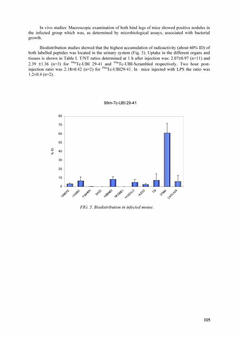
In vivo studies: Macroscopic examination of both hind legs of mice showed positive nodules in
the infected group which was, as determined by microbiological assays, associated with bacterial
growth.
Biodistribution studies showed that the highest accumulation of radioactivity (about 60% ID) of
both labelled peptides was located in the urinary system (Fig. 5). Uptake in the different organs and
tissues is shown in Table I. T/NT ratios determined at 1 h after injection was: 2.07±0.97 (n=11) and
2.39 ±1.36 (n=3) for 99m
Tc-UBI 29-41 and 99m
Tc-UBI-Scrambled respectively. Two hour post-
injection ratio was 2.18±0.42 (n=2) for 99m
Tc-UBI29-41. In mice injected with LPS the ratio was
1.2±0.4 (n=2).
99m-Tc-UBI 29-41
0
10
20
30
40
50
60
70
80
% ID
FIG. 5. Biodistribution in infected mouse.
105105
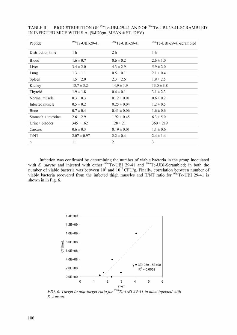
TABLE III. BIODISTRIBUTION OF 99m
Tc-UBI-29-41 AND OF 99m
Tc-UBI-29-41-SCRAMBLED
IN INFECTED MICE WITH S.A. (%ID/gm, MEAN ± ST. DEV)
Peptide99m
Tc-UBI-29-4199m
Tc-UBI-29-41 99m
Tc-UBI-29-41-scrambled
Distribution time 1 h 2 h 1 h
Blood 1.6 ± 0.7 0.6 ± 0.2 2.6 ± 1.0
Liver 3.4 ± 2.0 4.3 ± 2.9 5.9 ± 2.0
Lung 1.3 ± 1.1 0.5 ± 0.1 2.1 ± 0.4
Spleen 1.5 ± 2.0 2.3 ± 2.6 1.9 ± 2.5
Kidney 13.7 ± 3.2 14.9 ± 1.9 13.0 ± 3.8
Thyroid 1.9 ± 1.8 0.4 ± 0.1 3.1 ± 2.3
Normal muscle 0.3 ± 0.3 0.12 ± 0.01 0.6 ± 0.2
Infected muscle 0.5 ± 0.2 0.25 ± 0.04 1.2 ± 0.5
Bone 0.7 ± 0.4 0.41 ± 0.06 1.6 ± 0.6
Stomach + intestine 2.6 ± 2.9 1.92 ± 0.45 6.3 ± 5.0
Urine+ bladder 345 ± 162 128 ± 21 360 ± 219
Carcass 0.6 ± 0.3 0.19 ± 0.01 1.1 ± 0.6
T/NT 2.07 ± 0.97 2.2 ± 0.4 2.4 ± 1.4
n 11 2 3
Infection was confirmed by determining the number of viable bacteria in the group inoculated
with S. aureus and injected with either 99m
Tc-UBI 29-41 and 99m
Tc-UBI-Scrambled; in both the
number of viable bacteria was between 103
and 1010
CFU/g. Finally, correlation between number of
viable bacteria recovered from the infected thigh muscles and T/NT ratio for 99m
Tc-UBI 29-41 is
shown in in Fig. 6.
y = 3E+08x - 5E+08
R2
= 0,6652
0,0E+00
2,0E+08
4,0E+08
6,0E+08
8,0E+08
1,0E+09
1,2E+09
1,4E+09
0 1 2 3 4 5 6
T/NT
CFU/mL
FIG. 6. Target to non-target ratio for 99m
Tc-UBI 29-41 in mice infected with
S. Aureus.
106
3.2.99m
Tc labelling and control of BPTI and HNE2
Following the techniques described above, a single peak was obtained from the purification
profile when using a molar ratio of BPTI:NHS-MAG3 of 1:4 (83%, 5th
mL) and two peaks when using
molar ratios of 1:7 (40%, 5th
mL and 60%, 10th
) and 1:20 (35%, 5th
mL and 65%, 12 th
mL). The
fractions labelled and purified by gel permeation P4 showed two peaks of activity: 1th
(5,5 mL and 2nd
(23 mL).
ITLC-SG: At 1 to 2 h post-labelling showed 44±3%, (n=5), determined by MEK and 40±2%
(n=5) by 0.9% NaCl, respectively, in a sample of the initial reaction vial. The control on the fractions
collected after purification on P4 column, at 2–5 h post labelling gave an RCP of 89±5% (n= 4)
determined by MEK and 90±5% (n=5) by 0.9% NaCl chromatography.
Sep-pak C18: RCP determined as percent activity in the ethanolic fraction was 60-74%. When
using this same procedure for control of the fractions collected at the gel permeation on P4 column,
radiochemical purities of 78-95% were achieved.
HPLC: The radiochemical purity determined by HPLC of the initial reaction mixture was 61%,
RT: 14.76 min (Superdex peptide Column) and 58-63%, RT: 27.75 (C 18 Column). After purification
by gel permeation on P4 column, an increase to 93%, RT: 14.8 min (Superdex Column) of
radiochemical purity was achieved.
The results obtained with HNE2 are: Labelling yield determined by ITLC-SG in MEK was 50%
(Rf 0.0). The purification on P4 column, showed an expanded peak from 5 to 15 mL (10 fractions)
which, upon chromatographic control in ITLC-SG using MEK gave 90-100% of activity at Rf 0.0.
Sep-pak: Fraction 6 from P4 purification (corresponding to an eluted volume of 5.8 mL) and
fraction 10 (corresponding to an eluted volume of 10.3 mL) gave 58% and 71.1% of total activity
collected in the ethanolic fractions respectively.
3.3.99m
Tc labelling and control of EB1
The results obtained for the various labelling conditions tested are shown in Table II. The
technique of labelling was using a molar ratio 100:1 EB1/SnCl2.2H
20, with immediate heating at 90-
95ºC, for 5 min.
Chromatographic control in Whatman1 and MEK gave RCP: 92.6±5% (n=3).
Stability evaluation by chromatographic control in MEK showed slight decrease in RCP up to
4 h at RT as can be seen in Fig. 7.
Stability in human serum evaluated as percentage at origin in chromatography in MEK
(Whatman 1) showed no significant change for 2 h.
Challenge to cysteine showed no tranchelation of 99m
Tc-EB1 to cysteine up to molar ratio 1/50
EB1/Cys (1mg/mL).
Specific binding of 99m
Tc-EB1 to avidin was evidenced by gel permeation (Fig. 8). Specific
Binding of 99m
Tc-EB1 to S.aureus and S.aureus Virulent were 1,4±0.3 (n=2) and 1,5±0.1 (n=2)
respectively.
107107
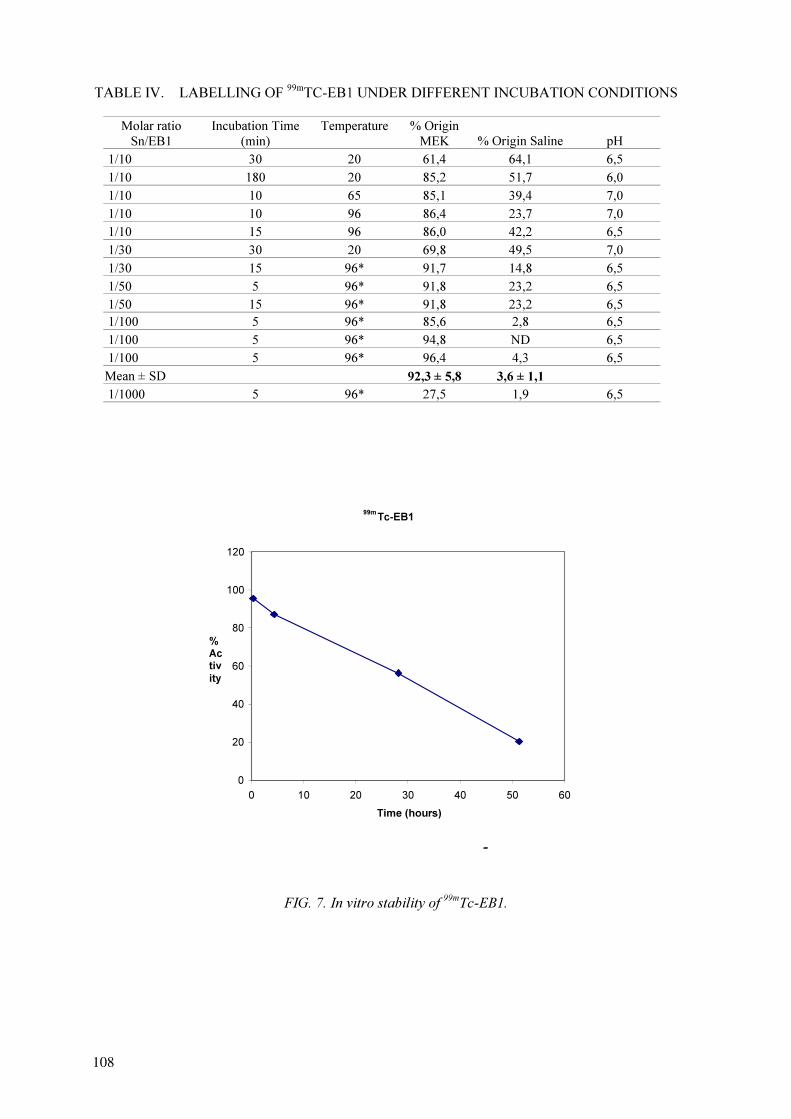
TABLE IV. LABELLING OF 99m
TC-EB1 UNDER DIFFERENT INCUBATION CONDITIONS
Molar ratio
Sn/EB1
Incubation Time
(min)
Temperature % Origin
MEK % Origin Saline pH
1/10 30 20 61,4 64,1 6,5
1/10 180 20 85,2 51,7 6,0
1/10 10 65 85,1 39,4 7,0
1/10 10 96 86,4 23,7 7,0
1/10 15 96 86,0 42,2 6,5
1/30 30 20 69,8 49,5 7,0
1/30 15 96* 91,7 14,8 6,5
1/50 5 96* 91,8 23,2 6,5
1/50 15 96* 91,8 23,2 6,5
1/100 5 96* 85,6 2,8 6,5
1/100 5 96* 94,8 ND 6,5
1/100 5 96* 96,4 4,3 6,5
Mean ± SD 92,3 ± 5,8 3,6 ± 1,1
1/1000 5 96* 27,5 1,9 6,5
FIG. 7. In vitro stability of 99m
Tc-EB1.
99m
Tc-EB1
0
20
40
60
80
100
120
0 10 20 30 40 50 60
Time (hours)
%
Ac
tiv
ity
-
108
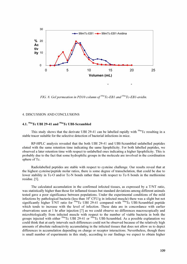
FIG. 8. Gel permeation in PD10 column of 99m
Tc-EB1 and 99m
Tc-EB1-avidin.
4. DISCUSSION AND CONCLUSIONS
4.1.99m
Tc UBI 29-41 and 99m
Tc UBI-Scrambled
This study shows that the derivate UBI 29-41 can be labelled rapidly with 99m
Tc resulting in a
stable tracer suitable for the selective detection of bacterial infections in mice.
RP-HPLC analysis revealed that the both UBI 29-41 and UBI-Scrambled unlabelled peptides
eluted with the same retention time indicating the same lipophilicity. For both labelled peptides, we
observed a later retention time with respect to unlabelled ones indicating a higher lipophilicity. This is
probably due to the fact that some hydrophilic groups in the molecule are involved in the coordination
sphere of Tc.
Radiolabelled peptides are stable with respect to cysteine challenge. Our results reveal that at
the highest cysteine/peptide molar ratios, there is some degree of transchelation, that could be due to
lower stability in Tc-O and/or Tc-N bonds rather than with respect to Tc-S bonds in the methionine
residue. [5].
The calculated accumulation in the confirmed infected tissues, as expressed by a T/NT ratio,
was statistically higher than those for inflamed tissues but standard deviations among different animals
tested gave a poor significance between populations. Under the experimental conditions of the mild
infections by pathological bacteria (less than 106
CFU/g in infected muscle) there was a slight but not
significantly higher T/NT ratio for 99m
Tc UBI 29-41 compared with 99m
Tc UBI-Scrambled peptide
which tends to increase with the level of infection. These data are in concordance with earlier
observations seen at 1 hr after injection [7] as we could observe no differences macroscopically and
microbiologically from infected muscle with respect to the number of viable bacteria in both the
groups injected with either 99m
Tc UBI 29-41 or 99m
Tc UBI-Scrambled. As a possible explanation we
could think that at early intervals such differences could not be observed because of the relatively high
amounts of absolute radioactivity accumulating in the infected tissues that does not allow us to depict
differences in accumulation depending on charge or receptor interactions. Nevertheless, though there
is small number of experiments in this study, according to our findings we expect to obtain higher
- -
0
10
20
30
0 5 10 15 20 25
Volumen (mL)
%
Ac
tiv
ity
99mTc-EB1 99mTc-EB1-Avidina
109109

T/NT ratios by increasing the number of viable bacteria in the inoculum that would give us a clearer
view on the differences in accumulation of both tracers at early intervals.
In the in vitro binding studies we observed a high specific binding for both S.aureus strains with
99m
Tc-UBI 29-41 (38.1% for S. aureus and 32.7% for S. aureus Virulent).
4.2.99m
Tc HNE2 and BPTI
99m
Tc labelling MAG3-BPTI was achieved with high yield as evidenced by HPLC control on
reverse phase column with radiometric and UV detection. However we could not show that the 99m
Tc –
BPTI had intact biological properties because we could not evaluate its activity in the presence of
trypsin.
Through the analysis of the labelling of various conjugates and radiochemical purity attained,
the best molar ratio for the conjugate production was determined as 1:7. Therefore future work will be
stressed on optimizing this step.
Gel permeation through P4 column represents an effective separation method even though it is
time consumming.
4.3.99m
Tc-EB1
The direct labelling of EB1 was simple, quick and efficient. 99m
Tc-EB1 had good stability for at
least 4 h. 99m
Tc-EB1 didn’t show good binding to S. aureus. and S. aureus Virulent. So it is not a
promising tracer for differential bacterial infection diagnosis.
ACKNOWLEDGEMENTS
We thank the IAEA for inviting us to participate in the CRP. Also, we are grateful to
Dr. Ernest Pauwels for his advice and for sharing his knowledge with us, and to Dr. Donald
Hnatovich, Dra. Renata Mikolajczak and Dr. Jehangir. We thank Dr. M. M. Welling of LUMC, The
Netherlands, for his advise and cooperation.
This work was done with the collaboration of Pablo Cabral, Department of Radiopharmacy,
C.I.N., Montevideo, Uruguay.
REFERENCES
[1] BECKER, W., “The contribution of nuclear medicine to patient with infection”, Eur. J. Nucl.
Med., (1995) 22 1195–1211.
[2] NIBBERING, P.H., et al., “Radiolabelled antimicrobial peptides for imaging of infections: a
review”, Nucl. Med. Comm. 19 (1998) 1117–1121.
[3] VALLABHAJOSULA, S.; “Technetium-99m labelled chemotactic peptides: specific for
imaging infection?”, J. Nucl. Med. 38 (1997) 1322–1326.
[4] SIGNORE, A., et al., “Peptide radiopharmaceuticals for diagnosis and therapy”, Eur. J. Nucl.
Med. 28 (2001)1555–1565.
[5] WELLING, M.M,, PAULUSMA-ANNEMA, A., BALTER, H.S., PAUWELS, E.K.J.,
NIBBERING, P.H., “Technetium labelled antimicrobial peptides discriminate between bacterial
infections and sterile inflammations”, Eur. J. Nucl. Med. 27 (2000) 292–301.
[6] DE KOSTER, H.S., ET AL., “The use of dedicated peptide libraries permits the discovery of
high affinity binding peptides”, J. Immunol. Methods 187 (1995,) 179–188.
[7] WELLING, M.M., et al., “Radiochemical and biological characteristics of 99m
Tc-UBI 29-41 for
imaging of bacterial infections”, Nucl. Med. Biol. 29 (2002) 413–422.
110

[8] INTERNATIONAL ATOMIC ENERGY AGENCY, Development of Kits for 99m
Tc
Radiopharmaceuticals for Infection Imaging, Summary Report of the 1st
RCM, 7–10 August
2000, Budapest (internal report).
[9] HNATOWICH, D.J., et al., “Labelling peptides with technetium-99m using a bifunctional
chelator of a N-hydroxysuccinimide ester of mercaptoacetyltriglycine”, J. Nucl. Med. 39 (1998)
56–64.
[10] INTERNATIONAL ATOMIC ENERGY AGENCY, Development of Kits for 99m
Tc
Radiopharmaceuticals for Infection Imaging, Summary Report of the 2nd
RCM, 19–22 February
2002, Mexico (internal report).
[11] HIEMSTRA, P.S., et al., “Ubiquicidin,, a novel murine microbicidal protein present in the
citosolic fraction of macrophages”, J. Leukocyte Biol. 66 (1999) 423–428.
[12] WELLING, M.M., et al., “99m
Tc-labelled antimicrobial peptides for detection of bacterial and
Candida albicans infections”, J. Nucl. Med. 42 (2001) 788–794.
111111


LIST OF PARTICIPANTS
Crudo, J.L. Comisión Nacional de Energía Atómica, Radioquímica,
Centro Atómico Ezeiza Radiofármacos,
Buenos Aires, Argentina
Ferro Flores, G. Instituto Nacional de Investigaciones Nucleares,
Departamento de Materiales Radioactivos,
Salazar Estado de Mexico, Mexico
Hnatowich, D. University of Massachusetts, Medical School,
Division of Nuclear Medicine, Department of Radiology,
Worcester, MA, 01655, United States of America
Jehangir, M. Pakistan Atomic Energy Commission,
Pakistan Institute of Nuclear Science and Technology,
Nuclear Chemistry Division, Islamabad, Pakistan
Körösi, L. Fodor József National Center for Public Health,
“Frederic Joliot-Curie” National Research Institute for
Radiobiology and Radiohgiene,
Department of Applied Isotopes, Budapest, Hungary
Mikolajczak, R. Institute of Atomic Energy, Department of Chemical Analysis,
Radioisotope Centre, Otwock Swierk, Poland
Narasimhan, D.V.S. International Atomic Energy Agency
Pauwels, E.K.J. State Univesity of Leiden, Leiden University Medical Centre,
Department of Radiology, Division of Nuclear Medicine,
Leiden, Netherlands
Poramatikul, N. Ministry of Science and Technology,
Office of Atoms for Peace,
Division of Isotope Production, Bangkok, Thailand
Ramamoorthy, N. Department of Atomic Energy,
Board of Radiation and Isotope Technology,
Radiopharmaceutical Programme, Mumbai, India
Souto Pais, B. Universidad de la República, Facultad de Ciencias,
Centro de Investigaciones Nucleares,
Departamento de Radiofarmacia, Montevideo, Uruguay
Widjaksana, W. National Nuclear Energy Agency,
Radioisotope Production Centre,
Radioisotope and Radiopharmaceutical Development Centre,
Serpong, Indonesia
Zhifu, Luo China National Nuclear Corporpation,
China Institute of Atomic Energy, Department of Isotopes,
Beijing, China
113113