development of hybrid antibacterial membrane by … · membran yang terhasil dicirikan bagi menilai...
TRANSCRIPT

DEVELOPMENT OF HYBRID ANTIBACTERIAL MEMBRANE BY
INCORPORATING SILVER PARTICLE WITH 2,4,6-TRIAMINOPYRIMIDINE
AS COMPATIBILIZER
HATIJAH BINTI BASRI
A thesis submitted in fulfilment of the
requirements for the award of the degree of
Doctor of Philosophy (Chemical Engineering)
Faculty of Chemical Engineering
Universiti Teknologi Malaysia

ABSTRACT
The objective of this study was to develop and characterize the
polyethersulfone (PES) incorporated with silver (Ag) as an antibacterial membrane
which can remove and disinfect bacteria in a single step for environmental
application. The PES-Ag membrane was developed from PES, silver nitrate as an
antibacterial agent and 2,4,6-triaminopyrimidine (TAP) as compatibilizer. The
influence of AgNO3 loading, molecular weights (MW) of polyvinylpyrrolidone
(PVP) as dispersant and type of compatibilizer have been investigated. The resulting
membranes were characterized based on their thermal, tensile and structural
properties which were used in correlation with the membrane antibacterial properties.
The incorporation of Ag in PES membrane has increased the tensile strength doubled
as compared to the unmodified PES. Furthermore, it was observed that the highest
AgNO3 loading (2 wt%) and the highest MW (360,000) of PVP as dispersant has led
to higher silver content on membrane surfaces. This is evidenced from energy
dispersive X-ray (EDX) analysis and X-ray photoelectron spectroscopy (XPS). These
properties have induced a better antibacterial activity in a disc-diffusion test against
Escherichia coli (E.coli) and Staphylococcus aureus (S.aureus). The structural
characterization by field emission scanning electron microscope (FESEM) revealed
that by incorporating TAP as compatibilizer, smaller Ag particles size with improved
distribution and average pore size of 0.174 µm was obtained. In addition, the silver
residue during fabrication monitored by inductive coupled plasma-mass spectrometry
(ICP-MS) was significantly reduced (62.6%). These parameters have led to E.coli
removal of log reduction value (LRV) 3.59 and 100% growth inhibition tested on
E.coli suspension of 1×10⁶ colony forming unit (CFU/mL). From the adhesion test,
this membrane exhibited the least E.coli adherence which in turn evidenced its anti-
adhesion property. In conclusion, the PES-Ag membrane with TAP as compatibilizer
produced was potential in bacteria removal and disinfection below the CFU
maximum range for water and waste water treatment.
ABSTRAK
Objektif kajian ini ialah untuk membangun dan mencirikan selaput
(membran) poliethersulfona (PES) yang digabungkan dengan perak (argentum)
sebagai selaput anti-bakteria yang akan dapat menyingkir dan menyahjangkit
bakteria dalam satu langkah untuk aplikasi alam sekitar. Membran PES-Ag
dibangunkan daripada PES, garam nitrat perak (AgNO3) sebagai agen anti-bakteria
dan 2,4,6-triaminopyrimidine (TAP) sebagai bahan bantu serasi. Pengaruh muatan
AgNO3, berat molekul polivinilpirolidone (PVP) yang bertindak sebagai bahan bantu
serak (dispersant) dan jenis bahan bantu serasi (compatibilizer) juga telah dikaji.
Membran yang terhasil dicirikan bagi menilai sifat terma, kekuatan tegangan (tensil)
dan struktur yang kemudiannya dikorelasi kepada sifat anti-bakteria membran
tersebut. Penggabungan Ag ke dalam membran PES telah meningkatkan kekuatan
tegangan membran dua kali ganda berbanding membran PES tidak terubahsuai.
Muatan AgNO3 tertinggi (2 wt%) dan PVP pada berat molekul tertinggi (360,000
Da) didapati telah berjaya menghasilkan kandungan Ag yang lebih tinggi. Ciri ini
telah dibuktikan melalui analisis yang menggunakan kaedah spektroskopi penyebar
tenaga sinar-X (EDX) dan kaedah spektroskopi elektron-foto sinar-X (XPS). Sifat-
sifat ini seterusnya telah mencetuskan sifat anti-bakteria yang lebih baik, dibuktikan
melalui ujian pembauran-cakera (disc-diffusion) terhadap bakteria Escherichia coli
(E.coli) dan Staphylococcus aureus (S.aureus). Pencirian struktur dengan
menggunakan mikroskopi imbasan elektron pemancaran medan (FESEM) telah
memberi maklumat bahawa dengan menggunakan TAP sebagai bahan bantu serasi,
partikel Ag yang lebih kecil dengan taburan yang lebih baik pada saiz liang purata
0.174 m telah dperolehi. Di samping itu, sisa Ag yang terlarut resap (leach) semasa
pembuatan membran yang dikawal dengan menggunakan spektrometri jisim-
berganding plasma teraruh (ICP-MS) didapati menurun dengan nyata sebanyak
62.6%. Keseluruhan parameter yang dikaji telah menunjukkan bahawa penyingkiran
E.coli adalah pada nilai penurunan log (LRV) 3.59 dan 100% perencatan
pertumbuhan apabila diuji pada 1×10⁶ unit koloni terbentuk per mL (CFU/mL).
Membran ini juga didapati menunjukkan lekatan bakteria (bacterial adherence) yang
terkecil dalam ujian lekatan terhadap E.coli sekaligus membuktikan sifat anti-
lekatan. Kesimpulan daripada kajian ialah membran PES-Ag dengan TAP sebagai
bahan bantu serasi adalah sangat berpotensi dalam penyingkiran dan perencatan
bakteria di bawah julat CFU untuk air dan rawatan air.

TABLE OF CONTENTS
CHAPTER TITLE
PAGE
DECLARATION ii
ACKNOWLEDGEMENT iii
ABSTRACT iv
ABSTRAK v
TABLE OF CONTENTS vi
LIST OF TABLES xi
LIST OF FIGURES xiii
LIST OF ABBREVIATIONS xvii
LIST OF SYMBOLS xix
LIST OF APPENDICES xx
1 INTRODUCTION 1
1.1 Research Background 1
1.2 Problem statements 3
1.3 Objectives of the study 4
1.4 Research scopes 4
1.5 Research significance 5
1.6 Organization of the thesis 6
2 ANTIBACTERIAL MEMBRANE FOR BACTERIA
REMOVAL: A REVIEW
7
2.1 Introduction 7
2.2 Disinfection in water and wastewater treatment 10
2.3 Membrane technology in bacteria removal 14

2.4 Physicochemical interaction between bacteria
and surface
16
2.5 Bacteria retention in porous media 18
2.5.1 Bacteria retention via straining 19
2.5.2 Bacteria retention via adsorption 20
2.6 Bacterial adhesion and biofouling 22
2.7 Advantages of antibacterial membrane over the
other bacteria removal method
24
2.8 Silver incorporation in polymer composites
25
3 METHODOLOGY 27
3.1 Operational framework 27
3.2 Material selection 27
3.3 Membrane fabrication 31
3.3.1 Dope preparation 31
3.3.2 Casting of asymmetric membrane 32
3.4 Membrane characterization 33
3.4.1 Attenuated total reflection-Fourier
transform infra-red
33
3.4.2 Miscibility analysis 33
3.4.3 Field emission scanning electron
microscope (FESEM) and atomic force
microscopy (AFM)
34
3.4.4 Energy dispersive x-ray (EDX) and
inductive coupled plasma-mass
spectrometry (ICP-MS)
35
3.4.5 Tensile strength analysis 35
3.4.6 X-ray photoelectron spectroscopy (XPS) 36
3.4.7 Surface porosity and pore size analysis 36
3.4.8 Contact angle measurement 37
3.5 Performance testing 38
3.5.1 Pure water permeation (PWP) 38
3.5.2 Antibacterial tests 39

3.5.2.1 Disc diffusion method 39
3.5.2.2 Filtration of E.coli suspension 40
3.5.2.3 Filtration of environmental
sample
41
3.5.2.4 Anti-adhesion test 42
4 THE EFFECT OF SILVER LOADING IN
POLYETHERSULFONE (PES)
ULTRAFILTRATION (UF) MEMBRANES FOR
BACTERIA REMOVAL
43
4.1 Silver loaded-PES UF membranes for bacteria
Removal
43
4.2 Experimental 44
4.2.1 Fabrication of PES-UF membranes
loaded with silver nitrate
44
4.2.2 Membrane characterization 45
4.2.3 Performance testing of membranes 46
4.3 Results and discussion 47
4.3.1 Effects of Ag-loading in thermal
properties of membrane
47
4.3.2 Effects of Ag-loading in membranes
morphology and silver loss
49
4.3.3 Pure water permeability (PWP) and pore
sizes of PES and PES-AgNO3 membranes
52
4.3.4 Contact angle and mechanical strength
of PES membranes
54
4.3.5 Antibacterial properties of membrane 57
4.4 Conclusion 60
5 POLYETHERSULFONE-SILVER ANTIBACTERIAL
MEMBRANE: EFFECT OF PVP MOLECULAR
WEIGHT ON MORPHOLOGY AND
ANTIBACTERIAL ACTIVITY

61
5.1 PES-silver antibacterial membrane: Effect of
PVP addition
61
5.2 Preparation of PES-Ag with PVP 63
5.3 Characterization of PES-silver membranes 64
5.4 Results and discussion 67
5.4.1 Miscibility analysis 67
5.4.2 ATR-FTIR analyses 69
5.4.3 Morphology analyses using FESEM 71
5.4.4 Contact angle 74
5.4.5 X-Ray photoelectron spectroscopy (XPS) 76
5.4.6 Pure water permeation (PWP) and
antibacterial property of membrane
78
5.4.7 Silver loss in membrane fabrication 81
5.5 Conclusion 82
6 SILVER-FILLED POLYETHERSULFONE
MEMBRANES FOR ANTIBACTERIAL
APPLICATIONS – EFFECT OF
COMPATIBILIZER
83
6.1 Silver-filled PES membranes: Effect of
Compatibilizer
83
6.2 Experiment 85
6.2.1 Materials and membrane preparation 85
6.2.2 Compatibility analysis 86
6.2.3 Analysis of silver loss 87
6.2.4 Morphological properties 88
6.2.5 Antibacterial activity 88
6.2.6 Anti-adhesion 90
6.3 Results and discussion 90
6.3.1 Compatibility analysis 90
6.3.2 Analysis of silver-loss 93
6.3.3 Binding energy (BE) of silver atoms 94

6.3.4 Membrane morphology 98
6.3.5 AFM analysis 101
6.3.6 Antibacterial activity 103
6.4 Conclusion 109
7 GENERAL CONCLUSION AND
RECOMMENDATION
110
7.1 General conclusion 110
7.2 Recommendations for future works 111
REFERENCES 113
Apendices A-J 133-151
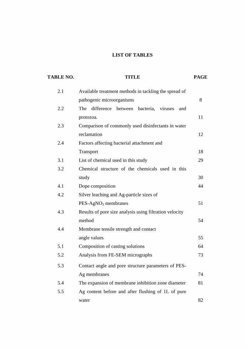
LIST OF TABLES
TABLE NO. TITLE PAGE
2.1 Available treatment methods in tackling the spread of
pathogenic microorganisms
8
2.2 The difference between bacteria, viruses and
protozoa.
11
2.3 Comparison of commonly used disinfectants in water
reclamation
12
2.4 Factors affecting bacterial attachment and
Transport
18
3.1 List of chemical used in this study 29
3.2 Chemical structure of the chemicals used in this
study
30
4.1 Dope composition 44
4.2 Silver leaching and Ag-particle sizes of
PES-AgNO3 membranes
51
4.3 Results of pore size analysis using filtration velocity
method
54
4.4 Membrane tensile strength and contact
angle values
55
5.1 Composition of casting solutions 64
5.2 Analysis from FE-SEM micrographs 73
5.3 Contact angle and pore structure parameters of PES-
Ag membranes
74
5.4 The expansion of membrane inhibition zone diameter 81
5.5 Ag content before and after flushing of 1L of pure
water
82

6.1 Composition of membrane formulation 86
6.2 Ag-loss during fabrication 94
6.3 Averaged membrane surface roughness via AFM 102
C1 Weights of wet and dried membrane, membrane
thickness and porosity for pore size analysis
138
C2 The mean pore size of prepared membranes 140
E1 Fluxes data of prepared membranes at TMP (1-6) bar 142
E2 Detailed data for flux calculations 143
I1 OD600 values for feed and permeates in the E.coli
filtration onto prepared membranes
using vacuum filtration cell
148
I2 CFU/mL values for feed and permeates in the E.coli
filtration onto prepared membranes using cross-flow
rig-system
149

LIST OF FIGURES
FIGURE NO. TITLE PAGE
2.1 Pressure driven membrane processes classified
principally by average pore diameter
14
2.2 Simplified concept schematic of membrane
separation. A desired component (water) is allowed to
pass through while non-desired component (bacteria)
is retained.
15
2.3 Schematic of the physicochemistry between bacteria-
nanoparticles and membrane matrices.
17
2.4 Stages of biofilm formation and the two-stages
bacterial adsorption
21
2.5 Total interaction energy between a bacterial cell and a
surface depending on ionic strength
22
3.1 Schematic diagram of the research framework 28
3.2 Experimental setting for dope preparation 31
3.3 (a) Nitrogen gas tank; (b) solution dope reservoir; (c)
air compressor; (d) pneumatic system; (e) trolley and
glass plate; (f) gear pump; (g) casting knife; (h)
evaporation chamber; (i) coagulant bath
32
3.4 Schematic diagram of the cross-flow permeation
testing system.
39
3.5 Experimental set-up for the antibacterial test – E.coli
filtration.
40
3.6 Environmental waste samples were kept in low
temperature before analysis
41
4.1 PES dope in yellowish transparent color becoming
chrome after AgNO3 addition.
47

4.2 Thermogravimetric curves of PES and
PES-AgNO3 membranes
48
4.3 DSC curves of PES and PES-AgNO3 membranes 49
4.4 FESEM micrographs of surfaces (left) and cross
sectional structure (right) of (a) PES, (b) PES-
0.5AgNO3 and (c) PES-2.0AgNO3 membranes
50
4.5 EDX analyses of (a) PES-0.5AgNO3 and (b) PES-
2.0AgNO3 membranes according to the FESEM
images
52
4.6 Pure water permeability (Lm-2
hr-1
) for PES and PES-
AgNO3 membranes against pressure (bar).
54
4.7 The scheme of interactions between PES as polymer
and AgNO3 as additive.
56
4.8 Various mechanisms of antibacterial activities exerted
by Ag+
58
4.9 Images of PES-0.5AgNO3 (a) and (c), PES-2.0AgNO3
(b) and (d) showing inhibition zone against E.coli (a),
(b) and S.aureus (c), (d). The red arrows are pointing
at the inhibition ring possessed around membrane
circular disc.
59
5.1 Thermo gram of (a) PES-Ag and (b) PES-Ag-P10,
(c) PES-Ag-P40, (d) PES-Ag-P360 membranes
68
5.2 ATR-FTIR spectra of (a) PES-Ag-P10, (b) PES-Ag-
P40, (c) PES-Ag-P360, (d) PES, (e) PES-Ag
70
5.3 FE-SEM surface and cross section images of (a) PES-
Ag, (b) PES-Ag-P10, (c) PES-AgP40 and (d) PES-
Ag-P360 membranes
72
5.4 Illustration of Ag in filling membrane surface pores
with representative of FESEM micrographs of cross-
section (a) PES-Ag-P40, (b) PES-Ag-P360
75
5.5 Stacked XPS-spectra for all prepared membranes 77
5.6 Pure water permeation of prepared membranes 78
5.7 Antibacterial results of prepared membranes 80

6.1 Chemical structure of (a) polyvinyl pyrrolidone,
(b) 2,4,6 triaminopyrimidine
85
6.2 X-Ray diffraction patterns of (a) PES, (b) PES-
AgNO3, (c) PES-PVP-AgNO3 and (d) PES-TAP-
AgNO3 membranes
91
6.3 Tg curves of PES and PES-modified membranes 92
6.4 XPS spectra of PES-modified membranes 96
6.5 Possible interactions between PVP-Ag+,
TAP-Ag+ and PES-TAP.
97
6.6 FE-SEM cross-section and surface images of
membranes
99
6.7 EDX spectra of PES-modified membranes (a) PES-
AgNO3, (b) PES-PVP-AgNO3 (c) PES-TAP-AgNO
100
6.8 3-D AFM images of (a) PES, (b) PES-Ag, (c) PES-
Ag-PVP, (d) PES-Ag-TAP membranes
102
6.9 Results of the antibacterial activity by using
agar diffusion method
105
6.10 Agar plates of feed and permeate from the filtration of
E.coli suspension. Images on left are feed and images
on the right are permeate for (a) PES-Ag (LRV3.25),
(b) PES-Ag with PVP (LRV2.93) and (c) PES-Ag
with TAP (LRV3.59)
106
6.11 Results of the antibacterial activity on the E.coli
filtration (a) PES (b) PES-AgNO3, (c) PES-PVP-
AgNO3 (d) PES-TAP-AgNO3
107
6.12 Agar plates of feed and permeate of environmental
waste filtration
108
B1 Rigaku X-Ray diffractometer 136
B2 Atomic Force Microscopy 136
B3 X-Ray photoelectron spectroscopy 137
B4 Contact angle goniometer 137
D1 The whole set-up for PWP and bacteria removal test 141
D2 Membrane test cell 141

G1 Vacuum filtration cell set-up for E.coli filtration 145
I1 Agar plates inoculated with feed (left) and permeates
(right) in the environmental waste analysis
147
J1 Schematic structure of E.coli bacterium 150
J2 The difference in cell wall of bacteria gram positive
and bacteria gram negative
151

LIST OF ABBREVIATIONS
MF - Microfiltration
DBPs - Disinfection by-products
DOM - Dissolved organic matter
UF - Ultrafiltration
LRV - Log-reduction value
UV - Ultra-violet
E.coli - Escherichia coli
S.aureus - Staphylococcus aureus
DNA - Deoxyribonucleic acid
TGA - Thermogravimetric analysis
DSC - Differential scanning calorimetry
XRD - X-ray diffraction
XPS - X-ray photoelectron spectroscopy
EDX - Energy dispersive X-ray
ICP-MS - Inductive coupled plasma-mass spectrometer
CFU - Colony forming unit
NOM - Natural organic matter
DOC - Dissolved organic carbon
SS - Suspended solids
TDS - Total dissolved solid
pH - -log[H+]
WHO - World of Health Organization
DLVO - Deryaguin-Landau and Vervey-Overbeck
MWCO - Molecular weight cut-off
Da - Dalton
AFM - Atomic force microscopy
ATR-
FTIR
- Attenuated total reflection-Fourier transform
infra-red spectroscopy
FESEM - Field emission scanning electron microscope
ppt - Part per trillion
ppm - Part per million
PWP - Pure water permeation
TOC - Total organic carbon
MW - Molecular weight
NA - Nutrient agar
OD - Optical density
MD - Minimal Davis
rpm - Revolution per minute
v/v - Volume per volume
Fig. - Figure
MTS - Mechanical testing system

Wt. - Weight
Eq. - Equation
SD - Standard deviation
DMFC - Direct methanol fuel cell
cps - Count per second
BE - Binding energy

LIST OF SYMBOLS
% - Percent
˚C - Degree Celcius
h - hour
mA - miliampere
kV - Kilo Volt
- degree
θ - theta
M - Micro molar
wt. % - Weight percent
d - Diameter
Jv - Pure water permeation (Flux)
rm - Mean pore diameter (m)
ε - Porosity (%)
Q - Volume of permeate per unit
time (m3s
-1)
A - Membrane surface area (m2)
ΔT - Permeation time (s)
µm - Micrometer
nm - Nanometer
Cp - Concentration of permeate
(ppm)
Cf - Concentration of feed (ppm)
mg/L - Miligrams per litre
mm/min - Milimeter per minute
VR - Repulsive energy
VA - Attractive energy
VT - Total energy
Tg - Glass transition temperature

LIST OF APENDICES
APPENDIX TITLE PAGE
A List of publications 140-142
B The instruments used in the characterization 143-144
C Pore size measurement by using Guerout-Elford-Ferry
equation
145-147
D Experimental set-up for flux and bacteria removal
measurement
148
E Pure water permeation in asymmetric membrane 149
F Preparation of agar plates for antibacterial test 151
G Experimental set-up for bacteria removal and colony
forming unit (CFU/mL) enumeration
152
H UTM map showing environmental spot 153
I Enumeration of colony forming unit (CFU) for agar
plates
154-156
J Structure of E.coli bacterium and the difference of cell
wall structure between gram positive and gram
negative
157-158

CHAPTER 1
INTRODUCTION
1.1 Research Background
Microbiological contamination of water sources has long been a concern to
the public. According to some authors, there were numbers of various bacterial
species available (ranging from 102 to 10
4 mL
−1) in raw water as well as sewage
effluents (Bonnélye et al., 2008; Goldman et al., 2009) tend to adhere to surfaces and
grow mainly at the expense of nutrients accumulated from the water phase.
Microbiological contamination in any sources should be avoided at any cost since in
the production of potable water, only a limited number of bacteria (depends on the
type of bacteria) are acceptable. The process for the removal of contaminants
depends not only on the nature of the microorganisms but also on the desired levels
of purity.
The use of membrane filtration in water treatment has greatly contributed to
greener technology. For example, microfiltration (MF) membrane has been widely
applied in water purification process due to its capability to remove microorganisms
and to treat harmful pollutants as well as dissolved organic matters (DOM) (Ghayeni
et al.,1996; Oh et al., 2007). Laine et al. reported that ultrafiltration (UF)
applications represent 74% of the total installed low-pressure membrane full-scale
plants (identified world-wide) in water industry in order to meet more stringent
regulations in producing drinking water (Laine et al., 2000).
It was reported in the open literature that membrane technology is one of the
disinfection technique where microorganisms are retained without any chemicals

engagement. However, the problem of biofouling aroused when membrane is
applied, due to the accumulation of microorganisms on membrane surfaces. In
addition, the current practice of membrane filtration required additional step
addressed as disinfection step via techniques such as chlorination (the most common
one), ozonation and UV. There were many and thorough discussion available in the
use of chlorination recently. The use of chlorination may lead to the disinfection by-
products (DBPs) release which in turn exposed consumers to potential carcinogenic
compounds such as the derivatives of chloramines.
Many studies have been conducted to overcome/meet the restrictions as well
as to resolve membrane fouling problems due to the uncontrolled accumulations of
micro-species. In handling biofouling problems, membrane modification, low-flux
operation and chemical cleaning are areas to be explored (Chang et al., 2002; Khor et
al., 2007). The effective prevention of microbial growth in a membrane system can
only be achieved when continuous and sufficiently high chlorine concentration is
maintained. However, due to stricter legislative regulation on chlorine usage, other
effective and environmental-friendly alternative is needed.
In membrane modification, the research in combining inorganics into
polymer matrices has been expanding since 1990-s. The inorganics chosen were
tailored with the application such as catalysis, biochemistry, separation and sensing.
In gas separation, the inorganic fillers namely zeolite, carbon molecular sieve, silica
and metal oxides has contributed to enhance membrane separation performance
specifically in addressing flux decline and selectivity (Rafizah et al., 2008; Kusworo
et al., 2008; Ismail et al., 2009; Mataram et al., 2010).The combination of inorganics
in polymer matrices or well-known as mixed-matrix provides the solution for highly
cost-maintenance and brittleness of inorganic membranes. The inorganic fillers in
mixed matrix membrane act to create preferential permeation pathways for selective
permeability while posing a barrier for undesired permeation in order to improve the
separation performance (Goh et al., 2011).
In water application, attempts were made in addressing flux decline due to the
accumulation of (micro- or macro-)species onto membrane surfaces which in turn
affecting the separation performance. The important issue in membrane
manufacturing is to develop membrane with suitable pore size in order to attain

various sizes of contaminants. In addition, membrane must also show sufficient
resistance towards the feed components as well as the operating condition. In
antibacterial application, a number of researches have been conducted in exploring
silver-incorporation to polymeric materials such as cellulose acetate (CA),
polacrylonitrile (PAN), polysulfone (PSf) and chitosan for the application of water
treatment, nano-fibre and food-packaging (Chou et. al., 2005; Wang et. al., 2005; Ma
et. al., 2008, Zodrow et. al., 2009). Silver was found to leach in the reported articles
and to date; attempts on overcoming this problem are still not published in the open
literature.
1.2 Problem statements
The conventional UF membrane in water and waste water treatment
established in bacteria removal has achieved a promising rejection value of >99% or
to be specific, a log reduction value (LRV) of >3. However, after the membrane
filtration process, it is necessary to perform an extra step which is disinfection as a
secondary bacteria control barrier and distribution system protection (Ghayeni et al.,
1996; Zio et al., 2005). The options available for disinfection are: UV, ozonation and
chlorination. The most commonly used method is chlorination due to the easy-
handling process and cost effectiveness. However, the major drawbacks of this
method is when greater number of bacteria present, higher concentration of chlorine
is needed, hence more disinfection by-products (DBPs) will be released in the water
distribution system. Current waste water treatment with microbial burden is facing
the problem of biofouling due to the accumulation of microorganisms over operation
time. In order to address the issues, current research is conducted to explore the
possibility and effectiveness of using a UF membrane incorporated with an
antibacterial agent in removing bacteria as well as to investigate its potential in
behaving anti-biofouling properties.
1.3 Objectives of the study

Based on the existing problem statements, the current study has been
performed with the following objectives:
i. To develop an antibacterial membrane by incorporating silver (Ag) as an
antibacterial agent without sacrificing membrane fluxes and removal abilities and to
characterize the membrane in terms of mechanical, morphological, water permeation,
hydrophilicity and pore sizes.
ii. To study the effect of incorporating a compatibilizer, PVP of different
molecular weights in membrane properties and performances.
iii. To evaluate the effect of incorporating different compatibilizers in membrane
properties and performances.
iv. To evaluate the fluxes of prepared membranes using pure water permeation
test on the custom-made test-rig.
v. To evaluate the antibacterial performance of membrane using disc diffusion
method, filtration of bacterial suspension and anti-biofouling tests.
1.4 Research scopes
In order to achieve the above mentioned objectives, the following scopes of
study were drawn.
i. Preparation of dope using PES as polymeric material, NMP as solvent and
AgNO3 as an additive or precursor of antibacterial agent, silver (Ag).
ii. Selection of AgNO3-loading and compatibilizer based on the evaluation in the
miscibility and antibacterial tests.
iii. Fabrication of PES-AgNO3 asymmetric membrane using phase inversion
technique and characterization of membranes in terms of mechanical strength,
hydrophilicity, overall porosity, pore sizes and water permeation.
iv. Evaluation of Ag-entrapment in prepared membranes by using ICP-MS, EDX
and XPS techniques.
v. Membrane fluxes measurement was carried out by using custom-made test rig
at pressure range 1-6 bar.

vi. Performance measurement of prepared membranes was conducted in terms of
antibacterial activity by using disc diffusion method and bacteria removal via
the filtration of bacterial suspension.
vii. Performance measurement of prepared membranes was conducted in terms of
anti-biofouling properties through an anti-adhesion test.
viii. Comparison of PES pristine membrane with PES antibacterial membrane in
all characterization and antibacterial tests.
1.5 Research significance
This study is of significance to the research of water treatment which
involves disinfection steps. The antibacterial membrane extends the multi-steps
options for water treatment to a stand-alone removal and disinfection of bacteria. The
results obtained in the study also provide the information in bacteria-removal and
bacteria-killing mechanisms which lead to the most effective options in treating
polluted water. Furthermore, the information on silver entrapment obtained in this
study would be beneficial to the other related fields such as in medicinal and
electrical field where silver is optimized in wound dressings and conducting material.
1.6 Organization of the thesis
The thesis is divided into six chapters. The first chapter presents the research
background as well as the problem statement. The research objectives, scopes and
significance are also highlighted in first chapter. Chapter two provides the literature
review on bacteria removal which includes the theories of the whole process and the
options available for bacteria removal. The advantages of antibacterial membrane,
current status and future direction of the technology are also discussed in this
chapter. Chapter three is dedicated to the detailed description of the research
methodology. The material selection for dope preparation, membrane fabrication and
performance testing conducted in this work are explained in this chapter. In chapter

four, the effect of silver content on the properties and performance of fabricated
membranes are explored and discussed. Subsequently, Chapter five describes the
effect of polyvinylpyrrolidone (PVP) of various molecular weights on antibacterial
properties of the resulting membranes. Chapter six discusses the significant
contribution of different compatibilizers in membrane antibacterial activities. Other
improved properties and the comprehensive discussions on the anti-adhesion
properties of resultant membranes are also included. Finally in Chapter seven,
conclusion of the research is drawn and the potential future works are proposed.

CHAPTER 2
ANTIBACTERIAL MEMBRANE FOR BACTERIA REMOVAL:
A REVIEW
2.1 Introduction
In conventional treatment of contaminated water sources, several methods
have been applied to provide multiple barriers to the spread of pathogenic
microorganisms and have minimize the spread of waterborne disease. Table 2.1 lists
each treatment which can either be used individually or couple with other methods in
a multi- step process.
Amongst the processes listed, sedimentation is considered as an easy and
widely accepted technique due to its natural principle which uses earth gravitational
force to settle down particles/suspended solids (SS) from a suspension. The process
consumes low energy therefore reduce the processing cost. However, some
drawbacks quoted by Guazzelli (1984) was the low loading rates which is only 1- 2
m3m
-2hr
-1 plus large space needed, hence higher construction cost will be required.
Other than the natural force, sedimentation applies coagulants as an aid to cater high
sludge loading. The efficient sweep coagulation can only be achieved at higher
coagulant dose. Excessive slime bacteria which result in filter clogging are another
drawback in sedimentation (Horan and Mara, 2003).

Table 2.1: Available treatment methods in tackling the spread of pathogenic microorganisms
Treatment
method
Processes Effectiveness References
Coagulation Coagulation or coagulation-flocculation is a conventional
technique to remove organic and inorganic suspension,
colloids and other natural organic matter (NOM). This
process uses chemicals addressed as coagulants and
coagulant aids. Examples of common coagulants are
Al2(SO4)3 (alum), Fe2(SO4)3 (copperas), FeSO4 and FeCl3
while examples of the aids are bentonite,
(Al2O3)4SiO2.2H2O, sodium silicate, Na2SiO3, lime,
Ca(OH)2 and calcium carbonate, CaCO3. Primary
coagulants neutralizes the electrical charges of particles
in the water which cause the particles to clump together,
while the aids are generally used to reduce flocculation
time and specifically used for clear water with very low
turbidity that does not coagulate well with usual
procedures.
Removes
Turbidity at ~ 99%
NOM (in terms of dissolved
organic carbon, DOC (up to
59.5%)
Bacteria at >3 log reduction
value, LRV
(Konieczny et al.,
2009)
(www.thewatertrea
tments.com)
(Qin et al., 2006)
Sedimentation Sedimentation is a physical water treatment process to
settle suspended solid that has been deposited by natural
processes in water under the influence of gravity. The
settling rate of sedimentation can be dramatically
improved by the addition of small dosages of polymeric
flocculants. The higher the solid concentration, the faster
the flocculation occurs and hence larger flogs will be
produced.
E.coli removal of >50%
(depending on turbidity and
the characteristics of
particular sediment).
For 50 mL of E.coli
suspension, population was
reduced to 1/120, 1/100,000
and 1/1,400,000 after 24, 48
and 72 h respectively.
(Gutbai and
Gregory, 1991)
(Milne et al.,
1986)
(Kawabata &
Tanabe, 2005)

Filtration Filtration is a process to separate matters from fluid by
passing the mixture through a porous media that entraps
the solids in its matrix or retains them on its surface. The
amount of removal is a function of the filtering media.
The removal was subject to several factors including
mechanisms (straining or adsorption), the grain size of
porous media, organic matter content, bacteria species
and etc.
The median reduction is 104.
Soil-filtration has reduced
total bacteria up to 99.94%.
Filtration of E.coli
suspension on an
antibacterial membrane has
resulted in 100% growth
inhibition.
(Sterik et al.,
2004)
(Vanderbroucke et
al, 1995)
(Gilbert et al.,
1976)
(Basri et al., 2010)
Disinfection
Disinfection is considered as a primary mechanism for
the inactivation of pathogenic organisms to prevent the
spread of waterborne disease to downstream users and the
environment. Disinfection is performed via physical or
chemical techniques. Radiation, filtration and heating are
some examples of physical disinfection while
chlorination and ozonation are chemical disinfecting
method.
By UV disinfection, the
N
N0
10log:
E.coli 3.8
Total coliform 3.3
Enterococci 3.3
(Madaeni, 1999)
(Olanczuk-Neyman
et al., 2001)

Sedimentation and further flocculation were needed to produce good quality
water while the use of powder activated carbon (PAC) was possibly needed to
remove taste and odor (Hagen, 1998). In order to complement the conventional water
treatment processes, disinfection was performed by using chemical disinfectants or
physical methods. The objective of disinfection is to render an object or field free
from infection in which the infection may represent a risk to persons or environment
(Gilbert and Brown, 1995).
2.2 Disinfection in water and wastewater treatment
Diseases caused by pathogenic bacteria, viruses, protozoa or helminthes are
the most common and wide-spread health risk associated with drinking water. For
this reason, the World of Health Organization (WHO) has placed the greatest
importance on the microbiological quality of drinking water emphasized the potential
consequences of microbial contamination are such that its control must never be
compromised (Gorchev, 1996). To the least, water sources must be protected from
contamination by human and animal wastes which contain a variety of bacterial,
viral, protozoa and helminthes pathogens which are the sources of the waterborne
disease. The characteristics of the main waterborne disease source are listed in Table
2.3.
Waterborne disease is disease resulted from improper sanitary disposal of
human feces. The feces of healthy persons contain 1 to 1000 million per gram of
each of the following groups of bacteria: enterobacteria (e.g E.coli), enterococci,
lactobacilli, clostridia, bacteriodas, bifidobacteria and eubacteria (Hammer and
Hammer Jr, 2008).
In order to meet the stringent regulations by WHO, disinfection methods
(chemical and/or physical) has been adopted in water and wastewater treatment. The
commonly used disinfectants are listed in Table 2.2 with their risks, advantages and
efficiency against different contaminants for general comparison. Several studies
have shown that the efficiency of UV as a disinfection method is highly dependent
on the concentration of SS (Narkis et al., 1995; Hurst, 1996; Blume et al., 2002).

This is due to the fact that SS can protect the bacteria through a ‘sheltering’ flogs
which prevented the UV-light from penetrating and destroyed by the disinfectants
(LeChevallier, 1988). The UV-light cannot penetrate large particles from ~50 m in
diameter, thus the required energy will be raised drastically (Neis and Blume, 2003).
Table 2.2: The difference between bacteria, viruses and protozoa
(Madigan et al., 2000).
Organism Size Description Examples (waterborne)
Viruses 20-120 nm Biological agents
consisting of
molecules of nucleic
acids and protein
envelope.
Enterovirus,
coxsackievirus,
echovirus, rotavirus,
hepatitis A & B.
Bacteria 1-6 m Unicellular and
organism with
nucleus
E.coli, Salmonella sp.,
Shigella sp.
Protozoa Cryptosporidium
sp. (4-6 m)
Giardia sp. (8-12
length) × (7-10
m width)
Protozoa are single-
celled eukaryotes
(organisms whose
cells have nuclei)
that show some
characteristics
usually associated
with animals, most
notably mobility and
heterotrophy
Giardia duodenalis,
Cryptosporidium sp.,
Entamoeba

Table 2.3: Comparison of commonly used disinfectants in water
reclamation (Asano et al., 2007)
Characteristics Chlorine
gas
Sodium
hypochlorite
Chlorine
dioxide
ozone UV radiation
Deodorizing ability
High Moderate High High na*
Interaction with
organic matters
Oxidizes
organic
matter
Oxidizes
organic
matter
Oxidizes
organic
matter
Oxidizes
organic
matter
Absorbance
of UV
irradiation
Corrosiveness Highly
corrosive
Corrosive Highly
corrosive
Highly
corrosive
na*
Toxic to higher
forms of life
Highly
toxic
Highly toxic Toxic Toxic Toxic
Penetration of
particles
High High High High Moderate
Safety concern High Moderate to
Low
High Moderate Low
Solubility Moderate High High High na*
Stability Stable Slightly
unstable
Unstable Unstable na*
Effectiveness as disinfectant
Bacteria Excellent Excellent Excellent Excellent Good
Protozoa Fair to
poor
Fair to poor Good Good Excellent
Viruses Excellent Excellent Excellent Excellent Good
Byproduct
formation
THMs and
HAAs
THMs and
HAAs
Chlorite
and
chlorate
Bromate None known
in measurable
concentrations
Increases TDS Yes Yes Yes No No
Use as a
disinfectant
Common Common Occasional Occasional Increasing
rapidly
*na = not applicable
The recent development in the area of disinfection has been discovered to
reflect a great diversity and complexity of product. As an example, for chlorination,
chlorine in its free form may react with a group of organic acid available in water
and result in trihalomethanes (THM) or other DBPs formation (Asano et al., 2007).
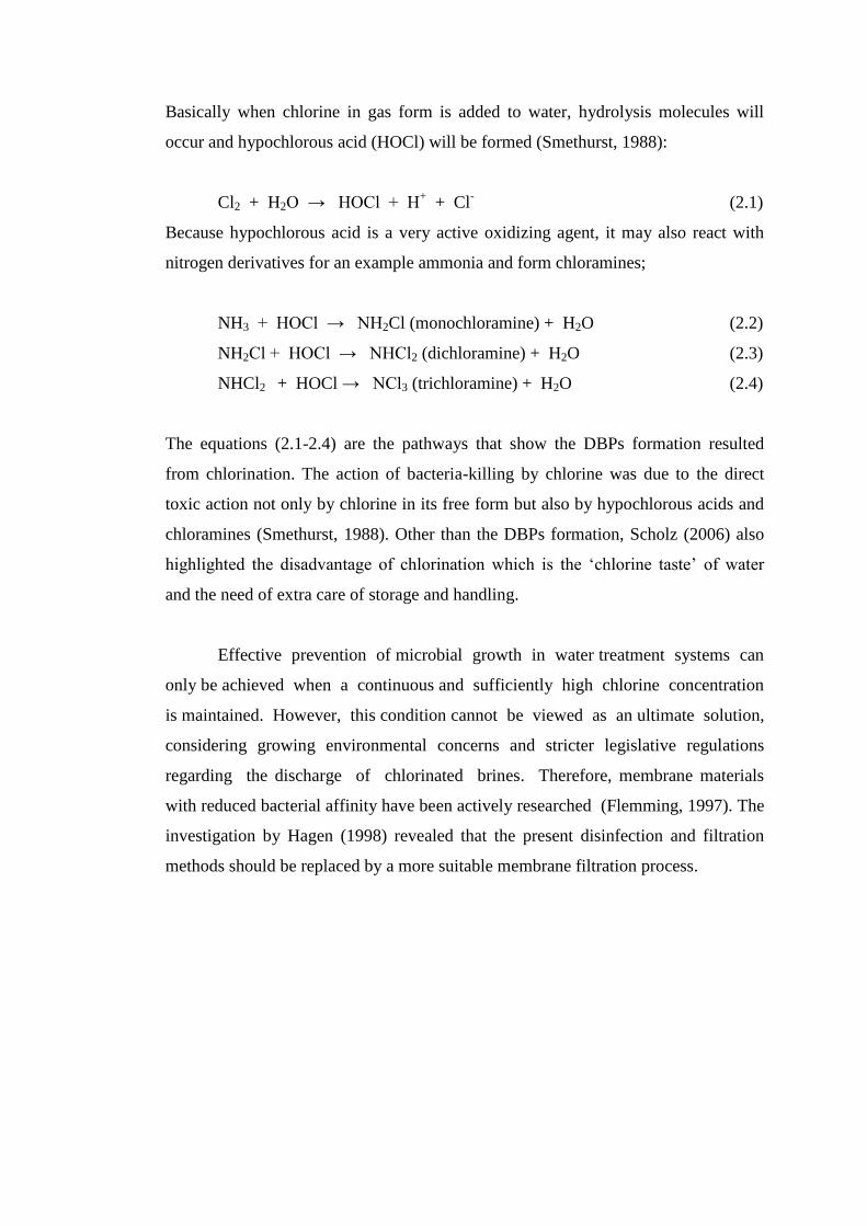
Basically when chlorine in gas form is added to water, hydrolysis molecules will
occur and hypochlorous acid (HOCl) will be formed (Smethurst, 1988):
Cl2 + H2O → HOCl + H+ + Cl
- (2.1)
Because hypochlorous acid is a very active oxidizing agent, it may also react with
nitrogen derivatives for an example ammonia and form chloramines;
NH3 + HOCl → NH2Cl (monochloramine) + H2O (2.2)
NH2Cl + HOCl → NHCl2 (dichloramine) + H2O (2.3)
NHCl2 + HOCl → NCl3 (trichloramine) + H2O (2.4)
The equations (2.1-2.4) are the pathways that show the DBPs formation resulted
from chlorination. The action of bacteria-killing by chlorine was due to the direct
toxic action not only by chlorine in its free form but also by hypochlorous acids and
chloramines (Smethurst, 1988). Other than the DBPs formation, Scholz (2006) also
highlighted the disadvantage of chlorination which is the ‘chlorine taste’ of water
and the need of extra care of storage and handling.
Effective prevention of microbial growth in water treatment systems can
only be achieved when a continuous and sufficiently high chlorine concentration
is maintained. However, this condition cannot be viewed as an ultimate solution,
considering growing environmental concerns and stricter legislative regulations
regarding the discharge of chlorinated brines. Therefore, membrane materials
with reduced bacterial affinity have been actively researched (Flemming, 1997). The
investigation by Hagen (1998) revealed that the present disinfection and filtration
methods should be replaced by a more suitable membrane filtration process.

Figure 2.1: Pressure driven membrane processes classified principally by
average pore diameter (Eykamp, 1995).
2.3 Membrane technology in bacteria removal
In general, membrane is a barrier that separates two phases and restricts the
transport of various species in a specific manner when a driving force is applied. In
other words, when driving force is applied, the membrane placed in a fluid system
will retain one component by sieving or size-exclusion mechanism and produce
purified solution. Pressure-driven membrane processes which are reverse osmosis,
nano-, ultra- and microfiltration are now being extensively used for the purification
of natural and waste waters. Figure 2.1 illustrates the pore size range of pressure
driven membrane processes that are used to separate particles of various size range.
Ultrafiltration (UF) is a pressure driven membrane process whose nature lies
between nanofiltration and microfiltration (MF). MF is typically known and used for
turbidity reduction and removal of suspended solids within the approximately size of
1-30 m (Li et al., 2003) meanwhile UF membranes are commonly used to remove
some viruses, color, odor, and some colloidal natural organic matter. Both processes
H2O
(2 Å)
Na+
(3.7 Å)
Sucrose
(10 Å)
Hemoglobin
(70 Å)
Influenza
virus
(1000 Å)
Pseudomonas
diminuta
(0.28 m) Staphylococcus
bacteria
(1 m)
Starch
(10 m)
1 Å 10 Å 100 Å 1000 Å 1 10 100
Microfiltration
Conventional
filtration Ultrafiltration Reverse
Osmosis
Pore diameter

require low transmembrane pressure (1- 30 psi) to operate, and both are now being
used as pretreatment in desalination processes such as reverse osmosis,
nanofiltration, and electrodialysis. As a pressure driven membrane process, UF
membrane normally possesses asymmetric structure with thin but relatively dense
top layer (thickness 0.1-1.0 µm), supported by a porous substructure (thickness ≈50-
150 µm) in which suspended colloids and particles in the approximate size range of
10-1000 Å are retained. An illustration in Figure 2.2 simplified the separation
concept in membrane. Although UF has been commonly used in current market, the
improvement on the available system is still necessary.
Transportation of molecules or particles via membrane occurs due to the
driving forces applied. This driving force can be chemical potential gradient, e.g
concentration gradient or pH gradient; pressure difference; electrical potential
difference or combination of these (Mulder, 1991). In bacteria removal, bacteria
transport is generalized to occur by advection, diffusion (for small bacteria) and
chemotaxis (Corapcioglu, 1996). Chemotaxis is the preferential movement of
bacteria in response to chemical gradients such as areas of higher nutrient
concentrations.
Figure 2.2: Simplified concept schematic of membrane separation. A
desired component (water) is allowed to pass through while
non-desired component (bacteria) is retained.
Feed
Permeate

When membrane with smaller pore size is used or bigger solute species need
to be retained, the higher pressure has to be applied in the operating system. The
proportional relationship between the membrane fluxes and the driving force is:
Flux = Proportionality factor × Driving force
J = A × X (2.5)
where A is the proportionality factor determines how fast the components or particles
is transported through the membrane. In other words, A is a resistance measurement
exerted by membrane as a diffusion medium when force is applied to the components
or particles.
According to Song and Elimetech (1995), the net velocity of bacterial cell
normal to the membrane surface is largely determined by normal convection with
small contributions by tangential correction and Brownian diffusion. The interaction
force profile suggested that aggregation were enhanced in acidic medium even
though the bacterial deposition rate is lower. The model studied also suggested that
the increment of permeation velocity resulted in higher bacterial deposition rate.
2.4 Physicochemical interaction between bacteria and surface
The physicochemical interaction between bacteria and surfaces has been
highlighted. The schematic in Figure 2.3 illustrates the interaction among bacterial
cell, inorganic particles and the surface of porous media. Removal of bacteria from
the flowing liquid phase generally occurs by filtration and adsorption or cell death
(Corapcioglu, 1996). The various bacterial attachment and detachment mechanisms
are affected by one or more factors as listed in Table 2.4. According to Yuehuei and
Friedman (2000), bacteria surface hydrophobicity is an important physical factor for
adhesion. Generally, hydrophobic bacteria prefer materials with hydrophobic
surfaces while hydrophilic characteristics prefer hydrophilic surfaces. However,
hydrophobic bacteria adhere to a greater extent than hydrophilic bacteria.

Figure 2.3: Schematic of the physicochemistry between bacteria-
nanoparticles and membrane matrices.
(Yuehei and Friedman, 2000)
When approaching the entrance of a pore, Leblue et al. (2009) explained that
bacteria are submitted to the shear and drag forces created by the trans-membrane
pressure (TMP) applied during the filtration step. Such stress may lead to bacteria
volume reduction and surface deformation (governed by the cell wall Young
Modulus value) which would allow the cell to penetrate into the membrane pore.
Whether the cell membrane is disrupted or not, the bacteria still have the possibility
to pass the pore. If the bacteria penetrate the membrane and retain its integrity, these
bacteria will keep their pathogenicity and hence risk consumers. To address the
problem, the inorganic antimicrobial agent attached on the membrane surfaces and in
membrane pores will act and perform biocidal action. The system which combined
membrane technology with inorganic antimicrobial is efficient in such a way that the
metal ion is bound within a delivery system that stabilizes them and then releases
them through a process of ion exchange at the surface (Peinemann and Nunes, 2010).
H2O M
+
Bacterial cell
Metal ions
M
Membrane
surface

Table 2.4: Factors affecting bacterial attachment and transport
(Corapcioglu, 1996)
Factor Effect on transport or attachment
pH An attachment favors low pHs.
Ionic strength Attachment increases with higher ionic strength due to the ‘particle
double-layer’ size reduction.
Clay-content Attachment increases with high clay content due to larger specific area
for adsorption.
Oxygen
limitations
Oxygen-limited biofilms exhibit lower shear removal rates but higher
sloughing.
Change on media Attachment of negative bacteria will be high in positive charges media.
Flow rate Higher flow rates reduce bacterial attachment.
Nutrient
concentration
Bacterial size reduced in higher nutrient concentrations.
Bacterial size Smaller bacteria may interact with media less and may not be removed
by filtration as easy as bigger bacteria. On the other hand, larger
bacteria have been shown to move faster than small bacteria.
Cell
concentration
At low cell density, attachment is favored. Bacteria tend to move from
high concentration areas to low concentration areas by a tumbling
diffusive flux.
Bacterial motility Motile bacteria may migrate faster than non-motile bacteria through
chemotaxis.
Water content Bacteria moves faster through unsaturated soil at higher water content.
2.5 Bacteria retention in porous media
According to Dunne Jr., (2002) the process of bacterial adhesion is dictated
by the variables including the species of bacteria, surface composition,
environmental factors and the essential gene products. From an evolutionary
standpoint, the selective advantage of bacterial adhesion was postulated to favor a
nutritional and non-hostile environment and provide a level of protection. Frimmel et
al. (2007) discusses the two types of deposition mechanism namely straining and
interception. Straining is generally about retaining big agglomerates while
interception is about retaining small aggregates on porous surface after collision. The
interception mechanism which is dominated by physicochemical interactions
between the cell surface and the porous medium has been reported to govern the

adhesion of cells. In order to understand the process of bacterial adhesion, the two
mechanisms in bacteria removal are discussed.
2.5.1 Bacteria retention via straining
According to Stevik et al. (2004) straining mechanism depends on the grain
size of the porous media. Generally, the extent to which the bacteria are retained by
straining is inversely proportional to the size of the filter media particles. In other
words, the smaller the filter media size, the more bacteria will be retained via
straining. By considering filter-media factors, straining will become a dominant
mechanism when the average cell size of the bacteria is greater than the size of 5% of
the grains that compose the porous material (Stevik et al., 2004). The presence of
macropores in filtration has been found to result in poor volume utilization and allow
a more rapid and distant bacteria movement (Chandler et al., 1981). In brief, the
transport for most of the bacteria in a system on saturated flow (e.g waste water
treatment) is found to favorably take place in the smaller pores. Generally, larger
cells will be more efficiently removed by filtration.
Weiss et al. (1995) studied the effect of bacterial cell shape on the transport
in porous media and suggested a preferential removal of long, rod-shaped cells
during transport. Bacterial straining can also be influenced by flow rates and
hydraulic loading. A high flow-rate may increase the average water suction in an
unsaturated filter medium. As a result, greater transport may occur via larger pores
which in turn decrease the effect of bacterial straining by porous material. Ausland et
al. (2002) observed a higher removal of fecal coliform bacteria in filtration systems
using uniform pressure distribution as compared to gravity dosing. Another factor to
be considered in bacteria removal via straining is clogging (Vandevivere and
Baveye, 1992). Clogging occurs due to the biomass growth in the porous media.
Bacteria removal is more efficient in clogged filtration system due to the hydraulic
disfunction which diminishes the purification of wastewater (Bouwer, 1974; Gannon
et al., 1991).

2.5.2 Bacteria retention via adsorption
In contrast to straining mechanism, adsorption is the dominant mechanism in
bacteria retention when media pores sizes are larger than that of bacteria (Sharma et
al., 1985). The bacterial adsorption on solid surfaces involves two stages
mechanisms that conform to the classical Derjaguin-Landau and Verwey-Overbeek
(DLVO) theory which has been first suggested on charged colloidal particles. The
stages were illustrated in Figure 2.4. The first stage is a reversible mechanism
controlled by electrostatic interactions between the cell surface and the adsorbent
(porous media). Weak interaction is present between the bacterium and porous
material. During this stage, reversibly attached bacteria can detach from the surface
of a particle and return to the water phase, depending on the conductance and
chemical properties of the fluid or aqueous solution.
In second stage, bacterial adsorption forms a much more persistent bond
between adsorbent (porous media) and adsorbate (bacterium). This mechanism is
irreversible and sometimes referred as adhesion (Olson et al., 1991). According to
the classical DLVO theory, the energy of interaction (VT) between a bacterium and
solid surface is (Derjaguin and Landau, 1941):
ART VVV (2.6)
VR = repulsive energy resulting from the overlapping of the electrical double layer of
cell and substratum (generally repulsive), VA = attractive energy resulting from van
der Waals interactions (generally attractive).

Figure 2.4: Stages of biofilm formation and the two-stages bacterial
adsorption (Katsikogianni and Missirlis, 2004; Houdt and
Michiels, 2005).
According to Hermansson, who extended the classical DLVO theory, VA
(Hermansson, 1999):
d
ArVA
6
(2.7)
where A is the Hamaker constant, d is the separation distance between the cell and
the solid surface and r is the cell radius (assuming cells are spherical). The DLVO
theory states that the distance of separation between colloidal adsorbents (porous
media) and adsorbates (bacterium) is the distance at which the repulsive (VR) and
attractive (VA) energy are balanced.
Hori and Matsumoto (2010) explained the two steps mechanism in DLVO
theory by relating them to the ionic strength as illustrated in Figure 2.3. At low ionic
strength, the energy barrier prevents the bacterium from approaching solid surfaces
via Brownian motion.When the energy barrier becomes higher and further from the
solid surface (at lower ionic strengths), the bacterial cells is found unable to adhere
on the surface. In contrast, at high ionic strength, the energy disappears and bacterial
cells can easily and rapidly attain irreversible adhesion.
First stage: Bacterial
cells-attachment to
surface, involving:
Hydrophobic
interaction
Electrostatic
interaction
Van der Waals
forces
Second stage:
Adhesion of cell to
cell and
proliferations to
mature biofilm
formation
Cells detachment
due to the
degradation of
extracellular
macromolecules.
Repetition of colonization process on
new sites

Figure 2.5: Total interaction energy between a bacterial cell and a surface
depending on ionic strength (Hori and Matsumoto, 2010).
The retention of bacteria via adsorption mechanism is influenced by porous
media designation (Huysman and Verstraete, 1993). The smaller the particle sizes,
the larger the surface area, hence more adhesion sites will be provided (DeFlaun and
Mayer, 1983; McDowell-Boyer, 1986).The surface roughness of the porous media
may increase the adsorption (as a result of reduced sheer forces) and lower
desorption rates, thus increase the media surface area (Donlan, 2002).
2.6 Bacterial adhesion and biofouling
Water filtration based on membrane technology is frequently coupled with an
undesired decline in flux which is caused by membrane fouling. Fouling is generally
defined as a reduction in water transport per unit area of membrane caused by the
accumulation of substances including microorganisms, inorganic, particulates,
colloidal and organic matter on or in the membrane (Lee et al., 2010). Biofouling
during bacteria removal may cause significant effect to osmotic pressure and hence
require frequent chemical cleaning which eventually shorten membrane life.
G
0 r
(nm)
G
0 r
(nm)
G
0 r
(nm)
Ionic strength high low
Energy barrier
<10nm

The adhesion/interaction between particles, including both inorganic colloidal
particles and bacterial cells has led to biofilm formation. The particles adhered on or
in the membrane form biofilm and reduce the flow through the membrane, which in
turn result in great reduction in the filtration efficiency and working lifetime of the
membranes (Hilal et al., 2009). Biofilms that composed primarily of microorganisms
and extracellular polymeric substances is the major hindrance in membrane filtration
and cause severe loss of performance.
A study of Lee et al. (2010) which explored the PES UF membrane (350 Da)
and polyamide (PA) NF membrane performances on Staphylococcus epidermis
(S.epidermis) (0.5 m, gram positive, sphere), Flavobacterium lutescens
(F.lutescens) (2.5 m × 0.3m, gram negative, rod) and Escherichia coli (E.coli) (1.5
m × 0.5 m, gram negative, rod) has resulted in few conclusions:
i) In terms of particles retention (under a high ionic strength condition),
the PA NF membrane exhibited a much lower fouling than that of
PES UF membrane.
ii) PES UF membrane which is rougher and more hydrophilic surface
showed lower retention time in which the lower the retention time, the
lower propensity for membrane biofouling.
iii) Bacteria retention on membrane surfaces were longer in KCl solution
(stronger ionic) compared to deionized water.
iv) Among the three bacteria sp., E.coli and F.lutescens exhibited the
highest potential of fouling for both membranes.
Kochkodan et al. (2008) studied the adhesion of different microorganisms to
polymeric membrane of various chemical natures. Results revealed that membranes
deposited with TiO2 particles reduced the number of cells in colony forming unit per
mL (CFU/mL) about 98.1 % under uv-irradiation for six hours. The mechanism of
bactericidal action of TiO2 under black uv-irradiation is based on the formation of
OH, O2- and HO2 radicals in aqueous system (Salih, 2002). It was also found that
microorganisms adhered more on hydrophobic PES or PSf than on hydrophilic
cellulose membrane. Hydrophilic E.coli was found to adhere less compared to

hydrophobic P.putida. In addition, the fluxes of membrane deposited with
microorganisms was found to decrease with time and TiO2 particles presence has
provided a strong photo bactericidal under black uv-irradiation.
According to Hori and Matsumoto (2010), the bacterial adhesion can be
controlled by antimicrobial agents’ addition, surface modifications or electro-
classical methods.
2.7 Advantages of antibacterial membrane over the other bacteria removal
method
Conventional membranes have optimized pore sizes and other membrane
properties such as hydrophilicity to remove bacteria. The key advantage of an
antibacterial membrane is the enhanced antibacterial action by the incorporated
antibacterial agent. Permeability on the inner and outer antibacterial membranes will
lead to the release of antibacterial agent and ultimately disrupt the bacterial cell wall
membrane. Therefore, instead of bacteria removal via retaining them on membrane
surfaces, an antibacterial membrane offers removal (via suitable pore size ranges)
and disinfection in a standalone system.
As a result, biofilm formation can be substantially hindered and biofouling
can be obstructed. The formation of smoother and anti-adherence membrane surfaces
is another value added which then extend an antibacterial membrane as a promising
candidate in bacteria removal for wastewater treatment. It has been proposed that the
interaction energy between a colloidal particle and a rough membrane surface has
considerable lateral variations thus particles will have greater tendency to accumulate
(Rizwan and Bhattacharjee, 2007). In contrast, a smooth surface reduces or
eliminates any non-contacting areas thus increases the repulsive interaction energy
barrier between a colloidal particle and membranes surfaces (Hoek et al., 2003).

REFERENCES
Ah, U., D. Wirz, U. Pieles and A.U. Daniels, (2008), Effects of silver nitrate and a
silver nanoparticle biomaterial additive on E. coli growth determined by
isothermal micro-nano calorimetry (IMNC), Eur. Cells Mater., 16: 9.
Antonelli, M., S. Rossi, V. Mezzanotte and C. Nurizzo, (2006), Secondary effluent
disinfection: PAA long term efficiency, Environ. Sci. Technol., 40: 4771–
4775.
Arenas, M. F. and V. L. Acoff, (2004), Contact angle measurements of Sn-Ag and
Sn-Cu lead-free solders on copper substrates, J. Electronic Mater., 33(12):
1452-1458.
Arthaneeswaran, G., P. Thanikaivelan, K. Srinivasn, D. Mohan and M. Rajendran,
(2004), Synthesis, characterization and thermal studies on cellulose acetate
membranes with additive, Eur. Polym. J., 40: 2153-2159.
Asano, T., F. L. Burton, H. L. Leverenz, R. Tsuchihashi and G. Tchobanoglous,
Water reuse: Issues, technologies and applications, Metcalf&Eddy
Inc., USA: McGraw-Hills Profesional, 602-617; 2007.
Atiyeh, S. B., M. Costagliola, S. N. Hayek and S. A. Dibo, (2007), Effect of silver on
burn wound infection control and healing: Review of the literature, Burns, 33:
139-148.
Atterbury, R. J., M. A. P. Van Bergen, F. Ortiz, M. A. Lovell, J. A. Harris, A. De
Boer, J. A. Wagenaar, V. M. Allen and P. A. Barrow, (2007), Bacteriophage
therapy to reduce Salmonella colonization of broiler chickens, Appl. Environ.
Microbiol., 73: 4543–4549.
Ausland, G., T. K. Stevik, J. F. Hanssen, J. C. Kohler and P. D. Jenssen, (2002),

Intermittent filtration of wastewater – removal of fecal coliforms and fecal
streptococci, Water Res., 26: 3507-3516.
Baker, C., A. Pradhan, L. Pakstis, D. J. Pochan and S. I. Shah, (2005), Synthesis
and antibacterial properties of silver nanoparticles, J. Nanosci.
Nanotechnol., 5 : 244–249.
Barth, C., M. C. Goncalves, A.T. N. Pires, J. Roeder and B. A. Wolf, (2000),
Asymmetric polysulfone and polyethersulfone membranes: effects of
themodynamic conditions during formation on their performance, J. Membr.
Sci., 169(2): 287–299.
Basri, H., A.F. Ismail, M. Aziz, K. Nagai, T. Matsuura, M.S. Abdullah and B.C.
Ng, (2010), Silver-filled Polyethersulfone Membranes for Antibacterial
Applications -Effect of PVP and TAP addition on silver dispersion,
Desalination, 261(3): 264-271.
Bing, P., W. Jia, C. Li-Yuan, M. Ai-Li, W. Yum-Yan, (2008), Preparation of Nano-
Ag/TiO2 thin film, Trans Non-ferrous Met. Soc. China, 18: 986-994.
Blanco, J. F., Q. T. Nguyen and P. Schaetzel, (2001), Novel hydrophilic membrane
materials: sulfonated polyethersulfone Cardo, J. Membr. Sci., 186(2): 267-
279.
Blume, T., I. Martinez and U. Neis, Wastewater disinfection using ultrasound and
UV light, TU Hamburg-Harburg Reports on Sanitary Engineering, 35,
2002: 117-128.
Bonnélye, V., L. Gueya and J. Del Castillo, (2008), UF/MF as RO pre-treatment:
the real benefit, Desalination, 222: 59–65.
Bouwer, H., (1974), Design and operation of land treatment systems for minimum
contamination of ground water, Ground Water, 12(3): 26-30.
Cao, K., B-G. Li and Z-R. Pan, (1999), Micron-size uniform
poly(methylmethacrylate) particles by dispersion polymerization in polar
media, IV Monomer partition and locus of polymerization, Colloids Surf. A,
153: 179–187.
Chakrabarty, B., A. K. Ghoshal and M. K. Purkait, (2008), Effect of molecular
weight of PEG on membrane morphology and transport properties, J.
Membr. Sci., 309: 209–221.
Chandler, D. S., I. Farran and J. A. Craven, (1981), Persistence and Distribution of

Pollution Indicator Bacteria on Land Used for Disposal of Piggery Effluent,
Appl. Environ. Microbiol., 42(3): 453-460.
Chang, I-S., P. L. Clech, B. Jefferson and S. Judd, (2002), Membrane Fouling in
Membrane Bioreactors for Wastewater Treatment, J. Environ. Eng., 128(11):
1018–1029.
Chatterjee, U., S. K. Jewrajka and S. Guha, (2009), Dispersion of functionalized
silver nanoparticles in polymer matrices: stability, characterization and
physical properties, Polym. Compos., 30(6): 827-834.
Chen, Z., M. Deng, Y. Chen, G. He, M. Wu and J. Wang, (2004), Preparation and
performance of cellulose acetate/polyethyleneimine blend microfiltration
membranes and their applications, J. Membr. Sci., 235: 73–86.
Choi, B.-H., H.-H. Lee, S. Jin, S. Chun and S.-H. Kim, (2007), Characterization of
the optical properties of silver nanoparticle films, Nanotechnology, 18: 1-5.
Choi, O., K. K. Deng, N.-J. Kim, L. Jr. Ross and R.Y. Surampalli, (2008), The
inhibitory effects of silver nanoparticles, silver ions and silver chloride
colloids on microbial growth, Water Res., 42(12): 3066-3074.
Chou, K.-S. and Y.-S. Lai, (2004), Effect of polyvinyl pyrrolidone molecular
weights on the formation of nanosized silver colloids, Mater. Chem. Phys.,
83: 82-88.
Chou,W.-L., D.-G. Yu and M.-C. Yang, (2005), The preparation and
characterization of silver-loading cellulose acetate hollow fiber membrane
for water treatment, Polym. Adv. Technol., 16: 600–607.
Clement, J. L. and P. S. Jarrett, (1994), Antibacterial Silver, Met.-Based Drugs, 1:
467–482.
Corapcioglu, M.Y., (1996), Advances in porous media, The Netherlands:
Elsevier Science B.V., Vol. 3, 65-66.
Costerton, J. W., P. S. Stewart and E. P. Greenberg, (1999), Bacteria biofilms: A
common cause of persistent infections, Science, 284(5418): 1318-1322.
Curtin, J. J. and R. M. Donlan, (2006), Using bacteriophages to reduce formation
of catheter-associated biofilms by Staphylococcus epidermidis, Antimicrob.
Agents Chemother., 50: 1268–1275.
Damm, C and H, Munstedt, (2008), Kinetic aspects of the silver ion release from

antimicrobial polyamide/silver nanocomposite, Appl. Phys. A, 91(3): 479-
486.
Dastjerdi, R. and M. Montazer, (2010), A review on the application of inorganic
nano-structured materials in the modification of textiles: Focus on
antimicrobial properties, Colloid Surf. B: Biointerfaces, 79: 5-18.
DeFlaun, M. F. and L. M. Mayer, (1983), Relationships between bacteria and grain
surfaces in intertidal sediments, Limnol. Oceanogr., 28(5): 873-888.
Deng, Y., G. Dang, H. Zhou, X. Rao and C. Chen, (2008), Preparation and
characterization of polyimide membranes containing Ag nanoparticles in
pores distributing on one side, Mater. Lett., 62: 1143-1146.
Deng, B., J. Li, Z. Hou, S. Yao, L. Shi, G. Liang and K. Sheng, (2008),
Microfiltration membranes prepared from polyethersulfone powder grafted
with acrylic acid by simultaneous irradiation and their pH dependence,
Radiat. Phys. Chem., 77: 898-906.
Derjaguin, B. and L. Landau, (1941), Theory of the stability of strongly charged
lyophobic sols and the adhesion of strongly charged particles in solution of
electrolytes, Acta Physicochim., 14: 633-662.
Donlan, R. M., (2002), Biofilms: Microbial life on surfaces, Emerg. Infect. Dis.,
8(9): 1-14.
Donovan, D. M., (2007), Bacteriophage and peptidoglycan degrading enzymes
with antimicrobial applications, Recent Pat. Biotechnol., 1: 113–122.
Dunkelberg, H. and F. Fleitmann-Glende, (2006), Measurement of the microbial
barrier effectiveness of sterilization containers in terms of the log reduction
value for prevention of nasocomial infections, Am. J. Infection Control,
34(5): 285-289.
Dunn, K. and V. E. Jones, (2004), The role of ActicoatTM
with nanocrystalline
silver in the management of burns, Burns, 30(1): S1-S9.
Dunne Jr., W. M., (2002), Bacterial adhesion: Seen any good adhesion lately? Clin.
Microbiol. Rev., 15(2): 155-166.
Evens, R. P., Drug and biological development from molecule to product and
beyond, USA: Springer, 292-305; 2007.
Eykamp, W., Chapter 1: Microfiltration and ultrafiltration in R. D. Noble and S. A.
Stern, Membrane separations technology, Principles and applications, USA:
Elsevier Science B.V., 2; 1995.
Fraser, J. F., J. Bodman, R. Sturgess, J. Faoagali and R. M. Kimble, (2004), An in
vitro study of the anti-microbial efficacy of a 1% silver sulphadiazine and
0.2% chlorhexidine digluconate cream, 1% silver sulphadiazine cream and
silver coated dressing, Burns, 30(1): 35-41.
Freger, V., J. Gilron and S. Belfer, (2002), TFC polyamide membranes modified
by grafting of hydrophilic polymers: an FT-IR/AFM/TEM study, J.
Membr. Sci., 209: 283–292.
Frimmel, F. H., F. Van der Kammer and H.-C. Flemming, Colloidal transport in
porous media, New York: Springer Berlin Heidelberg, 131-140; 2007.
Gannon, J., Y. Tan, P. Baveye and M. Alexander, (1991), Effect of sodium
chloride on transport of bacteria in a saturated aquifer material, Appl.
Environ. Microbiol., 57(9): 2497-2501.
Gao, X.-Y., S.-Y. Wang, J. Li, Y.-X. Zheng, R.-J. Zhang, P. Zhou, Y.-M. Yang and
L.-Y. Chen, (2004), Study of structure and optical properties of silver oxide
films by ellipsometry, XRD and XPS methods, Thin Solid Films, 455-456:
438-442.
Gasaymeh, S. S., S. Radimon, L. Y. Heng, E. Saion and G. H. Mohamed Saeed,
(2010), Synthesis and characterization of silver/polyvinilpirrolidone
(Ag/PVP) nanoparticles using gamma irradiation techniques, American J.
Appl. Sci., 7: 892-901.
Ghayeni, S. S. B., S. S. Madaeni, A. G. Fane and R. P. Schneider, (1996), Aspects
of microfiltration and reverse osmosis in municipal wastewater reuse,
Desalination, 106: 25–29.
Gilbert, P. and M. R. W. Brown, (1995), Some perspective on preservation and
disinfection in the present day, Int. Biodeterior. Biodegrad., 219-226.
Gilbert, R. G., C. P. Gerba, R. C. Rice, H. Bouwer, C.Wallis and J. L. Melnick,
(1976), Virus and bacteria removal from wastewater by land treatment,
Water Res., 32(3): 333-338.
Gino, E., Prevention and rehabilitation of clogged wells by physicochemical and
biological treatment as a function of chemical and biological composition
of ground water. Technion-Israel Institute of Technology. Ph.D. thesis,
2003.

Godet, T., C. Vaxelaire, C. Michel, A. Milet and P. Belmont, (2007), Silver versus
gold catalysis in tandem reactions of carbonyl functions onto alkynes: a
versatile access to furoquinoline and pyranoquinoline cores, Chemistry,
13(19): 5632-5641.
Goldman, G., J. Starosvetsky and R. Armon, (2009), Inhibition of biofilm
formation on UF membrane by use of specific bacteriophages, J. Membr.
Sci., 342(1-2) : 145–152.
Gomez H. E. and S. W. Lin, 2003, Development of hydrophilic ultrafiltration
membrane from polysulfone-polyvinylpyrrolidone, J. Mex. Chem. Soc., 47:
52–57.
Goosen M. F. A., S. S. Sablani, H. Ai-Hinai, S. Ai-Obeidani, R. Al-Belushi and D.
Jackson, (2004), Fouling of reverse osmosis and ultrafiltration membranes: a
critical review, Sep. Sci. Technol., 39: 2261–2297.
Gorchev, H.G., (1996), Chlorine in water disinfection, Pure Appl. Chem., 68(9):
1731-1735.
Gorski, A. J. Borysowski, R. Miedzybrodzki and B. Weber-Dabrwoska,
Bacteriophages in medicine in S. Mc Grath and D. Van Sinderen (Eds.),
Bacteriophage, UK: Caister Academic Press,Wymondham, 125–158; 2007.
Guazzelli, E., (2006), Sedimentation of small particles: How can such a simple
problem be so difficult?, Comptes Rendus Mecanique, 334(8-9), 539-544.
Gulrajani, M. L., D. Gupta, S. Periyasamy and S. G. Muthu, (2008), Preparation
and application of silver nanoparticles on silk for imparting antimicrobial
properties, J. Appl. Polym. Sci., 108: 614-623.
Gupta, P., M. Bajpai and S. K. Bajpai, (2008), Textile Technology : Investigation
of antibacterial properties of silver nanoparticle-loaded poly(acrylamide-co-
itaconic acid)-grafted cotton fabric, J. Cotton Sci., 12: 280-286.
Gutbai, L. and J. Gregory, (1991), Flocculation and sedimentation off high
turbidity water, Water Res., 25(9): 1137-1143.
Hagen, K., (1998), Removal of particles, bacteria and parasites with ultrafiltration
for drinking water treatment, Desalination, 119:85-91.
Hagens, S. and M. J. Loessner, (2007), Application of bacteriophages for detection
and control of foodborne pathogens, Appl. Microbiol. Biotechnol., 76: 513–
519.
Hammer, M. J. and M. J. Hammer Jr, Water and wastewater technology, 6th
edition, Amazon: Pearson Prentice Hall, 58-59; 2008.
Han, L., R. Wang, D. Yuan, B. Wu, B. Lou and M. Hong, (2005), Hierarchical
assembly of a novel luminescent silver coordination framework with 4-(4-
pyridylthiomethyl)benzoic acid, J. Mol. Struct.,737: 55-59.
Han, M.-J. and S.-T. Nam, (2002), Thermodynamic and rheological variation in
polysulfone solution by PVP and its effect in the preparation of phase
inversion membrane, J. Membr. Sci., 202: 55–61.
Hermansson, M., (1999), The DLVO theory in microbial adhesion, Colloids Surf.
B: Biointerfaces, 14: 105-119.
Hilal, N., W. R. Bowen, D. Johnson and H. Yin, Chapter 5. AFM and
Development of (Bio)Fouling-Resistant Membranes in Atomic Force
Microscopy in Process Engineering, An introduction to AFM for improved
processes and products, Butterworth Heinemann Publictions: Elsevier Ltd.,
139-150; 2009.
Hirano, S., Y. Wakasa, A. Saka, S. Yoshizawa, Y. Oya-Seimiya, Y. Hishinuma, A.
Nishimura, A. Matsumoto and H. Kumakura, (2003), Preparation of Bi-2223
bulk composed with silver-alloy wire, Physica C, 392–396: 458–462.
Hoek, E. M. V., S. Bhattacharjee and M. Elimelech, (2003), Membrane surface
roughness on colloid-Membrane DLVO interactions, Langmuir, 19: 4836-
4847.
Horan, N. J. and D. Mara, Part 3: Microbiology of wastewater treatment:
Introduction to microbiological wastewater treatment in Handbook of
water and wastewater microbiology, Academic Press, Great Britain:
Elsevier, 321; 2003.
Hori, K., S. Matsumoto, (2010), Bacterial adhesion: From mechanism to control,
Biochemical. Eng. J., 48: 424-434.
http://www.saltlakemetals.com/silver_Antibacterial.htm
http://www.tdshealthcare.co.uk/
Hurst, C.J., Modelling disease transmission and its prevention by disinfection,
Great Britain: Cambridge University Press, 357-359; 1996.
Huysman, F. and W. Verstraete, (1993), Effect of cell surface characteristics on the
adhesion of bacteria to soil particles, Biol. Fertil. Soils, 16: 21-26.

Idris, A., N. M. Zain and M. Y. Noordin, (2007), Synthesis, characterization and
performance of asymmetric polyethersulfone (PES) ultrafiltration membranes
with polyethylene glycol of different molecular weights as additives,
Desalination, 207: 324-339.
Ismail, A. F. and A. R. Hassan, (2007), Effect of additive contents on the
performances and structural properties of asymmetric polyethersulfone
(PES) nanofiltration membranes, Sep. Purif. Technol., 55: 98–109.
Ismail, A. F., B. C. Ng and W.A. W. Abdul Rahman, (2003), Effects of shear rate
and forced convection residence time on asymmetric polysulfone
membranes structure and gas separation performance, Sep. Purif. Technol.,
33(3): 255-272.
Jaafar, J., A. F. Ismail and T. Matsuura, (2009), Preparation and barrier properties
of SPEEK/Cloisite 15A/TAP nanocomposite membrane for direct methanol
fuel cell application, J. Membr. Sci., 345: 119–127.
Johnson, J., (2003), A Novel Polyethersulphone microporous membrane, Feature
Article Membrane Technology, 5–10.
Jones, J. B., L. E. Jackson, B. Balogh, A. Obradovic, F. B. Iriarte and M. T.
Momol, (2007), Bacteriophages for plant disease control, Ann. Rev.
Phytopathol., 45: 245–262.
Kahn, L., J. Hulls and P. Aschwanden, Chapter 3- The Soil in The septic system
owner’s manual, Revised ed., USA: Shelter Publication, Inc., 28-32; 2007.
Kang, S.W., J. H. Kim, K. Char and Y. S. Kang, (2005), Long-term separation
performance of phthalate polymer/silver salt complex membranes for
olefin/paraffin separation, Macromolecular Res., 13(2): 162-166.
Kawahara, K., K. Tsuruda, M. Morishita and M. Uchida, (2000), Antibacterial
effect of silver–zeolite on oral bacteria under anaerobic conditions, Dent.
Mater., 16: 452–455.
Kawabata, N. and E. Tanabe, (2005), Removal of water-borne bacteria by
coagulation and sedimentation using sawdust coated with an equimolar
copolymer of N-benzyl-4-vinylpyridinium chloride with styrene, React.
Funct. Polym., 65: 293-299.
Khanna P. K., N. Singh, S. Charan, V.V.V.S. Subbarao, R. Gokhale and U. P.
Mulik, (2005), Synthesis and characterization of Ag/PVA anocomposite by
chemical reduction method, Mater. Chem. Phys., 93: 117–121.

Khanna, P. K., N. Singh, S. Charan and A. K. Viswanath, (2005), Synthesis of
Ag/polyaniline nanocomposite via an in situ photo-redox mechanism,
Mater. Chem. Phys., 92: 214–219.
Khanna, P. K., N. Singh, D. Kulkarni, S. Deshmukh, S. Charan and P. V.
Adhyapak, (2007), Water based simple synthesis of re-dispersible silver
nano-particles, Mater. Lett., 61: 3366-3370.
Khanna, P. K., N. Singh, S. Charan, V. V. V. S. Subbarao, R. Gokhale and U. P.
Mulik, (2005), Synthesis and characterization of Ag/PVA nanocomposite by
chemical reduction method, Mater. Chem. Phys., 93: 117–121.
Khayet, M., C. Y. Feng, K. C. Khulbe and T. Matsuura, (2002), Study on the effect
of a non-solvent additive on the morphology and performance of
ultrafiltration hollow-fiber membrane, Desalination, 148: 321-327.
Khayet, M., C.Y. Feng and T. Matsuura, (2003), Morphological study of
fluorinated asymmetric polyetherimide ultrafiltration membranes by surface
modifying macromolecules, J. Membr. Sci., 213: 159-180.
Khor, S. L., D. D. Sun, Y. Liu and J. O. Leckie, (2007), Biofouling development
and rejection enhancement in long SRT MF membrane bioreactor, Process
Biochem., 42: 1641–1648.
Khulbe, K. C., T. Matsuura, C. Y. Feng, G. Lamarche and A.-M. Lamarche,
(2002), Characterization of ultrafiltration membrane prepared from PES by
using electron spin resonance technique, Sep. Purif. Technol., 29: 15-22.
Kim, H. S., S. J. Park, D. Q. Nguyen, J. Y. Bae, H. W. Bae, H. Lee, S. D. Lee and
D. K. Choi, (2007), Multifunctional zwitterionic compounds as new
membrane materials for separating olefin-paraffin mixtures, Green Chem.,
9: 599-604.
Kim, J. H., M. S. Kang and C. K. Kim, (2005), Fabrication of membranes for the
liquid separation Part I Ultrafiltration membranes prepared form novel
miscible blends of polysulfone and poly(1-vinylpyrrolidone-co-acrylonitrile)
copolymers, J. Membr. Sci., 265: 167–175.
Kim, J. S., E. Kuk, K. N. Yu, J.-H. Kim, S. J. Park, H. J. Lee, S. H. Kim, Y. K.
Park, Y. H. Park, C.-Y. Hwang Y.-K. Kim, Y.-S. Lee, D. H. Jeong and M.-H.
Cho, (2007), Antimicrobial effects of silver nanoparticles, Nanomed.
Nanotechnol. Biol. Med., 3: 95-101.
Kim, N., C-S. Kim and Y-T. Lee, (2008), Preparation and characterization of
polyethersulfone membranes with p–toluenesulfonic acid and
polyvinylpyrrolidone additives, Desalination, 233: 218–226.
Kistemann, T., T. Classen, C. Koch, F. Dangendorf, R. Fischeder, J. Gebel, V.
Vacata and M. Exner, (2002), Microbial load of drinking water reservoir
tributaries during extreme rainfall and runoff, Appl. Environ. Microbiol., 68:
2188–2197.
Kochkodan, V., S. Tsarenko, N. Potapchenko, V. Kosinova and V. Gocharuk,
(2008), Adhesion of microorganisms to polymer membranes: a
Photobactericidal effect of surface treatment with TiO2, Desalination, 220:
380-385.
Kohler, J. and M.-H. Whangbo, (2008), Electronic structure study of the [Ag-Ag]4-
,
[Au-Au]4-
and [Hg-Hg]2-
zintl anions in the intermetallic compounds
Yb3Ag2, Ca5Au4 and Ca3Hg2: Transition metal anions as p-metal elements,
Chem. Mater., 20: 2751-2756.
Kong, H. and J. Jang, (2006), One-step fabrication of silver nanoparticle embedded
polymer nanofibers by radical-mediated dispersion polymerization, Chem.
Commun., 3010-3012.
Konieczny, K., D. Sakol and M. Bodzek, (2006), Efficiency of the hybrid
coagulation-ultrafiltration water treatment process with the use of intensed
hollow-fiber membranes, Desalination, 198: 102-110.
Kora, A. J., R. Manjusha and J. Arunachalam, (2009), Superior bactericidal activity
of SDS capped silver nanoparticles: Synthesis and characterization, Mater.
Sci. Eng. C, 29: 2104-2109.
Kumar R. and H. Münstedt, (2005), Silver ion release from antimicrobial
polyamide/silver composites, Biomed. Mater., 26: 2081–2088.
Kusworo, T. D., A. F. Ismail, A. Mustafa and T. Matsuura, (2008), Dependence of
membrane morphology and performance on preparation conditions: The
shear rate effect in composite casting, Sep. Purif. Technol., 61: 249–257.
Lafreniere, L.Y., F. D. F. Talbot, T. Matsuura and S. Sourirajan, (1987), Effect of
PVP additive on the performance of polyethersulfone ultrafiltration
membranes, Ind. Eng. Chem. Res., 26: 2385–2389.
Laine, J.-M., D. Vial and P. Moulart, (2000), Status after 10 years of operation-
overview of UF technology today, Desalination, 131: 17–25.

LeChevallier, M.W., C. D. Cawthon and R. G. Lee, (1988), Factors promoting
survival of bacteria in chlorinated water supplies, Appl. Environ.
Microbiol., 54(3): 649-654.
Lee, T.-H., Silver nanocluster single molecule opto-electronics and its applications,
Georgia Institute of Technology, PhD Thesis; 2004.
Lee, E., H. K. Shon and J. Cho, (2010), Biofouling characteristics using flow field-
flow fractionation: Effect of bacteria and membrane properties, Bioresour.
Technol., 101: 1487-1493.
Lee, K. H., S. C. Rah and S-G. Kim, (2008), Formation of monodisperse silver
nanoparticles in poly(vinylpyrrolidone) matrix using spray pyrolysis, J. Sol-
Gel Sci. Technol., 45: 187–193.
Li, H., A. G. Fane, H.G.L. Coster and S. Vigneswaran, (2003), Observation of
deposition and removal behavior of submicron bacteria on the membrane
surface during crossflow microfiltration, J. Membr. Sci., 217: 29-41.
Li, J.-F., Z.-L. Xu, H. Yang, C.-D. Feng and J.-H. Shi, (2008), Hydrophilic
microporous PES membranes prepared by PES/PEG/DMAc casting
solutions, J. Appl. Polym. Sci., 107: 4100-4108.
Li, J.-F., Z-L. Xu, H. Yang, Y. Li, Y. Yu and M. Liu, (2009), Effects of TiO2
nanoparticles on the surface morphology and performance of microporous
PES membrane, Appl. Surf. Sci., 255: 4725–4732.
Li, Q., S. Mahendra, D. Y. Lyon, L. Brunet, M.V. Liga, D. Li and P. J. J. Alvarej,
(2008), Antimicrobial nanomaterials for water disinfection and microbial
control: Potential applications and implications, Water Res., 42: 4591-4602.
Li, Z., H. Huang, T. Shang, F. Yang, W. Zheng, C. Wang and S. K. Manohar,
(2006), Facile synthesis of single-crystal and controllable sized silver
nanoparticles on the surface of polyacrylonitrile nanofibres, Nanotechnol.,
17: 917-920.
Lin, H.W., W. H. Hwu and M. D. Ger, (2008), The dispersion of silver
nanoparticles with physical dispersal procedures, J. Mater. Process
Technol., 206(1-3): 56-61.
Liu, C. X., D. R. Zhang, Yi He, X. S. Zhao and R. Bai, (2010), Modification of
membrane surface for anti-biofouling performance: Effect of anti-adhesion
and anti-bacteria approaches, J. Membr. Sci., 346: 121–130.

Liu, Q.-L. and J. Xiao, (2004), Silicate-filled poly(siloxone imide) membranes for
removal of VOCs from water by pervaporation, J. Membr. Sci., 230: 121–
129.
Liu, S. X., J.-T. Kim, and S. Kim, (2008), Effect of polymer surface modification
on polymer-protein interaction via hydrophilic polymer grafting, J. Food Sci.
E: Food Engineering and Physical Properties, 73(3): E143-E150.
Liu, X., X. Cai, J. F. Mao, C. Jifu, (2001), ZnS/Ag/ZnS nano-multilayer films for
transparent electrodes in flat display application, Appl. Surf. Sci., 183(1-
2):103-110.
Liu, Y., S. Hashimoto, K. Yasumoto, K. Takei, M. Mori, Y. Funahashi, Y. Fijishiro,
A. Hirano, Y. Takeda, (2009), Preparation and application of nano-dispersed
Ag in La0.6Sr0.4CoxFe1-xO3- perovskites for intermediate temperature
solid oxide fuel cell, Current Appl. Phys., 9(1): S51-S53.
Ma, X., Y. Su, Q. Sun, Y. Wang and Z. Jiang, (2007), Enhancing the antifouling
property of polyethersulfone ultrafiltration membranes through surface
adsorption-crosslinking of poly(vinyl alcohol), J. Membr. Sci., 300: 71–78.
Ma, Y., T. Zhou and C. Zhao, (2008), Preparation of chitosan–nylon-6 blended
membranes containing silver ions as antibacterial materials, Carbohydr. Res.,
343(2): 230-237.
Madaeni S. S., (1999), The application of membrane technology for water
disinfection, Review Paper, Water Res., 33: 301-308.
Madigan, M. T., J. M. Martinko and J. Parker, Chapter 3: Overview of cell
structure and the significance of smallness in Brock Biology of
Microorganisms, 9th
edition, Upper Saddle River, NJ: Prentice-Hall, 58-59;
2000.
Malaisamy, R., D. R. Mohan and M. Rajendran, (2002), Polyurethane and
sulfonated polysulfone blend ultrafiltration membranes (I. Preparation and
characterization studies), J. Colloid Interface Sci., 254: 129-140.
Martinez-Castanon, G.A., N. Nino-Martinez, F. Martinez-Gutierrez, J.R. Martinez-
Mendoza and F. Ruiz, (2008), Synthesis and antibacterial activity of silver
nanoparticles with different sizes, J. Nanopart. Res. (Brief Comm.), 10: 1343-
1348.
McDowell-Boyer, L.M., J. R. Hunt and N. Sitar, (1986), Particle transport through
porous media, Water. Resour. Res., 22(13): 1901-1921.

Mimi Sakinah, A. M., A. F. Ismail, R. M. Illias and O. Hassan, (2007), Fouling
characteristics and autopsy of a PES ultrafiltration membrane in cyclodextrins
separation, Desalination, 207: 227-242.
Milne, D. P., J. C. Curran and L. Wilson, (1986), Effects of sedimentation on
removal of fecal coliform bacteria from effluents in estuarine water, Water
Res., 20(12): 1493-1496.
Mohd Norddin, M. N. A., A. F. Ismail, D. Rana, T. Matsuura, A. Mustafa and A.
Tabe-Mohammadi, (2008), Characterization and performance of proton
exchange membranes for direct methanol fuel cell: Blending of sulfonated
poly(ether ether ketone) with charged surface modifying macromolecule, J.
Membr. Sci., 323: 404-413.
Monteiro, D.R., L. F. Gorup, A. S. Takamiya, A. C. Ruvolla, E. R. de Camargo, D.
B. Barbosa, (2009), The growing importance of materials that prevent
microbial adhesion- Antimicrobial effect of medical devices containing
silver, Int. J. Antimicrob. Agents, 34: 103-110.
Mu, L.-J., K. S. Kim, K.H. Lee, K. Cho and C. E. Park, (2002), Surface
modification of polysulfone ultrafiltration membrane by oxygen plasma
treatment, J. Membr. Sci., 199: 133–145.
Mulder, M., Basic Principles of Membrane Technology, The Netherlands: Kluwer
Academic Publisher; 1991.
Narkis, N., R. Armon, R.Offer, F. Orsnansky and E. Friedland, (1995), Effect of
suspended solids on wastewater disinfection efficiency by chlorine dioxide,
Water Res., 29(1): 227-236.
Nasef, M. M., H. Saidi and K. Z. Mohd. Dahlan, (2009), Single-step radiation
induced grafting for preparation of proton exchange membranes for fuel
cell, J. Membr. Sci., 339(1–2): 115–119.
Neis, U. and T. Blume, (2003), Ultrasonic disinfection of wastewater effluents for
high-quality reuse, Water Sci. Technol.: Water Supply, 3(4):261-267.
Nielsen, P. H., B. Frolund and K. Keiding, (1996), Changes in the composition of
extracellular polymeric substances in activated sludge during anaerobic
storage, App. Microbiol. Biotechnol., 44: 823–830.
Oh, H. K., S. Takizawaa, S. Ohgaki, H. Katayama, K. Oguma and M. J. Yu,
(2007), Removal of organics and viruses using hybrid ceramic MF system
without draining PAC, Desalination, 202: 191–198.

Olanczuk-Neyman, K., H.Stosik-Fleszar and S.Mikolajski, (2001), Evaluation of
indicator bacteria removal in wastewater treatment process, Pol. J. Environ.
Stud., 10(6): 457-461.
Olson, B. H., R. McCleary and J. Meeker, Chapter 12: Background and models for
bacterial biofilm formation and function in water distribution system in C.
J. Hurst, Modelling the environmental fate of microorganisms, Washington
DC: ASM Press, 358-363; 1991.
Oron, G., R. Armon, R. Mandelbaum, Y. Manor, C. Campos, L. Gllierman, M.
Salgot, C. Gerba, I. Klein and C. Enriquez, (2001), Secondary wastewater
disposal for crop irrigation with minimal risks, Water Sci. Technol.
(Wastewater Reclamation, Recycling and Reuse), 43: 139–146.
Ortiz-Ibarra, H., N. Casillas, V. Soto, M. Barcena-Soto, R. Torres-Vitela, W. de la
Cruz and S. Gomez-Salazar, (2007), Surface characterization of
electrodeposited silver on activated carbon for bactericidal purposes, J.
Colloid Interface Sci., 314(2): 562-571.
Park, K. J., D. S. Seo and J. K. Lee, (2008), Conductivity of silver paste prepared
from nanoparticles, Colloids Surf. A: Physicochem. Eng. Aspects, 313-314:
351-354.
Peitzman, S. J. and Felix d’Herelle, (1969), Bacteriophage therapy, Trans. Stud.
Coll. Physicians Phila., 37: 115–123.
Pihlajamaki, A., P. Vaisuren and M. Nystom, (1998), Characterization of clean and
fouled polymeric ultrafiltration membranes by fourier transform IR
spectroscopy–attenuated total reflection, Colloids Surf. A: Physicochemical
and Engineering Aspects, 138: 323-333.
Qin, J. and T. S. Chung, (1999), Effect of dope flow rate on the morphology,
separation performance, thermal and mechanical properties of ultrafiltration
hollow fibre membranes, J. Membr. Sci., 157: 35-51.
Qin, J.-J., M. H. Oo, K. A. Kekre, F. Knops and P. Miller, (2006), Reservoir water
treatment using hybrid coagulation-ultrafiltration, Desalination, 193: 344-
349.
Qingwen, S., L. Yi, X. Jianwei, J. Y. Hu and M. Yuen, (2007), Thermal stability of
composite phase change material microcapsules incorporated with silver
nano-particles., Polymer, 48: 3317-3323.

Rafizah, W. A. W. and A. F. Ismail, (2008), Effect of carbon molecular sieve sizing
with poly(vinyl pyrrolidone) K-15 on carbon molecular sieve–polysulfone
mixed matrix membrane, J. Membr. Sci., 307: 53–61.
Rahimpour, A., S. S. Madaeni and S. Mehdipour-Ataei, (2008), Synthesis of a
novel poly(amide-imide)(PAI) and preparation and characterization of PAI
blended polyethersulfone (PES) membranes, J. Membr. Sci., 311: 349-359.
Rajniak, P., S. C. Tsinontides, D. Pham, W. A. Hunke, S. D. Reynolds and R. T.
Chern, (2008), Sterelizing filtration-Principles and practice for successful
scale-up to manufacturing, J. Membr. Sci., 325: 223–237.
Raju, K. S., (1994), Bonding characteristics in silver vanadate: an XPS study,
Mater. Lett., 19: 65-68.
Remize, P. J., C. Guigui and C. Cabassud, (2006), From a new method to consider
backwash efficiency to the definition of remaining fouling, Desalination, 199:
86–88.
Reddy, A. S., C.-Y. Chen, S. C. Baker, C.-C. Chen, J.-S. Jean, C.-W. Fan, H.-R.
Chen and J.-C. Wang, (2009), Synthesis of silver nanoparticles using
surfactin: A biosurfactant as stabilizing agent, Mater. Lett. 63: 1227–1230.
Ridgway, H. F. and H.-C. Flemming, Biofouling of membranes in Membrane
Processes for water treatment, New York: McGraw Hill; 1996.
Rizwan, T. and S. Bhattacharjee, (2007), Initial deposition of colloidal particles on
a rough nanofiltration membrane, Can. J. Chem Eng., 85(5): 570-579.
Salih, F.M., (2002), Enhancement of solar inactivation of Escherichia coli by
titanium dioxide photocatalytic oxidation, J. Appl. Microbiol., 92: 923.
Saljoughi E. and T. Mohammadi, (2009), Cellulose acetate (CA)
polyvinylpyrrolidone (PVP) blend asymmetric membrane-Preparation,
morphology and performance, Desalination, 249: 850–854.
Sambhy, V., M. M. Macbride and B. R. Peterson, (2006), Silver bromide
nanoparticles/polymer composites: dual action tunable antimicrobial
materials, J. Am. Chem. Soc., 128: 9798-9808.
Sarkar, S., A. D. Jana, S. K. Samanta and G. Mostafa, (2007), Facile synthesis of
silver nano particles with highly efficient anti-microbial property,
Polyhedron, 6(15): 4419-4426.
Scholz, M., Wetland systems to control urban runoff, The Netherlands: Elsevier,
159-162; 2006.

Seo, D., W. Yoon, S. Park, J. Kim and J. Kim, (2007), the preparation of
hydrophobic silver nanoparticles via solvent exchange method, Colloids
and Surf. A: Physicochem. Eng. Aspects, 313-314: 158-161.
Sharma, M. M., Y. I. Chang and T. F. Yen, (1985), Reversible and irreversible
surface charge modification of bacteria for facilitating transport through
porous media, Colloids and Surf., 16: 193-206.
Shephard, J. J., D. M. Savory, P. J., Bremer and A. J. McQuillan, (2010), Salt
Modulates bacterial hyrophobicity and charge properties influencing
adhesion of Pseudomonas aeruginosa (PA01) in aqueous suspensions,
Langmuir, 26(11): 8659-8665.
Shigenobu, M., R. Mohammad, K. Masayuki, H. Hiroshi, U. Takako, A. Kazue, U.
Junpei, S. Shingo and I. Shosuke, (2006), Bacteriophage therapy- A short
review, Recent Res. Develop. Virol., 7: 1–11.
Singh, N. and P. K. Khanna, (2007), In situ synthesis of silver nano-particles in
polymethylmethacrylate, Mater. Chem. Phys., 104: 367-372.
Singh, R., Hybrid membrane systems for water purification- Technology,
System, Design and Operation, Oxford, UK: Elsevier Ltd, 94; 2006.
Slistan-Grijalva, A., R., Herrera-Urbina, J. F. Rivas-Silva, M. Avalos-Borja, F. F.
Castillon Barraza and A. Posada-Amarillas, (2008), Synthesis of silver
nanoparticles in a polyvinylpyrrolidone (PVP) paste and their optical
properties in a film and in ethylene glycol, Mater. Res. Bull., 43: 90–96.
Smethurst, G., Basic water treatment for application world-wide, 2nd
edition,
London: Thomas Telford Ltd., 137-138; 1988.
Soffer, Y., A. Adin and J. Gilron, (2004), Threshold flux in fouling of UF
membranes by colloidal iron, Desalination, 161: 207–221.
Son, W. K., J. H. Youk and W. H. Park, (2006), Antimicrobial cellulose acetate
nanofibers containing silver nanoparticles, Carbohydr. Polym., 65: 430-434.
Song, I. F. and M. Elimetech, (1995), Particle deposition onto a permeable surface
in laminar-flow, J. Colloid Interf. Sci., 173: 165-180.
Songping, W., (2007), Preparation of micron size flake silver powders for
conductive thick films, J. Mater. Sci. Mater. Electron, 18: 447–452.
Stamm, M., Chapter 4: X-ray photoelectron spectroscopy in Polymer
surfaces and interfaces: characterization, modification and application,
Verlag Berlin Heidelberg: Springer, 76-79; 2008.

Steen, M. L. L. Hymas, E. D. Havey, N. E. Capps, D. G. Castner and E. R. Fisher,
(2001), Low temperature plasma treatment of asymmetric polysulfone
membranes for permanent hydrophilic surface modification, J. Membr. Sci.,
188: 97–114.
Stenstrom, T.A., (1989), Bacterial hydrophobicity, an overall parameter for the
measurement of adhesion potential to soil particles, Appl. Environ.
Microbiol., 55(1): 142-147.
Stevik, T.K., K. Aa, G. Ausland and J.F. Hanssen, (2004), Retention and removal
of pathogenic bacteria in wastewater percolating through porous media: A
review, Water Res., 38: 1355-1367.
Su, C., K. Kuraoka and T. Yazawa, (2002), Increasing the stability of silver(I) ions
in inorganic-organic hybrid membranes for C2H4/C2H6 separation by using
weakly self-coordinating anions of the silver salts, J. Mater. Sci. Lett., 21:
525-527.
Suk, D. C., G. Chowdhury and T. Matsuura, (2002), Study in the kinetics of
surface migration of surface modifying macromolecules in membrane
preparation, Macromolecules, 35: 3017-3021.
Tan, K. and S. K. Obendorf, (2007), Development of a microbial microporous
polyurethane composite, J. Membr. Sci., 289: 199-209.
Taurozzi, S. J., H. Arul, V. Z. Bosak, A. F. Burban, T. C. Voice, M. L. Bruening
and V.V. Tarabara, (2008), Effect of filler incorporation route on the
properties of polysulfone–silver nanocomposite membranes of different
porosities, J. Membr. Sci., 325: 58–68.
Tenover, F. C., (2006), Mechanisms of antimicrobial resistance in bacteria, Am. J.
Med., 119(6-1): S3–S10.
Thomas, J. A., J. A. Soddell and D. I. Kurtboke, (2002), Fighting foam with
phages? Water Sci. Technol., 46: 511–518.
Torrestiana-Sanchez, B., R. I. Ortiz-Basurto and E. Brito-De La Fuente, (1999),
Effect of nonsolvent on properties of spinning solutions and
polyethersulfone hollow fiber ultrafiltration membranes, J. Membr. Sci.,
152: 19–28.
Valette, G., (1982), Hydrophilicity of metal surfaces: silver, gold and copper
electrodes, J.Electroanal.Chem. Interfacial Electrochem.,139(2):285-301.

Vanderbroncke-Grauls, C.M., K.B. Teeuw, K. Ballemans, C. Lavooij, P.B.
Cornelisse and J. Verhoef, (1995), Bacterial and viral removal efficiency,
heat and moisture exchange propertiesof four filtration devices, J. Hosp.
Infect., 29(1): 45-46.
Vandivevivere, P. and P. Baveye, (1992), Relationship between transport of
bacteria and their clogging efficiency in sand columns, Appl. Environ.
Microbiol., 58(8): 2523-2530.
Virender, K. S., A. Y. Ria and L. Yekaterina, (2009), Silver nanoparticles : Green
synthesis and their antimicrobial activities, Adv. Colloid Interface Sci., 145:
83-96.
Wakida, S-I. and Y. Ujihira, (1986), A new silver ion sensor with silver-7,7,8,8-
Tetra cyanoquinodimethane film coupled to a conventional field effect
transistor, Anal. Sci., 2: 231-233.
Waldor, M. K., D. I. Friedman and S. Adhya, Chapter 22: Use of phages in
therapy and bacterial detection in M. McKinstry and R. Edgar, Phages:
Their role in bacterial pathogenesis and biotechnology, Washington, DC:
American Society for Microbiology: ASM Press, 430–440; 2005.
Wan, L.-S., Z.-K., Xu, X.-J., Huang, A.-F., Che and Z.-G., Wang, (2006), A novel
process for the post-treatment of polyacrylonitrile-based membranes:
Performance improvement and possible mechanism, J. Membr. Sci., 277(1-
2): 157-164.
Wang, H., X. Qiao, J. Chen, X. Wang and S. Ding, (2005), Mechanisms of PVP in
the preparation of silver nanoparticles (Review), Mater. Chem. Phys., 94:
449–453.
Wang, Y., Q. Yang, G. Shan, C. Wang, J. Du, S. Wang, Y. Li, X. Chen, X. Jing
and Y. Wei, (2005), Preparation of silver nanoparticles dispersed in
polyacrylonitrile nanofiber film spun by electrospinning, Mat. Lett., 59:
3046-3049.
Watts, J. F. and J. Wolstenholme, An introduction to Surface Analysis by
XPS and AES, England: John Wiley and Sons Ltd.; 2003.
Xu, Z.-L. and F. A. Qusay, (2004), Effect of polyethylene glycol molecular
weights and concentrations on polyethersulfone hollow fiber ultrafiltration
membranes, J. Appl. Polym. Sci., 91(5): 3398-3407.

Xu, Z.-L. and F. A. Qusay, (2004), Polyethersulfone (PES) hollow fiber
ultrafiltration membranes prepared by PES/non-solvent/NMP solution, J.
Membr. Sci., 233(1-2): 101-111.
Xu, Z.-L., T.-S. Chung and Y. Huang, (1999), Effect of polyvinyl pyrrolidone
molecular weights on morphology, oil/water separation, mechanical and
thermal properties of polyetherimide/polyvinylpyrrolidone hollow fiber
membrane, J. Appl. Polym. Sci., 74(9): 2220-2223.
Yao, C., X. Li, K. G. Neoh, Z. Shib and E. T. Kang, (2008), Surface modification
and antibacterial activity of electrospun polyurethane fibrous membranes
with quaternary ammonium moieties, J. Membr. Sci., 320: 259-267.
Yong, H., H. H. C. Park, Y. S. Kang, J. Won and W. N. Kim, (2001), Zeolite-filled
polyimide membrane containing 2,4,6-triaminopyrimidine, J. Membr. Sci.,
188: 151–163.
Yoo, S. H., J. H. Kim, J. Y. Jho, J. Won and Y. S. Kang, (2004), Influence of the
addition of PVP on the morphology of asymmetric polyimide phase inversion
membranes: Effect of PVP molecular weight, J. Membr. Sci., 236: 203–207.
Yu, D.-G., M.-Y. Teng, W.-L. Chou and M.-C. Yang, (2003), Characterization and
inhibitory effect of antibacterial PAN-based hollow fiber loaded with ilver
nitrate, J. Membr. Sci., 225: 115-123.
Yu, H., X. Xu, X. Chen, T. Lu, P. Zhang and X. Jing, (2007), Preparation and
antibacterial effects of PVA-PVP hydrogels containing silver nanoparticles,
J. Appl. Polym. Sci., 103(1): 125-135.
Yuehuei, H. A. and R. J. Friedman, Handbook of bacterial adhesion: Principles,
methods and application, USA: Humana Press, 84-87; 2000.
Yuqing, X. L., L. Liming, L. Weigang, Y. Dequan and D. Daoan, (2001), Atomic
force Microscopy and X-ray photoelectron spectroscopy study on
nanostructured silver thin films irradiated by atomic oxygen, Mater. Sci.
Eng., B79: 68-70.
Zhang, Z., B. Zhao and L. Hu, (1996), PVP protective mechanism of ultrafine
silver powder synthesized by chemical reduction processes, J. Solid State
Chem., 121: 361-371.
Zhao, Y., J. S. Wang, C. Zhao and D. Xia, (2009), Synthesis and electrochemical
performance of LiCoPO4 micron-rods by dispersant-aided hydrothermal
method for lithium ion batteries, Rare Met., (28-2): 117–121.

Zheng, M., M. Gu, Y. Jin and G. Jin, (2001), Optical properties of silver-dispersed
PVP thin film, Mater. Res. Bull., 36: 853–859.
Zheng, R., J. Ozisik and R.W. Siegel, (2005), Disruption of self-assembly and
altered mechanical behavior in polyurethane/zinc oxide nanocomposites,
Polymer, 46: 10873-10882.
Zio, A. D., M. Prisciandaro, and D. Barba, (2005), Disinfection of surface waters
with UF membranes, Desalination, 179: 297-305.
Zodrow, K., L. Brunet, S. Mahendra, D. Li, A. Zhang, Q. Li and P. J. J. Alvarez,
(2009), Polysulfone ultrafiltration membranes impregnated with silver
nanoparticles show improved biofouling resistance and virus removal, Water
Res., 43: 715-723.
Zou, J., Y. Xu, B. Hou, D. Wu and Y. Sun, (2007), Controlled growth of silver
nanoparticles in a hydrothermal process, China Particuology, 5: 206-212.