development of frog (rana tigrina)
TRANSCRIPT

Development of Frog (Rana tigrina) Breeding season of frog starts from on arrival of monsoon or, in rainy season.
(From July to Sept.)
On arrival of breeding season male frog starts to produce croaking sound to
attract female for breeding. Male frog gets the position on dorsal surface of female frog
and hold the female frog with the help of copulatory pad present in fore limbs of male
frog.
During this copulation male frog remains inactive but female frog is active. Its
copulation is called amplexus. While amplexus female frog releases a group of ovum
(3,000 to 4,000 eggs) from uterus to atmosphere or, in water. Such group of ovum is
called spawn and the process to release spawn is called spawning.
Male frog also releases semen over the spawn for fertilization.
Each and every egg is surrounded by jelly. As soon as jelly comes in contact of water it
becomes enlarge and performs following functions:-
Functions of jelly:-
1. It works as germicidal.
2. It helps to egg float on surface of water.
3. It is distasteful. So, it prevents the egg being eaten by predator.
4. It maintains the temperature of egg.
Fertilization:-
Fertilization is external it takes place in water.
Many spermatozoa penetrate the outer layer of jelly but only one of them enters
into ovum. Spermatozoon penetrates the ovum in animal hemisphere. Vitelline
membrane and egg cell membrane is digested by secretion of spermlysin by acrosome.
Spermatozoon may enter with tail piece or, without tail piece.
Spermatozoon moves towards the center of ovum to form penetration path. From
the center it turns and moves towards the female pronucleus present in animal
hemisphere. Such changed path is called copulation path or, fertilization path.

Structure of a fertilized ovum or, zygote :-
After fertilization ovum undergoes some changes. Some of them are followings :-
1. Vitelline membranes shifts outwards or, away the egg cell membrane. Now, this
membrane is called fertilization membrane.
2. The region where spermatozoon penetrates the ovum is bulged outside to form
fertilization cone or, reception cone.
3. The space created between egg cell membrane and fertilization membrane is
called perivitelline space. Zygote is free to rotate in peri vitelline space.
4. Cortical cytoplasm moves from animal hemisphere to vegetal hemisphere and
vice – versa. As a result grey crescent is formed where cortical cytoplasm moves
from vegetal hemisphere to animal hemisphere.

Cleavage or, segmentation :-
In this developmental stage zygote undergoes repeated mitosis but there is no
change in size of embryo.
Cleavage begins after about 2.5 hours of fertilization.
The 1st cleavage of frog is equal and holoblastic. It is meridional in plane, it
means it passes from animal pole and vegetal pole. It divides the gray crescent and
fertilization cone into two equal halves.
The 2nd cleavage begins after 1 hour of completion of 1st cleavage. 2nd cleavage
is also equal and holoblastic. It is also meridional in plane and remains
perpendicular to the 1st cleavage. As a result of 2nd cleavage four blastomeres of
equal sized are formed.

The 3rd cleavage starts after 30 minute of 2nd is completed. The 2rd cleavage is
horizontal in plane and perpendicular to the both 1st and 2nd cleavage. It is
holoblastic and unequal. It shifts slightly towards the animal hemisphere from
equator. As a result of 3rd cleavage 8 – cell stage is formed.
Four smaller blastomeres formed towards the animal pole are called micromeres
and four larger towards the vegetal pole is called megameres.
Fourth cleavage is also meridional in plane and equal holoblastic.
Now micromeres divide rapider or, faster than megameres. Micromeres are
metabolically most active because of presence of more amount of cytoplasm.
Morula:-
The solid mass of cell is called morula.
This stage is absent in development of frog.

Blastula
The size of embryo starts to increase because of a development of a cavity
called segmentation cavity or, Blastocoel.
The embryo in this stage is called blastula. The blastula of frog is adequal
coeloblastula. Adequal means blastocoel is eccentric in position and coeloblastula
means blastocoel is large in size. The blastocoel is infiltered by an albuminous fluids
secreted by surroundings cells.
Presumptive areas in Blastula
In blastula stage different regions can be marked as a part of future
development.
Ectoderm is developed from micromeres, endoderm from megameres and
mesoderm from both micromeres and megameres. The entire surface of blastula of
frog may be roughly divided into three regions. These are:-
1. Animal region:- on and around the animal pole.
2. Marginal zone :- all around the equator.
3. Vegetal region :- around the vegetal pole.
Animal region:-
It consists of two main areas:-

i) Neural ectoderm:-
This is the area present on dorsal side of ectoderm. It develops into nervous
system of the embryo.
ii) Epidermal Ectoderm:-
This areas present antereo – ventral side of neural ectoderm. It develops into
epidermis of the skin.
2. Marginal Zone :-
This is the intermediate zone which contains following prospective organ
regions:-
i) Notochord :- It occupies a large area on the mid dorsal side of the blastula.
ii) Prechordal plate:- Notochord is followed by an area containing the material for the
prechordal connective tissue (At anterior tip of the notochord).
iii) Endoderm lining :-
Below to prechordal plate towards the vegetal pole endodermal lining is present.
It forms lining of anterior part of alimentary canal like buccal cavity and pharynx.
iv) Segmental muscle :-

On either side of notochordal area on dorsal side are taken up by the mesoderm
for segmental muscle of the body.
v) Ventrolateral mesoderm :-
The lateral and ventral parts of the marginal zone give rise to the mesodermal
lining of the body cavity.
3. Vegetal region :-
It is composed of cells that are later found in midgut and hindgut.
Gastrulation
The events by which blastula is converted into gastrula comprise gastrulation.
This is the phase in which the cells of blastoderm migrate or, redistributed to form
different germ layers. This development stage of embryo is called gastrula.
It can be studied under following headings:-
1. Epiboly
2. Invagination and formation of blastopore
3. Involution
4. Rotation
1. Epiboly :-
In this stage micromeres divide faster and covers whole of the embryo or,
megameres except a small region of megameres on dorsal side. Such uncovered area
of megameres is called yolk plug.

2. Invagination and formation of blastopore :-
This stage is started by formation of a depression or, groove on the dorsal
side of embryo, at the boundary between gray crescent and vegetal hemisphere.
Groove is formed by migration of megameres towards the blastocoel. The cavity
or, groove formed on dorsal surface is called archaenteron which will form alimentary
canal of future. Its opening is called blastopore. The area of blastoderm immediately
above the blastopore is called dorsal lip of blastopore.
3. Involution and closure of blastopore :-
Involution can be described as the rolling of the superficial cells (future
notochord and mesoderm) over the rim of blastopore. Micromeres divide and migrate to
the lateral side of dorsal lip of blastopore. Gradually lateral lips grow and meet ventrally
to form ventral lips of blastopore. So, that the blastopore assumes the shape of ring.
The rim of blastopore continuous to contract and moves gradually towards the
vegetal pole.
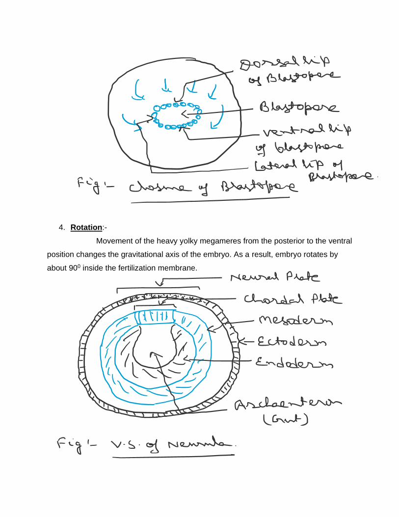
4. Rotation:-
Movement of the heavy yolky megameres from the posterior to the ventral
position changes the gravitational axis of the embryo. As a result, embryo rotates by
about 900 inside the fertilization membrane.

Now the embryo is called gastrula because it has three primary germinal
layers i. e. ectoderm, mesoderm and endoderm.
Neurulation
The formation and closure of the neural plate is called neurulation.
The embryonic stage which possesses neural plate is called neurula.
After the closure of blastopore, neural plate situated along the mid – dorsal axis of the
embryo becomes differentiated from the rest of ectoderm. The cells of neural plate
becomes thick and elongated. The margins of the neural plate become thickened and
elevated above the general level as neural folds or, crest. The midline of the neural
plate is depressed, forming a shallow groove along the length of the body. Later, the
neural folds become higher and meet dorsally in the median line forming a tube which
subsequently separates from the parent ectoderm. This is the neural tube whose
anterior portion is swallowed to form brain with cavities and the posterior part develops
into spinal cord.

A certain number of the cells of neural plate fails to associate with neural tube.
These cells are called neural crest cells. These cells give rise to the branchial skeleton,
dorsal root ganglia, sensory nerves, sympathetic nervous systemand medullary part of
adrenal gland.
Formation of notochordal splits
The median – dorsal region of the mesoderm segregates from its lateral parts
lying on both sides. As the chordal plate is separated from the mesoderm to form
notochord. It is located just below to neural tube.

Differentiation of mesoderm and
formation of coelom
The dorsal part of the mesoderm lying on the sides of the notochord becomes
subdivided into a series of mesodermal segments called somites. There are three
distinct regions :-
1. A dorsal epimere :- It is further subdivided into three parts
i) Dermatome :- It gives rise dermis of skin.
ii) Myotome:- It gives rise muscular tissue.
iii) Sclerotome – It gives rise appendicular skeleton.
2. An intermediate mesomere :- It is further divided into
i) Neprotome – It gives rise nephron of kidney.
ii) Genital ridges :- It gives rise gonads.
3. A ventral hypomere :-
It splits into two layers an external applied to ectoderm known as the
parietal or, somatic layer. An internal layer applied to the endoderm called
visceral or, splanchnic layer. The narrow space between these two layers is

called splanchnocoel. In later stage the cavity expands and become the body
cavity or, coelom of the adult animal.