development of an optical immunosensor based on the fluorescence of cyanine-5 for veterinarian...
TRANSCRIPT

Biotechnology Letters 26: 993–997, 2004.© 2004 Kluwer Academic Publishers. Printed in the Netherlands.
993
Development of an optical immunosensor based on the fluorescence ofCyanine-5 for veterinarian diagnostics
Marta Silva1, Helder Cruz1,2, Osvaldo Rossetti3, Alicia Arese3 & Abel Oliva1,∗1Biosensors Laboratory, IBET/ITQB- Instituto de Biologia Experimental e Tecnologica/Instituto de TecnologiaQuımica e Biologica, Apartado 12, P-2781-901 Oeiras, Portugal2 Also: ECBio – R&D in Biotechnology, Apartado 98, 2781-901 Oeiras, Portugal3Instituto de Biotecnologia, Instituto Nacional de Tecnologia Agropecuaria, Los Reseros y Las Cabañas, 1712Castelas, Buenos Aires, Argentina∗Author for correspondence (Fax: +351-214421161; E-mail: [email protected])
Received 19 March 2004; Revisions requested 29 March 2004; Revisions received 13 April 2004; Accepted 13 March 2004
Key words: Brucella sp. diagnostics, controlled pore glass, immobilization, optical biosensor, photobleaching
Abstract
A model optical immunosensor was developed to quantify an antibody present in a sample by measuring thefluorescence of Cyanine-5 conjugated with the antibody, using a competitive and a sandwich immunoreactionconfiguration, with the antigen immobilised in controlled pore glass beads. At pH 2, 94% of the antigen-antibodycomplex was dissociated, allowing reutilisation. Photobleaching had no effect on the fluorescence. This modelsystem was used to detect Brucella sp. infection and could quantify anti-Brucella sp. antibodies in ovine serumsamples in the range from 0.005 to 0.11 mg ml−1.
Introduction
For veterinarian diagnostics, the identification of an-imal diseases is often based on immunological tech-niques, including immunofluorescence and ELISA.However, these methods are often expensive. Simplerantibody-based diagnostics can be developed, whichcombine the specificity and sensitivity of laboratory-based immunoassays with higher versatility, with re-spect to portability and reusability, and low cost (Cruzet al. 2002, Oliva et al. 2001). In the present work,a model system to be used in veterinarian diagnosticswas designed to allow the detection of different anti-gens or antibodies present in serum samples by usingdifferent immunoreagents. Assays were first carriedout in 96 well plates to study and optimise immunore-action. After that, the assays were transposed to andoptimised in the fibre optics immunosensor.
As a model application, a competitive optical im-munosensor was developed using an antibody (goatIgG) immobilised in controlled pore glass (CPG). Thesample IgG was detected by fluorescence measure-ment using an optical fibre, after the competition in the
sensor’s flow cell of the sample IgG with a monoclonalmouse anti-goat IgG-Cyanine-5 (IgG-Cy5).
As a first application, a sandwich immunoreactionto detect anti-Brucella sp. antibodies in infected anim-als was used with an ovine serum sample infected withBrucella sp. For this assay a recombinant antigenicprotein from Brucella sp., BP26, was immobilisedinto CPG. This immobilisation support, isothiocy-anate derivatised CPG, is composed of translucentporous beads where protein amine groups bind co-valently, and thus stably, presenting a large specificarea and showing high efficiency and capacity for pro-tein immobilisation (Stabel et al. 1992, Zhou & Cass1991).
Materials and methods
Goat IgG immobilisation in controlled pore glass(CPG)
The immobilisation of goat IgG in CPG (isothiocy-anato glass, average pore of 70 nm) was performed

994
following a procedure developed at Biosensors Labor-atory (Rosa et al. 2002).
Regeneration experiment
Eleven µl of CPG with GIgG were added to each wellof a 96 well plate and blocked with 200 µl 5% (v/v)skim milk containing 80% bovine serum in PBST(0.05% Tween 20 in PBS) for 1 h at 37 ◦C. Afterwashing, 50 µl monoclonal anti-goat IgG (MAGIgG),diluted to 10 µg ml−1 in dilution buffer (0.1% bovineserum albumin in Tris buffer saline pH 7.5), were ad-ded to each well, except in the negative control wherePBS was used. After incubation for 1 h at 37 ◦C andwashing, the plate was incubated 1 h at 37 ◦C with200 µl per well of different elution buffers: i) 30% di-oxane pH 2; ii) 30% dioxane pH 12; iii) glycine buffer(0.1 M pH 1.8); iv) glycine buffer (0.1 M pH 11.7);v) phosphate buffer (7.5 mM Na2HPO4, 2.5 mM
NaH2PO4, pH 2); vi) phosphate buffer (7.5 mM
Na2HPO4, 2.5 mM NaH2PO4, pH 12), vii) phosphatebuffer (15 mM Na2HPO4, 5 mM NaH2PO4, pH 2)and viii) citrate buffer (0.1 M, pH 2.1). Absorbance ofthe supernatants was measured at 405 nm after usinganti-mouse IgG alkaline phosphatase conjugate andp-nitrophenyl phosphate as substrate.
Conjugation of Cyanine-5C (Cy5) with MAGIgG
The conjugation procedure used followed the Fluoro-Link Cy5 reactive dye 5-pack protocol from Amer-sham Pharmacia Biotech. One ml of the antibody to beconjugated (monoclonal anti-goat IgG) at 1 mg ml−1
in 0.1 M sodium carbonate buffer pH 9.3 was added tothe dye vial. After conjugation, the labelled antibodywas separated from the excess unconjugated dye bygel permeation chromatography.
Optical immunosensor set-up
The immunosensor system was configured as a distal-phase optode (Figure 1). A multimode optical fibre(Thorlabs) guides the excitation radiation from a HeNelaser into the flowcell, illuminating the CPG where theantibody is immobilised and the immunoreaction takesplace. From the side (at 90◦) of the flowcell wall, thefluorescent radiation emitted by Cy5 as a consequenceof the laser excitation is coupled to a fibre bundle thatguides the optical signal to the detector system (OceanOptics) that monitors the spectral profile of the radi-ation obtained. The measured signal is then processed
Fig. 1. Scheme of the optical immunosensor set up.
by a dedicated software for data acquisition and quan-tification and stored in a computer. The glass flowcell,built in-house, has a total volume of 300 µl to whichwas added 15 µl CPG with immobilised GIgG.
Saturation of GIgG immobilised in CPG withMAGIgG-Cy5
Sequential additions of MAGIgG-Cy5 were per-formed in the flowcell to 15 µl GIgG immobilised inCPG. A sample with 0.015 mg ml−1 of MAGIgG-Cy5 was introduced into the flowcell and incubatedfor 30 min. After incubation, the flowcell was washedwith 2 ml PBST and 1 ml PBS and the fluorescencewas measured, being this process repeated until themeasured fluorescence did not increase.
Photobleaching studies
The photobleaching study was carried out for rabbitanti-mouse IgG-Cy5 (AMIgG -Cy5) at 1 × 10⁻⁷ M inPBS under three different binding conditions: i) free insolution; ii) mixed with 15 µl of CPG; and iii) boundto mouse IgG (MIgG) immobilised in CPG. Sampleswere irradiated during 5 h with the laser and the totalenergy, measured at the CPG-optical fibre interface,was of 3.5 mW (Thorlabs).
Competitive immunoreaction
The calibration curve was performed with MAGIgG-Cy5 and MAGIgG diluted in 0.1% BSA in TBS.Fifteen µl CPG containing immobilised GIgG andblocked with 5% (v/v) skim milk containing 80%(v/v) bovine serum in PBST were introduced into the

995
Fig. 2. Regeneration experiment using different elution buffers: (1)30% dioxane, pH 2; (2) 30% dioxane, pH 12; (3) 0.1 M glycinebuffer, pH 1.8; (4) 0.1 M glycine buffer, pH 11.7; (5) 10 mM phos-phate buffer, pH 2; (6) 10 mM phosphate buffer, pH 12; (7) 20 mM
phosphate buffer, pH 2; (8) 0.1 M citrate buffer, pH 2.12.
flowcell. In the sequential competitive configuration,the flowcell was incubated with 0, 0.03, 0.055 and0.11 mg ml−1 of MAGIgG for 60 min, washed andincubated for 60 min with 0.11 mg ml−1 of MAGIgG-Cy5. After washing, the fluorescence was measuredat 670 nm. In the simultaneous competitive configura-tion, the flowcell was incubated with 0.11 mg ml−1
of MAGIgG-Cy5 mixed with 0, 0.03, 0.055 and0.11 mg ml−1 of MAGIgG.
BP26 immobilisation in CPG
The Brucella sp. antigenic protein BP26, produced re-combinantly in E. coli was obtained from Instituto deBiotecnologia/Instituto Nacional de Tecnologia Agro-pecuaria, Buenos Aires, Argentina. The method usedto immobilize the BP26 in CPG was the same methoddescribed before for GIgG immobilization in CPG.
Sandwich immunoreaction
For the sandwich immunoreaction, a calibration curvewas performed with ovine serum samples from an-imals infected with Brucella sp. and MAGIgG-Cy5diluted in 0.1% BSA in TBS. Fifteen µl CPG with im-mobilised BP26, blocked with 30% (v/v) skim milk inPBST, were introduced into the flowcell and incubatedfor 30 min with the serum sample diluted 1:200. Afterincubation and washing, a sample with 0.005 mg ml−1
of MAGIgG-Cy5 was added, incubated for 30 minand, after washing, the fluorescence was measured at670 nm. This procedure was used for all points of thecalibration curve.
Results and discussion
The experimental studies were performed in twophases: first, the immunoreaction was designed, testedand optimized in 96 well plates containing CPG,where different conditions were tested simultaneouslyin a faster and more economical way; then, the optimalconditions found in 96 well plates were transposed tothe immunosensor.
Plate assays
Antibody immobilisation in CPG and regenerationexperimentsThe obtained 30 mgGigG/gCPG saturation capacity ofthe CPG for the immobilisation of GIgG was a some-what lower value than the 57 mgcd1/gCPG obtainedin the immobilisation of a nitrite reductase (cd1) alsoin CPG (Rosa et al. 2002), but much higher thanthe 1 mgIgG/gCPG obtained in the immobilisation ofanti-IgG-FITC in CPG (Vidal et al. 2000).
For biosensor reutilisation, it is necessary to regen-erate the antibody-antigen binding (Wijesuriya 1994).As shown in Figure 2, the acidic buffers were moreefficient to remove the bound antigen from the anti-body, being 20 mM phosphate buffer pH 2 the mosteffective. The incubation time in that buffer necessaryfor complete dissociation was found to be 20 min andafter four consecutive cycles of washing and bindingof the antigen, the regenerated fraction of immobilisedantibody yielded 95% in fluorescence per regenerationcycle.
Immunosensor assays
Saturation of GIgG immobilised in CPG withMAGIgG-Cy5As can be observed in Figure 3, the concentrationof bound MAGIgG-Cy5, given by the fluorescenceintensity at 670 nm, increased with increasing con-centration of incubated antibody until 0.11 mg ml−1,and then tends to saturation. Since 0.11 mg ml−1 ofMAGIgG-Cy5 was enough to saturate all availablemolecules of GIgG immobilised in the CPG presentin the flowcell, that concentration was selected for usein the subsequent work.

996
Fig. 3. Saturation curve obtained by sequential additions ofMAGIgG-Cy5 to the GIgG immobilised in CPG.
Fig. 4. Photobleaching of AMIgG-Cy5 at 10−7 M: i) free in solu-tion (solid circle); ii) mixed with CPG (solid square); iii) bound tomouse IgG immobilised in CPG (solid triangles).
PhotobleachingPhotobleaching, a specific characteristic of a fluores-cent probe (Dittrich & Schwille 2001) is characterizedby a decrease in fluorescence signal. As can be ob-served in Figure 4, the prolonged exposure of theconjugated antibody AMIgG-Cy5 to the laser light ledto photobleaching, leading to an overestimation of theantibody present in a sample. The results patent inTable 1 show that AMIgG-Cy5 in solution has a decayhalftime of 57 min, while AMIgG-Cy5 mixed withfree CPG has 67 min and AMIgG-Cy5 bound to MIgGimmobilised in CPG has 166 min. The longer decayhalftime when AMIgG-Cy5 is bound to MIgG immob-ilised in CPG is due to the protective effect of CPG
Table 1. Photobleaching: fluorescence decay halftime ofAMIgG-Cy5 at 10−7 M at different binding conditions.
Condition Half-time (min)
AMIgG-Cy5 free in solution 57
AMIgG-Cy5 mixed with CPG 67
AMIgG-Cy5 bound to mouse
IgG immobilised in CPG 166
(Holden & Cremer 2003), which contains relativelyfew electron-rich sites.
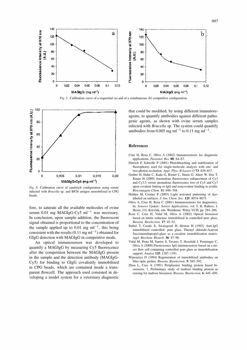
Competitive immunoreactionFigures 5a and 5b show the calibration curves of MA-GIgG using a sequential and a simultaneous competit-ive configuration, respectively, being both calibrationcurves obtained by measuring fluorescence signal ofMAGIgG-Cy5. The difference between the initial andthe final fluorescence signals, corresponds to the totalMAGIgG bound to the immobilised GIgG, i.e. theamount of MAGIgG bound to GIgG was inverselyproportional to the amount of bound MAGIgG-Cy5remaining in the immunosensor’s flowcell. Figure 5ashows the calibration curve of MAGIgG when a se-quential configuration was adopted. The capacity ofbinding to GIgG immobilised in CPG was the same,with the increase in MAGIgG concentration leadingto a decrease in the fluorescence signal. When a sim-ultaneous configuration was adopted, as shown inFigure 5b, the calibration curve shows exponential de-cay behaviour. After analyzing the results, a decreasein the affinity of MAGIgG-Cy5 in comparison to MA-GIgG was observed. Usually the fluorescent label isstatically coupled to the lysine residues of the target-specific antibodies (Gruber et al. 2000). From Figure 5it is possible to conclude that the immunosensor usinga competitive immunoreaction, either in sequential orin simultaneous mode, quantifies the sample antibody(MAGIgG) in concentrations up to 0.11 mg ml−1.
Sandwich immunoreactionThis study was performed in the flowcell using a sand-wich configuration of the immunoreaction with Bru-cella sp. antigen BP26. Figure 6 shows the calibrationcurve of positive ovine serum sample when detectedwith MAGIgG-Cy5. The results show that the positiveovine serum binds to the recombinant protein, BP26,immobilised in CPG and after incubation of 20 µlsample, all molecules of BP26 were occupied. There-
997
Fig. 5. Calibration curve of a sequential (a) and of a simultaneous (b) competitive configuration.
Fig. 6. Calibration curve of sandwich configuration using seruminfected with Brucella sp. and BP26 antigen immobilised in CPGbeads.
fore, to saturate all the available molecules of ovineserum 0.01 mg MAGIgG-Cy5 ml−1 was necessary.In conclusion, upon sample addition, the fluorescentsignal obtained is proportional to the concentration ofthe sample applied up to 0.01 mg ml−1, this beingconsistent with the results (0.11 mg ml−1) obtained forGIgG detection with MAGIgG in competitive mode.
An optical immunosensor was developed toquantify a MAGIgG by measuring Cy5 fluorescenceafter the competition between the MAGIgG presentin the sample and the detection antibody (MAGIgG-Cy5) for binding to GIgG covalently immobilisedin CPG beads, which are contained inside a trans-parent flowcell. The approach used consisted in de-veloping a model system for a veterinary diagnostic
that could be modified, by using different immunore-agents, to quantify antibodies against different patho-genic agents, as shown with ovine serum samplesinfected with Brucella sp. The system could quantifyantibodies from 0.005 mg ml−1 to 0.11 mg ml−1.
References
Cruz H, Rosa C, Oliva A (2002) Immunosensors for diagnosticapplications. Parasitol. Res. 88: S4–S7.
Dittrich P, Schwille P (2001) Photobleaching and stabilization offluorophores used for single-molecule analysis with one- andtwo-photon excitation. Appl. Phys. B-Lasers O 73: 829–837.
Gruber H, Hahn C, Kada G, Riener C, Harás G, Ahrer W, Dax T,Knaus H (2000) Anomalous fluorescence enhancement of Cy3and Cy3.5 versus anomalous fluorescence loss of Cy5 and Cy7upon covalent linking to IgG and noncovalent binding to avidin.Bioconjugate Chem. 11: 696–704.
Holden M, Cremer P (2003) Light activated patterning of dye-labeled on surfaces. J. Am. Chem. Soc. 125: 8074–8075.
Oliva A, Cruz H, Rosa C (2001) Immunosensors for diagnostics.In: Sensors Update: Sensor Applications, vol. 9, H. Baltess, J.Hesse, J.G. Korvink, eds. Weinheim: Wiley-VCH, pp. 284–286.
Rosa C, Cruz H, Vidal M, Oliva A (2002) Optical biosensorbased on nitrite reductase immobilised in controlled pore glass.Biosens. Bioelectron. 17: 45–52.
Stabel T, Casale E, Swaisgood H, Horton H (1992) Anti-IgGimmobilized controlled- pore glass. Thionyl chloride-ActivedSuccinamidopropyl-glass as a covalent immobilization matrix.Appl. Biochem. Biotech. 36: 87–96.
Vidal M, Prata M, Santos S, Tavares T, Hossfeld J, Preininger C,Oliva A (2000) Fluorescence IgG immunosensor based on a mi-cro flow cell containing controlled pore glass as immobilisationsupport. Analyst 125: 1387–1391.
Wijesuriya D (1994) Regeneration of immobilized antibodies onfiber optic probes. Biosens. Bioelectron. 9: 585–592.
Zhou L, Cass A (1991) Periplasmic binding protein based bi-osensors. 1. Preliminary study of maltose binding protein assensing for maltose biosensor. Biosens. Bioelectron. 6: 445–450.