development of a low cost fetal heart sound monitoring ... · indicator of stress on unborn in the...
TRANSCRIPT

Vol.2, No.6, 380-389 (2009)doi:10.4236/jbise.2009.26055
SciRes Copyright © 2009 Openly accessible at http://www.scirp.org/journal/JBISE/
JBiSE
Development of a low cost fetal heart sound monitoring system for home care application
Arun Kumar Mittra1, Nitin K. Choudhari2 1MIET, Gondia: Department of Electronics Engineering, Manoharbhai Patel Institute of Engineering and Technology, Gondia (MS), India; 2Smt. Bhugwati College of Engineering, Nagpur, India. Email: [email protected] Received 6 January 2008; revised 26 April 2009; accepted 26 June 2009.
ABSTRACT Variations in fetal heart rate (FHR) is a potential indicator of stress on unborn in the womb of mother. In hospitals, FHR surveillance is per-formed by ultrasound based Doppler equip-ments. However, recent studies show that fre-quent exposure to ultrasound radiations is not recommended for the fetal well-being. Because of this and many other reasons, these instru-ments are not recommended for prolonged home monitoring applications. This work is fo-cused around development of a prototype sys-tem for fetal home monitoring application. Pre-sented system can record the abnormal FHR and alert the pregnant women to report to a physician. Recorded data is then processed by a novel methodology for deriving results of di-agnostic importance. The instrument has been tested on pregnant women in the clinical envi-ronment and has gone through an extensive clinical trial at local hospitals. The results show that the technique is suitable and effective for long-term FHR home monitoring application. Keywords: Ambulatory Monitoring; Phonocardio-graphy; Cardiography; Simulation; Signal Processing 1. INTRODUCTION
To diagnose pre-term labor, ambulatory monitoring for abnormal FHR has proven to be an effective method [1]. Abnormality in fetal heart rate (FHR) is an indicator of pre-maturity and miscarriage. It is very important to monitor, such abnormalities in pregnant women, which are at high risk, with history of miscarriage. These ab-normalities are unpredictable and may occur at any time, especially in the case of pre-term labor. The pathogene-sis of pre-term labor is still poorly understood, however, the unusual occurrence of pre-maturity and miscarriage can be largely prevented by the timely diagnosis of
pre-term labor and its arrest with tocolytic medication. If the unborn heart rate increases very high or drops to a very low, it calls for urgent attention. In both the cases, it is obvious that the baby is in stress and special urgent medical attention is needed. For this reason, the electronic monitoring of the FHR has become one of the most fa-miliar methods used in the antenatal period [2,3]. The ultrasound based Doppler instruments are widely used for this purpose in hospitals, but for varied reasons, they are not suitable for home monitoring application and long-term surveillance of unborn [4,5]. It is imperative to note that these instruments are also invasive in nature.
There is still a gap between existing technologies and the user requirement for safe, convenient, and reliable fetal monitoring [6,7]. In view of these considerations, a strong need is felt for the development of a FHR moni-toring machine which will be non-invasive, cost effec-tive, simple to operate and which can be used by a preg-nant woman for prolonged home monitoring application [8]. Preliminary experiments in this regard have been conducted [9,10] and a cost effective prototype is de-veloped for at home long term FHR recording and monitoring. This instrument can record fetal heart sound for hours in a standard MP3 format. In case of abnormal symptoms, the subject visits the physician where the recorded data can be analyzed with the help of associ-ated software and computer. The audiovisual display of fetal heart sound will provide valuable additional infor-mation to the physician for diagnosis and treatment.
The basic technique used in the presented instrument is called fetal Phonocardiography (fPCG) [11,12]. In this technique a specially designed microphone is placed on the abdomen of the subject, which primarily detects and records the fetal heart sound. However, phonocardio-graphy is extremely susceptible to ambient noise [13,14, 15]. Unfortunately, the fetal heart activity produces much less acoustic energy and moreover it is surrounded by highly noisy environment. This noise has a direct consequence on the signal that often changes remarkably from one beat to next, with the additional characteristic
A. K. Mittra et al. / J. Biomedical Science and Engineering 2 (2009) 380-389
SciRes Copyright © 2009 Openly accessible at http://www.scirp.org/journal/JBISE/
381
of poor signal to noise ratio (SNR). These unwanted disturbance signals contribute an additional difficulty in the detection of the principal fetal heart sounds (S1 and S2). Hence it is necessary to develop and evaluate sig-nal-processing technique to improve SNR before ob-taining reliable time references of the fetal heart sound signal [16]. In order to improve SNR, and accomplish an optimal external noise cancellation performance, phono- cardiographic devices need a very superior and advanced noise reducer in the preprocessing stage of the instru-ment. In this work a very effective noise reducer is de-signed and used in the presented prototype system. The design is implemented with help of Matlab Simulink and intensive tests are carried out in order to evaluate the performance of resultant method. The instrument has been found effective from 30th week to final term of gestation with satisfactory level of sensitivity.
The paper is organized as follows: The next section discusses the hardware of the presented system, used for the detection and recording of fetal heart sound and am-bient noise. The following section, describes software and different signal processing techniques used by the system. The article then presents a comprehensive com-puter simulation. The last section deals with experimen-tal and clinical trial results with conclusions.
2. SYSTEM DESCRIPTION
The Fetal heart sound monitoring system presented in this study, primarily comprises of two main modules:
1) Detection and Recording Module (DRM). 2) Processing and Display Module (PDM). The DRM is a small hardware, which is placed on the
subject’s abdomen for detection and recording of the fetus heart sound signal. This module records the ab-normal FHR and alerts pregnant woman, that it is time to seek some medical attention.
The PDM is software, developed for physician’s per-sonal computer. When the subject approaches the physi-cian along with DRM, recorded data is down loaded and then processed with the use of PDM, to generate the results of diagnostic importance.
2.1. Detection and Recording Module (DRM)
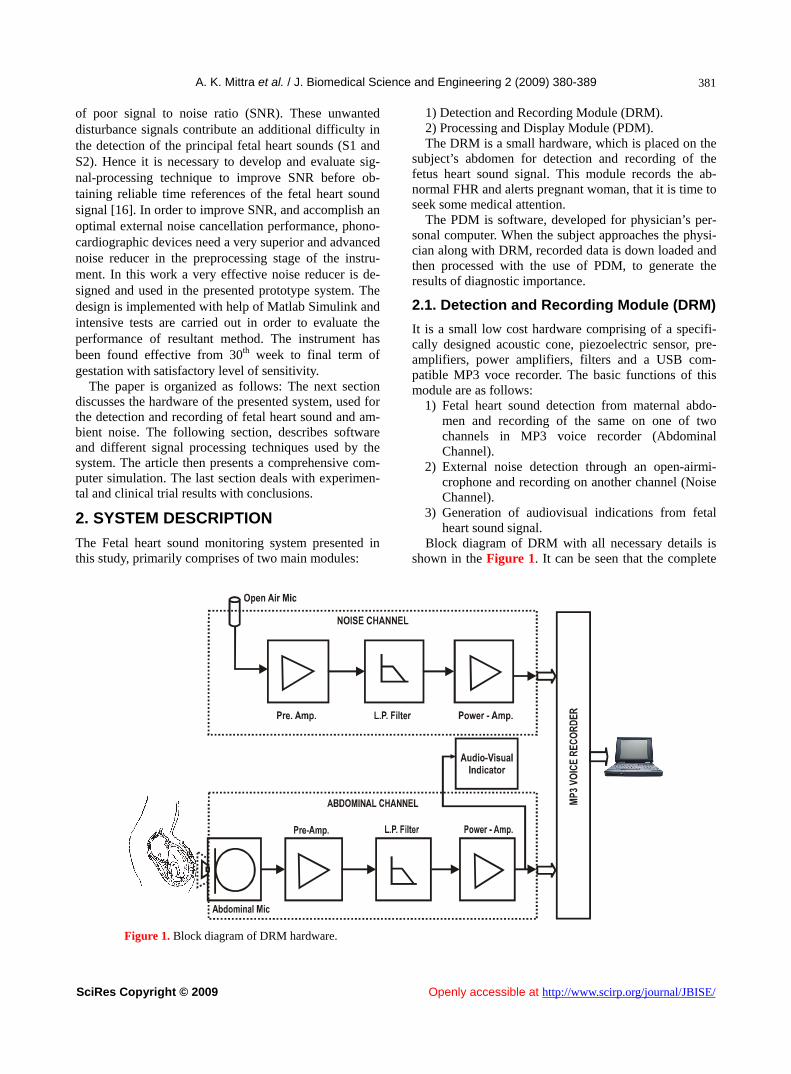
It is a small low cost hardware comprising of a specifi-cally designed acoustic cone, piezoelectric sensor, pre- amplifiers, power amplifiers, filters and a USB com- patible MP3 voce recorder. The basic functions of this module are as follows:
1) Fetal heart sound detection from maternal abdo-men and recording of the same on one of two channels in MP3 voice recorder (Abdominal Channel).
2) External noise detection through an open-airmi-crophone and recording on another channel (Noise Channel).
3) Generation of audiovisual indications from fetal heart sound signal.
Block diagram of DRM with all necessary details is shown in the Figure 1. It can be seen that the complete
Figure 1. Block diagram of DRM hardware.

A. K. Mittra et al. / J. Biomedical Science and Engineering 2 (2009) 380-389
SciRes Copyright © 2009 Openly accessible at http://www.scirp.org/journal/JBISE/
382
system is divided in two basic sections: a) Abdominal Channel b) Noise Channel.
a) Abdominal Channel: Fetal phonocardiography re-quires the conversion of mechanical vibration on sub-ject's abdomen to electrical signal by microphone. Fetus heart sound is extremely weak hence it cannot be sensed properly by putting a sensor immediately on the subject's abdomen. To overcome this problem a particular acous-tic cone is developed which is a direct extension of the chest piece of standard stethoscope. The air enclosed in the cone acts as a transmission media between the mem-brane and electro-mechanical transducer device. The output of the sensor is fed to pre-amplifier for high am-plification and better noise rejection. IC LM 381 is used here for this particular purpose, which raises the signal from the transducer level to the line level.
It is essential to keep ambient noise as low as possible; this is carried out by an active low pass filter with cut-off frequency of 200 Hz. This value of cut off fre-quency is selected, because most of the fetus heart sound spectrum lies below this frequency limit. Active filter is implemented using an easily available operational am-plifier IC 741 along with suitable resistor capacitor net-work. IC TBA810 based power amplifier further streng- thens the output signal from filter, and provides audible fetus heart sound. It is then recorded on any one channel of MP3 voice-recorder. This amplifier further provides driving power to audio-visual indicator of the DRM de-vice.
b) Noise Channel: In fetal phonocardiographic meas-urement, ambient noise creates major problem at signal processing stage [17,18]. To overcome this problem, special signal processing techniques are used in this study, which require a primary sample of the noise, cre-ating disturbance in the signal of interest. To facilitate this, ambient noise is detected through an independent open-air microphone. After pre-amplification and filter-
ing, noise signals are recorded on another channel of the memory device.
It is important to note that abdominal channel micro-phone detects the sound primarily originating from the maternal abdomen, but these signals get mixed with a damped version of the external noise. The open air Noise Channel microphone detects only the ambient noise and does not contain any traces of fetal heart sound signal. Photographs of prototype experimental model are shown in the Figures 2(a) & 2(b).
2.2. Processing and Display Module (PDM)
The PDM is the software part of presented system, made available in the physician’s personal computer. When pregnant woman feels some abnormality and the DRM alerts her, she may go to the hospital with the DRM. Stored data within the device is used for further proc-essing and investigation through PDM. Block diagram of PDM with all necessary signal flow details is shown in the Figure 3.
A brief description of each signal-processing module incorporated in the block diagram is given below:
a) Data Acquisition: The output of the MP3 voice re-corder is directly fed to the Line-in of the multimedia card, which contains on board signal conditioning, ana-log to digital conversion and digital signal processing hardware. Matlab data acquisition toolbox is used to download both channels of MP3 voice recorder and to store it in two separate *.wav files. These files carry noised fetal heart sound signal and external noise sepa-rately.
b) Adaptive Filter: Adaptive filters track the dynamic nature of a system and allow elimination of unwanted part of the signal. In this study adaptive filters are used for external noise cancellation i.e. removal of external unwanted background sound signal from the fetal heart
(a) (b)
Figure 2. Photographs of prototype.

A. K. Mittra et al. / J. Biomedical Science and Engineering 2 (2009) 380-389
SciRes Copyright © 2009 Openly accessible at http://www.scirp.org/journal/JBISE/
383
Figure 3. Block diagram of PDM.
sound signal. As shown in Figure 3, desired signal d(k), the one to clean up, comes from abdominal channel, carries noise n(k) and wanted signal s(k). The Noise Channel carries only background noise n'(k), is applied as input signal x(k) of the filter. As long as the input noise to the filter n'(k) remains correlated to the un-wanted noise n(k), the adaptive filter adjusts its weights w(k) to reduce the value of the difference between y(k) and d(k), this results in elimination of external noise and a clean fetal heart signal s(k) will appear on the error port. The generalized mathematical relationship be-tween different signals and filter weights can be de-picted as:
d(k) = s(k) + n(k) y(k) = Filter{x(k), w(k)}
or y(k) = w(k) . n’(k) e(k) = d(k) – y(k)
= s(k) + n(k) – w(k) . n’(k) s(k)
w(k+1) = w(k) + e(k) x(k)
Notice that in this implementation, the error signal actually converges to the input data signal, rather than converging to zero.
c) Band Pass Filter: Adaptive filtering of recorded signal removes only the external noise from the compos-ite abdominal signal. A band pass digital filter is de-signed to limit the signal in 35 Hz < f < 200 Hz fre-quency band. The selection of lower frequency limit is based on the experimental result, that a considerable part of the disturbing maternal heart and digestive sound lies below this border, while the fetal heart sounds are not dominantly present there any more. Upper limit is set to remove maternal respiratory sound and remaining traces
of external noise signals. d) Phonocardiogram: Fetal Phonocardiograph is a
time versus amplitude plot of fetal heart sound and is considered as the primary time domain characteristic of the fetal heart sound signal. It is a graphical representa-tion of vibration or sound signal detected from the ma-ternal abdominal wall, caused by the contractile activity of the fetal heart. The general fPCG wave pattern of the signal over cardiac cycles may be readily appreciated by visual inspection and can be used as a potential indicator of few congenital diseases.
e) Spectrogram: fPCG signals posses multiple reso-nance frequencies. This leads towards the need to de-scribe the fPCG signals, not only in terms of time but also in terms of frequency domain, also known as spec-trogram. It provides distribution of the signal’s energy or power over a wide band of frequencies.
f) Envelop Generation: In order to find out exact pe-riodicity of heartbeat, it is proposed to use envelop of the fetal heart sound signal. This envelope encompasses and traces the peaks of signal under consideration. In this application it is derived by the method of squaring and low pass filtering [19]. The resultant envelope signal reflects the amplitude dynamics of signal on the same time scale as of the original signal.
g) Burst Generator: After the process of envelope generation, exact positioning of amplitude burst is car-ried out by the method of thresholding and relaying. Whenever amplitude of signal envelops goes above a pre-defined limit it is relayed over as a maximum value otherwise it is taken as zero. This process results in a series of discrete pulses representing occurrences, and duration of fetal heartbeats.

A. K. Mittra et al. / J. Biomedical Science and Engineering 2 (2009) 380-389
SciRes Copyright © 2009 Openly accessible at http://www.scirp.org/journal/JBISE/
384
h) FHR Calculation: The last element of the sig-nal-processing block is for the FHR determination. This is carried out by counting numbers of discrete pulses in the signal for a pre-defined time interval.
3. SYSTEM SIMULATION
Processing and Display Module (PDM) described above is simulated using the Simulink modeling tool of Matlab version 7. The model is built by interconnecting requi-site blocks, which are available in the software library and their parameters are entered while designing them for simulation. The inputs of the simulating system are the recorded signals of DRM module, where as the out-puts are Phonocardiogram, Spectrogram and record of FHR in Beats Per Minutes (BPM). System simulation of developed PDM module is shown in Figure 4.
After data acquisition, signals are de-noised with the help of adaptive filter. Fetal heart sound signals, detected from mother's abdomen are fetched from corresponding *.wav file and applied to the desired port of the filter block. This signal carries fetal heart sound and a damped version of the external noise. The unwanted external noise is available in another *.wav file and is applied to the input port of the filter. De-noised signal comes out
through the error port of the filter block. It should be noted that the output port is not used in the simulation process; however this port provides internal feedback for error calculation in the filter block.
Adaptive filtering eliminates only the external noise. For suppressing remaining artifacts, digital band pass filter is used in the simulation. This block allows signals of the specified range only, while signals of all other frequencies are attenuated. After the band pass filtering signals are relatively less distorted and applied to the time scope block of the module. This block provides the time-domain response of the fetal heart signal, which is also called as phonocardiogram. The frequency domain response i.e. cardio-spectrogram of de-noised signal is produced through periodogram and vector scope block icons. The periodogram block computes a non-paramet-ric estimate of the spectrum. The block averages the squared magnitude of the FFT computed over windowed sections of the input and normalizes the spectral average by the square of the sum of the window samples. The vector scope block is a comprehensive display tool similar to a digital oscilloscope. It is used here to plot frequency-domain response of the de-noised fetal heart sound signal.
Figure 4. System simulation of PDM.
Figure 5. Sub-system for envelop generation.

A. K. Mittra et al. / J. Biomedical Science and Engineering 2 (2009) 380-389
SciRes Copyright © 2009 Openly accessible at http://www.scirp.org/journal/JBISE/
385
Figure 6. FHR calculator sub-system.
The simulation block diagram showing the bottom-
most blocks (refer Figure 4) are related with the FHR calculation. The first block is for envelope generation and it is a simulink subsystem as shown in Figure 5.
This sub system is based on the concept of squaring and low pass filtering. The input signal is first multiplied by itself. Squaring the signal effectively modulate the input by using itself as the carrier wave. This means half the energy of the signal is pushed towards higher fre-quencies and half is shifted towards DC. The envelope can then be obtained by keeping all the DC low fre-quency energy and eliminating the high frequency en-ergy. In this sub-system, a simple minimum-phase low pass filter is used to get rid of the high frequency energy.
Output of envelope generator is connected to the relay block (refer Figure 4). This block allows its output to switch between two threshold values. Once the relay is ON, it remains ON until the input drops below the value of the switch-off point parameter and when the relay is OFF, it remains OFF until the input exceeds the value of the switch-on point parameter. This block converts the envelope signal into a series of discrete pulses. These pulses are fed to FHR Calculator sub-subsystem. Details of this sub-system are shown in Figure 6, in which the Counter block increments an internal counter each time it receives a trigger at the click (Clk) port. A trigger sig-nal at the reset (Rst) port brings the counter to its initial state.
Counter output is converted to Beats Per Minute (BPM) and is then finally displayed on the output device.
4. EXPERIMENTAL TESTING AND RESULTS
Experimental testing is necessary to verify the reliability and performance of any system under a developmental stage. An artificial womb, which give simulated testing conditions for fetal monitoring system is very appropri-ate and useful for initial testing in comparison to actual clinical testing on pregnant women. In this work, an arti-ficial womb is prepared for simulated performance test-ing of monitoring systems under study. Through Matlab signal processing toolbox, simulated signals are gener-ated for fetal heart sound, maternal heart sound, maternal
respiratory sound and for external noise. After amplifi-cation these signals are applied to different speakers placed underneath a rubber balloon filled with water. DRM hardware under test is placed on the opposite side of the balloon. This arrangement simulates a fetal heart-beat passing through amniotic fluid to the wall of the mother's abdomen. For providing external support, the complete assembly is housed in a solid wooden tub. It is then placed in a thick glass envelop for elimination of outside noise interference. Presented system was initially tested on above-mentioned artificial womb. This was a totally subjective test, performed only to check viability of the instrument.
After satisfactory performance with artificial womb, system has been tested on the pregnant women in clini-cal environment. More than 15-fetal heart sound re-corded samples were taken from different women, who were between 36 to 40th week of singleton pregnancy. Recorded data was transferred to personal computer in *.wav file through multimedia card. Matlab Simulation discussed above was used to process and display the recorded sound from wave files. Figure 7 shows signal waveforms at various stages of the simulation, obtained from abdominal recording of a pregnant woman at 39 weeks of gestation (Subject No. 1). In these graphs, X- axis represents the time in seconds whereas Y-axis represents amplitude of signal in volts. Uppermost wave- form (Graph a) represents 2 seconds time span of repre-sentative sample of fetal heart sound, practically re-corded through the abdominal microphone.
Second waveform (Graph b) is the corresponding ex-ternal noise, recorded through external microphone. It may be noted that noise level is very high, which con-taminates the fetal heart sound to a larger extent. This representative sample of noise is used as a reference input for adaptive filters in signal processing stage of simulation. Third waveform (Graph c) describes the de-noised signal coming out from the adaptive filter. It can be observed that external noise is considerably re-duced and amplitude burst are distinctly visible in the waveform. In order to further increase the signal to noise ratio, band pass filters are used. Fourth waveform (Graph d) is the signal after band pass filtering and this

A. K. Mittra et al. / J. Biomedical Science and Engineering 2 (2009) 380-389
SciRes Copyright © 2009 Openly accessible at http://www.scirp.org/journal/JBISE/
386
Figure 7. Simulation output (a) Signals from abdominal channel (b) Signals from external channel (c) Signal after adaptive filtering (d) Signal after band pass filtering (e) Signal after envelope generator (f) Signal after thresh holding.
is the final phonocardiographic signal, which the instru-ment provides. Fifth waveform (Graph e) is signal enve-lope provided by the complex process of envelope gen-eration. This envelope is then passed through an ampli-tude thresholding process that in turn converts amplitude burst of the envelope signal into discrete pulses. The last waveform (Graph f) indicates the final processed signal,
used for the FHR calculation. It is believed that a more precise examination of
phonocardiographic signal may be useful for pre-detec-tion of intra uterine growth retardation and other abnor-malities of fetal. To facilitate this, a separate time scope block is provided in the simulation. This block provides a zoomed-in version of signal for any specified period of

A. K. Mittra et al. / J. Biomedical Science and Engineering 2 (2009) 380-389
SciRes Copyright © 2009 Openly accessible at http://www.scirp.org/journal/JBISE/
387
Figure 8. Fetal phonocardiogram.
Figure 9. Fetal spectrogram.
time. Figure 8 shows such display for a time interval of 0.8 seconds.
Phonocardiogram is the time domain response of the fetal heart sound signal. Presented system is in addition; capable of providing frequency domain response of the signal, called Spectrogram. It represents the contribution of every frequency of the spectrum to the power of over-all signal. Spectrogram of fetal heart sound for a small time window around 0.55 sec instant of previous illus-tration is shown in the Figure 9.
In order to support the performance of developed sys-tem, phonocardiogram signals recorded through proto-
type were compared with signals of simultaneously used ultrasound Doppler based instrument (Model: Coddle-Graph–L of Maestros Mediline Systems). In this com-parative experimentation following parameters were measured:
N’ = Total number of amplitude bursts detected by prototype.
N = Total number of amplitude bursts detected by ref-erence instrument.
M = Total number of missed bursts by the prototype. F = Total number of false bursts detected by the pro-
totype.

A. K. Mittra et al. / J. Biomedical Science and Engineering 2 (2009) 380-389
SciRes Copyright © 2009 Openly accessible at http://www.scirp.org/journal/JBISE/
388 Table 1. Outcome of comparative measurement.
Subject No Number of Amplitude Burst from Prototype
Number of Amplitude Burst from Reference Instrument
Number of Missed Amplitude Burst
Number of False Amplitude Burst
Performance Index %
1 260 270 10 0 96.30
2 290 286 0 4 98.60
3 274 280 6 0 97.86
4 278 284 6 0 97.89
5 310 302 0 8 97.35
6 294 298 4 0 98.66
7 290 296 0 6 97.97
8 318 324 0 6 98.15
9 292 290 2 0 99.31
10 312 302 10 0 96.69
11 266 280 0 14 95.00
12 302 306 0 4 98.69
13 286 288 0 2 99.31
14 298 304 0 6 98.03
15 314 310 4 0 98.71
16 304 308 0 4 98.70
From these data, performance of the instrument [20]
can be derived with the help of formula:
Performance Index (N M F
N
)X 100
This comparative measurement is performed on 16 pregnant women between 36th to 40th week of gestation age, and average recording duration was stayed limiting to one minute. It is observed that in most of the cases, phonocardiographic-based prototype signal quality is almost at par with the ultrasound Doppler based signals. Table 1 shows result of these measurements and calcula-tion of corresponding performance indices.
In light of these recorded values, the overall Perform-ance Index of the system is found around 97% in corre-lation of ultrasound based Doppler instrument. This per-formance value is fairly good for a prototype model and will certainly improve in commercially advanced sys-tem.
5. CONCLUSIONS
This work presents development of a very powerful, non-invasive, portable and low cost battery operated standalone fetal heart sound recording and monitoring system that can be used in prevailing home environment. The hardware of prototype model is of the size 9 X 8 X 4.4 cm and of the weighs 205 grams with 9 V alkaline
battery. Signal recorded through this prototype model are digitally processed and analyzed on a personal com-puter. Using enhanced adaptive and band pass filtering techniques, a remarkable improvement in signal to noise ratio is achieved by the system. Processed signals are finally used to produce impressive results of significant diagnostic and clinical importance. Instrument has been tested on pregnant women with varied gestational period and also validated by simultaneous measurement with a standard ultrasonic Doppler device. From the results it can be concluded that the presented system is viable and can effectively be used in the development of commer-cial phonocardiographic-based fetal home care monitor-ing system.
6. ACKNOWLEDGEMENTS
The fetal heart sound recordings were done at district government women hospital and at Ratnaparkhi Nursing Home Gondia (M.S.). The authors of this paper would also like to thank Dr. Shrish Ratnaparkhi (Gyneacologist), Dr. (Mrs.) Megha Ratnaparkhi (Obstetrician), Prof. Vijay Chourasia and Prof. (Mrs.) Vijaya Rahangdale for their kind support in carrying out observations with the help of developed proto-type instrument. Pregnant women who volunteered to participate in clinical test are also appreciated for their kind gesture.
REFERENCES
[1] M. Godinez, et al., (2003) On-line fetal heart rate moni-tor by phonocardiography, Proceedings of 25th annual
A. K. Mittra et al. / J. Biomedical Science and Engineering 2 (2009) 380-389
SciRes Copyright © 2009 Openly accessible at http://www.scirp.org/journal/JBISE/
389
international conference-IEEE, Cancun Mexico, 3141– 3144.
[2] M. Moghavvemi, et al., (2003) A non-invasive PC based measurement of fetal phonocardiography, Journal of Sensors and Actuators, 1(107), 96–103.
[3] A. K. Mittra, et al., (2007) Fetal heart rate detection and monitoring techniques: A comparative analysis and lit-erature review, Proceedings of National Conference- INVENT–2007, M. P. Institute of Engineering and Tech-nology, Gondia (M. S.), 124–140.
[4] F. Javed, et al., (2006) A signal-processing module for the analysis of heart sounds and heart murmurs, Pro-ceedings of International MEMS Conference, Singapore, 34, 1098–1105.
[5] B. H. Tan, et al., (2000) Real time analysis of fetal phonocardiograohy, Proceedings of IEEE International Conference–TENCON–2000, Kualalumpur, 2(2000), 135– 140.
[6] A. K. Mittra, et al., (2006) Design & development of PC-Based fetal heart sound monitoring system, The In-dian Journal of Information Science & Technology, 1(2), 1–8.
[7] A. K. Mittra, et al., (2004) Function analysis of sensors used in cardiotocograph: A trans-abdominal fetal heart rate and uterine contraction monitoring machine, Na- tional Conference on Sensors Technology–Gwalior, 28– 31.
[8] A. K. Mittra, et al., (2006) Functional analysis of fetus heart sound and uterous contraction monitoring machine using quality function deployment, I-Manager: Journal of Engineering and Technology, 2(2).
[9] A. K. Mittra, et al., (2006) Development of non-invasive portable fetus heart sound monitoring machine: An ex-perimental approach, The Journal of Lab Experiments, 6(2), 104–110.
[10] A. K. Mittra, et al., (2005) Improvisation in technique for trans abdominal monitoring of fetal heart rate and uterus contraction, Proceedings of national conference–BIO- CON-2005, Bharati Vidyapeeth Deemed University Pune, 25–28.
[11] V. Nigam, et al., (2004) Cardiac sound separation, Pro-ceedings of IEEE International Conference on Com-puters in Cardiology, Chicago, 497–500.
[12] C. Horvath, B. Uveges, F. Kovacs, and G. Hosszu, (2007) Application of the matching pursuit method in a fetal phonocardiographic telemedicine system, 29th Annual International Conference of the IEEE Engineering in Medicine and Biology Society, EMBS, 1892–1895.
[13] A. Jimenez-Gonzalez and C. J. James, (2008) Blind source separation to extract foetal heart sounds from noisy abdominal phonograms: A single channel method, 4th IET International Conference on Advances in Medi-cal, Signal and Information Processing, MEDSIP, 1–4.
[14] K. K. Spyridou and L. J. Hadjileontiadis, (2007) Analysis of fetal heart rate in healthy and pathological pregnancies using wavelet-based features, 29th Annual International Conference of the IEEE-Engineering in Medicine and Biology Society EMBS, 1908–1911.
[15] F. Kovacs, et al., (2000) A rule based phonocardiographic method for long term fetal heart rate monitoring, IEEE Transactions on biomedical engineering, 47(1), 124–130.
[16] M. Brusco, et al., (2004) Digital phonocardiography: A PDA-based approach, Proceedings of the 26th Annual In-ternational Conference of the IEEE EMBS, San Fran-cisco California, 1, 2299–2302.
[17] Y. M. Lee, et al., (2002) Remote heart rate monitoring system based on phonocardiography, Proceedings of Student Conference on Research and Development-IEEE, Shah Alam Malaysia, 27–30.
[18] P. Varady, (2001) Wavelet-based adaptive de-noising of phonocardiographic records, Proceedings of IEEE-23 Annual EMBS International Conference, Istanbul Turkey, 2, 1846–1849.
[19] http://www.mathworks.com/products/demos/shipping/dspblks/dspenvdet.html.
[20] E. C. Karvounis, M. G. Tsipouras, D. I. Fotiadis, and, K. K. Naka, (2007) An automated methodology for fetal heart rate extraction from the abdominal electrocardio-gram, IEEE Transactions on Information Technology in Biomedicine, 11(6), 628–638.