development and validation of a hplc-dad separation method for
TRANSCRIPT
International Journal of Applied Science and Engineering 2013. 11, 2: 195-201
Int. J. Appl. Sci. Eng., 2013. 11, 2 195
Development and Validation of a HPLC-DAD Separation Method for Determination of Bioactive Antrocin in Medicinal
Mushroom Antrodia camphorata
Madamanchi Geethangili, Yerra Koteswara Rao, and Yew-Min Tzeng*
Department of Applied Chemistry, Chaoyang University of Technology, Taichung, Taiwan
Abstract: Mushrooms are a food with high nutritional value. Antrodia camphorata (AC), is a medicinal mushroom being widely used as food dietary supplement for cancer prevention. The sesquiterpene lactone, antrocin reported as a novel dual Akt and mTOR inhibitor in metastatic breast cancer cells and, is the most potent among more than one hundred secondary metabolites isolated from AC. For the first time, this study developed and validated a simple high-performance liquid chromatography coupled with diode-array detector (HPLC-DAD) method for determination of antrocin in AC extracts. Separation of antrocin was achieved within 8 min on a J’sphere ODS-M80 C18 column using the gradient mobile phase acetonitrile and water containing 0.1% formic acid with a flow rate of 1.0 mL/min and DAD detection at 205 nm. The method produced linear response in the concentration range of 100-1600 μg/mL with a detection limit of 64.3μg/mL and a quantification limit of 194.8 µg/mL. The method was validated in terms of intra- and inter-day precision (within 6.3% and 8.9%, respectively). At the fortified levels of 200, 400, 600 and 800 µg/mL, the mean recoveries of antrocin ranged from 98.4% to 101.2%. The developed method successfully was applied for the determination of antrocin in AC preparations. Keywords: Antrocin; Antrodia camphorata extract; HPLC-DAD; method development.
* Corresponding author; e-mail: [email protected] Received 13 November 2012 © 2013 Chaoyang University of Technology, ISSN 1727-2394 Accepted 25 February 2013
1. Introduction
Medicinal mushrooms belonging to higher Basidiomycetes reported to have wide range of biological activities [1, 2]. Antrodia camphorata (AC), named “Niu-chang-chih” in Chinese is a medicinal mushroom being widely used as food dietary supplement for cancer prevention and hepatoprotection in several Asian and European countries [3]. According to ancient traditional preparations AC has been considered to be safe for clinical applications. Pharmacological researchers also revealed that it has huge range of bioactivities from anti-cancer to vasorelaxation [1, 3]. The chemistry of this medicinal mushroom has been extensively studied leading to the identification of more than hundred secondary metabolites [1, 3]. They mainly consist of terpenoids, benzenoids, lignans, benzoquinone derivatives, succinic and maleic derivatives which are unique for AC. Among these, particular attention has been directed to a sesquiterpene lactone, antrocin (Figure 1(a)). Presently this compound is reputed as the most potent that contribute AC pharmacological efficacy [4]. Antrocin has been reported as a potent antagonist in various cancer types including breast, lung, liver, and colon cancer cells, being

Madamanchi Geethangili, Yerra Koteswara Rao, and Yew-Min Tzeng
196 Int. J. Appl. Sci. Eng., 2013. 11, 2
highest in metastatic breast cancer MDA-MB-231 cells (MMCs) [4]. Additionally, compared with cancer drugs doxorubicin and cisplatin, antrocin has a much better antiproliferation potency in MMCs. Antrocin efficiently suppressed the phosphorylation of Akt and its downstream effectors mTOR, GSK-3β, and NF-κB in cellular settings, thus inhibited the protein expression of anti-apoptotic Bcl-2, Bcl-xL, survivin, and their mRNA, with concomitant increase in pro-apoptotic Bax and cytosolic cytochrome c [4].
In general, the quantities of active compounds in mushrooms are dependent on intraspecies variability, environmental conditions, harvest period, and processing method [5, 6]. Besides these factors, the extraction methods used to process the mushrooms can also affect the quantities of biologically active compounds in the extract [6]. Besides the peculiar chemical properties of antrocin, its lower quantity (0.005–0.04% dw), in the fruiting bodies of AC has represented an important feature to be investigated. Reversed-phase high-performance liquid chromatography coupled with diode-array detectors (HPLC-DAD) are more frequently adopted for mushroom analysis, owing its superior separation efficiency and commercial availability of various stationary phases and packed columns [7, 8]. To the best of our knowledge, until now, there has been no published analytical method for the determination of antrocin. Thus, the objective of this study was to develop a HPLC–DAD separation method for determination of antrocin in extracts of AC. The developed method was validated for routine use also demonstrated. 2. Materials and methods 2.1. Materials
All reagents used were analytical reagent grade. HPLC grade acetonitrile and methanol (Fisher Scientific Co., Pittsburg, PA, USA) was used for preparation of the mobile phase. Water for HPLC was treated with a Milli-Q A10 purification system (Millipore, Bedford, MA, USA). Antrocin (Figure 1(a)), standard (purity > 95%) was obtained from the fruiting bodies of AC following the extraction and isolation procedures as we described previously [4]. 2.2. Equipment and chromatographic conditions
A Hitachi L-7100 series HPLC instrument (Hitachi, Tokyo, Japan) consisting of a L-7400 diode array detector (DAD) detector at 205 nm and an automatic injector with 20-µL loop were used. Chromatography separations were performed with a J’sphere ODS-M80 C18 reversed phase column (250 × 4.6 mm, 4 µm, YMC Sep. Technol., Japan). The mobile phase consisted of acetonitrile (solvent A) and water containing 0.1% formic acid (solvent B). A linear gradient program was used as follows: 80% A in the first 0 min, linearly gradient to 90% A over 20 min. The sample volume of 5 µL was injected into the HPLC system, flow-rate was set at 1.0 mL/min and the antrocin peak was recorded using DAD absorbance at 205 nm. This was followed by 10 min equilibration period prior to the injection of another sample. Experiments were performed at ambient temperature. All the operations, the acquiring and analysis of data were controlled by Chromatographic Data Station Software (Hitachi-D-7000, Japan). 2.3. Sample preparation
A stock solution was prepared by dissolving approximately 1 mg of antrocin into 1 mL of
Development and Validation of a HPLC-DAD Separation Method for Determination of Bioactive Antrocin in Medicinal Mushroom Antrodia camphorata
Int. J. Appl. Sci. Eng., 2013. 11, 2 197
ethanol. The stock standard solution was diluted with mobile phase A to obtain five calibration standards of 100, 200, 400, 800 and 1600 μg/mL of antrocin.
Air dried and powdered fruiting bodies or mycelium or commercial sample of AC (1 g) were extracted three times by refluxing with n-hexane (20 mL) for 1 h. The extraction solutions were combined to be filtered and then the n-hexane was removed under reduced pressure. This residue was reconstituted in 1.0 mL of mobile phase A and then filtered through 0.22 µm pore size polymeric PTFE filters, 5 µL was injected into the HPLC system for analysis. The identification of antrocin was performed by comparing its retention time and standard spiking with that of reference standard. 2.4. Method validation
Ethanol stock solution was diluted with mobile phase A to appropriate concentration for the construction of calibration curves. Five concentrations of antrocin solution were injected in triplicate, and the calibration curve was constructed by plotting the value of peak area versus the value of concentration of antrocin. The limit of detection (LOD) and limit of quantitation (LOQ) under the present chromatographic condition were determined at signal-to-noise ratios (S/N) of 3 and 10, respectively. Intra-day and inter-day variations were chosen to determine the precision of the developed assay. Approximately 1.0 g of the pulverized samples of AC were weighed, extracted with n-hexane and analyzed as described in sections 2.2 and 2.3. For intraday variability test, the samples were analyzed in triplicate for three times within 1 day, while for inter-day variability test, the samples were examined in triplicate for consecutive 3 days. The relative standard deviations (RSDs) of the retention time and peak area were taken as the measures of precision. The stock and working solutions were stored at 4°C.
Recovery test was used to evaluate the accuracy of the method. Accurate amount of antrocin standard was added to approximate 1.0 g of AC, and then extracted with n-hexane and analyzed as described in sections 2.2 and 2.3. The mean recovery and repeatability were calculated on three assays for the standard antrocin. 3. Results and discussion 3.1. Method development
In order to develop an efficient HPLC separation method for antrocin analysis, preliminary tests were performed with the objective to select adequate and optimal conditions. These include mobile phase selection, detection wavelength and flow rate. A standard antrocin solution at concentration level 0.5 mg/mL, reversed-phase J’sphere ODS-M80 C18 column was used throughout the analyses. At first, the use of water alone as mobile phase did not allow determination of antrocin, it was therefore necessary to use an organic modifier in the mobile phase. Then, different kinds of solvent systems, such as acetonitrile and methanol with a variety of modifiers were tested. It was observed that gradient elution with acetonitrile: 0.1% formic acid in water (acetonitrile gradient change from 80–85%) for a runtime 20 min at 234 nm, there was a noticeable antrocin peak was obtained. Therefore, a mobile phase of acetonitrile: 0.1% formic acid in water was the optimal choice for HPLC-DAD analysis of antrocin. Next, an appropriate detection wavelength was explored. The wavelength variation of the DAD detector during the analysis was based on the maximum wavelength (λmax) absorbance of antrocin. Ultraviolet (UV) and visible absorbance scanning between wavelengths ranging from 200 to 400

Madamanchi Geethangili, Yerra Koteswara Rao, and Yew-Min Tzeng
198 Int. J. Appl. Sci. Eng., 2013. 11, 2
nm showed that antrocin has absorption maxima at wavelength of 205 nm. Thus, the DAD detector was set at 205 nm at the beginning of the elution to achieve high enough sensitivity for antrocin detection, while keeping the background noise as low as possible. Using mobile phase of acetonitrile: 0.1% formic acid in water (acetonitrile gradient change from 80–85%), and the detection wavelength of 205 nm, antrocin peak observed with higher intensity and good base line separation (Figure 1(b)). In order to reduce the retention time from 8.08 min, next we tried to use the acetic acid instead of formic acid without changing the mobile phase composition. Although the retention time reduced to 7.33 min from 8.08 min, however, there was no good base line separation and peak symmetry. Therefore, in the present study acetic acid was replaced by the more volatile formic acid. Finally, different flow rates 0.6, 0.8, 1.0, 1.5 mL/min were tested and found that the flow rate of 1.0 mL/min was optimal. Thus, the final optimized conditions for antrocin analysis were: J’sphere ODS-M80 C18 (250 4.6 mm, i.d, 4 µm particle size) column by gradient elution using acetonitrile-0.1% formic acid in water (acetonitrile gradient change from 80–85%) as a mobile phase. The flow rate and detection wavelength was set at 1 mL/min and 205 nm, respectively. A typical chromatogram of the separation of antrocin under these conditions was presented in Figure 1(b).
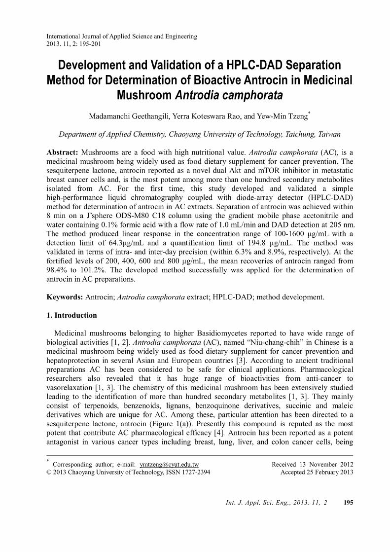
OO
H
H
Antrocin
Antrodia camphorata fruiting bodies (a)
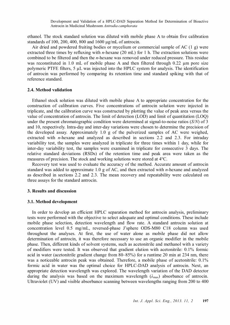
0 2 4 6 8 10 12 14 16 18 20-0.1
0.0
0.1
0.2
0.3
0.4
0.5
Antrocin
Abs
orba
nce
(AU
)
Retention time (min)
(b)
Figure 1. (a) Fruiting bodies of A. camphorata and chemical structure of antrocin. (b) HPLC-DAD chromatogram of standard antrocin. Conditions: column: J’sphere ODS-M80 C18 (250×4.6 mm i.d, 4 μm); gradient mobile phase: A-acetonitrile (ACN); B-0.1% formic acid (HCOOH) in water (gradient change 0–20 min, A: 80–85%). Flow rate of 1.0 mL/min, injection volume of 5 μL, and

Development and Validation of a HPLC-DAD Separation Method for Determination of Bioactive Antrocin in Medicinal Mushroom Antrodia camphorata
Int. J. Appl. Sci. Eng., 2013. 11, 2 199
DAD detection at 205 nm 3.2. Method validation
The developed method was validated [9], in terms of linearity, LOD and LOQ, precision, reproducibility and recovery test. Calibration curve was constructed for standard antrocin using five concentration levels. In the range of 100–1600 μg/mL, calibration curve showed good linearity with correlation coefficient (r2) of 0.9993.
The intra-day and inter-day precisions of the proposed method were determined by estimating the corresponding responses for four different concentrations (200, 400, 600 and 800 µg/mL) of antrocin five times during the same day and over three consecutive days, respectively. The intra-day precision was ≤6.3%, and inter-day precision was ≤8.9% for each level of antrocin (Table 1). The results demonstrate that the values were within the acceptable range and that the method was both accurate and precise.
Table 1. Precision and recovery of antrocin using HPLC-DAD Added
(µg/mL) Intra-day Inter-day Recovery
Detecteda RSD (%) Detecteda RSD (%) Mean (%) RSD (%) 200 202±1.3 4.7 199±2.7 5.2 99.6 4.6 400 399±0.8 3.9 401±1.2 6.7 101.2 5.3 600 596±8.6 6.3 598±7.1 7.5 99.3 3.9 800 809±11.7 5.4 803±13.2 8.9 98.4 3.5
Sensitivity of the method was based on the LOD and LOQ determined by the present
chromatographic condition. The stock solution (1 mg/mL) of antrocin was diluted to a series of appropriate concentrations with mobile phase, and an aliquot of the diluted solutions were injected into HPLC for analysis. The LOD and LOQ were determined at a signal-to-noise ratio (S/N) of around 3 and 10, respectively. These values for antrocin were 64.3 μg/mL, and 194.8 μg/mL, respectively.
Accuracy of the method was confirmed by performing recovery experiments. The recovery test was done by the standard addition method. Low (200 µg/mL), medium ( 400 and 600 µg/mL) and large amounts (800 µg/mL) of antrocin was added to the n-hexane extract from fruiting bodies of AC and then analysis was done as described in sections 2.2 and 2.3. The mean recovery was counted according to the following formula: recovery (%) = (amount found−original amount)/amount spiked × 100%, and RSD (%) = (SD/mean) × 100%. The mean recovery of antrocin in the range 98.4% to 101.2% and their RSD value was less than 5.3% (Table 1).
Last, we have evaluated antrocin stability in the concentration range of 200−800 µg/mL at room temperature for 7 days prior to analysis. After 7 days, it was found that antrocin was retained in the range of 99.8 to 100.7%, when compared to freshly prepared samples (Table 2). This indicated that antrocin was quite stable over one week at room temperature.
Table 2. Stability of antrocin by HPLCa
Concentration (μg/mL) Stability SD RSD (%) 200 100.7 2.61 2.58 400 100.3 0.76 0.63 600 99.2 1.32 1.45
Madamanchi Geethangili, Yerra Koteswara Rao, and Yew-Min Tzeng
200 Int. J. Appl. Sci. Eng., 2013. 11, 2
800 99.8 0.48 0.67 3.3. Application of the method
The content of antrocin in different AC preparations was successfully determined and the results presented in Table 3. In order to obtain optimal extraction efficiency, extractable solvent was optimized. Hexane, chloroform, ethanol, methanol and water solutions were tried as the extraction solvent. In the present study, water extract does not resulted antrocin peak. The extracts of chloroform and hexane resulted higher yield of antrocin as compared with ethanol and methanol extracts. However, chloroform extract results more complexity in antrocin separation, in addition to its toxicity as compared with hexane. Therefore, hexane was chosen as the extraction solvent since the antrocin could not only efficiently be extracted but also well resolved from background. Three replicate measurements were executed for each sample. Because of the low antrocin content in AC products, its quantitation was only possible in the natural fruiting bodies of I and II (A and B in Table 3). The higher content of antrocin (0.45 mg/g) was determined in the natural fruiting bodies II sample (B in Table 3). In contrast, AC samples cultivated fruiting bodies (CFs), CFs+Chinese medicine, 1 year old cultivated primary mycelium and fermented broth could not be confirmed for antrocin in the present study (C−F in Table 3).
Table 3. Contents of antrocin in different A. camphorata samplesa
A. camphorata samplesa Antrocin mean contentb
A 0.22±0.05
B 0.45±0.16
C ND
D ND
E ND
F ND ND means=not detectable a Analyzed samples were labeled as (A) Natural fruiting bodies I. (B) Natural
fruiting bodies II. (C) Cultivated fruiting bodies (CFs). (D) CFs + Chinese medicine. (E) Cultivated primary mycelium. (F) Fermented broth.
b The average±SD of three experiments are shown in mg/g. In conclusion, in this study a HPLC–DAD method was developed for separation of antrocin.
To the best of our knowledge, this was the first description of antrocin separation in the literature. The method used a simple mobile phase composition that was easy to prepare with little or no variation. The relatively rapid run time of within 8 min allows the analysis of a large number of samples in a single day. Based on the validation results of good accuracy, repeatability and precision, the proposed method could be used as a prerequisite for quality control of A. camphorata. Acknowledgements
This study was supported by the grants from National Science Council of Taiwan (NSC

Development and Validation of a HPLC-DAD Separation Method for Determination of Bioactive Antrocin in Medicinal Mushroom Antrodia camphorata
Int. J. Appl. Sci. Eng., 2013. 11, 2 201
100-2113-M-324-001-MY3 and 100-2811-M-324-001). References [ 1] Yue, P. Y., Wong, Y. Y., Chan, T. Y., Law, C. K., Tsoi, Y. K., and Leung, K. S. 2012. Review
of biological and pharmacological activities of the endemic Taiwanese bitter medicinal mushroom, Antrodia camphorata (M. Zang et C. H. Su) Sh. H. Wu et al. (Higher Basidiomycetes). International Journal of Medicinal Mushrooms, 14: 241-256.
[ 2] Chang, S. T. and Wasser, S. P. 2012. The role of culinary-medicinal mushrooms on human welfare with a pyramid model for human health. International Journal of Medicinal Mushrooms, 14: 95-134.
[ 3] Geethangili, M. and Tzeng, Y. M. 2011. Review of pharmacological effects of Antrodia camphorata and its bioactive compounds. Evidence-Based Complementary and Alternative Medicine, ID: 212641, 17 pages.
[ 4] Rao, Y. K., Wu, A. T., Geethangili, M., Huang, M. T., Chao, W. J., Wu, C. H., Deng, W. P., Yeh, C. T., and Tzeng Y. M. 2011. Identification of antrocin from Antrodia camphorata as a selective and novel class of small molecule inhibitor of Akt/mTOR signaling in metastatic breast cancer MDA-MB-231 cells. Chemical Research in Toxicology, 24: 238-245.
[ 5] Wasser, S. P. 2011. Current findings, future trends, and unsolved problems in studies of medicinal mushrooms. Applied Microbiology and Biotechnology, 89: 1323-1332.
[ 6] Lo, H. C. and Wasser, S. P. 2011. Medicinal mushrooms for glycemic control in diabetes mellitus: history, current status, future perspectives, and unsolved problems (review). International Journal of Medicinal Mushrooms, 13: 401-426.
[ 7] Fang, S. H., Rao, Y. K., and Tzeng, Y. M. 2004. Quantitative determination, cytotoxic and inhibitory effects of trans-cinnamaldehyde from Cinnamomum osmophloeum on human cancer cell lines but not peripheral blood mononuclear cells. International Journal of Applied Science and Engineering, 2: 136-147.
[ 8] Rao, Y. K., Lin, H. Y., Wu, W. S., and Tzeng, Y. M. 2008. Evaluation of HPLC and MEKC methods for the analysis of lipopeptide antibiotic iturin A produced by Bacillus amyloliquefaciens. International Journal of Applied Science and Engineering, 6: 85-96.
[ 9] ICH. 2005. Validation of Analytical Procedures: Text and Methodology. ICH Guidelines, Q2 (R1), International Conference on Harmonisation: Geneva (www.emea.europa.eu/pdfs/human/ich/038195en.pdf).