development and identification of fully human scfv-fcs ... · ... is the most common...
TRANSCRIPT
RESEARCH ARTICLE Open Access
Development and identification of fullyhuman scFv-Fcs against StaphylococcusaureusSiji Nian, Tong Wu, Yingchun Ye, Xu Wang, Wenfeng Xu and Qing Yuan*
Abstract
Background: Staphylococcus aureus, a gram-positive pathogen, causes many human infections. Methicillin-resistantS. aureus (MRSA) is the most common drug-resistance bacteria. Nearly all MRSA bacteria are resistant to severaldrugs. Specific antibodies are the main components of the host’s humoral immunity, and play a significant role inthe process of the host’s resistance to bacterial infection.
Results: A single-chain variable fragment (scFv) library was constructed using mRNA from the peripheral bloodmononuclear cells of S. aureus infected volunteers. After the scFv library DNA was transformed into Escherichia coliTG1, ~1.7 × 10⁷ independent clones with full-length scFv inserts. The scFv library was screened by phage display forthree rounds using S. aureus as an antigen. The single clones were chosen at random and the scFvs were expressedfor enzyme-linked immunosorbent assay (ELISA) assessment. Approximately 50 % of the clones were positive withgood binding activity to S. aureus. To improve the stability of scFvs, scFv-fragment crystallizable regions (-Fcs) wereconstructed and expressed in E. coli DH5α. The expressed scFv-Fcs were purified and identified by western blot.These antibodies were further characterized and analyzed for bioactivity. The results showed that the expressionlevel and folding of scFv-Fcs induced at 25 °C without isopropyl β-D-1-thiogalactopyranoside (IPTG) were higherthan that induced at 32 °C with 1.0 mmol/L IPTG. scFv-Fcs had good bioactivity and could specifically bind with S. aureus.
Conclusion: scFv-Fcs against S. aureus were successfully constructed and are good candidates for the development offuture adjunctive therapy for severe S. aureus infections.
Keywords: Staphylococcus aureus, Single-chain variable fragment, Fragment crystallizable regions, scFv-Fc, Phage display
BackgroundStaphylococcus aureus, a gram-positive pathogen, causesan large array of diverse human infections, rangingfrom relatively minor skin and wound infections tomore serious life-threatening diseases, such as deep-tissue infections, pneumonia, and bacteremia [1, 2].Based on data from the U.S Centers for Disease Con-trol and Prevention, S. aureus infection has beendeemed the most lethal of all infectious diseases. Theemergence and spread of multidrug-resistant strainsin communities and even in hospitals are makingtherapeutic intervention increasingly difficult and ex-pensive [3–5]. Meticillin-resistant S. aureus (MRSA) is
the most common drug-resistant bacteria and causesclinical diseases such as skin infections, pneumonia,and septicemia. Nearly all MRSAs are also multidrugresistant and are not sensitive to β-lactam antibiotics,such as penicillin and cephalosporins, or to chloromy-cetin, lincomycin, aminoglycoside antibiotics, tetracyc-line antibiotics, and macrolide antibiotics. Some S.aureus are also not sensitive to vancomycin [6–9].With only a few new antibiotics in development, con-siderable interest and efforts have been directed to-ward exploring active and passive immune-mediatedtherapeutic approaches to prevent and treat staphylo-coccal infections.Specific antibodies are the main component of a host’s
humoral immunity, and play a significant role in theprocess of creating a host’s resistance to bacterial infec-tion. Antibodies can bind with the antigens of bacteria
* Correspondence: [email protected] School of Basic Medical Sciences, Sichuan medical university, Room 218,Hanguang building, No 319, Zhongshan road, Luzhou, Sichuan 646000,China
© 2016 Nian et al. Open Access This article is distributed under the terms of the Creative Commons Attribution 4.0International License (http://creativecommons.org/licenses/by/4.0/), which permits unrestricted use, distribution, andreproduction in any medium, provided you give appropriate credit to the original author(s) and the source, provide a link tothe Creative Commons license, and indicate if changes were made. The Creative Commons Public Domain Dedication waiver(http://creativecommons.org/publicdomain/zero/1.0/) applies to the data made available in this article, unless otherwise stated.
Nian et al. BMC Immunology (2016) 17:8 DOI 10.1186/s12865-016-0146-z

and kill or eliminate the bacteria through neutralization,activating complements, promoting phagocytosis, andantibody-dependent cellular cytotoxicity (ADCC). Fornearly 30 years, antibodies have gone through fourstages of development—murine antibodies, human-mouse chimeric antibodies, humanized antibodies, andfully human antibodies. Fully human antibodies are usedto reduce the rejection reaction when treating humandiseases [10, 11].Traditional monoclonal antibodies were murine. If
used to treat human diseases, the human anti-mouseantibody (HAMA) reaction would occur; therefore,fully human antibodies were perfected to treat humandiseases. A fully human antibody is prepared in oneof two ways—using transgenic mice or constructing ahuman phage single-chain variable fragment (scFv)library. The molecular weight of the fully human anti-body is too high to use phage display for selection.scFv has the property of low molecular weight, whichis suitable for phage display, and has antigen-bindingproperties; therefore, scFvs is the best block for con-structing other types of antibodies. However, scFvsare not stable and do not have the properties of frag-ment crystallizable regions (Fcs), such as ADCC, soFcs were fused with scFvs, creating scFv-Fc, to in-crease stability and recover Fc function.In this study, an scFv library was constructed using
mRNA from peripheral blood mononuclear cells(PBMCs) of volunteers infected with S. aureus and spe-cific fully human scFv-Fvs against S. aureus were devel-oped with the hope that an adjunctive therapy isdeveloped for severe S. aureus infections.
MethodsPBMCs of the five volunteers enrolled in this studywho were infected with S. aureus were used for theconstruction of a scFv library. The criterion forselecting volunteers was only that they were infectedwith S. aureus, which was confirmed by tests fromthe clinical laboratory in the first affiliated hospitalof Sichuan medical university, China. The experimentwas approved by the ethics committee of Sichuanmedical university (No. 5105025012142). All volun-teers were adults and provided written informedconsent.
Construction of the scFv libraryThe scFv library was constructed by referring to pre-vious reports by our research group [12]. Briefly,PBMCs from volunteers infected with S. aureus wereisolated by Ficoll Paque Plus (Amersham PharmaciaBiotech, Inc., Piscataway, NJ, USA) according to themanufacturer’s instructions. mRNA was extractedfrom the PBMCs using the Dynabeads mRNA
DIRECT kit (Invitrogen, USA) and was used tosynthesize full-length cDNA using the SMART cDNAlibrary construction kit (Clontech, USA). The variableregions of heavy-chain (VH) and light-chain (VL) im-munoglobulin (Ig) were amplified by four subsequentpolymerase chain reactions (PCRs) using a set ofprimers and following Qing Yuan [12] and the VH
and VL gene repertoires were linked by overlappingextension PCR [12].The phage display vector pCANTAB-5E, Escheri-
chia coli strain TG1, and helper phage M13K07(Amersham Pharmacia Biotech, Inc., Piscataway, NJ,USA) were used to create the phage library. The scFvlibrary DNA (VH-linker-VL) and phagemid pCANTAB-5Evector were digested with Sfi I and Not I. Thedigested scFv fragments were inserted into the vectorto generate a scFv-gene III fusion library using T4DNA ligase (New England BioLabs, UK) at 4.0 °Covernight. The ligated products were transformed intoE. coli TG1 by electroporation and grown at 37 °C onculture plates containing lysogeny broth (LB) mediumsupplemented with 100 μg/mL ampicillin and 2.0 %glucose. Thirty clones from the library were selectedat random and identified by PCR to estimate the pro-portion of full-length scFv clones. The PCR productswere digested by BstN I for producing a fingerprintmap to estimate the diversity of scFvs. All clones onthe plates were scraped and suspended in LB contain-ing 15 % glycerol.
Phage amplificationEighty microliters of scraped bacterial cells or frozencell suspensions under glycerol were incubated in40 mL LB containing 100 μg/mL ampicillin and2.0 % glucose until the optical density at a wave-length of 600 nm (OD600) = 0.2 while shaking at 37 °C.The bacteria were collected by centrifugation andsuspended in 40 mL LB with ampicillin without glu-cose. Approximately 6 × 10⁹ transducing unit (TU) ofhelper M13K07 (Amersham Pharmacia Biotech, Inc.,Piscataway, NJ, USA) were added to each milliliter ofcell suspension, incubated for 15 min at 37 °C with-out agitation, and incubated for another 2.0 h withagitation. Kanamycin was added to obtain a finalconcentration of 20 μg/mL, after which the cellswere incubated overnight at 32 °C. The phage wassubsequently precipitated with polyethylene glycol(PEG)-NaCl (20 % PEG, 2.5 mol/L NaCl) and resus-pended in 0.01 mol/L phosphate-buffered saline(PBS) buffer.
Affinity selectionImmunotubes were coated with ~1.0 × 10⁸ cfu/mL S.aureus in coating buffer (0.1 mol/L Na2CO3/NaHCO3,
Nian et al. BMC Immunology (2016) 17:8 Page 2 of 9
pH 9.6) and left overnight at 4.0 °C; these were then se-quentially blocked with 4.0 % fat-free milk for 1.0 h at37 °C and gentle agitation. Approximately 1 × 10¹² TUfrom the freshly amplified scFv libraries were added tothe blocked immunotubes and incubated at roomtemperature for 2.0 h. The immunotubes were rinsed 10times with 0.01 mol/L PBST (0.05 % Tween-20 in0.01 mol/L PBS) and 0.01 mol/L PBS separately, theneluted with 0.1 mol/L glycine-HCl (pH 2.2) and ampli-fied as above. In the second and third rounds of affinityselection, 1 × 10⁷ cfu/mL and 1 × 10⁶ cfu/mL bacteriaantigen, respectively, were coated and the immunotubeswere washed separately 15 to 20 times with 0.01 mol/LPBST and 0.01 mol/L PBS.
Phage enzyme-linked immunosorbent assayFollowing affinity selection of the scFv library in threerounds, individual clones were randomly chosen andgrown at 37 °C. The phage was amplified accordingto the previously described protocols. The amplifiedphage preparation (1012 TU) was blocked with 4.0 %fat-free milk in 0.01 mol/L PBS for 30 min and addedto an immunoassay plate coated with S. aureus forenzyme-linked immunosorbent assay (ELISA) and thenegative control (0.01 mol/L PBS) was set up. Plateswere incubated for 1.0 h, washed three times withwashing buffer PBST, and finally incubated withhorseradish peroxidase (HRP)-conjugated anti-M13mouse monoclonal antibody (Amersham PharmaciaBiotech, Inc., Piscataway, NJ, USA). The immunoreac-tions were developed by incubating in TMB liquidsubstrate (Sigma-Aldrich Company, Inc., St. Louis,MO, USA) for 15 min. The reactions were stoppedby adding 2.0 mol/L H2SO4. Absorbance at 450 nmwas recorded and used to select the phage thatexpressed scFv for recognizing the target antigens.
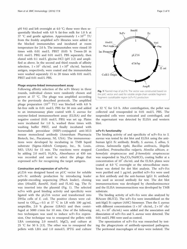
Construction and expression of scFv-FcspLZ16 was designed based on pUC vector for solublescFv-Fc antibody production by introducing leaderpeptide-encoding sequencing (PhoA leader), cloningsites (HindIII, BamHI, NotI) and FLAG tag. The Fcwas inserted into the plasmid (Fig. 1). The selectedscFvs with good binding activity and specificity wereligated with the pLZ16 vector and transformed intoDH5α cells of E. coli. The positive clones were cul-tured to OD600 = 0.5 at 37 °C in LB with 100 μg/mLampicillin, 2.0 % glucose (LBAG) and then centri-fuged at 5000 r/min for 10 min. One of the followingtwo techniques was used to induce scFv-Fcs expres-sion. One technique was to resuspend the pellets withLBA containing 2.0 mmol/L MgCl2 and culture at25 °C for 50 h [13]. The other was to resuspend thepellets with LBA and 1.0 mmol/L IPTG and culture
at 32 °C for 5.0 h. After centrifugation, the pellet wascollected and resuspended in 0.01 mol/L PBS. Thesuspended cells were sonicated and centrifuged, andthe supernatant was detected by ELISA and westernblot.
scFv-Fc functionalityThe binding activity of and specificity of scFv-Fcs to S.aureus was tested by dot blot and ELISA using the anti-human IgG1 Fc antibody. Briefly, S. aureus, S. albus, S.citreus, Salmonella typhi, Bacillus anthracis,, ShigellaCastellani, Proteusbacillus vulgaris, Monilia albican, α-hemolytic streptococcus and β-hemolytic streptococcuswas suspended in Na2CO3/NaHCO3 coating buffer at aconcentration of 107 cfu/mL and the ELSIA plates werecoated at 4.0 °C overnight or the nitrocellulose mem-brane was dotted for dot blot analysis. The scFv-Fcswere purified and 1 μg/mL purified scFv-Fcs were usedas first antibody and the anti-human IgG1 Fc antibodywas used as second antibody for test. The dot blotimmunoreactions was developed by chemiluminscenceand the ELISA immunoreactions was developed by TMBsubstrate.The binding activity of scFv-Fcs were also analyzed by
BIAcore (BLITZ). The scFv-Fcs were immobilized on theanti-hIgG Fc capture (AHC) biosensor. Then the S. aureuswith different concentration (1 × 10⁷ cfu/mL, 2 × 10⁷ cfu/mL, 4 × 10⁷ cfu/mL) were added and the association anddissociation of scFv-Fcs and S. aureus were detected. The0.01 mol/L PBS were used as control.The opsonization of scFv-Fcs was researched by test-
ing the phagocytosis of antibody-opsonized pathogens.The peritoneal macrophages of mice were isolated. The
Fig. 1 Plasmid map of pLZ16. The vector was constructed based onthe pUC vector and used for soluble single-chain variable fragment-fragment crystallizable region (scFv-Fc) production
Nian et al. BMC Immunology (2016) 17:8 Page 3 of 9

Fig. 2 Amplification of VH, Vκ, and Vλ gene repertoires. Lane M: DNA Marker II (Tiangen, CN). The expected size of the amplified VH and VL(including Vκ and Vλ) was ~350 bp. A part of amplified VH, Vκ, and Vλ were shown in Fig. 2
Fig. 3 a Thirty clones were chosen randomly from the single-chain variable fragment (scFv) library and the positive clones inserted into full scFvgenes were identified by polymerase chain reaction (PCR). Lane M: 2000 bp DNA Marker (TaKaRa, JP). Lanes 1–30: amplified scFvs from differentclones randomly picked. b PCR products of scFvs were digested by BstN I. 2000 bp DNA Marker (TaKaRa, JP). Lane M: 2000 bp DNA Marker(TaKaRa, JP). Lanes 1–30: Fingerprints of scFvs digested by BstN I
Nian et al. BMC Immunology (2016) 17:8 Page 4 of 9

macrophages and S. aureus (1 × 10⁷ cfu) was mixed atthe ratio 1:20 in RPMI-1640 medium containing 10 %fetal calf serum (FCS). Ten microliter of scFv-Fc(100 μg/mL) was added into the mixture and incu-bated at 37 °C for 2 h with gentle agitation. Themouse IgG1 was used as control and each group wasrepeat for three times. Then the 200 μL of mixturewas spread the LB plates for overnight culture. Thefollowing day, the clone numbers were calculated.
Statistical methodBackground noise correction was performed from ELISAby subtracting the absorbance. All the data were repeatedfor three times and the data sets the mean and the stand-ard error were calculated. Data were presented as means± SEM. Differences between groups were determined bythe two-tailed t-test. P < 0.05 was considered statisticallysignificant. *p < 0.05, **P < 0.01, ***P < 0.001.
Results and discussionConstruction of the scFv librarymRNA was extracted from PBMCs of volunteers in-fected S. aureus. The quality of the mRNA was the keyto synthesize the full-length cDNA. The integrity of theextracted mRNA was determined by testing the OD260/OD280 and electrophoresis. The synthesized ds cDNAappeared as a smear of from 0.1 to 8.0 kb on the gel andthe size of distribution, yield, and intensity of cDNAwere satisfied (data not shown). The full-length cDNAwas used as a template to amplify the regions of VH andVL, including Vκ and Vλ. The VH, Vκ, and Vλ gene reper-toires were amplified with 42, 16, and 18 optimized pri-mer sets, respectively. Nearly all PCR reactions wereeffective and the correct PCR products were obtainedfrom the gel picture; the expected size of the amplifiedVH and VL (including Vκ and Vλ) was ~350 bp. A part ofamplified VH, Vκ, and Vλ were shown in Fig. 2. The VH-linker gene and VL-linker gene repertories were amplified
Fig. 4 Amino acid sequences of 10 clones which were randomly picked from the library and analyzed by DNA sequence
Fig. 5 The specificity of selected single-chain variable fragment (scFvs) detected by enzyme-linked immunosorbent assay. Number 15 and 16were negative controls
Nian et al. BMC Immunology (2016) 17:8 Page 5 of 9

and the VH-linker-VL gene repertories (750 bp) were ob-tained by overlapping extension PCR. The DNA library ofhuman scFvs was developed.The DNA of the scFv library was ligated with phagemid
vector pCANTAB-5E and transformed into TG1 cells of E.coli. Thirty clones were randomly chosen and the insertedscFvs were identified by PCR. The results showed that allclones contained full-length scFv genes. By BstN I digestionof the scFv DNA, the fingerprint maps showed that almostall scFvs were different (Fig. 3); For further analysis of thediversity of the library, the 10 clones were picked randomlyfor sequencing. The results showed that the sequences ofthose scFvs were diverse (Fig. 4). The library was estimatedto contain 1.7 × 10⁷ unique scFv members.
Affinity selectionThe phage library was amplified and for each round of af-finity selection, ~1012 TU freshly amplified scFv antibodylibrary were used for screening. Approximately 1.1 × 106,3.1 × 106, and 1.2 × 10⁷ phages were recovered after the
first, second, and third cycles, respectively. Phage poolsbefore and after each round of affinity selection weretested by ELISA, which was performed by using the anti-M13 HRP-conjugated secondary antibody. The phage poolbefore affinity selection had no signal with the antigen,but after three rounds of screening, there was strong bind-ing activity between the library phage and antigen (datanot shown). Hundreds of clones were randomly chosenfrom the phage pool after three rounds of affinity selec-tion. The phages amplified from those clones were testedby ELISA and about 50 % showed positive whose absorb-ance of OD450 was two-fold those of the negative controls.
scFv specificity testFrom the hundreds of clones those with higher absorb-ance had better binding activity to the antigen and wereselected for testing scFv specificity. The S. aureus, S. albus,S. citreus, S. typhi, B. anthracis, S. castellani, P. vulgaris,M. albicans, α-H.streptococcus and β-H. streptococcus (1 ×10⁷ cfu/ml) were diluted in coating buffer and coated in
Fig. 6 a Constructed scFv-Fcs (S78, S128, S117, S182) were amplified and their sizes were ~1.5 kb. Lane M: 2000 bp DNA Marker (TaKaRa, JP).b Expression level of scFv-Fcs (S78, S128, S117, S182) expressed at 25 °C and 32 °C, respectively, were tested by enzyme-linked immunosorbent assay
Fig. 7 a The scFv-Fcs (S78, S117) expressed at 25 °C and 32 °C respectively were purified and run SDS-PAGE. Lane M: prestained protein ladder(Fermentas, USA); b The scFv-Fcs (S78, S128, S117, S182) were identified by western blot. Lane M: prestained protein ladder (Fermentas, USA)
Nian et al. BMC Immunology (2016) 17:8 Page 6 of 9
wells of ELISA plates at 4.0 °C overnight. The ELISA re-sults showed that 14 clones had good specificity with S.aureus and no binding reaction with the control bacteria(Fig. 5). Number 15 and 16 (0.01 mol/L PBS was added,not scFvs) were used as the negative controls. Each testwas repeated for three times, the value of the blank wasremoved, and the standard errors were calculated. ThescFvs with better binding activity and specificity (S78,S117, S128, S182) were inserted into the pLZ16 vectorwith human IgG1 Fc.
Construction and expression of scFv-FcsTo overcome the instability of scFvs, scFv-Fcs were con-structed. The Fc region comprises the CH2 and CH3 do-mains and the hinge region of the human IgG1. The hingeserves as a flexible spacer between the two parts of the Fc-fusion protein, allowing each part of the molecule to func-tion independently. After transformation, the clones wereidentified by PCR amplification. The results showed thatthe bands at 1.5 kb were amplified (Fig. 6a). The sequen-cing results showed that the scFv-Fcs were correct. Next,the scFv-Fcs were induced to express at 25 °C for 50 hwithout IPTG or 32 °C for 5.0 h with 1.0 mmol/L IPTG.The expressed scFv-Fcs were coated on the ELISA platesand detected with goat anti-human IgG1 Fc HRP-conjugated antibody. The results showed that OD450 ofscFv-Fcs expressed at 25 °C was higher than thatexpressed at 32 °C (Fig. 6b). The positive results of detec-tion with human IgG1 Fc HRP-conjugated antibody
showed that the Fc regions were successfully fused intoscFv. The expressed scFv-Fc (S78, S117) under the twoconditions were purified. From sodium dodecyl sulfatepolyacrylamide gel electrophoresis staining (SDS-PAGE)(Fig. 7a), the scFv-Fcs was pure and not degraded after50 h expression at lower temperatures (25 °C). The lowertemperature will degrade the aggregation of expressedprotein and stabilize the bioactivity of scFv-Fc. MgCl2 wasadded to the medium to maintain the cell wall. Later, theexpressed scFv-Fcs (S78, S117, S128, S182) at 25 °C weredetected by western blot with the anti-FLAG HRP-conjugated antibody and a single band of ~67 kDa ap-peared (Fig. 7b). Based on this, it was concluded that alower temperature was beneficial to the expression andfolding of scFv-Fcs and that the system used is suitable forthe production of Fc-fused antibody fragments.
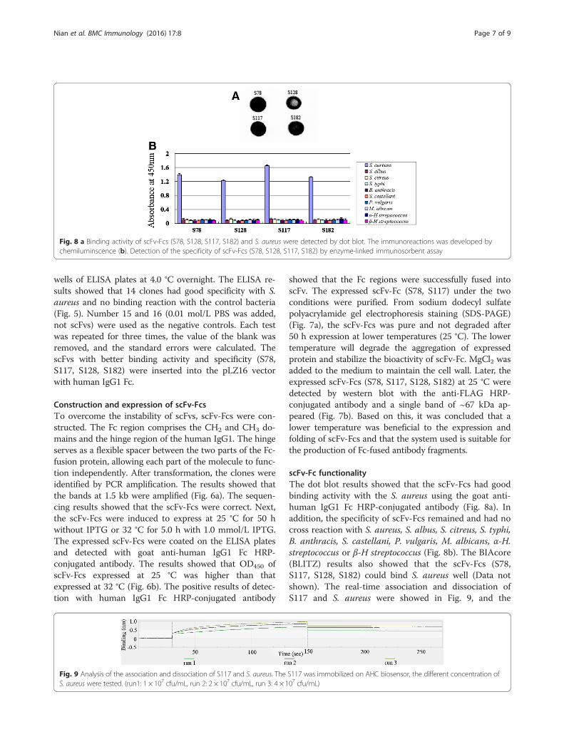
scFv-Fc functionalityThe dot blot results showed that the scFv-Fcs had goodbinding activity with the S. aureus using the goat anti-human IgG1 Fc HRP-conjugated antibody (Fig. 8a). Inaddition, the specificity of scFv-Fcs remained and had nocross reaction with S. aureus, S. albus, S. citreus, S. typhi,B. anthracis, S. castellani, P. vulgaris, M. albicans, α-H.streptococcus or β-H streptococcus (Fig. 8b). The BIAcore(BLITZ) results also showed that the scFv-Fcs (S78,S117, S128, S182) could bind S. aureus well (Data notshown). The real-time association and dissociation ofS117 and S. aureus were showed in Fig. 9, and the
Fig. 8 a Binding activity of scFv-Fcs (S78, S128, S117, S182) and S. aureus were detected by dot blot. The immunoreactions was developed bychemiluminscence (b). Detection of the specificity of scFv-Fcs (S78, S128, S117, S182) by enzyme-linked immunosorbent assay
Fig. 9 Analysis of the association and dissociation of S117 and S. aureus. The S117 was immobilized on AHC biosensor, the different concentration ofS. aureus were tested. (run1: 1 × 10⁷ cfu/mL, run 2: 2 × 10⁷ cfu/mL, run 3: 4 × 10⁷ cfu/mL)
Nian et al. BMC Immunology (2016) 17:8 Page 7 of 9

equilibrium dissociation constant (KD), the rate con-stants for association (ka) and dissociation (kd) betweenS. aureus and selected scFv-Fcs were listed in Table 1,which were determined by BIAcore (BLITZ).The opsonization of scFv-Fcs was detected. Com-
pared with mouse IgG controls, the plates spreadwith the mixture containing scFv-Fc (S78, S117) hadless clones (Table 2) and it has statistically significantfor S78 (P < 0.01) and S117 (P < 0.001). The resultsshowed the scFv-Fcs (S78, S117) against S. aureus couldplay the role of opsonization, and promoting the functionof phagocytosis of macrophage.
DiscussionCommercial murine IgG against S. aureus has been usedin research but no fully human antibody against S. aureushas been commercially available for treatment of the in-fection of S. aureus. Our scFv-Fc against S. aureus was afully human antibody, and the affinity of scFv was notevolved in vivo; therefore, after the functional scFv-Fc wasselected, the affinity of scFv-Fc will be improved by affinitymaturation in vitro and the antigenic determinant of S.aureus also will be researched. We hope to establish anadjunctive therapy for severe S. aureus infections usingthose antibodies.scFv comprised only Ig VH and VL connected by a sol-
uble and flexible oligopeptide. scFv use is easy for the de-sign and construction of a phage display antibody library,and phage display technology makes possible the directisolation of monovalent scFv antibody fragments; however,scFv has disadvantages of being unstable, having a shortserum half-life, and lacking avidity because of monovalentbinding, all of which could limit its effectiveness. Reducingthese disadvantages would be useful in making scFv moresuccessful as an antibody. The scFv-Fc format has certainproperties of IgGs, such as bivalency, tag-free detection,
and Fc-mediated effector functions. Moreover, scFv-Fcantibodies have been successfully used to neutralize viral,bacterial, and fungal pathogens in vitro and in vivo, whichsuggests its potential uses in therapy [14–16].In this study, the scFv against S aureus was screened
and merged with Fc. The scFv-Fc was expressed with thevector pLZ16 and the better expression condition was in-cubated at 25 °C for 50 h without IPTG. According toHiroyoshi (2008) [13], optimum conditions were estab-lished for bacterial cultivation and protein expression, util-izing an unusually long cultivation time (>50 h), very lowtemperature (25 °C), and no IPTG, thereby leading to theproduction and extracellular secretion of fragmentantigen-binding (Fab) proteins in a high-yield using thevector pComb43C9 with lactose promoter.Based on the report of Hiroyoshi [13], the vector
pLZ16 was designed in this study for expressing thesoluble scFv-Fc antibody. The results showed that theoverall gene expression level under the conditions of 25 °Cfor 50 h and without IPTG induction was higher than theexpression under the conditions of 32 °C for 5.0 h withIPTG. The production yield of the functional scFv-Fc at25 °C was highly dependent on the following points: (1)freshness of a single colony of the phagemid-harboring E.coli cells; (2) temperature controls, including pre-warmingof the culture medium in the inoculation steps; and (3)medium exchange to one with no glucose at the inductionstep for protein expression [13].Another study reported that auto-induction without
IPTG allows efficient screening many clones in parallelfor expression and solubility, as cultures need only to beinoculated and grown to saturation, and yields of targetprotein are typically several-fold higher than that ob-tained by conventional IPTG induction [17].The expressed and purified scFv-Fcs have good bio-
activity and specificity with S. aureus. In subsequentstudies, the antigenic determinant mechanisms of actionwill be researched.
ConclusionsIn this work, a human scFv library with a volume of1.7 × 107 was constructed. From the library, specificscFvs against S. aureus were selected using three roundsof phage display. The scFv-Fcs with stable structure de-veloped and expressed in E. coli DH5α, and the suitable
Table 1 The kinetic constants determined by BIAcore (BLITZ)analysis for scFvs
scFv ka (1/Ms) kd (1/s) kd/ka(KD, M)
S78 4.937e4 5.731e-4 1.161e-8
S128 2.123e4 8.231e-4 3.877e-8
S117 5.539e5 4.804e-4 8.673e-9
S182 2.213e4 7.648e-4 3.456e-8
Table 2 Test of the opsonization of scFv-Fcs
clones clones
S. aureus +macrophage + S78 (42 ± 5) × 104 ** S. aureus +macrophage + IgG (78 ± 7) × 104
S. aureus +macrophage + S128 (62 ± 6) × 104 S.aureus +macrophage (82 ± 8) × 104
S. aureus +macrophage + S117 (26 ± 5)104***
S. aureus +macrophage + S182 (56 ± 6) × 104
**P <0.01, ***P <0.001
Nian et al. BMC Immunology (2016) 17:8 Page 8 of 9

expression system induced at 25 °C was beneficial forthe correct folding of scFv-Fcs. The purified scFv-Fcsalso had the functionality of good binding activity andspecificity to S. aureus. These antibodies could be usedas candidates for the development of future adjunctivetherapy for severe S. aureus infections.
Availability of data and materialsConsidering the data has not been used for applyingpatent, the data will not be shared.
AbbreviationsADCC: antibody-dependent cellular cytotoxicity; ELISA: enzyme-linkedimmunosorbent assay; Fc: fragment crystallizable region; FCS: fetal calfserum; Ig: immunoglobulin; IPTG: isopropyl β-D-1-thiogalactopyranoside;HAMA: human anti-mouse antibody; MRSA: meticillin resistant Staphylococcusaureus; PBMC: peripheral blood mononuclear cell; PBS: phosphate-bufferedsaline; PCR: polymerase chain reaction; PEG: polyethylene glycol; scFv: single-chain variable fragment; VH: variable regions of heavy-chain; VL: variableregions of light-chain.
Competing interestsThe authors declare that they have no competing interest.
Authors’ contributionsSJ made the main contributions to this study, and acquired, analyzed, andinterpreted the data. TW participated in the design and coordination ofthe study and in the construction of the vector for expressing scFv-Fcs. YYparticipated in the screening of scFvs. XW constructed the scFv library. WXparticipated in the immunoassays. QY contributed to the conception anddesign of the study, the analysis and interpretation of the data, and draft-ing and revising the manuscript. All authors read and approved the finalmanuscript.
AcknowledgmentsThis work was supported by the Sichuan Youth Science and TechnologyFoundation (No. 2012JQ0018) and the Luzhou City Bureau of science andtechnology (2013LZLY-K77).
Received: 2 January 2015 Accepted: 19 April 2016
References1. Pezato R, Bottura L, de Paula SR, Voegels RL, Bachi AL, Gregório LC. Bone:
the final frontier for Staphylococcus aureus penetration in chronicrhinosinusitis. J Otolaryngol Head Neck Surg. 2013;42(1):45.
2. Yamada K, Wanchun J, Ohkura T, Murai A, Hayakawa R, Kinoshita K, MizutaniM, Okamoto A, Namikawa T, Ohta M. Detection of methicillin-resistantStaphylococcus aureus using a specific anti-PBP2a chicken IgY antibody. JpnJ Infect Dis. 2013;66(2):103–8.
3. Faghri J, Shahbazzadeh D, Pooshang Bagheri K, Moghim S, Ghase mianSafaei H, Nasr Esfahani B, Fazeli H, Yazdani R, Mirmohammad Sadeghi H.Two Dimensional Structural Analysis and Expression of a NewStaphylococcus aureus Adhesin Based Fusion Protein. Iran J Basic Med Sci.2012;15(2):725–38.
4. Liang SY, Khair HN, McDonald JR, Babcock HM, Marschall J. Daptomycinversus vancomycin for osteoarticular infections due to methicillin-resistantStaphylococcus aureus (MRSA): a nested case–control study. Eur J ClinMicrobiol Infect Dis. 2013;33(4):659–6.
5. Pulido Pérez A, Baniandrés Rodríguez O, Ceballos Rodríguez MC, MendozaCembranos MD, Campos Domínguez M, Suárez Fernández R. Skin InfectionsCaused by Community-Acquired Methicillin-Resistant Staphylococcusaureus: Clinical and Microbiological Characteristics of 11 Cases. ActasDermosifiliogr, 2013, 29. pii: S0001-7310(13)00321-9. doi: 10.1016/j.ad.2013.09.002.
6. Kali A, Stephen S, Umadevi S, Kumar S, Joseph NM, Srirangaraj S. ChangingTrends in Resistance Pattern of Methicillin Resistant Staphylococcus aureus.J Clin Diagn Res. 2013;7(9):1979–82.
7. Jurke A, Kock R, Becker K, Thole S, Hendrix R, Rossen J, Daniels-Haardt I,Friedrich A. Molecular epidemiology of meticillin-resistant Staphylococcusaureus (MRSA): think regionally but use globally uniform typing languages.Euro Surveill. 2013;18(43):20617.
8. Krakauer T, Stiles BG, Krakauer T, Stiles BG. The staphylococcal enterotoxin(SE) family: SEB and siblings. Virulence. 2013;4(7):19.
9. Salgado-Pabón W, Case-Cook LC, Schlievert PM. Molecular analysis ofstaphylococcal superantigens. Methods Mol Biol. 2014;1085:169–85.
10. Chen W, Gong R, Ying T, Prabakaran P, Zhu Z, Feng Y, Dimitrov DS.Discovery of Novel Candidate Therapeutics and Diagnostics Based onEngineered Human Antibody Domains. Curr Drug Discov Technol.2013;11(1):28–40.
11. Mandrup OA, Friis NA, Lykkemark S, Just J, Kristensen P. A novel heavydomain antibody library with functionally optimized complementaritydetermining regions. PLoS One. 2013;8(10):e76834.
12. Qing Y, Li H, Xu W, Yuchuan W, Yan G, Chengwen L, Siji N. Construction ofhuman non-immune library and selection of scFvs against IL-33. ApplBiochem Biotechnol. 2012;167:498–509.
13. Kuba H, Furukawa A, Okajima T, Furukawa K. Efficient bacterial production offunctional antibody fragments using a phagemid vector. Protein Expr Purif.2008;58:292–300.
14. Moutel SEI, Marjou A, Vielemeyer O, Nizak C, Benearoch P, Dubel S, Perez F.A multi-Fc-species system for recombinant antibody production. BMCBiotechnol. 2009;9:14.
15. Rülker T, Voß L, Thullier P, O’ Brien LM, Pelat T, Perkins SD, Langermann C,Schirrmann T, Dübel S, Marschall HJ, Hust M, Hülseweh B. Isolation andcharacterization of a human-like antibody fragment (scFv) that inactivatesVEEV in vitro and in vivo. Plos one. 2012;7:e37242.
16. West Jr AP, Galimidi RP, Gnanapragasam PNP, Bjorkman PJ. Single-chainFv-based anti-HIV proteins: potential and limitations. J Virol. 2012;86:195–202.
17. Studier William F. Protein production by auto-induction in high-densityshaking cultures. Protein Expr Purif. 2005;41:207–34.
• We accept pre-submission inquiries
• Our selector tool helps you to find the most relevant journal
• We provide round the clock customer support
• Convenient online submission
• Thorough peer review
• Inclusion in PubMed and all major indexing services
• Maximum visibility for your research
Submit your manuscript atwww.biomedcentral.com/submit
Submit your next manuscript to BioMed Central and we will help you at every step:
Nian et al. BMC Immunology (2016) 17:8 Page 9 of 9