development and disease - tohoku forum for creativity · input from the thalamus creates diversity...
TRANSCRIPT

Yasuyuki Taki Graduate School of Medicine / IDAC, Tohoku University
Tomomi ShimogoriRiken BSI
Organizers
General OrganizersToshio Iijima
Graduate School of Life Sciences, Tohoku UniversityNoriko Osumi
Graduate School of Medicine, Tohoku University
Tohoku Forum for Creativity Thematic Program 2015
Frontiers of Brain Science
August 24, Mon. ‒ 26, Wed., 2015Auditorium of the International Center for Smart Aging Research
Seiryo Campus, Tohoku University
AbstractDevelopment and Disease
Symposium on


1
Contents
Scope 2
Committees 3
Access 4
Congress Information 6
Registration 7
Social Programs 8
Time Table 9
Abstracts and Profiles
Principal Speakers 13
Short Talks and Posters 48
2
Scope of the Symposium
All of the information required to generate the intricate design of our
complex brains is present in the zygote after fertilization. These genetic and
epigenetic programs are carefully implemented during embryonic
development, during which each cell is correctly assigned its location and
function as the embryo grows. Small alterations in this seemingly impossible
task sometimes occur, and can have profound consequences which may
ultimately result in neurodevelopmental diseases. In the second symposium,
we will discuss how the program of neural development unfolds, with the
aim of understanding how errors in these processes can result in the
development of brain disease.

3
Committees
Supported by
Tohoku Forum for Creativity (TFC)
Tohoku University, Institute for Development, Aging & Cancer (IDAC)
Tohoku Brain Science Center (BSC)
Tohoku University Graduate School of Medicine, United Center for Advanced
Research & Translational Medicine (ART)
General Organizers
Toshio Iijima (Professor, Graduate School of Life Sciences, Tohoku University)
Noriko Osumi (Professor, Graduate School of Medicine, Tohoku University)
Symposium Organizers
Yasuyuki Taki (Professor, Graduate School of Medicine / IDAC, Tohoku University)
Tomomi Shimogori (Team Leader, Riken BSI)


5
Transportation
Seiryo Campus
Smart Ageing International
Research Center
Seiryo Campus
6
Congress Information
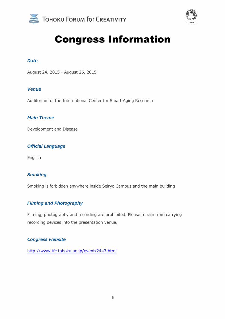
Date
August 24, 2015 - August 26, 2015
Venue
Auditorium of the International Center for Smart Aging Research
Main Theme
Development and Disease
Official Language
English
Smoking
Smoking is forbidden anywhere inside Seiryo Campus and the main building
Filming and Photography
Filming, photography and recording are prohibited. Please refrain from carrying
recording devices into the presentation venue.
Congress website
http://www.tfc.tohoku.ac.jp/event/2443.html

7
Registration
Registration Desk
Entrance of the Auditorium on 1st floor in the Smart Aging Research Building, Seiryo
Campus.
Open hours
Mon. August 24: 13:00-
Tue. August 25: 9:10-17:30
Wed. August 26: 9:30-15:50
Registration Fee
Free (Participants who will join the get together party or the reception are requested to
pay the admission fee)

8
Social programs @ Lobby
Mon. August 24: Get Together party (18:30 - )
Tue. August 25: Lunch & Poster (12:10 - ), Reception (17:30 - )
Wed. August 26: Lunch & Poster (12:10 - )

9
Time Table
Mon. August 24
Lab Tour / discussion
Welcome (Dean, IDAC, Tohoku University)
Session 1: Brain Development
13:10
14:10
14:50
15:20
15:40
16:20
16:50
18:00
Francois Guillemot (Institute for Medical Research, UK)Signals and factors controlling stem cell activity in the adult brain
Shubha Tole (Tata Institute of Fundamental Research, India)Early patterning of the cortical primordium
Short talk 1 (1) Takako Kikkawa
Dmrt genes differentially participate in Cajal-Retzius cell development of the cerebral cortex
(2) Dan Ohtan WangImaging RNA in Living Neural Circuits with Hybridization-sensitive Fluorescent Probes
Break
Tomomi Shimogori (RIKEN BSI, Japan)Input from the thalamus creates diversity of the cortical neurons
Goichi Miyoshi (@Fishell Lab, New York University, USA)Assembly of neocortical circuitry by FoxG1, a gene associated with neurocognitive disorders
Yusuke Hirabayashi (@Pollex Lab, Columbia University, USA)Exploring the role of the mitochondria/endoplasmic reticulum (ER) interface in axonal development
Get together @ Lobby of the same building
AM
13:00

10
Tue. August 25
Session 2: Brain Evolution
09:10
10:10
10:40
11:00
11:40
12:25
Wieland Huttner (Max Planck Institute, Dresden, Germany)Neural stem and progenitor cells and neocortex expansion in development and evolution.
Short talk 2(1) Takuya Imamura
Gene-activation-associated long non-coding RNAs for species-dependent epigenome formation
(2) Kouta KannoIndividual differences of courtship ultrasonic vocalizations and its neuronal correlates in male mice
Break
Erich Jarvis (Duke University Medical Center, USA)Brain evolution of complex behavioral traits: vocal learning and spoken language
Short talk 3(1) Asuka Matsui
Neural activity dependent BTBD3 translocation to the cytoskeleton is essential for proper dendrite development
(2) Carina HanashimaNeuronal subtype specification in establishing the cerebral cortex
Lunch & Poster session

11
Tue. August 25
Session 4: Clinical Studies of Neurodevelopmental Disease
15:40
16:10
16:50
17:30
Short talk 4 (1) Mikio Hoshino
Analysis of autism susceptibility candidate 2 gene during development
Stephan Sanders (UCSF, USA)Genomic architecture and gene discovery in autism spectrum disorder
Hidenori Yamasue (Tokyo University, Japan)Crosstalk between neuroscience and clinical psychiatry with oxytocin in a neurodevelopmental disorder
Reception@Lobby of the same building
Session 3: Models for Neurodevelopmental Disease
14:00
14:40
15:20
Noboru Hiroi (Albert Einstein College of Medicine, USA)Postnatal neurogenesis and dimensional features of autism in a genetic mouse model of 22q11.2 copy number variants
Noriko Osumi (Tohoku University, Japan)Crosstalk between Pax6 haploinsuffciency and prenatal aging in modulating offspring behavior: a possible role for epigenetic modification
Break

12
Wed. August 26
09:30
10:10
10:50
11:10
12:00
13:10
13:50
14:30
14:50
15:50
Yasuyuki Taki (Tohoku University, Japan)Brain development using magnetic resonance imaging in healthy children
Kenji Tsuchiya (Hamamatsu Medical University, Japan)Neurodevelopmental Trajectories of Children with Autism Spectrum Disorder
Break
Atsushi Senju (Birkbeck University of London, UK)Development of spontaneous social cognition and autism
Lunch & Poster session
Yoko Kamio (National Center for Neurology and Psychiatry, Japan)Early detection and early intervention for ASD during life course development: Not only for social communication problems but also for diverse mental health issues
Masahiro Hirai (Jichi Medical University, Japan)Embodied cognition from inside out in atypical development
Break
Paul Matthews (Imperial College, UK)Imaging in large populations: better defining later life brain disease risks in terms of development, exposure and aging
Closing Remarks (TBA)


14
François Guillemot
The Francis Crick Institute, Mill Hill laboratory, London, UK
【Biographical Sketch】
2015 – present Group leader, The Francis Crick Institute, London UK
2009 - 2015 Head of the Division of Molecular Neurobiology
National Institute for Medical Research, London UK
2002 - 2009 Programme Leader
National Institute for Medical Research, London UK
2002 - present Honorary Reader, Department of Anatomy and Developmental
Biology, University College, London UK
1996 - 2002 Research Director, Centre National de la Recherche
Scientifique, IGBMC, Strasbourg, France
1994 - 1996 Research Investigator, Centre National de la Recherche
Scientifique, IGBMC, Strasbourg, France
1991 - 1994 Postdoctoral Fellow, Samuel Lunenfeld Research Institute
Mt Sinai Hospital, Toronto, Canada
1989 - 1991 Postdoctoral Fellow, Department of Genetics
Harvard Medical School, Boston MA, USA

15
Signals and factors controlling stem cell activity in the adult brain
François Guillemot
The Francis Crick Institute, Mill Hill laboratory, London, UK
Neural stem cells can adopt diverse fates, including quiescence, self-renewal and neuronal or glial differentiation. The selection of appropriate stem cell fates is crucial for the normal growth of the embryonic brain and for the functional integrity of the mature brain.
The control of neural stem cells fates by extracellular signals has been thoroughly investigated. In contrast, little is known of the transcription factors that mediate the activity of these extrinsic signals and implement appropriate fate decisions.
Using high throughput genomic analysis and genetic approaches in neural stem cell cultures and in the embryonic and adult mouse brain in vivo, we have identified two transcription factors, ASCL1 and NFIX, which respectively promote neural stem cell activity and neural stem cell quiescence in the adult brain and in stem cell culture. We are currently investigating the regulation of the activity of these factors and the nature of the gene expression programmes that they activate.

16
ShubhaTole
Department of Biological Sciences
Tata Institute of Fundamental Research
Mumbai-400,005, India
【Biographical Sketch】
2011- present Professor, Department of Biological Sciences Tata Institute of Fundamental Research, India
2014 Visiting Professor, University of Geneva
2008 - 2009 Visiting Associate Professor, Stanford University
2007 - 2011 Associate Professor, Tata Institute of Fundamental Research, India
2002 - 2007 Reader, Tata Institute of Fundamental Research, India
1999 - 2002 Fellow, Tata Institute of Fundamental Research, India
1994 - 1999 Post Doctoral Fellow, University of Chicago

17
Early Patterning of the Cortical Primordium
Shubha Tole,
Department of Biological Sciences, Tata Institute of Fundamental Research, Mumbai, India
The cerebral cortex arises from a simple sheet of neuroepithelial tissue in the embryo. How this sheet is patterned to produce multiple cortical structures in a reliable and reproducible manner is a question of both evolution and development. We explored interactions between three regulators of early patterning, transcription factors Foxg1, Lhx2, and Pax6. Using combinations of double mutants, we identified mechanisms that regulate the formation and position of the cortical hem, a signaling center that is responsible for inducing hippocampal fate in adjacent cortical tissue. These genetic interactions provide insight into the early steps of patterning of the cortical primordium. Further, we found that hem is itself part of a forebrain hem system that may have arisen as part of an evolutionary mechanism to regulate cortical development.
References:
Mangale et al., 2008: Lhx2 selector activity specifies cortical identity and suppresses hippocampal organizer fate. Science 319: 304-309, 2008
Roy et al., 2013: Lhx2 regulates the development of the forebrain hem system. Cerebral Cortex 10.1093/cercor/bhs421
Work in Dr. Tole’s lab has been supported by a Wellcome Trust Senior Fellowship (056684/Z/99/Z), Swarnajayanti Fellowship (Dept. of Science and Technology, Govt. of India), and grants from the Department of Biotechnology, and the Department of Science and Technology, Govt. of India.

18
Tomomi Shimogori
RIKEN Brain Science Institute, Wako, Japan
【Biographical Sketch】
2010 - present RIKEN BSI Team Leader of Lab for Molecular Mechanisms of Thalamus Development
2004 – 2010 RIKEN BSI Unit Leader of Shimogori Research Unit
1998 – 2004 Dept. Neurobiology, Pharmacology and Physiology, University of Chicago, USA Laboratory of Dr. Elizabeth A. Grove

19
Input from the thalamus creates diversity of the cortical neuron
Tomomi Shimogori
RIKEN Brain Science Institute, Wako, Japan
Brain development at the embryonic stage is heavily controlled by intrinsic factors. Such as, number of cells to generate and types of cells to differentiate, are controlled by cues at ventricular/ subventricular zone and at their early postmitotic stages. Axons are guided to their target area and make connections by guidance cues. However, this blueprint of neuronal circuit development needs to be modified and rewired at their postnatal stages. This plasticity is important for animal to obtain better neuronal circuit that control animals behavior in most efficient way for survival. Meanwhile, negative factors such as stress in early postnatal stage can also change circuit development which causes abnormal behavior later in their adult stages. Therefore, revealing mechanism of experience dependent brain development is important to understand how we can grow healthy brain or avoid developing sick brain. In my talk, I would like to discuss about molecular mechanism of experience dependent neuronal circuit development take place in postnatal brain, especially how cortical neurons are specified by input coming from thalamus.
References:
Mashiko et al.: Comparative anatomy of marmoset and mouse cortex from genomic expression. J Neurosci. 32(15):5039-53, 2012
Matsui et al.: BTBD3 controls dendrite orientation toward active axons in mammalian neocortex. Science 342(6162), 1114-1118, 2013

20
Goichi Miyoshi
Work Address: 507, 5th Floor, NYU Neuroscience Institute,
Smilow Research Center,
New York University School of Medicine, 522 First Avenue,
New York, NY, 10016
【Biographical Sketch】
2004 - present NYU Neuroscience Institute, Smilow Research Center / Skirball Institute
New York UniversitySchool of Medicine, New York, NY
Postdoctoral fellow, laboratory of Dr. Gordon Fishell
1999 - 2004 Graduate School of Biostudies, Kyoto University
Graduate student, laboratory of Dr. Ryoichiro Kageyama
Ph.D., BiostudyMay 2004

21
Assembly of neocortical circuitry by FoxG1, a gene associated with neurocognitive disorders
Goichi Miyoshi
NYU Neuroscience Institute, New York University School of Medicine, New York, USA,
The mammalian cerebral cortex is composed of a sophisticated neuronal network that processes higher order information such as perception, consciousness and memory. Thus, mutations in genes involved in the specification and migration of neurons as well as the formation of the correct synapses within the six-layered neocortex often lead to neurological diseases.
Recent discoveries of both gain- and loss-of-function mutations in the transcription factor FoxG1 in patients with neurocognitive disorders strongly suggest that proper FoxG1 gene dosage is essential for mental health. By taking advantage of mouse genetic strategies, I have revealed that FoxG1 expression levels change dramatically during the course of embryonic brain development in a manner that is tightly correlated with the differentiation and maturation stage of neurons. I have demonstrated that these dynamic changes in FoxG1 expression are critical in the determination of the laminar identity of pyramidal neurons. Furthermore, I have found that FoxG1 is required at distinct developmental stages of GABAergic interneurons that play key inhibitory roles in the neocortical circuit.
These findings provide clarity as to the dose-dependent requirement for FoxG1 and why even relatively minor changes in its expression during development result in severe neurological impairment.
References:
Miyoshi, G., and Fishell, G. (2012). Dynamic FoxG1 Expression Coordinates the Integration of Multipolar Pyramidal Neuron Precursors into the Cortical Plate. Neuron 74, 1045-1058.

22
Yusuke Hirabayashi
Department of Neuroscience, Columbia University
【Biographical Sketch】
2013 – present Post Doctoral Fellow, Franck Polleux lab, Department of Neuroscience, Columbia University
2013 – 2013 Research Associate, Franck Polleux lab, Department of Cell Biology, The Scripps Research Institute
2007 – 2013 Research Associate, Laboratory of Cell Signaling (Yukiko Gotoh Lab.), Institute of Molecular and
Cellular Biosciences, The University of Tokyo
2006 - 2007 Research Fellow, Laboratory of Cell Signaling (Yukiko Gotoh Lab.), Institute of Molecular and Cellular Biosciences, The University of Tokyo
23
Exploring the role of the mitochondria/endoplasmic reticulum (ER) interface in axonal development
Yusuke Hirabayashi and Franck Polleux
Columbia University- Department of Neuroscience- Mortimer B. Zuckerman Mind Brain Behavior Institute- Kavli Institute for Brain Science, New York, NY 10032
Axon arborization and assembly of presynaptic terminals are critical for the formation of functional neural circuits. Our group studies the role of the serine/threonine kinase LKB1 (also called STK11 or Par4) in axon morphogenesis (Barnes et al. Cell 2007) and we recently discovered that LKB1 and one of its 14 downstream kinase, NUAK1, controls axon branching by promoting mitochondria immobilization at nascent presynaptic sites (Courchet, Lewis et al. Cell 2013). Importantly, the LKB1-NUAK1 pathway immobilized mitochondria specifically at nascent presynaptic sites. These results suggest that presynaptic mitochondria capture plays an important role in axonal/presynaptic development. However, what anchors mitochondria at specific presynaptic sites is currently unknown.
The physical coupling of mitochondria with ER represents one of the best- characterized organelle interface at least in simple eukaryotic cells such as yeast. We are currently testing the potential role of mitochondria/ER coupling in the control of axon and presynaptic development. In the current study, we developed novel ways to visualize this organelle interface in developing neurons and we examined the dynamics of ER/mitochondria interaction in developing axons. Our results suggest that the mitochondria-ER interface might play a role in mitochondria localization presynaptically and thereby might play a role during axon morphogenesis.
This work is supported by NINDS grant 9R01NS089456-06 (FP) and a JSPS grant for research abroad (YH).

24
Wieland B. Huttner
Max Planck Institute of Molecular Cell Biology and Genetics,
Dresden, Germany
【Biographical Sketch】
1976 – 1977 Postdoc, MPI for Experimental Medicine, Göttingen, Germany
1977 – 1980 Postdoc with Paul Greengard (Nobel Laureate 2000), Department of Pharmacology, YaleUniversity, New Haven, USA
1981 – 1985 Junior Group Leader, MPI for Psychiatry, Munich, Germany
1985 – 1990 Group Leader, EMBL Cell Biology Programme, Heidelberg, Germany
1987 – 1991 Dean of Graduate Studies, EMBL PhD Programme, Heidelberg, Germany
1991 – 2000 Professor of Neurobiology and Chair, Institute of Neurobiology,
University of Heidelberg, Germany
1991 – 1999 Speaker, SFB 317 Molecular Biology of Neural Mechanisms and Interactions
University of Heidelberg, Germany
1992 – 2000 Speaker, “Graduiertenkolleg“ Molecular and Cellular Neurobiology
University of Heidelberg, Germany
1998 - present Director, MPI of Molecular Cell Biology and Genetics, Dresden, Germany
2001 - present Speaker, International Max Planck Research School
IMPRS-CellDevoSys, Dresden, Germany
2002 – present Honorary Professor of Neurobiology, Technische Universität Dresden, Germany
2004 – 2010 Member of the German Council of Science and Humanities ("Wissenschaftsrat")
2009 – 2012 Chair of the Scientific Council of the Max Planck Society

25
Neural stem and progenitor cells and neocortex expansion in development and evolution
Wieland B. Huttner
Max Planck Institute of Molecular Cell Biology and Genetics, Dresden, Germany
Our group studies the molecular and cellular mechanisms of neurogenesis in the developing neocortex in the context of mammalian brain evolution, specifically the various types of cortical stem and progenitor cells (CSPCs), their modes of division, their lineages, and the neuron production resulting therefrom. With regard to (i) the site of mitosis and (ii) the absence or presence of ventricular contact at mitosis, three principal classes of CSPCs can be distinguished. First, CSPCs that reside in the ventricular zone (VZ) and that contact the ventricle where they undergo mitosis, i.e. the neuroepithelial cells, apical radial glial cells and apical intermediate progenitor cells, collectively referred to as apical progenitors (APs). Second, CSPCs that reside in the subventricular zone (SVZ) where they typically undergo mitosis and that have delaminated from the ventricle, i.e. the basal (or outer) radial glial cells and basal intermediate progenitor cells, collectively referred to as basal progenitors (BPs). Third, CSPCs that undergo mitosis in the basal VZ or in the SVZ and that retain ventricular contact at mitosis, called subapical progenitors.
Our group has been studying the following issues related to these CSPCs in the developing mouse, ferret, marmoset, macaque and human neocortex: (1) the various lineages from APs to BPs; (2) the machinery underlying BP delamination; (3) symmetric versus asymmetric cell divisions; (4) the microcephaly gene Aspm; (5) the AP marker prominin-1/CD133; (6) membrane particles released into the ventricle; (7) extracellular matrix, integrins, and progenitor self-renewal; (8) cell cycle length; (9) transcriptomes of embryonic mouse and fetal human neocortical germinal layers and specific progenitor subpopulations.
Recent insights into the cell biology of CSPCs, molecular pathways and factors, and their role in neocortex expansion in development and evolution will be presented.

26
Erich D. Jarvis
Associate Professor, Department of Neurobiology
Investigator, Howard Hughes Medical Institute
Duke University Medical Center, Box 3209
Durham, NC 27710
【Biographical Sketch】
1979-1983 Scholarships to Geoffrey Ballet and Alvin Ailey Dance Schools, NY
1979-1983 Dance Major, High School of the Performing Arts, NY
1983-1988 B.A., Double: Biology & Mathematics. Minor: Chemistry. Hunter College, NY
1988-1995 Ph.D., Molecular Neurobiology & Animal Behavior, The Rockefeller University, NY
1995-1998 Postdoc. Molecular Neurobiology & Animal Behavior, The Rockefeller University, NY
1984-1988 Undergraduate: Molecular biology of protein synthesis genes in bacteria with Dr. Rivka Rudner Hunter College, NY
1988-1995 Graduate, Molecular behavioral mechanisms of song-associative learning in songbirds with Dr. Fernando Nottebohm. The Rockefeller University, NY
1995-1998 Postdoc, Molecular biology of learned vocal communication in songbirds with Dr. Fernando Nottebohm. The Rockefeller University, NY
1998-2002 Assistant Professor, Adjunct: The Rockefeller University, NY
1998-2005 Assistant Professor, Department of Neurobiology, Duke University Medical Center (DUMC), NC
1999-2005 Assistant Professor, Fellow: Center for Cognitive Neuroscience, Duke University, NC
2000-2005 Assistant Professor, Center for Bioinformatics & Computational Biology, Duke University
2000-2005 Assistant Professor, Allied Faculty: Psychological & Brain Sciences, Duke University, NC
2001-2005 Assistant Professor, Faculty: Development Biology Program, DUMC, NC
2005-present Associate Professor, Tenure: Neurobiology & departments above, Duke University, NC
2008-present Investigator, Howard Hughes Medical Institute (HHMI)

27
Brain evolution of complex behavioral traits: vocal learning and spoken language
Erich D. Jarvis
Department of Neurobiology, Duke University, Durham North Carolina USA; Howard Hughes Medical Insitutes, Chevy Chase, Maryland USA
Understanding the evolution and mechanisms of how brain pathways for complex behaviors evolve as been mysterious. One such trait is vocal learning, which is critical for song in song learning birds and spoken language in humans. Vocal learners have forebrain to brainstem vocal control systems, hereas vocal non-learners only have brainstem vocal systems. We found that the specialized song learning systems of songlearning birds (songbirds, parrots, hummingbirds) are embedded within an ancient ertebrate motor system that controls limb and body movements. The song learning and adjacent motor systems share many features in common, including motor-driven gene expression networks, and an anterior pathway necessary for motor learning and a posterior pathway necessary for production of learned movements. In addition, the song and speech learning pathways show convergent expression changes in axon guidance molecules and other genes in song-learning birds and humans. To explain these findings, I propose a motor theory of vocal learning origin, where ancient brain systems used to control movement and motor learning gave rise, by brain pathway duplication, to brain systems to learn and produce song and spoken language. The duplicated motor system is connected to muscles of the vocal organ to control a specialized form of learned movement control - song and speech, which has specialized changes in genes involved in neural connectivity and neural activity. The auditory-motor connectivity of the vocal learning system in turn influences the adjacent motor system to allow vocal learners to synchronize their body movements to rhythms in sounds heard, to learn to dance. In this manner, the evolution of brain pathways for vocal learning have evolved independently of a common ancestor, but dependent on a pre-existing motor learning pathway scaffold that then diverged.
References:
Pfenning et al. Convergent transcriptional specializations in the brains of humans and song learning birds. (2014) Science 346 (6215): 1333 & 1256846-1 to -13.
Petkov and Jarvis. Birds, primates, and spoken language origins: behavioral phenotypes and neurobiological substrates. (2012) Front Evol Neurosci. 4(12):1-24.
Jarvis. Unsolved Mysteries: Vocal learning and spoken language: PLoS Biology (in review).

28
Noboru Hiroi
Professor of Psychiatry and Neuroscience
Albert Einstein College of Medicine of Yeshiva University
【Biographical Sketch】
1995-1998 Associate Research Scientist, Division of Molecular Psychiatry, Department of Psychiatry, Yale University School of Medicine, New Haven, CT
1998-2005 Assistant Professor of Psychiatry, Assistant Professor of Neuroscience, Director of Laboratory of Molecular Psychobiology, Department of Psychiatry and Behavioral Sciences and Department of Neuroscience, Albert Einstein College of Medicine, Bronx, NY
2005-2011 Associate Professor of Psychiatry, Associate Professor of Neuroscience, Director of Laboratory of Molecular Psychobiology, Department of Psychiatry and Behavioral Sciences and Department of Neuroscience, Albert Einstein College of Medicine, Bronx, NY
2006-2009 Visiting Scientist, RIKEN-Brain Science Institute (BSI), Wako, Saitama, Japan.
Noboru Hiroi, Ph.D.
2011-present Associate Professor. Department of Genetics, Albert Einstein College of Medicine, Bronx, NY
2011-present Professor of Psychiatry, Professor of Neuroscience, Director of Laboratory of Molecular Psychobiology, Department of Psychiatry and Behavioral Sciences and Department of Neuroscience, Albert Einstein College of Medicine, Bronx, NY

29
Ontogeny of Autism: altered developmental trajectories of dimensional features of autism in genetic mouse models of 22q11.2 copy number variants
Noboru Hiroi, PhD
Department of Psychiatry and Behavioral Sciences, Dominick P. Purpura Department of Neuroscience, Department of Genetics, Albert Einstein College of Medicine, Bronx, NY, USA
The precise mechanisms of autism spectrum disorders (ASDs) remain elusive. However, recently discovered genetic variants have provided an unprecedented entry point to delve into the genetic mechanisms of this developmental neuropsychiatric disorder. Chromosomal deletions and duplications, termed copy number variants (CNVs), are robustly and reproducibly associated with ASDs. While CNVs at human chromosome 22q11.2 are associated with high rates of ASDs, how 22q11.2-encoded genes contribute to this developmental neuropsychiatric disorder is still poorly understood. Gene dose alterations of small segments and individual genes in mice have identified several 22q11.2 genes that contribute to various dimensional aspects of developmental neuropsychiatric disorders. Our studies have identified the T-box transcription factor 1 (Tbx1) and Catechol-O-methyltransferase (COMT) as potential causative genes for dimensional aspects of ASDs. Tbx1 heterozygous pups emit individually invariable call sequences as early as postnatal day 8 and such call sequences are ineffective in eliciting maternal approach. Moreover, Tbx1 heterozygosity impairs reciprocal social interaction and working memory and enhances a repetitive behavioral trait. COMT over-expression selectively impairs the developmental maturation of working memory capacity during adolescence in mice. Our in vivo and in vitro data show that Tbx1 heterozygosity and COMT over-expression affects postnatal neurogenesis. Taken together, defective postnatal neurogenesis is a potential neuronal mechanism through which various dimensional features of ASDs developmentally emerge.
References:
N. Hiroi, et al. A 200-kb region of human chromosome 22q11.2 confers antipsychotic-responsive behavioral abnormalities in mice. Proc Natl Acad Sci U S A. 102(52),19132-7, 2005.
T. Hiramoto, et al. Tbx1: identification of a 22q11.2 gene as a risk factor for autism spectrum disorder in a mouse model. Hum. Mol. Genet. 20, 4775-4785 2011.
N. Hiroi, et al. Copy number variation at 22q11.2: from rare variants to common mechanisms of developmental neuropsychiatric disorders. Mol. Psychiatry 18, 1153-1165 2013.

30
Noriko Osumi
Department of Developmental Neuroscience Chair,
Center for Neuroscience Director, United Centers for Advanced Research & Translational Medicine (ART) Tohoku University School of Medicine [email protected]
【Biographical Sketch】
1979-1985 School of Dentistry, Tokyo Medical & Dental University
1985-1989 Graduate School of Dentistry, Tokyo Medical & Dental University
1989-1996 Research Associate, Department of Craniofacial Morphogenesis & Anomalies,
Tokyo Medical & Dental University
1996-1998 Associate Professor, National Institute of Neuroscience, National Center of
Neurology & Psychiatry
1998- Present position
2007- Special Advisor for Gender Equality (Tohoku University)
2007- Member of Science Council of Japan
2008-2011 Distinguished Professor (Tohoku University)
2010-2011 Visiting Professor, MCB, Harvard University
2011- Director of Core Center for Neuroscience
2012- TWAS Associate Member
2014- Associate Member of Science Council of Japan
2015- Director of United Centers for Advanced Research & Translational Medicine

31
Crosstalk between Pax6 haploinsufficiency and paternal aging in modulating offspring behavior: a possible role for epigenetic modification
Noriko Osumi
Department of Developmental Neuroscience, Tohoku University Graduate School of Medicine, Sendai, Japan
Human epidemiological studies have indicated that advanced paternal age is related in their offspring to elevated rates of various psychiatric disorders such as schizophrenia, autism, early onset of bipolar disorder, reduced IQ, and impaired social functioning in adolescence. Both genetic and epigenetic mechanisms are assumed to be involved in these transgenerational effects. Currently, we are focusing on elucidation of the epigenetic mechanism how paternal aging and paternal Pax6 haploinsufficiency cause vocal communication deficits in F1 offspring, which was abolished when F2 offspring was born to young F1 male mice. We have identified a common change of H3K79 tri-methylation (H3K79me3) in both wild type (WT) and Pax6 mutant spermatocytes and sperm. Furthermore, a notable association was observed between H3K79 tri-methylation of sperm versus age of male mice, and versus vocal communication deficits in offspring. Based on these lines of evidence we favor to propose a scenario that altered regulation of gene expression by H3K79me3 might be involved in a pathophysiological basis of vocal communication deficits by advanced paternal aging. Identification of target genes whose expressions are regulated by H3K79me3 will be warranted.
References:
Maekawa et al.: A novel missense mutation (Leu46Val) of PAX6 found in an autistic patient. Neurosci Lett, 462(3), 267-271, 2009
Umeda et al.: Evaluation of Pax6 mutant rat as a model for autism. PLoS ONE, 5(12), e15500, 2010
Kimura et al.: Dynamic expression patterns of Pax6 during spermatogenesis in the mouse. J Anat (in press)

32
Stephan J. Sanders
Department of Psychiatry, UCSF, San Francisco, USA
【Biographical Sketch】
2003-2004 Pre-registration House Officer, Internal Medicine, General Surgery, Paediatrics, Queens Medical Center, Nottingham, UK
2004-2005 Senior House Officer, Emergency Medicine, Derbyshire Royal Infirmary, Derby, UK
2005-2006 Senior House Officer, Paediatrics and Neonatal Intensive Care, Northwick Park Hospital, Harrow, London, UK
2006-2007 Senior House Officer, Paediatrics and Neonatal Intensive Care, Chelsea and Westminster Hospital, London, UK
2008-2011 Postdoctoral Research Associate, Child Study Center and Genetics, Yale University Child Study Center, New Haven, CT
2011-2014 Graduate Student, Genetics, Yale University, New Haven, CT
2014-present Assistant Professor, Psychiatry, University of California, San Francisco (UCSF), San Francisco, CA, USA
33
Genomic architecture and gene discovery in autism spectrum disorder
Stephan J Sanders, MD PhD
Department of Psychiatry, UCSF, San Francisco, USA
Autism spectrum disorder (ASD) is characterized by impairments in social communication and restricted or repetitive behavior or interests. Heritability studies demonstrate a high genetic contribution to ASD risk. Advances in genomic technology and statistical methods, along with the availability of large cohorts of ASD families, have revolutionized our understanding of the genomic architecture of ASD and provided a mechanism for robust gene discovery. By integrating new and previously published data from ~2,500 families in the Simons Simplex Collection with published data from the Autism Sequencing Consortium and Autism Genome Project, we identify 71 unique risk loci (6 multigenic loci and 65 genes) associated with ASD based on de novo and rare inherited variants. Phenotypic analysis shows that the de novo mutations are observed more frequently in the presence of female sex, low IQ, or seizures. The 65 risk genes form a cohesive network of protein-protein interactions that is enriched for genes associated with chromatin regulation and synaptic function.
References:
Iossifov, I., O'Roak, B.J., Sanders, S.J., Ronemus, M., Krumm, N., Levy, D., Stessman, H.A., Witherspoon, K.T., Vives, L., Patterson, K.E., et al. (2014). The contribution of de novo coding mutations to autism spectrum disorder. Nature 515, 216-221.
De Rubeis, S., He, X., Goldberg, A.P., Poultney, C.S., Samocha, K., Cicek, A.E., Kou, Y., Liu, L., Fromer, M., Walker, S., et al. (2014). Synaptic, transcriptional and chromatin genes disrupted in autism. Nature 515, 209-215.
Pinto, D., Delaby, E., Merico, D., Barbosa, M., Merikangas, A., Klei, L., Thiruvahindrapuram, B., Xu, X., Ziman, R., Wang, Z., et al. (2014). Convergence of genes and cellular pathways dysregulated in autism spectrum disorders. Am J Hum Genet 94, 677-694.

34
Hidenori Yamasue
Department of Neuropsychiatry
Graduate School of Medicine
University of Tokyo
7-3-1, Hongo, Bunkyo-ku Tokyo 113-8655, Japan
【Biographical Sketch】
2009-current Associate Professor and Co-director, Department of Neuropsychiatry,Graduate School of Medicine, University of Tokyo
2008 Associate Professor, Department of Neuropsychiatry, Graduate School of Medicine, University of Tokyo
2006-2008 Assistant Professor, Department of Neuropsychiatry, Graduate School of Medicine, University of Tokyo
2002-current Visiting Researcher, Laboratory for Molecular Dynamics of Mental Disorders, Brain Science Institute, RIKEN, Wako, Japan
2000- 2006 Specialty Doctor, Clinical and Research Fellow, Department of
Neuropsychiatry, Graduate School of Medicine, University of Tokyo, Tokyo,
Japan

35
Crosstalk between neuroscience and clinical psychiatry with oxytocin in a neurodevelopmental disorder
Hidenori Yamasue
Department of Neuropsychiatry, School of Medicine, The University of Tokyo, Tokyo, Japan
Autism spectrum disorders, a highly prevalent neurodevelopmental disorder, currently have no established treatment for its core symptoms. The disorders are characterized by two core symptoms including deficits in social communication and interaction, and repetitive and restricted behavior. Since accumulating evidence supports the concept that oxytocin can induce effects on social and affiliative behaviors, the neuropeptide is thought to be a potential therapeutic approach for deficits in social communication and interaction in individuals with autism spectrum disorders. In fact, our previous studies have revealed oxytocin-induced temporal improvements of autistic behavior and its neural basis such as brain activity. Ongoing studies are further conducting to examine several unresolved issues such as 1) clinically meaningful effects after long-term administrations of oxytocin, 2) bio-markers predicting individual differences in therapeutic effects in advance, and 3) potential genetic and molecular mechanisms of effects of oxytocin on autism spectrum behaviors. In the forum, integration of previous findings and introductions of ongoing studies will be presented to promote productive interactions with other speakers and audiences from various research fields.
References:
Yamasue H et al.: Integrative approaches utilizing oxytocin to enhance prosocial behavior: from animal and human social behavior to autistic social dysfunction. J Neurosci 32(41), 14109-14117, 2012.
Watanabe T et al.: Mitigation of Sociocommunicational Deficits of Autism Through Oxytocin-Induced Recovery of Medial Prefrontal Activity: A Randomized Trial. JAMA psychiatry 71(2), 166-175, 2014
Aoki Y et al.: Oxytocin improves behavioural and neural deficits in inferring others’ social emotions in autism. Brain 137(Pt 11), 3073-3086, 2014
Aoki Y et al.: Oxytocin's neurochemical effects in the medial prefrontal cortex underlie recovery of task-specific brain activity in autism: a randomized controlled trial. Mol Psychiatry 20(4), 447-453, 2015
Aoki Y, Yamasue H: Reply: Does imitation act as an oxytocin nebulizer in autism spectrum disorder? Brain, First published online: 11 March 2015.

36
Yasuyuki Taki
Department of Nuclear Medicine and Radiology,
Institute of Development, Aging and Cancer,
Tohoku University
4-1 Seiryo-cho, Aoba-ku, 980-8575, Sendai Japan
【Biographical Sketch】
2003 – 2004 Medical Stuff, Tohoku University Hospital, Japan
2004 – 2008 Assistant Professor, Institute of Development, Aging and Cancer, Tohoku University, Japan
2008 – 2012 Associate Professor, Institute of Development, Aging and Cancer, Tohoku University, Japan
2012 - 2013 Professor, Tohoku Medical Megabank Organization, Tohoku University, Japan
2013 -present Professor, Institute of Development, Aging and Cancer, Tohoku University, Japan

37
Brain development using magnetic resonance imaging in healthy children
Yasuyuki Taki
Department of Nuclear Medicine and Radiology, Institute of Development, Aging and Cancer, Tohoku University, Sendai, Japan
Brain development continues throughout childhood and adolescence. It has been revealed that human brain development is structurally and functionally a non-linear process. In this review, we reviewed the studies of brain development in healthy children, from the viewpoint of structure and perfusion of gray matter and white matter. The trajectory of gray matter volume with age shows increase then decrease, and the age at peak volume is different among brain regions in the first and second decades of age. On the other hand, white matter volume increases mostly linearly during those periods. As for the fractional anisotropy, most regions shows exponential trajectory with age during those periods. In addition, the correlation between cerebral blood flow and age showed similar tendency of that of the gray matter volume. In addition, there are gender differences of brain structure and brain perfusion in regions such as the precuneus. Still, there are number of uninvestigated important issues, and future studies are expected to solve these issues.
References:
Taki Y, et al.: Linear and curvilinear correlations of brain white matter volume, fractional anisotropy, and mean diffusivity with age using voxel-based and region-of-interest analyses in 246 healthy children. Hum Brain Mapp, A34(8), 1842-56, 2013.
Taki Y, et al.: Linear and curvilinear correlations of brain gray matter volume and density with age using voxel-based morphometry with the Akaike information criterion in 291 healthy children. Hum Brain Mapp, 34(8), 1857-71, 2013.
Sassa Y, et al.: The correlation between brain gray matter volume and empathizing and systemizing quotients in healthy children. NeuroImage, 60, 2035–2041, 2012.

38
Kenji J. Tsuchiya
Associate Professor
Hamamatsu University School of Medicine
Research Center for Child Mental Development and
United Graduate School of Child Development
【Biographical Sketch】
2009 - Associate Professor (PI), Research Center for Child Mental Development,
Hamamatsu University School of Medicine
2009- Associate Professor (PI), Department of Epidemiology and Biostatistics, United Graduate
School of Child Development, Osaka University, Kanazawa University and Hamamatsu
University School of Medicine
2006- Assistant Professor, Research Center for Child Mental Development, Hamamatsu University
School of Medicine
2003- Assistant Professor, Department of Psychiatry and Neurology, Hamamatsu University School
of Medicine
2001- Administrator and Staff Psychatrist, Tokyo Metropolitan Tama Centre for Mental Health
2000- Research Fellow, National Centre for Register-based Research, University of Aarhus
1999- Researcher Fellow, Department of Psychiatric Demography, University of Aarhus

39
Neurodevelopmental Trajectories of Children with Autism Spectrum Disorder
Kenji J. Tsuchiya
Hamamatsu University School of Medicine, Research Center for Child Mental Development, Hamamatsu, Japan
Symptoms of autism spectrum disorder (ASD) can be detected as early as during the first two years of life. The earliest symptoms of ASD include lack of a range of social skills and communicative skills, although no single sign at any specific time point is sufficient to predict emergence of ASD. Considering this lack of knowledge, researchers have addressed how neurodevelopmental trajectories are different between children with and without ASD using (birth) cohort studies of high-risk children.
Birth cohorts of general population are also expected to address this lack of knowledge. Notably, cohort studies of general population instead of selected population are highly beneficial because contributions of risk factors to each neurodevelopmental trajectory can be estimated without biases. In an attempt to do this, we founded a birth cohort study, Hamamatsu Birth Cohort for Mothers and Children (HBC Study) with a sample of 1258 children born between 1 December, 2007 and 31 October, 2011 and their 1138 mothers. The participating children have been followed up for 7 times during the first two years of life and 5 times during four more years (Tsuchiya et al., 2010; Takagai et al., under review). We have succeeded in extracting five classes of neurodevelopmental trajectories: high normal, normal, low normal, delayed, markedly delayed. Further, each class has a different set of risk factors (Nishimura et al., under review). Finally, we have found that children who belong to low normal, delayed, and markedly delayed class are solely associated with a risk for having ASD in a long run. This suggests ASD can be subtyped according to early neurodevelopmental trajectory patterns.
Reference:
Tsuchiya, K. J., Matsumoto, K., Suda, S., et al. (2010). Searching for very early precursors of autism spectrum disorders: the Hamamatsu Birth Cohort for Mothers and Children (HBC). J Dev Origins Health Disease, 1, 158-173.

40
Atsushi Senju
MRC Career Development Award Fellow Reader in Social Neuroscience
Centre for Brain and Cognitive Development Department of Psychological Sciences, Birkbeck College The Henry Wellcome Building, Torrington Square, London WC1E 7HX
Research interests
My main research interest is the developmental origin of the "Social Brain" network, and its atypical development in Autism Spectrum Disorder (ASD). Especially, I'm interested in how infants and children attend to, evaluate and react to others' social and communicative signal such as eye contact, contingent movement and referential gaze/pointing. Currently I'm utilizing both behavioural and electrophysiological measurement to tackle these questions.
【Biographical Sketch】
2002-2005 Research Fellow (DC1), Japan Society for the Promotion of Science, Tokyo, Japan (Supervisor: Toshikazu Hasegawa)
2005, 2006 Researcher, Japan Agency of Science and Technology, Fukuoka, Japan
2005-2006 Leverhulme Trust Visiting Research Fellow, Birkbeck, University of London, London, UK
2006-2008 ESRC/MRC Interdisciplinary Postdoctoral Fellow, Birkbeck, University of London, London, UK
2008-2011 ESRC Research Fellow, Birkbeck, University of London, London, UK
2011-present MRC Career Development Award Fellow, Birkbeck, University of London, London, UK
2014-present Reader in Social Neuroscience, Department of Psychological Sciences, Birkbeck, University of London, London, UK

41
Development of Spontaneous Social Cognition and Autism
Atsushi Senju
Centre for Brain and Cognitive Development, Birkbeck, University of London, London, UK
Most of human social interaction depends on spontaneous processing of social information embedded in a natural and complex environment, and appropriate response in time. It contrasts with the majority of standard assessment, in which the stimuli are well controlled and often isolated, the task and the instruction is highly explicit and well instructed, and the participants often have enough time to think and react. Here, I would like to present a series of researches exploring how humans process social information spontaneously, which is demonstrated with eye-tracking methodology showing how participants spontaneously shift their gaze adaptively when they observe social scene. I will also summarize our research which explored how spontaneous social cognition develops, how the development could be affected by postnatal environment such as cultural and familial background, and how it is disturbed by developmental disorder such as autism.
References:
Senju, A., Southgate, V., White, S., & Frith, U. (2009). Mindblind eyes: An absence of spontaneous theory of mind in Asperger syndrome. Science, 325, 883-885.
Senju, A., Tucker, L., Pasco, G., Hudry, K., Elsabbagh, M., Charman, T., & Johnson, M. H. (2013). The importance of the eyes: communication skills in infants of blind parents. Proceedings of the Royal Society B: Biological Sciences, 280(1760).
Senju, A., Vernetti, A., Kikuchi, Y., Akechi, H., & Hasegawa, T. (2013). Cultural modulation of face and gaze scanning in young children, PLOS ONE, 8, e74017.

42
Yoko Kamio
Department of Child and Adolescent Mental Health,
National Institute of Mental Health,
National Center of Neurology and Psychiatry, Tokyo, Japan
Academic field
Child psychiatry, particularly autism and related developmental disorders
【Biographical Sketch】
1992-2001 Junior Lecturer, Department of Psychiatry, Kyoto University;
2000-2001 Visiting Research Fellow, Department of Psychology, University of Connecticut
2001-2006 Associate Professor, Department of Clinical Psychology, Kyushu University
2006–present Director of Department of Child and Adolescent Mental Health, National Institute of
Mental Health, National Center of Neurology and Psychiatry, Japan

43
Early detection and early intervention for ASD during life course development: Not only for social-communication problems but also for diverse mental health issues
Yoko Kamio
Department of Child and Adolescent Mental Health, National Institute of Mental Health, National Center of Neurology and Psychiatry, Tokyo, Japan, [email protected]
Autism spectrum disorder (ASD) is a heterogeneous syndrome that includes both syndromic autism (to a lesser degree) and idiopathic autism (to a much greater degree), which has diverse manifestations across the life course not only at a group level but also at an individual level. This diversity seems to be common across ethnicities and cultures; that is, ASD may be culture free, although there are no official representative data in Japan at present. Nonetheless, it is essential to consider the socio-cultural context when developing evidenced-based early interventions for ASD and disseminating them to the entire child population with ASD and their families across Japan.
Given this, this presentation will address; 1) enhancement of early detection by complementing the existing community developmental surveillance system with an ASD screening tool; 2) the unmet clinical needs of community toddlers with high autistic symptoms including those relating to sleep, irritability, fear and anxiety, and sensory problems; 3) how early symptoms found at around 2 years of age can predict later psychopathology; 4) the long-term impact of early intervention for individuals with normal IQ ASD. Finally, instead of targeting the entire ASD population, an endophenotype approach is required for translational research. This presentation will also include our findings about sensory hypersensitivity.
References:
Kamio et al.: Acta Psychiatrica Scandinavica, 128(1), 45-53, 2013
Kamio et al.: Autism, 17 (1): 16-27, 2013
Kamio et al.: J Aut Dev Disord, 44 (1), 194-203, 2014
Takahashi et al.: Molecular Autism, 5(1):23, 2014

44
Masahiro Hirai
Functional Brain Science Lab,
Center for Development of Advanced Medical Technology,
Jichi Medical University, Tochigi, Japan
【Biographical Sketch】
2013 – present Associate Professor, Jichi Medical University
2012 Visiting Researcher, JSPS Researcher Exchange Program
(Central European University, Hungary)
2011 – 2013 Researcher (Institute for Developmental Research, Aichi
Human Service Center, Japan)
2009 – 2011 JSPS Postdoctoral Fellowship for Research Abroad (Queen's
University, Canada)
2006 – 2009 Japan Society for the Promotion of Science (JSPS) Research
Fellowship for Young Scientists (National Institute for
Physiological Sciences, Japan)
2005 – 2006 Postdoctoral Fellow (University of Tokyo)

45
Embodied cognition from inside out in atypical development
Masahiro Hirai
Functional Brain Science Lab, Center for Development of Advanced Medical Technology, Jichi Medical University, Tochigi, Japan
Understanding another’s behavior and how other people see the world is one of the vital abilities for us to live in our dynamic and fluid social world. In this talk, two aspects of the body perception from two different views in atypical development will be discussed. The first view for observing the body and bodily motion from the outside, our series of electrophysiological and behavioral studies on action perception in people with autism spectrum disorders (ASD) and Williams syndrome (WS) will be reported. The second view for observing the body from the inside, a series of studies on visual perspective taking (VPT) where the ability of understanding, taking into account what and how they see will be presented. The results showed that the ability of VPT was impaired compared to that of the mental rotation task in people with WS and this difficulty may be due to an inability to accurately simulate mental body motion. These results indicate both aspects of body perception – inside and outside - may be impacted in people with neurodevelopmental disorders. The relationship between the atypical body perception and social cognition will be discussed further.
References:
Hirai et al.: Body configuration modulates the usage of local cues to direction in biological motion perception. Psychological Science, 22(12), 1543-1549, 2011
Hirai et al.: Developmental changes in mental rotation ability and visual perspective-taking in children and adults with Williams Syndrome, Frontiers in Human Neuroscience, 7, 856 (13 pages), 2013
Hirai et al.: Differential electrophysiological responses to biological motion in children and adults with and without autism spectrum disorders. Research in Autism Spectrum Disorders, 8(12) 1623-1634, 2014

46
Paul McMAHAN Matthews
Edmond and Lily Safra Chair, Head,
Division of Brain Science
Department of Medicine Imperial College London
and Centre for Neurotechnology
【Biographical Sketch】
2014- present Edmond and Lily Safra Professor of Translational Neuroscience and Therapeutics, Division of Brain Sciences, Department of Medicine, Imperial College London
2012- present Professor and Head, Division of Brain Sciences, Department of Medicine, Imperial College London
2012- 2014 Vice-President for Integrative Medicines Development, Neurosciences and Medicine Development Lead, GlaxoSmithKline (joint appointment with Imperial College)
2011-2012 Vice-President for Imaging; Head, Global Imaging Unit (GIU), Center for Clinical Studies Excellence, GlaxoSmithKline.
2005-2011 Vice-President for Imaging; Head, GSK Clinical Imaging Centre (CIC), GlaxoSmithKline.
2006-2008 Vice-President for Imaging, Neurology and Genetics
2006-2013 Professor of Clinical Neurosciences, Centre for Neuroscience, Department of Medicine
Postgraduate Training
Research
1982-1983 Postdoctoral Fellow, Dept. of Biochemistry and Clinical Magnetic Resonance Laboratory, Radcliffe Infirmary, University of Oxford
1983-1984 Postdoctoral Fellow, Dept. of Pharmacology, Stanford University
1989-1990 Clinical Research Fellow (Neurogenetics), Montreal Neurological Institute, McGill University
1990-1993 MRC (Canada) Clinician-Scientist, Genetics Laboratory, Department of Biochemistry, University of Oxford

47
Imaging in large populations: better defining later life brain disease risks in terms of development, exposure and aging
Paul M. Matthews, OBE, MD, DPhil, FRCP, FMedSci
Edmond and Lily Safra Chair, Head, Division of Brain Sciences, Department of Medicine Imperial College London and Centre for Neurotechnology
Imaging, family history, genetics, lifestyle and exposure data promise new opportunities for understanding disease risk as an integrated outcome of developmental factors and environment. New capabilities applied at large scale are offering an unprecedented opportunity to address this challenge.
One of the most recent of these studies is the most ambitious to date. It is being developed within the UK Biobank (www.ukbiobank.ac.uk/), a large prospective cohort that was established by the UK Medical Research Council and Wellcome Trust as a resource for the investigation of risk factors for major diseases and morbidities of middle and older age. 500,000 men and women aged 40-69 years were recruited nationwide between 2006 and 2010. The baseline assessment was extensive, with detailed information gathered on prevalent disease, diet, lifestyle, socioeconomic factors, education, medications/ supplements and specific measurements such as blood pressure, weight, height, bio-impedance, grip strength and ultrasound measures of heel bone density and cognitive testing. Biomarker and genetic data also are avalaible. Longitudinal follow up to death will continue with linkage to medical records.
The UK Biobank thus combines great size, breadth and depth for a prospective longitudinal cohort study. As incident cases accrue, it allows a broad range of health outcomes- particularly including those for late life brain diseases to be related to a uniquely broad range of risk factors through case-control studies “nested” within the overall cohort.
Last year, the UK Biobank Imaging Enhancement was initiated with funding from the UK Medical Research Council and the Wellcome Trust. The objective of the Imaging Enhancement is to provide a comprehensive imaging assessment that will include 3T MRI of the brain including advanced DTI and resting state fMRI for connectivity analysis; 1.5T MRI of the heart and upper abdomen; carotid doppler and DEXA of whole body, on a total of 100,000 participants across England. During the first year, processes have been optimised ethical issues explored and the feasibility of this complex, high throughput imaging demonstrated.
For my group, this resource will enable investigations of brain disease based on genetic and observations of longitudinal changes in probands, along with observations of forme fruste of disease across clinically healthy individuals sharing risk factors. One area of focus will be further testing of the hypothesis that healthy age-related brain degeneration mirrors development, with the areas of the brain thought to develop later also degenerating earlier. To illustrate how such large imaging datasets can be used, I will review a pilot study of 484 healthy subjects that revealed a transmodal network, the lifespan pattern of age-related changes in which highlighted ways in aging is related to the patterns of development of the brain. This work provided evidence that the network of brain regions which develops relatively late during adolescence shows accelerated degeneration in old age compared with the rest of the brain and is characterized by areas of heightened vulnerability to unhealthy developmental and aging processes. This link between development, processes of aging and cognitive outcomes provides a broad framework for consideration of many diseases of the brain. Availability of large imaging datasets from UK Biobank and similar initiatives in the near future should enable powerful explorations of this to be undertaken.

48
Abstracts for
Short Talks and Posters

49
Dmrt genes differentially participate in Cajal-Retzius cell development of the cerebral cortex Takako Kikkawa1, Nobuyuki Sakayori2, Hayato Yuuki1, Yu Katsuyama3, Noriko Osumi1 1. Department of Developmental Neuroscience, United Center for Advanced Research and Translational Medicine (ART), Tohoku University School of Medicine, Sendai, Japan 2. Department of Molecular Genetics, Institute of Biomedical Sciences, Fukushima Medical University, Fukushima, Japan 3. Department of Organ Anatomy, Tohoku University School of Medicine, Sendai, Japan
For the development of the central nervous system, a large variety of neuronal cell types are needed to be generated at defined times and locations. Cajal-Retzius (CR) cells are the first neurons generated during corticogenesis and are essential pioneer neurons that control neuronal migration in the cortex. CR cells are derived from specific regions within the cortex, i.e., the pallial septum (PS), pallial-subpallial boundary (PSB), and cortical hem (CH). However, the molecular mechanism underlying the generation of CR cell subtype in distinct CR cell origins is poorly understood. We have previously shown that Dmrta1 (double-sex and mab-3 related transcription factor like family A1) is expressed in neural stem/progenitor cells in the neocortex and regulates proneural genes downstream of Pax6. In this study, we found that Dmrta1 was expressed in the PS, PSB, and a part of the CH, whereas Dmrt3 was strongly expressed in the CH. To reveal functions of Dmrta1 and Dmrt3 in the production of CR cells from distinct origins, we observed CR cells in Dmrta1 and Dmrt3 knockout (KO) mice. We found that Dmrt3 ablation decreased the number of p73-positive CR cells derived from the CH compared with wild type (WT) mice because Dmrt3 KO mice have abnormal the CH structure. However, there were no differences between WT and Dmrta1 KO mice in the numbers of p73-positive CR cells in the caudal cortex. These results suggest that Dmrt3 is involved in the development of the CH-derived CR subtype. To more explore the functions among Dmrt family members in CR cell development, measurement of the PS- and PSB-derived CR subtypes using Dmrta1 and Dmrt3 KO mice is now in progress.

50
Imaging RNA in Living Neural Circuits with Hybridization-sensitive Fluorescent Probes Dan Ohtan Wang iCeMS, Kyoto University
If we can “see” newly synthesized RNA in a living brain, we will have a sense of real-time interactions between environment and genome. If we can do this at single-cell levels, we will know what neural circuits are integrating synaptic inputs to genome and are making long-lasting changes to their connectivity with newly expressed genetic information. In my presentation, I will introduce two newly developed RNA labeling technologies that may be potentially used to achieve such goals in living animal brains.
51
Gene-activation-associated long non-coding RNAs for species-dependent epigenome formation Takuya Imamura Department of Stem Cell Biology and Medicine, Graduate School of Medical Sciences, Kyushu University, Fukuoka 812-8582, Japan
Brain morphology and function differ depending on mammalian species. Recent transcriptome analyses have shown that long non-coding RNAs (ncRNAs) play prevalent roles in transcriptional regulation. In particular, we have reported that promoter-associated ncRNAs (pancRNAs) activate the partner gene expression via local epigenetic changes. Here, we identify thousands of genes under the pancRNA-mediated transcriptional regulation in five mammalian species in common. In the mouse, 1) pancRNA-partnered genes show tissue-specific expression pattern, 2) expression of pancRNAs significantly enriched H3K4me3 and H3K27ac marks towards the partner gene expression, 3) H3K4me1 marks the pancRNA-partnered genes regardless of their expression level, and 4) C- or G-skewed motifs were exclusively overrepresented between -200 and -1 bp relative to the transcription start sites of pancRNA-partnered genes. More importantly, the comparative transcriptome analysis among five different mammalian species using a total of 25 counterpart tissues showed that overall pancRNA expression profile exhibited extremely high species-specificity compared to that of mRNA, suggesting that a significant number of pancRNAs contributed to the enhancement of a set of partner genes' expression in a sequence-specific manner. We conclude that the gain and/or loss of gene-activation-associated pancRNA repertories, caused by formation or disorganization of the genomic GC-skewed structure, finely shapes tissue-specific pattern of gene expression according to a given species.

52
Individual differences of courtship ultrasonic vocalizations and its neuronal correlates in male mice Kouta Kanno1.2, Takefumi Kikusui1 1. Companion Animal Research, School of Veterinary Medicine, Azabu University 2. Research Fellow of Japan Society for the Promotion of Science
Sexually dimorphic displays observed in various animals have been shaped in the process of evolution. Recently, among the displays, courtship vocalizations in mice has been noted as models for vocal communication and social behavior in the fields of biolinguistics and autism research as well as instinctive behavior. Male mice emit ultrasounds when encountering females. The ultrasound vocalizations (USVs) contain song-like structure and recent studies have focused on the detail analysis of the characteristics of the songs, such as syllable compositions or temporal sequence of syllables, which are regarded as a biolinguistic model of syntax. However, the point is, biological or behavioral significance and neural mechanisms of the USVs has been poorly understood. Here we demonstrated correlation of amount of emission in the USVs with emotional behaviors and neuronal activity. First, we quantified differences in amount of USVs among individuals (C57BL6/j males), when encountering females. We conducted a behavioral battery and it revealed that amount of USVs was an expression of sexual motivation because the amount of the emission was negatively correlated with mount latency; males that emitted more showed mounting on a female with a shorter latency. Furthermore, comparing males before and after castration, USVs was significantly decreased after castration and testosterone replacement restored the USVs. Next, we screened responsible brain regions for the USVs emission using immunohistochemistry for neuronal activity maker c-Fos. As a result, the number of c-Fos immunoreactive (ir) cells in the ventral tegmental area (VTA) was significantly higher in the males that exhibited USVs than that in exhibited no USVs. Additionally, characteristic distribution of c-Fos-ir cells was observed in the posterior part of preoptic area (POA). The POA-VTA pathway has been known as an important one for sexual motivation. On the other hand, c-Fos immunoreactivity was not observed in the bed nucleus, which is known as the one that is activated by sexual experience. The present study suggests that courtship vocalization is expression of sexual motivation related to sex hormones and can be applied for the tool to distinguish neuronal representation of sexual motivation from that of sexual experience. It possibly contributes investigation for information processing in the brain and its relation to the behavior as the outcome of the processing.

53
Neuronal activity dependent translocation of Btbd3 to the cytoskeleton is essential for proper dendrite development Asuka Matsui, Mami U, Aya C Yoshida, Satomi S Kikuchi, Tomomi Shimogori Lab for Molecular Mechanisms of Thalamus Development
Proper dendrite formation controlled by neuronal activity is essential for efficient neuronal circuit formation, contributing to proper and effective information processing. In our previous study, we reported that BTBD3 (BTB/POZ domain containing 3) plays a critical role on spiny stellate neurons in mouse barrel cortex to control dendrite orientation toward higher neuronal activity. However, detail molecular mechanism remains unknown how dendrite morphology is controlled by neuronal activity and BTBD3. First we analyzed subcellular distribution of BTBD3 in primary dissociated cortical neurons and revealed that BTBD3 translocation to cytoskeleton is triggered by neuronal activity. BTBD3 was localized with neuronal intermediate filaments but not with actin and microtubule. Next we introduced variety of BTBD3 deletion variants to identify critical domain of BTBD3 for neuronal activity dependent cytoskeletal translocation. We found that BTB domain is indispensable for activity dependent subcellular translocation. Moreover, BTB domain lacking mutant lost ability to remove excess dendrite in mouse barrel cortex. These results suggest that translocation of BTBD3 to the cytoskeleton is required for morphological change of dendrites. We will further examine how neuronal activity triggers BTBD3 translocation, and also visualize the localization of BTBD3 and dendrite pruning process simultaneously in living neurons. These results will reveal detail molecular pathway for activity dependent dendrite patterning.
54
Neuronal subtype specification in establishing the cerebral cortex Carina Hanashima Laboratory for Neocortical Development, RIKEN Center for Developmental Biology, Kobe, Japan
The functional integrity of the vertebrate brain systems relies on the precisely coordinated production of diverse neuron populations during development. Specifically in the mammalian neocortex, progenitors produce distinct neuronal subtypes in a stereotypical order and establish a characteristic six-layer structure. Although homologous domains exist in sauropsids, neither avian nor reptilian dorsal pallium exhibits identical laminar structure despite some of the common gene expressions. Therefore, an interesting question remains how the developmental program is uniquely orchestrated in mammals to control the number and subtypes of neurons to assemble an elaborate neocortical circuit.
Previous work from the laboratory has shown that Foxg1, a forkhead transcription factor expressed in the telencephalon, plays a key role in neocortical specification. Using conditional Foxg1 mutant mice, we demonstrated that the onset of Foxg1 is necessary and sufficient to establish early neocortical gene program and simultaneously switch neurogenesis from earliest Cajal-Retzius (CR) cells to projection neurons of the neocortex. Genome-wide transcriptome and chromatin immunoprecipitation further revealed that Foxg1 binds to mammalian-conserved non-coding sequences to regulate early gene program in the neocortex. Consistent with these data, comparative studies using chick, gecko and marmoset embryos show that Foxg1 target genes are differentially regulated amongst the mammalian and non-mammalian vertebrates. We further identified mammalian-specific CR-expressing genes that may be involved in instructing the neocortical cytoarchitecture. These results imply that the acquisition of novel regulatory system and molecular diversity of CR cells may be a critical step in establishing a laminated neocortex in mammalian vertebrates.
55
Analysis of Autism Susceptibility Candidate 2 gene during development
M. Hoshino, K. Hori National Center of Neurology and Psychiatry, Department of Biochemistry and Cellular Biology, Tokyo, Japan.
Previous studies have demonstrated that mutations in Autism susceptibility candidate 2 gene (AUTS2) are associated with wide range of psychiatric disorders including autism spectrum disorders, schizophrenia, intellectual disability, ADHD, dyslexia and epilepsy, its functional roles have not been well understood. In this study, we reveal that AUTS2 protein activates Rac1 to induce lamellipodia, but downregulates Cdc42 to suppress filopodia in neuronal cells. Loss-of-function experiments show that AUTS2 is involved in neuronal migration and neuritogenesis in the developing brain. Moreover, Auts2-deficient mice display behavioral abnormalities including anxiety-related emotion and memory formation. Thus, our findings indicate that AUTS2 contributes to cortical development and is critical for the acquisition of neurocognitive functions.

56
Progress report of the Tohoku Medical Megabank Project Birth and Three-Generation Cohort Study (The BirThree Cohort Study) Hirohito Metoki, Mami Ishikuro, Taku Obara, Yuki Sato, Masahiro Kikuya, Shinichi Kuriyama, Atsushi Hozawa, Noriko Osumi, Hideyasu Kiyomoto, Junichi Sugawara, Yoichi Suzuki, Teiji Tominaga, Nobuo Fuse, Naoko Minegishi, Ichiro Tsuji, Shigeo Kure, Nobuo Yaegashi, Masayuki Yamamoto Tohoku Medical Megabank Organization, Tohoku University, Sendai, Japan.
Background
Great East Japan Earthquake and subsequent Tsunami Disaster attacked devastatingly wide area of north east coast of Japan on March 11th, 2011. In general, cause of diseases were combination of genetic factors and environment factors. There is no reports whether the big disaster was associated with individual diseases beyond genetic factors. Especially in children, detailed analysis is needed to divide genetic factors and environment factors.
The Tohoku Medical Megabank Project Birth and Three-Generation Cohort Study (the BirThree Cohort Study) is a population-based prospective cohort study with genetic data and family relationships. This cohort is planned as comprehensive cohort regarding disease in a subject of all ages including child development.
Study design
The BirThree Cohort Study is a population-based prospective cohort study which consists of three-generation. At first, this study includes pregnant women and their fetus as probands. Next, we participate the partner of the pregnant women and his/her parents (if present) as well as the children of the pregnant women (if any). The BirThree Cohort Study will include 70,000 participants: 20,000 pairs of pregnant women and their fetus, 10,000 fathers, 15,000 grandparents, and 5,000 siblings of fetus respectively.
Recruitment
By the end of July, 32,647 subjects participated: 12,670 pairs of pregnant women and their fetus, 3,708 fathers, 3,209 grandparents, and 4,466 siblings of fetus. Babies given birth by the end of July were 8,223.

57
Decreased volume in the brain of Pax6 heterozygous mutant rats: A morphometric MRI study Kotaro Hiraoka1, Akira Sumiyoshi2, Hiroi Nonaka2, Ryuta Kawashima2, Noriko Osumi3 1. Division of Cyclotron Nuclear Medicine, Cyclotron and Radioisotope Center, Tohoku University, Sendai, Japan, [email protected] 2. Department of Functional Brain Imaging, Institute of Development, Aging and Cancer, Tohoku University, Sendai, Japan 3. Department of Developmental Neuroscience, Tohoku University Graduate School of Medicine, Sendai, Japan
Pax6 is a highly conserved transcription factor among vertebrates and plays important roles in development of organs. In development of the central nervous system, Pax6 pleiotropically regulates various developmental processes. It regulates patterning of the neural tube, proliferation and differentiation of neuroepithelial cells, migration of neurons, and formation of neural circuits. Human subjects with PAX6 mutations show various brain malformations and psychiatric and cognitive disorders. We have revealed that Pax6 heterozygous mutant (rSey2/+) rats exhibit abnormal social interaction in adult stage [1]. However, brain malformations in adult mutants underlying the behavioral disorders are unknown. In this study, we investigated abnormal morphology of the brain in rSey2/+ rats using small-animal magnetic resonance imaging (MRI). Sixty 10-week-old rats underwent brain MRI (29 rSey2/+ rats and 31 wild-type rats). T2-weighted images were acquired using a 7.0-T small-animal MRI. The software SPM8 was used for image preprocessing and statistical image analysis. Through the preprocessing of images, normalized maps of the Jacobian determinant which is a parameter for the expansion and/or contraction of brain regions were obtained for each rat. In the voxel-by-voxel analysis, rSey2/+ rats showed significant volume decrease in the various regions in the brain including the isocortex, cingulum, corpus callosum, hippocampus, thalamus, and midbrain compared to wild-type rats. In the ROI analysis, rSey2/+ rats showed 3.4% volume decrease in the whole gray matter and 3.6% volume decrease in the whole white matter compared to wild-type rats. In conclusion, volume is decreased in various gray and white matter regions of the brain in rSey2/+ rats. The mechanism undelying the volume decrease and concern of the volume decrease to manifestation of the abnormal behaviors in rSey2/+ rats are to be clarified in future studies.

58
Dynamic expression pattern of Pax6 in the testis
Ryuichi Kimura, Kaichi Yoshizaki, Kohei Koike, Hitoshi Inada, Noriko Osumi
Department of Developmental Neuroscience, United Center for Advanced Research and Translational Medicine (ART), Tohoku University School of Medicine, Sendai, Japan
Pax6 transcriptional factor is expressed in various stem/progenitor cells and control balance between their proliferation and differentiation, although neither expression nor function of Pax6 has been addressed yet in the adult testis where spermatogenesis continues throughout life. In the present study, we examined the expression and function of Pax6 in male germ cell line. The expression of Pax6 was confirmed in 67.0% (40/58 cells) of Plzf positive spermatogonia. The expression of Pax6 was also observed in almost all p63 positive primary spermatocytes (99.1%, 335/338 cells).To examine the expression dynamics of Pax6 with progression of meiosis, we classified seminiferous epithelium into 12 stages based on cellular organization of seminiferous tubular in the cross section. Pax6 expression was observed in the nucleus of round spermatids at the seminiferous epithelial stages I-V and pachytene spermatocytes in all seminiferous stages. Pax6 was not observed in pre-leptotene, leptotene and zygotene spermatocyte. We found that Pax6 formed single dot-like structure in mid pachytene spermatocytes that were observed in stage V-VIII seminiferous tubules. This dot like structure was revealed as XY body, a nuclear sub-region in which X and Y chromosome are silenced and compartmentalized during meiosis. Pax6 accumulation on XY body was restricted in mid pachytene spermatocytes, and Pax6 was excluded from XY body in late pachytene spermatocytes that were observed in stage X seminiferous tubules. These results may suggest a novel role of Pax6 in spermatogenesis other than a conventional transcriptional factor.

59
Paternal Pax6 haploinsufficiency precociously accelerated vocal communication deficits by advanced paternal aging
Kaichi Yoshizaki, Ryuichi Kimura, Kohei Koike, Hitoshi Inada, Noriko Osumi
Department of Developmental Neuroscience, United Center for Advanced Research and Translational Medicine (ART), Tohoku University School of Medicine, Sendai, Japan
Human epidemiological studies have indicated that advanced paternal age is associated with various psychiatric disorders in their offspring through genetic and/or epigenetic mechanisms. How paternal aging affects specific traits in offspring is becoming a fundamental issue in biology. Here we found that vocal communication by maternal separation was impaired in F1 offspring derived from old father than that in F1 offspring derived from young or middle-aged. In addition, paternal Pax6 haploinsufficiency precociously induced the vocal communication deficits by advanced paternal age in offspring that inherited Pax6 mutation. Intriguingly, these transgenerational phenotypes disappeared in F2 offspring when they were born to young F1 male mice. Regarding the neuronal activity, we focused on the paraventricular thalamus (PVT), amygdala, and piriformcortex because they are the brain regions related with anxiety and also express Pax6. We found that the number of c-fos+ cells in the PVT and amygdala was significantly decreased in F1 offspring derived from old father. These results suggest that vocal communication defects due to advanced paternal age might be caused by the absence of anxiety-related neural activity through epigenetic mechanisms. To uncover underlying epigenetic mechanisms, we are analyzing DNA methylation and/or histone modification in sperm and would like to examine gene expression profiles in the pup brain derived from young and old male mice.

60
Dendritic morphology of cortical pyramidal neurons is regulated by the dopaminergic signal in early postnatal mice
Sato, T.1, 2, Saito, T.3 and Osumi, N.1
1) Dev Neurosci, Grad Sch of Med, Tohoku Univ, 2) FRIS, Tohoku Univ, 3) Dev Biol, Grad Sch of Med, Chiba Univ
It has been suggested that dopamine plays an important role in various brain functions including cognition, reward and executive function in humans. Abnormality of the dopaminergic signal has been thought to cause mental disorder, such as attention deficit hyperactivity disorder (ADHD) and schizophrenia. While many reports have investigated roles of the dopaminergic signal in adolescent and adult stages, there are few reports that show a developmental role of the dopaminergic signal in early postnatal stages. We are trying to elucidate the role of the dopaminergic signal in neural circuit formation in early postnatal mice. It has been shown that there are two types of dopamine receptors. One is the D1-like family of dopamine receptors that activates the trimeric GTP-binding protein Gs and increases the intracellular level of cAMP. Another is the D2-like family of dopamine receptors that activates Gi and decreases the intracellular level of cAMP. We misexpressed dopamine receptor D1 (Drd1a) or D2 (Drd2) in pyramidal neurons of the mouse prefrontal cortex by the in vivo electroporation-based tetracycline system (Sato et al., Journal of Neuroscience Methods, 2013). When Drd1a was misexpressed, basal dendrites became shorter than those of control. In contrast, misexpression of Drd2 caused longer and straighter basal dendrites than those of control. These results suggest that the dopaminergic signal may regulate dendritic morphology of cortical pyramidal neurons in early postnatal stages. The dopaminergic signal may play an important role not only in adolescent and adult stages but also in early postnatal stages. Now we are trying to analyze the morphology of basal dendrites of Drd1a- or Drd2-deficient pyramidal neurons.


www.tfc.tohoku.ac.jp
The Tohoku Forum for Creativity is the first international visitor research institute in Japan. Our aim is to identify important problems across all fields of research,
and to develop innovative ideas and deeper theoretical foundations through intensive, focused discussions.
Organizers
David Amaral
Francois Guillemot
Jeff Lichtman
Shinsuke Shimojo
Howard Eichenbaum
Wieland Huttner
Georg Northoff
Susumu Tonegawa
Speakers Include
Tools and Technologies Development and DiseaseGeneral Organizers Memory and Mind
UC DAVIS
MRC
Harvard University
CalTech
Boston University
Max Planck
University of Ottawa
MIT / RIKEN
Boston University
Max Planck
University of Ottawa
MIT / RIKEN
Frontiers of Brain ScienceJuly 2015 – September 2015
Tohoku Forum for Creativity Thematic Program 2015
Tools and TechnologiesDevelopment and DiseaseMemory and Mind
(July 21 – 27, 2015)
(August 24 – 26, 2015)
(September 28 – 30, 2015)
International Symposia
Organizers
David Amaral
Francois Guillemot
Jeff Lichtman
Shinsuke Shimojo
UC DAVIS
MRC
Harvard University
CalTech
Speakers Include
Tools and Technologies Development and DiseaseGeneral Organizers Memory and Mind
Howard Eichenbaum
Wieland Huttner
Georg Northoff
Susumu Tonegawa
Supported by