development and cell polarity of the c. elegans intestine€¦ · development and cell polarity of...
TRANSCRIPT
15
Development and Cell Polarity of the C. elegans Intestine
Olaf Bossinger1 and Michael Hoffmann2
1Institute of Molecular and Cellular Anatomy (MOCA), RWTH Aachen University, Aachen,
2Department of General Pediatrics, University Children's Hospital, Heinrich-Heine-University Düsseldorf, Düsseldorf,
Germany
1. Introduction
1.1 The nematode C. elegans as a model organism
Much of our knowledge on development of multicellular organisms and the underlying cellular and molecular processes is derived from the studies of model organisms, like C. elegans, Drosophila, Xenopus, zebrafish and mouse. These model organisms were selected based on their amenability to experimental studies.
In 1963, Sydney Brenner realized that “Part of the success of molecular genetics was due to the use of extremely simple organisms which could be handled in large numbers: bacteria and bacterial viruses.” He further argued “…..that the future of molecular biology lies in the extension of research to other fields of biology, notably development and the nervous system”. Thus, he proposed to the Medical Research Council: ”we want a multicellular organism which has a short life cycle, can be easily cultivated, and is small enough to be handled in large numbers, like a micro-organism. It should have relatively few cells, so that exhaustive studies of lineage and patterns can be made, and should be amenable to genetic analysis.
We think we have a good candidate in the form of a small nematode worm, Caenorhabditis…..” (cited after: Wood, 1988).
C. elegans genetics started in October 1967 with Sydney Brenner’s first mutant hunt, which produced two mutants showing a “dumpy” and a “variable abnormal” phenotype (Brenner, 2009). In 1974, the article entitled “The genetics of Caenorhabditis elegans” (Brenner, 1974) reported a study of 300 EMS-induced mutants and a map of about 100 genes on six linkage groups, which provided an excellent starting point for future C. elegans research.
Since that time many key steps towards the total description of C. elegans have been undertaken:
- complete description of cellular development (cell lineage, Fig.2) from egg to adult (Sulston and Horvitz, 1977; Sulston et al., 1983)
- complete description of the nervous system: all branches and connections determined (White et al., 1986)
www.intechopen.com
Current Frontiers and Perspectives in Cell Biology 336
- first use of green fluorescent protein as a marker for gene expression in a multicellular organism (Chalfie et al., 1994; Hunt-Newbury et al., 2007)
- first draft genome sequence of a multicellular organism completed (The_C_elegans_Sequencing_Consortium, 1998)
- basic mechanism of double-stranded(ds) RNA-mediated interference worked out (Fire et al., 1998)
- nearly all predicted genes tested for function by RNAi (Fraser et al., 2000; Gönczy et al., 2000; Kamath et al., 2003)
- comprehensive databases on WormBase (http://www.wormbase.org) (Harris et al., 2010), Wormatlas (http://www.wormatlas.org) and WormBook (http://www.wormbook.org).
In the 1990s, the popularity of C. elegans climbed sharply, as indicated by the increase in the number of research publications per year. Thirteen and 744 research articles were published in 1974 and 2009, respectively (Han, 2010). Over the past decade, research on the nematode C. elegans was granted three Nobel prizes for groundbreaking discoveries such as programmed cell death (apoptosis), dsRNA-mediated interference and the use of the green fluorescent protein. The Nobel prize for Physiology or Medicine went to H. Robert Horvitz, John Sulston and Sydney Brenner in 2002 (Brenner, 2003; Horvitz, 2003; Sulston, 2003) and to Andrew Fire and Craig Mello in 2006 (Fire, 2007; Mello, 2007). The Nobel prize for Chemistry went to Martin Chalfie (with Osamu Shimamura and Roger Tsien) in 2008 (Chalfie, 2009; Tsien, 2009).
Caenorhabditis elegans is a small, free-living nematode (Blaxter, 2011) that survives by feeding primarily on bacteria. In the laboratory C. elegans normally grows at temperatures between 12 °C and 26 °C on agar plates, which are seeded with E. coli bacteria as a food source (Fig.1A). The animals can also be grown in liquid culture for biochemical analyses. Starved worm cultures retain their viability for months and strains can be frozen and stored at -80 °C or lower (http://www.cbs.umn.edu/CGC/). Such frozen stocks are stable for > 40 years. C. elegans is an important model system for biological research in many fields including genomics, cell biology, neuroscience and aging (http://www.wormbook.org). Among its many advantages for study are its short life cycle (Fig.1B), compact genome (100 x 106 base pairs, Fig.1C), invariance in cell number and anatomy, ease of propagation and small size. The simplicity and invariance permit complete and exhaustive descriptions. There are two C. elegans sexes: a self-fertilizing hermaphrodite (Fig.1A) and a male. The adult body plan is anatomically simple with about 1031 and 959 somatic cells in hermaphrodites and males, respectively. The C. elegans hermaphrodite produces a large number of progeny per adult (> 200) and is amenable to genetic crosses. C. elegans can be examined at the cellular level in vivo by Nomarski differential interference contrast microscopy, because it is transparent throughout its life cycle. The life cycle is temperature dependent and by a temperature shift from 16 °C to 25 °C the time needed for development can be accelarated about 100% (Fig.1B).
Since 1974, when Sydney Brenner published his pioneering genetic screen (Brenner, 1974), researchers have developed increasingly powerful methods for identifying genes and genetic pathways in C. elegans (Jorgensen and Mango, 2002). The long history of C. elegans as a genetic model organism means that there are a large number of mutants available. The C. elegans Genetics Center (CGC) houses the community collection of C. elegans mutant strains and related nematode strains (http://www.cbs.umn.edu/CGC/). Due to the efforts of the C. elegans Gene Knockout Consortium (http://www.celeganskoconsortium.omrf.org/) in
www.intechopen.com
Development and Cell Polarity of the C. elegans Intestine 337
the United States and Canada and the National BioResource Project in Japan, deletion alleles have been obtained for about 5,500 out of 20,000 predicted genes (Mitani, 2009; Moerman and Barstead, 2008).
Working with existing mutants can be advantageous for several reasons: First, temperature-sensitive conditional alleles allow the analysis of otherwise lethal mutations. They may also provide a way of analyzing gene function during a specific developmental process. Second, genetic mutants avoid inconsistencies sometimes observed in RNAi phenotypes that may arise from variability in the bacterial expression of dsRNA or from the amount of bacteria ingested by the worm strain used. Third, genetic alleles may encode partially functional proteins or gain-of-function gene products, thus providing additional information about the structure-function features of the gene product.
To further analyze the function of a gene product, it is often helpful to have a complete loss-of-function allele. If such a mutant is not available, there are three knock-out consortiums (see above) that are generating large collections of deletion alleles for the C. elegans community. If a knock-out of your gene-of-interest does not exist, one can request a new screen through the websites. With new approaches to generate targeted deletion mutants and to control gene expression the arsenal of methods to investigate gene functions in C. elegans is growing (Boulin and Bessereau, 2007; Calixto et al., 2010b; Frokjaer-Jensen et al., 2010; Robert and Bessereau, 2007).
Obtaining strains containing heritable null mutations in every gene (see above) is complementary to RNAi, a so-called reverse genetics approach (Baylis and Vazquez-Manrique, 2011). RNAi in C. elegans (Fig.3) was first described in the 1990s (Guo and Kemphues, 1996) and quickly became an important laboratory tool for investigating gene function. RNAi is easily achieved in the worm and the availability of the genome sequence (The_C_elegans_Sequencing_Consortium, 1998) helped to make RNAi the reverse genetics tool of choice, particularly for genome-wide studies of developmental processes (Fraser et al., 2000; Gönczy et al., 2000; Kamath et al., 2003; Sönnichsen et al., 2005). The effectiveness of RNAi in C. elegans is even maintained during spaceflight (Etheridge et al., 2011). RNAi seems to be an evolutionary conserved cellular response to dsRNA, and the mechanism is thought to originate from an ancient endogenous defense mechanism against viral and other heterologous dsRNAs (Lu et al., 2005; Schott et al., 2005; Wilkins et al., 2005). In mammalian cells, introduction of dsRNAs longer than 30 bp activates antiviral pathways, leading to nonspecific inhibition of translation and cytotoxic responses.
To inactivate gene expression in early C. elegans embryos and to analyze the resulting phenotype, worms can e.g. be fed bacteria expressing dsRNA corresponding to the gene of interest (Fig.3). Because the adult hermaphrodite continuously produces oocytes, pre-existing mRNA is eliminated with each egg that is laid. Embryos born early after the initiation of RNAi are only mildly depleted of the gene product whereas embryos born later are usually highly depleted. The time required for efficient depletion varies among target genes, but generally 24 - 30 hours after the initiation of feeding, mRNA levels are reduced significantly, protein levels are almost undetectable and phenotypes are visible.
A problem often arises when looking for phenotypes by RNAi in later embryogenesis. If the gene product of interest is involved in a developmental process prior to the one to be observed or in multiple cell types, making specificity of the phenotype unclear. Worm
www.intechopen.com
Current Frontiers and Perspectives in Cell Biology 338
strains that are sensitive to RNAi only in a particular tissue have now been generated (Calixto et al., 2010a; McGhee et al., 2009; Pilipiuk et al., 2009; Qadota et al., 2007). One strategy relies on a genetic background that is resistant to RNAi due to a mutation in an essential RNA processing protein, e.g. RDE-1 (Fig.3) and complementation in the tissue of interest by tissue-specific promoter induction of wild-type protein. Tissue-specific RNAi largely circumvents the problems mentioned but does rely on having promoters that turn on early enough in the tissue to have sufficient depletion by the developmental stage of interest. Nevertheless, RNAi has a few intrinsic limitations. First, RNAi efficiency is sensitive to the experimental conditions, and the result can be variable. Second, residual gene expression persists to an extent that is difficult to predict for a given gene. Third, some tissues are partially resistant to RNAi (Zhuang and Hunter, 2011).
In summary, the discovery of RNAi has led to a much greater reliance on the reverse genetics approach but with the advent of next-generation DNA sequencing technologies and the ensuing ease of whole-genome sequencing are reviving the use of classical genetics to investigate C. elegans development (Bowerman, 2011; Hobert, 2010).
1.2 Introduction to epithelial tissues
Epithelia are polarized tissues (Fig.4A) that outline the cavities (e.g. the digestive tract) and surfaces (e.g. the epidermis, Fig.4B-C) of the body (de Santa Barbara et al., 2003; Fuchs, 2007; Noah et al., 2011). They are specialized for secretion, absorption, protection or sensory functions. Polarization of epithelial cells is manifested by distinct apical and basolateral membrane domains, which are separated by cell junctions that form belt-like structures around the apex of the cells (Fig.4A; Knust and Bossinger, 2002; Nelson, 2003; Nelson, 2009; Weisz and Rodriguez-Boulan, 2009). Epithelial cell junctions serve the adhesion, communication, vectorial transport, and morphogenetic properties of epithelia. Two of the most important features for the functions of epithelia are to create a diffusion barrier between two biological compartments and to build a cell adhesion system between their cells. Cell-cell adhesion is regulated by cell-specific mechanical and biochemical constraints. For instance, fibroblasts and neuronal cells are involved in more labile and plastic interactions, whereas endothelial and epithelial cells require a strong adhesion.
During the process of epithelial polarization the organization and maintenance of the boundary between apical and basolateral membranes must be regulated. In vertebrate epithelia, this fence function is established by a specific intercellular junction, the tight junction (TJ; Anderson and Van Itallie, 2009; Ebnet, 2008; Eckert and Fleming, 2008; Tsukita et al., 2001). TJs are the most apical cell junction in vertebrate epithelia and lie adjacent to the more basally localized zonula adherens (ZA; Harris and Tepass, 2010; Wang and Margolis, 2007). TJs provide a fence to lateral diffusion of membrane proteins and a barrier to the diffusion of molecules in between the individual epithelial cells. In invertebrates, TJs have not been found thus far. However, a region just apical to the ZA in Drosophila epithelia harbors a probably larger protein complex, called the subapical region (SAR; Bulgakova and Knust, 2009). It has been suggested that one of the functions of this protein complex is the fence function of vertebrate TJs (Müller, 2000; Wodarz et al., 2000). In many invertebrate epithelia the paracellular transport through the epithelium is controlled by a unique invertebrate structure, the septate junction (SJ; Müller and Bossinger, 2003). In the nematode C. elegans SJ (Lints and Hall, 2009) have thus far only been found in the spermatheca
www.intechopen.com
Development and Cell Polarity of the C. elegans Intestine 339
epithelium (Pilipiuk et al., 2009), raising the interesting question as to how embryonic epithelia in these animals maintain a diffusion barrier. Claudins with four transmembrane domains are major cell adhesion molecules working at TJs in vertebrates. In C. elegans four claudin-related proteins (CLC-1 to -4) exist and two of them, CLC-1 and CLC-2, seem to be involved in the pharynx and epidermis barrier, respectively (Asano et al., 2003).
2. Development and differentiation of the C. elegans embryonic intestine
The C. elegans digestive tract is one of the most complex portions of the nematode anatomy and is composed of a large variety of tissues and cell types (Altun and Hall, 2009c; Bird and Bird, 1991; Kormish et al., 2010; White, 1988). It forms a separate epithelial tube running inside the cylindrical body wall, separated from it by the pseudocoelomic body cavity, and placed parallel to the gonad. The C. elegans digestive tract is divided into the foregut (stomodeum; buccal cavity and the pharynx; Altun and Hall, 2009d; Mango, 2007), the midgut (intestine; Altun and Hall, 2009b; McGhee, 2007), and the hindgut (proctodeum; rectum and anus in hermaphrodites and cloaca in males; Altun and Hall, 2009a) and contains a total of 127 cells (Schnabel et al., 1997; Sulston et al., 1983). In comparison to human digestive tracts, it lacks both an intestine-sheathing innervated muscle layer and a renewable/regenerating stem cell population. In C. elegans, ingested E. coli bacteria flow through the digestive tract by the muscular pumping and peristalsis of the pharynx at the anterior end, and the waste material is discarded through the opening of the anus at the posterior end by the action of the enteric muscles. Developmentally, the intestine (midgut) is endodermal in origin, deriving clonally from the E-lineage whereas the foregut and hindgut have a mixed lineage from ectodermal and mesodermal origins (Fig.2).
The C. elegans intestine is a large organ (~ 1/3 of the somatic tissue) that carries out multiple functions executed by distinct organs in higher eukaryotes (McGhee, 2007): digestion of food, absorption of processed nutrients, synthesis and storage of macromolecules, nurturing of oocytes by producing yolk, and initiation of an innate immune response to pathogens (Kimble and Sharrock, 1983; Schulenburg et al., 2004). Remarkably, despite a large increase in tissue volume during larval and adult development (Fig.1B), the intestine continues to grow without further cell or nuclei divisions. Intestinal cells become binucleate and polyploid during post-embryonic development. By the adult stage, the intestine is composed of only 20 (Fig.5A-E) cells with a total of 30-34 nuclei, which have increased their ploidy to 32C (Hedgecock and White, 1985; Sulston and Horvitz, 1977). Age-related changes in the intestine include the loss of critical nuclei, the degradation of intestinal microvilli, and changes in the size, shape, and cytoplasmic contents of the intestine (McGee et al., 2011).
The intestinal epithelium consists of 20 cells that are mostly positioned as bilaterally symmetric pairs to form a long tube around a lumen. Each of these cell pairs forms an intestinal ring (II-IX int rings). The anteriormost intestinal ring (int ring I) is an exception and is comprised of four cells (Fig.5E Leung et al., 1999; Sulston et al., 1983). The intestine is composed of large cells, with distinct apical, lateral and basal membrane domains. Each intestinal cell forms part of the intestinal lumen at its apical pole (Fig.5E’-E’’) and contains a basal lamina at its basal pole (Kramer, 2005), whose constituents are either made by the intestine itself (laminin ┙ and ┚ nidogen/entactin) or by the muscle and somatic gonad (type IV collagen). Many microvilli extend into the lumen from the apical surface, forming a brush border. The microvilli are anchored into a strong cytoskeletal network of intermediate
www.intechopen.com
Current Frontiers and Perspectives in Cell Biology 340
filaments at their base, called the terminal web. The core of each microvillus has a bundle of actin filaments that connects to this web (Bossinger et al., 2004; Carberry et al., 2009; Hüsken et al., 2008; MacQueen et al., 2005). Each intestinal cell is sealed laterally to its neighbors by large apical adherens junctions and connects to the neighboring intestinal cells via gap junctions on the lateral sides (Altun et al., 2009; Bossinger and Schierenberg, 1992; Cox and Hardin, 2004; Hardin and Lockwood, 2004; Labouesse, 2006; Michaux et al., 2001).
The molecular and cellular events that lead to the formation of the intestinal epithelial tube have been described and reviewed in great detail elsewhere. In brief, these events include the correct specification and asymmetric division of the intestinal founder cell EMS (Bossinger and Schierenberg, 1996; Goldstein, 1992; Han, 1997; Kormish et al., 2010; Schierenberg, 1987; see Fig.2 for further details), the ingression of the intestinal precusor cell Ea and Ep during gastrulation (Fig.5B-C; Chisholm, 2006; Putzke and Rothman, 2003; Rohrschneider and Nance, 2009; Sawyer et al., 2009; Schierenberg, 2005; Schierenberg, 2006), the cytoplasmic polarization of intestinal primordial cells (Fig.5D; Achilleos et al., 2010; Bossinger et al., 2001; Leung et al., 1999; Totong et al., 2007), the formation of apical adherens junction and the generation of the future lumen within the primordium (Fig.5E'-E''; Leung et al., 1999), the intercalation of specific sets of cells (Hoffmann et al., 2010; Leung et al., 1999), the invariant ‘twist’ in the anterior of the intestinal primordium (Hermann et al., 2000), and finally the differentiation of the late embryonic, larval and adult intestine that has been proposed to be under the control of the GATA-factor ELT-2 (McGhee et al., 2009; McGhee et al., 2007; Pauli et al., 2006).
3. Apicobasal polarity complexes in the C. elegans intestine
From genetic studies on Drosophila ectoderm and mammalian culture cells, it appears that at least four spatially restricted membrane associated protein-scaffolds are required for regulating the maturation of the ZA in epithelial cells: the PAR-3–PAR-6–aPKC (PPC) complex, the Crumbs–Stardust–Patj complex, the Scribble–Dlg–Lgl complex, and the Yurt–Coracle group (Betschinger et al., 2003; Bilder et al., 2003; Harris and Peifer, 2005; Harris and Peifer, 2007; Krahn et al., 2010a; Krahn et al., 2010b; Laprise et al., 2009; Plant et al., 2003; Tanentzapf and Tepass, 2003; Yamanaka et al., 2003).
In the C. elegans embryo, a single electron-dense structure, the “C. elegans apical junction” (CeAJ, McMahon et al., 2001), is a prerequisite for correct epithelial cell functions (Cox and Hardin, 2004; Labouesse, 2006; Lynch and Hardin, 2009; Michaux et al., 2001; Müller and Bossinger, 2003). The CeAJ is a belt-like junctional structure that encircles the apex of polarized epithelial cells and resembles the ZA in other systems. By immunohistochemistry, the apicolateral membrane domain can be subdivided into four subdomains (Fig.6): the PPC together with the Drosophila Crumbs homolog CRB-1 and the multi PDZ-domain containing protein MAGI-1 (Achilleos et al., 2010; Aono et al., 2004; Bossinger et al., 2001; Stetak and Hajnal, 2011; Totong et al., 2007), the catenin–cadherin complex (CCC; Costa et al., 1998; Grana et al., 2010; Kwiatkowski et al., 2010), the DLG-1–AJM-1 complex (DAC; Bossinger et al., 2001; Firestein and Rongo, 2001; Köppen et al., 2001; Lockwood et al., 2008; McMahon et al., 2001) and the LET-413 protein (Bossinger et al., 2004; Legouis et al., 2000; Legouis et al., 2003; Lockwood et al., 2008; Pilipiuk et al., 2009; Segbert et al., 2004).
Epithelial polarization of the C. elegans intestine can be subdivided into three processes, first the appearance of junctional complexes, i.e. the CCC and DAC (Köppen et al., 2001;
www.intechopen.com
Development and Cell Polarity of the C. elegans Intestine 341
Kwiatkowski et al., 2010; Lockwood et al., 2008) at the future apical pole (Achilleos et al., 2010), second the assembly of a junctional belt around the apex of epithelial cells (Totong et al., 2007), third and fourth the maintenance of epithelial cell polarity (Bossinger et al., 2004; Legouis et al., 2000) and cell-cell adhesion (Segbert et al., 2004; van Fürden et al., 2004).
4. Targeting of junctional complexes
At the end of the C. elegans proliferation phase, when the intestinal primordium consists of 16, so-called E-cells (E16, Fig.5D), foci of the CCC and DAC accumulate at the apical surface (Fig.7A-C) under the control of par-3 and let-413 gene functions, respectively (Fig.1C-E; Achilleos et al., 2010; Legouis et al., 2000). In very elegant experiments, a targeted protein degradation strategy was used to remove both maternal and zygotic PAR-3 (par-3M/Z) from C. elegans embryos before epithelial polarization starts (Achilleos et al., 2010; Totong et al., 2007).
While localization of the CCC is mainly PAR-3 regulated, the DAC is under control of PAR-3 and LET-413. Interestingly, apical but not basolateral localization of LET-413 in intestinal primordial cells seems to be PAR-3 dependent too (Achilleos et al., 2010), suggesting that PAR-3 presumably acts via LET-413 to promote apical targeting of the DAC (Fig.7I). Consistent with this idea in let-413(RNAi) embryos the DAC reaches its apical position less efficiently (compare Figs.1E and 1F; Köppen et al., 2001; Legouis et al., 2000; McMahon et al., 2001; Segbert et al., 2004), a phenotype reminescent of embryos depleted for maternal and zygotic PAR-3 (Achilleos et al., 2010).
Using RNAi to deplete PAR-3 and LET-413 in developing larvae of C. elegans, Aono et al. (2004) and Pilipiuk et al. (2009) only discovered a requirement for these proteins in spermathecal development but not in other epithelia. Spermathecal precursor cells are born during larval development and differentiate into an epithelial tube for the storage of sperm. In PAR-3 and LET-413–depleted worms, the distribution of the DAC and apical microfilaments are severely affected in spermathecal cells, suggesting that the primary defect is in the organization of the apical domain.
How PAR-3 and LET-413 become localized apically in intestinal primordial and spermathecal cells is not known. In Drosophila membrane targeting of Bazooka/PAR-3 is mediated by direct binding to phosphoinositide lipids (Krahn et al., 2010b). Recent deletion and point mutation analyses of three LAP proteins, using C. elegans LET-413, human Erbin and human Scribble demonstrate that their LRR domain is crucial for membrane targeting (Legouis et al., 2003). Importantly, functional studies of LET-413 in C. elegans show that the LRR domain but not the PDZ domain is necessary for LET-413 to function during embryogenesis (Legouis et al., 2003).
5. Assembly of the junctional belt
During the early morphogenesis phase of C. elegans, the assembly of junctional complexes into an adhesive belt encircling the apex of epithelial cells (Figs.5E’’,7F) depends on LET-413, DLG-1 and PAR-6 gene functions (black arrows in Fig.7I). In mid-morphogenesis of let-413(RNAi) embryos, long stretches of normal DAC localization form at the subapical cortex of epithelial cells, which are separated intermittently by gaps completely lacking DAC (Legouis et al., 2000). In contrast, the AJM-1 pattern in DLG-1 depleted embryos is
www.intechopen.com
Current Frontiers and Perspectives in Cell Biology 342
characterized by small aggregates separated by large regions in which AJM-1 is almost completely missing (Bossinger et al., 2001; McMahon et al., 2001). In let-414;dlg-1(RNAi) embryos AJM-1 localization is nearly completely abolished (Köppen et al., 2001).
The N-terminal leucine-rich repeats of LET-413, which mediate basolateral localization, show good similarity with the Ras-interacting protein SUR-8 (Legouis et al., 2003). Among the small GTPase families, the Rab proteins are well known for their role in vesicle trafficking (Jordens et al., 2005) and it has been postulated that many charcteristics of LET-413 qualify this protein for acting as a docking platform in a trafficking pathway, which is controlled by small GTPases and ensures assembly of the CeAJ (Legouis et al., 2000).
For several reasons, and consistent with data from cell culture (see above), we do not favor
the F-actin network as a major player in early steps of CeAJ biogenesis. First, C. elegans
mutants defective in components of the CCC show severe defects in actin filament bundling
without interfering with the formation of an adhesive junctional belt (Costa et al., 1998).
Second, depletion of ERM-1, the only Ezrin-Radixin-Moesin homolog in C. elegans, almost
completely abolishes establishment of the F-actin network in the apical cortex. Nevertheless,
the CeAJ continuously forms around the apex of intestinal cells (van Fürden et al., 2004).
Third, both described phenotypes are quite different from let-413/dlg-1 induced defects, in
which clustering of CeAJ proteins becomes the predominant phenotype (Bossinger et al.,
2001).
There are nine ┙-tubulins (TBA-1-9) and six ┚-tubulins (TBB-1-6) in the C. elegans genome.
Microtubules (MTs) are oriented circumferentially in dorsal and ventral epidermal cells, but
are less well-organized in lateral seam cells (Costa et al., 1998). During organogenesis of the
C. elegans intestine, MTs are concentrated near the apical cortex, where they appear to
emerge in a fountain-like array and extend along the lateral surfaces of the cells. Numerous
MTs are in the vicinity of the centrosomes, suggesting that there might be a MT organizing
center at the apical cortex (Leung et al., 1999). By contrast, in many other epithelial cells
most MTs are noncentrosomal and align along the apicobasal polarity axis. They create
asymmetry by orienting their minus- and plus-ends towards the apical and basal membrane
domains, respectively (Bacallao et al., 1989; Bre et al., 1990).
The polarized MT cytoskeleton in the C. elegans embryonic intestine is ideally suited to
transport vesicles from the basally located Golgi toward the apical surface (Leung et al.,
1999). During Drosophila cellularization, strong MT nucleation from apical centrosomes is
likely necessary for the assembly of lateral MTs that promote the apical transport of
lipids/proteins to form cell membranes and the initial apical positioning of AJs (Harris and
Peifer, 2005; Lecuit and Wieschaus, 2000; Papoulas et al., 2005). In the C. elegans intestine,
centrosomal MTs might also help direct the symmetric positioning of the CeAJ around the
subapical domain. MT motors have been previously implicated in AJ assembly. For
example, dynein interacts with ┚-catenin and may tether MTs to AJs assembling between
cultured epithelial cells (Ligon et al., 2001). Kinesin transports AJs proteins to nascent AJs in
cell culture (Chen et al., 2003; Mary et al., 2002) and MKLP-1/ZEN-4 is required for apical
targeting of AJM-1 in the C. elegans pharynx epithelium (Portereiko et al., 2004). During
early epithelial development in Drosophila positioning of Bazooka/PAR-3 relies on
cytoskeletal cues, including an apical scaffold and dynein-mediated basal-to-apical transport
(Harris and Peifer, 2005).
www.intechopen.com
Development and Cell Polarity of the C. elegans Intestine 343
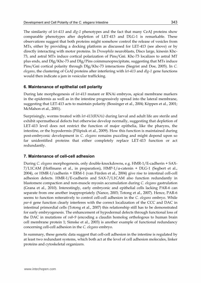
The similarity of let-413 and dlg-1 phenotypes and the fact that many CeAJ proteins show
comparable phenotypes after depletion of LET-413 and DLG-1 is remarkable. These
observations suggest that both proteins might somehow control the release of vesicles from
MTs, either by providing a docking platform as discussed for LET-413 (see above) or by
directly interacting with motor proteins. In Drosophila neuroblasts, Discs large, kinesin Khc-
73, and astral MTs induce cortical polarization of Pins/Gαi. Khc-73 localizes to astral MT
plus ends, and Dlg/Khc-73 and Dlg/Pins coimmunoprecipitate, suggesting that MTs induce
Pins/Gαi cortical polarity through Dlg/Khc-73 interactions (Siegrist and Doe, 2005). In C.
elegans, the clustering of CeAJ proteins after interfering with let-413 and dlg-1 gene functions
would then indicate a jam in vesicular trafficking.
6. Maintenance of epithelial cell polarity
During late morphogenesis of let-413 mutant or RNAi embryos, apical membrane markers
in the epidermis as well as in the intestine progressively spread into the lateral membrane,
suggesting that LET-413 acts to maintain polarity (Bossinger et al., 2004; Köppen et al., 2001;
McMahon et al., 2001).
Surprisingly, worms treated with let-413(RNAi) during larval and adult life are sterile and
exhibit spermathecal defects but otherwise develop normally, suggesting that depletion of
LET-413 level does not restrict the function of major epithelia, like the pharynx, the
intestine, or the hypodermis (Pilipiuk et al., 2009). How this function is maintained during
post-embryonic development in C. elegans remains puzzling and might depend upon so
far unidentified proteins that either completely replace LET-413 function or act
redundantly.
7. Maintenance of cell-cell adhesion
During C. elegans morphogenesis, only double-knockdowns, e.g. HMR-1/E-cadherin + SAX-
7/L1CAM (Hoffmann et al., in preparation), HMP-1/┙-catenin + DLG-1 (Segbert et al.,
2004), or HMR-1/cadherin + ERM-1 (van Fürden et al., 2004) give rise to intestinal cell-cell
adhesion defects. HMR-1/E-cadherin and SAX-7/L1CAM also function redundantly in
blastomere compaction and non-muscle myosin accumulation during C. elegans gastrulation
(Grana et al., 2010). Interestingly, early embryonic and epithelial cells lacking PAR-6 can
separate from one another inappropriately (Nance, 2003; Totong et al., 2007). Hence, PAR-6
seems to function reiteratively to control cell-cell adhesion in the C. elegans embryo. While
par-6 gene function clearly interferes with the correct localization of the CCC and DAC in
intestinal primordial cells (Totong et al., 2007) this relationship still has to be demonstrated
for early embryogenesis. The enhancement of hypodermal defects through functional loss of
the DAC in mutations of vab-9 (encoding a claudin homolog orthologous to human brain
cell membrane protein 1; Simske et al., 2003) is another example of functional redundancy
concerning cell-cell adhesion in the C. elegans embryo.
In summary, these genetic data suggest that cell-cell adhesion in the intestine is regulated by
at least two redundant systems, which both act at the level of cell adhesion molecules, linker
proteins and cytoskeletal organizers.
www.intechopen.com
Current Frontiers and Perspectives in Cell Biology 344
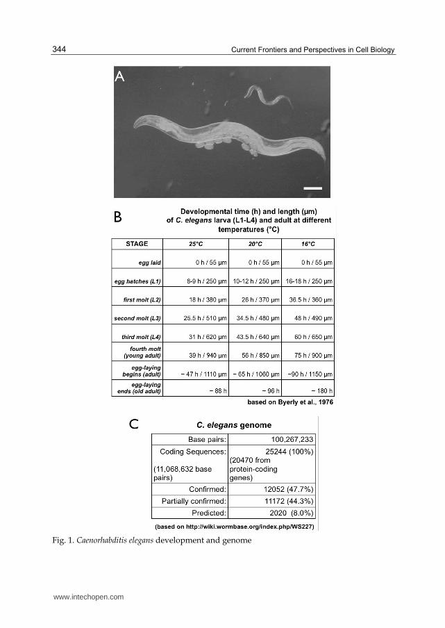
Fig. 1. Caenorhabditis elegans development and genome
www.intechopen.com
Development and Cell Polarity of the C. elegans Intestine 345
(A) Shows a DIC micrograph of a C. elegans larva (top) an adult hermaphrodite (middle) and embryos (bottom) maintained on agar plates with E. coli as food source (scale bar: 100 µm). (B) The table summarizes the developmental time (in hours) of C. elegans at different temperatures (°C), starting with the eggs released from the mother’s uterus (0 h), completing embryogenesis (8-18 h), passing through four larval stages (L1-L4) and finally reaching adulthood (47-90 h). The length of the egg, larva and adult at each stage is given in micrometers (µm). (C) provides a short summary of the C. elegans genome (The_C_elegans_Sequencing_Consortium, 1998) that contains 100,267,633 base pairs and is estimated to have 25244 coding sequences (CDS) from which 47.7% have been confirmed (every base of every exon has transcription evidence). 44.3% CDS are partially confirmed (some, but not all exon bases are covered) and 8.0% CDS show no transcriptional evidence at all. Recent meta-analysis of results from four orthology prediction programs has yielded a set of 7633 C. elegans genes (“OrthoList”) having human orthologs (Shaye and Greenwald, 2011).
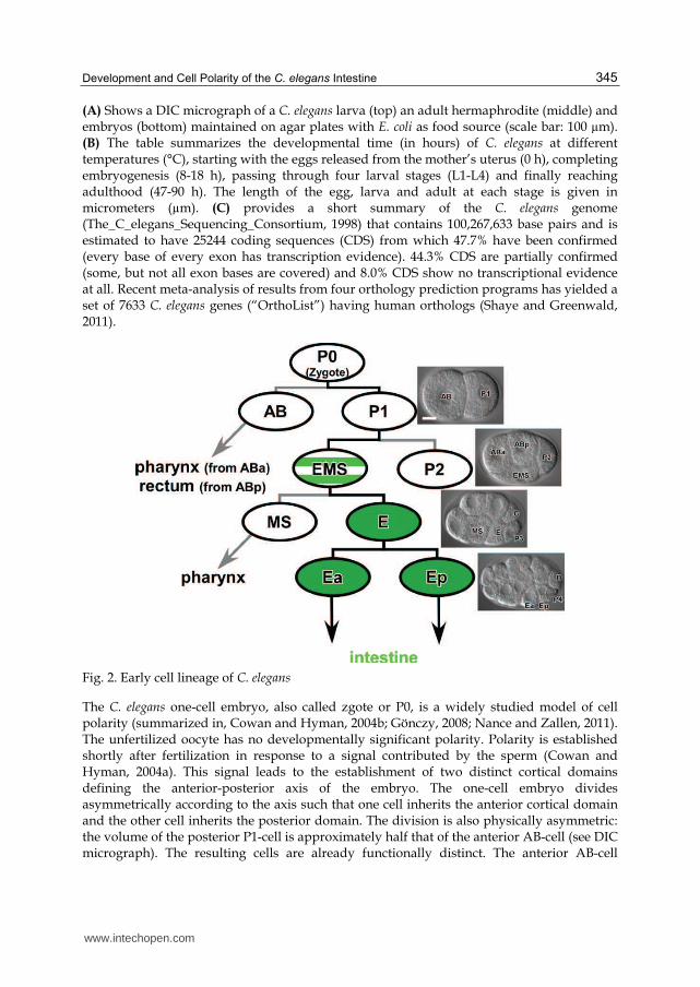
Fig. 2. Early cell lineage of C. elegans
The C. elegans one-cell embryo, also called zgote or P0, is a widely studied model of cell polarity (summarized in, Cowan and Hyman, 2004b; Gönczy, 2008; Nance and Zallen, 2011). The unfertilized oocyte has no developmentally significant polarity. Polarity is established shortly after fertilization in response to a signal contributed by the sperm (Cowan and Hyman, 2004a). This signal leads to the establishment of two distinct cortical domains defining the anterior-posterior axis of the embryo. The one-cell embryo divides asymmetrically according to the axis such that one cell inherits the anterior cortical domain and the other cell inherits the posterior domain. The division is also physically asymmetric: the volume of the posterior P1-cell is approximately half that of the anterior AB-cell (see DIC micrograph). The resulting cells are already functionally distinct. The anterior AB-cell
www.intechopen.com

Current Frontiers and Perspectives in Cell Biology 346
proceeds along a differentiation pathway producing ectoderm (hypodermis, pharynx, and neurons). The posterior P1-cell re-establishes anterior-posterior polarity and again divides asymmetrically (into P2 and EMS; see DIC micrograph) in a stem cell-like mode of division. These stem cell-like divisions establish the founder cells for the somatic lineages of the worm (AB, MS, E, C and D; see DIC micrographs) and maintaining a single stem cell (P4; see DIC micrographs) for the germline, which finally produces sperms and oocytes in the adult hermaphrodite.
The complete C. elegans digestive tract consists of three “organs” derived from four distinct
embryonic cell lineages (Sulston et al., 1983): pharynx (57 cells from ABa; 38 cells from MS),
intestine (20 cells from E; green), and rectum (11 cells from ABp; Sewell et al., 2003). Only
the intestine is a pure clone of 20 E-cells; the three other lineages produce cells both inside
and outside of the digestive tract. The intestine is one of the few cell lineages in the C. elegans
embryo where a plausible sequence of direct molecular interactions can be proposed
throughout the life cycle (Kormish et al., 2010; McGhee, 2007), beginning with maternally-
derived factors in the cytoplasm of the early embryo (e.g. SKN-1 and SYS-1/POP-1),
progressing through a small number of zygotic transcription factors (e.g. END-1/3 and ELT-
2), and ending with the transcription of e.g. vitellogenin genes in the adult intestine. ELT-2
has been proposed to participate directly in the regulation of most intestinal genes
expressed from the E2 cell stage (Ea and Ep, see DIC micrograph) and later (McGhee et al.,
2009; McGhee et al., 2007). The molecular mechanisms that lead to the asymmetric division
of the EMS blastomere (green striated) into a larger MS- and a smaller E blastomere (see DIC
micrograph) and the correct specification of their cell fates, central to the formation of the
pharynx and intestine has been describe in great detail elsewhere (Maduro, 2010; Mango,
2007; Sugioka et al., 2011). Orientation (DIC micrographs): anterior, left, dorsal top; scale
bar: 10 µm.
Fig. 3. RNA-mediated interference (RNAi) in C. elegans
www.intechopen.com
Development and Cell Polarity of the C. elegans Intestine 347
Over the last decades, RNAi has been found not only be effective in C. elegans but also in other
organisms and cell culture. The cartoon depicts a very simplified scheme of the exogenous
RNAi-mechanism in C. elegans (for detailed reviews see: Ahringer, 2006; Fischer, 2010; Maine,
2008) that leads to targeted destabilization of endogenous, homologous mRNA molecules by
double stranded RNA (dsRNA; Fire et al., 1998). (A) In a cell, RNA is used as a "messenger"
(mRNA) to carry genetic information from the nucleus into the cytoplasm, where it is
translated into proteins. (B) In C. elegans, exogenous dsRNA can be either applied by injection,
“feeding“ or “soaking” (Maeda et al., 2001; Mello et al., 1991; Timmons and Fire, 1998). dsRNA
is then cut into ~22 nt primary siRNAs by a protein complex containing the RNAse III enzyme
Dicer (DCR-1) and the dsRNA binding protein RDE-4 (Ketting et al., 2001; Tabara et al., 2002).
The Argonaute protein RDE-1 (Tabara et al., 1999) binds siRNAs and seems only required for
their stability (Parrish and Fire, 2001). Finally, RDE-1 slicer activity removes the passenger
strand from the guide strand in the siRNA duplex (Steiner et al., 2009), which is necessary to
allow guide-strand accessibility to the mRNA target. (C) RNAi in C. elegans includes an
amplification step (Alder et al., 2003; Fire et al., 1998). The mRNA that is targeted by siRNAs
serves as a template for the generation of secondary siRNAs mediated by RNA-dependent
RNA polymerases (RdRPs). Secondary siRNAs are always antisense and have 5′ triphosphates
instead of the 5′ monophosphate characteristic of Dicer cleavage. Secondary siRNAs are made
by unprimed RNA synthesis by RdRPs, which are recruited to the target mRNA bound to the
primary siRNA in complex with RDE-1 (Pak and Fire, 2007; Sijen et al., 2007). In vitro studies
suggest that secondary siRNA generation is Dicer-independent (Aoki et al., 2007). (D) siRNAs
present in the cell are associated with an effector complex called the RISC (RNA-induced
silencing complex). In C. elegans multiple such complexes exist (Caudy et al., 2003; Chan et al.,
2008; Gu et al., 2007), which finally drive mRNA destabilization.
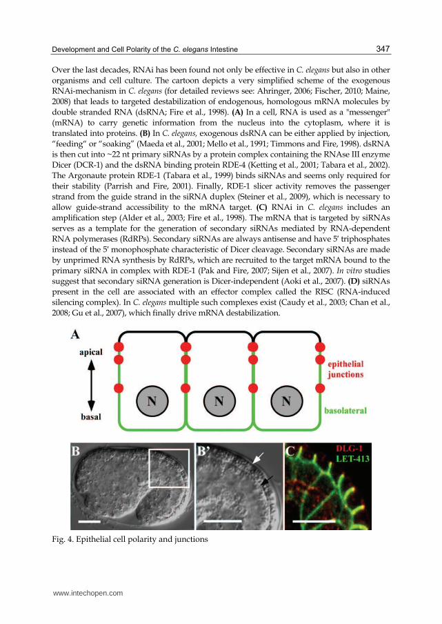
Fig. 4. Epithelial cell polarity and junctions
www.intechopen.com
Current Frontiers and Perspectives in Cell Biology 348
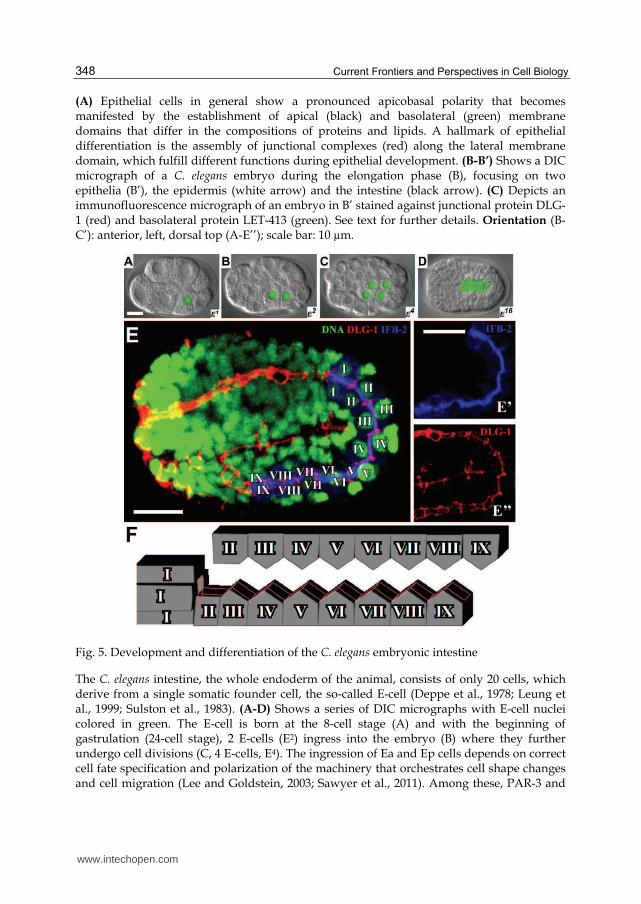
(A) Epithelial cells in general show a pronounced apicobasal polarity that becomes manifested by the establishment of apical (black) and basolateral (green) membrane domains that differ in the compositions of proteins and lipids. A hallmark of epithelial differentiation is the assembly of junctional complexes (red) along the lateral membrane domain, which fulfill different functions during epithelial development. (B-B’) Shows a DIC micrograph of a C. elegans embryo during the elongation phase (B), focusing on two epithelia (B’), the epidermis (white arrow) and the intestine (black arrow). (C) Depicts an immunofluorescence micrograph of an embryo in B’ stained against junctional protein DLG-1 (red) and basolateral protein LET-413 (green). See text for further details. Orientation (B-C’): anterior, left, dorsal top (A-E’’); scale bar: 10 µm.
Fig. 5. Development and differentiation of the C. elegans embryonic intestine
The C. elegans intestine, the whole endoderm of the animal, consists of only 20 cells, which derive from a single somatic founder cell, the so-called E-cell (Deppe et al., 1978; Leung et al., 1999; Sulston et al., 1983). (A-D) Shows a series of DIC micrographs with E-cell nuclei colored in green. The E-cell is born at the 8-cell stage (A) and with the beginning of gastrulation (24-cell stage), 2 E-cells (E2) ingress into the embryo (B) where they further undergo cell divisions (C, 4 E-cells, E4). The ingression of Ea and Ep cells depends on correct cell fate specification and polarization of the machinery that orchestrates cell shape changes and cell migration (Lee and Goldstein, 2003; Sawyer et al., 2011). Among these, PAR-3 and
www.intechopen.com
Development and Cell Polarity of the C. elegans Intestine 349
PAR-6 proteins regulate apical accumulation of myosin heavy chain, and a Wnt-Frizzled signaling pathway modulates contraction of the actomyosin network that drives apical constriction and finally leads to correct ingression of endodermal precursor cells (Cabello et al., 2010; Grana et al., 2010; Lee et al., 2006). Gastrulation in C. elegans later continues with the internalization of other cells including mesoderm and germline progenitors (Chisholm and Hardin, 2005; Nance et al., 2005). During early morphogenesis, the intestinal precursor cells (E16) start to polarize (D, 16 E-cells, E16, only 10 E-cells in focal plane) and finally an intestinal tube of 20 E-cells forms during ongoing morphogenesis of C. elegans. (E-E’’) Shows micrographs of a mid-morphogenesis stage (similar to D) stained against DNA (E, green, YoYo), the intestinal-specific intermediate filament protein IFB-2 localized in the apical cortex (E’, blue, mabMH33), and the junctional protein DLG-1 (E’’, red, anti-DLG-1 antibodies). (F) The cartoon depicts the organization of the intestinal epithelial tube in nine units (I-IX), which are connected by the CeAJ (red). Orientation (A-E’’): anterior, left, dorsal top (A-E’’); scale bar: 10 µm.
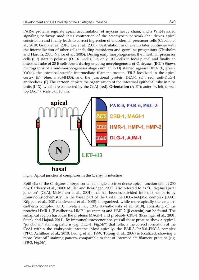
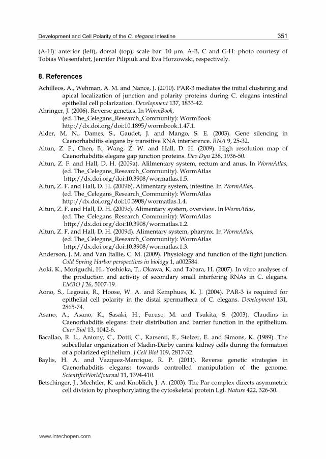
Fig. 6. Apical junctional complexes in the C. elegans intestine
Epithelia of the C. elegans embryo contain a single electron-dense apical junction (about 250 nm; Carberry et al., 2009; Müller and Bossinger, 2003), also referred to as “C. elegans apical junction“ (CeAJ; McMahon et al., 2001) that has been subdivided into distinct parts by immunohistochemistry. In the basal part of the CeAJ, the DLG-1–AJM-1 complex (DAC; Köppen et al., 2001; Lockwood et al., 2008) is organized, while more apically the catenin–cadherin complex (CCC; Costa et al., 1998; Kwiatkowski et al., 2010), consisting of the proteins HMR-1 (E-cadherin), HMP-1 (α-catenin) and HMP-2 (β-catenin) can be found. The subapical region harbours the proteins MAGI-1 and probably CRB-1 (Bossinger et al., 2001; Stetak and Hajnal, 2011). By immunofluorescence analysis all these proteins show a typical, “junctional” staining pattern (e.g. DLG-1, Fig.5E’’) that reflects the correct formation of the CeAJ within the embryonic intestine. Most apically, the PAR-3–PAR-6–PKC-3 complex (PPC; Achilleos et al., 2010; Leung et al., 1999; Totong et al., 2007) is localized, showing a more “cortical” staining pattern, comparable to that of intermediate filament proteins (e.g. IFB-2, Fig.5E’).
www.intechopen.com
Current Frontiers and Perspectives in Cell Biology 350
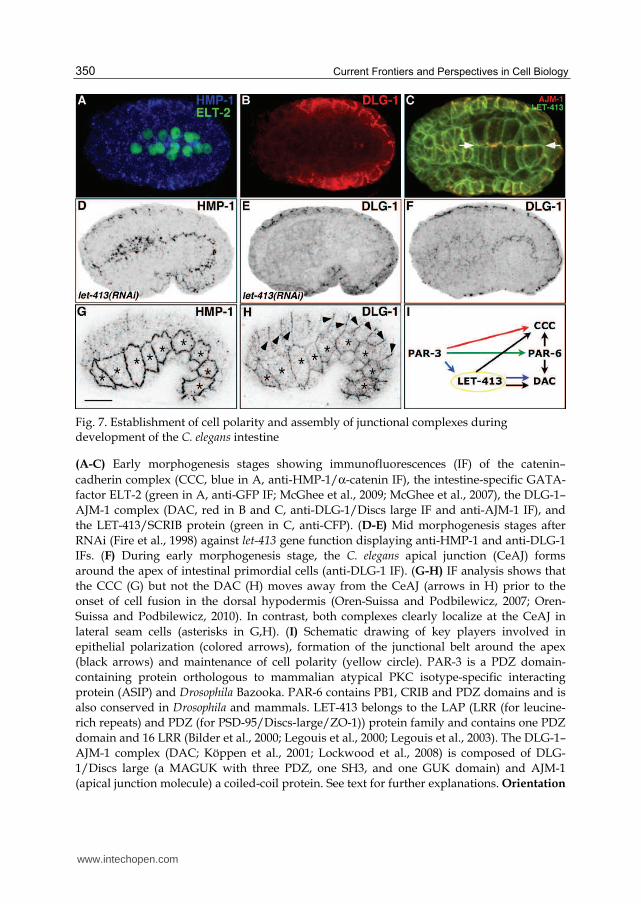
Fig. 7. Establishment of cell polarity and assembly of junctional complexes during development of the C. elegans intestine
(A-C) Early morphogenesis stages showing immunofluorescences (IF) of the catenin–
cadherin complex (CCC, blue in A, anti-HMP-1/α-catenin IF), the intestine-specific GATA-
factor ELT-2 (green in A, anti-GFP IF; McGhee et al., 2009; McGhee et al., 2007), the DLG-1–
AJM-1 complex (DAC, red in B and C, anti-DLG-1/Discs large IF and anti-AJM-1 IF), and
the LET-413/SCRIB protein (green in C, anti-CFP). (D-E) Mid morphogenesis stages after
RNAi (Fire et al., 1998) against let-413 gene function displaying anti-HMP-1 and anti-DLG-1
IFs. (F) During early morphogenesis stage, the C. elegans apical junction (CeAJ) forms
around the apex of intestinal primordial cells (anti-DLG-1 IF). (G-H) IF analysis shows that
the CCC (G) but not the DAC (H) moves away from the CeAJ (arrows in H) prior to the
onset of cell fusion in the dorsal hypodermis (Oren-Suissa and Podbilewicz, 2007; Oren-
Suissa and Podbilewicz, 2010). In contrast, both complexes clearly localize at the CeAJ in
lateral seam cells (asterisks in G,H). (I) Schematic drawing of key players involved in
epithelial polarization (colored arrows), formation of the junctional belt around the apex
(black arrows) and maintenance of cell polarity (yellow circle). PAR-3 is a PDZ domain-
containing protein orthologous to mammalian atypical PKC isotype-specific interacting
protein (ASIP) and Drosophila Bazooka. PAR-6 contains PB1, CRIB and PDZ domains and is
also conserved in Drosophila and mammals. LET-413 belongs to the LAP (LRR (for leucine-
rich repeats) and PDZ (for PSD-95/Discs-large/ZO-1)) protein family and contains one PDZ
domain and 16 LRR (Bilder et al., 2000; Legouis et al., 2000; Legouis et al., 2003). The DLG-1–
AJM-1 complex (DAC; Köppen et al., 2001; Lockwood et al., 2008) is composed of DLG-
1/Discs large (a MAGUK with three PDZ, one SH3, and one GUK domain) and AJM-1
(apical junction molecule) a coiled-coil protein. See text for further explanations. Orientation
www.intechopen.com
Development and Cell Polarity of the C. elegans Intestine 351
(A-H): anterior (left), dorsal (top); scale bar: 10 µm. A-B, C and G-H: photo courtesy of
Tobias Wiesenfahrt, Jennifer Pilipiuk and Eva Horzowski, respectively.
8. References
Achilleos, A., Wehman, A. M. and Nance, J. (2010). PAR-3 mediates the initial clustering and apical localization of junction and polarity proteins during C. elegans intestinal epithelial cell polarization. Development 137, 1833-42.
Ahringer, J. (2006). Reverse genetics. In WormBook, (ed. The_Celegans_Research_Community): WormBook http://dx.doi.org/doi:10.1895/wormbook.1.47.1.
Alder, M. N., Dames, S., Gaudet, J. and Mango, S. E. (2003). Gene silencing in Caenorhabditis elegans by transitive RNA interference. RNA 9, 25-32.
Altun, Z. F., Chen, B., Wang, Z. W. and Hall, D. H. (2009). High resolution map of Caenorhabditis elegans gap junction proteins. Dev Dyn 238, 1936-50.
Altun, Z. F. and Hall, D. H. (2009a). Alilmentary system, rectum and anus. In WormAtlas, (ed. The_Celegans_Research_Community). WormAtlas http://dx.doi.org/doi:10.3908/wormatlas.1.5.
Altun, Z. F. and Hall, D. H. (2009b). Alimentary system, intestine. In WormAtlas, (ed. The_Celegans_Research_Community): WormAtlas
http://dx.doi.org/doi:10.3908/wormatlas.1.4. Altun, Z. F. and Hall, D. H. (2009c). Alimentary system, overview. In WormAtlas,
(ed. The_Celegans_Research_Community): WormAtlas http://dx.doi.org/doi:10.3908/wormatlas.1.2.
Altun, Z. F. and Hall, D. H. (2009d). Alimentary system, pharynx. In WormAtlas, (ed. The_Celegans_Research_Community): WormAtlas http://dx.doi.org/doi:10.3908/wormatlas.1.3.
Anderson, J. M. and Van Itallie, C. M. (2009). Physiology and function of the tight junction. Cold Spring Harbor perspectives in biology 1, a002584.
Aoki, K., Moriguchi, H., Yoshioka, T., Okawa, K. and Tabara, H. (2007). In vitro analyses of the production and activity of secondary small interfering RNAs in C. elegans. EMBO J 26, 5007-19.
Aono, S., Legouis, R., Hoose, W. A. and Kemphues, K. J. (2004). PAR-3 is required for epithelial cell polarity in the distal spermatheca of C. elegans. Development 131, 2865-74.
Asano, A., Asano, K., Sasaki, H., Furuse, M. and Tsukita, S. (2003). Claudins in Caenorhabditis elegans: their distribution and barrier function in the epithelium. Curr Biol 13, 1042-6.
Bacallao, R. L., Antony, C., Dotti, C., Karsenti, E., Stelzer, E. and Simons, K. (1989). The subcellular organization of Madin-Darby canine kidney cells during the formation of a polarized epithelium. J Cell Biol 109, 2817-32.
Baylis, H. A. and Vazquez-Manrique, R. P. (2011). Reverse genetic strategies in Caenorhabditis elegans: towards controlled manipulation of the genome. ScientificWorldJournal 11, 1394-410.
Betschinger, J., Mechtler, K. and Knoblich, J. A. (2003). The Par complex directs asymmetric cell division by phosphorylating the cytoskeletal protein Lgl. Nature 422, 326-30.
www.intechopen.com
Current Frontiers and Perspectives in Cell Biology 352
Bilder, D., Birnbaum, D., Borg, J. P., Bryant, P., Huigbretse, J., Jansen, E., Kennedy, M. B., Labouesse, M., Legouis, R., Mechler, B. et al. (2000). Collective nomenclature for LAP proteins. Nature cell biology 2, E114.
Bilder, D., Schober, M. and Perrimon, N. (2003). Integrated activity of PDZ protein complexes regulates epithelial polarity. Nat Cell Biol 5, 53-58.
Bird, A. F. and Bird, J. (1991). The structure of nematodes. California: Academic Press. Blaxter, M. (2011). Nematodes: the worm and its relatives. PLoS Biol 9, e1001050. Bossinger, O., Fukushige, T., Claeys, M., Borgonie, G. and McGhee, J. D. (2004). The apical
disposition of the Caenorhabditis elegans intestinal terminal web is maintained by LET-413. Dev Biol 268, 448-456.
Bossinger, O., Klebes, A., Segbert, C., Theres, C. and Knust, E. (2001). Zonula adherens formation in Caenorhabditis elegans requires dlg-1, the homologue of the Drosophila gene discs large. Dev Biol 230, 29-42.
Bossinger, O. and Schierenberg, E. (1992). Cell-cell communication in the embryo of Caenorhabditis elegans. Dev Biol 151, 401-9.
Bossinger, O. and Schierenberg, E. (1996). Early embryonic induction in C. elegans can be inhibited with polysulfated hydrocarbon dyes. Dev Biol 176, 17-21.
Boulin, T. and Bessereau, J. (2007). Mos1-mediated insertional mutagenesis in Caenorhabditis elegans. Nat Protoc 2, 1276-87.
Bowerman, B. (2011). The near demise and subsequent revival of classical genetics for investigating Caenorhabditis elegans embryogenesis: RNAi meets next-generation DNA sequencing. Mol Biol Cell 22, 3556-8.
Bre, M. H., Pepperkok, R., Hill, A. M., Levilliers, N., Ansorge, W., Stelzer, E. H. and Karsenti, E. (1990). Regulation of microtubule dynamics and nucleation during polarization in MDCK II cells. J Cell Biol 111, 3013-21.
Brenner, S. (1974). The genetics of Caenorhabditis elegans. Genetics 77, 71-94. Brenner, S. (2003). Nature's gift to science (Nobel lecture). Chembiochem 4, 683-7. Brenner, S. (2009). In the beginning was the worm. Genetics 182, 413-5. Bulgakova, N. A. and Knust, E. (2009). The Crumbs complex: from epithelial-cell polarity to
retinal degeneration. J Cell Sci 122, 2587-96. Cabello, J., Neukomm, L. J., Gunesdogan, U., Burkart, K., Charette, S. J., Lochnit, G.,
Hengartner, M. O. and Schnabel, R. (2010). The Wnt pathway controls cell death engulfment, spindle orientation, and migration through CED-10/Rac. PLoS Biol 8, e1000297.
Calixto, A., Chelur, D., Topalidou, I., Chen, X. and Chalfie, M. (2010a). Enhanced neuronal RNAi in C. elegans using SID-1. Nature methods 7, 554-9.
Calixto, A., Ma, C. and Chalfie, M. (2010b). Conditional gene expression and RNAi using MEC-8-dependent splicing in C. elegans. Nat Methods 7, 407-11.
Carberry, K., Wiesenfahrt, T., Windoffer, R., Bossinger, O. and Leube, R. E. (2009). Intermediate filaments in Caenorhabditis elegans. Cell Motil Cytoskeleton 66, 852-64.
Caudy, A. A., Ketting, R. F., Hammond, S. M., Denli, A. M., Bathoorn, A. M., Tops, B. B., Silva, J. M., Myers, M. M., Hannon, G. J. and Plasterk, R. H. (2003). A micrococcal nuclease homologue in RNAi effector complexes. Nature 425, 411-4.
Chalfie, M. (2009). GFP: lighting up life (Nobel Lecture). Angewandte Chemie 48, 5603-11. Chalfie, M., Tu, Y., Euskirchen, G., Ward, W. and Prasher, D. (1994). Green
fluorescent protein as a marker for gene expression. Science 263, 802-5.
www.intechopen.com
Development and Cell Polarity of the C. elegans Intestine 353
Chan, S. P., Ramaswamy, G., Choi, E. Y. and Slack, F. J. (2008). Identification of specific let-7 microRNA binding complexes in Caenorhabditis elegans. RNA 14, 2104-14.
Chen, X., Kojima, S., Borisy, G. G. and Green, K. J. (2003). p120 catenin associates with kinesin and facilitates the transport of cadherin-catenin complexes to intercellular junctions. J Cell Biol 163, 547-57.
Chisholm, A. D. (2006). Gastrulation: Wnts signal constriction. Curr Biol 16, R874-6. Chisholm, A. D. and Hardin, J. (2005). Epidermal morphogenesis. In WormBook, (ed.
The_Celegans_Research_Community), pp. 1-22: WormBook http://dx.doi.org/doi:10.1895/wormbook.1.35.1.
Costa, M., Raich, W., Agbunag, C., Leung, B., Hardin, J. and Priess, J. (1998). A putative catenin-cadherin system mediates morphogenesis of the Caenorhabditis elegans embryo. J Cell Biol 141, 297-308.
Cowan, C. R. and Hyman, A. (2004a). Centrosomes direct cell polarity independently of microtubule assembly in C. elegans embryos. Nature 431, 92-6.
Cowan, C. R. and Hyman, A. A. (2004b). Asymmetric cell division in C. elegans: cortical polarity and spindle positioning. Annual review of cell and developmental biology 20, 427-53.
Cox, E. A. and Hardin, J. (2004). Sticky worms: adhesion complexes in C. elegans. J Cell Sci 117, 1885-97.
de Santa Barbara, P., van den Brink, G. R. and Roberts, D. J. (2003). Development and differentiation of the intestinal epithelium. Cell Mol Life Sci 60, 1322-32.
Deppe, U., Schierenberg, E., Cole, T., Krieg, C., Schmitt, D., Yoder, B. K. and von Ehrenstein, G. (1978). Cell lineages of the embryo of the nematode Caenorhabditis elegans. Proc Natl Acad Sci U S A 75, 376-80.
Ebnet, K. (2008). Organization of multiprotein complexes at cell-cell junctions. Histochem Cell Biol 130, 1-20.
Eckert, J. J. and Fleming, T. P. (2008). Tight junction biogenesis during early development. Biochim Biophys Acta 1778, 717-728.
Etheridge, T., Nemoto, K., Hashizume, T., Mori, C., Sugimoto, T., Suzuki, H., Fukui, K., Yamazaki, T., Higashibata, A., Szewczyk, N. J. et al. (2011). The effectiveness of RNAi in Caenorhabditis elegans is maintained during spaceflight. PLoS ONE 6, e20459.
Fire, A., Xu, S., Montgomery, M., Kostas, S., Driver, S. and Mello, C. (1998). Potent and specific genetic interference by double-stranded RNA in Caenorhabditis elegans. Nature 391, 806-11.
Fire, A. Z. (2007). Gene silencing by double-stranded RNA (Nobel Lecture). Angewandte Chemie 46, 6966-84.
Firestein, B. L. and Rongo, C. (2001). DLG-1 Is a MAGUK Similar to SAP97 and Is Required for Adherens Junction Formation. Mol Biol Cell 12, 3465-75.
Fischer, S. E. (2010). Small RNA-mediated gene silencing pathways in C. elegans. The international journal of biochemistry & cell biology 42, 1306-15.
Fraser, A. G., Kamath, R. S., Zipperlen, P., Martinez-Campos, M., Sohrmann, M. and Ahringer, J. (2000). Functional genomic analysis of C. elegans chromosome I by systematic RNA interference. Nature 408, 325-30.
www.intechopen.com
Current Frontiers and Perspectives in Cell Biology 354
Frokjaer-Jensen, C., Davis, M. W., Hollopeter, G., Taylor, J., Harris, T. W., Nix, P., Lofgren, R., Prestgard-Duke, M., Bastiani, M., Moerman, D. G. et al. (2010). Targeted gene deletions in C. elegans using transposon excision. Nat Methods 7, 451-3.
Fuchs, E. (2007). Scratching the surface of skin development. Nature 445, 834-42. Goldstein, B. (1992). Induction of gut in Caenorhabditis elegans embryos. Nature 357, 255-7. Gönczy, P. (2008). Mechanisms of asymmetric cell division: flies and worms pave
the way. Nat Rev Mol Cell Biol 9, 355-66. Gönczy, P., Echeverri, G., Oegema, K., Coulson, A., Jones, S. J., Copley, R. R., Duperon, J.,
Oegema, J., Brehm, M., Cassin, E. et al. (2000). Functional genomic analysis of cell division in C. elegans using RNAi of genes on chromosome III. Nature 408, 331-6.
Grana, T. M., Cox, E. A., Lynch, A. M. and Hardin, J. (2010). SAX-7/L1CAM and HMR-1/cadherin function redundantly in blastomere compaction and non-muscle myosin accumulation during Caenorhabditis elegans gastrulation. Developmental biology 344, 731-44.
Gu, S. G., Pak, J., Barberan-Soler, S., Ali, M., Fire, A. and Zahler, A. M. (2007). Distinct ribonucleoprotein reservoirs for microRNA and siRNA populations in C. elegans. RNA 13, 1492-504.
Guo, S. and Kemphues, K. J. (1996). A non-muscle myosin required for embryonic polarity in Caenorhabditis elegans. Nature 382, 455-8.
Han, M. (1997). Gut reaction to Wnt signaling in worms. Cell 90, 581-4. Han, M. (2010). Advancing biology with a growing worm field. Developmental dynamics : an
official publication of the American Association of Anatomists 239, 1263-4. Hardin, J. and Lockwood, C. (2004). Skin tight: cell adhesion in the epidermis of
Caenorhabditis elegans. Curr Opin Cell Biol 16, 486-92. Harris, T. J. and Peifer, M. (2005). The positioning and segregation of apical cues during
epithelial polarity establishment in Drosophila. J Cell Biol 170, 813-23. Harris, T. J. and Peifer, M. (2007). aPKC controls microtubule organization to balance
adherens junction symmetry and planar polarity during development. Dev Cell 12, 727-38.
Harris, T. J. and Tepass, U. (2010). Adherens junctions: from molecules to morphogenesis. Nat Rev Mol Cell Biol 11, 502-14.
Harris, T. W., Antoshechkin, I., Bieri, T., Blasiar, D., Chan, J., Chen, W. J., De La Cruz, N., Davis, P., Duesbury, M., Fang, R. et al. (2010). WormBase: a comprehensive resource for nematode research. Nucleic Acids Res 38, D463-7.
Hedgecock, E. M. and White, J. G. (1985). Polyploid tissues in the nematode Caenorhabditis elegans. Dev Biol 107, 128-33.
Hermann, G. J., Leung, B. and Priess, J. R. (2000). Left-right asymmetry in C. elegans intestine organogenesis involves a LIN-12/Notch signaling pathway. Development 127, 3429-40.
Hobert, O. (2010). The impact of whole genome sequencing on model system genetics: get ready for the ride. Genetics 184, 317-9.
Hoffmann, M., Segbert, C., Helbig, G. and Bossinger, O. (2010). Intestinal tube formation in Caenorhabditis elegans requires vang-1 and egl-15 signaling. Dev Biol 339, 268-279.
Horvitz, H. R. (2003). Worms, life, and death (Nobel lecture). Chembiochem 4, 697-711.
www.intechopen.com
Development and Cell Polarity of the C. elegans Intestine 355
Hunt-Newbury, R., Viveiros, R., Johnsen, R., Mah, A., Anastas, D., Fang, L., Halfnight, E., Lee, D., Lin, J., Lorch, A. et al. (2007). High-throughput in vivo analysis of gene expression in Caenorhabditis elegans. PLoS Biol 5, e237.
Hüsken, K., Wiesenfahrt, T., Abraham, C., Windoffer, R., Bossinger, O. and Leube, R. (2008). Maintenance of the intestinal tube in Caenorhabditis elegans: the role of the intermediate filament protein IFC-2. Differentiation 76, 881-896.
Jordens, I., Marsman, M., Kuijl, C. and Neefjes, J. (2005). Rab proteins, connecting transport and vesicle fusion. Traffic 6, 1070-7.
Jorgensen, E. M. and Mango, S. E. (2002). The art and design of genetic screens: Caenorhabditis elegans. Nat Rev Genet 3, 356-69.
Kamath, R. S., Fraser, A. G., Dong, Y., Poulin, G., Durbin, R., Gotta, M., Kanapin, A., Le Bot, N., Moreno, S., Sohrmann, M. et al. (2003). Systematic functional analysis of the Caenorhabditis elegans genome using RNAi. Nature 421, 231-7.
Ketting, R. F., Fischer, S. E., Bernstein, E., Sijen, T., Hannon, G. J. and Plasterk, R. H. (2001). Dicer functions in RNA interference and in synthesis of small RNA involved in developmental timing in C. elegans. Genes Dev 15, 2654-9.
Kimble, J. and Sharrock, W. J. (1983). Tissue-specific synthesis of yolk proteins in Caenorhabditis elegans. Dev Biol 96, 189-96.
Knust, E. and Bossinger, O. (2002). Composition and formation of intercellular junctions in epithelial cells. Science 298, 1955-9.
Köppen, M., Simske, J. S., Sims, P. A., Firestein, B. L., Hall, D. H., Radice, A. D., Rongo, C. and Hardin, J. D. (2001). Cooperative regulation of AJM-1 controls junctional integrity in Caenorhabditis elegans epithelia. Nat Cell Biol 3, 983-91.
Kormish, J. D., Gaudet, J. and McGhee, J. D. (2010). Development of the C. elegans digestive tract. Current opinion in genetics & development 20, 346-54.
Krahn, M. P., Buckers, J., Kastrup, L. and Wodarz, A. (2010a). Formation of a Bazooka-Stardust complex is essential for plasma membrane polarity in epithelia. The Journal of cell biology 190, 751-60.
Krahn, M. P., Klopfenstein, D. R., Fischer, N. and Wodarz, A. (2010b). Membrane targeting of Bazooka/PAR-3 is mediated by direct binding to phosphoinositide lipids. Current Biology 20, 636-42.
Kramer, J. M. (2005). Basement membranes. In WormBook, (ed. The_Celegans_Research_Community), pp. 1-15: WormBook http://dx.doi.org/doi:10.1895/wormbook.1.16.1.
Kwiatkowski, A. V., Maiden, S. L., Pokutta, S., Choi, H. J., Benjamin, J. M., Lynch, A. M., Nelson, W. J., Weis, W. I. and Hardin, J. (2010). In vitro and in vivo reconstitution of the cadherin-catenin-actin complex from Caenorhabditis elegans. Proc Natl Acad Sci U S A 107, 14591-6.
Labouesse, M. (2006). Epithelial junctions and attachments. In WormBook, (ed. The_Celegans_Research_Community): WormBook http://dx.doi.org/doi:10.1895/wormbook.1.56.1.
Laprise, P., Lau, K. M., Harris, K. P., Silva-Gagliardi, N. F., Paul, S. M., Beronja, S., Beitel, G. J., McGlade, C. J. and Tepass, U. (2009). Yurt, Coracle, Neurexin IV and the Na(+),K(+)-ATPase form a novel group of epithelial polarity proteins. Nature 459, 1141-5.
www.intechopen.com
Current Frontiers and Perspectives in Cell Biology 356
Lecuit, T. and Wieschaus, E. (2000). Polarized insertion of new membrane from a cytoplasmic reservoir during cleavage of the Drosophila embryo. J Cell Biol 150, 849-60.
Lee, J. and Goldstein, B. (2003). Mechanisms of cell positioning during C. elegans gastrulation. Development 130, 307-20.
Lee, J., Marston, D. J., Walston, T., Hardin, J., Halberstadt, A. and Goldstein, B. (2006). Wnt/Frizzled signaling controls C. elegans gastrulation by activating actomyosin contractility. Curr Biol 16, 1986-97.
Legouis, R., Gansmuller, A., Sookhareea, S., Bosher, J. M., Baillie, D. L. and Labouesse, M. (2000). LET-413 is a basolateral protein required for the assembly of adherens junctions in Caenorhabditis elegans. Nat Cell Biol 2, 415-422.
Legouis, R., Jaulin-Bastard, F., Schott, S., Navarro, C., Borg, J. P. and Labouesse, M. (2003). Basolateral targeting by leucine-rich repeat domains in epithelial cells. EMBO Rep 4, 1096-1100.
Leung, B., Hermann, G. J. and Priess, J. R. (1999). Organogenesis of the Caenorhabditis elegans intestine. Dev Biol 216, 114-34.
Ligon, L. A., Karki, S., Tokito, M. and Holzbaur, E. L. (2001). Dynein binds to beta-catenin and may tether microtubules at adherens junctions. Nat Cell Biol 3, 913-7.
Lints, R. and Hall, D. H. (2009). Reproductive system, somatic gonad. In WormAtlas, (ed. The_Celegans_Research_Community). WormAtlas http://dx.doi.org/doi:10.3908/wormatlas.1.22.
Lockwood, C. A., Lynch, A. M. and Hardin, J. (2008). Dynamic analysis identifies novel roles for DLG-1 subdomains in AJM-1 recruitment and LET-413-dependent apical focusing. J Cell Sci 121, 1477-87.
Lu, R., Maduro, M. F., Li, F., Li, H. L., Broitman-Maduro, G., Li, W. and Ding, S. W. (2005). Animal virus replication and RNAi-mediated antiviral silencing in Caenorhabditis elegans. Nature 436, 1040-3.
Lynch, A. M. and Hardin, J. (2009). The assembly and maintenance of epithelial junctions in C. elegans. Front Biosci 14, 1414-32.
MacQueen, A. J., Baggett, J. J., Perumov, N., Bauer, R. A., Januszewski, T., Schriefer, L. and Waddle, J. A. (2005). ACT-5 is an essential Caenorhabditis elegans actin required for intestinal microvilli formation. Molecular biology of the cell 16, 3247-59.
Maduro, M. F. (2010). Cell fate specification in the C. elegans embryo. Dev Dyn 239, 1315-29. Maeda, I., Kohara, Y., Yamamoto, M. and Sugimoto, A. (2001). Large-scale analysis of gene
function in Caenorhabditis elegans by high-throughput RNAi. Curr Biol 11, 171-6. Maine, E. M. (2008). Studying gene function in Caenorhabditis elegans using RNA-mediated
interference. Brief Funct Genomic Proteomic 7, 184-94. Mango, S. E. (2007). The C. elegans pharynx: a model for organogenesis. In WormBook, (ed.
The_Celegans_Research_Community): WormBook http://dx.doi.org/doi:10.1895/wormbook.1.129.1.
Mary, S., Charrasse, S., Meriane, M., Comunale, F., Travo, P., Blangy, A. and Gauthier-Rouviere, C. (2002). Biogenesis of N-cadherin-dependent cell-cell contacts in living fibroblasts is a microtubule-dependent kinesin-driven mechanism. Mol Biol Cell 13, 285-301.
www.intechopen.com
Development and Cell Polarity of the C. elegans Intestine 357
McGee, M. D., Weber, D., Day, N., Vitelli, C., Crippen, D., Herndon, L. A., Hall, D. H. and Melov, S. (2011). Loss of intestinal nuclei and intestinal integrity in aging C. elegans. Aging Cell 10, 699-710.
McGhee, J. D. (2007). The C. elegans intestine. In WormBook, (ed. The_Celegans_Research_Community): WormBook http://dx.doi.org/doi:10.1895/wormbook.1.133.1.
McGhee, J. D., Fukushige, T., Krause, M. W., Minnema, S. E., Goszczynski, B., Gaudet, J., Kohara, Y., Bossinger, O., Zhao, Y., Khattra, J. et al. (2009). ELT-2 is the predominant transcription factor controlling differentiation and function of the C. elegans intestine, from embryo to adult. Dev Biol 327, 551-65.
McGhee, J. D., Sleumer, M. C., Bilenky, M., Wong, K., McKay, S. J., Goszczynski, B., Tian, H., Krich, N. D., Khattra, J., Holt, R. A. et al. (2007). The ELT-2 GATA-factor and the global regulation of transcription in the C. elegans intestine. Dev Biol 302, 627-45.
McMahon, L., Legouis, R., Vonesch, J. L. and Labouesse, M. (2001). Assembly of C. elegans apical junctions involves positioning and compaction by LET-413 and protein aggregation by the MAGUK protein DLG-1. J Cell Sci 114, 2265-77.
Mello, C. C. (2007). Return to the RNAi world: rethinking gene expression and evolution (Nobel Lecture). Angewandte Chemie 46, 6985-94.
Mello, C. C., Kramer, J. M., Stinchcomb, D. T. and Ambros, V. (1991). Efficient gene transfer in C.elegans: extrachromosomal maintenance and integration of transforming sequences. EMBO J 10, 3959-70.
Michaux, G., Legouis, R. and Labouesse, M. (2001). Epithelial biology: lessons from Caenorhabditis elegans. Gene 277, 83-100.
Mitani, S. (2009). Nematode, an experimental animal in the national BioResource project. Exp Anim 58, 351-6.
Moerman, D. G. and Barstead, R. J. (2008). Towards a mutation in every gene in Caenorhabditis elegans. Brief Funct Genomic Proteomic 7, 195-204.
Müller, H. A. (2000). Genetic control of epithelial cell polarity: lessons from Drosophila. Dev Dyn 218, 52-67.
Müller, H. A. and Bossinger, O. (2003). Molecular networks controlling epithelial cell polarity in development. Mech Dev 120, 1231-56.
Nance, J. (2003). C. elegans PAR-3 and PAR-6 are required for apicobasal asymmetries associated with cell adhesion and gastrulation. Development 130, 5339-5350.
Nance, J., Lee, J. Y. and Goldstein, B. (2005). Gastrulation in C. elegans. In WormBook, (ed. The_Celegans_Research_Community), pp. 1-13: WormBook http://dx.doi.org/doi:10.1895/wormbook. 1.23.1
Nance, J. and Zallen, J. A. (2011). Elaborating polarity: PAR proteins and the cytoskeleton. Development 138, 799-809.
Nelson, W. J. (2003). Adaptation of core mechanisms to generate cell polarity. Nature 422, 766-74.
Nelson, W. J. (2009). Remodeling epithelial cell organization: transitions between front-rear and apical-basal polarity. Cold Spring Harbor perspectives in biology 1, a000513.
Noah, T. K., Donahue, B. and Shroyer, N. F. (2011). Intestinal development and differentiation. Exp Cell Res 317, 2702-10.
Oren-Suissa, M. and Podbilewicz, B. (2007). Cell fusion during development. Trends Cell Biol 17, 537-46.
www.intechopen.com
Current Frontiers and Perspectives in Cell Biology 358
Oren-Suissa, M. and Podbilewicz, B. (2010). Evolution of programmed cell fusion: common mechanisms and distinct functions. Dev Dyn 239, 1515-28.
Pak, J. and Fire, A. (2007). Distinct populations of primary and secondary effectors during RNAi in C. elegans. Science 315, 241-4.
Papoulas, O., Hays, T. S. and Sisson, J. C. (2005). The golgin Lava lamp mediates dynein-based Golgi movements during Drosophila cellularization. Nat Cell Biol 7, 612-8.
Parrish, S. and Fire, A. (2001). Distinct roles for RDE-1 and RDE-4 during RNA interference in Caenorhabditis elegans. RNA 7, 1397-402.
Pauli, F., Liu, Y., Kim, Y. A., Chen, P. J. and Kim, S. H. (2006). Chromosomal clustering and GATA transcriptional regulation of intestine-expressed genes in C. elegans. Development 133, 287-95.
Pilipiuk, J., Lefebvre, C., Wiesenfahrt, T., Legouis, R. and Bossinger, O. (2009). Increased IP3/Ca2+ signaling compensates depletion of LET-413/DLG-1 in C. elegans epithelial junction assembly. Dev Biol 327, 34-47.
Plant, P. J., Fawcett, J. P., Lin, D. C., Holdorf, A. D., Binns, K., Kulkarni, S. and Pawson, T. (2003). A polarity complex of mPar-6 and atypical PKC binds, phosphorylates and regulates mammalian Lgl. Nat Cell Biol 5, 301-8.
Portereiko, M. F., Saam, J. and Mango, S. E. (2004). ZEN-4/MKLP1 is required to polarize the foregut epithelium. Curr Biol 14, 932-41.
Putzke, A. P. and Rothman, J. H. (2003). Gastrulation: PARtaking of the bottle. Curr Biol 13, R223-5.
Qadota, H., Inoue, M., Hikita, T., Koppen, M., Hardin, J. D., Amano, M., Moerman, D. G. and Kaibuchi, K. (2007). Establishment of a tissue-specific RNAi system in C. elegans. Gene 400, 166-73.
Robert, V. J. and Bessereau, J. L. (2007). Targeted engineering of the Caenorhabditis elegans genome following Mos1-triggered chromosomal breaks. EMBO J 26, 170-83.
Rohrschneider, M. R. and Nance, J. (2009). Polarity and cell fate specification in the control of Caenorhabditis elegans gastrulation. Dev Dyn 238, 789-96.
Sawyer, J. M., Glass, S., Li, T., Shemer, G., White, N. D., Starostina, N. G., Kipreos, E. T., Jones, C. D. and Goldstein, B. (2011). Overcoming Redundancy: an RNAi Enhancer Screen for Morphogenesis Genes in Caenorhabditis elegans. Genetics 188, 549-64.
Sawyer, J. M., Harrell, J. R., Shemer, G., Sullivan-Brown, J., Roh-Johnson, M. and Goldstein, B. (2009). Apical constriction: A cell shape change that can drive morphogenesis. Dev Biol 34, 5-19.
Schierenberg, E. (1987). Reversal of cellular polarity and early cell-cell interaction in the embryos of Caenorhabditis elegans. Dev Biol 122, 452-63.
Schierenberg, E. (2005). Unusual cleavage and gastrulation in a freshwater nematode: developmental and phylogenetic implications. Dev Genes Evol 215, 103-8.
Schierenberg, E. (2006). Embryological variation during nematode development. In WormBook, (ed. The_Celegans_Research_Community), pp. 1-13: WormBook http://dx.doi.org/doi:10.1895/wormbook.1.55.1.
Schnabel, R., Hutter, H., Moerman, D. G. and Schnabel, H. (1997). Assessing normal embryogenesis in Caenorhabditis elegans using a 4D microscope: variability of development and regional specification. Dev Biol 184, 234-65.
www.intechopen.com
Development and Cell Polarity of the C. elegans Intestine 359
Schott, D. H., Cureton, D. K., Whelan, S. P. and Hunter, C. P. (2005). An antiviral role for the RNA interference machinery in Caenorhabditis elegans. Proc Natl Acad Sci U S A 102, 18420-4.
Schulenburg, H., Kurz, C. L. and Ewbank, J. J. (2004). Evolution of the innate immune system: the worm perspective. Immunol Rev 198, 36-58.
Segbert, C., Johnson, K., Theres, C., van Fürden, D. and Bossinger, O. (2004). Molecular and functional analysis of apical junction formation in the gut epithelium of Caenorhabditis elegans. Dev Biol 266, 17-26.
Sewell, S. T., Zhang, G., Uttam, A. and Chamberlin, H. M. (2003). Developmental patterning in the Caenorhabditis elegans hindgut. Dev Biol 262, 88-93.
Shaye, D. D. and Greenwald, I. (2011). OrthoList: A Compendium of C. elegans Genes with Human Orthologs. PLoS ONE 6, e20085.
Siegrist, S. E. and Doe, C. Q. (2005). Microtubule-induced pins/galphai cortical polarity in Drosophila neuroblasts. Cell 123, 1323-35.
Sijen, T., Steiner, F. A., Thijssen, K. L. and Plasterk, R. H. (2007). Secondary siRNAs result from unprimed RNA synthesis and form a distinct class. Science 315, 244-7.
Simske, J. S., Köppen, M., Sims, P. A., Hodgkin, J., Yonkof, A. and Hardin, J. (2003). The cell junction protein VAB-9 regulates adhesion and epidermal morphology in C. elegans. Nat Cell Biol 5, 619-25.
Sönnichsen, B., Koski, L. B., Walsh, A., Marschall, P., Neumann, B., Brehm, M., Alleaume, A. M., Artelt, J., Bettencourt, P., Cassin, E. et al. (2005). Full-genome RNAi profiling of early embryogenesis in Caenorhabditis elegans. Nature 434, 462-9.
Steiner, F. A., Okihara, K. L., Hoogstrate, S. W., Sijen, T. and Ketting, R. F. (2009). RDE-1 slicer activity is required only for passenger-strand cleavage during RNAi in Caenorhabditis elegans. Nat Struct Mol Biol 16, 207-11.
Stetak, A. and Hajnal, A. (2011). The C. elegans MAGI-1 protein is a novel component of cell junctions that is required for junctional compartmentalization. Dev Biol 350, 24-31.
Sugioka, K., Mizumoto, K. and Sawa, H. (2011). Wnt Regulates Spindle Asymmetry to Generate Asymmetric Nuclear beta-Catenin in C. elegans. Cell 146, 942-54.
Sulston, J. and Horvitz, H. (1977). Post-embryonic cell lineages of the nematode, Caenorhabditis elegans. Dev Biol 56, 110-56.
Sulston, J., Schierenberg, E., White, J. and Thomson, J. (1983). The embryonic cell lineage of the nematode Caenorhabditis elegans. Dev Biol 100, 64-119.
Sulston, J. E. (2003). Caenorhabditis elegans: the cell lineage and beyond (Nobel lecture). Chembiochem 4, 688-96.
Tabara, H., Sarkissian, M., Kelly, W. G., Fleenor, J., Grishok, A., Timmons, L., Fire, A. and Mello, C. C. (1999). The rde-1 gene, RNA interference, and transposon silencing in C. elegans. Cell 99, 123-132.
Tabara, H., Yigit, E., Siomi, H. and Mello, C. C. (2002). The dsRNA binding protein RDE-4 interacts with RDE-1, DCR-1, and a DExH-box helicase to direct RNAi in C. elegans. Cell 109, 861-71.
Tanentzapf, G. and Tepass, U. (2003). Interactions between the crumbs, lethal giant larvae and bazooka pathways in epithelial polarization. Nat Cell Biol 5, 46-52.
The_C_elegans_Sequencing_Consortium. (1998). Genome sequence of the nematode C. elegans: a platform for investigating biology. Science 282, 2012-8.
Timmons, L. and Fire, A. (1998). Specific interference by ingested dsRNA. Nature 395, 854.
www.intechopen.com
Current Frontiers and Perspectives in Cell Biology 360
Totong, R., Achilleos, A. and Nance, J. (2007). PAR-6 is required for junction formation but not apicobasal polarization in C. elegans embryonic epithelial cells. Development 134, 1259-68.
Tsien, R. Y. (2009). Constructing and exploiting the fluorescent protein paintbox (Nobel Lecture). Angewandte Chemie 48, 5612-26.
Tsukita, S., Furuse, M. and Itoh, M. (2001). Multifunctional strands in tight junctions. Nat Rev Mol Cell Biol 2, 285-93.
van Fürden, D., Johnson, K., Segbert, C. and Bossinger, O. (2004). The C. elegans ezrin-radixin-moesin protein ERM-1 is necessary for apical junction remodelling and tubulogenesis in the intestine. Developmental biology 272, 262-276.
Wang, Q. and Margolis, B. (2007). Apical junctional complexes and cell polarity. Kidney Int 72, 1448-58.
Weisz, O. A. and Rodriguez-Boulan, E. (2009). Apical trafficking in epithelial cells: signals, clusters and motors. J Cell Sci 122, 4253-66.
White, J. (1988). The anatomy. In The nematode C. elegans, (ed. W. B. Wood), pp. 81-122. New York: Cold Spring Harbor Laboratory Press.
White, J., Southgate, E., Thomson, J. and Brenner, S. (1986). The structure of the nervous system of Caenorhabditis elegans. Philos Trans R Soc Lond B Biol Sci 314, 1-340.
Wilkins, C., Dishongh, R., Moore, S. C., Whitt, M. A., Chow, M. and Machaca, K. (2005). RNA interference is an antiviral defence mechanism in Caenorhabditis elegans. Nature 436, 1044-7.
Wodarz, A., Ramrath, A., Grimm, A. and Knust, E. (2000). Drosophila atypical protein kinase C associates with Bazooka and controls polarity of epithelia and neuroblasts. J Cell Biol 150, 1361-74.
Wood, W. B. (1988). Preface/Front Matter. In The nematode C. elegans, (ed. W. B. Wood). Cold Spring Harbor, New York: Cold Spring Harbor Laboratory Press.
Yamanaka, T., Horikoshi, Y., Sugiyama, Y., Ishiyama, C., Suzuki, A., Hirose, T., Iwamatsu, A., Shinohara, A. and Ohno, S. (2003). Mammalian Lgl forms a protein complex with PAR-6 and aPKC independently of PAR-3 to regulate epithelial cell polarity. Curr Biol 13, 734-43.
Zhuang, J. J. and Hunter, C. P. (2011). Tissue-specificity of Caenorhabditis elegans Enhanced RNAi Mutants. Genetics 188, 235-7.
www.intechopen.com
Current Frontiers and Perspectives in Cell BiologyEdited by Prof. Stevo Najman
ISBN 978-953-51-0544-2Hard cover, 556 pagesPublisher InTechPublished online 25, April, 2012Published in print edition April, 2012
InTech EuropeUniversity Campus STeP Ri Slavka Krautzeka 83/A 51000 Rijeka, Croatia Phone: +385 (51) 770 447 Fax: +385 (51) 686 166www.intechopen.com
InTech ChinaUnit 405, Office Block, Hotel Equatorial Shanghai No.65, Yan An Road (West), Shanghai, 200040, China
Phone: +86-21-62489820 Fax: +86-21-62489821
How to referenceIn order to correctly reference this scholarly work, feel free to copy and paste the following:
Olaf Bossinger and Michael Hoffmann (2012). Development and Cell Polarity of the C. elegans Intestine,Current Frontiers and Perspectives in Cell Biology, Prof. Stevo Najman (Ed.), ISBN: 978-953-51-0544-2,InTech, Available from: http://www.intechopen.com/books/current-frontiers-and-perspectives-in-cell-biology/development-and-cell-polarity-of-the-c-elegans-intestine

© 2012 The Author(s). Licensee IntechOpen. This is an open access articledistributed under the terms of the Creative Commons Attribution 3.0License, which permits unrestricted use, distribution, and reproduction inany medium, provided the original work is properly cited.