development analysis transformation-defective …jvi.asm.org/content/54/2/586.full.pdfdevelopment...
TRANSCRIPT

Vol. 54, No. 2JOURNAL OF VIROLOGY, May 1985, p. 586-5970022-538X/85/050586-12$02.00/0Copyright © 1985, American Society for Microbiology
Development and Analysis of a Transformation-Defective Mutant ofHarvey Murine Sarcoma tk Virus and Its Gene Product
MAUREEN 0. WEEKS,'* GORDON L. HAGER,2 ROBERT LOWE,3 AND EDWARD M. SCOLNICK3Laboratory of Tumor Immunology and Biology' and Laboratory of Experimental Carcinogenesis,2 National Cancer
Institute, Bethesda, Maryland 20205, and Merck Sharp & Dohme Research Laboratories, West Point,Pennsylvania 194863
Received 19 June 1984/Accepted 1 February 1985
The Harvey murine sarcoma virus has been cloned and induces focus formation on NIH 3T3 cells.Recombinants of this virus have been constructed which include the thymidine kinase gene of herpes simplexvirus type 1 in a downstream linkage with the p21 ras gene of Harvey murine sarcoma virus. Harvey murinesarcoma tk virus rescued from cells transfected with this construct is both thymidine kinase positive and focusinducing in in vitro transmission studies. The hypoxanthine-aminopterin-thymidine selectability of thethymidine kinase gene carried by this virus has been exploited to develop three mutants defective in the p21 rassequence. All three are focus negative and thymidine kinase positive when transmitted to suitable cells. Ofthese, only one encodes a p22 that is immunologically related to p21. This mutant has been used to explore therelationship between the known characteristics of p21 and cellular transformation. Data presented hereinindicate that the p21 of Harvey murine sarcoma virus consists of at least two domains, one which specifies theguanine nucleotide-binding activity of p21 and the other which is involved in p21-membrane association intransformed cells.
That the 21,000-molecular-weight (M.W.) p21 protein ofHarvey murine sarcoma virus (HaMSV) is associated withcellular transformation has been known for some time (7, 19,42); together with other ras-encoded proteins it has beenimplicated in the ontogeny of bladder, lung, and coloncancer as well as in neuroblastoma (11, 26, 29, 44, 45).Furthermore, activation of the normal cellular ras homologby bringing it under the control of the HaMSV viral promo-tor results in cellular transformation in transfection studies(5, 10). It has been suggested that the transformation phe-nomenon is a result either of increased expression of rasgene products (5, 6, 10, 14, 35, 46) or of conformationalchanges induced by the alteration of a single amino acid inthe transforming gene product due to a point mutation withinthe encoding DNA sequence (12, 27, 48, 49, 51, 53).
Attempts at developing HaMSV mutants with which toexplore the transformation of cells as mediated by theexpression of the 21,000-dalton viral protein (p21) have beenlimited. The difficulty encountered in looking for this type ofmutant has been that there was no easy way to monitor forthe presence of inactive HaMSV genes or genes exhibitinglow levels of expression in this system; as HaMSV possessesno characteristic features other than the expression of p21,loss or alteration of p21 expression has therefore meant lossof viral identity in infected cells.Wei et al. (52) reported the construction of a derivative of
HaMSV which incorporates a functional thymidine kinasegene from herpes simplex virus type 1 in a downstreamlinkage with the transforming or p21-encoding region ofHaMSV; this derivative is HaMSVtk5. In this paper wereport both the use of the HaMSVtk virus to developtransformation-defective mutants in the ras gene with 5-bromodeoxyuridine and the nature of the resultant muta-tions.
* Corresponding author.
MATERIALS AND METHODS
Cells and viruses. Fibroblasts were grown in Dulbecco-Vogt modified Eagle minimal essential medium containing 10U of penicillin per ml and 100 ,ug of streptomycin per ml(DMEM) and 10% fetal calf serum. Transformed cells weregrown initially in DMEM-Ham F12 (1:1)-7.5% horse serum.All cells were grown in incubators maintained at 37°C and10% CO2. Cells containing the viral thymidine kinase genewere grown in hypoxanthine-aminopterin-thymidine (HAT)medium (DMEM-10% fetal calf serum supplemented with10-5 M hypoxanthine, 4 x 10⁻⁷ M aminopterin, and 1.6 x10⁻⁵ M thymidine [47]).The following cells were used in this study: NIH 3T3, a
highly contact-inhibited fibroblast line derived from NIHSwiss mouse embryo cultures (22); NIH 3T3 (TK-), athymidine kinase-deficient clonal NIH 3T3 cell line obtainedfrom Robert Goldberg, Merck Sharp & Dohme ResearchLaboratories, West Point, Pa.; B21, a second contact-inhib-ited thymidine kinase-deficient cell line derived from BALB/cembryo cultures (20); and 543 C19, a subclone of the originalNIH 3T3 cell line nonproductively transformed by HaMSVtk(52).
Viruses used were 21-1S, an ecotropic Moloney murineleukemia virus provided by Char-mer Wei, formerly of theNational Cancer Institute, Bethesda, Md.; 543 C19 21-1S, asubclone of the original HaMSVtk virus derived by superin-fection of 543 C19 cells and rescue of HaMSVtk from thesecells with 21-1S; FMuLV 543 C19, HaMSVtk rescued by theFriend strain of murine leukemia virus (50); and FMuLVHa821, HaMSV rescued by the Friend strain of murineleukemia virus (40). Titers of transforming virus were deter-mined by the focus formation assay on freshly infected NIH3T3 cells as described by Jainchill et al. (22). Leukemia virustiters were determined by the XC plaque test reported byRowe et al. (28).
Viral mutagenesis. NIH 3T3 cells seeded in DMEM-10%fetal calf serum plus 5 Fig of polybrene were infected with the
586
on June 6, 2018 by guesthttp://jvi.asm
.org/D
ownloaded from

HaMSVtk MUTANT AND ITS GENE PRODUCT 587
543 C19 21-1S virus at a multiplicity of 0.1. Included in themedium for viral infection were various concentrations of5-bromodeoxyuridine (0, 10, 20, 50, 100, and 200 ,ug/ml).After 72 h, all plates were exposed to white light for 1 hbefore the medium was replaced with fresh DMEM-10%fetal calf serum. Viral harvests were made at 24 and 48 h andstored at -70°C until they were used (37).
Selection for viral mutants. NIH 3T3 (TK-) cells wereinfected with potentially mutagenized 543 C19 21-1S viralstocks at log-scale dilutions (10-1 through 10-4). After 24 h,infected cells were fed with HAT selective medium (47).Conditions were chosen that would give rise to 10 thymidinekinase CFU/60-mm plate. Mutagenized viral clones wereisolated by using the following conditions: (i) adsorption ofpotentially mutagenized virus onto B21 cells was allowed for1 h at 37°C; (ii) cultures were fed with DMEM-10% fetal calfserum; (iii) 24 h later, infected cells were treated with 0.1%trypsin-1 mM EDTA for 10 min at 37°C and plated in 96-wellmicrotiter plates; (iv) single cells were grown up inDMEM-10% fetal calf serum; and (v) clonal selection wasthen based on flat morphology and continued growth whentransferred into HAT medium.
Cell labeling and lysate immunoprecipitation. Cell lines forlabeling were grown to approximately 80% confluency ineither HAT medium or DMEM-10% fetal calf serum. Label-ing conditions with L-[355]methionine or 32Pi have beendescribed elsewhere (43). The labeling medium was re-moved, and the cells were washed twice with cold phos-phate-buffered saline. Lysis was carried out in situ with 1 mlof BW buffer without sodium dodecyl sulfate (SDS) (1%Triton X-100, 0.5% deoxycholate, 0.01 M sodium phosphate[pH 8.0], 0.1 M NaCl, 1 mM EDTA). Lysates were centri-fuged at 4°C for 30 min at 100,000 x g. The supernatant wasrecovered and incubated with 250 ,ul of coated Staph A(16)-15 ,ul of normal rat serum at 4°C. After 2 to 4 h, themixture was centrifuged at 10,000 x g for 10 min at 4°C.Trichloroacetic acid-precipitable counts were determined ona sample of the supernatant, and the remainder was stored at-20°C. This procedure has been described with minormodifications by Shih et al. (42). Radiolabeling with[9,10-3H(N)]palmitic acid was performed for a minimum of15 h in accordance with Sefton et al. (38).The immunoprecipitation protocol was modified from that
of Furth et al. (16). Monoclonal antibody (10 ,ul) made toHaMSV p21 (YA6-238 or Y13-172) was mixed with 5 x 10⁶to 20 x 106 trichloroacetic acid-precipitable cpm of labeledcell lysate in a 1.5-ml Eppendorf microfuge tube and ad-justed to 300 ,ul with BW buffer without SDS. The reactionmixture was incubated for 2 h at 4°C. The addition of 50 ,ulof coated Staph A was followed by an additional 1 h ofincubation at 4°C. The reaction mixture was centrifuged at10,000 rpm for 1 min in an Eppendorf 5413 microfuge tocollect the immune complex. The resultant Staph A pelletwas suspended, washed five times with 1.0 ml of cold BWbuffer, and centrifuged. After the final wash, pellets weresuspended in 25 ,ul of SDS sample buffer (100 mM Tris-hy-drochloride [pH 6.8], 2% SDS, 2% 2-mercaptoethanol, 40%glycerol, 0.004% bromphenol blue), boiled for 3 min, andcentrifuged for 5 min to remove dissociated Staph A. Dis-continuous slab gel electrophoresis of proteins was carriedout by the method of Laemmli (23) with 12% SDS-polyacrylamide separating gels. Gels were fixed in 10%acetic acid-25% 2-propanol overnight at room temperature.Gels containing proteins labeled with L-[35S]methionine weresoaked twice in dimethyl sulfoxide for 30 min each. Gelsthen were impregnated with 2,5-diphenyloxazole by soaking
in a solution of 22% 2,5-diphenyloxazole in dimethyl sulf-oxide for 2 h at 37°C. Finally the gels were washed in coldwater for 1 h and dried. Gels containing 32P-labeled proteinwere simply fixed and dried. Dried gels then were autoradio-graphed (1).
Protein size designations were made in reference to themobilities of "4C-labeled protein standards (Bethesda Re-search Laboratories, Rockville, Md.).
Subcellular localization of viral gene products. Subcellularfractionation of infected and uninfected cells was performedessentially by the method of Courtneidge et al. (9). Labeledand unlabeled cell extracts were treated identically. Cellswere scraped into phosphate-buffered saline and washed.Packed cells were suspended in 10 volumes of 0.01 MTris-hydrochloride (pH 8.0)-0.1 mM dithiothreitol. Theywere disrupted with 20 strokes with a Wheaton overheadstirrer to propel a Dounce homogenizer. The nuclear frac-tion was removed by centrifugation at 600 x g for 15 min at4°C, and the supernatant was centrifuged at 100,000 x g for30 min at 4°C. Recovered supernatant or S100 was adjustedto 1.0% Triton X-100. The pellet or P100 fraction wassuspended in 0.01 M Tris-hydrochloride (pH 8.0)-0.1 mMdithiothreitol-1% Triton X-100 and disrupted by passagethree times through a 22.5-gauge needle. Particulate matterwas removed by centrifugation for 10 min at 10,000 x g.Extracts then were concentrated and purified by precipita-tion with an equal volume of saturated ammonium sulfate at4°C. The precipitate was centrifuged at 100,000 x g for 30min at 4°C. The precipitate (found on the walls of thecentrifugation tubes) was suspended in 0.01 M Tris-hydro-chloride (pH 7.5)-0.1% Triton X-100 and dialyzed exten-sively against 0.01 M Tris-hydrochloride (pH 7.5).Enzymatic analysis of protein products. The tryptic peptide
mapping procedure described by Shih et al. was followedprecisely (40). Samples for Staphylococcus aureus V8 cleav-age were excised from gels and digested with 1 or 5 jig of S.aureus V8 protease (Miles Laboratories, Inc., Elkhart, Ind.)at 25°C for 90 min. Products of enzymatic cleavage wereanalyzed on 15% SDS-polyacrylamide gels and autoradio-graphed (8, 40).Phosphoamino acid analysis. Phosphate-labeled protein
electrophoresed on SDS-polyacrylamide gels was elutedfrom unfixed gels as described previously (40). Eluted pro-tein was hydrolyzed at 110°C for 3 h in 6 N HCl. Sampleswere lyophilized twice and suspended for the first dimensionin 5 ,ul of the electrophoresis buffer (glacial acetic acid:formicacid:water, 78:25:897). Unlabeled internal markers includedin this mixture (phosphothreonine, phosphoserine, andphosphotyrosine) were supplied by Thomas Shih, NationalCancer Institute. Samples were spotted on Schleicher &Schuell G 1440 thin-layer chromatography plates and elec-trophoresed at 950 V for 90 min. Ascending chromatogra-phy, the second dimension, was carried out in 0.5 Mammonium hydroxide-isobutyric acid (3:5, vol/vol). The32P-phosphoamino acids were visualized by autoradiogra-phy, and the internal markers were visualized by ninhydrinstaining (13).Guanine nucleotide-binding assay for protein attivity. Sam-
ples of p21 and related proteins were purified by subcellularfractionation as described above. The protein concentra-tions were determined by the method of Lowry et al. (24).Up to 1,600 p.g of protein was mixed with 5 x 10⁻⁶ M[8-3H]GDP in 0.6 ml of GDP binding buffer (1% TritonX-100, 0.1 M NaCl, 0.005 M MgCI2, 0.05 M Tris-hydrochlo-ride [pH 7.6]) with 7 p.1 of HaMSV-specific monoclonalantibody (YA6-238)-65 ,ul of protein A-Sepharose coated
VOL. 54, 1985
on June 6, 2018 by guesthttp://jvi.asm
.org/D
ownloaded from

588 WEEKS ET AL.
with rabbit antibody to rat immunoglobulin G. The lastreagent, protein A-Sepharose C1-4B, had been reconstitutedin 14 volumes of GDP binding buffer and coated in themanner described for Staph A. Incubation was carried out at4°C with a Tektator V shaker for agitation. After incubation,the immune complexes were collected by centrifugation andwashed three times with GDP- and MgCl2-free bindingbuffer. The complexes were collected on 0.45-,um filters(Millipore Corp., Bedford, Mass.) and washed an additionalthree times with the same buffer at 4°C, and the filters weredissolved in 15 ml of Filtron-X (National Diagnostics,Somerville, N.J.). The amount of [8-3H]GDP retained wasdetermined by liquid scintillation counting. In these experi-ments 13,100 cpm was equivalent to 1 pmol of [8-3H]GDP(16, 36).
In vitro 32P-labeling of proteins by autophosphorylation.Protein from the S100 or P100 fractions was labeled with 32pdonated by [_y-32P]GTP in the following reaction. Sampleswere immunoprecipitated as described above. The Staph Abound immune complex was washed in GDP binding bufferand incubated for 30 min at 37°C in 25 [LI of 0.1 MNaCl-0.005 M MgCI2-0.05 M Tris-hydrochloride (pH 8.0).Included in the mixture was 1 mM [_y-32PIGTP. At theconclusion of the incubation, an equal volume of SDS-poly-acrylamide gel sample buffer was added, and the sampleswere boiled for 3 min and electrophoresed on 12% SDS-polyacrylamide gels. Phosphate-labeled proteins were visu-alized by autoradiography (40).
RESULTSAs described above, 24-h viral harvests from NIH 3T3
cells infected with 543 C19 21-1S were used to infect freshNIH 3T3 cells in the presence of various levels of 5-bromo-deoxyuridine. Cells were exposed to white light and thenrefed, and 24- and 48-h viral harvests were made. Afterpreliminary characterization of potentially mutagenized vi-rus on NIH 3T3 (TK-) cells, the 48-h viral stock made fromplates subjected to 20 ,ug of 5-bromodeoxyuridine wasjudged to result in sufficient infected cell mortality in HATmedium to make further studies practical. A sufficient mor-tality rate represented a 2-log reduction in virally inducedthymidine kinase colony-forming and focus-forming activi-ties in the appropriate cells.
Selection and characterization of viral mutants. As indi-cated above, B21 cells were infected with potentially muta-genized 543 C19 21-1S virus at a 1:100 dilution, that condi-tion established in preliminary studies known to give rise to10 thymidine kinase CFU/ml of virus applied to a 60-mmplate. Infected cells were plated in 96-well microtiter plates,and clones were selected by virtue of their growth in HATmedium. A total of 400 clones infected with potentiallymutagenized HaMSVtk virus were isolated in this manner.Of these, three independently derived clones were recov-ered which had survived in HAT medium and were notmorphologically transformed. These viral clones, designated729 C19, 729 C120, and 729 C145, were picked and propa-gated in HAT medium.
Viral supernatants from 729 C19, 729 C120, and 729 C145were used to infect fresh NIH 3T3 cells and NIH 3T3 (TK-)cells. This was done both to ensure that transmissibleretrovirus had been recovered and to determine viral titers infocus formation and thymidine kinase colony formation. Thetiter of the Moloney murine leukemia virus helper viruspresent in infected cells also was determined by XC plaqueassay of Rowe et al. (28).The parental or wild-type Moloney murine leukemia vi-
TABLE 1. Titers of viral activity of transformation-defectiveviruses related to HaMSVtk
FFU per ml tk+ coloniesVirus on NIH 3T3 per ml on NIH PFUb/Ml
cells ~ 3T3 (TK-)cells ~~~cells543 C19 21-1S 1.0 x 10⁶ to 4.5 x 10⁶ 4 x 10⁷
5.0 x 106729 C19 0.0 4.0 x 10⁶ to 1.4 x 10⁷
10.0 X 106729 C120 0.0 1.5 x 10⁶ 5.6 x 106729 C145 0.0 4.5 x 10⁵ 5.0 x 10521-1S 0.0 0.0 1.0 x 10⁷Control 0.0 0.0 0.0
a FFU, Focus-forming units.b Determined by the XC plaque assay of Rowe et al. (28).
rus-HaMSVtk (543 C19 21-1S) virus expresses approxi-mately equal levels of thymidine kinase CFU and focus-form-ing units per milliliter (Table 1). Clones 729 C19, 729 C120,and 729 C145 have no demonstrable focus-forming activitybut do express similar levels of thymidine kinase colonyformation. All four lines are active in the XC plaque-formingassay, although disparity is clearly visible between thenumbers of CFU and PFU in some cases. The Moloneymurine leukemia virus control (21-1S) is active only in theplaque-forming assay as would be expected. Uninfectedcells used as an absolute control for these studies have noobservable activity in any of the assays.
Characterization of recovered viral mutants. UninfectedB21 cells and cells infected with viral mutants 729 C19, 729C120, or 729 C145 were pulse-labeled with L-[35S]methionineto determine whether p21-related proteins were being syn-thesized in these cells. Also, to ascertain whether theparental HaMSVtk cells did indeed produce a transformingprotein identical to that found in HaMSV-infected cells,pulse-labeling was performed in cells infected with FMuLVHa821 as well. Labeling was carried out for 15 h at 37°C.Lysates were prepared as described above, immunoprecipi-tated with monoclonal antibody specific for p21 (YA6-238),and electrophoresed on 12% SDS-polyacrylamide gels for 6h at 120 V.
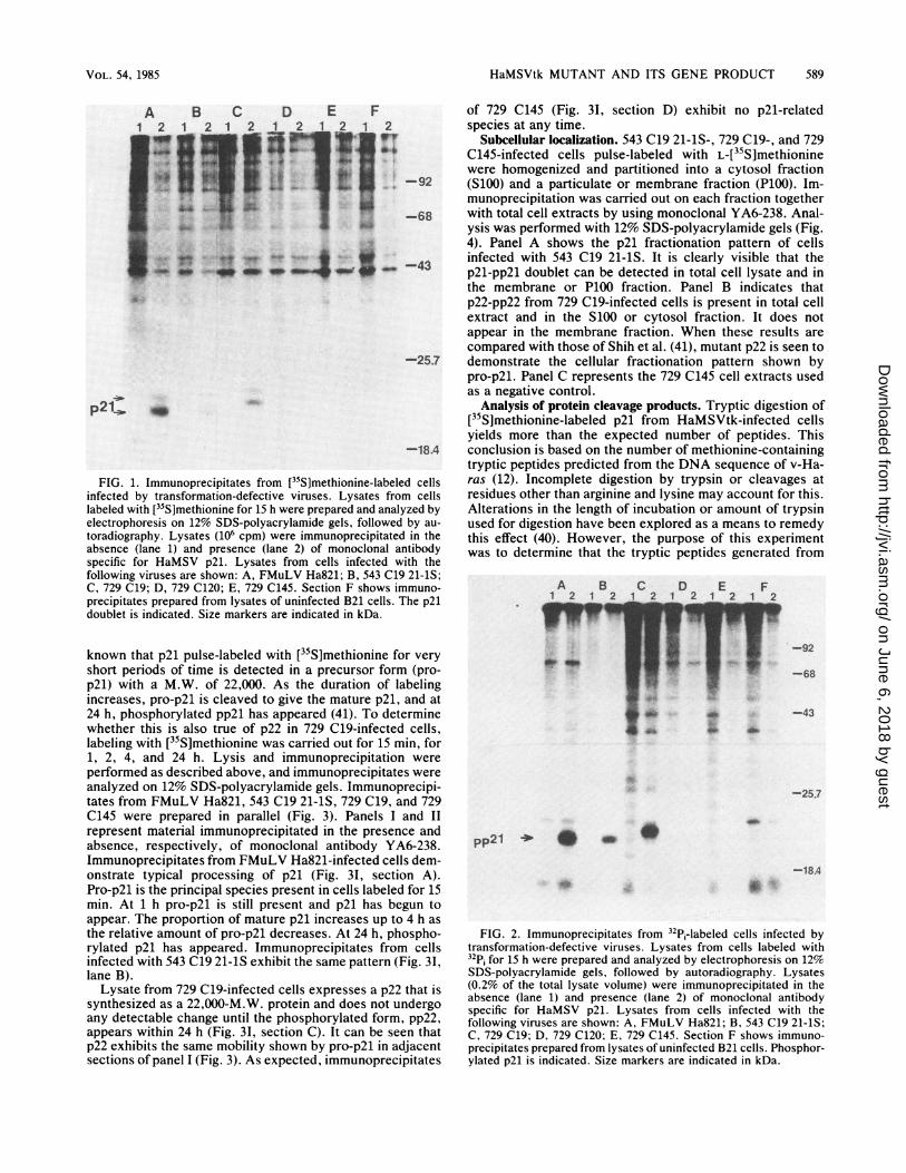
Immunoprecipitates of FMuLV Ha821-infected cellsproduce the characteristic p21 and pp2l doublet (Fig. 1A,lane 2). A doublet of the same mobility is also seen in cellsinfected with 543 C19 21-1S (Fig. 1B). In cells infected withclone 729 C19, however, a doublet of slower mobility is seen(Fig. 1C). This doublet consists of a 22,000-M.W. protein,p22, and a slower migrating species with a M.W. of 23,000.No p21-related protein is seen in immunoprecipitates fromcells infected with clone 729 C120 or 729 C145 or inuninfected B21 cells (Fig. 1D, E, and F).
In concert with the pulse-labeling of infected cells with[35S]methionine, a separate but identical study was per-formed with 32p, (Fig. 2). FMuLV Ha821-infected cellsproduce a phosphoprotein pp2l with a mobility equal to thatof the upper band in the [35S]methionine-labeled doublet(Fig. 2A). The same is true for cells infected with 543 C1921-1S (Fig. 2B). 729 C19-infected cells have a phosphopro-tein of 23,000 Mr. The mobility pattern seen here is like thatof the upper band of the characteristic p21 doublet, and thisphosphoprotein will be called pp22.
Examination of protein synthesis and processing. The p22encoded by the 729 C19 virus demonstrates a mobilityintermediate to that of the bands representing the character-istic [35S]methionine p21 doublet of HaMSV. However, it is
J. VIROL.
on June 6, 2018 by guesthttp://jvi.asm
.org/D
ownloaded from
HaMSVtk MUTANT AND ITS GENE PRODUCT 589
A B C D E F
1 2 1 2 1 2
-I
_ 0
-92
-68
-43
-25.7
p21= _
-18.4
FIG. 1. Immunoprecipitates from [35S]methionine-labeled cellsinfected by transformation-defective viruses. Lysates from cellslabeled with [35S]methionine for 15 h were prepared and analyzed byelectrophoresis on 12% SDS-polyacrylamide gels, followed by au-toradiography. Lysates (106 cpm) were immunoprecipitated in theabsence (lane 1) and presence (lane 2) of monoclonal antibodyspecific for HaMSV p21. Lysates from cells infected with thefollowing viruses are shown: A, FMuLV Ha821; B, 543 C19 21-1S;C, 729 C19; D, 729 C120; E, 729 C145. Section F shows immuno-precipitates prepared from lysates of uninfected B21 cells. The p21doublet is indicated. Size markers are indicated in kDa.
known that p21 pulse-labeled with [35S]methionine for veryshort periods of time is detected in a precursor form (pro-p21) with a M.W. of 22,000. As the duration of labelingincreases, pro-p21 is cleaved to give the mature p21, and at24 h, phosphorylated pp2l has appeared (41). To determinewhether this is also true of p22 in 729 C19-infected cells,labeling with [35S]methionine was carried out for 15 min, for1, 2, 4, and 24 h. Lysis and immunoprecipitation wereperformed as described above, and immunoprecipitates wereanalyzed on 12% SDS-polyacrylamide gels. Immunoprecipi-tates from FMuLV Ha821, 543 C19 21-1S, 729 C19, and 729C145 were prepared in parallel (Fig. 3). Panels I and IIrepresent material immunoprecipitated in the presence andabsence, respectively, of monoclonal antibody YA6-238.Immunoprecipitates from FMuLV Ha821-infected cells dem-onstrate typical processing of p21 (Fig. 31, section A).Pro-p21 is the principal species present in cells labeled for 15min. At 1 h pro-p21 is still present and p21 has begun toappear. The proportion of mature p21 increases up to 4 h asthe relative amount of pro-p21 decreases. At 24 h, phospho-rylated p21 has appeared. Immunoprecipitates from cellsinfected with 543 C19 21-1S exhibit the same pattern (Fig. 31,lane B).
Lysate from 729 C19-infected cells expresses a p22 that issynthesized as a 22,000-M.W. protein and does not undergoany detectable change until the phosphorylated form, pp22,appears within 24 h (Fig. 31, section C). It can be seen thatp22 exhibits the same mobility shown by pro-p21 in adjacentsections of panel I (Fig. 3). As expected, immunoprecipitates
of 729 C145 (Fig. 3I, section D) exhibit no p21-relatedspecies at any time.
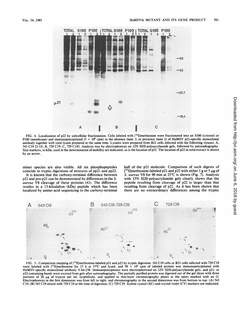
Subcellular localization. 543 C19 21-1S-, 729 C19-, and 729C145-infected cells pulse-labeled with L-[35S]methioninewere homogenized and partitioned into a cytosol fraction(S100) and a particulate or membrane fraction (P100). Im-munoprecipitation was carried out on each fraction togetherwith total cell extracts by using monoclonal YA6-238. Anal-ysis was performed with 12% SDS-polyacrylamide gels (Fig.4). Panel A shows the p21 fractionation pattern of cellsinfected with 543 C19 21-1S. It is clearly visible that thep2l-pp2l doublet can be detected in total cell lysate and inthe membrane or P100 fraction. Panel B indicates thatp22-pp22 from 729 C19-infected cells is present in total cellextract and in the S100 or cytosol fraction. It does notappear in the membrane fraction. When these results arecompared with those of Shih et al. (41), mutant p22 is seen todemonstrate the cellular fractionation pattern shown bypro-p21. Panel C represents the 729 C145 cell extracts usedas a negative control.
Analysis of protein cleavage products. Tryptic digestion of[35S]methionine-labeled p21 from HaMSVtk-infected cellsyields more than the expected number of peptides. Thisconclusion is based on the number of methionine-containingtryptic peptides predicted from the DNA sequence of v-Ha-ras (12). Incomplete digestion by trypsin or cleavages atresidues other than arginine and lysine may account for this.Alterations in the length of incubation or amount of trypsinused for digestion have been explored as a means to remedythis effect (40). However, the purpose of this experimentwas to determine that the tryptic peptides generated from
A B C D E F1 21r2 12 121t2 12"V__-
_P-92
-68
-43
*U
-25.7
pp21 * _-
-18.4
FIG. 2. Immunoprecipitates from 32Pi-labeled cells infected bytransformation-defective viruses. Lysates from cells labeled with32Pi for 15 h were prepared and analyzed by electrophoresis on 12%SDS-polyacrylamide gels, followed by autoradiography. Lysates(0.2% of the total lysate volume) were immunoprecipitated in theabsence (lane 1) and presence (lane 2) of monoclonal antibodyspecific for HaMSV p21. Lysates from cells infected with thefollowing viruses are shown: A, FMuLV Ha821; B, 543 C19 21-1S;C, 729 C19; D. 729 C120; E, 729 C145. Section F shows immuno-precipitates prepared from lysates of uninfected B21 cells. Phosphor-ylated p21 is indicated. Size markers are indicated in kDa.
VOL. 54, 1985
on June 6, 2018 by guesthttp://jvi.asm
.org/D
ownloaded from

590 WEEKS ET AL.
IB C I )D1234 5 1 2 3 4 5 11 2 3 4 5
468
8. rnbq! 0;" 43
A1 4 5 1
I~~~~~~~~~~~~~~~~~~~~~~~~~~~~~~~I
p1&-
44nFI X n~~~~~~~~~~~~~~~~~~~~~~~~~~~~~
FIG. 3. Protein processing in cells infected with wild-type and transformation-defective viruses. Immunoprecipitation was performed on
lysates prepared from cells that were labeled with [35S]methionine for 15 min and 1, 2, 4, and 24 h (lanes 1 to 5, respectively) in the presence
(panel I) and absence (panel II) of monoclonal antibody specific for HaMSV p21. Lysates were prepared from B21 cells infected with thefollowing viruses: A, FMuLV Ha821; B, 543 C19 21-1S; C, 729 C19; D, 729 C145. Analysis was by electrophoresis on 12% SDS-polyacryl-amide gels, followed by autoradiography. Size markers, in kDa, used in the determination of mobility are indicated, as is the location of p21.
HaMSVtk p21 and those generated by cleavage of 729 C19p22 under the same conditions were not widely variant. Forthis reason, HaMSVtk p21 or mutant p22 partially purifiedby SDS-polyacrylamide gel electrophoresis was digestedexactly as described previously (40). This was performedseparately on each protein and on a one-to-one mixture ofboth.
Examination of the peptide map produced from 543 C19p21 or from 729 C19 p22 reveals that there are two majorpeptides and a variety of minor peptides whose patterns are
very similar but may not be identical (Fig. SA and C).Digestion of a mixture of the two proteins, however, revealsthat the peptides generated by each protein do indeed havethe same mobilities in this system (Fig. SB). This wouldindicate that the sequences of these proteins are verysimilar, although there may be differences in regions that donot contain methionine.A similar study was performed with 32P1-labeled p21 and
p22. In this case there seem to be three principal phosphopep-tides that have a similar distribution pattern (Fig. 6). Three
l
*25.7
P21
*18.4
II
*25A
<18.4
J. VIROL.
on June 6, 2018 by guesthttp://jvi.asm
.org/D
ownloaded from
HaMSVtk MUTANT AND ITS GENE PRODUCT 591
P100
I
U-
P1001 vm
' -68
-43
-25.7
4-18.4
A I B l C
FIG. 4. Localization of p22 by subcellular fractionation. Cells labeled with [35S]methionine were fractionated into an S100 (cytosol) orP100 (membrane) and immunoprecipitated (5 x 10⁶ cpm) in the absence (lane 1) or presence (lane 2) of HaMSV p21-specific monoclonalantibody together with total lysate prepared at the same time. Lysates were prepared from B21 cells infected with the following viruses: A,543 C19 21-1S; B, 729 C19; C, 729 C145. Analysis was by electrophoresis on 12% SDS-polyacrylamide gels, followed by autoradiography.Size markers, in kDa, used in the determination of mobility are indicated, as is the location of p21. The location of p22 in total extract is shownby an arrow.
minor species are also visible. All six phosphopeptidescoincide in tryptic digestions of mixtures of pp2l and pp22.
It is known that the carboxy-terminal difference betweenp21 and pro-p21 can be demonstrated by differences in the S.aureus V8 cleavage of these proteins (41). The differenceresides in a 13-kilodalton (kDa) peptide which has beenlocalized by amino acid sequencing to the carboxy-terminal
half of the p21 molecule. Comparison of such digests of[35S]methionine-labeled p21 and p22 with either 1 g or 5 ,ug ofS. aureus V8 for 90 min at 25°C is shown (Fig. 7). Analysiswith 15% SDS-polyacrylamide gels clearly shows that thepeptide resulting from cleavage of p22 is larger than thatresulting from cleavage of p21. As it has been shown thatthere are no extraordinary differences among the tryptic
A 543 C19
cv
B 543 C19-729 C19
cv
XC-
a *
*40
+ 9
00
0
FIG. 5. Comparison mapping of [35S]methionine-labeled p21 and p22 by tryptic digestion. 543 C19 cells or B21 cells infected with 729 C19were labeled with [35S]methionine for 15 h at 37°C and lysed, and 50 x 106 cpm of labeled protein was immunoprecipitated withHaMSV-specific monoclonal antibody YA6-238. Immunoprecipitates were electrophoresed on 12% SDS-polyacrylamide gels, and p21- or
p22-containing bands were excised from gels after autoradiography. The partially purified protein was digested out of the gel slices with threeportions of 20 ,ug of trypsin per ml, lyophilized, and applied to thin-layer chromatography plates at the spots marked with an 0.
Electrophoresis in the first dimension was from left to right, and chromatography in the second dimension was from bottom to top. (A) 543C19; (B) 543 C19 mixed with 729 C19 at the time of digestion; (C) 729 C19. Xylene cyanol (XC) and crystal violet (CV) markers are indicated.
p21
C 729 C19cv
_ + 0
\ \\
VOL. 54, 1985
on June 6, 2018 by guesthttp://jvi.asm
.org/D
ownloaded from

592 WEEKS ET AL.
1-.xc,
A
A
+ 'xc.
I IX- v
+
A
.xc
AA
A lB ICFIG. 6. Comparison mapping of 32P-labeled p21 and p22 by tryptic digestion. 543 C19 cells or B21 cells infected with 729 C19 were labeled
with 32p; for 15 h at 37°C. Lysis and analysis were carried out as described in the legend to Fig. 3. (A) 543 C19; (B) 543 C19 mixed with 729C19 at the time of digestion; (C) 729 C19. Xylene cyanol (XC) markers are indicated. To facilitate photography, whole thin-layerchromatography plates are not shown.
peptides of these two proteins, these results iIsize difference between p21 and p22 must rcarboxy-terminal peptide (41).
Determination of the protein phosphorylatiphosphorylation site and surrounding amino aof HaMSV p21 have been determined by Shih eThe phosphorylated residue has been found toresidue 59 in the p21 amino acid sequence (12,
Protein samples were prepared by immunoprlysate from 543 C19 cells or cells infected withhad been labeled overnight with 32p;. Phospholand mutant pp22 were eluted from SDS-polyacr
A
1 2 3
25.7-
1 8.4-
1 4 -
FIG. 7. S. aureus V8 cleavage of p21 and p22. Pzp21 and p22 prepared from lysates of [35S]methionirby immunoprecipitation (50 x 106 cpm) and electrop]SDS-polyacrylamide gels were eluted from wet gelsography. Samples prepared from 543 C19 cells or cell729 C19 were digested with 1 or 5 ,ug of S. aureus25°C for 90 min. Cleavage products were analyzedpolyacrylamide gels and autoradiographed for 1 weep21; (B) 729 C19 p22. Lanes: 1, untreated; 2, 1 jig o13, 5 ,ug of S. aureus V8. Size markers are expresse
mply that the lyophilized, and hydrolyzed with 6 N HCl at 110HC for 3 h.eside in this By using the two-dimensional analysis system described by
Eckhart et al. (13) and comparing the mobility of theion site. The isotopically labeled amino acids with that of unlabeledLcid sequence internal phosphoamino acid standards, it can be seen thatet al. (39, 40). the residue phosphorylated in 729 C19-infected cells is alsobe threonine threonine (Fig. 8). Traces of phosphoserine can be seen in39). the mixture of both phosphopeptides (Fig. 8B). This proba-
*ecipitation of bly represents phosphorylation of cellular p21 (25). Whati729 C19 that appears to be a large residue to the left of phosphothreonineproteins pp2l in Fig. 8 represents incomplete hydrolysis products. Theylamide gels, material migrating toward the positive electrode is inorganic
phosphate.Examination of fatty acid association. Buss and Sefton (4)
B and Sefton et al. (38) have shown that the membrane-asso-ciated transforming proteins of Rous avian sarcoma virus
2 3 (pp6Osrc), HaMSV (p2lras), and Abelson murine leukemiavirus (p120gag-abl) contain tightly bound lipid (4, 38). It alsohas been shown by Garber et al. that species of Rous aviansarcoma virus that are temperature sensitive for transforma-tion are not lipid associated at nonpermissive temperatures(17). Therefore, to verify that the p21 expressed in HaMSVtkcells is lipid associated and to explore this aspect in relationto mutant p22, 543 C19 cells or cells infected with 729 C19 or
*"'f 729 C145 were labeled in parallel with L-[35S]methionine and[9,10-3H(N)]palmitic acid for 15 h. Lysate preparation andimmunoprecipitation were carried out as described above.Labeled proteins were resolved on 12% SDS-polyacryl-amide gels (Fig. 9). Panel 1 shows immunoprecipitated p21and p22 labeled with [35S]methionine (sections A and B, +).
artially purified Panel 2 shows the same material labeled with [3H]palmitateie-labeled cells and autoradiographed for 1 day. When p21 labeled withhoresis on 12% [3H]palmitate was autoradiographed for 1 week, it was
as infected with clearly visible as a doublet (panel 3, + in section A). The p22V8 protease at does not appear as a palmitate-labeled species at 1 week orlon 15% SDS- in longer exposures (panel 3, + in section B). Cells infectedk. (A) 543 C19 with 729 C145, therefore, do not exhibit any palmitate-la-f S. aureus V8; beled p21-related proteins.d in kDa. Guanine nucleotide binding and autophosphorylation. To
J. VIROL.
on June 6, 2018 by guesthttp://jvi.asm
.org/D
ownloaded from

HaMSVtk MUTANT AND ITS GENE PRODUCT 593
+
*. . P-TyrI
.-sz
..h;, , -PSx
0
A
+
p-Tyr
_'0.
p-l*1. ..
P-hr&r,
B
+
- P-Ty r
P-Thr .
& - P-Ser
0)
CFIG. 8. Phosphoamino acid analysis of p22. Samples of 32P -labeled p21 or p22 were prepared as described in the text (initial lysate, 50
x 106 cpm), hydrolyzed in 6 N HCI for 3 h at 110°C, lyophilized, and analyzed in two dimensions, with the internal markers indicated.Electrophoresis in the first dimension is the horizontal direction. Chromatography in the second dimension is from bottom to top. (A) 543 C19p21; (B) 543 C19 p21 mixed with 729 C19 p22 before hydrolysis; (C) 729 C19 p21. The origin is marked with an 0.
ascertain whether the GDP-binding activity associated withp2lras was also associated with the mutant p22 protein, thestandard binding assay was performed on extracts of NIH3T3 cells infected with FMuLV 543 C19, 729 C19, or 729C145 (16). These lysates represented total extract, S100, andP100 from each cell line. Since preliminary studies showedthat nonspecific binding in these extracts was relatively high,extracts were partially purified and concentrated with am-monium sulfate (39). It was assumed that the nucleotide-binding activity would track with the appearance of associ-ated proteins in similar fractionations. The results of bindingstudies carried out on these preparations are presented (Fig.10).
It can be seen that extracts of FMuLV 543 C19 do bindGDP in femtomole quantities when prepared under theconditions described. Furthermore, it can be seen that GDPbinding by HaMSVtk p21 increases when purified as a P100
A B C- + -
A B C
cell fraction (Fig. 10). There is no binding activity in an S100fraction prepared simultaneously.GDP-binding activity is very low in total extracts of cells
infected with 729 C19. As expected, binding activity in-creases almost sevenfold in an S100 preparation and isalmost negative in the corresponding P100 membrane frac-tion. Binding activity is also lacking in extracts of 729C145-infected cells at the levels tested.The published data on total extracts of HaMSV-infected
NIH 3T3 cells indicates that 0.9 pmol of [3H]GDP is boundper mg of protein in a total cell extract (39). This translatesinto 900 fmollmg or 0.9 fmolI,ug. In this assay, the bindingactivity of HaMSVtk p21 is therefore within the publishedrange. It is also clear that unpurified extracts of cells infectedwith 729 C19 demonstrate considerably less binding activity.To verify the specificity of nucleotide bindinf exhibited by
729 C19 p22, a 20-fold excess (4.1 x 10- M) of cold
A B C4- _ +_ W_
-43
-25.7
P21
-18.4
1 1 2 13JFIG. 9. Labeling of p21 and p22 by [3H]palmitate. Infected B21 cells were labeled with [9,10-3H(N)Ipalmitic acid for 15 h at 37°C. Lysis
and immunoprecipitation of 0.1% of the total lysate was performed in the presence (+) and absence (-) of monoclonal YA6-238 as describedin the text. Analysis was by electrophoresis on 12% SDS-polyacrylamide gels, followed by autoradiography for 24 h (panels 1 and 2) or 1 week(panel 3). (A) 543 C19 cell lysate; (B) 729 C19 infected cell lysate; (C) 729 C145 infected cell lysate. Size markers are expressed in kDa.
VOL. 54, 1985
I v:
on June 6, 2018 by guesthttp://jvi.asm
.org/D
ownloaded from

594 WEEKS ET AL.
nucleotide competitor was incubated with either FMuLV543 C19 P100 or 792 C19 S100 for 30 min before addinglabeled [3H]GDP. After the isotope was added, the assaywas carried out as usual (Table 2). It can be seen that GDP,GTP, dGDP, and dGTP are effective competitors for thenucleotide-binding site in either p21 or mutant p22. Itappears that there are some variations in the effects thatadenine nucleotides have on the binding of [3H]GDP by p21and p22 in this assay, but it cannot be said that adeninenucleotides prevent binding by either p21 or p22. UTP alsoappears to compete for the GDP-binding site in both p21 andp22. This was also shown by Scolnick et al. (36). GMP,cGMP, TTP, and CTP do not affect binding (data notshown).The upper band of the p21 doublet has been shown to be
phosphate labeled in infected cells and can be labeled invitro through phosphate donation by [-y-32P]GTP (39, 40). Itis also true that HaMSVtk p21 can be autophosphorylated invitro. Immunoprecipitates of 12 Fxg (lane 1) or 25 ,ug (lane 2)of an ammonium sulfate-purified P100 from FMuLV 543C19-infected cells are shown (Fig. 11A). Samples carriedthrough immunoprecipitation in the absence of monoclonalantibody to p21 are not detected in this assay (lane 3). S100from 729 C19-infected cells phosphorylates p22 visibly onlywhen immunoprecipitated at the higher concentration (Fig.liB, lane 2). It is also useful to note that a 30,000-M.W.protein is phosphorylated under these conditions. Such aspecies has been seen among proteins made from a simianvirus 40-HaMSV recombinant that enhances RNA transcrip-tion from in-phase initiation sites further upstream in the rasgene (18). In the absence ofmonoclonal antibody no phospho-
TABLE 2. Nucleotide competition: [3H]GDP binding[3H]GDP binding (fmol/p.g)
Competitor FMuLV 543 C19 P100 729 C19 S100
GDP 0.038 ± 0.005 0.029 ± 0.029GTP 0.060 ± 0.014 0.036 ± 0.036dGDP 0.018 ± 0.011 0.001 ± 0.0005dGTP 0.037 ± 0.012 0.022 ± 0.017ATP 0.650 ± 0.090 0.313 ± 0.237ADP 0.460 ± 0.160 0.212 ± 0.167dATP 0.560 ± 0.130 0.472 ± 0.377dADP 0.825 ± 0.175 0.225 ± 0.21UTP 0.320 0.200 ± 0.070No competitor 0.825 ± 0.175 0.565 ± 0.395
rylation is seen. A P100 prepared from 729 C145-infectedcells does not phosphorylate under these conditions.
DISCUSSION
Development of a ras gene mutant of HaMSVtk producingp22. It is evident that p22 must possess antigenic determi-nants intrinsic to p21, as the immunoprecipitability of a p22doublet when labeled with [35S]methionine has been con-firmed. The nonphosphorylated form of this doublet exhibitsa mobility intermediate to those of the standard p21 bandsand is therefore reminiscent of the pro-p21 precursor proteinfound in HaMSV-infected cells (41). The possibility that the
0zD0m
c]00
00I
a,0
200
100
G
200
100i
200 - * S1000 P100
100 _
O t
0 25 50 100 200pg PROTEIN
FIG. 10. [3H]GDP binding by subcellular protein fractions. Totallysate and subcellular fractions (S100 and P100) from NIH 3T3 cellsinfected with 729 C19 and from cells infected with FMuLV 543 C19were concentrated, partially purified by ammonium sulfate precipi-tation, dialyzed extensively, and incubated with [3H]GDP. BoundGDP is expressed in femtomoles, where 13,100 cpm = 1 pmol or1,000 fmol. Protein concentrations are expressed in micrograms.
A1 2 3
B C1 2 3 1 2 3
_.F.s..t-
i..W_...
-43
-25.7
-18.4
FIG. 11. Autophosphorylation activity of partially purified p21and p22. Partially purified p21 and p22 were immunoprecipitated inthe presence or absence of monoclonal YA6-238 as described in thetext. Immunoprecipitates were then labeled in vitro with phosphatedonated by [y-32P]GTP by incubation for 30 min at 37°C. Phosphor-ylated proteins were analyzed by SDS-polyacrylamide gel elec-trophoresis, followed by autoradiography. (A) FMuLV 543 C19P100; (B) 729 C19 S100; (C) P100 from 729 C145. Lanes: 1, 12 ,ugplus antibody; 2, 25 ,ug plus antibody; 3, 25 ,ug without antibody.Size markers are expressed in kDa.
J. VIROL.
on June 6, 2018 by guesthttp://jvi.asm
.org/D
ownloaded from

HaMSVtk MUTANT AND ITS GENE PRODUCT 595
729 C19 virus is a processing-defective mutant of HaMSVtkmust therefore be considered. In general, the characteriza-tion of p22 should contribute toward an utiderstanding of therelationship between p21 and cellular transformation byhelping to identify the cellular target and the transformingfunctions of p21.
Characterization of a mutant of HaMSVtk producing p22.The development of the p22 mutant of HaMSVtk has severalimportant implications for the study of transforming proteinsin relation to cell structure and function. In particular, thecharacterization of p22 generates certain conclusions aboutthe relationship between p21 and the maintenance of trans-formation in cells. Therefore, it is necessary to recapitulatethe known data on p22 in this context.
It is evident that p22 must possess many of the antigenicdeterminants intrinsic to p21. Aside from its immunoprecipi-tability as a doublet when labeled with [35S]methionine,tryptic digestion of p22 produces multiple peptides thatappear to be identical to those generated for p21 under thesame conditions. Both proteins exist in a phosphorylatedform, and threonine is the amino acid phosphorylated ineach case (40, 43). Furthermore, the kinetics of phosphor-ylation in pulse-labeling of cells with 32Pi are the same.Tryptic digests of these phosphorylated proteins are highlyrelated as well. This implies that p21 and p22 must also bevery similar in amino acid composition. However, it isapparent that p22 demonstrates a reduced mobility withrespect to that of p21. Comparison of protein processing,when monitored by labeling with [35S]methionine, indicatesthat p21 and p22 are both synthesized as "pro-proteins"with an Mr of 22,000. However, although pro-p21 is cleavedto generate the mature p21, the mobility of p22 does notchange. Both proteins still give rise to a second proteinmoiety of decreased mobility within 24 h, and this speciescomigrates with the band appearing during pulses with 32p,under the same conditions. This could suggest the presenceof a mutation within the p22-encoding nucleic acid sequenceresulting either in an aberrant cleavage site or in an alteredenzyme recognition site preventing enzymatic cleavage fromoccurring. Furthermore, p22 does not bind palmitate, sug-gesting a defect at the fatty acid binding site or a process-ing-membrane association-related failure of p22 to be trans-located to the fatty acid binding locus within the cell.
Pro-p21 is synthesized on free ribosomes in cell cytoplasmand can consequently be isolated by crude fractionation ofcells into an S100 or cytosol fraction (41). Mature p21 ismembrane associated (16, 41, 54). Mature p21 also can beisolated as part of the P100 or membrane fraction of cells.Examination of cells infected with mutant 729 C19 revealsthat p22 is found in the S100 from fractionated cells. Com-parison of fragments generated from [35S]methionine-labeledp21 and p22 cleavage with S. aureus V8 indicates that thesize difference between the two proteins is probably at thecarboxy terminus of p22 as is true for the size differencebetween p21 and pro-p21 (41). From this it could be con-strued that p22 is altered at a membrane binding site or thatfailure to be cleaved prevents exposure of the necessarybinding site. Therefore, these studies imply that p22 is aproduct of the mutagenized ras gene of HaMSV that isdefective for protein processing or membrane association.This mutant may be comparable to those developed byWillumsen et al. (55), in which a terminal cysteine residue isaltered. However, it is still not clear whether processing ormembrane association is the initial event.
It is significant to note that, although cells containing p22do not undergo transformation, p22 demonstrates GDP-bind-
ing activity and is capable of autophosphorylation with[_y-32P]GTP as a phosphate donor. However, p22 appears todiffer from p21 in specificity of nucleotide binding. Nucleo-tide competition studies reveal that p22 has some affinity foradenosine nucleotides that contain multiple phosphate moi-eties. This could be explained by a gene mutation resulting inamino acid differences in the nucleotide binding site of p22.Alternatively, the failure of p22 to undergo processing mayinterfere with the folding of the protein. This could result inan altered binding site that possesses the same nucleotiderecognition sites but one in which these recognition sites aremore flexible with respect to one another. As the presence oftrace impurities in the adenine nucleotide stocks used in thisassay cannot be excluded entirely, the data presented shouldbe viewed as suggestive rather than conclusive.The appearance of a transient phosphoprotein pp3O during
autophosphorylation of p21 has been noted previously (un-published data) so its appearance under the same conditionsin cell lysates containing p22 is not unusual. As mentionedbefore, it is known that a p30 can result from the transcrip-tion of an open reading frame further upstream in the rasgene (12, 18). Phosphorylation of this protein, which may bepresent at low levels in infected cells, will give rise to thepp30 seen in these studies.
Considerations of p21 and p22 in relation to other oncproteins. In these studies we have examined the biochemicalproperties of a mutant of the ras gene that is defective fortransformation. This gene is unique in that it produces aprotein highly related both structurally and functionally tothe transforming protein. The failure of mutant p22 and pp22to become membrane associated suggests that membraneassociation is vital to cell transformation.
Several laboratories have demonstrated that a variety ofmurine retroviral structural (gag-gene-encoded) polypro-teins undergo posttranslational myristylation (4, 21, 33, 34).The presence of an amino-terminal myristyl-amide linkageenhances the hydrophobicity of these proteins and is thoughtto facilitate their membrane association (17). Since myristyl-ated amino termini are found in gag precursor and cleavageproteins, it is suggested that fatty acid association occursduring protein biosynthesis and must therefore be a prereq-uisite to membrane association (30-32). A variety of gag-oncfusion proteins are also myristylated (33). This suggests thatmyristylation may play a role (specific membrane attach-ment) in the transformation process. Palmitate has not beenfound in association with any of the gag polyproteins tested,but palmitate labeling has been used to examine some of thegag-onc fusion proteins and with other transforming pro-teins. Among these are Rous sarcoma virus pp60src, Abelsonsarcoma virus p12Mag-abl, and HaMSV p2lras (38). Notably,in the case of pp6Osrc, it has been found that labeledpalmitate is metabolized to myristate which is found inassociation with the onc gene protein. Therefore, it has beensuggested that fatty acid association is necessary for mem-brane association. This is supported by Garber et al., whofound that temperature-sensitive mutants of Rous sarcomavirus fail to be palmitate or membrane associated at nonper-missive temperatures (17).
Fatty acid binding in pp6Osrc and p21ras is posttranslation-al, occurring in approximately the same time frame asmembrane association and cleavage (38). It is not known,however, whether palmitate binding is a prerequisite to or aresult of membrane association. The results of Schultz andOroszlan suggest that lipid binding may be the initial event inthe transformation process (33). If so, the principle defect inthe p22ras mutant may be failure to bind fatty acid. However,
VOL. 54, 1985
on June 6, 2018 by guesthttp://jvi.asm
.org/D
ownloaded from

596 WEEKS ET AL.
unpublished data (A. Papageorge, personal communication)suggest that palmitation of p21 occurs in the carboxy-termi-nal portion of the cleaved molecule. Although the palmita-tion site has only been localized to the terminal V8 cleavagefragment, one cannot rule out the possibility that an altera-tion of the p21 cleavage site could also prevent palmitationand interfere with membrane association. That these eventsare related and associated with transformation by p21 is notcontested. It is therefore postulated that p21 contains at leasttwo functional domains, one which is responsible for thenucleotide binding activity and a second which is affected byor results in the cleavage and activation of the maturemembrane-bound transforming protein. The mutant protein,p22, has been altered with respect to the second function.That p21 may consist of more than one domain is not a
novel idea. The concept of a transforming protein composedof multiple functional domains (a protein kinase and asecond distinct, but necessary, membrane-associated func-tion) is also proposed for the avian src protein (2, 3, 15).Whether these domains are cis acting in the case of HaMSVp21 is not known for certain at this time (25).
In conclusion, although the time sequence of these eventsis still unclear, it does appear that transformation in theHaMSV system is correlated with protein processing, mem-brane association, and lipid binding by the ras gene product.Furthermore, the transformation protein of HaMSV consistsof at least two domains which must function in tandem fortransformation to occur.
ACKNOWLEDGMENTAll work mentioned herein was performed in the Laboratory of
Tumor Virus Genetics, National Cancer Institute.
LITERATURE CITED1. Bonner, W. M., and R. A. Laskey. 1974. A film detection
method for tritium-labeled proteins and nucleic acids in poly-acrylamide gels. Eur. J. Biochem. 46:83-88.
2. Bryant, D., and J. T. Parsons. 1982. Site-directed mutagenesisof the src gene of Rous sarcoma virus: construction andcharacterization of a deletion mutant temperature sensitive fortransformation. J. Virol. 44:683-691.
3. Bryant, D., and J. T. Parsons. 1983. Site-directed point mutationin the src gene of Rous sarcoma virus results in an inactive srcgene product. J. Virol. 45:1211-1216.
4. Buss, J. E., and B. M. Sefton. 1985. Myristic acid, a rare fattyacid, is the lipid attached to the transforming protein of Roussarcoma virus and its cellular homolog. J. Virol. 53:7-12.
5. Chang, E. H., R. W. Ellis, E. M. Scolnick, and D. R. Lowy. 1980.Transformation of cloned Harvey murine sarcoma virus DNA:efficiency increased by long terminal repeat DNA. Science210:1249-1251.
6. Chang, E. H., M. E. Furth, E. M. Scolnick, and D. R. Lowy.1982. Tumorigenic transformation of mammalian cells inducedby a normal human gene homologous to the oncogene of Harveymurine sarcoma virus. Nature (London) 297:479-483.
7. Chang, E. H., J. M. Maryak, C.-M. Wei, T. Y. Shih, R. Shober,H. L. Cheung, R. W. Ellis, G. L. Hager, E. M. Scolnick, andD. R. Lowy. 1980. Functional organization of the Harvey mur-ine sarcoma virus genome. J. Virol. 35:76-92.
8. Cleveland, D. W., S. G. Fisher, M. W. Kirschner, and U. K.Laemmli. 1977. Peptide mapping by limited proteolysis in so-dium dodecyl sulfate and analysis by gel electrophoresis. J.Biol. Chem. 252:1102-1106.
9. Courtneidge, S. A., A. D. Levinson, and J. M. Bishop. 1980. Theprotein encoded by the transforming gene of avian sarcomavirus (pp60src) and a homologous protein in normal cells(pp60Proto-src) are associated with the plasma membrane. Proc.Natl. Acad. Sci. U.S.A. 77:3783-3787.
10. DeFeo, D., M. A. Gonda, H. A. Young, E. H. Chang, D. R.
Lowy, E. M. Scolnick, and R. W. Ellis. 1981. Analysis of twodivergent rat genomic clones homologous to the transforminggene of Harvey murine sarcoma virus. Proc. Natl. Acad. Sci.U.S.A. 78:3328-3332.
11. Der, C. J., T. G. Krontiris, and G. M. Cooper. 1982. Transform-ing genes of human bladder and lung carcinoma cell lines arehomologous to the ras genes of Harvey and Kirsten sarcomaviruses. Proc. Natl. Acad. Sci. U.S.A. 79:3637-3640.
12. Dhar, R., R. W. Ellis, T. Y. Shih, S. Oroszlan, B. Shapiro, J.Maizel, D. Lowy, and E. Scolnick. 1982. Nucleotide sequence ofthe p21 transforming protein of Harvey murine sarcoma virus.Science 217:934-936.
13. Eckhart, W., M. A. Hutchinson, and T. Hunter. 1979. Anactivity phosphorylating tyrosine in polyoma T antigen im-munoprecipitates. Cell 18:925-933.
14. Fasano, O., D. Birnbaum, L. Edlund, J. Fogh, and M. Wigler.1984. New human transforming genes detected by a tumorige-nicity assay. Mol. Cell. Biol. 4:1695-1705.
15. Fincham, V. J., D. J. Chiswell, and J. A. Wyke. 1982. Mappingof nonconditional and conditional mutants in the src gene ofPrague strain Rous sarcoma virus. Virology 116:72-83.
16. Furth, M. E., L. J. Davis, B. Fleurdelys, and E. M. Scolnick.1982. Monoclonal antibodies to the p21 products of the trans-forming gene of Harvey murine sarcoma virus and of a cellularras gene family. J. Virol. 43:294-304.
17. Garber, E. A., J. G. Krueger, H. Hanafusa, and A. R. Goldberg.1983. Only membrane-associated RSV-src proteins have amino-terminally bound lipid. Nature (London) 302:161-163.
18. Gruss, P., R. W. Ellis, T. Y. Shih, M. Konig, E. M. Scolnick, andG. Khoury. 1981. SV40 recombinant molecules express the geneencoding p21 transforming protein of Harvey murine sarcomavirus. Nature (London) 293:486-488.
19. Hager, G. L., E. H. Chang, H. W. Chan, C. F. Garon, M. A.Israel, M. A. Martin, E. M. Scolnick, and D. R. Lowy. 1979.Molecular cloning of the Harvey sarcoma virus closed circularintermediates: initial structural and biological characterization.J. Virol. 31:795-809.
20. Hampar, B., J. G. Derge, A. L. Boyd, M. A. Tainsky, and S. D.Showalter. 1981. Herpes simplex virus (type 1) thymidine kinasegene does not transform cells morphologically. Proc. Natl.Acad. Sci. U.S.A. 78:2616-2619.
21. Henderson, L. E., H.C. Krutzsch, and S. Oroszlan. 1983. My-ristyl amino-terminal acylation of murine retrovirus proteins: anunusual post-translational protein modification. Proc. Natl.Acad. Sci. U.S.A. 80:339-343.
22. Jainchill, J. L., S. A. Aaronson, and G. J. Todaro. 1969. Murinesarcoma and leukemia viruses: assay using clonal lines ofcontact-inhibited mouse cells. J. Virol. 4:549-553.
23. Laemmli, U. K. 1970. Cleavage of structural proteins during theassembly of the head of bacteriophage T4. Nature (London)227:680-685.
24. Lowry, 0. H., N. J. Rosebrough, A. L. Farr, and R. J. Randall.1951. Protein measurement with the Folin phenol reagent. J.Biol. Chem. 193:265-275.
25. Papageorge, A., D. Lowy, and E. M. Scolnick. 1982. Compara-tive biochemical properties of the p21 ras molecules coded forby viral and cellular ras genes. J. Virol. 44:509-519.
26. Parada, L. F., C. J. Tabin, C. Shih, and R. A. Weinberg. 1982.Human EJ bladder carcinoma oncogene is a homologue ofHarvey sarcoma virus ras gene. Nature (London) 297:474-478.
27. Reddy, E. P., R. K. Reynolds, E. Santos, and M. Barbacid. 1982.A point mutation is responsible for the acquisition of transform-ing properties by the T24 human bladder carcinoma oncogene.Nature (London) 300:148-152.
28. Rowe, W. P., W. E. Pugh, and J. W. Hartley. 1970. Plaque assaytechniques for murine leukemia viruses. Virology 42:1136-1139.
29. Santos, E., S. R. Tronick, S. A. Aaronson, S. Pulciani, and M.Barbacid. 1982. T24 human bladder carcinoma oncogene is anactivated form of the normal human homologue of BALB- andHarvey-MSV transforming genes. Nature (London) 298:343-347.
30. Schlesinger, M. J., A. I. Magee, and M. F. G. Schmidt. 1980.Fatty acid acylation of proteins in cultured cells. J. Biol. Chem.
J. VIROL.
on June 6, 2018 by guesthttp://jvi.asm
.org/D
ownloaded from

HaMSVtk MUTANT AND ITS GENE PRODUCT 597
255:10021-10024.
31. Schmidt, M. F. G., and M. J. Schlesinger. 1979. Fatty acidbinding to vesicular stomatitus virus glycoprotein: a new type ofpost-translational modification of the viral glycoprotein. Cell17:813-819.
32. Schmidt, M. F. G., and M. J. Schlesinger. 1980. Relation of fattyacid attachment to the translation and maturation of vesicularstomatitus and Sindbis virus membrane glycoproteins. J. Biol.Chem. 255:3334-3339.
33. Schultz, A. M., and S. Oroszlan. 1983. In vivo modification ofretroviral gag gene-encoded polyproteins by myristic acid. J.Virol. 46:355-361.
34. Schultz, A. M., and S. Oroszlan. 1984. Myristylation of gag-oncfusion proteins in mammalian transforming viruses. Virology133:431-437.
35. Schwab, M., K. Alitalo, H. E. Varmus, J. M. Bishop, and D.George. 1983. A cellular oncogene (c-Ki-ras) is amplified, over-
expressed, and located within karyotypic abnormalities in mouseadrenocortical tumour cells. Nature (London) 303:497-501.
36. Scolnick, E. M., A. G. Papageorge, and T. Y. Shih. 1979.Guanine nucleotide-binding activity as an assay for the src
protein of rat-derived murine sarcoma viruses. Proc. Natl.Acad. Sci. U.S.A. 76:5355-5359.
37. Scolnick, E. M., J. R. Stephenson, and S. A. Aaronson. 1972.Isolation of temperature-sensitive mutants of murine sarcomavirus. J. Virol. 10:653-657.
38. Sefton, B. M., I. S. Trowbridge, J. A. Cooper, and E. M.Scolnick. 1982. The transforming proteins of Rous sarcomavirus, Abelson sarcoma virus, and Harvey sarcoma virus con-tain tightly-bound lipid. Cell 31:465-474.
39. Shih, T. Y., A. G. Papageorge, P. E. Stokes, M. 0. Weeks, andE. M. Scolnick. 1980. Guanine nucleotide-binding and au-
tophosphorylating activities associated with the p21SrC protein ofHarvey murine sarcoma virus. Nature (London) 287:686-691.
40. Shih, T. Y., P. E. Stokes, G. W. Smythers, R. Dhar, and S.Oroszlan. 1982. Characterization of the phosphorylation sitesand the surrounding amino acid sequences of the p21 transform-ing proteins coded for by the Harvey and Kirsten strains ofmurine sarcoma viruses. J. Biol. Chem. 257:11767-11773.
41. Shih, T. Y., M. 0. Weeks, P. Gruss, R. Dhar, S. Oroszlan, andE. M. Scolnick. 1982. Identification of a precursor in thebiosynthesis of the p21 transforming protein of Harvey murinesarcoma virus. J. Virol. 42:253-261.
42. Shih, T. Y., M. 0. Weeks, H. A. Young, and E. M. Scolnick.1979. p21 of Kirsten murine sarcoma virus is thermolabile in aviral mutant temperature sensitive for the maintenance oftransformation. J. Virol. 31:546-556.
43. Shih, T. Y., M. 0. Weeks, H. A. Young, and E. M. Scolnick.1979. Identification of a sarcoma virus-coded phosphoprotein in
nonproducer cells transformed by Harvey or Kirsten murinesarcoma virus. Virology 96:64-79.
44. Shimizu, K., M. Goldfarb, M. Perucho, and M. Wigler. 1983.Isolation and preliminary characterization of the transforminggene of a human neuroblastoma cell line. Proc. Natl. Acad. Sci.U.S.A. 80:383-387.
45. Shimizu, K., M. Goldfarb, Y. Suard, M. Perucho, Y. Li, T.Kamata, J. Feramisco, E. Stavnezer, J. Fogh, and M. Wigler.1983. Three human transforming genes are related to the viralras oncogenes. Proc. Natl. Acad. Sci. U.S.A. 80:2112-2116.
46. Spandidos, D. A., and N. J. Agnantis. 1984. Human malignanttumours of the breast, as compared to their respective normaltissue have elevated expression of the Harvey ras oncogenes.Anticancer Res. 4:269-272.
47. Szybalska, E. H., and W. Szybalski. 1962. Genetics of humancell lines. IV. DNA-mediated heritable transformation of abiochemical trait. Proc. Natl. Acad. Sci. U.S.A. 48:2026-2034.
48. Tabin, C. J., S. M. Bradley, C. I. Bargmann, R. A. Weinberg,A. G. Papageorge, E. M. Scolnick, R. Dhar, D. R. Lowy, andE. H. Chang. 1982. Mechanism of activation of a human onco-gene. Nature (London) 300:143-148.
49. Taparowsky, E., Y. Suard, 0. Fasano, K. Shimizu, M. Goldfarb,and M. Wigler. 1982. Activation of the T24 bladder carcinomatransforming gene is linked to a single amino acid change.Nature (London) 300:762-765.
50. Troxler, D. H., J. K. Boyars, W. P. Parks, and E. M. Scolnick.1977. Friend strain of spleen focus-forming virus: a recombinantbetween mouse type-C ecotropic viral sequences and sequencesrelated to xenotropic virus. J. Virol. 22:361-372.
51. Tsuchida, N., and T. Ryder. 1982. Nucleotide sequence of theoncogene encoding the p21 transforming protein of Kirstenmurine sarcoma virus. Science 217:937-939.
52. Wei, C.-M., M. Gibson, P. G. Spear, and E. M. Scolnick. 1981.Construction and isolation of a transmissible retrovirus contain-ing the src gene of Harvey murine sarcoma virus and thethymidine kinase gene of Herpes simplex virus type 1. J. Virol.39:935-944.
53. Wierenga, R. K., and W. G. Hol. 1983. Predicted nucleotide-binding properties of p21 protein and its associated cancervariant. Nature (London) 302:842-844.
54. Willingham, M., I. Pastan, T. Y. Shih, and E. M. Scolnick. 1980.Localization of the src gene product of the Harvey strain ofmurinn sarcoma virus to the plasma membrane of transformedcells by electron microscopic immunocytochemistry. Cell19:1005-1014.
55. Willumsen, B., A. Christensen, N. L. Hubbert, A. G. Papa-george, and D. Lowy. 1984. The p21 ras C-terminus is requiredfor transformation and membrane association. Nature (London)310:583-586.
VOL. 54, 1985
on June 6, 2018 by guesthttp://jvi.asm
.org/D
ownloaded from