developing magsifter & nia to measure resistance to ... · developing magsifter & nia to...
TRANSCRIPT
Developing Magsifter & NIA to Measure Resistance to Targeted Therapy in CTCs
Viola Chen CCNE T32 presentation
Fan lab 1-18-17

Dilemma of measuring resistance to targeted therapy in renal cell carcinoma (RCC)
CT scan
How do I know it’s working?Empiric selection
Tumor Initiation
Diagnosis Treatment 3 months Resistance
Response
7 agents against VEGF pathway 2 growth inhibition drugs 1 immune modulator
10 targeted therapies
1
1.resistance mechanisms not well-understood
2.serial biopsies of tumor masses not feasible
3.Lack of sensitive, clinically validated protein signaling assays for clinical samples

Response
measure resistance earlier
Tumor Initiation
Diagnosis Treatmentuntil progression
Clinical Outcome
Resistance
2
Dilemma of measuring resistance to targeted therapy in RCC

Improving patient outcomes to targeted therapy in RCC
% survivors
3 6 9 12 15 18
100
50
0
80
PFS (months)
Standard CT scan
no benefit
3
Responders
non-responders
• reduce unnecessary toxicities • potentially increase time to tumor
resistance (or PFS) • better tumor burden control
benefits of “early failure” panel:

Project proposalHypothesis: Magsifting technology can be adapted to capture RCC circulating tumor cells (CTCs) and to measure resistance to targeted therapy in CTCs using nanoimmunoassay (NIA)
• Aim 1: Develop magnetic sifter technology to isolate CTCs from patients with RCC based on CAIX surface protein expression
• Aim 2: Profile RCC CTCs using nano immunoassay (NIA) to measure biologic resistance to drug therapy in VEGFR pathway, cell cycle and apoptosis proteins
4
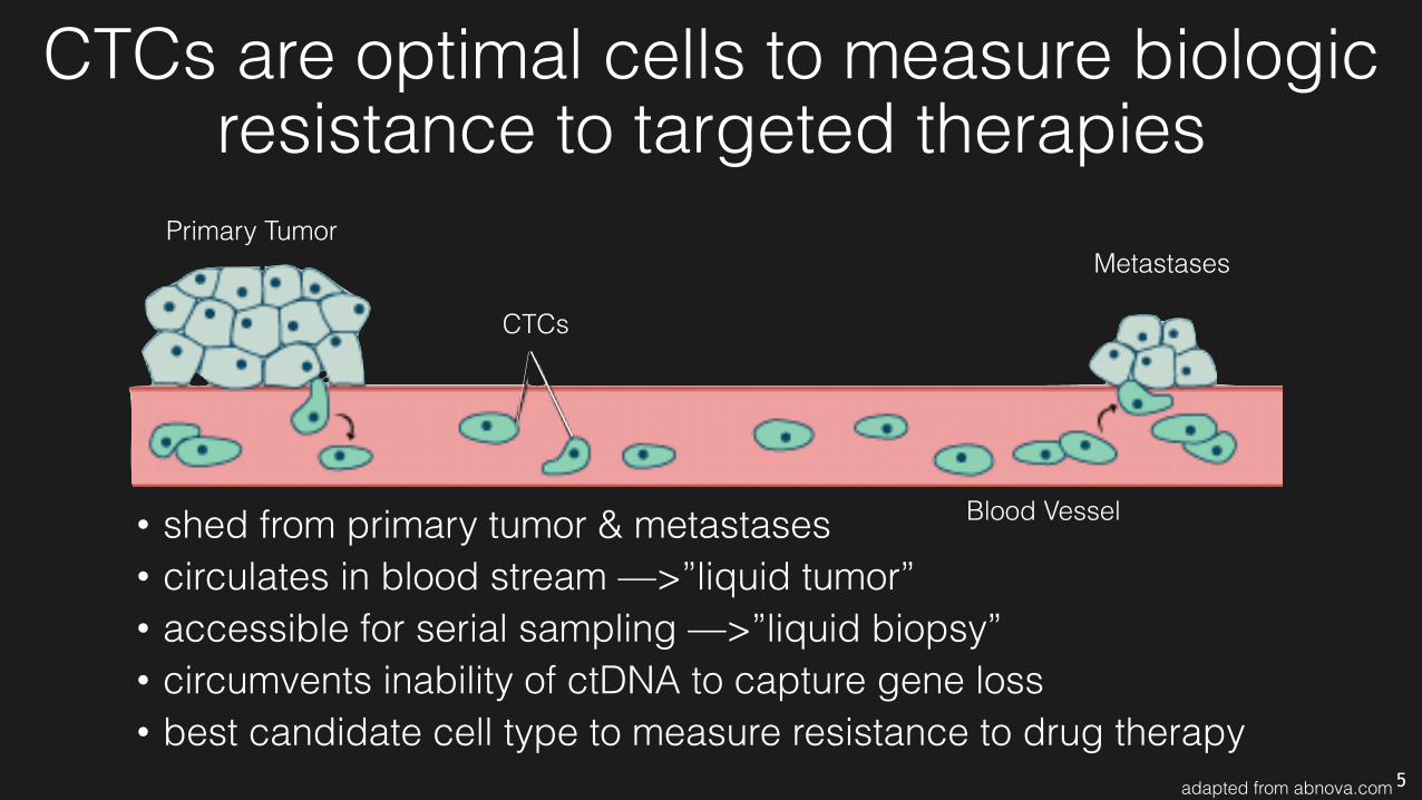
CTCs are optimal cells to measure biologic resistance to targeted therapies
• shed from primary tumor & metastases • circulates in blood stream —>”liquid tumor” • accessible for serial sampling —>”liquid biopsy” • circumvents inability of ctDNA to capture gene loss • best candidate cell type to measure resistance to drug therapy
Primary TumorMetastases
CTCs
Blood Vessel
5adapted from abnova.com

Project proposalHypothesis: We propose a novel approach to detect early therapeutic failure by assessing early signaling changes in circulating tumor cells (CTCs) before changes in tumor size are detectable by standard imaging criteria at 3 months.
• Aim 1: Develop magnetic sifter technology to isolate putative CTCs from patients with RCC based on CAIX surface protein expression
• Aim 2: Profile RCC CTCs using nano immunoassay (NIA) to measure biologic resistance to drug therapy in VEGFR pathway, cell cycle and apoptosis proteins
6

Patient blood can be magnetically “sifted” to isolate CTCs
Expansion from lung studies: Park, SM. PNAS. (2016)
Shan Wang
Jared Nesvet
Chin Chun Ooi Dawson Wong
Circulating Tumor Cell
CA9: Carbonic Anhydrase 9 (RCC cell surface marker)
Magnetic Nanoparticle
7
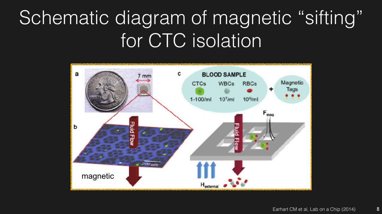
Schematic diagram of magnetic “sifting” for CTC isolation
magnetic
Earhart CM et al, Lab on a Chip (2014) 8

“Magsifter” processes multiple samples in parallel
Syringe assembly
Complete set-up
Magnetic chips
9

“Magsifted” cells from mRCC patients are distinct from WBCs by immunofluorescence
mRC
C P
atie
nts
Hea
lthy
Don
ors
LegendGreen: Pan-cytokeratin Red: CD45 Blue: DNA
Capture yield: ~60% Purity~30% Work in progress:
confirmatory immune staining of RCC-specific markers (PAX8)
10Unpublished data from the Wang Lab
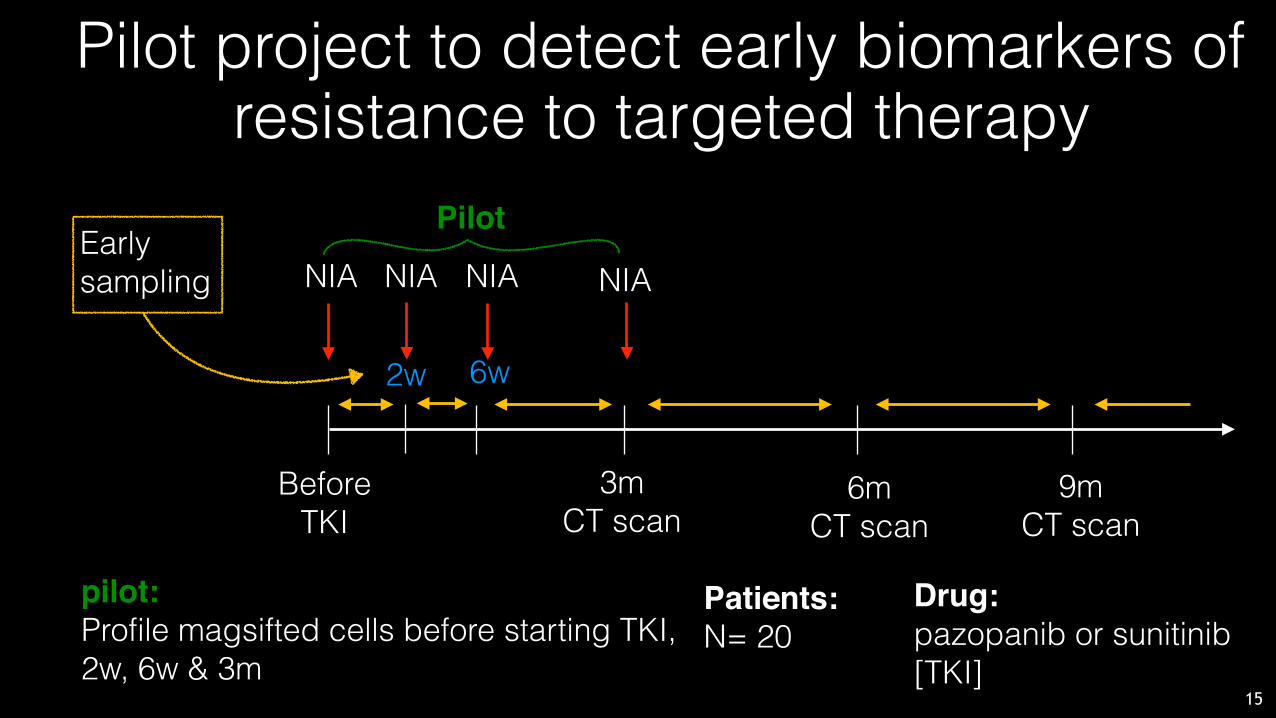
Pilot project to detect early biomarkers of resistance to targeted therapy
Patients:N= 20
15
Drug:pazopanib or sunitinib [TKI]
pilot:Profile magsifted cells before starting TKI, 2w, 6w & 3m
Pilot
NIA
6w
Before TKI
2w
NIA NIA
3m CT scan
6m CT scan
9m CT scan
Early sampling NIA

Project proposalHypothesis: We propose a novel approach to detect early therapeutic failure by assessing early signaling changes in circulating tumor cells (CTCs) before changes in tumor size are detectable by standard imaging criteria at 3 months.
• Aim 1: Develop magnetic sifter technology to isolate putative CTCs from patients with RCC based on CAIX surface protein expression
• Aim 2: Profile RCC CTCs using nano immunoassay (NIA) to measure biologic resistance to drug therapy in VEGFR pathway, cell cycle and apoptosis proteins
11
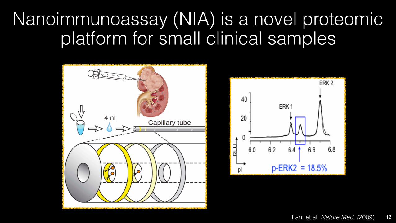
Nanoimmunoassay (NIA) is a novel proteomic platform for small clinical samples
12Fan, et al. Nature Med. (2009)
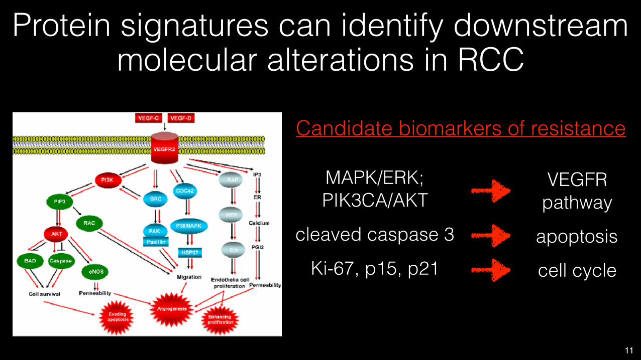
Protein signatures can identify downstream molecular alterations in RCC
11
Candidate biomarkers of resistance
MAPK/ERK; PIK3CA/AKT
cleaved caspase 3
Ki-67, p15, p21
VEGFR pathway
apoptosis
cell cycle

NIA can identify & quantify biologic response in “magsifted” cells vs PBMCs
Work in progress: • optimizing magsifting
pipeline to reliably measure ERK signaling
• in parallel, assay development for other markers in the panel
SignalIntensity
pI(isolectricpoint)
ppERK1pERK1
ppERK2ERK1
pERK2ERK2
CTCs
PBMCs
ERK2pERK2
ERK1ppERK2ppERK1
pERK1
Sifted cells
PBMCs
Sign
al in
tens
ity
pI (isoelectric point)
9.7%
0%
13
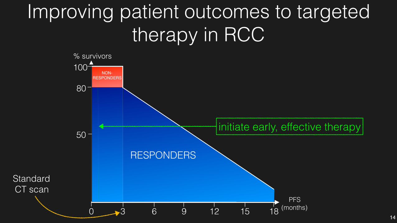
% survivors
3 6 9 12 15 18
100
50
0
80
RESPONDERS
PFS (months)
NON-RESPONDERS
Standard CT scan
14
Improving patient outcomes to targeted therapy in RCC
initiate early, effective therapy
Summary• “Liquid biopsies” are needed to track
patients’ therapeutic status on TKIs
• ‘MagSifter’ is a viable strategy to isolate putative CTCs from blood draws
• NIA can profile biologic response of sifted patient cells before & during therapy
• Biomarker panel for early failure reduces unnecessary side effects and may even lead to increased PFS or OS
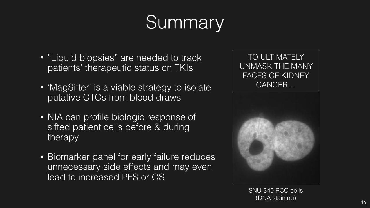
SNU-349 RCC cells (DNA staining)
TO ULTIMATELY UNMASK THE MANY FACES OF KIDNEY
CANCER…
16

AcknowledgementsAlice C. Fan (PI)Viola Chen, Christian Hoerner, Sangeeta Kowli
Materials Science & EngineeringJared Nesvet, Chin Chun Ooi, Dawson Wong, Shan X. Wang (PI)
FundingNCI T32 (Viola Chen) Stanford TRAM Program (Christian Hoerner) NCI R21 (Shan Wang/Alice Fan) NCI U54 (CCNE-TD)
ASCO (Alice Fan) Thanks for your attention!
Stanford Cancer CenterSandy Srinivas, Chia-Sui Kao
Participating patients
UrologyHongjuan Zhao, Donna M. Peehl (PI) Thomas Metzner, John Leppert (PI) CCTOYoriko Imae, Jared Bailey, Shermeen Poushnejad, Denise Haas StatisticsLaurel Stell, Chiara Sabatti (PI)
TASCShailja Patel, Joanna Liliental
CCNE-TDSam Gamhbir 17

The End

Plan to detect early clinical biomarkers
Patients:N= 20
Drug:pazopanib or sunitinib
phase I:Profile magsifted cells 2 wks after starting TKI & at 3 months
phase II:Profile at serial time points until median PFS reached for both TKIs to ensure a profile predictive of therapy failure
Before TKI
2 wks
NIA NIA NIA NIA
3m CT scan
6m CT scan
9m CT scan
Early sampling
phase I phase II
13
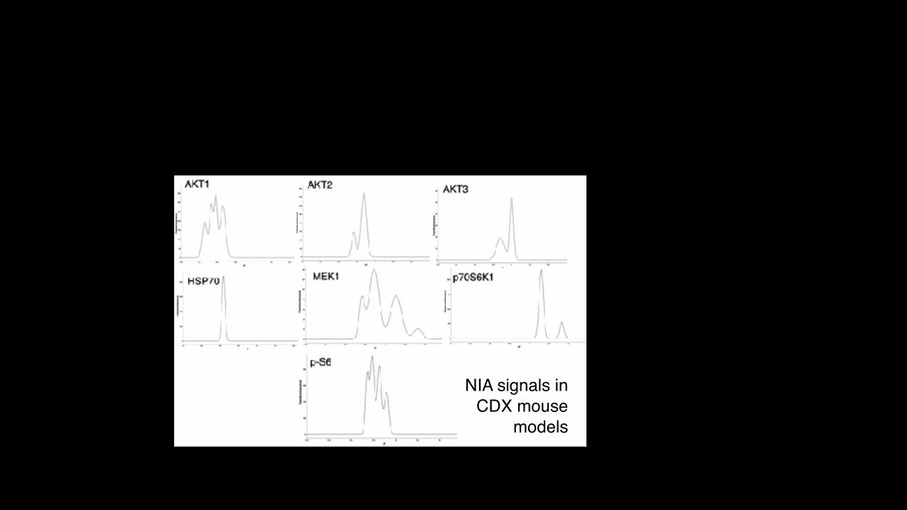
NIA signals in CDX mouse
models