determining the presence of peripheral arterial disease in
TRANSCRIPT

MEDITER
RA
NE
AN J
O U R N A L O F RH
EU
MATOLOGY
Determining the presence of Peripheral Arterial Disease in patients with Rheumatoid Arthritis
Andrea C. Grech, Alfred Gatt, Andrew A. Borg, Cynthia Formosa
Mediterr J Rheumatol 2017; 28(2):86-93
MEDITERRANEAN JOURNAL OF RHEUMATOLOGY June 2017 | Volume 28 | Issue 2
E-ISSN: 2529-198X
MEDITERRANEAN JOURNAL OF RHEUMATOLOGY
Volume 28 IssueJune 2017 2
AN EDITION OF GREEK RHEUMATOLOGY SOCIETY AND PROFESSIONAL ASSOCIATION OF RHEUMATOLOGISTS
http://www.mjrheum.org
e-ISSN: 2529-198X

MEDITERRANEAN JOURNAL OF RHEUMATOLOGY
2822017
86
This work is licensed under a Creative Commons Attribution-NonCommercial 4.0 International License. ORIGINAL PAPER
Keywords: Rheumatoid arthritis, ankle brachial pressure index, cardiovascular disease risk factors, screening.
ABSTRACTObjectives: The aim of the study was to determine the manifestations of PAD in a population of RA participants with no history of cardiovascular events.Methods: A prospective observational non-experimental study was conducted on 100 participants presenting with RA and no history of significant cardiovascular events. Vascular assessment includ-ing Doppler spectral waveform analysis and Ankle Brachial Pressure Index was conducted. Results: Triphasic waveforms was found in the Posterior Tibial Artery (PT) in 70% right foot, 66% left foot and Dorsalis Pedis Artery (DP) in both feet in the64% of the patients. Twenty-nine per cent of the participants had biphasic PT right foot and 33% had biphasic PT left foot. Thirty-six per cent had biphasic DP both feet whilst only one participant (1%) had a discontinuous monophasic PT of both feet. The ABPI readings were found to be normal in 96% of participants and mild PAD was found in only 4% of the study population. Conclusions: Results indicate that whilst the ABPI index was normal in the majority of participants, waveform analysis was suboptimal (biphasic) in approximately one-third of the study sample. These findings highlight that the assessment of peripheral arterial perfusion should utilize both modalities to identify patients with early PAD. Mediterr J Rheumatol 2017;28(2):86-93
https://doi.org/10.31138/mjr.28.2.86
Article Submitted 05/04/2017; Revised Form 08/06/2017; Article Accepted 21/06/2017
Cite this article as: Grech A C, Gatt A, Borg A A, Formosa C. Determining the presence of Peripheral Arterial Disease in patients with Rheu-matoid Arthritis. Mediterr J Rheumatol 2017;28(2):86-93.
@Grech A C, Gatt A, Borg A A, Formosa C
Determining the presence of Peripheral Arterial Disease in patients with Rheumatoid ArthritisAndrea C. Grech1,2, Alfred Gatt2, Andrew A. Borg1,3, Cynthia Formosa2
1Department of Health, Malta, 2Faculty of Health Sciences, 3Faculty of Medicine and Surgery, University of Malta
INTRODUCTIONRheumatoid arthritis (RA) is a complex inflammatory disease characterized mainly by systemic inflammation,
persistent synovitis and auto antibodies.1 Rheu-matoid arthritis has been described as one of the most severe and common conditions across the spectrum of inflammatory rheumatic
conditions2 with cardiovascular disease (CVD) represent-ing an extra-articular manifestation associated with in-creased morbidity and mortality.3
Atherosclerosis is common in RA4 and is associated with the chronic disease-related inflammation involving acti-vation of T- lymphocytes and macrophages and the pro-duction of pro-inflammatory cytokines.5 As a result, RA patients exhibit a higher risk of peripheral arterial disease (PAD) compared to the general population.6 Although PAD has a pivotal role in the development of vascular disease especially in the field of diabetes mellitus,7-9 in
Corresponding author: Cynthia Formosa, PhDFaculty of Health Sciences, Room 14 University of Malta, Msida MSD2080MaltaTel.: +356 99861396E-mail: [email protected]

87
DETERMINING THE PRESENCE OF PERIPHERAL ARTERIAL DISEASE IN PATIENTS WITH RHEUMATOID ARTHRITIS
RA, this entity appears to be under-diagnosed.10,11
Although screening for PAD is not routinely performed in RA subjects, diagnostic measures such as Ankle Brachi-al Pressure Index (ABPI) and Doppler Spectral Waveform Analysis12 are widely used to investigate such abnormal-ities in diabetic and other high-risk populations. Of note, ABPI measurement has been reported as an excellent marker to predict cardiovascular disease and mortali-ty.13,14
Since atherosclerosis may remain clinically silent for many years,15 early identification of subclinical PAD is of utmost importance in RA population. Therefore, the aim of the study was to determine the manifestations of PAD in a well-characterized population of RA individuals with no history of cardiovascular events.
SUBJECT SELECTION AND METHODSSubject SelectionRheumatoid arthritis individuals were recruited from out-patient Rheumatology Clinic in our centre. A hundred consecutive subjects were enrolled for this investigation, on a “first through the door basis”. The study protocol was approved by the University Research Ethics Com-mittee and all participants provided informed consent before data collection. All investigations were carried out in accordance with the principles of the Declaration of Helsinki as revised in 2000. Participants eligible for this study were adults aged >18 years, diagnosed with RA according to the 2010 Revised American College of Rheumatology and European League Against Rheu-matism (ACR/EULAR) Diagnostic criteria16 and with no known history of CVD events. Patients with history of diabetes mellitus, ulcerations or amputations or revas-cularization surgery as well as those on treatment with anti-platelet and anticoagulant regimens were excluded from the study.
Study DesignA prospective non-experimental observational study was conducted. The clinical tools used during this research were based on validated and previously published meth-ods17 following a thorough review of the literature on international guidelines and recommendations. A data-base was constructed to record all the information.
MethodsPatient CharacteristicsAfter informed consent, participants’ characteristics were recorded, including gender, age, duration of RA and Body Mass Index. In addition, medication history, blood tests including C-reactive protein, erythrocyte sed-imentation rate, rheumatoid factor, anti-cyclic citrullinat-ed peptide antibodies and lipid profile were documented. Hypertension and hypertensive therapy together with ad-ditional information such as smoking were also recorded.
Peripheral arterial diseaseVascular assessment including Doppler spectral wave-form analysis and ABPIs were conducted. The testing modalities and examination methods were carried out by the same experienced investigator (AS), who had over 10 years’ experience in the field, to ensure uniformity. Room temperature where assessment took place was kept at 21 to 23 degrees Celsius (68 to 75 F) to avoid vasoconstriction or vasodilatation of digital arteries due to ambient temperature. All participants underwent both measures because, al-though ABPI is the accepted ‘gold standard test’ for di-agnosing PAD, we have clearly demonstrated that it is not the case especially if the patients have calcification of arteries. In fact spectral waveform analysis may detect patients with PAD when ABPI may fail to do so.18
Participants were asked to undo all tight clothing around the waist and the arm. Measurements were carried out after a 5-minute rest in a supine position with the upper body as flat as possible. The following procedure was utilized to measure both brachial systolic pressure and dorsalispedis and posterialtibial pressures and to acquire the qualitative Spectral Waveforms. The Huntleigh® DopplexAssist vascular package (Cardiff, UK), which is composed of a continuous wave Doppler with an 8MHz probe, was used to measure the resting ABPI and to acquire the qualitative Spectral Waveforms of the posterior tibial and dorsalis pedis arteries. The probe was held steadily on the anatomical artery loca-tion at an angle between 45 to 60 degrees against the flow of arterial blood until an optimum Doppler signal was achieved. Interpretation of arterial spectral waveforms results was based on evidenced criteria obtained from the literature.19 Waveforms were classified as triphasic, biphasic, monophasic discontinuous and monophasic continuous. Triphasic waveforms were considered nor-mal, whereas the biphasic, monophasic discontinuous and monophasic continuous waveforms were consid-ered as abnormal and indicative of PAD.19 Values were interpreted according to the criteria proposed by the American Diabetes Association.20 ABPI calcula-tions were interpreted as 0.9-.29 normal, lower-extremity vascular disease was defined as an ankle brachial index < 0.90 in either foot. An ABPI of >1.3 was considered sig-nificantly elevated and indicative of vascular calcification.All data were recorded on a spreadsheet designed in Mi-crosoft Excel to group together the information required for interpretation of the results. Statistical analyses were carried out using SPSS Version 22 (IBM, Chicago, Illi-nois, USA). Normalcy for data was statistically tested us-ing the Kolmogorov-Smirnov test, which indicated that since the p-value exceeded the 0.05 level of significance, data were considered to be normally distributed. Thus, the one-way Analysis of Variance (ANOVA) was used to analyse the data.

MEDITERRANEAN JOURNAL OF RHEUMATOLOGY
2822017
88
MEDITERRANEAN JOURNAL OF RHEUMATOLOGY
2822017
88
RESULTSA total of 100 participants, including 16 males and 84 females were recruited in this study. The mean age of the study group (±SD) and duration of RA was 61 ±11.2 and 12.2±10.8 years respectively (Table 1).
Table 1 highlights the metabolic characteristics of the study population.
WAVEFORM SPECTRAL ANALYSISWhen analyzing the Posterior Tibial (PT) Artery, 70% of the participants had triphasic waveforms in the Right foot and 66% in the Left foot. Triphasic waveforms of the Dor-salis Pedis Arteries of both feet were recorded in 64% of the participants. Twenty-nine percent of participants had biphasic right and 33% had biphasic left Posterior Tibial Arteries. Thir-ty-six percent had biphasic Dorsalis Pedis of both feet,
Table 1. Metabolic parameters of the study population (N=100).
Metabolic parameters
Frequency (n=100) Percent Valid
Percent
Mean Duration
YearsSD
GenderMale 16 16 16
Female 84 84 84
BMI CategoryNormal 32 32 32
Overweight 34 34 34 Obese 34 34 34
HypertensionNo 57 57 57 Yes 43 43 43 9.058 7.6471
Hypertension Controlled by medication
No 10 10 23.3 Yes 33 33 76.7
Non-Hypertensive 57 57
HypercholesterolaemiaNo 70 70 70 Yes 30 30 30 4.2417 3.83467
Cholesterol Control
Diet 13 13 41.9 Diet &
Medication 18 18 58.1
No Cholesterol 69 69
Never smokedNo 34 34 34 Yes 66 66 66
Family member with RANo 63 63 63 Yes 37 37 37
Family member with HypertensionNo 51 51 51 Yes 49 49 49
Family member with Hypercholesterolaemia
No 67 67 67 Yes 33 33 33
Family member with CVDNo 51 51 51 Yes 49 49 49

89
TITLE
89
DETERMINING THE PRESENCE OF PERIPHERAL ARTERIAL DISEASE IN PATIENTS WITH RHEUMATOID ARTHRITIS
whilst only one participant (1%) had a discontinuous monophasic PT of both feet.
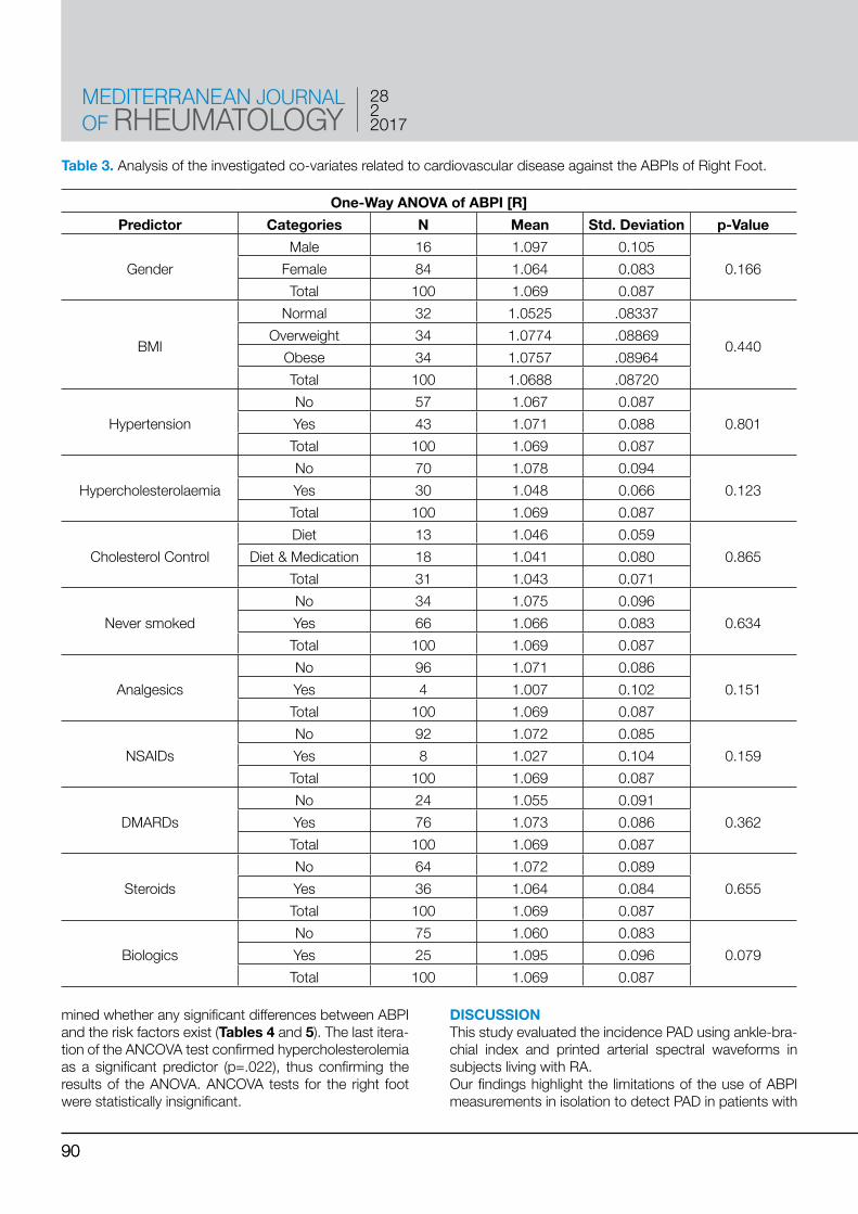
ANKLE BRACHIAL PRESSURE INDEXThe ABPI was found to be normal (0.9-1.29) in 96% of participants, whilst mild obstruction was found in only 4% of the subjects (mean 0.85, range 0.82-0.88). The One-way ANOVA (Analysis of Variance) was used to analyze the investigated co-variates related to cardiovas-
cular disease against the ABPIs of the Right and Left foot independently. The cardiovascular risk factors analyzed included gender, BMI, hypertension, hypercholesterol-emia, smoking and RA medications, amongst others. Only hypercholesterolemia was found to be significantly related to the ABPI (p=0.022) in the Left foot. (Tables 2 and 3).Further statistical analysis involved the Analysis of Cova-riance (ANCOVA) linear regression analysis which deter-
Table 2. Analysis of the investigated co-variates related to cardiovascular disease against the ABPIs of Left Foot.
One-Way ANOVA of ABPI [L]Predictor Categories N Mean Std. Deviation р-Value
GenderMale 16 1.097 0.093
0.157Female 84 1.065 0.080Total 100 1.070 0.083
BMI
Normal 32 1.0512 .07623
0.292Overweight 34 1.0752 .07243
Obese 34 1.0819 .09654Total 100 1.0698 .08274
HypertensionNo 57 1.072 0.088
0.757Yes 43 1.067 0.076Total 100 1.070 0.083
HypercholesterolaemiaNo 70 1.082 0.086
0.022*Yes 30 1.041 0.068Total 100 1.070 0.083
Never smokedNo 34 1.088 0.089
0.113Yes 66 1.060 0.079Total 100 1.070 0.083
AnalgesicsNo 96 1.073 0.083
0.075Yes 4 0.998 0.041Total 100 1.070 0.083
NSAIDsNo 92 1.073 0.083
0.201Yes 8 1.034 0.077Total 100 1.070 0.083
DMARDsNo 24 1.058 0.078
0.419Yes 76 1.074 0.084Total 100 1.070 0.083
steroidsNo 64 1.069 0.084
0.860Yes 36 1.072 0.081Total 100 1.070 0.083
BiologicsNo 75 1.064 0.081
0.204Yes 25 1.088 0.086Total 100 1.070 0.083
MEDITERRANEAN JOURNAL OF RHEUMATOLOGY
2822017
90
MEDITERRANEAN JOURNAL OF RHEUMATOLOGY
2822017
90
mined whether any significant differences between ABPI and the risk factors exist (Tables 4 and 5). The last itera-tion of the ANCOVA test confirmed hypercholesterolemia as a significant predictor (p=.022), thus confirming the results of the ANOVA. ANCOVA tests for the right foot were statistically insignificant.
DISCUSSIONThis study evaluated the incidence PAD using ankle-bra-chial index and printed arterial spectral waveforms in subjects living with RA. Our findings highlight the limitations of the use of ABPI measurements in isolation to detect PAD in patients with
Table 3. Analysis of the investigated co-variates related to cardiovascular disease against the ABPIs of Right Foot.
One-Way ANOVA of ABPI [R]Predictor Categories N Mean Std. Deviation р-Value
GenderMale 16 1.097 0.105
0.166Female 84 1.064 0.083Total 100 1.069 0.087
BMI
Normal 32 1.0525 .08337
0.440Overweight 34 1.0774 .08869
Obese 34 1.0757 .08964Total 100 1.0688 .08720
HypertensionNo 57 1.067 0.087
0.801Yes 43 1.071 0.088Total 100 1.069 0.087
HypercholesterolaemiaNo 70 1.078 0.094
0.123Yes 30 1.048 0.066Total 100 1.069 0.087
Cholesterol ControlDiet 13 1.046 0.059
0.865Diet & Medication 18 1.041 0.080Total 31 1.043 0.071
Never smokedNo 34 1.075 0.096
0.634Yes 66 1.066 0.083Total 100 1.069 0.087
AnalgesicsNo 96 1.071 0.086
0.151Yes 4 1.007 0.102Total 100 1.069 0.087
NSAIDsNo 92 1.072 0.085
0.159Yes 8 1.027 0.104Total 100 1.069 0.087
DMARDsNo 24 1.055 0.091
0.362Yes 76 1.073 0.086Total 100 1.069 0.087
SteroidsNo 64 1.072 0.089
0.655Yes 36 1.064 0.084Total 100 1.069 0.087
BiologicsNo 75 1.060 0.083
0.079Yes 25 1.095 0.096Total 100 1.069 0.087
91
TITLE
91
RA. Results from the current study demonstrate incon-sistencies when utilizing both ABPI and arterial spec-tral Doppler waveform analysis, as Doppler waveforms were different from ABPI interpretations in most of the recruited subjects. Despite a ‘normal ABPI’ examination
in most of the patients, waveform analysis showed im-paired vascular function as one third of subjects demon-strated biphasic waveforms in one or both feet. Results of the present study have demonstrated that only 1% of the recruited subject population had severe PAD, which
Table 4. ANCOVA test of Left ABPI against all variables.
Tests of Between-Subjects Effects (ABPI Left)
Source Sum of Squares df Mean Square F P-value
Corrected Model 0.134 19 0.007 1.036 0.432Intercept 1.199 1 1.199 176.351 0.000Gender 0.008 1 0.008 1.247 0.268BMI Scale 0.011 2 0.006 0.843 0.434Hypertension 0.005 1 0.005 0.808 0.371Cholesterol 0.047 1 0.047 6.914 0.010NSAIDs 0.001 1 0.001 0.202 0.655Analgesics 0.012 1 0.012 1.751 0.190DMARDs 0.007 1 0.007 0.983 0.324Steroids 0.000 1 0.000 0.063 0.802Biologics 0.008 1 0.008 1.224 0.272Never smoked 0.009 1 0.009 1.330 0.252Age 0.001 1 0.001 0.095 0.759RA Duration 0.007 1 0.007 0.996 0.321Total Blood cholesterol 0.001 1 0.001 0.094 0.760High density Lipoproteins 0.000 1 0.000 0.001 0.975Low density Lipoproteins 0.000 1 0.000 0.047 0.828Rheumatoid Factor 0.006 1 0.006 0.934 0.337C-Reactive Protein 0.009 1 0.009 1.295 0.258Erythrocyte Sedimentation Rate 0.004 1 0.004 0.635 0.428Error 0.544 80 0.007 Total 115.130 100 Corrected Total 0.678 99
Table 5. The last iteration of ANCOVA test for Left ABPI showing cholesterol as a significant predicator.
Tests of Between-Subjects Effects (ABPI Left)Source Sum of Squares df Mean Square F P-value
Corrected Model 0.035 1 0.035 5.416 0.022Intercept 94.667 1 94.667 14443.713 0.000
Cholesterol 0.035 1 0.035 5.416 0.022*Error 0.642 98 0.007 Total 115.130 100
Corrected Total 0.678 99
DETERMINING THE PRESENCE OF PERIPHERAL ARTERIAL DISEASE IN PATIENTS WITH RHEUMATOID ARTHRITIS
MEDITERRANEAN JOURNAL OF RHEUMATOLOGY
2822017
92
MEDITERRANEAN JOURNAL OF RHEUMATOLOGY
2822017
92
could possibly be symptomatic. On the other hand, 29% of patients presented with biphasic waveforms indicating initial stages of subclinical PAD. These observations sug-gest that almost one third of RA patients may suffer from early PAD and the silent clinical presentation may lead to underdiagnosis of this condition in this population. An important characteristic of the recruited sample is the lack of significant co-morbidities, which is the crucial factor when analysing the results of this study. We took care to exclude patients with co-morbidities such as di-abetes, treatment with anti-platelet and anti-coagulant treatments, a history of ulcerations or major cardiovas-cular events in order our sample to include individuals with low CVD risk, thus leaving RA as the main inde-pendent variable. The literature identifies CVD risk fac-tors that are common to both diabetes and RA.21 It has also been reported that RA patients have a higher risk of PAD than the normal population.6 Normally, production of new blood vessels may occur either by endothelial sprouting from pre-existing angioblasts (Angiogenesis) or by peripheral recruitment of the endothelial progenitor cells (EPCs) (vasculogenesis). Such procedures are im-portant to maintain healthy tissue and also as compen-satory methods for development of collateral circulation in regional ischaemia. The quantity of EPCs in the pe-ripheral blood is reported to be inversely correlated with cardiovascular risk.22 In active RA there is a decrease in circulating EPCs, resulting in increased susceptibility to vascular dysfunction. This is due to the fact that, if less EPCs are found, less vasculogenesis may occur leading to less blood vessel formation and thus increased CVD risk. Wolfe & Michaud confirmed23 that, in RA, functional and numerical EPC decline is attributed to the upregu-lated production of tumor necrosis factor-alpha as well as other mediators of inflammation that are known to be pathogenic in RA. This is further augmented by high grade systemic inflammation which accelerates vascular risk in RA24 and partially explains the peripheral vascular dysfunction noticed in our highly selective RA population.Since TNF is increased in RA patients’ suppression of systemic inflammatory load with biologic targeted ther-apies may lead to normalization of EPC and reduction of CVD risk. It could be speculated that the beneficial effects of biologic therapies on CVD risk described over the last years25 may be - amongst others - associated with the attenuation of systemic inflammations’ adverse action on EPC numbers and functionality. This study has shown that in a cohort of patients with RA with very minimal co-morbidities, nearly all participants (96%) presented with normal ABPI readings. Howev-er, qualitative Spectral Waveform Analysis demonstrat-ed suboptimal/mild arterial perfusion in one third of the study group indicative of PAD. These findings are congruent with those of Chuang et al.6 who reported that RA patients with various co-mor-
bidities showed a significantly higher risk of PAD when compared with their controls without co-morbidities. In contrast, in a case-control study, the authors reported a higher prevalence of abnormal ABPI in patients with RA when compared to matched healthy individuals (p=0.001).10 These inconclusive results provided the rationale for the conduction of our study to investigate whether RA is an independent risk factor for peripher-al arterial disease.11 This study now suggests that RA may be an independent risk factor for PAD. It has been reported that Doppler analysis offers a particular advan-tage over the ABPI, since the detection of a pulsatile flow using Doppler analysis may be possible even in calci-fied arteries. Doppler waveform analysis therefore allows the detection of early arterial disease when normal ABPI readings are recorded. Our observations also indicate that further physiological testing such as toe pressure and toe brachial pressure indices should be performed to establish whether PAD is actually present in case those inconsistencies occur between two screening modalities for PAD namely ABPI and Doppler analysis. Earlier diag-nosis of PAD in RA allows the prompt management of CVD risk factors and CVD risk stratification which im-proves long term outcomes in this population.26
The duration of RA does not seem to have any impact on arterial perfusion since in this present study, although the duration of the condition amongst our participants ranged from 2 to 24 years, no significant differences was found between RA duration and ABPI and Doppler Spectral Waveform analysis. The only statistically signif-icant association established between cardiovascular disease co-variates assessed was between high serum cholesterol levels and ABPI readings (p=0.022).A limitation of this study is that subjects use of anti-de-pressants were not recorded, thus authors are unaware of the extent of use of this type of medication, which may affect vasodilation and thus the assessment of vascu-lar supply. More research is required in this field in order to explore the relationship between RA and individual co-morbidities in order to identify which of these factors contribute to PAD in this population.
CONCLUSIONResults indicated that whilst the ABPI index was normal in the majority of participants, waveform analysis was abnormal (biphasic) in approximately one-third of the study sample. This led to the conclusion that some of the recruited subjects with normal ABPI index but abnor-mal waveforms could mistakenly be classified as normal. This research emphasizes that assess peripheral arte-rial perfusion assessment should utilize both modalities and when they do not correlate (with either one suggest-ing PAD), these patients should be monitored or further evaluated accordingly. Early diagnosis of PAD allows the prompt commencement CVD risk management which

93
TITLE
93
may delay long-term complications, improve outcomes and reduce the financial burden which this condition imposes on both patients and the healthcare system. Current recommendations about physiological testing of peripheral perfusion in RA should consider including spectral waveforms as part of the assessment in this group of patients.
ACKNOWLEDGEMENTSThe authors would like to thank all participants who con-sented to participate in this study. The authors would also like to thank Prof. Liberato Camilleri (University of Malta) for his help in statistical advice.
CONFLICT OF INTERESTThe authors declare no conflict of interest.
REFERENCES1. Scott D L, Wolfe F, Huizinga T W J. Rheumatoid arthritis. Lancet
2010;376(9746):1094-108. 2. Edmonds M, Wall B. Metabolic disorders. In: Lorimer D, French G,
O’Donnel M, Burrow J, editors. Naele’s Disorders of the Foot. 6th ed. New York: Churchill Livingstone; 2002.
3. DeMaria A N. Relative risk of cardiovascular events in patients with rheumatoid arthritis. Am J Cardiol 2002;89:33D-38D.
4. González-Gay M A, González-Juanatey C. Inflammation and lipid profile in rheumatoid arthritis: bridging an apparent paradox. Ann Rheum Dis 2014;731281-3.
5. Galkina E, Ley K. Immune and inflammatory mechanisms of ath-erosclerosis. Annu Rev Immunol 2009;27:165-97.
6. Chuang Y W, Yu M C, Lin C L, Yu T M, Shu K H, Huang S T, et al. Risk of peripheral arterial occlusive disease in patients with rheumatoid arthritis. A nationwide population-based cohort study. Thromb Haemost 2016;115:439-45.
7. Orchard T, Strandness D. Assessment of Peripheral Vascular Dis-ease in Diabetes: Report and Recommendations of an Internation-al Workshop Sponsored by the American Heart Association and the American Diabetes Association. Diabetes Care 1993;16:1199-209.
8. McIntosh C. Assessing the vascular status of the feet in patients with diabetes. Wounds Essent. 2006;1:143-7.
9. Horowitz E, Rehm K. PVD and DPM: Should Podiatrists Be the Referrer or the Referee? Pod Manag 2011;(April/May).
10. Fraidoon A, Merza R, Mohammad K, Amin O, Abdullah R. Rheu-matoid Arthritis and the Ankle-Brachial Pressure Index: Any Asso-ciation? Cukurova Med J 2014; 39:83-90.
11. Stamatelopoulos K S, Kitas G D, Papamichael C M, Kyrkou K, Zampeli E, Fragiadaki K, et al. Subclinical peripheral arterial disease in rheumatoid arthritis. Atherosclerosis 2010;212:305-9.
12. Lindhardsen J, Ahlehoff O, Gislason G H, Madsen O R, Olesen J B, Torp-Pedersen C, et al. The risk of myocardial infarction in rheu-matoid arthritis and diabetes mellitus: a Danish nationwide cohort study. Ann Rheum Dis 2011;70:929-34.
13. Fowkes F G R, Murray G D, Butcher I, Heald C L, Lee R J, Cham-bless L E, et al. Ankle brachial index combined with Framingham Risk Score to predict cardiovascular events and mortality: a me-ta-analysis. JAMA 2008;300:197-208.
14. Criqui M H, McClelland R L, McDermott M M, Allison M A, Blumen-thal R S, Aboyans V, et al. The ankle-brachial index and incident cardiovascular events in the MESA (Multi-Ethnic study of athero-sclerosis). J Am Coll Cardiol 2010;56:1506-12.
15. Kurt T, Temiz A, Gokmen F, Adam G, Ozcan S, Ozbudak E, et al. Can the ankle brachial pressure index (ABPI) and carotis intima media thickness (CIMT) be new early stage markers of subclinical atherosclerosis in patients with rheumatoid arthritis? Wien Klin Wo-
chenschr 2010;127:529-34. 16. Aletaha D, Neogi T, Silman A, Funovits J, Felson D, Bingham C.
Rheumatoid arthritis classification criteria: an American College of Rheumatology/European League Against Rheumatism collabora-tive initiative. Ann Rheum Dis 2010;69:1580-8.
17. Pham H, Armstrong D, Harvey C, Harkless L, Giurini J, Veves A. Screening techniques to identify people at high risk for diabetic foot ulceration: a prospective multicenter trial. Diabetes Care 2000;23:606-11.
18. Formosa C, Cassar K, Gatt A, Mizzi A, Mizzi S, Camileri K P, et al. Hidden dangers revealed by misdiagnosed peripheral arte-rial disease using ABPI measurement. Diabetes Res Clin Pract 2013;102:112-6.
19. Gorman P, De Nunzzio M, Donnelly R. Methods of arterial and ve-nous assessment. In: ABC of arterial and venous disease. 2nd ed. London: Blackwell Publishing Limited; 2009. p. 128.
20. American Diabetes Association. Standards of Medical Care in Dia-betes-2015. Diabetes Care 2015;38:1-94.
21. van Halm V P, Peters M J L, Voskuyl a E, Boers M, Lems W F, Visser M, et al. Rheumatoid arthritis versus diabetes as a risk fac-tor for cardiovascular disease: a cross-sectional study, the CARRE Investigation. Ann Rheum Dis 2009;68:1395-400.
22. Grisar J, Aletaha D, Steiner C W, Kapral T, Steiner S, Seidinger D, et al. Depletion of Endothelial Progenitor Cells in the Peripheral Blood of Patients With Rheumatoid Arthritis. Circulation 2005;111(2):204-11.
23. Wolfe F, Michaud K. Effect of body mass index on mortality and clin-ical status in rheumatoid arthritis. Arthritis Care Res 2012;64:1471-9.
24. Sattar N, McCarey D W, Capell H, McInnes I B. Explaining How High-Grade Systemic Inflammation Accelerates Vascular Risk in Rheumatoid Arthritis. Circulation. 2003;108:2957-63.
25. Low A S, Symmons D P, Lunt M, Mercer L K, Gale C P, Watson K D, et al., British Society for Rheumatology Biologics Register for Rheumatoid Arthritis (BSRBR-RA) and the BSRBR Control Centre Consortium. Relationship between exposure to tumour necrosis factor inhibitor therapy and incidence and severity of myocardi-al infarction in patients with rheumatoid arthritis. Ann Rheum Dis 2016;76:654-60.
26. Myasoedova E, Gabriel S E, Matteson E L, Davis J M 3rd, Ther-neau T M, Crowson C S. Decreased Cardiovascular Mortality in Patients with Incident Rheumatoid Arthritis (RA) in Recent Years: Dawn of a New Era in Cardiovascular Disease in RA? J Rheumatol 2017;44:732-9.
DETERMINING THE PRESENCE OF PERIPHERAL ARTERIAL DISEASE IN PATIENTS WITH RHEUMATOID ARTHRITIS